Embed Size (px)
Citation preview

Biochimica et Biophysica Acta 1812 (2011) 141–150
Contents lists available at ScienceDirect
Biochimica et Biophysica Acta
j ourna l homepage: www.e lsev ie r.com/ locate /bbad is
Review
Radical changes in multiple sclerosis pathogenesis
Jack van Horssen a,b,⁎,1, Maarten E. Witte b,1, Gerty Schreibelt c, Helga E. de Vries a
a Departments of Molecular Cell Biology and Immunology, VU University Medical Center, Amsterdam, The Netherlandsb Pathology, VU University Medical Center, Amsterdam, The Netherlandsc Department of Tumor Immunology, Radboud University Nijmegen Medical Centre, Nijmegen, The Netherlands
⁎ Corresponding author. Department of Molecular CelUniversity Medical Center Amsterdam, P.O. Box 705Netherlands. Tel.: +31 204448078; fax: +31 20444808
E-mail address: [email protected] (J. van Horsse1 Both authors contributed equally.
0925-4439/$ – see front matter © 2010 Elsevier B.V. Aldoi:10.1016/j.bbadis.2010.06.011
a b s t r a c t
a r t i c l e i n f oArticle history:Received 11 August 2009Received in revised form 8 June 2010Accepted 16 June 2010Available online 27 June 2010
Keywords:AntioxidantsMultiple sclerosisNeurodegenerationNeuroinflammationOxidative damageReactive oxygen species
Reactive oxygen species (ROS) contain one or more unpaired electrons and are formed as intermediates in avariety of normal biochemical reactions. However, when generated in excess amounts or not appropriatelycontrolled, ROS initiate extensive cellular damage and tissue injury. ROS have been implicated in theprogression of cancer, cardiovascular disease and neurodegenerative and neuroinflammatory disorders, suchas multiple sclerosis (MS). In the last decade there has been a major interest in the involvement of ROS in MSpathogenesis and evidence is emerging that free radicals play a key role in various processes underlying MSpathology. To counteract ROS-mediated damage, the central nervous system is equipped with an intrinsicdefense mechanism consisting of endogenous antioxidant enzymes. Here, we provide a comprehensiveoverview on the (sub)cellular origin of ROS during neuroinflammation as well as the detrimental effects ofROS in processing underlying MS lesion development and persistence. In addition, we will discuss clinicaland experimental studies highlighting the therapeutic potential of antioxidant protection in thepathogenesis of MS.
l Biology and Immunology, VU7, 1007 MB Amsterdam, The1.n).
l rights reserved.
© 2010 Elsevier B.V. All rights reserved.
1. Introduction
Reactive oxygen species (ROS) are small molecules which arehighly reactive due to presence of unpaired electrons or their ability togive rise to new free radicals. Many different forms of ROS exist, withsuperoxide (O2
−) and its derivatives hydroxyl radical (OH∙) andhydrogen peroxide (H2O2) being most abundant in eukaryotic cells.Superoxide can be formed both enzymatically by for instance NAD(P)H oxidase, and non-enzymatically as a by-product of oxidativephosphorylation [1]. Superoxide has a short life span and is eitherrapidly converted by superoxide dismutases (SODs) into hydrogenperoxide [2], or non-enzymatically converted into other ROS [3].Hydrogen peroxide is relatively stable and can travel further distancesthan superoxide and has the potential to generate reactive hydroxylradicals through interactions with iron or copper in cells by means ofthe Fenton reaction. Superoxide can also react with nitric oxide (NO),which is enzymatically produced by nitric oxide synthase (NOS), toform peroxynitrite (ONOO−) [4] (Fig. 1). Peroxynitrite itself isrelatively stable and can travel several micrometers before decom-posing into the highly reactive hydroxyl radical and nitrogen dioxide[4].
Physiological concentrations of ROS regulate various biologicalprocesses, e.g. control of ventilation and erythropoietin production [5].Furthermore, ROS are produced in vast quantities by immune cells inorder to kill pathogens and facilitate phagocytosis [6,7]. However, inneuropathological disorders, including Alzheimer's disease, Parkinson'sdisease, Huntington's disease and the neuroinflammatory disordermultiple sclerosis (MS), ROS production overwhelms the antioxidantcapacity and contributes to cellular injury (for review see [8–11]).
2. Origin of free radical formation in MS pathology
2.1. Classification of MS lesions
Multiple sclerosis (MS) is a chronic inflammatory disease of thecentral nervous system (CNS) affecting both white and gray matter[12,13]. The most predominant histopathological feature of MS is thepresence of focal demyelinated lesions scattered throughout the whitematter. The course of the disease is generally episodic, with frequentintervals of exacerbations followed by periods of remission. Therelapsing-remitting phase is characterized by immune-mediatedresponses, such aswidespreadmicroglial activation andmassive cellularinfiltrates in the CNS. In time, patients gradually develop secondaryprogressive MS, which is mainly characterized by neuronal and axonaldegeneration and extensive cortical demyelination [12,14,15].
The initial phase of MS lesion formation is mainly characterized byclusters of activatedmicroglia without evident signs of demyelination,whereas in the active phase of MS lesion development monocyte-

Fig. 1. Schematic overview of ROS formation and detoxification. Superoxide (O2. −) is
dismutated into hydrogen peroxide (H2O2) by superoxide dismutases, however incombination with nitric oxide (NO) forms peroxynitrite (ONOO.). Subsequently,hydrogen peroxide is either detoxified by endogenous antioxidant enzymes or in thepresence of transition metals converted into highly toxic hydroxyl radicals (OH·).
142 J. van Horssen et al. / Biochimica et Biophysica Acta 1812 (2011) 141–150
derived macrophages invade the lesion area and initiate thedemyelination process. Active lesions contain abundant activatedmyelin-laden microglia and macrophages throughout the lesion area.In time, active lesions gradually convert into chronic active lesions,which are characterized by a hypocellular demyelinated gliotic centerwith a hypercellular rim containing activated macrophages. In thechronic lesion stage when inflammation has subsided hypertrophicastrocytes form a dense network, the so-called astroglial scar [16].
2.2. Activated microglia and macrophages are major cell source of ROS
The cellular source of free radical production largely depends on thestage of the MS lesions. Both activated microglia and infiltratedmacrophages are able to generate vast amounts of proinflammatorymediators and oxidizing radicals, such as superoxide, hydroxyl radicals,hydrogen peroxide and nitric oxide [17]. Important sources of oxidizingspecies in activated microglia and macrophages are the ROS-generatingenzymes myeloperoxidase, xanthine oxidase and NADPH oxidase.Myeloperoxidase is a lysosomal enzyme which produces hypochlorousacid (HOCl) from hydrogen peroxide and chloride anion. In homo-genates ofMSwhite andgraymatter, demyelinationwas associatedwithsignificantly elevated myeloperoxidase activity [18,19]. In white matterlesionsmyeloperoxidasewas predominantly expressed bymacrophagesand activated microglia within and in close vicinity of MS plaques,emphasizing the key role of thesemyeloid cells in the generation of ROS.
Recent advances in the understanding of the pathology of MSrevealed that demyelinating lesions also exist within the graymatter. Ingeneral, cortical MS lesions lack significant macrophage infiltration,however the gray matter part of leukocortical (type I) lesions contain arim of activated microglia [12,20]. Gray and colleagues demonstratedthat myeloperoxidase was expressed by a subset of activated microgliain cortical lesions, suggesting that microglia-derived ROS mightcontribute to gray matter demyelination [19].
Xanthine oxidase catalyzes the oxidation of hypoxanthine toxanthine thereby generating large quantities of ROS, includingsuperoxide and its dismutation product hydrogen peroxide. In addition,NADPH oxidase is a multi-subunit enzyme complex that is activatedunder pathological conditions inmicroglia and catalyzes the productionof superoxide from oxygen. Evidence is accumulating that NADPHoxidase represents a common pathway ofmicroglia-mediated neuronaldamage [21]. Remarkably, nothing is known about the expressionpattern of xanthine oxidase and NADPH oxidase in brain tissue derivedof MS patients although it is plausible that both enzymes contribute toROS production during the early phase of MS lesion formation. Duringinflammatory conditions macrophages and microglia generate highlevels of nitric oxide by activation of inducible nitric oxide synthase.Nitric oxide reacts with superoxide to form highly toxic peroxynitrite,which in turn might attack tyrosine residues of proteins, therebyaffecting their function [22].
2.3. Mitochondrial dysfunction contributes to ROS production
In later stages of MS pathogenesis when inflammation has abated,other non-inflammatory pathogenic mechanisms, such as mitochondrialdysfunction contribute to the formation of free radicals. Mitochondrial-derived ROS are believed to play a major role in neurodegeneration, aspreviously observed in Alzheimer's and Parkinson's disease [8–10].Mitochondria are a constant source of ROS, due to electron leakage fromthe electron transfer chain (ETC) [1,23]. Normally, the amount of ROSproduced by mitochondria is counteracted by local antioxidant enzymes.However, mitochondrial ROS production can be markedly increased inresponse to pathological events leading to cellular oxidative stress. In theearly 1990's, it was suggested that mitochondriamight be involved inMSpathogenesis, as Leber's hereditary optic neuritis, which shares severalpathogeneticmechanismswithMS,was found to be caused bymutationsin mtDNA [24,25]. This led to a number of genetic studies in search of arelationbetweenmitochondrial genotype andMS.Although some reportsdescribed associations between mtDNA polymorphisms and MS [26,27],up tonownopathogenicmtDNAmutationsassociatedwithMShavebeendescribed [28].
More recently, mitochondrial alterations and mitochondria-derivedROS have been implicated in axonal degeneration inMS [29,30]. Axonaldegeneration inMScanbedivided into two stages, thefirst representingacute axonal injury in inflammatory MS lesion [31,32], and the secondbeing ‘slow burning’ axonal degeneration in non-inflammatory chroniclesions [33]. Acute axonal injury is directly mediated by inflammation-derived toxic molecules, including ROS and nitric oxide, whereas ‘slowburning’ axonal degeneration is believed to be the consequence of intra-axonal alterations caused by chronic demyelination. Upon demyelin-ation, axonal conduction is blocked due to the absence of sodiumchannels in acutely demyelinated segments. However, axonal conduc-tion can be restored by redistribution of sodium channels along thesedemyelinated segments [34]. This leads to an enormous influx ofsodium, which can be counteracted by increasing Na+/K+ ATPaseactivity. Na+/K+ATPase function depends on ATP, thusmitochondria indemyelinated axons need to increase their ATP production to maintainconduction. The increasedmitochondrial ATP production partially relieson an increase inmitochondrial number, aswe and others have recentlyshown, and is likely to lead to increased mitochondrial ROS production[35,36].
Interestingly, increasedmitochondrial density inMS lesions coincidedwith enhanced expression ofmitochondrial heat shock protein 70, awell-definedmarker ofmitochondrial stress. Mitochondrial heat shock protein70 was observed in axons and astrocytes in MS lesions, suggesting theoccurrence of ongoing mitochondrial oxidative stress [36]. Two, notmutually exclusive, causes for increasedmitochondrial ROS production inchronic demyelinated axons have been proposed. Firstly, increasednumbers of mitochondria may contribute to enhanced mitochondrialROS production, accumulating ROS damage tomitochondria and reducedATP production [37]. Alternatively, others suggested that mitochondriahave acquired oxidative damage during the inflammatory phase of MS,leading to increasedmitochondrial ROS production and eventually axonalenergy deprivation in the later stages of the disease [38]. This might bemediated by peroxynitrite, which is abundantly produced in theinflammatory stage of MS lesions [39,40]. In experimental autoimmuneencephalomyelitis (EAE), a validated animalmodel forMS,mitochondrialalterations and oxidative injury of mitochondrial proteins in axons evenprecede infiltration of leukocytes [41,42]. This finding indicates thatmitochondrial changes initiate a cascade of molecular events leading toaxonal neurodegeneration in EAE that is not mediated by inflammatorycells. Taken together, mitochondrial dysfunction and subsequent ROSproduction are key players in degeneration of chronically demyelinatedaxons inMS.Moreover, inflammation-derived ROS are a putative cause ofmitochondrial dysfunction in axons, although there is evidence support-ing that mitochondrial dysfunction occurs before or in the absence ofinflammation.

143J. van Horssen et al. / Biochimica et Biophysica Acta 1812 (2011) 141–150
3. ROS induce oxidative damage and contribute to MS pathogenesis
In the pathogenesis of MS, high amounts of ROS are produced whichoverpower the antioxidant capacity resulting in oxidative stress. In turn,oxidative stress will result in ROS-induced damage to biologicalmacromolecules suchaspolyunsaturated fatty acids (PUFA) inmembranelipids, essential proteins, and DNA/RNA. The CNS exhibits high oxygenconsumption and is enriched in PUFA,making it particularly vulnerable tolipid peroxidation. Increased levels of indicators of oxidative stress and/ordecreased levels of antioxidant enzymes and antioxidants have beendetected in blood and cerebrospinal fluid ofMS patients during the activephases of disease, indicating that increased levels of ROS may haveresulted in the depletion of cellular antioxidants [43–49].
3.1. Oxidative damage in MS lesions
The occurrence of widespread oxidative damage in demyelinatingMSplaques is supported by the expression ofmarkers of oxidative damage inMS brain tissue. Oxidative damage to cellular membrane lipids generates4-hydroxy-2-nonenal (4-HNE), a highly reactive aldehyde that is toxic toCNS cells. In active demyelinating MS lesions 4-HNE accumulates in bothphagocytic macrophages and large hypertrophic astrocytes [50]. Nitricoxide produced by inducible nitric oxide synthase can react withsuperoxide to generate peroxynitrite, a diffusible free radical that canreact with tyrosine residues in proteins to form nitrotyrosine. Nitrotyr-osine residues are present in high amounts in foamy macrophages andlargehypertrophic astrocytes throughout activedemyelinatingMS lesionssuggesting that both macrophages and astrocytes are a primary target ofoxidative damage [50–53]. Expression of 8-hydroxy-2’-deoxyguanosinereflects the occurrence of oxidative damage to nucleotides and ismarkedly increased in reactive astrocytes throughout inflammatory MSlesions, whichmight reveal either oxidative damagedmitochondrial DNAor increased RNA oxidation [50,54]. Interestingly, Bizzozero and cow-orkers demonstrated increased protein carbonylation and nitrosativestress in normal appearing white and gray matter in MS brain tissue[55,56]. Taken together, there is ample evidence that extensive oxidativeinjury occurs inMS plaques and that ROS-induced damage already occursin the earliest stages of neuroinflammation.
3.2. ROS induce pathological mechanisms of MS lesion formation
Alongside the damaging effect on essential biological molecules,free radicals contribute to several key processes underlying MSpathogenesis. In the initial phase of MS lesion formation, locallyproduced ROS induce tight junction and cytoskeletal changes in brainendothelial cells thereby facilitating transendothelial monocytemigration [57,58]. ROS also play a crucial role in myelin phagocytosisas blocking ROS productionwith NADPH oxidase inhibitors, lipoic acidor catalase reduced the phagocytosis of myelin by macrophages [59].Oligodendrocytes are highly susceptible to oxidative injury as theycontain high levels of PUFA that can react with ROS thereby triggeringlipid peroxidation. In addition, oligodendrocytes contain high intra-cellular concentrations of iron [60] and reduced levels of antioxidantenzymes and free radical scavengers [61–63].
Notably, immature oligodendrocyte progenitors (OPCs) are differen-tially vulnerable to oxidative stress, due to lower levels of antioxidantenzymes and anti-apoptotic proteins in combinationwith higher levels ofproapoptotic members [63,64]. Pro-oxidants like H2O2 strikingly inhibitexpression of myelin genes in human primary oligodendrocytes throughcellular redox alterations. Interestingly, French and colleagues recentlydemonstrated that oxidative stress impairs OPC differentiation byepigenetic mechanisms and decreased expression of genes that promotematuration and enhanced expression of genes involved in oligodendro-cyte dedifferentiation [65]. Together, these studies indicate that strategiesaimed at reinstating the redox balancemay offer protection against ROS-
induced oligodendrocyte damage but also promote the differentiation ofOPCs into mature, myelin-producing cells and thus remyelination.
4. Antioxidant protection as a potential therapeutic target for MS
Since ROS play a pivotal role in the initial phase as well as thechronic stage of MS, antioxidant therapy might be an attractiveapproach to limit disease progression. Experimental animal studieshave demonstrated that dietary intake of exogenous antioxidants,including flavonoids and α-lipoic acid, reduce the progression andclinical signs of EAE [58,66–74]. Despite promising results observed inanimal models for MS, data on successful antioxidant therapy in MSpatients is still limited emphasizing the need for epidemiological andclinical studies on antioxidant strategies. Notably, there are a numberof disadvantages to the use of exogenous antioxidants for MStreatment, as most antioxidant compounds do not efficiently crossthe blood–brain barrier and have a narrow therapeutic window.Consequently, high quantities are generally required to achieveprotective effects in animal models for MS [44,75], illustrating theneed for other protective antioxidant mechanisms.
4.1. Regulation of endogenous antioxidant enzyme systems
To maintain a proper tissue redox balance and counteract oxidativedamage the central nervous system is equipped with an endogenousantioxidant defense mechanism consisting of antioxidant enzymes.Production of these cytoprotective enzymes is induced upon exposureto ROS via a mechanism regulated at the transcriptional level (528).Genes that code for proteins involved in ROS detoxification share acommon promoter element, called the antioxidant response element(ARE).ARE-mediated geneactivation is coordinatedbynuclear factor E2related factor 2 (Nrf2), which upon exposure to electrophiles or ROS,translocates to the nucleuswhere it binds ARE and activates antioxidantenzyme gene transcription [76–78]. Under normal conditions Nrf2 islinked to the repressor protein Kelch-like ECH associating protein 1(Keap1) thereby promoting its degradation. Oxidative stress enablesNrf2 to escape Keap1-mediated proteasomal degradation, leading toNrf2 stabilization, nuclear translocation and subsequent production ofan array of cytoprotective enzymes [79]. So far, over 200 Nrf2-drivengenes involved in detoxification and antioxidant defense have beenidentified, including superoxide dismutases (SODs) [80], glutathioneperoxidases [81], peroxiredoxins [82], catalase [83] and NAD(P)H:quinone oxidoreductase 1 [84] (Fig. 2).
4.2. Regulation and potential protective roles of endogenous antioxidantenzymes in MS pathogenesis
Endogenous antioxidant enzymes may reflect the occurrence ofoxidative stress. Preliminary data from our group revealed a significantupregulation of antioxidant genes (Fig. 3) and protein levels (Fig. 4) inthe course of EAE (unpublished data) and recently we demonstratedthat Nrf2-driven enzymes are highly expressed in activeMS lesions [50].
Endogenous antioxidant enzyme systems have distinct roles indetoxification of ROS and may be differentially regulated in MS.Importantly, modulation of their expression of activity may exertprotective effects. For instance, superoxide dismutases (SODs) promotedismutation of superoxide anion into molecular oxygen and hydrogenperoxide. Increased gene and protein expression of SOD1 has beendetected in active demyelinating MS lesions [50,85]. In activedemyelinating MS lesions SOD1 staining was detected in foamymacrophages and astrocytes [50]. Dismutation of superoxide by SODsresults in the formation of hydrogen peroxide, which is subsequentlyconverted by catalase, glutathione peroxidases, and peroxiredoxins.
Catalase itself is ubiquitously expressed in the CNS and is mainlylocated in peroxisomes where it catalyzes the conversion of hydrogenperoxide into water and molecular oxygen. Impaired peroxisomal

Fig. 2. The Nrf2 pathway. Under normal conditions Nrf2 associates with Keap1 and the actin cytoskeleton. Subsequently, Nrf2 is ubiquitinated and targeted for proteasomalbreakdown. Free radicals are able to oxidize cysteine residues of Keap1, which enables Nrf2 to escape Keap1-mediated proteasomal degradation. Next, Nrf2 translocates into thenucleus where it binds to antioxidant response element (ARE) thereby inducing the production of antioxidant proteins.
144 J. van Horssen et al. / Biochimica et Biophysica Acta 1812 (2011) 141–150
function has been demonstrated in CNS homogenates of rats sufferingfromEAE, whichwas accompanied by reduced catalase gene expressionand activity [86]. Catalase treatment of guinea pigs suffering from EAEsignificantly reduced demyelination of the optic nerves, increased BBBintegrity, and ameliorated neurological symptoms [86,87]. Administra-tion of catalase reduced the severity of clinical signs in a rat model forEAE [88]. Furthermore, upregulation of catalase expression via viralvectors ameliorates EAE, implicating its positive effect in neuroinflam-matory disorders [89,90]. InMS brain tissue catalase immunostaining isobserved inmyelin-ladenmacrophages and astrocytes [50]. Tajouri andcoworkers demonstrated that glutathione peroxidase gene expressionis significantly increased in inflammatory MS lesions [85] and Guy andcolleagues showed that treatment with glutathione peroxidase reducesloss of BBB integrity in chronic EAE [87], suggesting that glutathioneperoxidase plays a protective role in neuroinflammation.
Peroxiredoxins are a class of antioxidant enzymes which areexpressed in distinct CNS cell types, including neurons, astrocytes andbrain endothelial cells [91,92] and differentially regulated in neurode-generative disorders [93]. Recently, we reported that peroxiredoxin-1overexpression protected brain endothelial cells from ROS-induced celldeath, reduced adhesion and subsequent transendothelial migration ofmonocytes and enhanced the integrity of the brain endothelial cell layer[92]. Furthermore, we observed a striking increase of vascular peroxir-edoxin-1 immunostaining in inflammatory lesions of experimental EAEanimals and inflammatory demyelinatingMS lesions. Enhanced vascularperoxiredoxin-1 expression may reflect ongoing oxidative stress in EAEand MS or it may function as a protective mechanism to limit ROS-mediated damage and leukocyte infiltration into the CNS [92].
Heme oxygenase-2 is constitutively expressed in the CNS,while hemeoxygenase-1 is inducible by a variety of stimuli, including oxidative stress[94]. Hemeoxygenases catalyze the rate-limiting step in the catabolismofheme and break down the porphyrin ring into biliverdin, free iron andcarbon monoxide. Various research groups have demonstrated neuro-protective effects of heme oxygenase-1 in vitro and in vivo [95,96].Induction of EAE in heme oxygenase-1 knockout mice led to enhancedCNS demyelination, paralysis, and mortality, as compared with wild typemice [97]. Upregulation of heme oxygenase-1 [97,98] reduced the clinicalseverity of EAE, whereas inhibition of heme oxygenase-1 markedlyexacerbated EAE. In contrast, treatment with the heme oxygenase-1inhibitor tin-protoporphyrinattenuatedclinical scores inmurineEAE [99].mRNA and protein levels of heme oxygenase-1 were strikingly upregu-lated in brain tissue of EAE rats, (Fig. 2). In MS lesions, heme oxygenaseimmunoreactivity was detected in activated microglia, infiltrated macro-phages and hypertrophic astrocytes [50,100–102]. Heme oxygenasedegrades heme into biliverdin, which is subsequently converted bybiliverdin reductase into bilirubin, while the pro-oxidant iron is
sequestered and inactivated by co-induced ferritin. Bilirubin exerts potentantioxidant activity, and both suppressed ongoing EAE and halted EAEprogressionwhenadministeredafter diseaseonset, likely by reducingBBBpermeability and oxidative damage [103]. Interestingly, bilirubin pro-tected rat oligodendrocytes against hydrogen peroxide-mediated celldeath. Treatment with biliverdin reductase, like bilirubin, reducedoxidative injury in EAE lesions and significantly suppressed clinicalsymptoms of EAE [104].
NQO1 is a cytosolic flavoprotein that has broad-spectrum antioxi-dant properties [105,106]. Besides their function as antioxidants, NQO1and NQO2 function to maintain both α-tocopherol and coenzyme Q10in their reduced antioxidant state. NQO1 is expressed in tissues thatrequire high levels of antioxidant protection, like lung respiratoryepithelium and the CNS [107,108]. NQO1 is predominantly found inastrocytes and brain endothelial cells of healthy human brain tissue andmarkedly upregulated in inflammatory MS lesions, particularly inhypertrophic astrocytes and myelin-laden macrophages [109].
Although within MS lesions, the expression of antioxidant enzymesystems is severely increased, there are still signs of ongoing oxidativedamage to brain tissue [50,51,53–56,110]. Future strategies to restore theredox equilibrium may protect CNS cells form oxidative cell death andlimit pathological processes underlying MS lesion formation and diseaseprogression.
4.3. Mitochondrial antioxidant enzymes
Due to high levels of intracellular ROS and their susceptibility tooxidative damage,mitochondria are equippedwith a robust antioxidantdefensemechanismconsistingof several antioxidant enzymes. SOD2, aniron/manganese superoxide dismutase, resides in the mitochondrialmatrix where it catalyzes the reaction of superoxide into hydrogenperoxide. Increased immunostaining of SOD2 in astroglia and microgliahas been observed in guinea pigs suffering from EAE [111] and SOD2protein expression is upregulated in reactive astrocytes in active MSlesions compared to surrounding normal appearing white matter [50].Increased SOD2 levels by viral mediated transfer rescued ATP synthesisand suppressed myelin degradation and neuronal injury in EAE [42].
In addition to SOD2, mitochondria contain specific enzyme systemscapable of detoxifying superoxide metabolites: the thioredoxin, gluta-thione and glutaredoxin system [112]. These antioxidant systems arewidely expressed in various cellular compartments, with specific familymemberswithinmitochondria. Themitochondrial thioredoxin system iscomposed of peroxiredoxin 3 (Prx3), peroxiredoxin (Prx5), thioredoxin2 (Trx2) and thioredoxin reductase 2 (TrxR2) [113]. Prx3 and Prx5convert hydrogen peroxide and peroxynitrite and are reduced by Trx2,which is subsequently reducedbyTrxR2 [113]. Prx5hasbeen reported to

Fig. 3. Gene expression of Nrf2-driven antioxidant enzymes in the course of acute EAE. Gene expression of antioxidant enzymes HO-1 (A), SOD2 (B), and NQO1 (C) in the brainstemof EAE animals at day 9 (n=2), 11 (n=7), 14 (n=7), and 17 (n=6) of EAE was analyzed using quantitative PCR as previously described [92]. mRNA expression is expressed as foldchange compared to control animals for each individual animal. Dotted line indicates fold change 1.0 and solid line indicates mean fold change per time point. (D) Summary of geneexpression of the antioxidant enzymes in A, B, and C. Data are expressed as fold change (mean ± SEM, compared to control). *, pb0.05, **, pb0.01, statistically different from 1.0, bymeans of single-column t-test.
145J. van Horssen et al. / Biochimica et Biophysica Acta 1812 (2011) 141–150
be upregulated in MS lesions, predominantly in astrocytes in active andchronicMS lesions [114]. In vitro and in vivo experiments have evidentlyshown the protective properties of members of the thioredoxin-peroxiredoxin system against neuronal damage [113,115,116], indicat-ing a putative role in countering neuro-axonal damage in MS. The othermitochondrial antioxidant systems, glutathione and glutaredoxinsystems work quite similar to the thioredoxin system (for review, see[112]). Although there is ample evidence supporting theneuroprotectiveeffects of mitochondrial antioxidant systems in neuronal survival [117–119], remarkably little is known about the expression and putativeprotective role of mitochondrial antioxidant enzymes in MS pathology.
4.4. Exogenous mitochondrial targeted antioxidants
Recently, antioxidant compounds have been developed whichselectively accumulate in themitochondrial matrix. Targeting of thesecompounds to mitochondria is achieved by covalent attachment ofantioxidants like ubiquinol and vitamin E to a lipophilic cation.Because of the large mitochondrial membrane potential, the anti-oxidants accumulate within the mitochondria [120,121]. The mito-chondrial targeted antioxidants MitoQ10 and MitoE2 have beenextensively tested in various models and have consistently provento protect cells from lipid peroxidation and ROS-mediated cell death[120,122–125]. A promising feature of mitochondrial targeted anti-oxidants is their widespread bioavailability. Upon intravenous andoral administration, mitochondria-targeted antioxidants are rapidlycleared from the bloodstream and accumulate in heart, brain, liverand other tissues [126]. So far, only a few studies have tested efficacyof mitochondrial targeted antioxidants in vivo. MitoQ10 attenuatedhypertension and improved endothelial function in hypertension
prone rats [127] and limited tissue damage and mitochondrialdysfunction in an ischemia-reperfusion model [128]. In contrast,MitoE2 did not prevent loss of striatal neurons following perinatalischemia-reperfusion injury [129], suggesting that mitochondrialoxidative damage is less important in this paradigm. Since mitochon-drial ROS production contributes to MS pathogenesis mitochondria-targeted antioxidant might be an attractive therapeutic strategy tocounteract axonal degeneration and neuronal injury. Therefore,animal studies are needed to explore the putative beneficial effectsof mitochondria-targeted antioxidants in animal models of neuroin-flammation and demyelination.
5. Antioxidant therapy: Future perspectives
Taken together, there is growing evidence that ROS play apivotal role in several processes underlying the formation andpersistence of MS lesions (Fig. 5). We postulate that restoring theredox balance in MS brain tissue might represent an attractivetherapeutic strategy to limit neuroinflammation and subsequentoxidative tissue damage. Nrf2/ARE regulated antioxidant enzymesare upregulated in CNS tissue of EAE animals and MS patients andmight act as an endogenous compensatory system to counteractROS-induced damage. Despite this cellular protective mechanismoxidative damage to essential biological macromolecules isabundant in inflammatory MS lesions, suggesting that althoughNrf2-mediated antioxidant enzymes are induced upon neuroin-flammation the response may be insufficient to reduce ROS-induced cellular damage. Hence, further activation of the Nrf2/AREsystem via monofunctional inducers might counteract oxidativestress in neuroinflammatory diseases, such as MS. Monofunctional
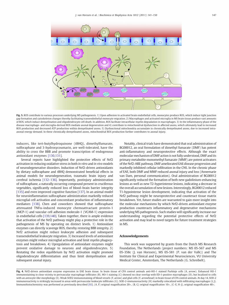
Fig. 5. ROS contribute to various processes underlying MS pathogenesis. 1) Upon adhesion to activated brain endothelial cells, monocytes produce ROS, which induce tight junctiongap formation and cytoskeleton changes thereby facilitating transendothelial monocyte migration. 2) Macrophages and activated microglia in MS brain tissue produce vast amountsof ROS, which induce demyelination and oligodendrocyte cell death. In addition, ROS facilitate intracellular myelin degradation in macrophages. 3) In the inflammatory phase of thedisease macrophage- and microglia-derived ROS mediate axonal degeneration and 4) contribute to mitochondrial dysfunction in affected axons, which ultimately lead to increasedROS production and decreased ATP production within demyelinated axons. 5) Dysfunctional mitochondria accumulate in chronically demyelinated axons, due to increased intra-axonal energy demand. In these chronically demyelinated axons, mitochondrial ROS production further contributes to axonal injury.
147J. van Horssen et al. / Biochimica et Biophysica Acta 1812 (2011) 141–150
inducers, like tert-butylhydroquinone (tBHQ), dimethylfumarate,sulforaphane and 3-hydroxycoumarin, are well-tolerated, have theability to cross the BBB and promote transcription of endogenousantioxidant enzymes [130,131].
Several reports have highlighted the protective effects of Nrf2activation in reducing oxidative stress in both in vitro and in vivomodelsof neurodegenerative disorders. Induction of Nrf2-driven antioxidantsby dietary sulforaphane and tBHQ demonstrated beneficial effects inanimal models for neurodegeneration, traumatic brain injury andcerebral ischemia [132–136]. Importantly, postinjury administrationof sulforaphane, a naturally occurring compound present in cruciferousvegetables, significantly reduced loss of blood–brain barrier integrity[135] and even improved cognitive function [137]. In an animal modelfor neuroinflammation sulforaphane administration markedly reducedmicroglial cell activation and concomitant production of inflammatorymediators [138]. Chen and coworkers showed that sulforaphaneattenuated TNFα-induced monocyte chemoattractant protein-1(MCP-1) and vascular cell adhesion molecule-1 (VCAM-1) expressionin endothelial cells [139,140]. Taken together, there is ample evidencethat activation of the Nrf2 pathway might play a protective role in thepathogenesis of MS by operating on distinct levels. 1) Antioxidantenzymes can directly scavenge ROS, thereby restoring BBB integrity. 2)Nrf2 activation might reduce leukocyte adhesion and subsequenttransendothelial leukocyte migration. 3) Increased levels of antioxidantenzymes might reduce microglial activation and limit myelin phagocy-tosis and breakdown. 4) Upregulation of antioxidant enzymes mightprevent oxidative damage to neurons and oligodendrocytes. 5)Restating the redox equilibrium by Nrf2 activation might promoteoligodendrocyte differentiation and thus limit demyelination andsubsequent axonal injury.
Fig. 4. Nrf2-driven antioxidant enzyme expression in EAE brain tissue. In brain tissue oimmunostaining in close vicinity to perivascular macrophage infiltrates (B). HO-1 staining (with an astrocyte-like morphology (E). Weak SOD2 immunostaining of blood vessels (F, arroimmunoreactivity is strikingly increased in areas with perivascular leukocyte infiltrates (G).Immunohistochemistry was performed as previously described [92]. (A, F) original magnifi
Notably, clinical trials have demonstrated that oral administration ofBG00012, an oral formulation of dimethyl fumarate (DMF) has potentanti-inflammatory and neuroprotective effects. Although the exactmolecularmechanismofDMFaction isnot fully understood,DMFand itsprimarymetabolite monomethyl fumarate (MMF) are potent activatorsof theNrf2-AREpathway. DMFamelioratedEAEdisease progression andmarkedly inhibited cellular infiltration in the CNS. In the chronic phaseof EAE, both DMF andMMF reduced axonal injury and loss (Annemarievan Dam, personal communication). Oral administration of BG00012significantly reduced the formation of both new gadolinium-enhancinglesions as well as new T2-hyperintense lesions, indicating a decrease intheoverall accumulation of new lesions. Interestingly, BG00012 reducedT1-hypointense lesion development, indicating that activation of theNrf2 pathway might be neuroprotective and counteract tissue matrixbreakdown. Yet, future studies are warranted to gain more insight intothe molecular mechanisms by which Nrf2-driven antioxidant enzymeproduction counteracts inflammatory and degenerative mechanismsunderlyingMSpathogenesis. Such studieswill significantly increase ourunderstanding regarding the potential protective effects of Nrf2activation and may lead to novel targets for future treatment strategiesin MS.
Acknowledgements
This work was supported by grants from the Dutch MS ResearchFoundation, The Netherlands (project numbers: MS 05-567 and MS05-358c (J. van Horssen), MS 05-581 (P. van der Valk)) and TheInstitute for Clinical and Experimental Neurosciences, VU UniversityMedical Center, Amsterdam, The Netherlands (G. Schreibelt).
f CFA control animals anti-HO-1 stained Purkinje cells (A, arrow). Enhanced HO-1C) showed no clear overlap with ED-1-positive macrophages (D), but localized to cellsw) and glial cells (F, arrowhead) in brain tissue of CFA control animals. At day 14, SOD-2SOD-2-immunoreactivity (H) markedly colocalized with infiltrating macrophages (I, J).cation 20×, (B, G) original magnification 10×, (C–E, H–J), original magnification 40×.

148 J. van Horssen et al. / Biochimica et Biophysica Acta 1812 (2011) 141–150
References
[1] E. Cadenas, A. Boveris, C.I. Ragan, A.O. Stoppani, Production of superoxideradicals and hydrogen peroxide by NADH-ubiquinone reductase and ubiquinol-cytochrome c reductase from beef-heart mitochondria, Arch. Biochem. Biophys.180 (1977) 248.
[2] C. Deby, R. Goutier, New perspectives on the biochemistry of superoxide anionand the efficiency of superoxide dismutases, Biochem. Pharmacol. 39 (1990)399.
[3] I. Fridovich, The biology of oxygen radicals, Science 201 (1978) 875.[4] J.S. Beckman, The double-edged role of nitric oxide in brain function and
superoxide-mediated injury, J. Dev. Physiol. 15 (1991) 53.[5] W. Droge, Free radicals in the physiological control of cell function, Physiol. Rev.
82 (2002) 47.[6] H.J. Forman, M. Torres, Redox signaling in macrophages, Mol. Aspects Med. 22
(2001) 189.[7] M.B. Hampton, A.J. Kettle, C.C. Winterbourn, Inside the neutrophil phagosome:
oxidants, myeloperoxidase, and bacterial killing, Blood 92 (1998) 3007.[8] M.T. Lin, M.F. Beal, Mitochondrial dysfunction and oxidative stress in
neurodegenerative diseases, Nature 443 (2006) 787.[9] P.I. Moreira, M.S. Santos, C.R. Oliveira, J.C. Shenk, A. Nunomura, M.A. Smith, X.
Zhu, G. Perry, Alzheimer disease and the role of free radicals in the pathogenesisof the disease, CNS, Neurol. Disord. Drug Targets 7 (2008) 3.
[10] M.F. Beal, Mitochondria, oxidative damage, and inflammation in Parkinson'sdisease, Ann. N.Y. Acad. Sci. 991 (2003) 120.
[11] A. Reynolds, C. Laurie, R.L. Mosley, H.E. Gendelman, Oxidative stress and thepathogenesis of neurodegenerative disorders, Int. Rev. Neurobiol. 82 (2007) 297.
[12] L. Bo, J.J. Geurts, S.J. Mork, P. van der Valk, Grey matter pathology in multiplesclerosis, Acta Neurol. Scand. Suppl. 183 (2006) 48.
[13] D.A. Hafler, Multiple sclerosis, J. Clin. Invest 113 (2004) 788.[14] E.M. Frohman, M.K. Racke, C.S. Raine, Multiple sclerosis–the plaque and its
pathogenesis, N. Engl. J. Med. 354 (2006) 942.[15] L. Bo, C.A. Vedeler, H.I. Nyland, B.D. Trapp, S.J. Mork, Subpial demyelination in the
cerebral cortex of multiple sclerosis patients, J. Neuropathol. Exp. Neurol. 62(2003) 723.
[16] P. van der Valk, C.J. De Groot, Staging of multiple sclerosis (MS) lesions:pathology of the time frame of MS, Neuropathol. Appl. Neurobiol. 26 (2000) 2.
[17] C.A. Colton, D.L. Gilbert, Microglia, an in vivo source of reactive oxygen species inthe brain, Adv. Neurol. 59 (1993) 321.
[18] E. Gray, T.L. Thomas, S. Betmouni, N. Scolding, S. Love, Elevated myeloperoxidaseactivity in white matter in multiple sclerosis, Neurosci. Lett. 444 (2008) 195.
[19] E. Gray, T.L. Thomas, S. Betmouni, N. Scolding, S. Love, Elevated activity andmicroglial expression of myeloperoxidase in demyelinated cerebral cortex inmultiple sclerosis, Brain Pathol. 18 (2008) 86.
[20] L. Bo, C.A. Vedeler, H. Nyland, B.D. Trapp, S.J. Mork, Intracortical multiple sclerosislesions are not associated with increased lymphocyte infiltration, Mult. Scler. 9(2003) 323.
[21] M.L. Block, NADPH oxidase as a therapeutic target in Alzheimer's disease, BMC.Neurosci. 9 (Suppl 2) (2008) S8.
[22] H. Rubbo, A. Trostchansky, V.B. O'Donnell, Peroxynitrite-mediated lipidoxidation and nitration: mechanisms and consequences, Arch. Biochem.Biophys. 484 (2009) 167.
[23] A. Boveris, B. Chance, The mitochondrial generation of hydrogen peroxide.General properties and effect of hyperbaric oxygen, Biochem. J. 134 (1973) 707.
[24] B. Kalman, F.D. Lublin, H. Alder, Mitochondrial DNA mutations in multiplesclerosis, Mult. Scler. 1 (1995) 32.
[25] N.K. Olsen, A.W. Hansen, S. Norby, A.L. Edal, J.R. Jorgensen, T. Rosenberg, Leber'shereditary optic neuropathy associated with a disorder indistinguishable frommultiple sclerosis in a male harbouring the mitochondrial DNA 11778 mutation,Acta Neurol. Scand. 91 (1995) 326.
[26] B. Kalman, F.D. Lublin, H. Alder, Characterization of the mitochondrial DNA inpatients with multiple sclerosis, J. Neurol. Sci. 140 (1996) 75.
[27] B. Kalman, S. Li, D. Chatterjee, J. O'Connor, M.R. Voehl, M.D. Brown, H. Alder,Large scale screening of the mitochondrial DNA reveals no pathogenic mutationsbut a haplotype associated with multiple sclerosis in Caucasians, Acta Neurol.Scand. 99 (1999) 16.
[28] B. Kalman, H. Alder, Is the mitochondrial DNA involved in determiningsusceptibility to multiple sclerosis? Acta Neurol. Scand. 98 (1998) 232.
[29] R. Dutta, J. McDonough, X. Yin, J. Peterson, A. Chang, T. Torres, T. Gudz, W.B.Macklin, D.A. Lewis, R.J. Fox, R. Rudick, K. Mirnics, B.D. Trapp, Mitochondrialdysfunction as a cause of axonal degeneration inmultiple sclerosis patients, Ann.Neurol. 59 (2006) 478.
[30] B. Kalman, K. Laitinen, S. Komoly, The involvement of mitochondria in thepathogenesis of multiple sclerosis, J. Neuroimmunol. 188 (2007) 1.
[31] B. Ferguson, M.K. Matyszak, M.M. Esiri, V.H. Perry, Axonal damage in acutemultiple sclerosis lesions, Brain 120 (Pt 3) (1997) 393.
[32] B.D. Trapp, J. Peterson, R.M. Ransohoff, R. Rudick, S. Mork, L. Bo, Axonaltransection in the lesions of multiple sclerosis, N. Engl. J. Med. 338 (1998) 278.
[33] B. Kornek, M.K. Storch, R. Weissert, E. Wallstroem, A. Stefferl, T. Olsson, C.Linington, M. Schmidbauer, H. Lassmann, Multiple sclerosis and chronicautoimmune encephalomyelitis: a comparative quantitative study of axonalinjury in active, inactive, and remyelinated lesions, Am. J. Pathol. 157 (2000) 267.
[34] M.J. Craner, J. Newcombe, J.A. Black, C. Hartle, M.L. Cuzner, S.G. Waxman,Molecular changes in neurons in multiple sclerosis: altered axonal expression ofNav1.2 and Nav1.6 sodium channels and Na+/Ca2+ exchanger, Proc. Nat. Acad.Sci. U.S.A. 101 (2004) 8168.
[35] D.J. Mahad, I. Ziabreva, G. Campbell, N. Lax, K. White, P.S. Hanson, H. Lassmann,D.M. Turnbull, Mitochondrial changes within axons in multiple sclerosis, Brain(2009).
[36] M.E. Witte, L. Bo, R.J. Rodenburg, J.A. Belien, R. Musters, T. Hazes, L.T. Wintjes, J.A.Smeitink, J.J. Geurts, H.E. de Vries, P. van der Valk, H.J. van, Enhanced number andactivity of mitochondria in multiple sclerosis lesions, J. Pathol. 2 (2009) 193.
[37] H.E. Andrews, P.P. Nichols, D. Bates, D.M. Turnbull, Mitochondrial dysfunctionplays a key role in progressive axonal loss in Multiple Sclerosis, Med. Hypotheses64 (2005) 669.
[38] S.G. Waxman, Ions, energy and axonal injury: towards a molecular neurology ofmultiple sclerosis, Trends Mol. Med. 12 (2006) 192.
[39] C.L. Gibson, T.C. Coughlan, S.P. Murphy, Glial nitric oxide and ischemia, Glia 50(2005) 417.
[40] H. Ischiropoulos, L. Zhu, J. Chen, M. Tsai, J.C. Martin, C.D. Smith, J.S. Beckman,Peroxynitrite-mediated tyrosine nitration catalyzed by superoxide dismutase,Arch. Biochem. Biophys. 298 (1992) 431.
[41] X. Qi, A.S. Lewin, L. Sun, W.W. Hauswirth, J. Guy, Mitochondrial protein nitrationprimes neurodegeneration in experimental autoimmune encephalomyelitis, J.Biol. Chem. 281 (2006) 31950.
[42] X. Qi, A.S. Lewin, L. Sun, W.W. Hauswirth, J. Guy, Suppression of mitochondrialoxidative stress provides long-term neuroprotection in experimental opticneuritis, Invest. Ophthalmol. Vis. Sci. 48 (2007) 681.
[43] M.E. van Meeteren, C.E. Teunissen, C.D. Dijkstra, E.A. van Tol, Antioxidants andpolyunsaturated fatty acids in multiple sclerosis, Eur. J. Clin. Nutr. 59 (2005)1347.
[44] Y. Gilgun-Sherki, E. Melamed, D. Offen, The role of oxidative stress in thepathogenesis of multiple sclerosis: the need for effective antioxidant therapy, J.Neurol. 251 (2004) 261.
[45] E. Karg, P. Klivenyi, I. Nemeth, K. Bencsik, S. Pinter, L. Vecsei, Nonenzymaticantioxidants of blood in multiple sclerosis, J. Neurol. 246 (1999) 533.
[46] A. Greco, L. Minghetti, G. Sette, C. Fieschi, G. Levi, Cerebrospinal fluid isoprostaneshowsoxidative stress inpatientswithmultiple sclerosis, Neurology53 (1999) 1876.
[47] V. Calabrese, R. Raffaele, E. Cosentino, V. Rizza, Changes in cerebrospinal fluidlevels of malondialdehyde and glutathione reductase activity in multiplesclerosis, Int. J. Clin. Pharmacol. Res. 14 (1994) 119.
[48] M. Koch, G.S. Ramsaransing, A.V. Arutjunyan, M. Stepanov, A. Teelken, D.J.Heersema, J. De Keyser, Oxidative stress in serum and peripheral bloodleukocytes in patients with different disease courses of multiple sclerosis, J.Neurol. 253 (2006) 483.
[49] G. Ferretti, T. Bacchetti, F. Principi, F. Di Ludovico, B. Viti, V.A. Angeleri, M. Danni, L.Provinciali, Increased levels of lipid hydroperoxides in plasma of patients withmultiple sclerosis: a relationshipwithparaoxonase activity,Mult. Scler. 11 (2005)677.
[50] J. VanHorssen, G. Schreibelt, J. Drexhage, T. Hazes, C.D. Dijkstra, P. Van der Valk, H.E.de Vries, Severe oxidative damage in multiple sclerosis lesions coincides withenhanced antioxidant enzyme expression, Free Radic. Biol. Med. 45 (2008) 1729.
[51] J.S. Liu,M.L. Zhao, C.F. Brosnan, S.C. Lee, Expressionof induciblenitric oxide synthaseand nitrotyrosine in multiple sclerosis lesions, Am. J. Pathol. 158 (2001) 2057.
[52] A.H. Cross, P.T. Manning, R.M. Keeling, R.E. Schmidt, T.P. Misko, Peroxynitriteformation within the central nervous system in active multiple sclerosis, J.Neuroimmunol. 88 (1998) 45.
[53] M. Diaz-Sanchez, K.Williams, G.C. DeLuca, M.M. Esiri, Protein co-expression withaxonal injury in multiple sclerosis plaques, Acta Neuropathol. 111 (2006) 289.
[54] F. Lu, M. Selak, J. O'Connor, S. Croul, C. Lorenzana, C. Butunoi, B. Kalman,Oxidative damage to mitochondrial DNA and activity of mitochondrial enzymesin chronic active lesions of multiple sclerosis, J. Neurol. Sci. 177 (2000) 95.
[55] O.A. Bizzozero, G. DeJesus, H.A. Bixler, A. Pastuszyn, Evidence of nitrosative damagein the brain white matter of patients with multiple sclerosis, Neurochem. Res. 30(2005) 139.
[56] O.A. Bizzozero, G. DeJesus, K. Callahan, A. Pastuszyn, Elevated protein carbonylationin the brain white matter and gray matter of patients with multiple sclerosis, J.Neurosci. Res. 81 (2005) 687.
[57] A. van der Goes, D. Wouters, S.M. Van Der Pol, R. Huizinga, E. Ronken, P.Adamson, J. Greenwood, C.D. Dijkstra, H.E. de Vries, Reactive oxygen speciesenhance the migration of monocytes across the blood–brain barrier in vitro,FASEB J. 15 (2001) 1852.
[58] G. Schreibelt, R.J. Musters, A. Reijerkerk, L.R. de Groot, S.M. van der Pol, E.M.Hendrikx, E.D. Dopp, C.D. Dijkstra, B. Drukarch, H.E. de Vries, Lipoic acid affectscellular migration into the central nervous system and stabilizes blood–brainbarrier integrity, J. Immunol. 177 (2006) 2630.
[59] A. van der Goes, J. Brouwer, K. Hoekstra, D. Roos, T.K. Van den Berg, C.D. Dijkstra,Reactive oxygen species are required for the phagocytosis of myelin bymacrophages, J. Neuroimmunol. 92 (1998) 67.
[60] J.R. Connor, Iron regulation in the brain at the cell and molecular level, Adv. Exp.Med. Biol. 356 (1994) 229.
[61] B.H. Juurlink, S.K. Thorburne, L. Hertz, Peroxide-scavenging deficit underliesoligodendrocyte susceptibility to oxidative stress, Glia 22 (1998) 371.
[62] O. Baud, A.E. Greene, J. Li, H. Wang, J.J. Volpe, P.A. Rosenberg, Glutathioneperoxidase-catalase cooperativity is required for resistance to hydrogenperoxide by mature rat oligodendrocytes, J. Neurosci. 24 (2004) 1531.
[63] B.D. Butts, C. Houde, H. Mehmet, Maturation-dependent sensitivity of oligoden-drocyte lineage cells to apoptosis: implications for normal development anddisease, Cell Death. Differ. 15 (2008) 1178.
[64] K. Blomgren, H. Hagberg, Free radicals, mitochondria, and hypoxia-ischemia inthe developing brain, Free Radic. Biol. Med. 40 (2006) 388.
[65] H.M. French, M. Reid, P. Mamontov, R.A. Simmons, J.B. Grinspan, Oxidative stressdisrupts oligodendrocyte maturation, J. Neurosci. Res. 14 (2009) 3076.

149J. van Horssen et al. / Biochimica et Biophysica Acta 1812 (2011) 141–150
[66] R.B. Kean, S.V. Spitsin, T. Mikheeva, G.S. Scott, D.C. Hooper, The peroxynitritescavenger uric acid prevents inflammatory cell invasion into the central nervoussystem in experimental allergic encephalomyelitis through maintenance ofblood-central nervous system barrier integrity, J. Immunol. 165 (2000) 6511.
[67] J.J. Hendriks, J. Alblas, S.M. Van Der Pol, E.A. van Tol, C.D. Dijkstra, H.E. de Vries,Flavonoids influence monocytic GTPase activity and are protective in experi-mental allergic encephalitis, J. Exp. Med. 200 (2004) 1667.
[68] M. Moriya, Y. Nakatsuji, K. Miyamoto, T. Okuno, M. Kinoshita, A. Kumanogoh, S.Kusunoki, S. Sakoda, Edaravone, a free radical scavenger, ameliorates experi-mental autoimmune encephalomyelitis, Neurosci. Lett. 440 (2008) 323.
[69] A.S. Basso, D. Frenkel, F.J. Quintana, F.A. Costa-Pinto, S. Petrovic-Stojkovic, L. Puckett, A.Monsonego, A. Bar-Shir, Y. Engel, M. Gozin, H.L. Weiner, Reversal of axonal loss anddisability in amousemodel of progressivemultiple sclerosis, J. Clin. Invest. 118 (2008)1532.
[70] P. Chaudhary, G.H. Marracci, D.N. Bourdette, Lipoic acid inhibits expression ofICAM-1 and VCAM-1 by CNS endothelial cells and T cell migration into thespinal cord in experimental autoimmune encephalomyelitis, J. Neuroimmunol.175 (2006) 87.
[71] Y. Gilgun-Sherki, Y. Barhum, D. Atlas, E. Melamed, D. Offen, Analysis of geneexpression in MOG-induced experimental autoimmune encephalomyelitis aftertreatmentwithanovel brain-penetratingantioxidant, J.Mol.Neurosci. 27 (2005)125.
[72] O. Aktas, T. Prozorovski, A. Smorodchenko, N.E. Savaskan, R. Lauster, P.M.Kloetzel, C. Infante-Duarte, S. Brocke, F. Zipp, Green tea epigallocatechin-3-gallate mediates T cellular NF-kappa B inhibition and exerts neuroprotection inautoimmune encephalomyelitis, J. Immunol. 173 (2004) 5794.
[73] G. Muthian, J.J. Bright, Quercetin, a flavonoid phytoestrogen, amelioratesexperimental allergic encephalomyelitis by blocking IL-12 signaling throughJAK-STAT pathway in T lymphocyte, J. Clin. Immunol. 24 (2004) 542.
[74] A. Ilhan, O. Akyol, A. Gurel, F. Armutcu, M. Iraz, E. Oztas, Protective effects ofcaffeic acid phenethyl ester against experimental allergic encephalomyelitis-induced oxidative stress in rats, Free Radic. Biol. Med. 37 (2004) 386.
[75] N.G. Carlson, J.W. Rose, Antioxidants in multiple sclerosis: do they have a role intherapy? CNS. Drugs 20 (2006) 433.
[76] K. Itoh, N. Wakabayashi, Y. Katoh, T. Ishii, T. O'Connor, M. Yamamoto, Keap1regulates both cytoplasmic-nuclear shuttling and degradation of Nrf2 inresponse to electrophiles, Genes Cells 8 (2003) 379.
[77] K. Itoh, K.I. Tong, M. Yamamoto, Molecular mechanism activating Nrf2-Keap1pathway in regulation of adaptive response to electrophiles, Free Radic. Biol.Med. 36 (2004) 1208.
[78] H. Motohashi, M. Yamamoto, Nrf2-Keap1 defines a physiologically importantstress response mechanism, Trends Mol. Med. 10 (2004) 549.
[79] T.W. Kensler, N. Wakabayashi, S. Biswal, Cell survival responses to environmen-tal stresses via the Keap1-Nrf2-ARE pathway, Annu. Rev. Pharmacol. Toxicol. 47(2007) 89.
[80] J.M. McCord, M.A. Edeas, SOD, oxidative stress and human pathologies: a briefhistory and a future vision, Biomed. Pharmacother. 59 (2005) 139.
[81] R.K. Thimmulappa, K.H. Mai, S. Srisuma, T.W. Kensler, M. Yamamoto, S. Biswal,Identification of Nrf2-regulated genes induced by the chemopreventive agentsulforaphane by oligonucleotide microarray, Cancer Res. 62 (2002) 5196.
[82] Y.J. Kim, J.Y. Ahn, P. Liang, C. Ip, Y. Zhang, Y.M. Park, Human prx1 gene is a target ofNrf2 and is up-regulated by hypoxia/reoxygenation: implication to tumor biology,Cancer Res. 67 (2007) 546.
[83] R. Dringen, P.G. Pawlowski, J. Hirrlinger, Peroxide detoxification by brain cells,J. Neurosci. Res. 79 (2005) 157.
[84] A.K. Jaiswal, Regulation of genes encoding NAD(P)H:quinone oxidoreductases,Free Radic. Biol. Med. 29 (2000) 254.
[85] L. Tajouri, A.S. Mellick, K.J. Ashton, A.E. Tannenberg, R.M. Nagra, W.W. Tourtellotte,L.R. Griffiths, Quantitative and qualitative changes in gene expression patternscharacterize the activity of plaques in multiple sclerosis, Brain Res. Mol. Brain Res.119 (2003) 170.
[86] J. Guy, E.A. Ellis, G.M. Hope, N.A. Rao, Antioxidant enzyme suppression ofdemyelination in experimental optic neuritis, Curr. Eye Res. 8 (1989) 467.
[87] J. Guy, E.A. Ellis, G.M. Hope, N.A. Rao, Antioxidant enzymes reduce loss of blood–brain barrier integrity in experimental optic neuritis, Arch. Ophthalmol. 107(1989) 1359.
[88] S.R. Ruuls, J. Bauer, K. Sontrop, I. Huitinga, B.A. 't Hart, C.D. Dijkstra, Reactiveoxygen species are involved in the pathogenesis of experimental allergicencephalomyelitis in Lewis rats, J. Neuroimmunol. 56 (1995) 207.
[89] J. Guy, X. Qi, H. Wang, W.W. Hauswirth, Adenoviral gene therapy with catalasesuppresses experimental optic neuritis, Arch. Ophthalmol. 117 (1999) 1533.
[90] J. Guy, X. Qi, W.W. Hauswirth, Adeno-associated viral-mediated catalaseexpression suppresses optic neuritis in experimental allergic encephalomyelitis,Proc. Nat. Acad. Sci. U.S.A. 95 (1998) 13847.
[91] T.A. Sarafian, M.A. Verity, H.V. Vinters, C.C. Shih, L. Shi, X.D. Ji, L. Dong, H. Shau,Differential expression of peroxiredoxin subtypes in human brain cell types, J.Neurosci. Res. 56 (1999) 206.
[92] G. Schreibelt, H.J. van, R.F. Haseloff, A. Reijerkerk, S.M. van der Pol, O.Nieuwenhuizen, E. Krause, I.E. Blasig, C.D. Dijkstra, E. Ronken, H.E. de Vries,Protective effects of peroxiredoxin-1 at the injured blood–brain barrier, FreeRadic. Biol. Med. 45 (2008) 256.
[93] K. Krapfenbauer, E. Engidawork, N. Cairns, M. Fountoulakis, G. Lubec, Aberrantexpression of peroxiredoxin subtypes in neurodegenerative disorders, Brain Res.967 (2003) 152.
[94] F.A. Wagener, H.D. Volk, D. Willis, N.G. Abraham, M.P. Soares, G.J. Adema, C.G.Figdor, Different faces of the heme-heme oxygenase system in inflammation,Pharmacol. Rev. 55 (2003) 551.
[95] S.Y. Hung, H.C. Liou, K.H. Kang, R.M. Wu, C.C. Wen, W.M. Fu, Overexpression ofheme oxygenase-1 protects dopaminergic neurons against 1-methyl-4-phenyl-pyridinium-induced neurotoxicity, Mol. Pharmacol. 74 (2008) 1564.
[96] K. Takata, Y. Kitamura, J. Kakimura, K. Shibagaki, T. Taniguchi, P.J. Gebicke-Haerter, M.A. Smith, G. Perry, S. Shimohama, Possible protective mechanisms ofheme oxygenase-1 in the brain, Ann. N.Y. Acad. Sci. 977 (2002) 501.
[97] A.A. Chora, P. Fontoura, A. Cunha, T.F. Pais, S. Cardoso, P.P. Ho, L.Y. Lee, R.A. Sobel,L. Steinman, M.P. Soares, Heme oxygenase-1 and carbon monoxide suppressautoimmune neuroinflammation, J. Clin. Invest. 117 (2007) 438.
[98] Y. Liu, B. Zhu, L. Luo, P. Li, D.W. Paty, M.S. Cynader, Heme oxygenase-1 plays animportant protective role in experimental autoimmune encephalomyelitis,Neuroreport 12 (2001) 1841.
[99] A. Chakrabarty, M.R. Emerson, S.M. LeVine, Heme oxygenase-1 in SJL mice withexperimental allergic encephalomyelitis, Mult. Scler. 9 (2003) 372.
[100] T. Stahnke, C. Stadelmann, A. Netzler,W. Bruck, C. Richter-Landsberg, Differentialupregulation of heme oxygenase-1 (HSP32) in glial cells after oxidative stressand in demyelinating disorders, J. Mol. Neurosci. 32 (2007) 25.
[101] K. Mehindate, D.J. Sahlas, D. Frankel, Y. Mawal, A. Liberman, J. Corcos, S. Dion, H.M. Schipper, Proinflammatory cytokines promote glial heme oxygenase-1expression and mitochondrial iron deposition: implications for multiplesclerosis, J. Neurochem. 77 (2001) 1386.
[102] M.R. Emerson, S.M. LeVine, Heme oxygenase-1 and NADPH cytochrome P450reductase expression in experimental allergic encephalomyelitis: an expandedview of the stress response, J. Neurochem. 75 (2000) 2555.
[103] Y. Liu, B. Zhu, X. Wang, L. Luo, P. Li, D.W. Paty, M.S. Cynader, Bilirubin as a potentantioxidant suppresses experimental autoimmune encephalomyelitis: implica-tions for the role of oxidative stress in the development of multiple sclerosis, J.Neuroimmunol. 139 (2003) 27.
[104] Y. Liu, J. Liu, W. Tetzlaff, D.W. Paty, M.S. Cynader, Biliverdin reductase, a majorphysiologic cytoprotectant, suppresses experimental autoimmune encephalo-myelitis, Free Radic. Biol. Med. 40 (2006) 960.
[105] D. Siegel, D.L. Gustafson, D.L. Dehn, J.Y. Han, P. Boonchoong, L.J. Berliner, D. Ross,NAD(P)H:quinone oxidoreductase 1: role as a superoxide scavenger, Mol.Pharmacol. 65 (2004) 1238.
[106] D. Ross, J.K. Kepa, S.L. Winski, H.D. Beall, A. Anwar, D. Siegel, NAD(P)H:quinoneoxidoreductase 1 (NQO1): chemoprotection, bioactivation, gene regulation andgenetic polymorphisms, Chem. Biol. Interact. 129 (2000) 77.
[107] D. Siegel, D. Ross, Immunodetection of NAD(P)H:quinone oxidoreductase 1(NQO1) in human tissues, Free Radic. Biol. Med. 29 (2000) 246.
[108] M. Schultzberg, J. Segura-Aguilar, C. Lind, Distribution of DT diaphorase inthe rat brain: biochemical and immunohistochemical studies, Neuroscience27 (1988) 763.
[109] J. van Horssen, G. Schreibelt, L. Bo, L. Montagne, B. Drukarch, F.L. van Muiswinkel,H.E. de Vries, NAD(P)H:quinone oxidoreductase 1 expression in multiplesclerosis lesions, Free Radic. Biol. Med. 41 (2006) 311.
[110] A.H. Cross, P.T. Manning, R.M. Keeling, R.E. Schmidt, T.P. Misko, Peroxynitriteformation within the central nervous system in active multiple sclerosis, J.Neuroimmunol. 88 (1998) 45.
[111] X. Qi, J. Guy, H. Nick, J. Valentine, N. Rao, Increase of manganese superoxidedismutase, but not of Cu/Zn-SOD, in experimental optic neuritis, Invest.Ophthalmol. Vis. Sci. 38 (1997) 1203.
[112] M. Mari, A. Morales, A. Colell, C. Garcia-Ruiz, J.C. Fernandez-Checa, Mitochondrialglutathione, a key survival antioxidant, Antioxid. Redox. Signal. 11 (2009) 2685.
[113] A. Patenaude, M.R. Murthy, M.E. Mirault, Emerging roles of thioredoxin cycleenzymes in the central nervous system, Cell Mol. Life Sci. 62 (2005) 1063.
[114] J.E. Holley, J. Newcombe, P.G. Winyard, N.J. Gutowski, Peroxiredoxin V inmultiple sclerosis lesions: predominant expression by astrocytes, Mult. Scler. 13(2007) 955.
[115] F. Hattori, N. Murayama, T. Noshita, S. Oikawa, Mitochondrial peroxiredoxin-3protects hippocampal neurons from excitotoxic injury in vivo, J. Neurochem. 86(2003) 860.
[116] F. Plaisant, A. Clippe, S.D. Vander, B. Knoops, P. Gressens, Recombinantperoxiredoxin 5 protects against excitotoxic brain lesions in newborn mice,Free Radic. Biol. Med. 34 (2003) 862.
[117] S. Karunakaran, U. Saeed, S. Ramakrishnan, R.C. Koumar, V. Ravindranath,Constitutive expression and functional characterization of mitochondrialglutaredoxin (Grx2) in mouse and human brain, Brain Res. 1185 (2007) 8.
[118] N.R. Sims, M. Nilsson, H. Muyderman, Mitochondrial glutathione: a modulator ofbrain cell death, J. Bioenerg. Biomembr. 36 (2004) 329.
[119] U. Wullner, J. Seyfried, P. Groscurth, S. Beinroth, S. Winter, M. Gleichmann, M.Heneka, P. Loschmann, J.B. Schulz, M. Weller, T. Klockgether, Glutathionedepletion and neuronal cell death: the role of reactive oxygen intermediates andmitochondrial function, Brain Res. 826 (1999) 53.
[120] G.F. Kelso, C.M. Porteous, C.V. Coulter, G. Hughes,W.K. Porteous, E.C. Ledgerwood, R.A.Smith,M.P.Murphy, Selective targeting of a redox-active ubiquinone tomitochondriawithin cells: antioxidant and antiapoptotic properties, J. Biol. Chem. 276 (2001) 4588.
[121] R.A. Smith, C.M. Porteous, C.V. Coulter, M.P. Murphy, Selective targeting of anantioxidant to mitochondria, Eur. J. Biochem. 263 (1999) 709.
[122] K. Chen, S.R. Thomas, A. Albano, M.P. Murphy, J.F. Keaney Jr., Mitochondrialfunction is required for hydrogen peroxide-induced growth factor receptortransactivation and downstream signaling, J. Biol. Chem. 279 (2004) 35079.
[123] A. Dhanasekaran, S. Kotamraju, S.V. Kalivendi, T. Matsunaga, T. Shang, A.Keszler, J. Joseph, B. Kalyanaraman, Supplementation of endothelial cellswith mitochondria-targeted antioxidants inhibit peroxide-induced mito-chondrial iron uptake, oxidative damage, and apoptosis, J. Biol. Chem. 279(2004) 37575.

150 J. van Horssen et al. / Biochimica et Biophysica Acta 1812 (2011) 141–150
[124] G. Saretzki, M.P. Murphy, Z.T. von, MitoQ counteracts telomere shortening andelongates lifespan of fibroblasts under mild oxidative stress, Aging Cell 2 (2003)141.
[125] K.I. Siler-Marsiglio, Q. Pan, M. Paiva, I. Madorsky, N.C. Khurana, M.B. Heaton,Mitochondrially targeted vitamin E and vitamin E mitigate ethanol-mediatedeffects on cerebellar granule cell antioxidant defense systems, Brain Res. 1052(2005) 202.
[126] R.A. Smith, C.M. Porteous, A.M. Gane, M.P. Murphy, Delivery of bioactivemolecules to mitochondria in vivo, Proc. Nat. Acad. Sci. U.S.A. 100 (2003)5407.
[127] D. Graham, N.N. Huynh, C.A. Hamilton, E. Beattie, R.A. Smith, H.M. Cocheme, M.P.Murphy, A.F. Dominiczak, Mitochondria-targeted antioxidantMitoQ10 improvesendothelial function and attenuates cardiac hypertrophy, Hypertension 54(2009) 322.
[128] V.J. Adlam, J.C. Harrison, C.M. Porteous, A.M. James, R.A. Smith, M.P. Murphy, I.A.Sammut, Targeting an antioxidant to mitochondria decreases cardiac ischemia-reperfusion injury, FASEB J. 19 (2005) 1088.
[129] M.V. Covey, M.P. Murphy, C.E. Hobbs, R.A. Smith, D.E. Oorschot, Effect of themitochondrial antioxidant, Mito Vitamin E, on hypoxic-ischemic striatal injuryin neonatal rats: a dose-response and stereological study, Exp. Neurol. 199(2006) 513.
[130] A. Wierinckx, J. Breve, D. Mercier, M. Schultzberg, B. Drukarch, A.M. Van Dam,Detoxication enzyme inducers modify cytokine production in rat mixed glialcells, J. Neuroimmunol. 166 (2005) 132.
[131] T. Nguyen, P.J. Sherratt, C.B. Pickett, Regulatory mechanisms controlling geneexpression mediated by the antioxidant response element, Annu. Rev.Pharmacol. Toxicol. 43 (2003) 233.
[132] A.Y. Shih, S. Imbeault, V. Barakauskas, H. Erb, L. Jiang, P. Li, T.H. Murphy,Induction of the Nrf2-driven antioxidant response confers neuroprotectionduring mitochondrial stress in vivo, J. Biol. Chem. 280 (2005) 22925.
[133] A.Y. Shih, P. Li, T.H. Murphy, A small-molecule-inducible Nrf2-mediatedantioxidant response provides effective prophylaxis against cerebral ischemiain vivo, J. Neurosci. 25 (2005) 10321.
[134] J. Zhao, N. Kobori, J. Aronowski, P.K. Dash, Sulforaphane reduces infarct volumefollowing focal cerebral ischemia in rodents, Neurosci. Lett. 393 (2006) 108.
[135] J. Zhao, A.N. Moore, J.B. Redell, P.K. Dash, Enhancing expression of Nrf2-drivengenes protects the blood brain barrier after brain injury, J. Neurosci. 27 (2007)10240.
[136] C. Yang, X. Zhang, H. Fan, Y. Liu, Curcumin upregulates transcription factor Nrf2,HO-1 expression and protects rat brains against focal ischemia, Brain Res. 1282(2009) 133.
[137] P.K. Dash, J. Zhao, S.A. Orsi, M. Zhang, A.N. Moore, Sulforaphane improvescognitive function administered following traumatic brain injury, Neurosci. Lett.460 (2009) 103.
[138] N.G. Innamorato, A.I. Rojo, A.J. Garcia-Yague, M. Yamamoto, M.L. de Ceballos, A.Cuadrado, The transcription factor Nrf2 is a therapeutic target against braininflammation, J. Immunol. 181 (2008) 680.
[139] X.L. Chen, S.E. Varner, A.S. Rao, J.Y. Grey, S. Thomas, C.K. Cook, M.A. Wasserman,R.M. Medford, A.K. Jaiswal, C. Kunsch, Laminar flow induction of antioxidantresponse element-mediated genes in endothelial cells. A novel anti-inflamma-tory mechanism, J. Biol. Chem. 278 (2003) 703.
[140] X.L. Chen, G. Dodd, C. Kunsch, Sulforaphane inhibits TNF-alpha-inducedactivation of p38 MAP kinase and VCAM-1 and MCP-1 expression in endothelialcells, Inflamm. Res. 8 (2009) 513.


