Embed Size (px)
Citation preview

Molecular Tweezers with Varying Anions: A Comparative StudySom Dutt,† Constanze Wilch,† Thomas Gersthagen,† Peter Talbiersky,† Kenny Bravo-Rodriguez,‡
Matti Hanni,§ Elsa Sanchez-García,*,‡ Christian Ochsenfeld,*,§ Frank-Gerrit Klarner,*,†
and Thomas Schrader*,†
†Department of Chemistry, University of Duisburg-Essen, Universitatsstr. 7, 45117 Essen, Germany‡Max-Planck-Institut fur Kohlenforschung, Kaiser-Wilhelm-Platz 1, 45470 Mulheim an der Ruhr, Germany§Chair of Theoretical Chemistry, Department of Chemistry, University of Munich (LMU), Butenandtstr. 7, 81377 Munich, Germanyand Center for Integrated Protein Science (CIPSM) at the Department of Chemistry, University of Munich (LMU), Butenandtstr.5-13, 81377 Munich, Germany
*S Supporting Information
ABSTRACT: Selective binding of the phosphate-substitutedmolecular tweezer 1a to protein lysine residues was suggestedto explain the inhibition of certain enzymes and the aberrantaggregation of amyloid petide Aβ42 or α-synuclein, which areassumed to be responsible for Alzheimer’s and Parkinson’sdisease, respectively. In this work we systematically investigatedthe binding of four water-soluble tweezers 1a−d (substitutedby phosphate, methanephosphonate, sulfate, or O-methylene-carboxylate groups) to amino acids and peptides containinglysine or arginine residues by using fluorescence spectroscopy,NMR spectroscopy, and isothermal titration calorimetry (ITC). The comparison of the experimental results with theoretical dataobtained by a combination of QM/MM and ab initio 1H NMR shift calculations provides clear evidence that the tweezers 1a−cbind the amino acid or peptide guest molecules by threading the lysine or arginine side chain through the tweezers’ cavity,whereas in the case of 1d the guest molecule is preferentially positioned outside the tweezer’s cavity. Attractive ionic, CH-π, andhydrophobic interactions are here the major binding forces. The combination of experiment and theory provides deep insightinto the host−guest binding modes, a prerequisite to understanding the exciting influence of these tweezers on the aggregation ofproteins and the activity of enzymes.
■ INTRODUCTION
Molecular recognition of specific amino acid residues such aslysine or arginine in peptides and proteins is essential for theregulation of many biological processes such as enzymaticreactions, protein folding, or protein aggregation.1 Thus, thedesign and synthesis of water-soluble artificial host molecules,which selectively bind certain amino acids in aqueous solution,represents an important issue of supramolecular chemistry todate. In this respect crown ethers,2 calixarene derivativessubstituted by sulfonate or phosphonate groups,3 polyanioniccyclophanes,4 polyaza-arenes (“the arginine cork”),5 galactosederivatives,6 and molecular tweezers containing either peptideside chains7 or porphyrine side walls8 shall be mentioned here.These compounds serve as host molecules binding lysine and/or arginine derivatives among other amino acid or petide guestmolecules, however, with only little selectivity toward one ofthese guest molecules. Recently, we reported a moleculartweezer bearing lithium methanephosphonate groups in thecentral benzene bridge (compound 1b in Figure 1), whichbinds lysine and its derivatives in buffered aqueous solution atneutral pH more strongly than arginine or histidine by a factorof ca. 2 and 5, respectively. Other amino acids (for example,Asp, Ser, Phe, Leu, Ala, or Gly) are not bound by tweezer 1b.9
The related, also water-soluble, phosphate-substituted tweezer1a binds lysine and its derivatives even about 10 times morestrongly than the phosphonate tweezer 1b.10 Large upfieldshifts of the 1H NMR signals assigned to the methylene protonsof the lysine or arginine side chain (up to 4 ppm to smaller δvalues) were observed for the host−guest complexes of thetweezers 1a and 1b with lysine or arginine guest molecules.Due to the magnetic anisotropy of tweezers’ arene units these1H NMR shifts were considered to be an indicator for thethreading of the guest side chain into the tweezers’ cavity.9−13
Received: May 18, 2013Published: June 10, 2013
Figure 1. Structures of molecular tweezers substituted in the centralbenzene bridge.
Article
pubs.acs.org/joc
© 2013 American Chemical Society 6721 dx.doi.org/10.1021/jo4009673 | J. Org. Chem. 2013, 78, 6721−6734

Recently, a single-crystal structure of the complex betweenphosphate tweezer 1a and 14-3-3 protein was reported,displaying the threading of a protein lysine side chain throughthe tweezer’s cavity in the crystalline state.14 1a inhibits theenzymatic ethanol oxidation by blocking strategic lysineresidues around the active site of the enzyme (alcoholdehydrogenase, ADH).10 The ability of 1a to cap critical lysineresidues has been also used to prevent the aberrant aggregationof peptides and proteins such as Aβ42 and α-synuclein, whichare assumed to be responsible for Alzheimer’s and Parkinson’sdisease, respectively.11−13
These remarkable biological effects need to be firmly basedon a deep understanding of the mechanistic details of lysine andarginine recognition by these unique receptor molecules. Inorder to characterize the molecular recognition event,particularly to prove the assumption that these moleculartweezers bind lysine or arginine by threading the positivelycharged guest side chains through tweezers’ cavity, we studied aseries of the closely related tweezers 1a−d, which differed onlyin the nature of their anions. Using fluorescence and NMRspectroscopy as well as isothermal titration calorimetry (ITC),we elucidated the influence of the different anionic substituentson the molecular recognition of amino acid and peptide guestmolecules by these tweezers. The structures of the host−guestcomplexes were calculated by molecular dynamics simulations(MD) and quantum mechanics/molecular mechanics (QM/MM) methods and subsequently used for quantum chemical abinitio calculations of the complexation-induced shifts of the 1HNMR guest signals. The comparison of the theoretical andexperimental 1H NMR shift data offers further insight into thehost−guest complex structures. This is especially important forthe clarification of the already mentioned question of whetherthe side chain of the lysine or arginine guest molecules isindeed threaded into the tweezers’ cavity as was assumed fromthe experimental 1H NMR data of the previously reportedhost−guest complexes of the phosphate- and phosphonate-substituted tweezers 1a and 1b.9−11
■ RESULTS AND DISCUSSIONSynthesis and Characterization of Tweezers 1a−d.
The synthesis of the four tweezers 1a−d starts from the knownhydroquinone tweezer 1e (Scheme 1).15 The phosphate- andphosphonate-substituted tweezers 1a and 1b were prepared bythe reaction of 1e with POCl3 or MePOCl2 in the presence oftriethylamine followed by hydrolysis with dilute HCl andneutralization with LiOH as was already reported.9,10 Thereaction of 1e with sulfur trioxide pyridinium complex inanhydrous pyridine at 90 °C and subsequent workup with asaturated aqueous solution of NaHCO3 led to the sulfate-substituted tweezer 1c in an overall yield of 68%. Finally, thecarboxylate-substituted tweezer 1d was prepared by nucleo-philic substitution of 1e with methyl bromoacetate in thepresence of potassium carbonate and potassium iodide leadingto the tweezer 1g, followed by hydrolysis of the methylestergroups with sodium hydroxide in almost quantitative yield.16
The structures of all new compounds were assigned by theirspectral data listed in the experimental section of theSupporting Information. The 1H NMR spectra of thephosphate- and sulfate-substituted tweezers 1a and 1c areconcentration-dependent in aqueous buffer. Particularly, thechemical 1H NMR shifts assigned to the protons attached tothe tips of the terminal benzene rings are substantially upfield-shifted (toward smaller δ values) compared to the values
measured in CD3OD, whereas the 1H NMR spectra of thephosphonate- and carboxylate-substituted tweezers 1b and 1dare not significantly concentration-dependent. This findingindicates the formation of self-assembled dimers of the tweezers1a and 1c in aqueous solution comparable to the moleculartweezers bearing an extended central naphthalene spacer unit17
instead of the benzene bridge. The dimerization constants, Kdim,and maximum dimerization-induced shifts of the 1H NMRsignals of the protons of the tweezers 1a and 1c (Δδmax) weredetermined by dilution NMR titration experiments (Table 1).Geometry optimization of the monomers and dimers of
tweezers 1a and 1c by conformer search using the AMBER*/H2O force field led to intertwined dimer structures. Thecalculated dimer structure of the phosphate-substituted tweezeris shown in Table 1 as a representative example. In thestructures of the dimers the protons attached to the tips of onetweezer molecule were calculated to point toward the centralbenzene bridge of the other molecule and experience themagnetic anisotropy of this aromatic ring particularly strongly.These structures are, therefore, in good accord with theobserved large upfield shift of 1H NMR signal of the respectiveprotons of Δδmax ≥ 2 ppm.18 Contrary to the naphthalene-spaced tweezer, which forms a highly stable dimer (KDim = 2.3× 10⁵ M−1),17 the dimers of 1a and 1c are only weakly bound(Table 1) and dissociate in dilute solution. Thus, themonomeric tweezers can function as host molecules withoutpaying much energy for the dissociation of their dimers.
Scheme 1. Synthesis of Symmetrically Substituted Tweezers1a−d Starting from Hydroquinone Tweezer 1e
The Journal of Organic Chemistry Article
dx.doi.org/10.1021/jo4009673 | J. Org. Chem. 2013, 78, 6721−67346722

Tweezers with a central benzene bridge show a strongemission band at λmax ≈ 330 nm in the fluorescence spectra,which is slightly solvent- and substituent-dependent (1a, λmax =336 nm in H2O; 1f, λmax = 318 nm in CH3CN). Thecomparison with the spectrum of 1,4-dimethoxybenzene (λmax= 320 nm in CH3CN) allows the assignment of the tweezers’emission band to the central substituted hydroquinone bridgeas chromophor (see Supporting Information, Figure S1). Thebinding of guest molecules by these tweezers as host moleculesleads to partial quenching of this emission band. Thus, thehost−guest complex formation can here be detected byfluorescence spectroscopy. The binding constants Ka andhence the dissociation constants Kd (Kd = 1/Ka) can bedetermined by fluorimetric titration experiments.Characterization of Host−Guest Complexes by Fluo-
rometric and 1H NMR Titration Experiments. N/C-Protected lysine and arginine derivatives were selected asguest molecules besides parent lysine and arginine and smallbiologically interesting peptides containing either lysine orarginine residues (Figure 2). For example, tripeptide KAA19
builds bacterial cell walls, KLVFF20 is the central hydrophobiccluster within the Aβ amyloid peptide sequence, which isdiscussed as potential nucleation site for pathological proteinaggregation, and the pentapeptide KTTKS21 sends a signal tothe injured cell to regenerate its own collagen, with potentialapplications in anti-aging technology. Another attractive targetis the RGD22 sequence, which constitutes the key recognitionelement for numerous cell−protein and cell−cell communica-tion events.The partial quenching of the emission band at λmax = 336 nm
of the phosphate tweezer 1a by successive addition of Ac LysOMe or Ac Arg OMe as guest molecules in aqueous buffer atpH = 7.6 is shown as a representative example in Figure 3, leftcolumn. The quenching is here generally accompanied by aslight blue shift of the emission maximum. However, during the
titration of the sulfate tweezer 1c with peptide KLVFF a newband at λmax = 310 nm appears with increasing intensity, whichis even higher at the end of titration than that of the originalband of pure 1c (Figure 3). Since pure KLVFF shows anemission at λmax = 315 nm by itself, this new band at λmax = 310nm observed for its mixture with 1c can be certainly assigned tothe fluorescence of the adjacent phenylalanine residues ofKLVFF. In the titration of the phosphate tweezer 1a withKLVFF, however, the tweezer’s emission is only quenchedcomparable to all other experiments and no new band appearsat shorter wavelength. Evidently, in this case the phenylalanineresidues of KLVFF interact with tweezer 1a leading to theobserved quenching of the KLVFF fluorescence contrary to 1c.(The complete set of all titration experiments is shown in theSupporting Information.)The binding constants Ka and, hence, the dissociation
constants Kd were determined for host−guest complexes of allfour tweezers 1a, 1b, 1c, and 1d with guest moleculescontaining either lysine or arginine residues (Table 2) byfluorometric titration experiments from the dependencies of thetweezers’ emission band on the guest concentration. (Forrepresentative examples see Figure 3, right column. The curvesof all titration experiments are shown in the SupportingInformation).In buffered aqueous solution at almost neutral pH value, the
phosphate-substituted tweezer 1a is certainly partially proto-nated, whereas the phosphonate-, sulfate-, and carboxylate-substituted tweezers 1b,c,d exist as dianions under theseconditions. The phosphate tweezer 1a generally forms morestable host−guest complexes than the sulfate tweezer 1cfollowed by the phosphonate and carboxylate tweezers 1b and1d. In addition, the complexes of the lysine derivatives are morestable than those of the corresponding arginine derivatives. Thepeptides containing two adjacent K units at their N terminus(KKLVFF and KKLVFFAK) form the most stable complexeswith 1a. The complex stability is also dependent from the bufferconcentration. In more dilute buffered solution (10 mM) thecomplexes are more stable by a factor of 2−3 than those insolutions containing 200 mM buffer.The dissociation constants, Kd, were independently deter-
mined by 1H NMR titration experiments (Table 3), which arein good agreement with the data determined by fluorometrictitration experiments (Table 2). The observed differences in theKd values are certainly the result of the different concentrationsthat are necessary for the measurement of the fluorescence andNMR spectra. For the NMR experiments much higher host andguest concentrations are needed than for the fluorescencemeasurements (mM vs μM). The measurement of themaximum complexation-induced shifts of the 1H NMR guestproton signals, Δδmax, provides important information on thehost−guest complex structures in addition to the complexstability, which is characterized by the Kd values. In thecomplexes of the phosphate-, phosphonate-, and sulfate-substituted tweezers 1a−c large Δδmax values (up to 6 ppm)are observed for the signals of the guest methylene protonsassigned to the lysine or arginine side chain. This findingsuggests the threading of this side chain through the tweezers’cavity. In the complexes of the carboxylate-substituted tweezer1d the Δδmax values of the corresponding guest protons weredetermined to be substantially smaller (Δδmax < 1 ppm),indicating complex structures different from those of thetweezers 1a−c (Table 3). To gain further structuralinformation, the structures of the free tweezers, free guest
Table 1. Structure of the Dimer [1 (R1 = R2 =O(OH)PO2
−)]2, Dimerization Constants KDim [M−1], andMaximum Dimerization-Induced 1H NMR Shifts Δδmax =δdimer − δmonomer [ppm] of the Protons Attached to the Tipsof the Terminal Benzene Ringsa
reaction KDim Δδmax2 1a → [1a]2 60 ± 10 2.22 1c → [1c]2 370 ± 80 2.02 1d → [1d]2 <10 <0.02b
aStructure of the dimer was calculated by Monte Carlo conformersearch (MacroModel, AMBER*/H2O). Dimerization constants andmaximum dimerization-induced 1H NMR shifts were determined by1H NMR dilution titration experiments in buffered aqueous solution atpH = 7.4 for the molecular tweezers 1a, 1c, and 1d substituted byOPO3
2−2Li+, OSO3−Na+, or OCH2CO2
−Na+ groups in the centralbenzene bridge. bΔδobs at the highest measured concentration
The Journal of Organic Chemistry Article
dx.doi.org/10.1021/jo4009673 | J. Org. Chem. 2013, 78, 6721−67346723

molecules, and the corresponding host−guest complexes wereoptimized at the QM/MM level (Figures 5 and 6; Table 5).The resulting complex geometries were subsequently used for1H NMR shift calculations by the use of quantum chemical ab
initio methods. The comparison of the experimental and
calculated 1H NMR shift data provides good evidence for the
host−guest complex structures.Computation of Host−Guest Complex Structures by
QM/MM Methods and of Chemical 1H NMR Shifts byQuantum Chemical ab Initio Methods. The monomers and
Figure 2. Structures of lysine- and arginine-containing guest molecules (X = Cl).
The Journal of Organic Chemistry Article
dx.doi.org/10.1021/jo4009673 | J. Org. Chem. 2013, 78, 6721−67346724

the inclusion complexes of the anionic tweezers 1a′−d′ (Figure5, structures of 1a−d calculated without the positive counter-ions) with amino acids or short peptides were investigatedusing MD simulations and QM/MM calculations. MDsimulations were done using the CHARMM c33b1 programwith the CHARMM22 force field and the TIP3P model for
water.23−25 Since the phosphate tweezer 1a is partiallyprotonated in buffered aqueous solution at almost neutral pHvalue, the mono- and diprotonated structures 1a′ and 1a″,respectively, were used for the calculation of this system.Snapshots from the MD simulations were optimized at theQM(B3LYP-D2/SVP)/CHARMM level of theory using the
Figure 3. Selected dependencies of the emission band at λem = 336 nm of the tweezers 1a and 1c (λexc = 285 nm) on the guest concentration inaqueous phosphate buffer (10 mM, pH = 7.6).
The Journal of Organic Chemistry Article
dx.doi.org/10.1021/jo4009673 | J. Org. Chem. 2013, 78, 6721−67346725
Chemshell v3.2 code.23,24,26−31 The distances between theopposite C atoms of the tweezers’ terminal benzene rings
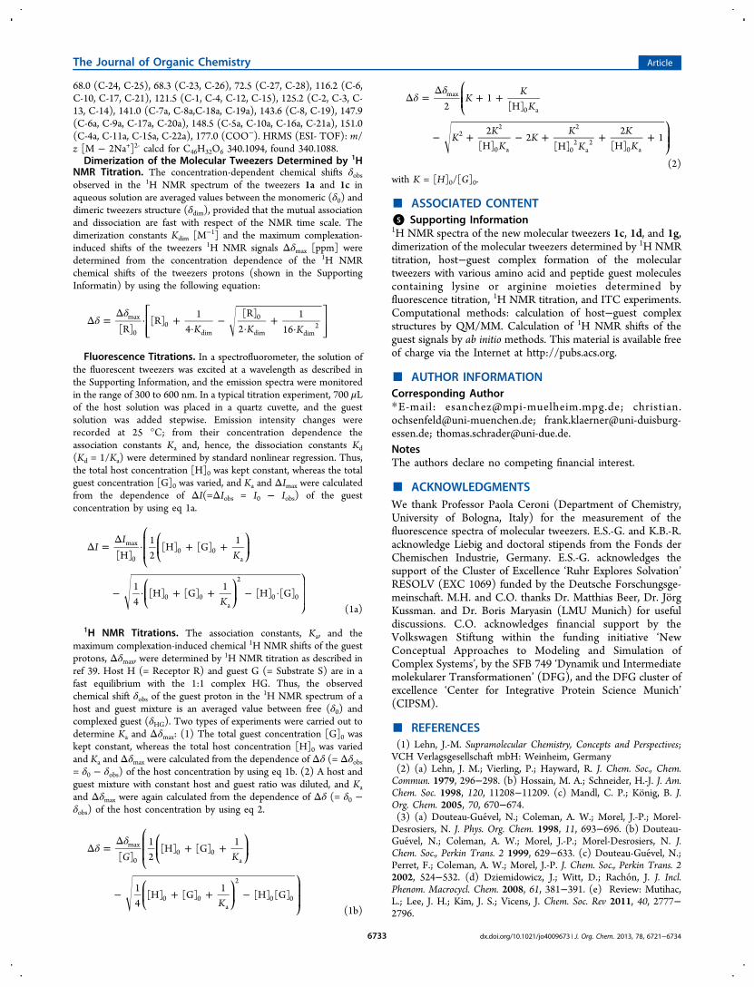
(Figure 4, Cp−Cp′ and Cm−Cm′) provide a measure of howopen the tweezers are. The X−Y distance between the tweezers’
X atom (X = P, S, C) and the Y atom of the guest side chainand the C1−C2−X angle between the opposite C atoms of thetweezers’ central hydroquinone bridges and the X atom of theguest side chain illustrate the degree of insertion of the aminoacid/peptide inside the tweezers’ cavity (Figure 4 andSupporting Information, Table S1). The predicted Cp−Cp′and Cm−Cm′ values for the isolated tweezers evidence a smalldependence on the nature of the substituent. For tweezers 1a′,1a″, 1c′, and 1d′, which have negatively charged substituents(not prone to interact with the cavity of the tweezers) thedistances Cp−Cp′ and Cm−Cm′ are very similar (at the QM/MM level the Cp−Cp′ and Cm−Cm′ distances of these tweezershave values of 5.24−5.49 Å and 3.51−3.89 Å, respectively;Table S1, Supporting Information). In 1b′ the methyl group ofthe substituent is prone to interact with the tweezers’ cavity(Figure 5). This is reflected in the larger distances for Cp−Cp′(5.94 Å) and Cm−Cm′ (4.33 Å) that correspond to a moreopen conformation of 1b′. For the noninteracting tweezers, theQM/MM distances are generally shorter than the MD averageand the QM/MM optimized structures are less symmetric(Figure 5). This is related to the tendency of the hydrogenatoms at the end of the tweezers to interact with the π systemof the aromatic ring at the opposite end side (see π-H-cdistances in Figure 4 and Supporting Information, Table S1).Such behavior is best described by the QM/MM calculations inwhich the tweezers are treated at the quantum level withempirical dispersion corrections (B3LYP-D2).Due to the interaction of the amino acids/peptide models
with the tweezers, the Cp−Cp′ and Cm−Cm′ distances getsomewhat shorter, indicating less open conformations of thetweezers. This is expected due to the positive charge of the sidechain of Lys and Arg and the negative electronic density in thetweezers cavity. In addition, the nature of the terminal groupsattached to Lys or Arg and the substituents in the tweezers alsoaffect the tweezers structure. In the host−guest complex 1b′·AcLys OMe′ the simultaneous interaction of the positive charge atthe ammonium group of the Lys side chain with the anionictweezer substituent results in an unfavorable conformation of
Table 2. Dissociation Constants Kd [μM] of Host−GuestComplexes of Tweezers 1a, 1b, 1c, and 1d with Lysine- orArginine-Containing Amino Acid and Peptide Derivativesa
guest 1a 1b 1c 1d
Ac Lys OMe 17 ± 6b,e 68 ± 1b 28 ± 2b 226 ± 14b
9 ± 6c 19 ± 3d 643 ± 7d
H Lys OH 21 ± 4b,e 874 ± 1b 227 ± 6c 1170 ± 20d
KAA 30 ± 3b 905 ± 1b 303 ± 5c 33333 ± 83d
KLVFF 20 ± 5b 38 ± 11c
KKLVFF 4 ± 1b 71 ± 1b
KKLVFFAK 7 ± 1b
KKKK 10 ± 7b,
Ac Arg OMe 60 ± 2b 178 ± 4b 882 ± 26b
20 ± 5c 77 ± 5d 281 ± 18d
H Arg OH 699 ± 15c 609 ± 27d
H Arg OMe 160 ± 6c
RGD 86 ± 1b
cRGDfV 59 ± 11c
cGRGDfL 26 ± 8c
aDissociation constants were determined by fluorometric titrationexperiments in aqueous phosphate buffer. The standard deviation ofKd is given in [%] of the determined value of Kd.
bPhosphate buffer,200 mM, pH = 7.6. cPhosphate buffer, 10 mM pH = 7.6. dPhosphatebuffer, 10 mM, pH = 7.2. eReference 10.
Table 3. Dissociation constants Kd and MaximumComplexation-Induced 1H NMR Shifts of Guest ProtonsΔδmax = δ0,G − δC,G Determined for Host−Guest Complexesof the Phosphate-, Phosphonate-, Sulfate-, and OCH2-Carboxylate-Substituted Tweezers 1a, 1b, 1c, and 1d withLysine- or Arginine-Containing Amino Acid and PeptideDerivativesa
host−guestcomplex Kd [μM] Δδmax [ppm]
1a·Ac Lys OMe 17 ± 0 3.91 (6-H), 0.51 (2-H), −0.32 (NCOCH3)1a·H Lys OH 40 ± 7 4.51 (6-H), 4.47 (5-H), 0.76 (3-H), 0.24
(2-H)1a·KAA 11 ± 9 5.82, 5.92 (9-H)b, 3.22 (8-H), 2.28 (7-H)1a·Ac Arg OMe 22 ± 4 3.75 (5-H), 2.54 (4-H), 1.23 (3-H), 0.63
(2-H)1b·Ac Lys OMe 227 ± 22d >4 (6-H)c, 1.45, 1.57 (5-H)b, 0.57 (2-H)1b·H Lys OH 714 ± 8d >4 (6-H)c
1b·KAA 833 ± 14d 2.80 (6-H), 1.03 (5-H), 0.29 (2-H)(1b)2·KTTK 147 ± 18d 0.42 (5-H), 0.41 (2-H)(1b)2·KTTKS 200 ± 24d 2.62 (5-H), 0.39 (2-H)1b·Ts Arg OMe 556 ± 40d 3.90 (5-H), 3.29, 4.09 (4-H)b, 1.00 (2-H)1b·RGD 653 ± 5d 3.41 (5-H), 0.92 (4-H), 1.50 (3-H)1c·Ac Lys OMe 12 ± 11 3.75 (6-H), 4.41 (5-H), 2.64 (4-H), 1.29
(3-H), 0.37 (2-H)1c·Ac Arg OMe 88 ± 5 3.86 (5-H), 2.51 (4-H), 1.32 (3-H), 0.42
(2-H)1d·Ac Lys OMe 1164 ± 11 0.94 (6-H), 0.54 (5-H), 0.40 (4-H), 0.77,
0.52 (3-H)b
1d·Ac ArgOMe
1393 ± 18 0.96 (5-H), 0.62, 0.48 (4-H)b, 0.52 (3-H)
aValues were determined by 1H NMR titration experiments at pH =7.2 in aqueous phosphate buffer, 75 mM for 1a, 25 mM for 1b, and 10mM for 1a·Ac Arg OMe, 1c, and 1d. bThe diastereotopic methyleneprotons show two separate signals in the complex. cThe signalbroadening does not allow the exact 1H NMR chemical shiftdetermination of the complex. dReference 9.
Figure 4. (A) Distances (black dotted lines) and angle (blue dottedline) used to describe the interaction between the molecular tweezersand the amino acid and peptides models (Y = P, S, or C atom of theOCH2CO2
− group of the tweezers, X = the N atom or the central Catom of the guanidinium moiety in the lateral chain of Lys or Arg,respectively). (B) Atoms of the lateral chains of Arg and Lys includedin the QM region used for the QM/MM optimizations are highlightedwith a sphere representation.
The Journal of Organic Chemistry Article
dx.doi.org/10.1021/jo4009673 | J. Org. Chem. 2013, 78, 6721−67346726

the lateral chain of Lys that causes the tweezer to adopt a moreopen conformation (Cp−Cp′ distance is 7.83 Å and Cm−Cm′distance is 6.82 Å, QM/MM values, Table S1). The host−guestorientation in 1b′·Ac Lys OMe′ and 1c′·Ac Lys OMe′ is
indicated by the X−Y distance and the angle C1−C2−X (4.06,4.06 Å and 65°, 62° respectively, Table S1). However, the sidechain in 1c′·Ac Lys OMe′ is better oriented, and the tweezereffectively traps the amino acid side chain. The effect of theterminal group is further illustrated in 1a′·KAA′, 1a′·Ac LysOMe′, and 1a′·Ac Arg OMe′. For these systems the Lys or Argside chain is very much threaded through the tweezer cavitywith the Lys ammonium or Arg guanidinium group pointingtoward the most negatively charged phosphate group of thetweezer as indicated by the value of C1−C2−X (89°, 98°, and89°, respectively, Table S1). This results in some repulsionbetween the amino acid terminal groups and the tweezershydrophobic body so the tweezers adopt more openconformations. For 1a′·H Lys OH the value of C1−C2−X is88°, but due to the small size of the substituent, no widening ofthe tweezer is observed (Table S1).Using the QM/MM optimized structures (with an explicit,
static 4-Å water layer around both the host−guest complex andthe pure guest molecule as well), the 1H NMR isotropicshielding constants of the guest protons in the host−guestcomplexes, δC, and in the pure guest molecules, δ0, werecomputed using ab initio methods. The computed results arecompared with the experimental values in Table 4. The fullNMR shielding tensors were calculated for all nuclei withinboth molecular entities with the HF method using the SVPbasis set,32 employing a local development version of the Q-Chem quantum chemical software package33 to yield thecomplexation-induced chemical shifts Δδmax. The reliability ofthe HF/SVP approach utilizing gauge including atomic orbitals(GIAO)35−37 has been shown elsewhere for comparablesystems.17,38−40 For the calculation of the nuclear shieldings,we apply linear-scaling methods,41,42 together with the recently
Figure 5. Structures of molecular tweezers 1a′ (≡ 1: R1 = OPO32‑, R2
= OP(OH)O2−), 1a″ (≡ 1: R1 = R2 = OP(OH)O2
−), 1b′ (≡ 1: R1 =R2 = OP(Me)O2
−), 1c′ (≡ 1: R1 = R2 = OSO3−), or 1d′ (≡ 1: R1 = R2
= OCH2CO2−) optimized by QM/MM calculations.
Table 4. Comparison of Experimental and Computational (HF/SVP) Complexation-Induced Chemical Shifts Δδmax (ppm) forGuest Protons in the Host−Guest Complexes of Tweezers 1a, 1b, 1c, and 1d with Lysine and Arginine Derivatives (Table 3)a
host−guest complex 6-H 5-H 4-H 3-H 2-H
exptl: 1a·H Lys OH 4.51 4.47 0.76 0.24calcd: 1a′·H Lys OH′ 6.04 4.11 1.99 0.38 0.37exptl: 1a·Ac Lys OMe 3.91 0.51b
calcd: 1a′·Ac Lys OMe′ 3.62 5.51 4.62 1.24 2.20b
exptl: 1a·KAA 5.92 3.22 2.28 1.09 0.20c
calcd: 1a′·KAA′ 5.71 5.08 2.55 0.36 0.06c
exptl: 1b·Ac Lys OMe >4 1.57, 1.45d 0.57calcd: 1b′·Ac Lys OMe′ 3.46 3.42, 3.21d 1.72 0.39 2.30exptl: 1c·Ac Lys OMe 3.75 4.41 2.64 1.29 0.37calcd: 1c′·Ac Lys OMe′ 4.39 3.19 1.10 0.33 0.27exptl: 1d·Ac Lys OMe 0.94 0.54 0.40 0.77, 0.52d
calcd: (1d′·Ac Lys OMe′)in 5.44 3.05 1.69 0.18, −0.92 1.33calcd: (1d′·Ac Lys OMe′)out 0.03 0.72 0.41 0.85, 0.08 0.46exptl: 1a·Ac Arg OMe 3.75 2.54 1.23 0.63calcd: 1a′·Ac Arg OMe′ 5.46 2.46 0.56 1.07exptl: 1b·Ts Arg OMe 3.90 4.09, 3.29d 1.00calcd: 1b′·Ts Arg OMe′ 4.30 2.51, 1.67d 0.87 −0.50exptl: 1c·Ac Arg OMe 3.86 2.51 1.32 0.42calcd: 1c′·Ac Arg OMe′ 3.86 0.63 0.41 0.42exptl: 1d·Ac Arg OMe 0.96 0.62, 0.48d 0.52calcd: (1d′·Ac Arg OMe′)in 3.36 1.39, 1.04d −0.20 1.93calcd: (1d′·Ac Arg OMe′)out 0.26 0.38, 0.36d 0.31 0.67
aThe ab initio data were calculated for the complex structures of 1a′, 1b′, 1c′, and 1d′ with the corresponding lysine and arginine derivatives shownin Figure 6. bΔδmax: −0.32 (exptl), −0.28 (calcd), NCOCH3; −0.23 (exp), −0.16 (calcd), CO2CH3.
c6-, 5-, 4-, 3- ,2-H ≡ 9-, 8-, 7-, 6-, 5-H in KAA.dDiastereotopic H atoms.
The Journal of Organic Chemistry Article
dx.doi.org/10.1021/jo4009673 | J. Org. Chem. 2013, 78, 6721−67346727

developed density matrix-based Laplace reformulation ofcoupled self-consistent field equations (DL-CPSCF)42,43 Incases where the flexibility of two or more close-by rotatinghydrogens yields only one peak in the experimental NMRspectrum, the calculated results were averaged accordingly overthese hydrogen shifts. For diastereotopic hydrogens in host−guest species 1b·Ac Lys OMe and 1b·Ts Arg OMe labeled withfootnote “d” in Table 4, the computed hydrogen shifts areassigned in two different possible ways and the values closer tothe experimental ones are chosen to represent the calculateddiastereotopic NMR shifts. This simple scheme was chosen,since there was no clear evidence that either of thediastereotopic hydrogens would favor a certain position inthe optimized host−guest and pure guest structures, forexample, close to the tweezers’ bridge or near the tweezers’tips. The final complexation-induced NMR chemical shifts, asdepicted in Table 4, were calculated as differences between theshieldings of specific nuclei in these structures. Representativecalculations of complexation-induced NMR shifts for the host−guest complexes 1a′·H Lys OH′, 1a′·Ac Lys OMe′, and1a′·KAA′ with a 6-Å water layer show a maximum absolutedeviation of roughly 0.4 ppm from the 4-Å results presented inTable 4.While some of the complexation-induced shifts listed in
Table 4 agree reasonably well with the experimental 1H NMRchemical shifts, the mean absolute deviations (MAD) from theexperimental values range from 0.6 to 1.5 ppm depending onthe host−guest case (the carboxylate tweezer complexes are notconsidered here). Here, one should note that the complex-induced shifts range up to 6 ppm. The general trend is that theinfluence of the complexation on the chemical shifts increasesas a function of a hydrogen position more deeply located in thehost cavity. Thus, in general, the part of the guest moleculemore inside the host cavity provides the largest Δδmax, and theparts furthest away from the cavity the smallest.It should be noted that for guest molecules interacting with
the carboxylate tweezer 1d, there are two sets of differentconformers. Both of these host−guest complexes wereoptimized by QM/MM methods and correspond to differentregions in the conformational space. For these conformers, twosets of complexation-induced shifts were computed. As seen inTable 4, the computed shifts are very different for structures(1d′·Ac Lys OMe′)in and (1d′·Ac Lys OMe′)out or (1d′·Ac ArgOMe′)in and (1d′·Ac Arg OMe′)out. Here most likely a rapidequilibrium between the “in” and “out” conformers of thesehost−guest species is expected that proceeds by rapid mutualcomplex formation and dissociation. Therefore, the exper-imental NMR shift data are evidently the average between thedata of the equilibrating “in” and “out” conformers. These typesof equilibria are not ruled out in the more stable cases either,although they appear to be most unlikely due to the large Δδmaxvalues and the better correspondence between computed andexperimental data. The situation here is thus rather differentfrom the previously studied molecular tweezers,17,38−40 wherethe guest molecules were more rigid and did not have the sameconformational freedom as the flexible lysine or arginine sidechains. Thus, dynamical equilibria play a more important rolefor the present systems.The basis set was confirmed to be adequate by carrying out
HF/def2-TZVP45 calculations, which yielded only minutechanges in the Δδmax values, and electron correlation effects,as studied with second-order Møller−Plesset perturbationtheory (MP2), also play a minor role based on previous
studies on similar systems.37 Therefore, the remainingdeviations between experimental and calculated chemical shiftsare thus most likely attributed to the present static treatment ofboth the solvent-including host−guest structures and freeguests. A snapshot type of averaging of NMR chemical shiftscould provide a closer agreement with the experimental NMRchemical shifts, as seen for example in refs 46−50, but wouldrequire a substantial computational effort for the complexsystems of the present work.The large theoretical shifts of 1H NMR guest signals, Δδmax,
calculated for the methylene protons 6-H or 5-H (adjacent tothe ammonium or guanidinium moiety of the lysine or arginineside chain) in the host−guest complexes with the tweezers 1a′,1b′, and 1c′ agree well (within the limit of roughly 0.5 ppm)with the experimental values (Δδmax = 3.9 − 5.9 ppm)determined for the corresponding complexes of the tweezers1a, 1b, and 1c (Table 4). Exceptions are the complexes 1a·HLys OH and 1a·Ac Arg OMe, in which the differences betweenthe theoretical and experimental Δδmax values of these protonsare larger (up to 1.5 ppm). Larger differences between thetheoretical and experimental Δδmax values were also determinedfor the other side chain guest protons in the complexes of 1a,1b, and 1c, but the trend of decreasing complexation-inducedshifts, Δδmax, of the 1H NMR signals of the guest protons 6-H >5-H > 4-H > 3-H > 2-H of the lysine side chain and 5-H > 4-H> 3-H > 2-H of the arginine side chain, respectively, are wellreproduced by the calculations. These f indings conf irm theassumption that the lysine and arginine guest molecules are boundby the phosphate-, methanephosphonate-, and sulfate-substitutedtweezers 1a, 1b, and 1c by threading the side chains through thetweezers’ cavity with the positively charged ammonium orguanidinium end group pointing toward one of the anionic groupsattached to the central benzene bridge of the tweezers. The stabilityand structures of these host−guest complexes are, evidently,determined by attractive ionic, CH-π, and hydrophobicinteractions. The differences between the theoretical andexperimental Δδmax values indicate that due to the flexibilityof the guest side chains these host−guest complexes consist ofmore than one structure that exist in a dynamic equilibrium sothat in the NMR spectrum only averaged signals are observedresulting from the rapidly equilibrating structures. The selectivebroadening of the shifted guest signals observed in the 1HNMR spectra of these complexes provides further evidence forsuch a dynamic exchange between several structures proceedingat a rate that is similar to the NMR time scale. In the case of thephosphate tweezer 1a, Δδmax values of several complexstructures of the phosphate and the partial protonatedphosphate tweezers (1a′, 1a″, 1a‴ ≡ 1: R1 = R2 = OPO3
2‑,1: R1 = OPO3
2‑, R2 = OP(OH)O2−, or 1: R1 = R2=
OP(OH)O2−) with several lysine and arginine guest molecules
(Supporting Information Figure S7 and Table S2) werecalculated to be different from those shown in Table 4, butthe trend in the shifts of the side chain 1H NMR signalsremains the same. This finding supports the assumption thateach host−guest complex consists of several structures with theguest side chain, however, threaded through the tweezers’cavity in all cases. In the host−guest complexes of thecarboxylate-substituted tweezer 1d with Ac Lys OMe or AcArg OMe as guest molecules, the Δδmax values were determinedto be substantially smaller (< 1 ppm; Table 4) for thecorresponding lysine or arginine side chain protons indicatingcomplex structures in which the guest side chain is positionedoutside the tweezer’s cavity. The comparison of the
The Journal of Organic Chemistry Article
dx.doi.org/10.1021/jo4009673 | J. Org. Chem. 2013, 78, 6721−67346728

experimental Δδmax values with theoretical values calculated forthe complex structures, where the guest side chain is positionedeither inside or outside the tweezer’s cavity, provides goodevidence that both complexes exist as rapid equilibria, (1d′·AcLys OMe′)in ⇌ (1d′·Ac Lys OMe′)out or (1d′·Ac Arg OMe′)in⇌ (1d′·Ac Arg OMe′)out, in each complex with preference forthe outside structure. Apparently, the extended OCH2CO2
−
groups of 1d direct the guest molecule to a position outside ofthe tweezer’s cavitiy, and the major host−guest binding force isthe electrostatic attraction between the carboxylate groups ofthe tweezer and the cationic ammonium or guanidinium moietyof the guest molecule. QM/MM calculations produce chelatearrangements between both carboxylates in 1d and thecomplexed amino acid cation outside the tweezer’s cavity;these are possible only because of the extra methylene group inthe OCH2CO2
− side chain, which is absent in 1a, 1b, and 1c(Figure 6). The loss of CH-π and hydrophobic interactions inthis geometry explains why the complexes of 1d aresignificantly less stable than those with phosphate or sulfatetweezers 1a or 1c. Evidently, these host−guest bindinginteractions inside the tweezers’ cavities of 1a−c deliver animportant contribution to the complex stability besides theionic interaction of the tweezers’ phosphate or sulfate group
and the amino acid cationic side chain. A special case ismethanephosphonate-substituted tweezer 1b. The methylgroup of one OP(CH3)O2
− side chain was calculated byQM/MM to point toward the tweezer’s cavity (Figure 5).Thus, the binding of a guest side chain by threading requires arotation around the O−P bond in 1b. This may explain why thehost−guest complexes of 1b are less stable than thecorresponding complexes of 1a and 1c. A similar weakeningof the complex stability has been observed for related hostmolecules bearing O-alkyl groups in the central benzene bridgein organic media. In these cases the O-alkyl groups were alsocalculated to point toward the host cavity and, hence, block theguest inclusion.51 Finally the different complex stabilities of thephosphate and sulfate tweezers 1a and 1c certainly result fromthe larger negative charge of the phosphate compared to thesulfate.
Solvent Dependence of the Host−Guest ComplexFormation. The solvent dependence of the amino acidbinding to the tweezers is remarkable, because not only is thecomplex stability solvent-dependent but also the complexstructure. For example, the very small complexation-induced 1HNMR shifts (Δδobs) observed for the binding of Ac Lys OMeby the phosphate tweezer 1a in methanol (Table 5) indicate a
Figure 6. Two viewing angles of the host−guest complex structures of the phosphate, phosphonate, sulfate, and carboxylate tweezers 1a′, 1b′, 1c′,and 1d′ with lysine and arginine derivatives (without counterions) optimized by QM/MM calculations. Each structure contains a 60 Å water layer(not shown).
The Journal of Organic Chemistry Article
dx.doi.org/10.1021/jo4009673 | J. Org. Chem. 2013, 78, 6721−67346729

complex structure where the guest molecule is positionedoutside the tweezer cavity comparable to the host−guestcomplex structures of the carboxylate tweezer 1d discussed inthe previous paragraph. Evidently, the ionic interaction betweenone of the negatively charged host phosphate groups and thepositively charged guest ammonium group is the dominatingbinding force in methanol and the weakened contribution ofdispersive and hydrophobic interactions gained by amino acidside chain threading seems to be less important in methanol.52
At the same time, the intensity of the tweezer’s emissionband is increased on guest binding in methanol but quenched
in aqueous buffer (1a·Ac Lys OMe). We conclude that thedirection and extent of emission intensity change also providesstructural information. In the mixture of methanol withaqueous phosphate buffer (1:2) containing 1a and Ac LysOMe, relatively large Δδobs values were observed (Table 5)indicating that the complex is at least partially formed bythreading of the lysine side chain into the tweezer’s cavitycomparable to the complex formation in pure aqueousphosphate buffer. In this solvent mixture, however, no changein the tweezer’s emission intensity could be detected by theaddition of Ac Lys OMe. These findings strongly suggest that
Table 5. Solvent Dependence of Change in Intensities of the Tweezers’ Emission Bands (ΔImax [%] = 100(I0 − Imax)/I0, of KdValues (Resulting from Fluorometric Titration Experiments), and of the Complexation-Induced 1H NMR Shifts of the GuestProtons Δδobs at Host and Guest Concentration [H]0 = [G]0 = 1.0 mMa
Δδobshost guest solvent ΔImax [%] Kd [μM] 6-H 5-H 4-H
1a Ac Lys OMe MeOH −6 66 ± 11 0.24 0.28 0.26MeOH/PB (1:2) nd 2.80 2.30 0.74PB 40 9 ± 6 3.60 3.40 1.60
Ac Arg OMe MeOH 0 0.76 0.44MeOH:PB (2:1) 0 0.72 0.46MeOH:PB (1:2) nd nd 1.45 0.96MeOH:PB (1:9) 27 157 ± 8 2.16 1.47PB 47 20 ± 5 3.30 2.19
1c Ac Lys OMe MeOH −44 20 ± 6 2.96 3.25 1.75PB 44 19 ± 3 3.40 3.30 1.30
Ac Arg OMe MeOH −22 276 ± 7 0.67 0.30PB 30 77 ± 5 2.67 1.70
1d Ac Lys OMe PB 36 643 ± 7 0.94b 0.54b 0.40b
Ac Arg OMe PB 13 281 ± 18 0.96b 0.62b
aI0 = emission intensity of free tweezers, Imax = emission intensity of host-guest complexes. The standard deviation of Kd is given in %. PB = aqueousphosphate buffer (10 mM) at pH = 7.2; nd = not determined. bΔδmax.
Figure 7. Plots of the isothermal titration calorimetry (ITC) measurements in aqueous phosphate buffer (10 mM, pH = 7.6) for the complexformation between phosphate tweezer 1a and Ac Lys OMe (left) and 1a and Ac Arg OMe (right).
The Journal of Organic Chemistry Article
dx.doi.org/10.1021/jo4009673 | J. Org. Chem. 2013, 78, 6721−67346730

the fluorescence is quenched if the guest is bound inside thetweezer’s cavity and increased if the guest is bound outside thecavity. In certain mixtures of methanol and aqueous buffer thesetwo effects seem to compensate each other, so that no intensitychange of the tweezer’s emission band occurs on the addition ofthe guest molecule.Determination of the Thermodynamic Parameters of
the Host−Guest Complex Formation by IsothermalTitration Calorimetry (ITC). The thermodynamic parametersfor the host−guest complex formation of the phosphate- andsulfate-substituted tweezers 1a and 1c with several lysine andarginine guest molecules were also determined by isothermaltitration calorimetry (ITC). Figure 7 shows the plots oftitration for the binding of Ac Lys OMe and Ac Arg OMe bytweezer 1a as representative examples. The dissociationconstants (Kd, Table 6) agree well with the data determinedindependently by fluorometric or 1H NMR titration experi-ments (Table 2 and 3). The additional thermodynamicparameter, the binding enthalpy ΔH and entropy TΔS,obtained by the ITC measurements, provide further insightinto the binding mode. The more negative ΔH valuedetermined for the formation of 1a·Ac Arg OMe indicatesthat the arginine guest is enthalpically bound tighter to 1a thanthe corresponding lysine guest. This tighter enthalpic bindingof Ac Arg OMe is overcompensated by a less favorable entropyso that the complex 1a·Ac Lys OMe is more stable (has asmaller dissociation constant, Kd) than 1a·Ac Arg OMe. Thiskind of enthalpy−entropy compensation is often found insupramolecular systems.53 Here the arginine guest loses degreesof rotational freedom inside the tweezer cavity due to its rigiddelocalized guanidinium ion, as opposed to lysine’s smalllocalized ammonium ion that continues to rotate around its C−N and C−C bonds inside the tweezer cavity. In addition, thelocalized lysine ammonium cation is certainly solvated by morewater molecules than the delocalized arginine guanidiniumcation so that in the former case more water molecules arereleased to the bulk than in the case of arginine; this is alsoentropically favorable. Similar results were earlier obtained forthe complex formation of a sulfonatocalix[4]arene derivativewith lysine or arginine in water.3b Also in this case, lysinebinding to the calixarene was entropically favored whereasarginine complexation produced the larger enthalpy gain.Transition to host−guest complex formation between thesulfate-substituted tweezer 1c and Ac Lys OMe or Ac Arg OMefurnishes Kd values determined by ITC that again agree wellwith those determined by fluorescence or 1H NMR titrationexperiments. In these cases, however, both enthalpy andentropy changes are more negative for Ac Lys OMecomplexation than those measured for Ac Arg OMe. There is
no obvious explanation for these differences between 1a and 1c.Assistance comes from close inspection of the calculatedcomplex structures (Figure 6): the guanidinium cation of thearginine side chain is more tightly bound to the doubly chargedphosphate anion from 1a than to the singly charged sulfateanion from 1c, which coincides with the superior enthalpy gainof the phosphate tweezer. On the other hand, the calculatedstructure of 1c·Ac Lys OMe suggests a chelate-type interactionbetween lysine’s ammonium inside the tweezer’s cavity andboth sulfate arms from 1c - which leads to a powerful enthalpygain at the cost of a substantial entropy loss.
■ CONCLUSION
The behavior of the water-soluble molecular tweezers 1a−d(bearing phosphate, methanephosphonate, sulfate, or OCH2-carboxylate groups in their central benzene bridge) wasinvestigated in buffered aqueous solution at almost neutralpH by three independent methods (fluoroscence, NMR, andITC titration experiments). They all form stable host−guestcomplexes with various amino acid and peptide guestscontaining either lysine or arginine moieties. In the case oftweezers 1a−c large complexation-induced shifts of the 1HNMR guest signals (Δδmax ≤ 6 ppm) were found for themethylene protons of the lysine or arginine side chain. Theseagree well with theoretical data calculated by a combination ofQM/MM and ab initio methods for the host−guest complexstructures shown in Figure 6. This correlation providesexperimental evidence for the postulated binding mode: thelysine or arginine side chain is literally threaded through thetweezers’ cavity with its positively charged ammonium orguanidinium end group pointing toward one of the anionicgroups on the tweezers’ central benzene bridge. Thus, attractiveionic, CH-π, and hydrophobic interactions are the majorbinding forces that determine the stability and structures ofthese host−guest complexes. Substantially smaller complex-ation-induced shifts of the corresponding 1H NMR guestsignals (Δδmax < 1 ppm) were found for the host−guestcomplexes of the carboxylate-substituted tweezer 1d with AcLys OMe or Ac Arg OMe as guest molecule. Comparison ofexperimental with theoretical Δδmax values calculated for thecomplex structures with included or externally bound guest(Figure 6) indicates that both complexes exist as rapidequilibria, (1d′·Ac Lys OMe′)in ⇌ (1d′·Ac Lys OMe′)out or1d′·Ac Arg OMe′)in ⇌ (1d′·Ac Arg OMe′)out, in each complexwith a significant preference for the external binding mode. Theextended OCH2CO2
− groups of 1d appear to direct the guestside chain to an external position (outside of the tweezer’scavity), and the major binding force becomes the electrostaticattraction between the tweezer carboxylates and the amino acid
Table 6. Thermodynamic Parameters for Complex Formation between Molecular Tweezers 1a or 1c and Lysine- or Argine-Containing Guest Moleculesa
host guest Ka ΔG ΔH −TΔS Kd
1a Ac Lys OMe 6.86 ± 0.07 −6.6 ± 0.1 −5.6 ± 0.1 −1.0 ± 0.1 14.6 ± 0.21a Ac Arg OMe 2.96 ± 0.21 −6.1 ± 0.1 −7.0 ± 0.2 +0.9 ± 0.3 34.0 ± 0.31a KLVFF 6.56 ± 0.26 −6.6 ± 0.1 −6.4 ± 0.1 −0.2 ± 0.1 15.2 ± 0.11c Ac Lys OMe 3.46 ± 0.16 −6.2 ± 0.1 −8.3 ± 0.1 +2.1 ± 0.1 28.9 ± 1.31c Ac Arg OMe 1.04 ± 0.09 −5.5 ± 0.1 −6.3 ± 0.4 +0.8 ± 0.5 96.9 ± 8.4
aParameters include binding constant Ka [104 M−1], binding Gibbs enthalpy ΔG [kcal/mol], binding enthalpy ΔH [kcal/mol], binding entropy TΔS
[kcal/mol], and dissociation constant Kd [μM] (= 1/Ka).Values were determined by isothermal titration calorimetry (ITC) measurements inaqueous phosphate buffer (10 mM, pH = 7.6). In each case, the listed values are the average of two independent measurements, and the listed errorsare the deviations from the mean values of the two measurements.
The Journal of Organic Chemistry Article
dx.doi.org/10.1021/jo4009673 | J. Org. Chem. 2013, 78, 6721−67346731

ammonium or guanidinium moiety. The loss of CH-π andhydrophobic interactions explains why these complexes aresignificantly less stable than those with phosphate or sulfatetweezers 1a or 1c. QM/MM calculations produce chelatearrangements between both carboxylates in 1d and thecomplexed amino acid cation; these are preferred for 1dcomplexes because of the extra methylene group, which isabsent in 1a and 1c. Solvent-dependence and ITC measure-ments provide further insight into the noncovalent host−guestbinding modes. In methanol ionic interactions between aminoacid cation and tweezers anions become the dominatingbinding force for all tweezers. Now, the guest side chain ispreferentially positioned outside each tweezers’ cavity; this ismost likely caused by the absence of the hydrophobic effect inthis solvent; in addition, methanol molecules may occupy thecavity. As a consequence, the hydrophobic effect in water seemsto be the major force that drags the guest molecule into thecavity of the tweezers. The thermodynamic parameters(enthalpy, ΔH, and entropy, TΔS) determined by ITC(Table 6) indicate powerful enthalpy-driven guest attraction,featuring an enthalpy−entropy compensation that is oftenobserved in supramolecular systems.53 In complex 1a·Ac ArgOMe a stronger enthalpic host−guest binding (ΔH(1a·Ac Arg OMe)< ΔH(1a·Ac Lys OMe)) is overcompensated by an unfavorablemore negative entropic parameter (TΔS(1a·Ac Arg OMe) <TΔS(1a·Ac Lys OMe)) compared to 1a·Ac Lys OMe, so that thelater complex is more stable at room temperature. Thethermodynamic parameters of the corresponding complexesof the sulfate tweezer 1c show no similar enthalpy−entropycompensation. This different behavior of complexes of 1c canbe explained by inspection of the calculated complex structures(Figure 6). In the structure of 1c′·Ac Lys OMe′ the guestammonium group is calculated to interact with both tweezersulfate groups, leading to a restriction of the rotation of guestside chain inside the tweezer cavity, which may explain thenegative entropy parameter compared to 1a·Ac Lys OMe. Theexperimentally observed properties of the tweezers 1a, 1b, and1c on one side and 1d on the other side are in good accordwith the calculation by means of molecular mechanics andquantum chemical methods. These findings provide goodconfidence into the methods applied here for the elucidation ofthe host−guest structures and stabilities.Thus, the combinationof experiment and theory provide deep insight into the bindingmodes of these molecular tweezers to the amino acids lysineand arginine, which is an essential prerequisite for theunderstanding of the intriguing effect of these tweezers onthe aggreation of proteins and the activity of enzymes.
■ EXPERIMENTAL SECTIONGeneral Experimental Details. 1H NMR, 13C NMR, DEPT
H,H−COSY, C,H−COSY, NOESY, HMQC, and HMBC titrationexperiments were carried out by using a 500 MHz spectrometer. Theundeuterated amount of the solvent was used as an internal standard.The 1H and 13C NMR signals were assigned by the 2D experimentsmentioned above. Positions of the protons of the methano bridges areindicated by the letters i (innen, toward the center of the molecule)and a (aussen, away from the center of the molecule). The numberingof the atoms in the tweezers is shown in the Supporting Information.Fluorescence spectra were measured on a spectrofluorometer. Massspectra were recorded on an ESI-TOF mass spectrometer. Isothermaltitration calorimetry (ITC) measurements were carried out by the useof a microcalorimeter at 25.0 °C. All melting points (mp) areuncorrected. All solvents were distilled prior to use.
Sulfate Tweezer 1c. A stirred solution of 75 mg (0.132 mmol) ofthe dihydroxy tweezer 1e and 85 mg (0.53 mmol) of sulfur trioxidepyridinium complex in 7 mL of dry pyridine was heated under reflux at90 °C for 24 h. Then, an additional 63 mg (0.397 mmol) of SO3·Pycomplex was added to this solution, and the mixture was stirred foranother 36 h at the same temperature. The mixture was cooled to rtand quenched with saturated aqueous NaHCO3 solution. The excessof inorganic salts was filtered off by a glass filter (D4), and the aqueousfiltrate was extracted with diethyl ether (3 × 50 mL). The solvent ofthe aqueous phase was distilled off in vacuum in a rotary evaporator.The solid residue was suspended in ethanol and filtered off. Thesolvent of the filtrate was removed in a rotary evaporator, and theremaining solid was dried in oil pump vacuum. The tweezer 1c wascollected as white solid. Yield: 70 mg, 68%; mp >229 °C(decomposition). 1H NMR (500 MHz, CD3OD): δ [ppm] 2.26 (td,2H, H-24a, H-25a), 2.33 (s, 4H, H-23, H-26), 2.51 (td, 2H, H-24i, H-25i), 4.00 (4H, H-5, H-11, H-16, H-22), 4.48 (s, 4H, H-7, H-9, H-18,H-20), 6.78 (m, 4H, 4H, H-2, H-3, H-13, H-14), 7.02 (m, 4H, H-4, H-12, H-1, H-15), 7.10 (s, 4H, H-6, H-10, H-17, H-21). 1H NMR (500MHz,D2O): δ [ppm] 2.34 (d, 2H, H-24a, H-25a), 2.39 (m, 4H, H-23,H-26), 2.48 (d, 2H, H-24i, H-25i), 4.20 (4H, H-5, H-11, H-16, H-22),4.45 (s, 4H, H-7, H-9, H-18, H-20), 6.06 (br, s, 4H, 4H, H-2, H-3, H-13, H-14), 7.03 (m, 4H, H-4, H-12, H-1, H-15), 7.18 (s, 4H, H-6, H-10, H-17, H-21). 13C NMR (125,7 MHz, CD3OD): δ [ppm] 50.0 (C-7, C-9, C-18, C-20), 52.4 (C-5, C-11, C-16, C-22), 69.0, 69.4 (C-23,C-24, C-25, C-26), 117.3 (C-6, C-10, C-17, C-21), 122.1 (C-4, C-12,C-1, C-15), 125.8 (C-2, C-3, C13, C-14), 139.3 (C-8, C-19) 144.8 (C-7a, C-8a, C-18a, C-19a), 148.6 (C-6a, C-9a, C-17a, C-20a), 149.2 (C-5a, C-10a, C-16a, C-21a), 151.9 (C-4a, C-11a, C-15a, C-22a). HRMS(ESI-TOF) m/z: [M + Na]+ calcd for C42H28O8S2Na3 793.0913,found 793.0944; m/z [M − Na]− calcd for C42H28O8S2Na 747.1129,found 747.1131; m/z [M − 2Na]2‑ calcd for C42H28O8S2 362.0618,found 362.0635.
Methyl Carboxylate Tweezer 1g. Under argon atmosphere 20mg (0.035 mmol) of the dihydroxy tweezer 1e was dissolved in 20 mLof dry acetone. Then 21.5 mg (0.14 mmol, 0.013 mL) of methylbromoacetate, 19 mg of potassium carbonate (0.14 mmol), and a fewgranules of potassium iodide were added to this solution, and themixture was stirred for 4 days at room temperature. Dichloromethane(50 mL) was added to this mixture, and the solution was washed withsaturated aqueous NH4Cl, saturated aqueous NaHCO3, and distilledwater. The organic phase was dried over Na2SO4, and the solvent wasremoved in vacuum in a rotary evaporator. The oily residue waspurified by column chromatography (silica gel, cyclohexane/EtOAc3:1) leading to tweezer 1g substituted by OCH2CO2Me groups ascolorless solid. Yield: 25 mg of 1g (0.030 mmol, 99%); mp 240 °C(decomposition). 1H NMR (500 MHz,CDCl3): δ [ppm] 2.35 (m, 4H,H-23, H-26), 2.43 (m, 4H, H-24, H-25), 3.66 (s, 6H, H-29, H-30),4.08 (m, 4H, H-5, H-11, H-16, H-22), 4.27 (m, 4H, H-7, H-9, H-18,H-20), 4.38 (s, 4H, H-27, H-28), 6.76 (m, 4H, H-2, H-3, H-13, H-14),7.08 (m, 4H, H-1, H-4, H-12, H-15), 7.14 (s, 4H, H-6, H-10, H-17, H-21). 13C NMR(125 MHz,CDCl3): δ [ppm] 14.4 (C-29, C-30), 48.5(C-7, C-9, C-18, C-20), 51.5 (C-5, C-11, C-16, C-22), 69.3 (C-24, C-25), 69.9 (C-23, C-26), 70.7 (C-27, C-28), 116.4 (C-6, C-10, C-17, C-21), 121.6 (C-1, C-4, C-12, C-15), 124.7 (C-2, C-3, C-13, C-14),140.3 (C-7a, C-8a,C-18a, C-19a), 144.5 (C-8, C-19), 147.1 (C-6a, C-9a, C-17a, C-20a), 147.8 (C-5a, C-10a, C-16a, C-21a), 150.6 (C-4a, C-11a, C-15a, C-22a), 170.1 (C-27a, C-28a). HRMS (ESI-TOF): m/z[M + Na+] calcd for C48H38O6Na 733.2561, found 733.2567.
Carboxylate Tweezer 1d. NaOH·H2O (0.082 mmol) was addedto the stirred solution of 29 mg (0.041 mmol) of methyl carboxylatetweezer 1g in 5 mL of dry methanol. After 1 h of stirring the solventwas removed in a rotary evaporator, and the solid residue was dried invacuum. Yield of 1d: 30 mg (0.041 mmol, >99%); mp > 255 °C(decomposition). 1H NMR (500 MHz, D2O): δ [ppm] 2.36 (m, 8H,H-23, H-24, H-25, H-26), 4.03 (s, 4H, H-5, H-11, H-16, H-22), 4.18(s, 4H, H-7, H-9, H-18, H-20), 4.33 (s, 4H, H-27, H-28), 6.75 (br s,4H, H-2, H-3, H-13, H-14), 7.16 (br s, 4H, H-1, H-4, H-12, H-15),7.27 (s, 4H, H-6, H-10, H-17, H-21). 13C NMR (125 MHz, D2O): δ[ppm] 47.8 (C-7, C-9, C-18, C-20), 50.8 (C-5, C-11, C-16, C-22),
The Journal of Organic Chemistry Article
dx.doi.org/10.1021/jo4009673 | J. Org. Chem. 2013, 78, 6721−67346732
68.0 (C-24, C-25), 68.3 (C-23, C-26), 72.5 (C-27, C-28), 116.2 (C-6,C-10, C-17, C-21), 121.5 (C-1, C-4, C-12, C-15), 125.2 (C-2, C-3, C-13, C-14), 141.0 (C-7a, C-8a,C-18a, C-19a), 143.6 (C-8, C-19), 147.9(C-6a, C-9a, C-17a, C-20a), 148.5 (C-5a, C-10a, C-16a, C-21a), 151.0(C-4a, C-11a, C-15a, C-22a), 177.0 (COO−). HRMS (ESI- TOF): m/z [M − 2Na+]2‑ calcd for C46H32O6 340.1094, found 340.1088.Dimerization of the Molecular Tweezers Determined by 1H
NMR Titration. The concentration-dependent chemical shifts δobsobserved in the 1H NMR spectrum of the tweezers 1a and 1c inaqueous solution are averaged values between the monomeric (δ0) anddimeric tweezers structure (δdim), provided that the mutual associationand dissociation are fast with respect of the NMR time scale. Thedimerization constants Kdim [M−1] and the maximum complexation-induced shifts of the tweezers 1H NMR signals Δδmax [ppm] weredetermined from the concentration dependence of the 1H NMRchemical shifts of the tweezers protons (shown in the SupportingInformatin) by using the following equation:
δδ
Δ =Δ
· +·
−·
+·
⎡⎣⎢⎢
⎤⎦⎥⎥K K K[R]
[R]1
4[R]
21
16max
00
dim
0
dim dim2
Fluorescence Titrations. In a spectrofluorometer, the solution ofthe fluorescent tweezers was excited at a wavelength as described inthe Supporting Information, and the emission spectra were monitoredin the range of 300 to 600 nm. In a typical titration experiment, 700 μLof the host solution was placed in a quartz cuvette, and the guestsolution was added stepwise. Emission intensity changes wererecorded at 25 °C; from their concentration dependence theassociation constants Ka and, hence, the dissociation constants Kd
(Kd = 1/Ka) were determined by standard nonlinear regression. Thus,the total host concentration [H]0 was kept constant, whereas the totalguest concentration [G]0 was varied, and Ka and ΔImax were calculatedfrom the dependence of ΔI(=ΔIobs = I0 − Iobs) of the guestconcentration by using eq 1a.
Δ =Δ
· + +
− · + + − ·
⎛
⎝⎜⎜
⎛⎝⎜
⎞⎠⎟
⎛⎝⎜
⎞⎠⎟
⎞
⎠⎟⎟
II
K
K
[H]12
[H] [G]1
14
[H] [G]1
[H] [G]
max
00 0
a
0 0a
2
0 0
(1a)
1H NMR Titrations. The association constants, Ka, and themaximum complexation-induced chemical 1H NMR shifts of the guestprotons, Δδmax, were determined by 1H NMR titration as described inref 39. Host H (= Receptor R) and guest G (= Substrate S) are in afast equilibrium with the 1:1 complex HG. Thus, the observedchemical shift δobs of the guest proton in the 1H NMR spectrum of ahost and guest mixture is an averaged value between free (δ0) andcomplexed guest (δHG). Two types of experiments were carried out todetermine Ka and Δδmax: (1) The total guest concentration [G]0 waskept constant, whereas the total host concentration [H]0 was variedand Ka and Δδmax were calculated from the dependence of Δδ (= Δδobs= δ0 − δobs) of the host concentration by using eq 1b. (2) A host andguest mixture with constant host and guest ratio was diluted, and Ka
and Δδmax were again calculated from the dependence of Δδ (= δ0 −δobs) of the host concentration by using eq 2.
δδ
Δ =Δ
+ +
− + + −
⎛
⎝⎜⎜
⎛⎝⎜
⎞⎠⎟
⎛⎝⎜
⎞⎠⎟
⎞
⎠⎟⎟
G K
K
[ ]12
[H] [G]1
14
[H] [G]1
[H] [G]
max
00 0
a
0 0a
2
0 0
(1b)
δδ
Δ =Δ
+ +
− + − + + +
⎛⎝⎜⎜
⎞⎠⎟⎟
KK
K
KK
KK
KK
KK
21
[H]
2[H]
2[H]
2[H]
1
max
0 a
22
0 a
2
02
a2
0 a
(2)
with K = [H]0/[G]0.
■ ASSOCIATED CONTENT*S Supporting Information1H NMR spectra of the new molecular tweezers 1c, 1d, and 1g,dimerization of the molecular tweezers determined by 1H NMRtitration, host−guest complex formation of the moleculartweezers with various amino acid and peptide guest moleculescontaining lysine or arginine moieties determined byfluorescence titration, 1H NMR titration, and ITC experiments.Computational methods: calculation of host−guest complexstructures by QM/MM. Calculation of 1H NMR shifts of theguest signals by ab initio methods. This material is available freeof charge via the Internet at http://pubs.acs.org.
■ AUTHOR INFORMATIONCorresponding Author*E-mail: [email protected]; [email protected]; [email protected]; [email protected] authors declare no competing financial interest.
■ ACKNOWLEDGMENTSWe thank Professor Paola Ceroni (Department of Chemistry,University of Bologna, Italy) for the measurement of thefluorescence spectra of molecular tweezers. E.S.-G. and K.B.-R.acknowledge Liebig and doctoral stipends from the Fonds derChemischen Industrie, Germany. E.S.-G. acknowledges thesupport of the Cluster of Excellence ‘Ruhr Explores Solvation’RESOLV (EXC 1069) funded by the Deutsche Forschungsge-meinschaft. M.H. and C.O. thanks Dr. Matthias Beer, Dr. JorgKussman. and Dr. Boris Maryasin (LMU Munich) for usefuldiscussions. C.O. acknowledges financial support by theVolkswagen Stiftung within the funding initiative ‘NewConceptual Approaches to Modeling and Simulation ofComplex Systems’, by the SFB 749 ‘Dynamik und Intermediatemolekularer Transformationen’ (DFG), and the DFG cluster ofexcellence ‘Center for Integrative Protein Science Munich’(CIPSM).
■ REFERENCES(1) Lehn, J.-M. Supramolecular Chemistry, Concepts and Perspectives;VCH Verlagsgesellschaft mbH: Weinheim, Germany(2) (a) Lehn, J. M.; Vierling, P.; Hayward, R. J. Chem. Soc., Chem.Commun. 1979, 296−298. (b) Hossain, M. A.; Schneider, H.-J. J. Am.Chem. Soc. 1998, 120, 11208−11209. (c) Mandl, C. P.; Konig, B. J.Org. Chem. 2005, 70, 670−674.(3) (a) Douteau-Guevel, N.; Coleman, A. W.; Morel, J.-P.; Morel-Desrosiers, N. J. Phys. Org. Chem. 1998, 11, 693−696. (b) Douteau-Guevel, N.; Coleman, A. W.; Morel, J.-P.; Morel-Desrosiers, N. J.Chem. Soc., Perkin Trans. 2 1999, 629−633. (c) Douteau-Guevel, N.;Perret, F.; Coleman, A. W.; Morel, J.-P. J. Chem. Soc., Perkin Trans. 22002, 524−532. (d) Dziemidowicz, J.; Witt, D.; Rachon, J. J. Incl.Phenom. Macrocycl. Chem. 2008, 61, 381−391. (e) Review: Mutihac,L.; Lee, J. H.; Kim, J. S.; Vicens, J. Chem. Soc. Rev 2011, 40, 2777−2796.
The Journal of Organic Chemistry Article
dx.doi.org/10.1021/jo4009673 | J. Org. Chem. 2013, 78, 6721−67346733

(4) Ngola, S. M.; Kearney, P. C.; Mecozzi, S.; Russell, K.; Dougherty,D. A. J. Am. Chem. Soc. 1999, 121, 1192−1201.(5) Bell, T. W.; Khasanov, A. B.; Drew, M. G. B.; Filikov, A.; James,T. L. Angew. Chem., Int. Ed. 1999, 38, 2543−2547.(6) Oberg, C. T.; Noresson, A.-L.; Leffler, H.; Nilsson, U. J. Chem.Eur. J. 2011, 17, 8139−8144.(7) Braxmeier, T.; Demarcus, M.; Fessmann, T.; McAteer, S.;Kilburn, J. D. Chem.Eur. J. 2001, 7, 1889−1898.(8) Villari, V.; Mineo, P.; Scamporrino, E.; Micali, N. Chem. Phys.2012, 402, 118−123.(9) Fokkens, M.; Schrader, T.; Klarner, F.-G. J. Am. Chem. Soc. 2005,127, 14415−14421.(10) Talbiersky, P.; Bastkowski, F.; Klarner, F.-G.; Schrader, T. J. Am.Chem. Soc. 2008, 130, 9824−9828.(11) (a) Sinha, S.; Lopes, D. H. J.; Du, Z.; Pang, E. S.; Shanmugam,A.; Lomakin, A.; Talbiersky, P.; Tennstaedt, A.; McDaniel, K.; Bakshi,R.; Kuo, P.-Y.; Ehrmann, M.; Benedek, G.-B.; Loo, J. A.; Klarner, F.-G.;Schrader, T.; Wang, C.; Bitan, G. J. Am. Chem. Soc. 2011, 133, 16958−16969. (b) Sinha, S.; Du, Z.; Maiti, P.; Klarner, F.−G.; Schrader, T.;Wang, C.; Bitan, G. ACS Chem. Neurosci. 2012, 3 (6), 451−458.(12) Prabhudesai, S.; Sinha, S.; Attar, A.; Kotagiri, A.; Fitzmaurice, A.G.; Lakshmanan, R.; Ivanova, M. I.; Loo, J. A.; Klarner, F.-G.;Schrader, T.; Stahl, M.; Bitan, G.; Bronstein, J. M. Neurotherapeutics2012, 9, 464−476.(13) Review: Klarner, F.-G.; Schrader, T. Acc. Chem. Res. 2013, 46,967−978.(14) Bier, D.; Rose, R.; Bravo-Rodriguez, K.; Bartel, M.; Ramirez-Anguita, J. M.; Dutt, S.; Wilch, C.; Klarner, F.-G.; Sanchez-Garcia, E.;Schrader, T.; Ottmann, C. Nat. Chem. 2013, 5, 234−239.(15) Klarner, F.-G.; Burkert, U.; Kamieth, M.; Boese, R.; Benet-Buchholz, J. Chem.Eur. J. 1999, 5, 1700−1707.(16) 1g is analoguous to the known ethylester-substituted tweezer 1(R1 = R2 = OCH2CO2Et): Klarner, F.-G.; Benkhoff, J.; Boese, R.;Burkert, U.; Kamieth, M.; Naatz, U. Angew. Chem., Int. Ed. Engl. 1996,35, 1130−1133. The OCH2CO2
−Cs+-substituted tweezer, which isanalogous to 1d, was prepared by Klarner, F.-G.; Burkert, U.; Kamieth,M.; Boese, R. J. Phys. Org. Chem. 2000, 604−611.(17) Klarner, F.-G.; Kahlert, B.; Nellesen, A.; Zienau, J.; Ochsenfeld,C.; Schrader, T. J. Am. Chem. Soc. 2006, 128, 4831−4841; J. Am. Chem.Soc. 2010, 132, 4029.(18) The results of the calculation obtained for the dimer structuresof the benzene-bridged tweezers 1a and 1c agree well with thoseobtained for the dimer of the related naphthalene-bridged tweezerwhere quantum chemical ab initio 1H NMR shift calculationsconfirmed the assumed structure.17
(19) Review. Interaction of KAA with vancomycin: Williams, D. H.;Bardsley, B. Angew. Chem., Int. Ed. 1999, 38, 1172−1193.(20) Tjernberg, L. O.; Lilliehook, C.; Callaway, D. J. E.; Naslund, J.;Hahne, S.; Thyberg, J.; Terenius, L.; Nordstedt, C. J. Biol. Chem. 1997,272, 12601−12605.(21) Guttman, C. Dermatol. Times 2002, Sept 1.(22) Hynes, R. O. Cell 1992, 69, 11−25.(23) Brooks, B. R.; Bruccoleri, R. E.; Olafson, B. D.; States, D. J.;Swaminathan, S.; Karplus, M. J. Comput. Chem. 1983, 4, 187−217.(24) Mackerell, A. D.; Feig, M.; Brooks, C. L. J. Comput. Chem. 2004,25, 1400−1415.(25) Jorgensen, W. L.; Chandrasekhar, J.; Madura, J. D.; Impey, R.W.; Klein, M. L. J. Chem. Phys. 1983, 79, 926−935.(26) Zoete, V.; Cuendet, M. A.; Grosdidier, A.; Michielin, O. J.Comput. Chem. 2011, 32, 2359−2368.(27) Chemshell, a Computational Chemistry Shell (www.chemshell.org).(28) Lee, C.; Yang, W.; Parr, R. G. Phys. Rev. B 1988, 37, 785−789.(29) Grimme, S. J. Comput. Chem. 2006, 27, 1787−1799.(30) Schafer, A.; Horn, H.; Ahlrichs, R. J. Chem. Phys. 1992, 97,2571−2577.(31) Senn, H. M.; Thiel, W. Angew. Chem. I. E. 2009, 48, 1198−1229.(32) Schafer, A.; Horn, H.; Ahlrichs, R. J. Chem. Phys. 1992, 97,2571−2577.
(33) Development version of the Q-Chem program package, 2012(http://www.q-chem.com).(34) London, F. J. Phys. Radium 1937, 8, 397−409.(35) Ditchfield, R. Mol. Phys. 1974, 27, 789.(36) Wolinski, K.; Hinton, J. F.; Pulay, P. J. Am. Chem. Soc. 1990,112, 8251−8260.(37) Zienau, J.; Kussmann, J.; Koziol, F.; Ochsenfeld, C. Phys. Chem.Chem. Phys. 2007, 9, 4552−4562.(38) Zienau, J.; Kussmann, J.; Ochsenfeld, C. Mol. Phys. 2010, 108,333−342.(39) Polkowska, J.; Bastkowski, F.; Schrader, T.; Klarner, F.-G.;Zienau, J.; Koziol, F.; Ochsenfeld, C. J. Phys. Org. Chem. 2009, 22,779−790.(40) Flaig, D.; Beer, M.; Ochsenfeld, C. J. Chem. Theory Comput.2012, 8, 2260−2271.(41) Ochsenfeld, C.; Kussmann, J.; Koziol, F. Angew. Chem., Int. Ed.2004, 43, 4485−4489.(42) Kussmann, J.; Ochsenfeld, C. J. Chem. Phys. 2007, 127, 054103.(43) Beer, M.; Ochsenfeld, C. J. Chem. Phys. 2008, 128, 221102.(44) Beer, M.; Kussmann, J.; Ochsenfeld, C. J. Chem. Phys. 2011, 134,074102.(45) Weigend, F.; Ahlrichs, R. Phys. Chem. Chem. Phys. 2005, 7, 3297.(46) (a) Buhl, M.; Mauschick, F. T. Phys. Chem. Chem. Phys. 2002, 4,5508−5514. (b) Buhl, M.; Parrinello, M. Chem.Eur. J. 2001, 7,4487−4494.(47) Pennanen, T. S.; Vaara, J.; Lantto, P.; Sillanpaa, A. J.; Laasonen,K.; Jokisaari, J. J. Am. Chem. Soc. 2004, 126, 11093−11102.(48) Kongsted, J.; Nielsen, C. B.; Mikkelsen, K. V.; Christiansen, O.;Ruud, K. J. Chem. Phys. 2007, 126, 034510−1−034510−8.(49) (a) Aidas, K.; Møgelhøj, A.; Nielsen, C. B.; Mikkelsen, K. V.;Ruud, K.; Christiansen, O.; Kongsted, J. J. Phys. Chem. A 2007, 111,4199−4210. (b) Aidas, K.; Mikkelsen, K. V.; Kongsted, J. Phys. Chem.Chem. Phys. 2009, 12, 761−768.(50) Bulo, R. E.; Jacob, C. R.; Visscher, L. J. Phys. Chem. A 2008, 112,2640−2647.(51) (a) Klarner, F.-G; Panitzky, J.; Blaser, D.; Boese, R. Tetrahedron2001, 3673−3687. (b) Klarner, F.-G.; Polkowska, J.; Panitzky, J.;Seelbach, U. P.; Burkert, U.; Kamieth, M.; Baumann, M.; Wigger, A.E.; Boese, R.; Blaeser, D. Eur. J. Org. Chem. 2004, 7, 1405−1423.(52) Gallivan, J. P.; Dougherty, D. A. J. Am. Chem. Soc. 2000, 122,870−874.(53) Ruloff, R.; Seelbach, U. P.; Merbach, A. E.; Klarner, F.-G. J. Phys.Org. Chem. 2002, 15, 189−196 and references therein.
The Journal of Organic Chemistry Article
dx.doi.org/10.1021/jo4009673 | J. Org. Chem. 2013, 78, 6721−67346734