Embed Size (px)
Citation preview
321 Indian Journal of Orthopaedics | May 2012 | Vol. 46 | Issue 3
IntRoductIon
Civinni and Durlacher1,2 described an association between neuroma and a specific pain in the foot for the first time. Morton, in 1876,3 defined this
neuroma as “a peculiar and painful affectation of the fourth metatarsophalangeal articulation.” An entrapment neuropathy of the interdigital nerve by the distal extent of the transverse intermetatarsal ligament, which produces a perineural fibrosis on the histological evaluation, has
been proposed as the cause of this lesion.4‑7 Morton’s neuroma is more prevalent in women than in men, with the onset of symptoms in the fifth decade of life.7‑9 The third intermetatarsal space is the most frequently involved.10,11
Although the diagnosis of Morton’s neuroma is primarily based on clinical findings, magnetic resonance imaging (MRI) and ultrasound (US) studies are sometimes performed to confirm the diagnosis.12‑14 Morton’s neuroma is identified by US visualizing a focal hypoechoic nodule replacing the normally hyperechoic interdigital fat of the web spaces of the forefoot at the level of the metatarsal heads. MRI typically demonstrates a small lesion with intermediate signal intensity on T1‑weighted images and low signal intensity on T2‑weighted images. Moreover, image techniques are useful in assessing the location and size of neuromas as well as the presence of double lesions.12,14
Some authors indicated that ancillary tests are only required in cases in which it is difficult to determine the site of the lesion or in patients with an atypical history.15 Others are defenders of the systematic use of ancillary tests.13,16 It has been shown that not all the lesions can be diagnosed with these techniques. The fact is that the size of the lesion17 and
Orthopaedic Department of the Parc de Salut Mar. Passeig Marítim 25‑29, 08003 Barcelona, Spain
Address for correspondence: Mr. Torres‑Claramunt Raúl, Passeig Marítim 25‑29 Barcelona, Spain. E‑mail: [email protected]
Access this article onlineQuick Response Code:
Website: www.ijoonline.com
DOI: 10.4103/0019-5413.96390
Original Article
MRI and ultrasonography in Morton’s neuroma: Diagnostic accuracy and correlation
Torres-Claramunt R, Ginés A, Pidemunt G, Puig Ll, de Zabala S
AbstRActBackground: The diagnosis of Morton’s neuroma is based primarily on clinical findings. Ultrasonography (US) and magnetic resonance image (MRI) studies are considered complementary diagnostic techniques. The aim of this study was to establish the correlation and sensitivity of both techniques used to diagnose Morton’s neuroma.Materials and Methods: Thirty seven patients (43 intermetatarsal spaces) with Morton’s neuroma operated were retrospectively reviewed. In all cases MRI or ultrasound was performed to complement clinical diagnosis of Morton’s neuroma. In all cases, a histopathological examination confirmed the diagnosis. Estimates of sensitivity were made and correlation (kappa statistics) was assessed for both techniques.Results: Twenty seven women and 10 men participated with a mean age of 60 years. Double lesions presented in six patients. The second intermetatarsal space was affected in 10 patients and the third in 33 patients. An MRI was performed in 41 cases and a US in 23 cases. In 21 patients, both an MRI and a US were performed. With regard to the 41 MRIs performed, 34 were positive for Morton’s neuroma and 7 were negative. MRI sensitivity was 82.9% [95% confidence interval (CI): 0.679–0.929]. Thirteen out of 23 US performed were positive and 10 US were negative. US sensitivity was 56.5% (95% CI: 0.345–0.768). Relative to the 21 patients on whom both techniques were carried out, the agreement between both techniques was poor (kappa statistics 0.31).Conclusion: Although ancillary studies may be required to confirm the clinical diagnosis in some cases, they are probably not necessary for the diagnosis of Morton’s neuroma. MRI had a higher sensitivity than US and should be considered the technique of choice in those cases. However, a negative result does not exclude the diagnosis (false negative 17%).
Key words: Correlation, Morton, MRI, neuroma, ultrasonography
Torres-Claramunt, et al.: MRI/US in diagnose of Morton’s neuroma
Indian Journal of Orthopaedics | May 2012 | Vol. 46 | Issue 3 322
the position of the foot at the time of an MRI examination18 influence the detection of neuromas. On the other hand, there is a doubt as to the necessity of imaging evidence to make this diagnosis19,20 due to there being a non‑negligible prevalence of Morton’s neuroma in asymptomatic patients. The usefulness of US and MRI studies in the diagnosis of a Morton’s neuroma and the determination as to which technique is more advantageous is controversial. Moreover, there is a large variation in the percentages of sensitivity and specificity of MRI and US previously reported in the literature.21‑27
The primary objective of this study was to assess the diagnostic sensitivity of US and MRI in the diagnosis of patients with Morton’s neuroma. The secondary objective was to determine the level of agreement between both techniques.
MAteRIAls And Methods
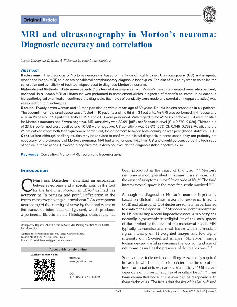
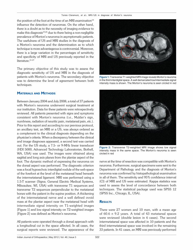
Between January 2004 and July 2008, a total of 37 patients with Morton’s neuroma underwent surgical treatment at our institution. Data for these patients were retrospectively reviewed. All patients presented with signs and symptoms consistent with Morton’s neuroma (i.e., Mulder’s sign, numbness, radiation of neuritic pain, metatarsal pain, etc.). Prior to this report and according to our previous protocol, an ancillary test, an MRI or a US, was always ordered as a complement to the clinical diagnosis depending on the physician’s criteria. When a discrepancy between the clinical and image diagnoses appeared, a second test was carried out. For the US study, a 7.5‑ or 9‑MHz linear transducer (HDI 5000, Advanced Technology Laboratories, Bothell, WA, USA) was used. The images were obtained along sagittal and long axis planes from the plantar aspect of the foot. The dynamic method of expressing the neuroma on the dorsal aspect was performed. The diagnostic criterion was a focal hypoechoic interdigital nodule of the web space of the forefoot at the level of the metatarsal head beneath the intermetatarsal ligament. MRI was performed using a 1.5‑T scanner (Signa; General Electric Medical Systems, Milwaukee, WI, USA) with transverse T1 sequences and transverse T2 sequences perpendicular to the metatarsal bones with the patient in the supine position. A thickening of the intermetatarsal nerve and a well defined ovoid mass at the plantar aspect near the metatarsal head with intermediate signal intensity on T1‑weighted images [Figure 1] and low signal intensity on T2‑weighted images [Figure 2] was defined as Morton’s neuroma.
All patients were operated through a dorsal approach with a longitudinal cut in the space affected. In all cases, the surgical reports were reviewed. The appearance of the
nerve at the time of resection was compatible with Morton’s neuroma. Furthermore, surgical specimens were sent to the Department of Pathology and the diagnosis of Morton’s neuroma was confirmed by histopathological examination in all of them. The sensitivity and 95% confidence interval (CI) of MRI and US were estimated. Kappa statistics was used to assess the level of concordance between both techniques. The statistical package used was SPSS 12 (SPSS Inc., Chicago, IL, USA).
Results
There were 27 women and 10 men, with a mean age of 60.6 ± 9.2 years. A total of 43 metatarsal spaces were reviewed (double lesion in 6 cases). The second intermetatarsal space was affected in 10 patients and the third intermetatarsal space was involved in the remaining 33 patients. In 41 cases, an MRI was previously performed
Figure 1: Transverse T1‑weighted MRI image reveals Morton’s neuroma in the third interdigital space. A well demarcated low/intermediate signal intensity mass is shown. The Morton’s neuroma is seen circled in red
Figure 2: Transverse T2‑weighted MRI image shows low signal intensity mass in the same space. The Morton’s neuroma is seen circled in red
Torres-Claramunt, et al.: MRI/US in diagnose of Morton’s neuroma
323 Indian Journal of Orthopaedics | May 2012 | Vol. 46 | Issue 3
and US was only done in 23 cases. In 21 patients, both an MRI and a US were performed. Considering histopathological diagnosis as a gold standard using MRI, 34 lesions were detected and 7 were missed. Sensitivity stood at 82.9% (95% CI: 0.68–0.93) (false negative, n=7). With regard to the US, 13 lesions were detected and 10 were missed. Here, the sensitivity was 56.5% (95% CI: 0.34–0.77) (false negative, n=10) [Table 1].
In the 21 lesions in which both radiological techniques were performed, the sensitivity of the MRI was higher than that of the US (85.7%, 95% CI: 0.64–0.98 vs. 52.4%, 95% CI: 0.30–0.7) [Table 2]. The level of agreement between both techniques was poor (kappa 0.31, 95% CI: 0.002–0.61). Table 3 shows the comparative data for MRI and US imaging findings.
dIscussIon
Different studies have been conducted to assess the usefulness of the MRI and US in diagnosing Morton’s neuroma. The results were controversial. Resch et al.21 compared US with MRI in patients in whom Morton’s neuroma was suspected and found poor sensitivity for both techniques. Although only nine patients were included in this study, these authors concluded that these techniques “were of little or no value.” Sharp et al.15 reported on the results of a prospective study of 29 cases comparing US, MRI, and clinical findings in the same cohort of patients in
order to determine their value in the diagnosis of Morton’s neuroma. They concluded that due to the higher sensitivity of the clinical findings, there is no requirement for MRI or US studies in patients in whom a suspicion of Morton’s neuroma was established on clinical grounds. In all our cases, the diagnosis was confirmed histopathologically. MRI studies missed 7 out of 41 cases and US missed 10 out of 23. On the other hand, both the US and MRI techniques were negative in three cases [Table 2]. However, surgical treatment was performed due to a high clinical suspicion and the pathologist confirmed the diagnosis.
For all these reasons, although we agree with Sharp and coworkers15 in the opinion that ancillary studies are not mandatory in the workup of patients with Morton’s neuroma, bearing legal aspects in mind, confirmation of the clinical diagnosis by a noninvasive technique before surgery might still be convenient.
On the other hand, in spite of considering clinical findings as a reliable means of diagnosis for Morton’s neuroma, ancillary tests may be helpful in the differential diagnosis or in the case of double lesions. Other diagnostic techniques, such as a CT scan, the injection of local anesthetics, or electrophysiology,18,19 have been reported as being useful tests in the confirmation of the diagnosis, but their sensitivity and specificity are low.
The US is a “user‑dependent test” and several studies have shown an overestimation of the size of neuromas.22,23 The sensitivity of US varies from study to study. In some of them, a 100% sensitivity has been reported,23,24 whereas lower Figures have been shown in the Resch et al. study.21 A 7.5‑MHz linear transducer is probably a low frequency for the detection of Morton’s neuroma and could be considered as one of the reasons why we report low US sensitivity in our department. On the other hand, we can increase the sensitivity of the US. Perini et al. published an interesting study using dynamic US to confirm the clinical suspicion of Morton’s neuroma.25 The main advantages of US were related to its high availability and the low cost.
MRI has been considered as an “accurate and operator‑independent modality,”26 even though variable sensitivity rates have been reported. Zanetti et al.14 found a sensitivity of 86.6%, which is similar to that obtained in our study. Lee et al.12 found 76% sensitivity. Other authors reported 100% sensitivity.27 Differences in the sensitivity rates are probably related to technical variables (the MRI equipment, sequences, etc.), the size of the neuroma, and position of the foot at the time of examination. The visibility of a Morton’s neuroma is best on MRI images obtained with the patient in a prone position.27 The radiologist’s experience
Table 1: Differences in sensitivity between US and MRIn Positive Negative Sensitivity (95% CI)
MRI 41 34 7 82.9 (0.679–0.929)US 23 13 10 56.5 (0.345–0.768)
Table 2: Imaging findings (US+MRI) of Morton’s neuromaMRI
Positive Negative TotalUS
Positive 11 0 11Negative 7 3 10Total 18 3 21
Table 3: MRI and US imaging findingsImaging findings MRI Imaging findings USn 41 n 23Neuroma diagnosis 34 Neuroma diagnosis 13Location
Second spaceThird space
6/3428/34
LocationSecond spaceThird space
2/1311/13
FindingIntermediate signal intensity in T1-weighted imageLow signal intensity in T2-weighted image
29/34
12/34
FindingHypoechoic interdigital nodule
13/13
Torres-Claramunt, et al.: MRI/US in diagnose of Morton’s neuroma
Indian Journal of Orthopaedics | May 2012 | Vol. 46 | Issue 3 324
is important in assessing and understanding pathologies of the foot.28 In summary, MRI is more technically independent than US, hence it has a major effect on diagnostic and therapeutic decisions by orthopedic surgeons when Morton’s neuroma is suspected.14
The low correlation of the tests studied means that both the US and the MRI enhance each other and higher sensitivity is achieved if both are performed.29,30 In agreement with other studies, with the fact that the sensitivity of each one is lower than the clinical exam,15 we cannot recommend performing either of the two studies in order to obtain greater sensitivity. We consider that they can never better the results obtained from a clinical exam. The low agreement obtained between both techniques reinforces the importance of the clinical diagnosis, and the higher sensitivity of the MRI than that of the US should give us reason to use it as the technique of choice in those cases in which an image test is necessary.29
The retrospective design is the main limitation as the radiologists and pathologists were not blinded to the clinical and intraoperative findings, respectively. In addition, the size of the neuroma was not systematically obtained, so its correlation with diagnostic accuracy cannot be assessed. The study only includes positive cases in the pathology diagnosis, the assessment of false positives and the specificity of imaging tests are not possible.
In conclusion, in the presence of a high clinical suspicion of Morton’s neuroma, imaging studies do not seem to be indispensable in confirming the diagnosis. In some particular circumstances like doubtful symptomatology, double lesions, and the medicolegal considerations of individual cases, imaging studies may be indicated. The higher sensitivity of the MRI compared to the US obtained in this series brings us to consider the MRI as the examination of choice for these cases.
RefeRences
1. Civinni F. Su di un gangliare rigonfiamento della piñata del piede. Mem Chir Archiespedale Pistoia. 1835:4-17.
2. Durlacher L. A treeatise on corns, bunions, the disease of nails, and the generalmanagement of the feet. London: Simpkin, Marshall; 1845. p. 52.
3. Morton TG. A peculiar and painful affection of the fourth metarsophalangeal articulation. Am J Med Sci 1876;71:37-9.
4. Levine SE, Myerson MS, Shapiro PP, Shapiro SL. Ultrasonographic diagnosis ofrecurrence after excision of an interdigital neuorma. Foot Ankle Int 1998;19:79-84.
5. Klenerman L, MacClellan GE, Gulloff RJ, Scadding JW. Mortons’s metatarsalgia: A retrospective and prospective study. In: Proceedings of the British OrthopaedicAssociation. J Bone Joint Surg Br 1983;28:78-82.
6. Reddy PD, Zellcof SB, Routolo C, Holder J. Interdigital neuroma. Local cutaneouschanges after corticosteroid injection. Clin Orthop 1995;317:185-7.
7. Wu KK. Morton’s interdigital neuorma: A clinical review of its etiology, treatment and results. J Foot Ankle Surg 1996;35:112-9.
8. Mann RA, Reynolds JC. Interdigital neuroma- a critical clinical analysis. Foot Ankle 1993;3:238-43.
9. Youngswick FD. Intermetatarsal neuroma. Clin Podiatr Med Surg 1994;11:579-92.
10. Bennett GL, Graham CE, Mauldin DM. Morton’s interdigital neuorma: A comprehensive treatment protocol. Foot Ankle Int 1995;16:760-3.
11. Coughlin MJ, Pinsonneault T. Operative treatment of interdigital neuroma. A long-term follow-up study. J Bone Joint Surg 2001;83:1321-8.
12. Lee MJ, Kim S, Huh YM, Song HT, Lee SA, Lee JW, et al. Morton Neuroma: Evaluated with ultrasonography and MR imaging. Korean J Radiol 2007;8:148-55.
13. Zanetti M, Ledermann T, Zollinger H, Hodler J. Efficacy of MR imaging in patientssuspected of having Morton’s neuroma. AJR Am J Roentgenol 1997;168:529-32.
14. Zanetti M, Strehle JK, Kundert HP, Zollinger H, Hodler J. Morton neuroma: Effect of MR imaging findings on diagnostic thinking and therapeutic decisions. Radiology 1999;213:583-8.
15. Sharp RJ, Wade CM, Hennessy MS, Saxby TS. The role of MRI and ultrasound imaging in Morton’s neuroma and the effect of size of lesion on symptoms. J Bone Joint Surg Br 2003;85:999-1005.
16. Biasca N, Zanetti M, Zollinger H. Outcomes after partial neurectomy of Morton’s neuorma related to preoperative case histories, clinical findings and findings in magnetic resonante imaging scans. Foot Ankle Int 1999;20:568-75.
17. Bencardino J, Rosenberg ZS, Liu X, Marty-Delfaut E. Morton’s Neuroma: Is always symptomatic? AJR Am J Roentolol 2000;175:649-53.
18. Guiloff RJ, Scadding JW, Klenerman L. Morton’s metatarsalgia: Clinical, electrophydiolicgical and histological observations. J Bone Joint Surg Br 1984;66:586-91.
19. Turan I, Lindgren LL, Sahlstedt T. Computed tomography for diagnsosis of Morton’s neuroma. J Foot Surg 1991;30:224-5.
20. Espinoza N, Schmitt JW, Saupe N, Maquieira GJ, Bode B, Vienne P, et al. Morton neuroma: MR imaging after resection- postoperative MRI and histologic findins in assymptomatic and symptomatic metatarsal spaces. Radiology 2010;255:850-6.
21. Resch S, Strenstrom A, Jonsson A, Jonsson K. The diagnostic efficacy of magnetic resonance imaging and ultrasonography in Morton’s neuroma: A radiological-surgicalcorrelation. Foot Ankle Int 1994;15:88-92.
22. Read JW, Noakes JB, Kerr D, Crichton KJ, Slater HK, Bonar F. Morton’s metatarsalgia: Sonographic findings andcorrelated histopathology. Foot Ankle Int 1999;20:153-61.
23. Jones S, Bygrave CJ, Betts RP, Smith TW. Morton’s neuroma: A sonographic-surgical evaluation.The Foot 1999;9:189-92.
24. Shapiro PP, Shapiro SL. Sonographic evaluation of interdigital neuromas. Foot Ankle Int 1995;16:604-6.
25. Perini L, Del Borrello M, Cipriano R, Cavallo A, Volpe A. Dynamic sonography of the forefoot in Morton’s síndrome: Correlation with magnetic resonante and surgery. Radiol Med 2006;111:897-905.
26. Kankanala G, Jain AS. The operational characteristics of ultrasonography for the diagnosis of plantar intermetatarsal neuroma. J Foot Ankle Surg 2007;46:213-7.
27. Weishaupt D, Treiber K, Kundert HP, Zollinger H, Vienne P, Hodler J, et al. Morton neuroma: MR imaging in prone, supine, and upright weight-bearing body positions. Radiology
Torres-Claramunt, et al.: MRI/US in diagnose of Morton’s neuroma
325 Indian Journal of Orthopaedics | May 2012 | Vol. 46 | Issue 3
2003;226:849-56.28. Llauger J, Palmer J, Monill JM, Franquet T, Bagué S, Rosón N.
MR imaging of benign soft-tissue masses of the foot and ankle. Radiographics 1998;18:1481-98.
29. Owengs R, Gougoulias N, Guthrie H, Sakellariou A. Morton’s neuroma: Clinical testing and imaging in 76 feet, compared to a control group. Foot Ankle Surg 2011;17:197-200.
30. Park HJ, Kim SS, Rho MH, Hong HP, Lee SY. Sonographic appearences of Morton’s neuroma. Differences from other
interdigital soft tissue masses. Ultrasound Med Biol 2011; 37:1204-9.
How to cite this article: Torres‑Claramunt R, Ginés A, Pidemunt G, Puig L, de Zabala S. MRI and ultrasonography in Morton’s neuroma: Diagnostic accuracy and correlation. Indian J Orthop 2012;46:321‑5.
Source of Support: Nil, Conflict of Interest: None.
Staying in touch with the journal
1) Table of Contents (TOC) email alert Receive an email alert containing the TOC when a new complete issue of the journal is made available online. To register for TOC alerts go to
www.ijoonline.com/signup.asp.
2) RSS feeds Really Simple Syndication (RSS) helps you to get alerts on new publication right on your desktop without going to the journal’s website.
You need a software (e.g. RSSReader, Feed Demon, FeedReader, My Yahoo!, NewsGator and NewzCrawler) to get advantage of this tool. RSS feeds can also be read through FireFox or Microsoft Outlook 2007. Once any of these small (and mostly free) software is installed, add www.ijoonline.com/rssfeed.asp as one of the feeds.
Copyright of Indian Journal of Orthopaedics is the property of Medknow Publications & Media Pvt. Ltd. and its
content may not be copied or emailed to multiple sites or posted to a listserv without the copyright holder's
express written permission. However, users may print, download, or email articles for individual use.


