Embed Size (px)
Citation preview

Three-dimensional Ultrasonography in the Diagnosis of Müllerian Duct Anomalies
21
Three-dimensional Ultrasonography in theDiagnosis of Müllerian Duct Anomalies1Carmina Bermejo, 1Rocío Cantarero, 1Dolores Díaz, 2Eva Labrador, 3Laura Ruiz López1Gabinete Médico Velázquez, C/Velázquez 25, 28001, Madrid, Spain2Centro de Resonancia Magnética de Alto Campo Abirem, C/Tomás López 3, 28009 Madrid, Spain3Gerencia de Atención Primaria, Talavera de la Reina (Toledo), Spain
Correspondence: Carmina Bermejo, Gabinete Médico Velázquez, Calle Fernán González 19, 2809, Madrid, SpainPhone: +34 619 712 380, e-mail: [email protected]
Donald School Journal of Ultrasound in Obstetrics and Gynecology, January-March 2009;3(1):21-30
AbstractAims: We studied the effectiveness of three-dimensional (3D)ultrasonography in the diagnosis of uterine malformations.Methods: 175 patients with clinical or ultrasonographic suspicion ofuterine malformation were studied between November 2004 and June2008. In all women we measured uterine volume for processing andlater reconstruction of 3D images. Cases in which no anomaly wasdetected with the 3D technique were excluded from study. A thoroughphysical genital examination with speculum was done before or afterultrasonography, except in three women with an imperforate hymen.Uterine malformations were recorded in detail, and the findings werecatalogued according to the American Fertility Society (AFS)classification. In 32 women magnetic resonance (MR) imaging wasalso used, and agreement between the two techniques was calculatedwith the kappa index.Results: The 175 müllerian anomalies we analyzed were diagnosed asagenesis (1 case), unicornuate uterus (1 genuine, 1 communicating),didelphys uterus (4), bicornuate uterus (22), septate uterus (80, 8 withtwo cervices) and arcuate uterus (68). For 1 unicornuate, 3 bicornuate,25 septate (4 with two cervices) and 3 arcuate uteri we performed MRimaging. Diagnostic correlation between 3D ultrasonography and MRimaging was obtained in all cases according to the AFS classification(kappa = 100%); concordance was also seen for descriptions ofassociated uterine malformations except for two cases: 1 bicornuateuterus with the additional presence of a cervical septum as detectedby MR imaging, which was not found on hysteroscopy for resection,and 1 septate uterus that appeared to have two cervices on 3Dultrasonography in a patient in whom physical examination was notpossible, and in whom MR imaging showed a complete septum withhypointense signals (kappa = 93.45%; 95% confidence interval 80.75-100%).Discussion: Owing to its ability to clearly render contours of anatomicalstructures, 3D ultrasonography provides detailed images of uterinemalformations and yields very similar results to those obtained withMR imaging. For an accurate comparison of the two techniques,ultrasonography should be accompanied by gynecologic physicalexploration, since the two methods yield equivalent results for therelation between the uterine cavity and the fundus. MR imaging isespecially useful to evaluate the cervix and vagina.
Keywords: Three-dimensional ultrasonography, müllerian ductanomalies, congenital uterine malformations, nuclear magneticresonance.
INTRODUCTION
The uterus and probably the upper part of the vagina(controversy over this point not withstanding)1 are derived fromthe paramesonephric or Müllerian ducts, after which anomaliesin these structures are named. The Müllerian ducts start todifferentiate to form the uterus in female embryos during thesixth week of gestation (until this time they are identical in maleand female embryos), and descend in parallel and externally tothe mesonephric ducts to drain into the urogenital sinus.Medially they cross the Wolffian or mesonephric ducts ventrally,and fuse along the midline to form the uterine primordium. Theseptum is subsequently reabsorbed in a craniocaudal direction,or according to an alternative hypothesis—fusion takes placebidirectionally simultaneously from the center toward both ends,with later resorption.2 This process accounts for certainanomalies that are not explained by the classic theory of uterinedevelopment.
Uterine anomalies make up a heterogeneous group ofcongenital malformations that can arise due to lack ofdevelopment of the Müllerian ducts, incomplete fusion, oralterations in septum resorption. It should be noted that anycombination of malformations can arise, and pure forms ofdifferent entities cannot always be distinguished.
The prevalence of uterine anomalies is difficult to establish.It is estimated that in the general population the prevalence ofuterine malformations is 0.4%, with different authors reportingrates that range from 0.1 to 3%,3,4 whereas a prevalence of 4%has been described among infertile women, although not allauthors distinguished between this group and the generalpopulation.5 Among patients with recurrent miscarriages therates vary from 3 to 38%.6-10 This disparity among differentpublications reflects the use of different diagnostic techniques,nonhomogeneous population samples, and differences in the
Carmina Bermejo et al
22
clinical entities of uterine malformation considered by differentauthors. Symptoms vary from asymptomatic to seriousgynecological (obstructive hematocolpos, hematometra) orobstetric problems in up to 25% of all cases, compared to 10%in the general population (recurrent miscarriage, placentalinsufficiency, incompetent cervix, etc.).11 The most frequentanomalies in asymptomatic women and patients with recurrentfirst trimester miscarriages are arcuate and subseptate uterus.There are no differences in the size of the indentation betweenthe two groups (except for nullipara), but in the recurrentmiscarriage group, the uterine cavities are smaller than in healthywomen regardless of parity.12 Arcuate uterus tends to beassociated with second trimester miscarriage, whereas firsttrimester miscarriage is more frequent in women with subseptateuterus.13-15 Other anomalies are often present, most of them areurologic in nature given the parallel embryologic origin of thetwo organ systems.2
Uterine malformations tend to be sporadic; howevermultiple factors can be involved in their origin. This situationhas been described in the etiology of septate uterus in theabsence of the Bcl2 gene.16 Some malformations appear indifferent chromosomal syndromes, although most women tendto have a normal karyotype and it is still unknown whethervertical inheritance is involved. Viral infections, teratogenicmedicines (mainly diethylstilbestrol, DES) and other causes alsoseem to be related to the etiology of these malformations.11,17, 18
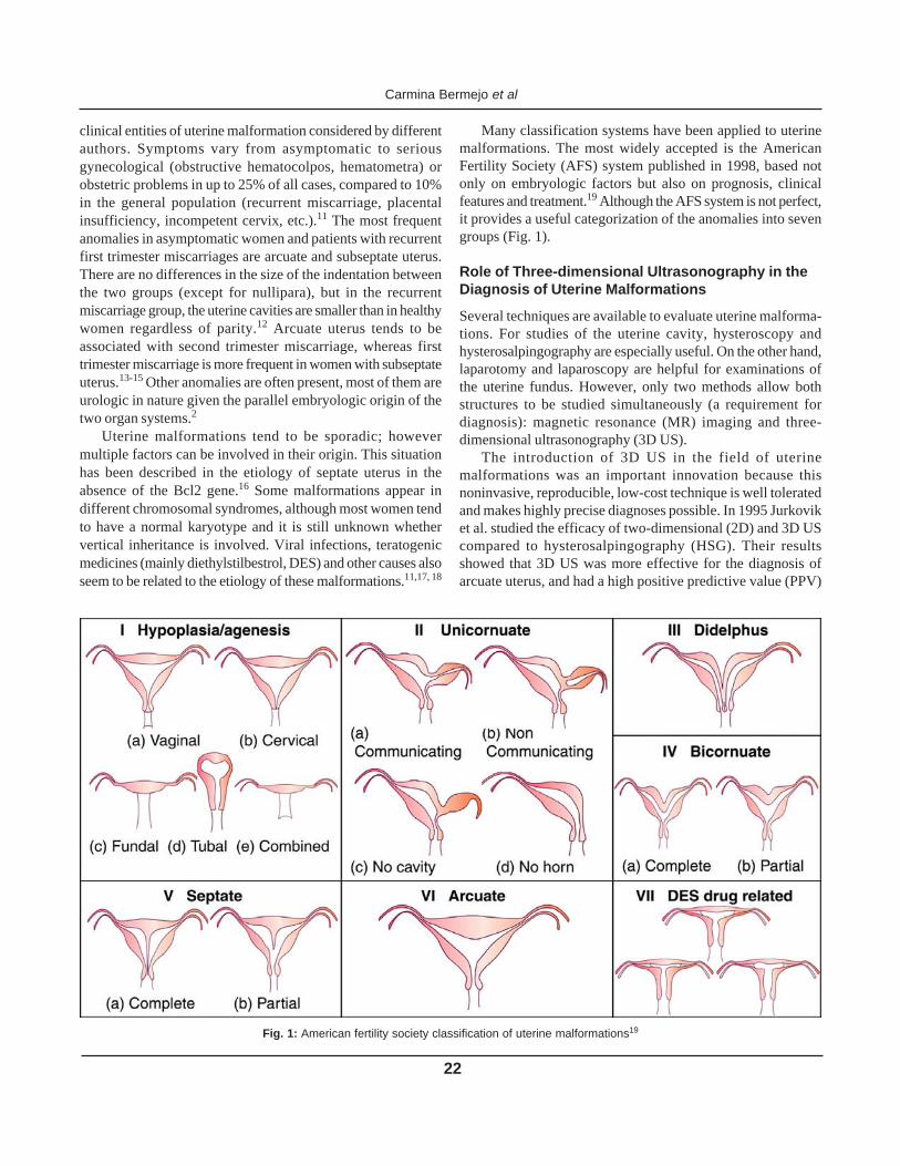
Many classification systems have been applied to uterinemalformations. The most widely accepted is the AmericanFertility Society (AFS) system published in 1998, based notonly on embryologic factors but also on prognosis, clinicalfeatures and treatment.19 Although the AFS system is not perfect,it provides a useful categorization of the anomalies into sevengroups (Fig. 1).
Role of Three-dimensional Ultrasonography in theDiagnosis of Uterine Malformations
Several techniques are available to evaluate uterine malforma-tions. For studies of the uterine cavity, hysteroscopy andhysterosalpingography are especially useful. On the other hand,laparotomy and laparoscopy are helpful for examinations ofthe uterine fundus. However, only two methods allow bothstructures to be studied simultaneously (a requirement fordiagnosis): magnetic resonance (MR) imaging and three-dimensional ultrasonography (3D US).
The introduction of 3D US in the field of uterinemalformations was an important innovation because thisnoninvasive, reproducible, low-cost technique is well toleratedand makes highly precise diagnoses possible. In 1995 Jurkoviket al. studied the efficacy of two-dimensional (2D) and 3D UScompared to hysterosalpingography (HSG). Their resultsshowed that 3D US was more effective for the diagnosis ofarcuate uterus, and had a high positive predictive value (PPV)
Fig. 1: American fertility society classification of uterine malformations19

Three-dimensional Ultrasonography in the Diagnosis of Müllerian Duct Anomalies
23
for major anomalies, especially for distinguishing bicornuatefrom subseptate uterus.20 More recent work by Raga et al.21
found 91.6% agreement between 3D US and HSG whenevaluating the fundus, and 100% efficacy with regard toexaminations of the uterine cavity when 3D US was comparedto laparoscopy. Wu et al.22 correlated the technique (also usingHSG and 2D US) with laparoscopy or hysteroscopy, and found92% efficacy for the diagnosis of septate uterus and 100%efficacy for bicornuate uterus. In 2007, Mohamed et al.published a study that compared 3D US versus laparoscopyand hysteroscopy. They achieved a sensitivity of 97%, aspecificity of 96%, a PPV of 92%, and a negative predictivevalue (NPV) of 99%.23 In 2001, Salim et al demonstrated thereproducibility of 3D US for the diagnosis of uterinemalformations, and modified the AFS classification accordingto ultrasonographic parameters.24 These authors noted that 3DUS was worthwhile as a screening method, since it improvedreproductive outcome.15 The same conclusion was reached byKupesic and Kurjak based on the importance of septostomy.These authors studied large series of cases, the largest of whichincluded 3850 patients from a tertiary infertility clinic where894 had a uterine malformation, and detected 689 septateuteri.25,26
Magnetic resonance imaging was considered the goldstandard for the diagnosis of müllerian duct anomalies, andmultiple studies documented its efficacy.27-30 However, 3D UScan be considered a useful alternative to this technique, becausein addition to its lower cost and the better tolerance by patients,it provides images of very similar quality that are almostsuperimposable with MR images for diagnostic purposes.31
Studies in progress to compare these two methods have validated3D US by documenting a high correlation between the findingsobtained with the two techniques. However, a good patienthistory and thorough gynecologic physical examination are anecessary complement to MR imaging.
Ultrasonographic Diagnosis of Müllerian DuctAnomalies
How can a uterine malformation be diagnosed byultrasonography? Usually the woman will have clinicalsymptoms or a suggestive physical exploration (vaginalpartitions, two cervices, etc.) and follow-up will be needed forthe diagnosis. Occasionally a malformation might be noticedduring routine ultrasonography. In most uterine anomalies, 2DUS will reveal two uterine cavities but little additionalinformation on which to base a diagnosis. This is generally thecase for nonextreme entities such as arcuate, septate andbicornuate uterus, and it is basically these forms that need to bedistinguished in order to indicate suitable treatment.
Three dimensional US is suitable to study the relationshipbetween the cavity and fundus, and almost always allows acorrect diagnosis of nonextreme entities. The addition of the
coronal plane (which cannot be acquired with conventional or2D US) is the key enhancement that makes it possible tovisualize these anomalies. Uterine volume needs to be measuredfor processing and later reconstruction into 3D images(rendering or reconstruction is part of the post-acquisition workrequired for diagnosis). By combining different planes theanomaly becomes available for study not only in the coronalplane (the most informative), but also in sagittal and axialsections (depending on the planes chosen for image capture),as with MR imaging. The data are not observer-dependent(any operator can use the acquired volumes) or exploration time-dependent (postacquisition rendering can be done later). Thevolumes obtained can be sent anywhere in the world to beprocessed and studied by any specialist with the required software.
Although one of the uses of 3D US is to evaluatemorphology, this technique also makes it possible to measuredistances of interest in the coronal plane24 (or any other plane)and to calculate cavity volumes [e.g. with Virtual OrganComputer-aided AnalysisTM (VOCAL)] (Fig. 2). Thisinformation can be used to obtain a reproductive prognosis,and to obtain informative images of blood supply.32,33
Figure 3 illustrates the shape of a normal uterusreconstructed with 3D US. This picture will serve as a referenceto facilitate interpretation of the images used here to illustratedifferent entities of the AFS classification.
Class I: Agenesis or hypoplasia
This group accounts for 5 to 10% of all uterine malformations.As seen in Figure 1, the most frequent type corresponds to theRokitansky-Kuster-Hauser syndrome, characterized by vaginaland uterine agenesis in 90% of cases. Three-dimensional USdoes not appear to be the most appropriate method for detectingagenesis or hypoplasia, since the diagnosis can be made byphysical examination and conventional 2D US.
Fig. 2: Virtual organ computer-aided analysis showing the volume ofthe uterine cavity in a septate uterus

Carmina Bermejo et al
24
Class II: Unicornuate Uterus
This anomaly results from poor or absent development of theparamesonephric ducts, and accounts for 20% of all uterinemalformations. Figure 1 shows the subgroups of unicornuateuterus. (Figs 4A to C) depicts a unicornuate uterus seen with2D US (note the single cavity, which is difficult to establishwith this technique), 3D US (in the coronal plane the cavitydoes not appear triangular), and MR imaging.
Class III: Didelphys Uterus
This malformation, which comprises 5% of all uterineanomalies, is caused by complete nonfusion of the müllerianducts, and is characterized by two completely separate hemiuteriwith two uterine bodies and two cervices with no connectionbetween the cervical cavities. The hemiuteri are tenuouslyconnected via an isthmus in the serosal layer. In some casesone of the hemiuteri may be obstructed (blind hemivagina dueto combined alteration of the urogenital sinus), resulting inhematocolpos or hematometra with acute clinical symptomsduring menarche similar to those produced by a unicornuatenoncommunicating uterus. It is not uncommon to find associatedrenal homolateral agenesis.34 A precise diagnosis can beaccomplished with physical examination (which disclosesvaginal septae) and later exploration with 2D US. Studies with3D US can be costly, since the two horns are generally separatedby a considerable distance and may therefore appear in differentplanes, making image acquisition and rendering difficult(Fig. 5).
Class IV: Bicornuate Uterus
When müllerian duct fusion is incomplete, a bicornuate uterusresults. The two hemiuteri are linked through a variable area in
Fig. 3: Normal uterus in 3D US
Figs 4A to C: Genuine unicornuate uterus in 2D US (A) 3D US (B)and MR imaging (C)

Three-dimensional Ultrasonography in the Diagnosis of Müllerian Duct Anomalies
25
the lower portion of the organ depending on the severity of theanomaly, and there is usually only one cervix although twocervices can be present. Bicornate uterus accounts for 10% ofall uterine malformations.
Symmetry of the two hemiuteri is one of its maincharacteristics of this anomaly, and is what distinguishes it fromcommunicating unicornuate uterus. In contrast to arcuate andseptate uterus, a central cleft is present in the serosa of thefundus.
The clinical features related to this class of anomaly aremainly obstetric and tend to be less marked that in septate uterus.Treatment should be based on the severity of the clinicalfeatures, hence the importance of a correct diagnosis todistinguish between the two entities (given that sometimes, inview of its poor prognosis, treatment should begin promptlyfor septate uterus). Troiano and McCarthy proposed a practicalformula to distinguish between septate and bicornuate uterus.35
If a line drawn from horn to horn crosses or is less than 5 mmfrom the fundus, bicornuate uterus should be diagnosed. Incontrast, if the line is further from the fundus, septate uterusshould be diagnosed regardless of the appearance of the fundus(i.e. convex, flat, or with a slight indentation). Figure 6 shows a3D US image illustrating bicornuate uterus. In this type ofanomaly the horns are usually more than 4 cm apart, althoughthis feature should not be used as an ultrasonographic criterionsince it is simply an indicator used in HSG to distinguishbetween types of anomalies.
Class V: Septate Uterus
This anomaly originates from incomplete septum resorptionresulting in an intermediate wall between the müllerian ducts;
as a result the uterine cavity is either partially (subseptate) orcompletely divided into two, but has a normal externalmorphology. Septate uterus is the most frequent malformation(55%), and the subseptate form is particularly frequent. Thisanomaly does not include arcuate uterus, since this should beconsidered a minor anomaly or a variation from normal. Froman obstetric point of view, septate uterus has a poor prognosissince it is related with a high percentage of fetal loss andplacental insufficiency.35
Figures 7A and B shows a subseptate uterus visualized with3D US and MR imaging. Figure 8 depicts a septate uterus withtwo cervices, an uncommon malformation first described in1994 by Brumsted and McBean, and explained by Müller’sbidirectional theory of duct fusion.36 Figures 9A and B showsanother case of septate uterus visualized with 3D US and MRimaging.
The complex malformation illustrated in Figure 10 consistsof a bicornuate uterus with a septum dividing the two cavities.This malformation can also be explained by Müller’s theory ofbidirectional fusion.
Class VI: Arcuate Uterus
Like the septate uterus, the arcuate uterus is considered aseparate anomaly in terms of prognosis and treatment. Itsexternal morphology is normal, and the uterine cavity showsonly a mild indentation rather than an actual septum. The depthof the indentation is not well-established. Syed and colleagues37
proposed a cutoff length no greater than 1.5 cm, but otherauthors such as Salim et al.24 considered the angle of theindentation (e.g. obtuse arcuate, acute subseptate) to be a moreimportant feature (Fig. 11).
Fig. 5: Didelphys uterus in 3D US Fig. 6: Bicornuate uterus in 3D US. A line drawn from horn to horntransects the fundus

Carmina Bermejo et al
26
Figs 7A and B: Subseptate uterus in 3D US (A) and MR imaging (B)
Fig. 8: Septate uterus with two cervices Fig. 10: Bicornuate uterus with complete septum
Figs 9A and B: Septate uterus with two cervices in 3D US (A) andMR imaging (B)

Three-dimensional Ultrasonography in the Diagnosis of Müllerian Duct Anomalies
27
Class VII: Iatrogenic Uterus (DES)
This type of malformation has become less common in recentdecades, since it was attributed to DES, which has not beenused for gestational complications for many years.38 The mostsubstantial change is the typical T-shape of the uterine cavity.(Fig. 12) illustrates a uterus with this alteration as visualizedwith 3D US (courtesy of Dr Pascual, Instituto UniversitarioDexeus, Barcelona, Spain).
PATIENTS AND METHODS
We studied 175 patients with clinical or ultrasonographicsuspicion of uterine malformation between November 2004 andJune 2008. Uterine volume was measured in all women for laterprocessing and reconstruction of 3D images. Images werescanned with two General Electric Voluson Expert systems
operated by a total of six observers. Volumes were capturedwith a volumetric vaginal transducer at 3.7-9.3 MHz. Imageswere acquired over a wide range of settings from medium tomaximum, at an angle of 90º, adjusting the size of the region ofinterest in the midsagittal plane of the uterus to included theentire organ in the image. A transverse plane was also used foranomalies of the major transverse diameter (didelphys uterus,wide septate uterus, certain bicornuate malformations with alarge separation between horns, and one communicatingunicornuate uterus) to be able to capture both horns and establishthe relationships between the cavity and fundus during 3D imagereconstruction.
Rendering or reconstruction of all captured images was donewith surface and light gradient modes. Sector planes were usedfor the diagnosis in three cases. If no anomaly was seen, thewoman was not included in the present analysis. A thoroughgenital physical exploration with speculum was done before orafter US except in three women with an imperforate hymen.The findings were catalogued according to the AFSclassification, and the associated uterine malformation wererecorded in detail. In the 32 patients for whom complete physicaland US explorations were feasible, MR imaging was also done.These cases did not differ significantly from the generalpopulation of cases in terms of types of anomalies or otherpatient characteristics. All MR images were obtained by thesame physician with a General Electric 0.2 tesla or a SiemensAvanto 1.5 tesla system, according to the usual sequence, withenhanced T2-weighted coronal planes (used because of theirimportance for the diagnosis). The kappa index was calculatedto determine agreement between the two diagnostic techniquesfor uterine malformations. Additional findings were alsoanalyzed in detail.
RESULTS
Of the 175 müllerian duct anomalies we analyzed, the diagnoseswere1 agenesis, 2 unicornuate uterus (1 genuine, 1 communi-cating), 4 didelphys uterus, 22 bicornuate uterus, 80 septateuterus (8 with two cervices) and 68 arcuate uterus. Magneticresonance imaging was also done in 1 woman with unicornuateuterus, 3 with bicornuate uterus, 25 with septate uterus (4 withtwo cervices) and 3 with arcuate uterus. The 3D US and MRfindings agreed in all cases regarding the relation between theuterine cavity and the fundus, so the results for AFSclassification based on 3D US were consistent (kappa = 100%).
However, in two patients we noted discrepancies in thecervix. In one woman with bicornuate uterus, MR imagingshowed the presence of a septum along the cervical canal, andin another woman a septum considered bicollis with 3D USwas diagnosed with MR imaging as a complete septumproducing a hypointense signal in the cervical canal. The firstdiscrepancy resulted in a false positive finding with MR imaging,
Fig. 11: Arcuate uterus in 3D US
Fig. 12: Diethylstilbestrol-exposed uterus in 3D US

Carmina Bermejo et al
28
revealed as such during hysteroscopy for resection. Duringhysteroscopy a fold was observed on the anterior cervical wall,which was probably responsible for the false septum imageobtained with MR imaging. In the second case, divergence ofthe cervical canals in the coronal plane of 3D US led to thediagnosis of a double cervix. Physical examination was notpossible because the patient had an imperforate hymen, so rectalUS was used. Magnetic resonance imaging showed a wideseptum dividing the whole uterus up to the external cervical os;the signals were compatible with fibrous tissue and differedfrom the myometrium signals that would have produced if adouble cervix had actually been present.
Even considering these two cases of discrepant findings,concordance between the two techniques was high (kappa =93.45%; 95% confidence interval 80.75%-100%).
DISCUSSION
Although 3D US is helpful as a complement to 2D US onmultiple occasions on the field of gynecology, it is onlyindispensable for the diagnosis of uterine malformations,therefore suspected malformations are the only absoluteindication for this technique. When a uterine malformation issuspected, our suggested protocol indicates initially 3D US,always accompanied by complete gynecologic exploration. Forcomplex or doubtful cases, MR imaging should also be used.Surgery should be reserved exclusively for malformations thatwould benefit from this treatment.
Images produced by 3D US and MR imaging are nearlyequivalent (see Figs 1 to 12). The relation between the fundusand uterine cavity can be well established with eitherultrasonographic reconstructions in the coronal plane or withcoronal sequences obtained by MR imaging. Hence the successof the combination of 3D US and MR imaging lies in the coronalsections, which are difficult to acquire with 2D US. In terms ofdiagnostic efficacy, neither technique is better than the other,and in our hands concordance based on classification of theanomalies according to the AFS system was complete.Differences may arise, however, in studies of the lower part ofthe uterus. Magnetic resonance distinguishes clearly betweendifferent tissues, and this is especially advantageous for studiesof the cervix and vagina, which should not be overlooked duringconventional ultrasonography. To analyze the cervix withultrasonography, the transducer needs to be withdrawn slightlyto visualize the cervical canal and myometrium. Sometimes thiscan disclose cervical cancer, which will unfortunately be in anadvanced stage since early diagnosis with imaging techniquesis not possible.
Excellent 3D images can be obtained from good 2D USimages, and the combination of the two techniques is useful toevaluate the existence of one or two cervices, or of a septum inthe cervical canal. The presence of two (generally wide) canals
that diverge in the lower region suggest a double cervix ratherthan a septate cervix, but although the ultrasonographicimpression is almost always correct, only verification byspeculoscopy can confirm the diagnosis. On the basis ofdifferences in MR imaging signal intensity, radiologists candiscriminate accurately between a septum (even if myometriumis present in its upper portion) and the cervical myometrium.Not unusually, MR imaging reports will describe vaginal septathat can be distinguished by their different signal intensitiescompared to the walls of the vagina. Gynecologists, however,cannot distinguish the latter with either of the techniquesavailable for them (2D US or 3D US), and given the highincidence of association between these and other uterinemalformations, is vital to use speculoscopy and manual vaginalexploration to search for septa. If the procedure is done asdescribed, the efficacy of the two techniques would beequivalent, hence 3D US would thus be a better option thanMR imaging because the former offers advantages such as lowercost and better tolerance by patients. A further advantage of3D US is its simplicity during gynecologic examinations in thesetting of everyday practice.
Although the cost of 3D US is substantial, this factor isoffset by its good tolerance. Patients reported fear of the contrastmedium, claustrophobia, and the duration of procedure as themain disadvantages of MR imaging. In the present study weencountered resistance to MR imaging and often needed toreassure women that contrast was not necessary, and use anopen MR imaging system. As a result we were only able to useboth techniques for a small sample, limited mainly to anomaliesthat did not pose a risk to the woman’s reproductive goals andthus did not require more “aggressive” diagnostic techniques.
One of the most useful applications of 3D US is indifferentiating between arcuate, septate and bicornuate uterus.Alcázar and colleagues, among others, reached the sameconclusion and also noted its lower efficacy in diagnosingdidelphys uterus.39 Two-dimensional images sometimes do notreveal differences between markedly arcuate, subseptate orpartial bicornuate uterus. The prognosis for these malformationvaries substantially, hence the importance of a correct diagnosisfor appropriate treatment. A number of publications advocatesystematic septostomy based upon the improved reproductiveoutcome in a considerable percentage of patients. Among thesepublications, the work by Kupesic and colleagues meritsparticular attention, especially their larger study in which theauthors advocate the efficacy of 3D US as a screening methodfor infertile patients, due to the high prevalence of septate uterusand the effectiveness of septostomy in the population theystudied.26,40 Other authors such as Tekes and colleagues41
suggested that the finding of a septate uterus was not necessarilyan indication for surgical treatment, which should be reservedfor women in whom the diagnosis might interfere with obstetric
Three-dimensional Ultrasonography in the Diagnosis of Müllerian Duct Anomalies
29
outcomes. We believe resection of large septa, which generallyhave a substantial myometrial component, could cause seriousuterine scarring which could lead in turn to placenta accreta.On the other hand, septate uteri tend to have larger cavities,and in our experience are associated with fewer reproductiveproblems than narrow uteri (possibly because of the better bloodsupply). In our experience the prognosis and most suitabletreatment for women with these anomalies can be determinedwith 3D US.
At our center hysteroscopic septostomy is practiced inwomen with antecedents of two or more miscarriages, with goodresults. The decision to use surgical correction for any uterinemalformation is based on clinical criteria. We emphasize thatour population of patients was not a preselected population,and that most studies that recommend systematic septostomyinvolved infertile women or women with a history of recurrentmiscarriages. Some authors such as Tomazevic and colleaguesfavor surgical intervention for the correction of even arcuateuterus, but this may be because their definition of this anomalyis closer to that of subseptate uterus, i.e. with a septum from1.3 to 1.5 cm long, for which resection could improve thereproductive prognosis.42 Generally speaking, the managementof to this type of anomaly is usually conservative. Authors suchas Golan and colleagues maintain that the only surgery indicatedfor these cases is cervical cerclage, although this will not preventfirst trimester miscarriages.43
We believe the most suitable approach to diagnosing auterine malformation should take into account the clinicalfeatures and the patient’s reproductive goals. We have used 3DUS to diagnose and plan the most appropriate treatment, withgood results.
ACKNOWLEDGMENTS
We thank K Shashok for revising the use of English in themanuscript.
REFERENCES
1. Acien P, Acien M. Malformations of the Female Genital Tract andEmbryological Bases. Curr Wom Health Rev 2007;3:248-88.
2. Muller P, Musset R, Netter A, Solal R, Vinourd JR, Gillet JY.Etat du haut appareil urinaire chez les porteuses de malformationsuterines: etude de 133 observations. Presse Med 1967;75:1331-36.
3. Byrne J, Nussbaum–Blask A, Taylor WS, et al. Prevalence ofmullerian duct anomalies detected at ultrasound. Am J MedGenet 2000;94:9-12.
4. March CM. Müllerian anomalies. Fertility news 24/1. EndocrFert Forum 1990;13:1.
5. Acien P. Incidence of müllerian defects in fertile and infertilewomen. Hum Reprod 1997;12:1372-76.
6. Stampe Sorensen S. Estimated prevalence of mullerian ductanomalies. Acta Obstet Gynecol Scand 1988;67:441-45.
7. Stray-Pedersen B, Stray-Pedersen S. Etiologic factors andsubsequent reproductive performance in 195 couples with a prior
history of habitual abortion. Am J Obstet Gynecol 1984;148:140-46.
8. Raga F, Bauset C, Remohí J, Bonilla-Musoles F, Simón C,Pellicer A. Reproductive impact of congenital mulleriananomalies. Hum Reprod 1997;12:2277-81.
9. Makino T, Hara T, Oka C, Toyoshima K, Sugi T, Iwasaki K,Umeuchi M, Iizuka R. Survey of 1120 japanese women with ahistory of recurrent spontaneous abortions. Eur J Obstet GynecolReprod Biol 1992;44:123-30.
10. Clifford K, Rai R, Watson H, Reagan L. An informative protocolfor the investigation of recurrent miscarriage: Preliminary experienceof 500 consecutive cases. Hum Reprod 1994; 9:1328-32.
11. Golan A, Langer R, Bukovsky I, Caspi E. Congenital anomaliesof the mullerian system. Fertl Steril 1989;51:747-55.
12. Salim R, Regan B, Woelfer B, Backos M, Jurkovic D. Acomparative study of the morphology of congenital uterineanomalies in women with and without a history of recurrentfirst trimester miscarriage. Hum Reprod 2003;18:162-66.
13. Tulandi T, Arronet GH, McInnes RA. Arcuate and bicornuateuterine anomalies and infertility. Fertil Steril 1980;34:362-64.
14. Acien P. Reproductive performance of women with congenitaluterine anomalies. Hum Reprod 1993;8:122-26.
15. Woelfer B, Salim R, Banerjee S, Elson J, Regan L, Jurkovic D.Reproductive outcomes in women with congenital uterineanomalies detected by three-dimensional ultrasound screening.Obstet Gynecol 2001;98:1099-1103.
16. Lee DM, Osathanondh R, Yeh J. Localization of Bcl-2 in thehuman fetal mullerian tract. Fertil Steril 1998;70:135-40.
17. Edwards JA, Gale RP. Camptobrachydactyly: A new autosomaldominant trait with two probable homozigotes. Am J Hum Genet1972;24:464-74.
18. Sarto GE, Simpson JL. Abnormalities of the mullerian and wolffianduct systems. Birth Defects Orig Artic Ser 1978;14: 37-54.
19. The American Fertility Society. Classifications of adnexaladhesions, distal tubal obstruction, tubal occlusion secondaryto tubal ligation, tubal pregnancies, mullerian anomalies andintrauterine adhesions. Fertil Steril 1988;49:944-55.
20. Jurkovic D, Geipel A, Gruboeck K, Jauniaux E, Natucci M,Campbell S. Three-dimensional ultrasound for de assessmentof uterine anatomy and detection of congenital anomalies: Acomparison with histerosalpingography and two-dimensionalsonography. Ultrasound Obstet Gynecol 1995;5:233-37.
21. Raga F, Bonilla-Mussoles F, Blanes J, Osborne NG. Congenitalmullerian anomalies: Diagnostic accuracy of three-dimensionalultrasound. Fertil Steril 1996;65:523-28.
22. Wu MH, Hsu CC, Huang KE. Detection of congenital müllerianduct anomalies using three-dimensional ultrasound. J ClinUltrasound 1997;25:487-92.
23. Mohamed M, Momtaz MD, Alaa N, Ebrashy MD, Ayman A,Marzouk MD. Three-dimensional ultrasonography in the evaluationof the uterine cavity. Middle East Fertil Soc J 2007; 12:41-46.
24. Salim R, Woelfer B, Backos M, Regan L, Jurkovik D.Reproducibility of three-dimensional ultrasound diagnosis ofcongenital uterine anomalies. Ultrasound Obstet Gynecol 2003;21:578-82.
25. Kupesic S, Kurjak A. Septate uterus: Detection and predictionof obstetrical complications by different forms ofultrasonography. J Ultrasound Med 1998;17:631-33.

Carmina Bermejo et al
30
26. Kupesic S, Kurjak A, Skenderovic S, Bjloes D. Screening foruterine abnormalities by three-dimensional ultrasound improvesperinatal outcome. J Perinat Med 2002;30:9-17.
27. Fedele L, Dorta M, Brioschi D, Massari C, Candiani GB.Magnetic resonance evaluation of double uteri. Obstet Gynecol1989;74:844-47.
28. Carrington BM, Hricak H, Nuruddin RN, Secaf E, Laros RK Jr,Hill EC. Müllerian duct anomalies: MR imaging evaluation.Radiology 1990;176:715-20.
29. Pellerito JS, McCarthy SM, Doyle MB, Glickman MG,DeCherney AH. Diagnosis of uterine anomalies: Relativeaccuracy of MR imaging, endovaginal sonography andhisterosalpingography. Radiology 1992;795-800.
30. Fischetti SG, Politi G, Lomeo E, Garozzo G. Magnetic resonancein the evaluation of mullerian duct anomalies. Radiol Med 1995;89:105-11.
31. Deutch TD, Abuhamad AZ. The Role of 3-DimensionalUltrasonography and Magnetic Resonance Imaging in theDiagnosis of Mullerian Duct Anomalies: A Review of theLiterature. J Ultrasound Med 2008;27(3):413-23.
32. Raine-Fenning N, Fleischer AC. Clarifying the role of three-dimensional transvaginal sonography in reproductive medicine:An evidence-based appraisal. Exp Clin Assist Reprod 2005; 2:10.
33. Timor-Tritsch IE, Monteagudo A, Tsymbal T, Strok I. Three-dimensional inversion rendering: a new sonographic techniqueand its use in gynecology. J Ultrasound Med 2005;24:681-88.
34. Madureira A, Mariz C, Bernardes J, Ramos I. Case 94: UterusDidelphys with Obstructing Hemivaginal Septum and IpsilateralRenal Agenesis. Radiology 2006;239:602-06.
35. Troiano R, McCarthy S. Müllerian duct anomalies: Imaging andclinical issues. Radiology 2004;233:19-34.
36. Mc Bean JH, Brumsted JR. Septate uterus with cervicalduplication: a rare malformation. Fertil Steril 1994;62:415-17.
37. Syed I, Hussain H, Weadock W, Ellis J. Uterus, Mullerian DuctAbnormalities. eMedicine. Article Last Updated: Oct 3, 2007.
38. Goldberg JM, Falcone T. Effect of diethylstilbestrol onreproductive function. Fertile Steril 1999;72:1-7.
39. Alcázar JL. Three-dimensional ultrasound in gynecology: currentstatus and future perspectives. Curr Wom Health Rev 2005;1:1-14.
40. Kupesic S. Clinical implications of sonographic detection ofuterine anomalies for reproductive outcome. Ultrasound ObstetGynecol 2001;18:387-400.
41. Tekes A, Macura K. Ultrasound and MR imaging correlation ofuterine anomaly. J Wom Imag 2004;6:91-93.
42. Tomazevic T, Ban-Frangez H, Ribic-Pucelj M, Premru-Srsen T,Verdenik I. Small uterine septum is an important risk variablefor preterm birth. Eur J Obstet Gynecol Reprod Biol 2007;135:154-57.
43. Golan A, Langer R, Wexler S, Segev E, Niv D, David M.Cervical cerclage—its role in the pregnant anomalous uterus.Int J Fertil 1990;35:164-70.