Embed Size (px)
Citation preview
Printed by Jouve, 75001 PARIS (FR)
Europäisches Patentamt
European Patent Office
Office européen des brevets
(19)
EP
1 36
1 43
6A
1*EP001361436A1*(11) EP 1 361 436 A1
(12) EUROPEAN PATENT APPLICATION
(43) Date of publication:12.11.2003 Bulletin 2003/46
(21) Application number: 03252921.6
(22) Date of filing: 09.05.2003
(51) Int Cl.7: G01N 33/543, C12Q 1/00,C12Q 1/37
(84) Designated Contracting States:AT BE BG CH CY CZ DE DK EE ES FI FR GB GRHU IE IT LI LU MC NL PT RO SE SI SK TRDesignated Extension States:AL LT LV MK
(30) Priority: 10.05.2002 US 144562
(71) Applicant: Lifescan, Inc.Milpitas, California 95035-6312 (US)
(72) Inventors:• Qian, Suyue
Fremont, California 94539 (US)• Guo, Sherry
San Jose, California 95132 (US)• Leong, Koon-wah
Sunnyvale, California 94086 (US)
(74) Representative: Mercer, Christopher Paul et alCarpmaels & Ransford43, Bloomsbury SquareLondon WC1A 2RA (GB)
(54) Multilayer reagent test strips and methods for using the same to quantify glycated proteinin a physiological sample
(57) Multilayer reagent test strips for quantitatingglycated protein in a fluid sample, as well as methodsfor using the same, are provided. The subject multilayertest strips include at least a filter layer, a proteinase layerand a ketoamine oxidase signal producing and fluid flowcontrol system layer. In using the subject test strips, afluid sample is applied to the test strip and a signal isgenerated that can be employed to quantitate the gly-cated protein level in the sample. The quantitated gly-
cated protein level can then be employed to determinethe amount of glycated protein in the fluid sample. Alsoprovided are kits and systems that include the subjecttest strips and find use in practicing the subject methods.The subject compositions and methods find use in gly-cated protein monitoring applications, among other util-ities.
EP 1 361 436 A1
5
10
15
20
25
30
35
40
45
50
55
2
Description
INTRODUCTION
Field of the Invention
[0001] The field of this invention is analyte detection, particularly glycated protein detection.
Background of the Invention
[0002] Individuals suffering from diabetes mellitus have an abnormally high blood sugar level generally because thepancreas does not secrete sufficient amounts of the active hormone insulin into the bloodstream to regulate carbohy-drate metabolism. If an abnormally high blood sugar level, known as a hyperglycemic condition, is allowed to continuefor prolonged periods, the individual will suffer from the chronic complications of diabetes, including retinopathy, ne-phropathy, neuropathy and cardiovascular disease. Studies indicate that diabetic patients who are able to maintainnear normal glycemic control greatly reduce the likelihood of these dire complications. Therefore, several tests havebeen developed to measure and control glycemic condition.[0003] One common medical test to control glycemic condition is the direct measurement of blood glucose levels bydiabetics. Because blood glucose levels fluctuate significantly throughout a given day, being influenced by diet, activity,and treatment, depending on the nature and severity of the individual case, some patients measure their blood glucoselevels up to seven times a day. Based on the observed pattern in the measured glucose levels, the patient and physiciantogether make adjustments in diet, exercise and insulin intake to better manage the disease. Clearly, this informationshould be available to the patient immediately.[0004] However, because of the frequent fluctuation of glucose levels in a given day, tests which are independentof a patient's diet, activity, and/or treatment and which provide longer term indications of blood glucose levels havealso been developed. These tests measure the concentration of glycated proteins or "protein-bound glucose" (PBG).Proteins, such as those present in whole blood, serum and other biological fluids react with glucose, under non-enzy-matic conditions, to produce glycated proteins. The extent of the reaction is directly dependent upon the glucose con-centration of the blood.[0005] One of the first glycated protein tests developed measures glycated hemoglobin, namely Hemoglobin A1c(HbA1c), which reflects glycemic control over approximately a 2 to 3 month period. Other such tests measure serumproteins, such as total glycated serum protein, or a specific glycated serum protein, namely glycated albumin. Glycatedalbumin reflects an intermediate glycemic control over approximately a 2 to 3 week period.[0006] Yet another way to indirectly assess blood sugar concentration is to analyze glycated protein concentration.The plasma proteins are glycated in vivo by a non-enzymatic reaction between glucose and available amino groupsof blood proteins, principally the γ-amino groups of lysine residues and the α-amino groups of the protein's terminalamino acid. The glucose binds to an amino group of the protein to form a Schiff base, i.e., aldimine, that undergoesmolecular rearrangement to form a stable ketoamine. In the art, such ketoamines are generically known as "fructos-amines." The degree of protein glycation and fructosamine formation is directly proportional to blood glucose concen-tration. Measurement of serum or plasma glycated protein levels is useful for monitoring diabetic control becauseglycated protein concentrations in serum or plasma reflect an average of blood glucose level over approximately a halfmonth period.[0007] One currently employed assay that provides accurate determinations of blood glycated proteins, levels is theGlyPro™ assay currently marketed by Genzyme Corporation, where this assay is described in U.S. Patent No.6,008,006. While this assay provides accurate results, it is performed in a clinical lab by a trained technician with asophisticated instrument, and is therefore not suitable for home or physician office use.[0008] U.S. Patent Nos. 5,470,752; 5,695,949 and 5,725,774 describe a multilayer reagent test strip for fructosaminequantification, where the test strip is designed for home or physician office use. However, measurements provided bythe test strips described herein tend to be inaccurate, as substances in the fluid sample other than the fructosamineanalyte also react with the signal producing system and affect the signal generated thereby, leading to inaccuracies inthe ultimate fructosamine quantification achieved with such test strips.[0009] Accordingly, there is continued interest in the development of additional multilayer reagent strips formats thatare suitable for glycated protein quantification, where the test strips are suitable for use in the home or physician officeand provide for the highly accurate measurements achieved with the currently employed clinical laboratory basedprotocols.
EP 1 361 436 A1
5
10
15
20
25
30
35
40
45
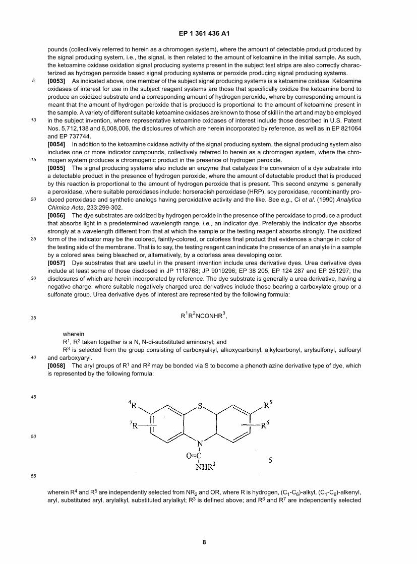
50
55
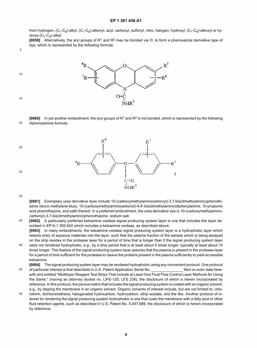
3
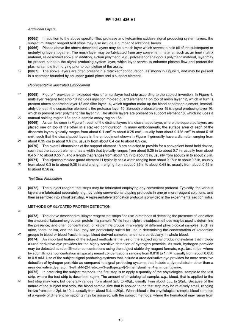
Relevant Literature
[0010] United States Patents of interest include: 5,470,752; 5,695,949; 5,725,774; 6,008,006. Also of interest are:WO 96/31270; WO 96/31619; WO 96/34977; EP 821064; and EP 737744.
SUMMARY OF THE INVENTION
[0011] Multilayer reagent test strips for quantitating glycated protein in a fluid sample, as well as methods for usingthe same, are provided. The subject multilayer test strips include at least a filter layer, a protease layer and a ketoamineoxidase signal producing and fluid flow control system layer. In using the subject test strips, a fluid sample is appliedto the test strip and a signal is generated that can be employed to quantitate the glycated protein level in the sample.Also provided are kits and systems that include the subject test strips and find use in practicing the subject methods.The subject compositions and methods find use in glycated protein monitoring applications, among other utilities.
BRIEF DESCRIPTION OF THE FIGURES
[0012]
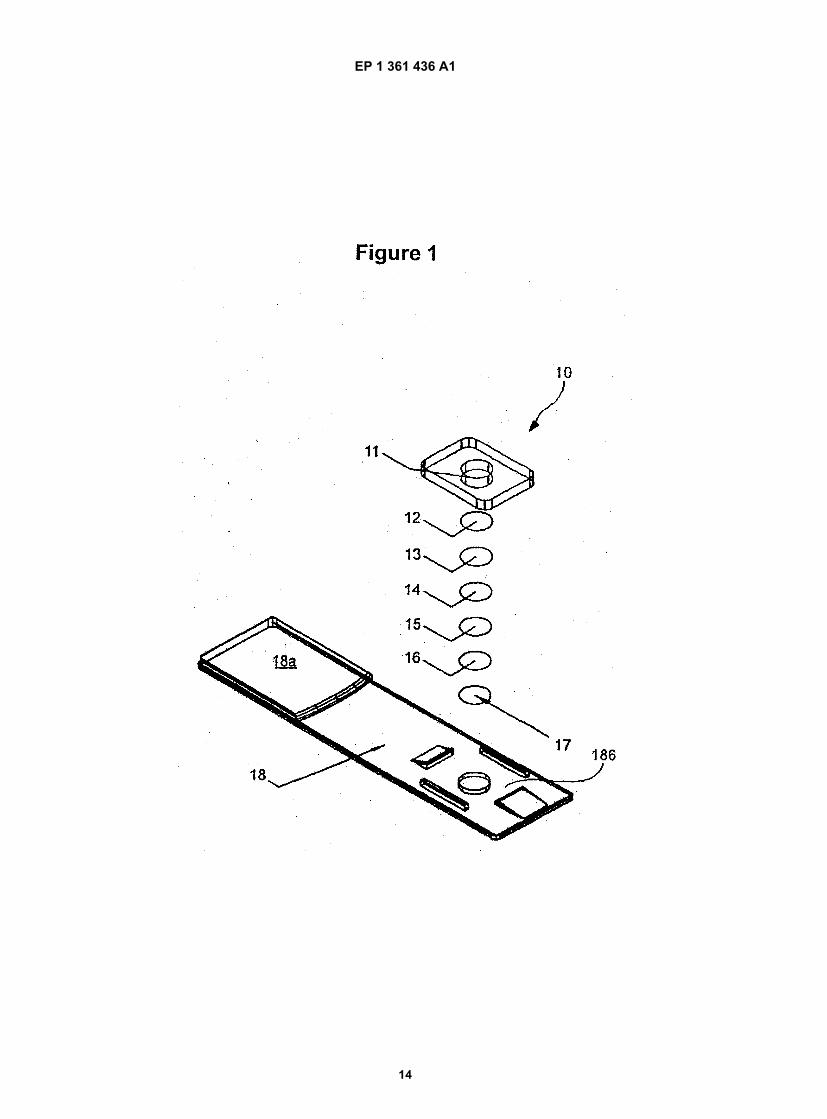
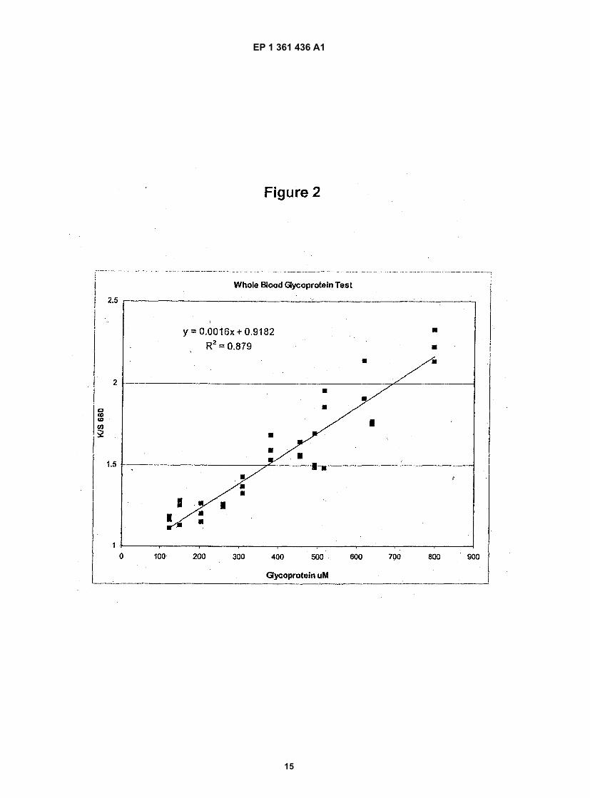
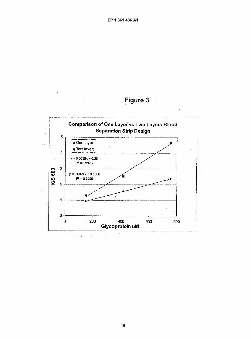
Figure 1 provides an exploded view of a multilayer reagent test strip according to one embodiment of the subjectinvention.Figure 2 provides graphical results of an assay performed with test strip with a one-layer blood separation layerconfiguration.Figure 3 provides graphical results of an assay with a two-layer blood separation layer configuration.
DESCRIPTION OF THE SPECIFIC EMBODIMENTS
[0013] Multilayer reagent test strips for quantitating glycated protein in a fluid sample, as well as methods for usingthe same, are provided. The subject multilayer test strips include at least a filter layer, a protease layer and a ketoamineoxidase signal producing fluid flow control system layer. In using the subject test strips, a fluid sample is applied to thetest strip and a signal is generated that can be employed to quantitate the glycated protein level in the sample. Alsoprovided are kits and systems that include the subject test strips and find use in practicing the subject methods. Thesubject compositions and methods find use in glycated protein monitoring applications, among other utilities.[0014] Before the subject invention is described further, it is to be understood that the invention is not limited to theparticular embodiments of the invention described below, as variations of the particular embodiments may be madeand still fall within the scope of the appended claims. It is also to be understood that the terminology employed is forthe purpose of describing particular embodiments, and is not intended to be limiting. Instead, the scope of the presentinvention will be established by the appended claims.[0015] In this specification and the appended claims, the singular forms "a," "an" and "the" include plural referenceunless the context clearly dictates otherwise. Unless defined otherwise, all technical and scientific terms used hereinhave the same meaning as commonly understood to one of ordinary skill in the art to which this invention belongs.[0016] Where a range of values is provided, it is understood that each intervening value, to the tenth of the unit ofthe lower limit unless the context clearly dictates otherwise, between the upper and lower limit of that range, and anyother stated or intervening value in that stated range, is encompassed within the invention. The upper and lower limitsof these smaller ranges may independently be included in the smaller ranges, and are also encompassed within theinvention, subject to any specifically excluded limit in the stated range. Where the stated range includes one or bothof the limits, ranges excluding either or both of those included limits are also included in the invention.[0017] Unless defined otherwise, all technical and scientific terms used herein have the same meaning as commonlyunderstood to one of ordinary skill in the art to which this invention belongs. Although any methods, devices andmaterials similar or equivalent to those described herein can be used in the practice or testing of the invention, thepreferred methods, devices and materials are now described.[0018] All publications mentioned herein are incorporated herein by reference for the purpose of describing anddisclosing the cell lines, vectors, and methodologies, which are described in the publications, which might be used inconnection with the presently described invention.[0019] As summarized above, the subject invention provides multilayer reagent test strips for quantitating glycatedprotein in a sample, as well as systems and kits that include the subject test strips. In further describing the invention,the test strips are described first in greater detail, followed by a review of the methods of using the test strips to quantitateglycated protein level. Finally, a review of representative systems and kits according to the subject invention is alsoprovided.
EP 1 361 436 A1
5
10
15
20
25
30
35
40
45
50
55
4
MULTILAYER REAGENT TEST STRIPS
[0020] As summarized above, the subject invention provides multilayer reagent test strips, where the subject teststrips find use in quantitating glycated protein in a fluid composition, as described in greater detail below. The subjectreagent test strips are multilayer reagent test strips, by which is meant that the subject reagent test strips include aplurality of different layers, where the layers are in sequential fluid communication, such that a fluid applied to a firstlayer of the plurality sequentially travels through the remaining layers of the plurality in a sequential manner.[0021] The number of distinct layers that make up the subject test strips may vary, typically ranging from about 2 to10, usually from about 3 to 7 in many embodiments. In many embodiments, the subject test strips include a minimumof three different layers, which layers, in sequential order, are: (a) a blood filter layer for separating red blood cells fromplasma; (b) a protease layer; and (c) a ketoamine oxidase signal producing and fluid flow control system layer. Assuch, many embodiments of the subject test strips include at least the above layers in sequential order, such that thesecond protease layer is in fluid communication with the first blood filter layer and the third ketoamine oxidase layer isin fluid communication with the second protease layer. In many embodiments, one or more additional layers that providefor additional functionality are also present.
Blood Separation Layer(s)
[0022] The first element of the subject multilayer test strips is the blood separation element, which element servesto produce plasma from whole blood, where the plasma then flows into subsequent layers for further treatment/analysis.This element may be present as a two layer separation element, or a single layer separation element. Each of thesedistinct embodiments is described in greater detail below.
Two Layer Blood Separation/Filter Element
[0023] In certain embodiments, a two layer structure is employed to produce plasma from whole blood, where thetwo layer structure includes a blood separation layer and a filter layer, which layers work in tandem to provide for redblood cell free plasma which is subsequently assayed in the subsequent layers. As used herein, the term "plasma"means the substantially colorless fluid obtained from a whole blood sample after red blood cells have been removedby the separation process and device of the present invention. Because plasma is serum plus the clotting proteinfibrinogen, the term "plasma" is used broadly herein to include both plasma and serum. The blood separation/filterlayers of the subject strips include a separation matrix and a filter layer.[0024] The separation matrix of the present invention is a permeable matrix which does not contain glass fibers and,therefore, is termed "a permeable non-glass fiber matrix." The term "permeable" means liquid-permeable, such aspermeable to plasma, as well as permeable or porous to red blood cells when the matrix is provided in the absenceof a polyol. As used herein, the phrase "matrix being porous to red blood cells in the absence of a polyol" means thatwithout the polyol contained in or on the matrix the red blood cells would simply pass through the matrix, virtuallyimmediately. In the absence of the polyol, red blood cells are not retained, by filtration or otherwise, in the matrix.[0025] The polyol contained within or on the matrix chemically reacts with the whole blood sample so as to clumpthe red blood cells. As used herein, "clump" or "clumping" means the collection into a mass or group, red blood cellsdistributed in a whole blood sample. While not wishing to be bound by any theory or mechanism, the clumping can bethe result of agglutination, coagulation, or the like, or some other chemical interaction between the polyol and the redblood cells.[0026] A useful permeable matrix can be a woven or non-woven material and can be an absorbent or a non-absorbentmaterial which may or may not be hydrophilic. Especially suitable materials for the matrix include, for example, wovenor non-woven, absorbent or non-absorbent, nylon, rayon, cotton, acrylic and polyester. In one embodiment of theinvention, the matrix is a non-woven, non-absorbent polyester. The polyester is preferably a poly(paraphenylene tereph-thalate), such as that used in a preferred polyester sold as Sontara® (DuPont, Inc., Wilmington, Del.). Another preferredmatrix is the woven, absorbent nylon Tetex®3-3710 (Tetko, Inc., Lancaster, N.Y.).[0027] Depending upon the porosity or other properties of the matrix, the clumped red blood cells either are retainedin the matrix or are filtered out by the filter material as described below. Some of the above-described matrix materials,such as the non-woven, non-absorbent polyesters, do not have "pores" in the traditional sense, i.e., that can be meas-ured, for example, by pore size (microns). In the absence of a polyol of the present invention such materials essentiallyhave no limit as the porosity and are porous to red blood cells, which have an average size of 5 µm. With such ma-croporous materials, if the polyol is not present the red blood cells pass through the matrix almost immediately. Forthose matrix materials which can be characterized based on pore size, the matrices used in the present invention canhave a pore size generally of from about 2 µm to about 10 µm. Such pores sizes can be useful for retaining the clumpedred blood cells. Depending upon the porosity, thickness, which is generally 200 to 1100 µm, and other properties of
EP 1 361 436 A1
5
10
15
20
25
30
35
40
45
50
55
5
the matrix, such as absorbency, the clumped red blood cells are either retained in the matrix or captured in a final filtermaterial as described below.[0028] The polyol-containing matrix has a first surface for sample application and a second surface where plasmais received or becomes available for additional separation. Generally, the first and second surfaces are presented asopposite sides of the matrix. The whole blood sample flows in a direction from the first surface toward the secondsurface, under conditions which provide such directional flow, such as, gravitation, vacuum, or external pressure. Toenhance the simplicity of the method, if desired, separation can be performed by gravity alone. Preferably, the sepa-ration matrix provides for flow in a vertical direction, preferably by gravitation.[0029] The separation method and device include a permeable non-glass fiber matrix containing a polyol. As usedherein, the terms "matrix containing a polyol" and "polyol-containing matrix" mean that the polyol is separately addedto the matrix and is not a component originally found in the composition or make up of the matrix, such as cellulosefilter paper. Further, "matrix containing a polyol" means a polyol can be impregnated into the matrix or coated into oronto the matrix or covalently or non-covalently bound to the matrix. In a preferred embodiment, the polyol is impregnatedinto the matrix.[0030] As used herein, the term "polyol" means a polyhydroxy alcohol which is an alkyl or aromatic containing morethan one hydroxyl group. The term "poly" as used in "polyol" does not infer that the alkyl or aromatic compound is alarge polymer made up of repeating monomeric units, but, instead, means that more than one hydroxyl group is presentin the compound. As discussed more fully below, with the exception of polysaccharides, the polyols used in the presentinvention are simple sugars or sugar alcohols, oligosaccharides, or other naturally or non-naturally occurring non-polymeric alkyl or aromatic compounds. Therefore, the term "polyol" encompasses sugars, alcohol derivatives of sug-ars, herein termed "sugar alcohols," and other naturally or non-naturally occurring non-polymeric polyols.[0031] As used herein, "sugar" includes monosaccharides, oligosaccharides, and polysaccharides. A monosaccha-ride is a simple sugar which is as a linear, branched, or cyclic polyhydroxy alcohol containing either an aldehyde or aketone group. Exemplary monosaccharides include, but are not limited to, mannose, glucose, talose, galactose, xylose,arabinose, lyxose, ribose and fructose. An oligosaccharide is a linear or branched carbohydrate that consists from twoto ten monosaccharide units joined by means of glycosidic bonds. Oligosaccharides which can be used in the presentinvention include, but are not limited to disaccharides such as sucrose, trehalose, lactose and maltose. Examples oflarger oligosaccharides which can be used in the invention include the cyclodextrins, such as alpha-cyclohexylamylose,beta-cycloheptaamylose, and gamma-cyclooctoamylose, as well as other oligosaccharides well known in the art. Apolysaccharide is any linear or branched polymer having more than ten monosaccharides linked together by glycosidicbonds. Exemplary polysaccharides include, but are not limited to, ficoll, polysucrose, and hydroxyethyl starch.[0032] Encompassed within "sugar" are those sugars which are naturally occurring as well as those which are knownbut which have not yet been identified as occurring naturally in plants or animals. For example, there are five knownnaturally occurring aldohexoses, including D-glucose, D-mannose, D-talose, D-galactose, and L-galactose. However,the aldohexose structure has four chiral carbons and thus, sixteen possible stereoisomers, all of which are known,although only the five listed above have been identified as occurring naturally in plants or animals. Thus, "sugar"encompasses enantiomers in either the D or L forms of a sugar as well as racemic mixtures thereof.[0033] A polyol of the present invention also can be a "sugar alcohol." A "sugar alcohol" is an alcohol derivative ofa mono- or an oligosaccharide which is generally formed by reduction of the aldehyde or ketone moiety on the mono-or oligosaccharide. Exemplary sugar alcohols include, but are not limited to, mannitol, sorbitol, arabitol, inositol, ga-lactitol, erythritol, and threitol. Also included within the definition of "sugar alcohol" are the alcohol derivatives of thosemono- and oligosaccharides described above.[0034] Where chiral carbons are present in the sugar alcohol, the sugar alcohol may be in the D or L form, such asD-threitol or L-threitol, or in a racemic mixture of both the D and L forms. The sugar alcohol can, but does not have to,be naturally occurring. That is, the sugar alcohol can be a derivative of a known, naturally occurring sugar, or, alterna-tively, it can have a D or L configuration known to exist but not necessarily identified as occurring in nature. The sugaralcohol also can be a sugar which is found naturally in its reduced alcohol form or it can be an alcohol derivative of asugar which derivative is not known to exist in nature.[0035] In addition to sugar or sugar alcohols, the polyol can be a non-polymeric naturally occurring or non-naturallyoccurring polyol, which includes linear, branched, or cyclic alkyl or aromatic compounds containing more than onehydroxyl group. As used herein the term "non-polymeric" means the alkyl or aromatic compounds are not polymers.Polymers are defined as high molecular weight compounds consisting of long chains that may be open, closed, linear,branched, or crosslinked, which chains are composed of repeating units, called monomers, which may be either iden-tical or different. As used herein, those polyols which are "naturally occurring" are ones which occur in nature and thosewhich are "non-naturally occurring" are not found in nature. Generally, these naturally occurring or non-naturally oc-curring alkyl or aromatic compounds range in size from three to twenty carbons (C3 to C20), and more preferably, fromthree to ten carbons (C3 to C10). Examples of such naturally occurring, non-polymeric polyols are glycerol, a three-carbon trihydroxy alcohol that occurs in many lipids, and quinic acid,1,3,4,5-tetrahydroxycyclohexanecarboxylic acid,
EP 1 361 436 A1
5
10
15
20
25
30
35
40
45
50
55
6
which acid can be in the salt form. Examples of non-naturally occurring, non-polymeric polyols include pentaerythritoland dipentaerythritol.[0036] In one embodiment, to apply the polyol to the matrix, the polyol can simply be dissolved in an aqueous solutiongenerally, at a concentration of about 20% when used alone, and at about 10% concentration when combined with apolycationic polymer, which is generally present in a concentration of about 0.5% to 5% as discussed more fully below.If desired, multiple layers of matrices containing polyol at lower concentrations, such as four layers of matrix containing5% polyol, also can be used. The polyol and, if present, the polycationic polymer can alternatively be dissolved inphysiological saline (0.85% NaCl), phosphate buffered saline (PBS), an organic solvent, or the like.[0037] In addition to the polyol, a polycationic polymer can, but does not have to, be added to the matrix. Similar tothe addition of a polyol to the matrix, the polycationic polymer can also be physically impregnated, coated into or onto,or covalently or non-covalently bound to the matrix. The polycationic polymer is also useful for clumping, as well asstabilizing clumped, red blood cells.[0038] The polycationic polymer component can be any polymer having more than one cationic site and are generallybased on monomers which contain an amine group. Suitable polycationic polymers include, for example, hexadimeth-rine bromide, trimethylenehexamethylenediammoniumbromide, polylysine, polyallylamine, polyarginine, poly(N,N-dimethylaminoethylmethacrylate, copolymers of N,N-dimethylaminoethylmethacrylate and methylmethacrylate, poly-ethyleneimine, poly(diallyldimethylammonium chloride), poly(1,1-dimethyl-3,5-dimethylenepiperidinium chloride), andmixtures thereof. The polymerized positively charged amino acids, such as polylysine, can have the amino acids ineither the D or L forms, such as poly-L-lysine or poly-D-lysine, or a racemic mixture thereof, such as poly-D,L-lysine.[0039] As described above, in one embodiment, to apply the cationic polymer to the matrix, the polymer can bedissolved in an a solution such as water, physiological saline, PBS, an organic solvent, or the like, and the matrix thendipped into the polymer containing solution. Generally, the polymer is in a concentration of about 0.5% to 5%. Whereboth polyol and polymer are contained in the matrix, the order of adding polyol and polymer to the matrix is irrelevant.For example, polyol and polymer can be simultaneously or sequentially dissolved in such aqueous solutions or solventsas those described above and both polyol and polymer simultaneously applied to the matrix, as described in the Ex-amples below. Alternatively, polyol and polymer can be applied to the matrix sequentially in any order.[0040] Non-hemolytic detergents, such as Pluronic (Pragmatics, Inc., Elkhart, Ind.), can be added to the aqueoussolutions or solvents described above, generally at a concentration of 0.01% to 0.1%. Such detergents help maximizeimpregnation of a polyol into the matrix, thereby improving the flow rate of the whole blood sample and the plasma.Other optional agents which can further enhance the flow rate, include, for example, polyvinylpyrrolidone or similarpolymers and other fillers which give the matrix and the below described filter material stiffness.[0041] In addition, the matrix may include one or more additional reagents to remove interference, e.g., KIO3, KMnO4,FeSO4for removing ascorbic acid interference, uricase for removing uric acid interference.[0042] As indicated above, a filter material can be used in combination with the matrix of the present invention.Suitable filter materials include, for example, nylon, cellulose acetate, polysulfone, synthetic fibers, and polycarbonate.The filter can, but does not have to, be a membrane. Illustrative filters and membranes include, for example, BTSpolysulfone membrane (Memtek, Inc., San Diego, Calif.), Ahlstrom synthetic fiber sheets, such as 94-30 A (AhlstromFiltration, Inc., Mt. Holly Spring, Pa.), Biodyne A®nylon membrane (Pall Corp., East Hills, N.Y.), Ultrabind 450 (Gelman,Ann Arbor, Mich.), and Nucleopore® polycarbonate (Costar, Corp., Cambridge, Mass.).[0043] The need for any additional filter material depends to a large extent on the porosity, thickness, absorbencyor other properties of the matrix. For example, the clumped red blood cells, depending upon the above properties ofthe matrix, can be retained in the matrix. Alternatively, or in addition thereto, a final filter material can be used to captureor retain any additional clumps of red blood cells. Where present, the filter material can generally have a porosity ofup to about 12 µm and preferably will have a pore size of less than 10 µm, and more preferably 5 µm or less.[0044] A filter material can be placed underneath the polyol-containing separation matrix, thereby supporting thematrix.[0045] In preferred embodiments of the invention, the blood separation method and device comprise a non-woven,non-absorbent polyester matrix impregnated with mannitol and either a nylon or polysulfone membrane below thematrix. Preferably, the matrix additionally contains hexadimethrine bromide and KIO3.[0046] The above-described two layer filtration element is further discussed in U.S. Patent Nos. 5,470,752; 5,695,949and 5,725774; the disclosures of which are herein incorporated by reference.
Single Layer Filtration Layer
[0047] In other embodiments, blood separation is achieve by an element made up of a single filtration layer. In theseembodiments, the single filtration layer is typically a porous matrix, wherein the separation takes place as the samplemoves through the matrix from one side to the other. A representative matrix to accomplish that separation may havepores that trap the red blood cells, generally pore sizes in the range from about 0.1 µm to about 5 µm. In certain
EP 1 361 436 A1
5
10
15
20
25
30
35
40
45
50
55
7
embodiments, the membrane is anisotropic, with a range of pore sizes; e.g., a broad range of pore sizes. When thematrix comprises an anisotropic membrane, the first side to which non-filtered blood is applied may be the large-poreside. For example, a gradient of pore sizes from about 0.1 µm to about 150 µm may extend through the membrane.On the large-pore side, pore size is preferably in the range from about 30 µm to about 40 µm. On the side of themembrane where the pores are smallest (i.e., the side that fluid exits in order to pass on to the next layer), the voidvolume is relatively small, and the material of the membrane is generally quite dense, within a layer that can typicallyconstitute up to 20% of the membrane's thickness. Within this layer, pore size is sometimes in the range from about0.1 to about 0.8 µm, with a nominal pore size often about 0.3 µm. In certain embodiments, the matrix is one that notonly traps red blood cells but also minimizes lysing of the cells, so that any portion of the sample that passes throughthe matrix to the downstream layers in the direction of fluid flow does not absorb light to any appreciable extent atabout 700 nm.[0048] The matrix of the separation layer is generally a hydrophilic porous membrane. The matrix allows for the flowof an aqueous medium through it. Polysulfones and polyamides (nylons) are examples of suitable matrix materials.Other polymers having comparable properties may also be used. A preferred method of preparing the porous materialthat forms the matrix of the separation layer is to cast the polymer without a supporting core. Such a matrix is, forexample, the anisotropic polysulfone membrane available from Memtec, Inc., San Diego, California . The terms "matrix"and "membrane" are used interchangeably herein. Each term is understood to not be limited to a single layer and mayinclude, for example, an absorbent layer. A matrix of less than about 500 µm thickness is usually employed with about115 to 155 µm being preferred. A thickness of about 130 to 140 µm is most preferred, particularly when the matrix isnylon or anisotropic polysulfone. The matrix generally does not deform on wetting, thus retaining its original confor-mation and size, and has sufficient wet strength to allow for routine manufacture.
Protease Layer
[0049] Downstream from the blood separation element/layer(s) in the direction of fluid flow is the protease layer ofthe subject multilayer reagent test strips. This layer comprises a matrix or membrane material and a protease enzyme.The matrix material is one that is porous and provides for flow of sample fluid through the material. The matrix that isemployed in this layer is typically an inert porous matrix that provides a support for protease component. As such, thematrix is one that is permissive of aqueous fluid flow through it and provides sufficient void space for the protease toexert its activity on proteins present in fluid that passes through the matrix. A number of different porous matrices havebeen developed for use in various analyte detection assays, which matrices may differ in terms of materials, pore sizes,dimensions and the like, where representative matrices include those described in U.S. Patent Nos: 55,932,431;5,874,099; 5,871,767; 5,869,077; 5,866,322; 5,834,001; 5,800,829; 5,800,828; 5,798,113; 5,670,381; 5,663,054;5,459,080; 5,459,078; 5,441,894 and 5,212,061; the disclosures of which are herein incorporated by reference. Thedimensions and porosity of the test strip may vary greatly, where the matrix may or may not have a porosity gradient,e.g., with larger pores near or at the sample application region and smaller pores at the detection region. Examples ofspecific matrix materials of interest include those prepared from polyamide (nylon), polysulfone, polyester, polyacrylate,cellulose, polycarbonate, nitrocellulose, etc.[0050] The protease layer also includes a protease, i.e., an enzyme having protease activity. The membrane ormatrix of the protease layer is typically coated with the protease in a manner that preserves the activity of the protease.The protease of the protease layer is one that cleaves protein molecules to yield accessible ketoamine bonds. Anyconvenient protease may be employed, where representative proteases include Protease XIV, Proteinase K, chymo-trypsin, substilisin, trypsin, and the like.
Signal Producing and Fluid Flow Control System Layer
[0051] In fluid communication with, and downstream of, the protease layer is the ketoamine oxidase signal producingand fluid flow control system layer. This signal producing system layer includes a porous matrix or membrane element,such as those described above, which includes a ketoamine oxidase signal producing system. In the subject test strips,the one or more members of the signal producing system are associated, e.g., covalently or non-covalently attachedto, at least a portion of (i.e., the detection region) the matrix, and in many embodiments to substantially all of the porousmatrix. In many embodiments, the matrix or membrane component is typically coated with the reagents of the ketoamineoxidase signal producing system.[0052] The ketoamine oxidase signal producing system of this particular layer of the multilayer reagent test strips isan oxidation signal producing system. By oxidation signal producing system is meant that in generating the detectablesignal from which the ketoamine concentration in the sample is derived, a ketoamine bond is oxidized by a ketoamineoxidase to produce an oxidized form of the substrate and a corresponding or proportional amount of hydrogen peroxide.The hydrogen peroxide is then employed, in turn, to generate the detectable product from one or more indicator com-
EP 1 361 436 A1
5
10
15
20
25
30
35
40
45
50
55
8
pounds (collectively referred to herein as a chromogen system), where the amount of detectable product produced bythe signal producing system, i.e., the signal, is then related to the amount of ketoamine in the initial sample. As such,the ketoamine oxidase oxidation signal producing systems present in the subject test strips are also correctly charac-terized as hydrogen peroxide based signal producing systems or peroxide producing signal producing systems.[0053] As indicated above, one member of the subject signal producing systems is a ketoamine oxidase. Ketoamineoxidases of interest for use in the subject reagent systems are those that specifically oxidize the ketoamine bond toproduce an oxidized substrate and a corresponding amount of hydrogen peroxide, where by corresponding amount ismeant that the amount of hydrogen peroxide that is produced is proportional to the amount of ketoamine present inthe sample. A variety of different suitable ketoamine oxidases are known to those of skill in the art and may be employedin the subject invention, where representative ketoamine oxidases of interest include those described in U.S. PatentNos. 5,712,138 and 6,008,006, the disclosures of which are herein incorporated by reference, as well as in EP 821064and EP 737744.[0054] In addition to the ketoamine oxidase activity of the signal producing system, the signal producing system alsoincludes one or more indicator compounds, collectively referred to herein as a chromogen system, where the chro-mogen system produces a chromogenic product in the presence of hydrogen peroxide.[0055] The signal producing systems also include an enzyme that catalyzes the conversion of a dye substrate intoa detectable product in the presence of hydrogen peroxide, where the amount of detectable product that is producedby this reaction is proportional to the amount of hydrogen peroxide that is present. This second enzyme is generallya peroxidase, where suitable peroxidases include: horseradish peroxidase (HRP), soy peroxidase, recombinantly pro-duced peroxidase and synthetic analogs having peroxidative activity and the like. See e.g., Ci et al. (1990) AnalyticaChimica Acta, 233:299-302.[0056] The dye substrates are oxidized by hydrogen peroxide in the presence of the peroxidase to produce a productthat absorbs light in a predetermined wavelength range, i.e., an indicator dye. Preferably the indicator dye absorbsstrongly at a wavelength different from that at which the sample or the testing reagent absorbs strongly. The oxidizedform of the indicator may be the colored, faintly-colored, or colorless final product that evidences a change in color ofthe testing side of the membrane. That is to say, the testing reagent can indicate the presence of an analyte in a sampleby a colored area being bleached or, alternatively, by a colorless area developing color.[0057] Dye substrates that are useful in the present invention include urea derivative dyes. Urea derivative dyesinclude at least some of those disclosed in JP 1118768; JP 9019296; EP 38 205, EP 124 287 and EP 251297; thedisclosures of which are herein incorporated by reference. The dye substrate is generally a urea derivative, having anegative charge, where suitable negatively charged urea derivatives include those bearing a carboxylate group or asulfonate group. Urea derivative dyes of interest are represented by the following formula:
whereinR1, R2 taken together is a N, N-di-substituted aminoaryl; andR3 is selected from the group consisting of carboxyalkyl, alkoxycarbonyl, alkylcarbonyl, arylsulfonyl, sulfoaryl
and carboxyaryl.[0058] The aryl groups of R1 and R2 may be bonded via S to become a phenothiazine derivative type of dye, whichis represented by the following formula:
wherein R4 and R5 are independently selected from NR2 and OR, where R is hydrogen, (C1-C6)-alkyl, (C1-C6)-alkenyl,aryl, substituted aryl, arylalkyl, substituted arylalkyl; R3 is defined above; and R6 and R7 are independently selected
R1R2NCONHR3,
EP 1 361 436 A1
5
10
15
20
25
30
35
40
45
50
55
9
from hydrogen, (C1-C6)-alkyl, (C1-C6)-alkenyl, acyl, carboxyl, sulfonyl, nitro, halogen, hydroxyl, (C1-C6)-alkoxyl or hy-droxy-(C1-C6)-alkyl.[0059] Alternatively, the aryl groups of R1 and R2 may be bonded via O, to form a phenoxazine derivative type ofdye, which is represented by the following formula:
[0060] In yet another embodiment, the aryl groups of R1 and R2 is not bonded, which is represented by the followingdiphenylamine formula:
[0061] Exemplary urea derivative dyes include 10-(carboxymethylaminocarbonyl)-3,7-bis(dimethylamino)phenothi-azine (leuco methylene blue), 10-(carboxymethylaminocarbonyl)-4,4'-bis(dimethylamino)diphenylamine, 10-propionicacid phenothiazine, and salts thereof. In a preferred embodiment, the urea derivative dye is 10-(carboxymethylamino-carbonyl)-3,7-bis(dimethylamino)phenothiazine, sodium salt.[0062] A particularly preferred ketoamine oxidase signal producing system layer is one that includes the layer de-scribed in EP-A-1 305 624 which includes a ketoamine oxidase, as described above.[0063] In many embodiments, the ketoamine oxidase signal producing system layer is a hydrophobic layer whichretards entry of aqueous materials into the layer, such that the plasma fraction of the sample which is being assayedon the strip resides in the protease layer for a period of time that is longer than if the signal producing system layerwere not rendered hydrophobic, e.g., by a time period that is at least about 5 times longer, typically at least about 10times longer. This feature of the signal producing system layer assures that the plasma is present in the protease layerfor a period of time sufficient for the protease to cleave the proteins present in the plasma sufficiently to yield accessibleketoamine.[0064] The signal producing system layer may be rendered hydrophobic using any convenient protocol. One protocolof particular interest is that described in U.S. Patent Application Serial No. filed on even date here-with and entitled "Multilayer Reagent Test Strips That Include at Least One Fluid Flow Control Layer Methods for Usingthe Same," (having an attorney docket no. LIFE-120; LFS 236), the disclosure of which is herein incorporated byreference. In this protocol, the porous matrix that includes the signal producing system is coated with an organic solvent,e.g., by dipping the membrane in an organic solvent. Organic solvents of interest include, but are not limited to: chlo-roform, dichloromethane, halogenated hydrocarbon, hydrocarbon, ethyl acetate, and the like. Another protocol of in-terest for rendering the signal producing system hydrophobic is one that coats the membrane with a fatty acid or otherfluid retention agents, such as described in U.S. Patent No. 5,447,689, the disclosure of which is herein incorporatedby reference.
EP 1 361 436 A1
5
10
15
20
25
30
35
40
45
50
55
10
Additional Layers
[0065] In addition to the above specific filter, protease and ketoamine oxidase signal producing system layers, thesubject multilayer reagent test strips may also include a number of additional layers.[0066] Placed above the above-described layers may be a mesh layer which serves to hold all of the subsequent orunderlying layers together. The mesh layer may be fabricated from any convenient material, such as an inert matrixmaterial, as described above. In addition, a clear polymeric, e.g., polyester or analogous polymeric material, layer maybe present beneath the signal producing system layer, which layer serves to enhance plasma flow and protect theplasma sample from drying prior to completion of the assay.[0067] The above layers are often present in a "stacked" configuration, as shown in Figure 1, and may be presentin a chamber bounded by an upper guard piece and a support element.
Representative Illustrated Embodiment
[0068] Figure 1 provides an exploded view of a multilayer test strip according to the subject invention. In Figure 1,multilayer reagent test strip 10 includes injection molded guard element 11 on top of mesh layer 12, which in turn ispresent above separation layer 13 and filter layer 14, which together make up the blood separation element. Immedi-ately beneath the separation element is the protease layer 15. Beneath protease layer 15 is signal producing layer 16,which is present over polymeric film layer 17. The above layers are present on support element 18, which includes amanual holding region 18a and a sample assay region 18b.[0069] As can be seen in Figure 1, each of the distinct layers is a disc shaped layer, where the separated layers areplaced one on top of the other in a stacked configuration. In many embodiments, the surface area of each of thedisparate layers typically ranges from about 0.1 cm2 to about 0.25 cm2, usually from about 0.125 cm2 to about 0.18cm2, such that the disc shaped layers in the embodiment shown in Figure 1 generally have a diameter ranging fromabout 0.35 cm to about 0.6 cm, usually from about 0.4 cm to about 0.5 cm.[0070] The overall dimensions of the support element 18 are selected to provide for a convenient hand held device,such that the support element has a width that typically ranges from about 0.25 in to about 0.7 in, usually from about0.4 5 in to about 0.55 in, and a length that ranges from about 1.5 in to about 3 in, usually from about 2 in to about 2.5 in.[0071] The injection molded guard element 11 typically has a width ranging from about 0.18 in to about 0.5 in, usuallyfrom about 0.3 in to about 0.38 in and a length ranging from about 0.35 in to about 0.68 in, usually from about 0.45 into about 0.56 in.
Test Strip Fabrication
[0072] The subject reagent test strips may be fabricated employing any convenient protocol. Typically, the variouslayers are fabricated separately, e.g., by using conventional dipping protocols in one or more reagent solutions, andthen assembled into a final test strip. A representative fabrication protocol is provided in the experimental section, infra.
METHODS OF GLYCATED PROTEIN DETECTION
[0073] The above described multilayer reagent test strips find use in methods of detecting the presence of, and oftenthe amount of ketoamine group on protein in a sample. While in principle the subject methods may be used to determinethe presence, and often concentration, of ketoamine groups in a variety of different physiological samples, such asurine, tears, saliva, and the like, they are particularly suited for use in determining the concentration of ketoaminegroups in blood or blood fractions, e.g., blood derived samples, and more particularly, in whole blood.[0074] An important feature of the subject methods is the use of the subject signal producing systems that includea urea derivative dye provides for the highly sensitive detection of hydrogen peroxide. As such, hydrogen peroxidemay be detected at submillimolar concentrations using the subject stable dry reagent formats, e.g., test strips, whereby submillimolar concentration is typically meant concentrations ranging from 0.010 to 1 mM, usually from about 0.050to 0.8 mM. Use of the subject signal producing systems that include a urea derivative dye provides for more sensitivedetection of hydrogen peroxide as compared to signal producing systems that include a dye substrate other than aurea derivative dye, e.g., N-ethyl-N-(2-hydroxy-3-sulfopropyl)-3-methylaniline, 4-aminoantipyrine.[0075] In practicing the subject methods, the first step is to apply a quantity of the physiological sample to the teststrip, where the test strip is described supra. The amount of physiological sample, e.g., blood, that is applied to thetest strip may vary, but generally ranges from about 2µL to 40µL, usually from about 5µL to 20µL. Because of thenature of the subject test strip, the blood sample size that is applied to the test strip may be relatively small, rangingin size from about 2µL to 40µL, usually from about 5µL to 20µL. Where blood is the physiological sample, blood samplesof a variety of different hematocrits may be assayed with the subject methods, where the hematocrit may range from
EP 1 361 436 A1
5
10
15
20
25
30
35
40
45
50
55
11
about 20% to 65%, usually from about 25% to 60%.[0076] Following application of the sample to the test strip, the sample is allowed to flow sequentially through thevarious layers of the strip, such that it separates in the separation element, is digested in the protease layer and reactswith the members of the signal producing system in the signal producing system layer to produce a detectable productthat is present in an amount proportional to the initial amount of the ketoamine groups of interest present in the sample.The amount of detectable product, i.e., the signal produced by the signal producing system, is then determined andrelated to the amount of glycated protein in the initial sample.
UTILITY
[0077] The above-described methods find use in any application where one wishes to test a fluid sample to determinethe concentration of ketoamine group on protein present therein. Of particular interest is use of the subject methodsto determine/monitor blood glucose levels. In such applications, the detected ketoamine group concentration providedby the subject methods is employed to determine the amount of glycated protein in the tested sample, as is known inthe art, which is turn is employed to monitor blood glucose levels, e.g., as is desirable in the treatment and managementof patients suffering from diabetes. Utilities for the subject test strips are further described in United States Patent Nos.5,470,752; 5,695,949; 5,725,774; and 6,008,006; the disclosures of which are herein incorporated by reference.
DETECTION SYSTEMS
[0078] Detection systems useful for practicing the subject methods include a reagent test strip as described aboveand an signal detection instrument or reader. In such systems, a physiological sample is applied to the test strip asdescribed above and the signal produced by the signal producing system is detected and related to the presence (andoften the amount) of glycated protein in the sample by the instrument. The above described reaction, detection andrelation steps, and instruments for practicing the same, are further described in U.S. Patent Nos. 4,734,360; 4,900,666;4,935,346; 5,059,394; 5,304,468; 5,306,623; 5,418,142; 5,426,032; 5,515,170; 5,526,120; 5,563,042; 5,620,863;5,753,429; 5,573,452; 5,780,304; 5,789,255; 5,843,691; 5,846,486; 5,902,731; 5,968,836 and 5,972,294; the disclo-sures of which are herein incorporated by reference. In the relation step, the derived glycated protein concentrationtakes into account the constant contribution of competing reactions to the observed signal, e.g., by calibrating theinstrument accordingly.
KITS
[0079] Also provided by the subject invention are kits for use in practicing the subject methods. The kits of the subjectinvention include a reagent test strip, as described above, and at least one of a means for obtaining said physiologicalsample, e.g., a lance for sticking a finger, a lance actuation means, and the like, and an standard, e.g., an glycatedprotein control solution that contains a standardized concentration of ketoamine or precursor thereof, e.g., glycatedprotein. In certain embodiments, the kits also include a detection instrument, as described above, for detecting theamount of product produced on the strip following sample application and relating the detected product to the presence(and often the amount) of analyte in the sample. Finally, the kits include instructions for using the subject kit componentsin the determination of glycated protein concentration in a physiological sample. These instructions may be presenton one or more of the packaging, a label insert, containers present in the kits, and the like.[0080] The following examples are offered by way of illustration and not by way of limitation.
EXAMPLES
I. Reagent Test Strip Preparation
A: Blood separation layer
[0081] Mesh: A Tetko mesh #7-280/44 was placed in detergent solution of 1% Pluronic (Pragmatics. Inc.) for oneminute. Excess detergent was removed and the mesh was dried by heating at 60°C for 10 minutes.[0082] Blood separation matrix: A solution containing 10% mannitol, 1.25% hexadimethrine bromide and 1% KIO3in physiological saline (0.85% NaCl) was impregnated onto Sontara® #8007 (DuPont. Inc.) on an automated impreg-nation/drying unit (AFM Engineering. Santa Anna. Calif.). The drying temperature was 100°C for approximately 10minutes.[0083] Memtec polysulfone membrane was untreated

EP 1 361 436 A1
5
10
15
20
25
30
35
40
45
50
55
12
B: Digestion layer
[0084] 200 mg/ml of protease XIV (Sigma) was dissolved in 100 mM EPPS, pH 8.0. The nylon mesh 3-200/39(SE-FAR) was immersed into the protease XIV solution. Excess solution was removed and the mesh was dried by heatingat 56°C for 10 minutes.
C: Color formation and fluid flow control layer
[0085]
A dip: It contains 300 U/ml of KAO in 20 mM PBS, pH 7.4, 1 mg/ml HRP, 1% PVP (MW=360K) and 50 mg/mlmannitol. The nylon membrane (Pall) was dipped into the solution and the excess solution was removed and themembrane was dried by heating at 56 °C for 5 minutes.B dip: After A dipping, the nylon membrane was further dipped into B dip containing 1 mM DA-60 in 70% methanol.The dipped membrane was dried by heating at 56°C for 5 minutes.C dip: A and B dipped membrane was quickly pull through into dichloromethane solvent. Excess solvent wasremoved and the membrane was dried under ventilation hood at room temperature for 2 minutes.D: Assembly of test strip
[0086] Each layer was cut into 0.476 cm (3/16 in) diameter discs which were assembled in sequence as indicatedin Figure 1 into final strip.
II. Glycated Protein Assay Employing the Reagent Test Strip as Prepared in Example I.
[0087] Figure 2: One layer blood separation layer strip design was used. i.e., blood separation layer has polysulfonemembrane only. 15 µl of whole patient blood was applied to test strip and the test strip was incubated at 37°C for 5minutes, then the color intensity was read on the Macbeth reflectance spectrometer.[0088] Figure 3: Comparison of one layer vs. two layer blood separation strip design. One layer is polysufone mem-brane only as blood separation layer. Two layers consist of Sontara® filter material and polysulfone membrane both.15 µl of whole blood with low, medium and high level glycoprotein was applied to test strip. The strip was incubated at40°C for 5 minutes. The color intensity was read on a Macbeth reflectance spectrometer. Significant increase of theslope of color intensity among different level of glycoprotein in two layer strip design definitely gives better resolutionrange. All data points in the Figure 3 represent the average of eight replicates and CV is around 10% for both strips.[0089] It is evident from the above results and discussion that the subject invention provides a highly sensitive andaccurate glycated protein detection system which is capable of providing results that are comparable in reliability andaccuracy to those currently achieved only in the clinical laboratory setting, where the system is one that can be employedby a patient or doctor in a non laboratory setting, e.g., at home or in the doctor's office. As such, the subject inventionrepresents a significant contribution to the art.[0090] All publications and patents cited in this specification are herein incorporated by reference as if each individualpublication or patent were specifically and individually indicated to be incorporated by reference. The citation of anypublication is for its disclosure prior to the filing date and should not be construed as an admission that the presentinvention is not entitled to antedate such publication by virtue of prior invention.[0091] Although the foregoing invention has been described in some detail by way of illustration and example forpurposes of clarity of understanding, it is readily apparent to those of ordinary skill in the art in light of the teachingsof this invention that certain changes and modifications may be made thereto without departing from the spirit or scopeof the appended claims.
Claims
1. A multilayer test strip comprising:
(a) a blood separation element for separating red blood cells from plasma;(b) a protease layer in fluid communication with said blood separation element; and(c) a ketoamine oxidase signal producing system layer in fluid communication with said protease layer.
2. The multilayer test strip according to Claim 1, wherein ketoamine oxidase signal producing system layer is hydro-phobic.
EP 1 361 436 A1
5
10
15
20
25
30
35
40
45
50
55
13
3. The multilayer test strip according to Claim 1 or Claim 2, wherein said layers are present in a stacked configuration.
4. The multilayer test strip according to any one of Claims 1 to 3, wherein said protease layer comprises a porousmatrix and a protease that cleaves proteins to produce ketoamine oxidase accessible ketoamine bonds.
5. The multilayer test strip according to any one of Claims 1 to 4, wherein said ketoamine oxidase signal producingsystem layer comprises a porous matrix, a ketoamine oxidase, a peroxidase and at least one indicator compound.
6. A measurement system for measuring an amount of glycated protein in a fluid sample, said system comprising:
(a) a multilayer test strip according to any one of Claims 1 to 5; and(b) a signal detection instrument for detecting signal produced on said multilayer test strip.
7. A method for quantifying the amount of glycated protein in a physiological sample, said method comprising:
(a) applying said physiological sample to a multilayer test strip according to any one of Claims 1 to 5;(b) detecting a signal produced on said test strip to quantify the amount of glycated protein in said physiologicalsample.
8. A kit for use in determining the concentration of glycated protein in a physiological sample, said kit comprising:
(a) a multilayer reagent test strip according to any one of Claims 1 to 5; and(b) at least one of:
(i) a means for obtaining a physiological sample and(ii) a control.
9. A signal detection instrument having present therein a multilayer reagent test strip according to any one of Claims1 to 5.
EP 1 361 436 A1
14
EP 1 361 436 A1
15
EP 1 361 436 A1
16
EP 1 361 436 A1
17
EP 1 361 436 A1
18


