Embed Size (px)
Citation preview
REVIEW ARTICLE
Multimodality imaging of pancreatic ductal adenocarcinoma:a review of the literatureShailesh V. Shrikhande1, Savio George Barreto1, Mahesh Goel1 & Supreeta Arya2
Departments of 1Hepato-Pancreato-Biliary Surgical Oncology, and 2Radiology, Tata Memorial Hospital, Mumbai, India
Abstracthpb_508 658..668
Background: Accurate pre-operative imaging in pancreatic cancer helps avoid unsuccessful surgical
explorations and forewarns surgeons regarding aberrant anatomy. This review aimed to determine the role
of current imaging modalities in the diagnosis and determination of resectability of pancreatic and
peri-ampullary adenocarcinomas.
Methods: A systematic search of the scientific literature was carried out using EMBASE, PubMed/
MEDLINE and the Cochrane Central Register of Controlled Trials for the years 1990 to 2011 to obtain
access to all publications, especially randomized controlled trials, reporting on the diagnostic accuracy of
ultrasonography, multi-detector computed tomography (MDCT), magnetic resonance imaging (MRI),
endoscopic ultrasonography (EUS) or positron emission tomography (PET)-computed tomography (CT)
and the evaluation of resectability of pancreatic and peri-ampullary adenocarcinomas.
Results: Based on 66 articles analysed in the review, MDCT and MRI/MRCP have comparable sensitivity
and specificity rates for diagnosis and staging of pancreatic cancers. EUS offers the best sensitivity and
specificity rates for lesions <2 cm. Improved staging has been noted when PET-CT scans are added to
pre-operative evaluation.
Conclusions: MDCT with angiography or MRI/MRCP should constitute the first imaging modality in
suspected pancreatic adenocarcinomas. EUS is recommended for assessing lesions not clearly
detected, but suspected, on CT/MRI and in tumours considered ‘borderline resectable’ on MDCT to
assess vascular involvement. PET-CT in locally advanced lesions will help rule out distant metastases.
Received 16 March 2012; accepted 16 May 2012
CorrespondenceShailesh V. Shrikhande, Department of Hepato-Pancreato-Biliary Surgical Oncology, Convener, GI –
DMG, Tata Memorial Hospital, Ernest Borges Marg, Parel, Mumbai 400 012, India. Tel: +91 22 2417 7173.
Fax: +91 22 2414 8114. E-mail: [email protected]; shailesh.shrikhande@
pancreaticcancerindia.org
Introduction
Carcinomas of the pancreas, although uncommon, are amongstthe leading causes of cancer-related deaths around the world.According to the American Cancer Society, the relative 1-yearsurvival is only 24%, and the overall 5-year survival rate is 5%1
and with similar outcomes reported worldwide.2–4 Complete sur-gical resection with chemotherapy (when indicated) offers thebest outcomes in this cancer.5 However, owing to the insidiousonset of the disease and the delayed presentation of patients,surgery with a curative intent may not always be possible. Owing
to the morbidity associated with a surgical exploration that islikely to be unsuccessful, pre-operative determination of theextent of the disease assumes significance. As a result of theincreased morbidity and technical difficulty associated with aber-rant arterial anatomy, an accurate pre-operative assessment of thevascular anatomy is important.6 Over the years, various radiologi-cal imaging modalities for the pancreas have been used for thediagnosis and staging of these cancers including abdominalultrasonography, computed tomography (CT) scans, magneticresonance imaging (MRI), laparoscopy and endoscopicultrasonography (EUS). The aims of this review were to define therole of pre-operative imaging in the evaluation of pancreaticadenocarcinomas by providing the evidence available to date oneach of these modalities.
This paper was presented at the 2010 IHPBA meeting in Buenos Aires,
Argentina.
DOI:10.1111/j.1477-2574.2012.00508.x HPB
HPB 2012, 14, 658–668 © 2012 International Hepato-Pancreato-Biliary Association
Methods
A systematic search of the scientific literature was carried outusing EMBASE, PubMed/MEDLINE and the Cochrane CentralRegister of Controlled Trials for the years 1990–2011 to obtainaccess to all publications, especially randomized controlled trials,that reported on the diagnostic accuracy of ultrasonography, CT,MRI or EUS and evaluation of resectability of pancreatic andperi-ampullary adenocarcinomas.
The search strategy was that described by Dickersin et al.7 withthe appropriate specific search terms, namely, ‘peri-ampullarycancer’, ‘pancreatic cancer’, ‘diagnosis’, ‘staging’, ‘resectability’,‘computed tomography’, ‘CT’, ‘magnetic resonance imaging’,‘MRI’, ‘ultrasonography’, ‘endoscopic ultrasonography’, ‘EUS’, ‘lap-aroscopy’, ‘positron emission tomography’, ‘PET’, ‘randomizedcontrolled trials’, ‘review’ and ‘meta-analysis’. All available majorpublications from the past 21 years were retrieved.
Inclusion criteria:
1 Original articles on the role of CT scan/MRI or MRCP/ultrasonography (abdominal and laparoscopic)/PET-CT/EUS in the diagnosis, determination of the extent ofdisease and resectability of pancreatic and peri-ampullaryadenocarcinoma.
2 Original articles comparing CT scans, MRI or MRCP, ultra-sonography (abdominal and laparoscopic), PET-CT and/or EUS in the diagnosis, determination of the extent ofdisease and resectability of pancreatic and peri-ampullaryadenocarcinoma.
Exclusion criteria:
1 Case reports.2 Articles on the imaging of tumours other than adenocarcicoma.
From the retrieved articles, only papers reporting on ultra-sonography, CT, MRI/MRCP, EUS or laparoscopy in the diagno-sis, staging and the determination of resectability in patientswith pancreatic and peri-ampullary cancer were included in thereview.
For clarification, the term staging has been used to indicate theability of the imaging modality to delineate the tumour, nodal andmetastatic disease whereas the term resectability is mainly used toindicate technical resectability in the absence of distant metastasesand vascular involvement (beyond the definition of borderlineresectable disease – for definition see below).
Results
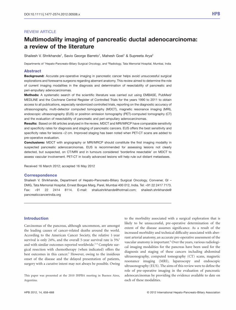
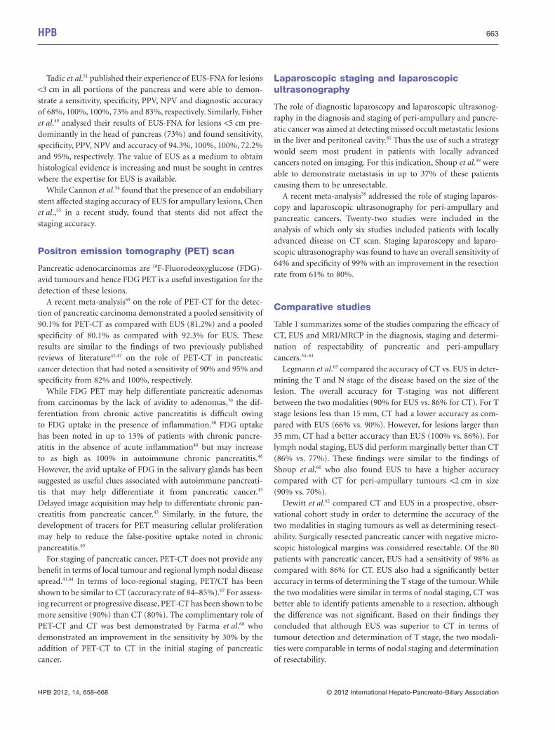
Using the above search strategy, 351 articles were identified ofwhich only 67 articles were analysed in this review (Fig. 1). Theseincluded three articles on abdominal ultrasonography,8–10 23 onCT and CT angiography,11–33 4 on MRI/MRCP,34–37 2 on laparo-scopic staging and laparosopcic ultrasonography,38,39 9 on PET
and PET-CT,40–48 3 on EUS49–51 and 23 comparing the variousmodalities.52–74
A recent review by Low et al.75 provides useful informationin differentiating benign from malignant lesions of the pancreasand hence this aspect has not been addressed in the presentreview. Other important reviews published on the topic in past11 years include the role of a standard MR in pre-operativeimaging of pancreatic masses76 and the use of an MR protocolincluding non-contrast T1-weighted fat-suppressed and dynamicgadolinium-enhanced gradient-echo imaging in pancreaticcancer by Vachiranubhap et al.77 Bipat et al.,78 in 2005, publisheda sentinel meta-analysis on the role of ultrasonography, CT andMRI for the diagnosis and determination of resectability of pan-creatic adenocarcinoma in which they concluded that helical CTwas the preferred imaging modality.
Diagnosis and staging: imaging modalitiesAbdominal ultrasonography in pancreatic cancerUltrasonography is amongst the most widely available and oftenthe first investigative imaging modality used to assess patients
Figure 1 Flow chart of the search strategy employed
HPB 659
HPB 2012, 14, 658–668 © 2012 International Hepato-Pancreato-Biliary Association
with hepatopancreatobiliary complaints or even non-specificabdominal pain. Karlson et al.8 reported a diagnostic sensitivity ofup to 90% for exocrine pancreatic tumours. Moreover, the pres-ence of obvious hepatic metastases on ultrasonography will oftenprevent the need for further imaging.
However, in general, body habitus, the retroperitoneal locationof the pancreas obscured often by bowel gas, as well as theoperator-dependant nature of the investigation preclude the useof ultrasonography as an accurate staging modality.
Morrin et al.,10 using gray scale and colour Doppler ultrasonog-raphy of the abdomen, were able to demonstrate results similar tohelical CT and CT angiography in detecting venous involvement.The role of the real-time imaging modality of contrast-enhancedultrasonography in pancreatic tumours has been trialled since thelate 1990s.9 Kitano et al.52 used coded phase inversion harmonicultrasonography to overcome the limitations from these previoustechniques and demonstrated a higher sensitivity of this tech-nique compared with contrast-enhanced CT but similar to EUSfor detecting lesions �2 cm.
Computed tomography (CT)CT scanners have developed tremendously over the past fewdecades resulting in the improved resolution and hence their diag-nostic capability. The thin-cut (64 section) intravenous contrast-enhanced multi-detector CT (MDCT) is the radiologicalinvestigation of choice.79 The scans are performed in phases, asfollows the non-contrast, arterial, pancreatic parenchymal and theportal venous phases. MDCT allows rapid anatomic coveragecoupled with excellent spatial resolution.12
The sensitivity of CT in the detection of pancreatic cancershas improved over the years with the advent of multi-phasicscans and lies between 75–100% with a specificity of70–100%.11,13,15,52,53,62,63,72 However, in spite of these improve-ments, the sensitivity of CT scan for lesions �2 cm is between68–77%13,52 with an accuracy of 77%.63 However, for tumours>2 cm, the sensitivity may be as high as 98%.52
Pancreatic carcinomas on CT appear mainly hypoattenuatingespecially on the arterial phase. However, they have also beennoted to be isoattenuating in 11% of individuals in which pan-
Figure 2 Algorithm outlining the role of the individual imaging modalities in the management of pancreatic and peri-ampullary cancers.MDCT, multi-detector computed tomography; MRI, magnetic resonance imaging; MRCP, magnetic resonance cholangiopancreatography;PET-CT, positron emission tomography-computed tomography; EUS, endoscopic ultrasonography; ERCP, endoscopic retrogradecholangiopancreatography
660 HPB
HPB 2012, 14, 658–668 © 2012 International Hepato-Pancreato-Biliary Association
creatic or biliary duct dilation are signs that may indicate thepresence of an underlying pathology.28 The pancreatic tumoursnoted to be hyperattenuating are mainly the neuroendocrinetumours.24 The pancreatic parenchymal and the portal venousphase appear to be similar but better than the arterial phase fordelineating pancreatic adenocarcinomas.17,26 However, for vascu-lar invasion, the sensitivity of images obtained in the portalvenous phase are better than those obtained in the pancreatic orarterial phases as images obtained in the pancreatic phase dem-onstrated more flow artefacts and decreased attenuation in thesuperior mesenteric vein, compared with the artefacts revealed onimages obtained in the portal venous (hepatic) phase.16
Imbriaco et al.18 found thin-section single-phase MDCT to bevery accurate for the diagnosis and assessment of resectability inpatients with a suspected pancreatic neoplasia owing to theoptimal tumour-to-pancreas contrast and maximal pancreaticparenchymal and peri-pancreatic vascular enhancement. Theyalso found that it allowed visualization of the entire liver and thewhole upper abdomen during the portal phase for accurate iden-tification of liver metastases and peritoneal seeding.
CT criteria for unresectable disease formerly included findingssuch as extrapancreatic disease involving the liver or peritoneum,and contiguous invasion of adjacent organs such as the stomachand colon, as well as involvement of peri-pancreatic vessels.27 In1997, Howard et al.56 reported a sensitivity and specificity of 63%and 100%, and an overall accuracy of 86%. However, Lu et al.25
and Diehl et al.15 found the sensitivity rates to be 84% and 91%,respectively. Karmazonovsky et al.,20 in 2005, found that spiral CThad a sensitivity, specificity, negative predictive value (NPV), posi-tive predictive value (PPV) and accuracy of 79%, 82%, 91%, 62%and 81%, respectively, in predicting unresectability. They alsofound that the sensitivity, specificity, positive predictive value,negative predictive value and accuracy of spiral CT in the diagno-sis of vascular invasion were 94%, 84.2%, 94%, 84%, and 91.3%,based on their correlation with the intra-operative findings.
The increasing surgical aggression in terms of pancreatic resec-tions that have gone hand in hand with the refinements in tech-niques of surgery, coupled with advancements in chemotherapyand radiotherapy have now led to the development of yet anotherintermediate disease stage: borderline resectable disease.21,29,31 Bydefinition,30 borderline resectable tumours are those tumours thatexhibit the following: (i) encasement of a short segment of thehepatic artery, without evidence of tumour extension to the celiacaxis, that is amenable to resection and reconstruction; and (ii)tumour abutment of the superior mesenteric artery involving<180° of the circumference of the artery or short-segment occlu-sion of the superior mesenteric vein, portal vein or their conflu-ence with a suitable option available for vascular reconstructionbecause the veins are normal above and below the area of tumourinvolvement. This stage strongly relies on the accurate delineationof peripancreatic vascular involvement based on CT imaging.
Brugel et al.14 confirmed that thin-slice multiplanar reconstruc-tions obtained with multislice helical CT provided an exact
depiction of the spatial relation between the tumour and thepotentially invaded vessels and thus had the capability to improvethe assessment of local resectability. Kaneko et al.19 retrospectivelycompared multidetector computed tomographic angiography(MDCTA) done pre-operatively to the actual surgical outcomeswith the aim of determining the value of MDCTA in predictingresectability of pancreatic head cancers. In this previous study,MDCTA was found to have a sensitivity, specificity, positive pre-dictive value, negative predictive values and accuracy of 100%,71%, 85%,100% and 89% which was similar to the resultsreported by Fusari et al.53 and Zamboni et al.32 The additionalfinding of the study by Zamboni et al.32 was the lack of evidencesuggesting varying results from the various generations of MDCTscanners used.
Shrikhande et al.29 identified 12 patients with borderline resec-table disease in a mixed pathological cohort of pancreatic adeno-carcinomas, solid pseudopapillary and neuroendocrine tumoursaccording to the MD Anderson Cancer Centre classification.30
They correlated the MDCT images with the intra-operative find-ings. Eight of the 12 patients actually underwent a curative R0resection while another 2 had microscopically positive margins(R1 resections). Based on this they determined that a gradingsystem based on three radiological features on MDCT, viz.maximum degree of circumferential contact (CC), length ofcontact of the tumour with major vessels (LC) and luminal nar-rowing of vessels at the point of contact with the tumour (venousdeformity, VD) is helpful to predict which patients with appar-ently borderline disease may actually be offered an up-front,potentially curative surgical resection. This grading system needsto be validated in a larger cohort of patients with pancreaticadenocarcinoma.
Similarly, Kent et al.22 have proposed a CT grading system forpancreaticobiliary tumours. This five-point scale describes theradiographical relationship of the pancreatobiliary mass to adja-cent vessels, viz. portal vein, superior mesenteric vein, superiormesenteric artery and celiac trunk. An increasing grade has beenshown to be associated with a higher probability of unresectabilityand microscopic positive resection margins.22
Zhao et al.33 performed a recent meta-analysis on the role of CTin the diagnosis and determination of vascular invasion in pan-creatic and periampullary cancer. Based on the results obtainedfrom the 18 studies selected, CT was found to have a sensitivityand specificity of 85% and 82%, respectively, in the diagnosis, anda sensitivity and specificity of 77% and 81% in the diagnosis ofvascular invasion. However, the results did indicate that the addi-tion of other imaging modalities, such as EUS, could furtherimprove the diagnostic accuracy of vascular invasion.
Kim et al.,23 on the other hand, compared the accuracy ofMDCT in the pre-operative evaluation of resectability betweenpatients who had received neoadjuvant chemo-radiotherapy withpatients who had not received any neoadjuvant treatment. Theyfound no difference between the two groups (83% versus 81%). Inthis previous study, the absence of para aortic lymph node
HPB 661
HPB 2012, 14, 658–668 © 2012 International Hepato-Pancreato-Biliary Association

metastasis (>1 cm in short axis) was considered a feature of resec-tability in addition to the definition of resectability provided inthe methods section of this review.
Magnetic resonance imaging (MRI) andmagnetic resonance cholangiopancreatography (MRCP)
In 1997, Trede et al.64 suggested that ultrafast MRI was the mostaccurate staging modality when compared with percutaneousultrasonography, dual-phase helical CT, selective visceral angiog-raphy and endoscopic retrograde cholangiopancreatography(ERCP) for pancreatic and peri-ampullary tumours.
Megibow et al.36 found that MRI had a diagnostic accuracy of70% for pancreatic carcinomas. Megibow et al. determined thatgradient-echo and T1-weighted spin-echo sequences rankedequally in the evaluation of vascular invasion, T1-weighted spin-echo sequences were preferred for assessing lymphadenopathyand T2-weighted spin-echo sequences were preferred for detect-ing hepatic metastases. Hanninen et al.34 noted that single-shotthick slab MRCP had superior image quality as compared withmulti-section MRCP images. They also noted a significantimprovement in the diagnostic accuracy of MRCP by two readers(89% and 84% vs. 72% and 69%) with the addition of T1 andT2-weighted images. This led them to infer that a comprehensiveMR approach should comprise both MRCP techniques andparenchymal sequences.34
More recently, Fusari et al.53 demonstrated sensitivity, specific-ity, accuracy, positive and negative predictive values of 100%,88%, 98%. 97% and 100%, respectively, for MRCP in terms ofdiagnosis, and sensitivity, specificity, accuracy, positive and nega-tive predictive values of 88%, 100%, 90%, 100% and 70%, respec-tively, for the evaluation of resectability of pancreatic carcinomas.
Tapper et al.37 determined that MRCP had a 100% and 83%,81% and 96%, and 87% and 95% sensitivity and accuracy rates inthe determination of resectability, arterial and venous involve-ment in pancreatic head carcinomas. These results were similar tothose of Hochwald et al.35 who determined that MRI and MRCPhad a sensitivity of 100%, specificity of 83%, PPV of 94%, NPV of100% and accuracy of 95% in determining resectability of pan-creatic cancer. Resectability in this previous study was defined assurgically removable with grossly negative margins. Hochwaldet al.35 noted that malignant lesions appeared as discrete,hypointense masses with respect to background pancreatic paren-chyma on post-contrast T1-weighted images. Based on a compari-son of pre-operatively performed MDCT and MR imagingwith angiography with the intra-operative findings, Lee et al.,65
however, noted that both modalities demonstrated an equalability in detection, prediction of vascular involvement and deter-mination of resectability for pancreatic ductal adenocarcinoma.
Miller et al.73 have provided specific scenarios in which MRCPmay add to the CT findings in the diagnosis and staging of tumours.In case of patients suspected to have pancreatic tumours on CT
which would appear as hypoattenuating lesions on the arterialphase but who have signs such as duct dilation or focal pancreaticatrophy, MRCP may be useful in such ‘non-contour deforming’lesions. The contrast resolution of MRI facilitates detection of suchsmall tumours on gadolinium-enhanced fat-suppressed images.Similarly, in patients with a ‘double duct’ sign, Miller et al.73 sug-gested that MRCP may be useful in differentiating a malignantfrom a benign aetiology. They also suggested a complementary rolefor MRCP in the characterization of suspected liver metastases andthe detection of omental and nodal lesions.
In patients with borderline resectable disease who receiveddown-staging chemotherapy, Donahue et al.74 studied the use ofCT and MRI signs pre-operatively. They noted that features sug-gestive of vascular involvement persisted even after chemotherapy.In spite of these features, patients underwent exploration with acurative intent based on other characteristics such as a reductionin tumour markers, a reduction in the size of the tumour onimaging and good functional status. In 83% of patients in whoma complete resection could be achieved, fibrosis, and not tumour,was the cause for the observed ‘involvement’ noted on preopera-tive imaging. Thus they concluded that CT and MRI had a lowsensitivity (71%) and specificity (58%) in predicting vascularinvolvement and resectability in the post-chemotherapy setting.
Endoscopic ultrasonography (EUS)
Studies reporting the diagnostic ability of EUS in the past werefraught with the bias that even benign lesions were included.Hence care should be exerted when interpreting earlier results ofEUS.80
The three types of echoendoscopes available today include:radial echoendoscopes that provide axial images that correspondto the familiar computed tomography (CT) or magnetic reso-nance imaging (MRI) slices, and provide images from 2700 or3600, depending on the manufacturer; curvilinear echoendo-scopes that provide an image parallel to the shaft of the endoscopeand allow for the visualization and directing of fine-needle aspi-ration (FNA), fine-needle injection (FNI), stent placement andother interventional EUS procedures; and finally the catheter-based specialty probes that are high-frequency probes that arepassed through either a forward-viewing endoscope or a side-viewing duodenoscope to provide direct visualization of a specificarea or lesion (i.e. submucosal mass, intraductal lesions or discretegastric lesion).50
EUS has emerged as a useful, albeit invasive, modality in thediagnosis of pancreatic tumours with sensitivities and accuracyapproaching 100% and specificity >95% even for lesions<2 cm.52,62,63,72 In ampullary tumours, EUS has been found to bemore sensitive and specific as compared with CT with a statisti-cally significant (P < 0.05) strength of tumour and nodalagreement with the final pathology in a cohort of 27 patients(7.4% – T1; 48.1% – T2; 44.4% – T3 and 63% – N0 and 37% – N1disease).71
662 HPB
HPB 2012, 14, 658–668 © 2012 International Hepato-Pancreato-Biliary Association
Tadic et al.51 published their experience of EUS-FNA for lesions<3 cm in all portions of the pancreas and were able to demon-strate a sensitivity, specificity, PPV, NPV and diagnostic accuracyof 68%, 100%, 100%, 73% and 83%, respectively. Similarly, Fisheret al.49 analysed their results of EUS-FNA for lesions <5 cm pre-dominantly in the head of pancreas (73%) and found sensitivity,specificity, PPV, NPV and accuracy of 94.3%, 100%, 100%, 72.2%and 95%, respectively. The value of EUS as a medium to obtainhistological evidence is increasing and must be sought in centreswhere the expertise for EUS is available.
While Cannon et al.54 found that the presence of an endobiliarystent affected staging accuracy of EUS for ampullary lesions, Chenet al.,55 in a recent study, found that stents did not affect thestaging accuracy.
Positron emission tomography (PET) scan
Pancreatic adenocarcinomas are 18F-Fluorodeoxyglucose (FDG)-avid tumours and hence FDG PET is a useful investigation for thedetection of these lesions.
A recent meta-analysis69 on the role of PET-CT for the detec-tion of pancreatic carcinoma demonstrated a pooled sensitivity of90.1% for PET-CT as compared with EUS (81.2%) and a pooledspecificity of 80.1% as compared with 92.3% for EUS. Theseresults are similar to the findings of two previously publishedreviews of literature42,47 on the role of PET-CT in pancreaticcancer detection that had noted a sensitivity of 90% and 95% andspecificity from 82% and 100%, respectively.
While FDG PET may help differentiate pancreatic adenomasfrom carcinomas by the lack of avidity to adenomas,70 the dif-ferentiation from chronic active pancreatitis is difficult owingto FDG uptake in the presence of inflammation.40 FDG uptakehas been noted in up to 13% of patients with chronic pancre-atitis in the absence of acute inflammation48 but may increaseto as high as 100% in autoimmune chronic pancreatitis.46
However, the avid uptake of FDG in the salivary glands has beensuggested as useful clues associated with autoimmune pancreati-tis that may help differentiate it from pancreatic cancer.45
Delayed image acquisition may help to differentiate chronic pan-creatitis from pancreatic cancer.43 Similarly, in the future, thedevelopment of tracers for PET measuring cellular proliferationmay help to reduce the false-positive uptake noted in chronicpancreatitis.40
For staging of pancreatic cancer, PET-CT does not provide anybenefit in terms of local tumour and regional lymph nodal diseasespread.41,44 In terms of loco-regional staging, PET/CT has beenshown to be similar to CT (accuracy rate of 84–85%).67 For assess-ing recurrent or progressive disease, PET-CT has been shown to bemore sensitive (90%) than CT (80%). The complimentary role ofPET-CT and CT was best demonstrated by Farma et al.68 whodemonstrated an improvement in the sensitivity by 30% by theaddition of PET-CT to CT in the initial staging of pancreaticcancer.
Laparoscopic staging and laparoscopicultrasonography
The role of diagnostic laparoscopy and laparoscopic ultrasonog-raphy in the diagnosis and staging of peri-ampullary and pancre-atic cancer was aimed at detecting missed occult metastatic lesionsin the liver and peritoneal cavity.81 Thus the use of such a strategywould seem most prudent in patients with locally advancedcancers noted on imaging. For this indication, Shoup et al.39 wereable to demonstrate metastasis in up to 37% of these patientscausing them to be unresectable.
A recent meta-analysis38 addressed the role of staging laparos-copy and laparoscopic ultrasonography for peri-ampullary andpancreatic cancers. Twenty-two studies were included in theanalysis of which only six studies included patients with locallyadvanced disease on CT scan. Staging laparoscopy and laparo-scopic ultrasonography was found to have an overall sensitivity of64% and specificity of 99% with an improvement in the resectionrate from 61% to 80%.
Comparative studies
Table 1 summarizes some of the studies comparing the efficacy ofCT, EUS and MRI/MRCP in the diagnosis, staging and determi-nation of respectability of pancreatic and peri-ampullarycancers.54–61
Legmann et al.63 compared the accuracy of CT vs. EUS in deter-mining the T and N stage of the disease based on the size of thelesion. The overall accuracy for T-staging was not differentbetween the two modalities (90% for EUS vs. 86% for CT). For Tstage lesions less than 15 mm, CT had a lower accuracy as com-pared with EUS (66% vs. 90%). However, for lesions larger than35 mm, CT had a better accuracy than EUS (100% vs. 86%). Forlymph nodal staging, EUS did perform marginally better than CT(86% vs. 77%). These findings were similar to the findings ofShoup et al.60 who also found EUS to have a higher accuracycompared with CT for peri-ampullary tumours <2 cm in size(90% vs. 70%).
Dewitt et al.62 compared CT and EUS in a prospective, obser-vational cohort study in order to determine the accuracy of thetwo modalities in staging tumours as well as determining resect-ability. Surgically resected pancreatic cancer with negative micro-scopic histological margins was considered resectable. Of the 80patients with pancreatic cancer, EUS had a sensitivity of 98% ascompared with 86% for CT. EUS also had a significantly betteraccuracy in terms of determining the T stage of the tumour. Whilethe two modalities were similar in terms of nodal staging, CT wasbetter able to identify patients amenable to a resection, althoughthe difference was not significant. Based on their findings theyconcluded that although EUS was superior to CT in terms oftumour detection and determination of T stage, the two modali-ties were comparable in terms of nodal staging and determinationof resectability.
HPB 663
HPB 2012, 14, 658–668 © 2012 International Hepato-Pancreato-Biliary Association
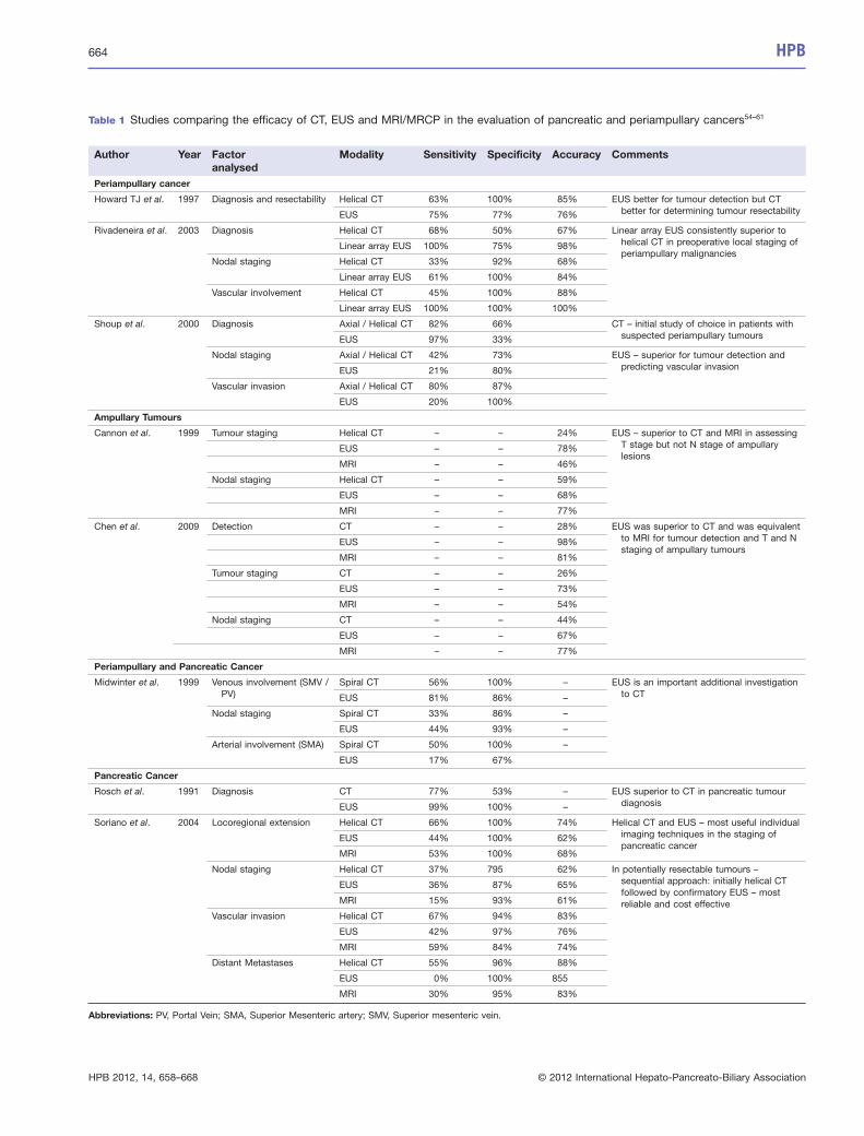
Table 1 Studies comparing the efficacy of CT, EUS and MRI/MRCP in the evaluation of pancreatic and periampullary cancers54–61
Author Year Factoranalysed
Modality Sensitivity Specificity Accuracy Comments
Periampullary cancer
Howard TJ et al. 1997 Diagnosis and resectability Helical CT 63% 100% 85% EUS better for tumour detection but CTbetter for determining tumour resectabilityEUS 75% 77% 76%
Rivadeneira et al. 2003 Diagnosis Helical CT 68% 50% 67% Linear array EUS consistently superior tohelical CT in preoperative local staging ofperiampullary malignancies
Linear array EUS 100% 75% 98%
Nodal staging Helical CT 33% 92% 68%
Linear array EUS 61% 100% 84%
Vascular involvement Helical CT 45% 100% 88%
Linear array EUS 100% 100% 100%
Shoup et al. 2000 Diagnosis Axial / Helical CT 82% 66% CT – initial study of choice in patients withsuspected periampullary tumoursEUS 97% 33%
Nodal staging Axial / Helical CT 42% 73% EUS – superior for tumour detection andpredicting vascular invasionEUS 21% 80%
Vascular invasion Axial / Helical CT 80% 87%
EUS 20% 100%
Ampullary Tumours
Cannon et al. 1999 Tumour staging Helical CT – – 24% EUS – superior to CT and MRI in assessingT stage but not N stage of ampullarylesions
EUS – – 78%
MRI – – 46%
Nodal staging Helical CT – – 59%
EUS – – 68%
MRI – – 77%
Chen et al. 2009 Detection CT – – 28% EUS was superior to CT and was equivalentto MRI for tumour detection and T and Nstaging of ampullary tumours
EUS – – 98%
MRI – – 81%
Tumour staging CT – – 26%
EUS – – 73%
MRI – – 54%
Nodal staging CT – – 44%
EUS – – 67%
MRI – – 77%
Periampullary and Pancreatic Cancer
Midwinter et al. 1999 Venous involvement (SMV /PV)
Spiral CT 56% 100% – EUS is an important additional investigationto CTEUS 81% 86% –
Nodal staging Spiral CT 33% 86% –
EUS 44% 93% –
Arterial involvement (SMA) Spiral CT 50% 100% –
EUS 17% 67%
Pancreatic Cancer
Rosch et al. 1991 Diagnosis CT 77% 53% – EUS superior to CT in pancreatic tumourdiagnosisEUS 99% 100% –
Soriano et al. 2004 Locoregional extension Helical CT 66% 100% 74% Helical CT and EUS – most useful individualimaging techniques in the staging ofpancreatic cancer
EUS 44% 100% 62%
MRI 53% 100% 68%
Nodal staging Helical CT 37% 795 62% In potentially resectable tumours –sequential approach: initially helical CTfollowed by confirmatory EUS – mostreliable and cost effective
EUS 36% 87% 65%
MRI 15% 93% 61%
Vascular invasion Helical CT 67% 94% 83%
EUS 42% 97% 76%
MRI 59% 84% 74%
Distant Metastases Helical CT 55% 96% 88%
EUS 0% 100% 855
MRI 30% 95% 83%
Abbreviations: PV, Portal Vein; SMA, Superior Mesenteric artery; SMV, Superior mesenteric vein.
664 HPB
HPB 2012, 14, 658–668 © 2012 International Hepato-Pancreato-Biliary Association
Park et al.66 found that gadolinium-enhanced dynamic three-dimensional–gradient echo MRI with MRCP showed superiortumour conspicuity and similar diagnostic performance com-pared with MDCT in evaluating the resectability of pancreaticcancer.
Discussion
While abdominal ultrasonography may be the first investigationin patients with upper gastrointestinal and biliary complaints(which could arise as a result of an underlying pancreatic malig-nancy), CT and MRI/MRCP constitute the most commonly per-formed primary investigations performed for the diagnosis andstaging of pancreatic and peri-ampullary cancers. The choicebetween CT or MRI/MRCP is more often determined by the avail-ability of the individual modality as well as the technical expertisein reporting them at the individual centres.
The major advantage of CT in comparison with EUS is itsability to provide an assessment of the entire abdominal cavitythus providing more information than EUS on distant metastases.
In interpreting the results from the earlier studies, it should beremembered that these studies were comparing EUS with oldergeneration CT scanners.
EUS appears to be most important in the assessment of lesionsnot clearly detected, but suspected, on CT/MRI, as well as inampullary tumours.71 It thus serves as a useful complementaryinvestigation to CT82 or even MRI/MRCP.
The role of CT angiography in confirming vascular anatomy/involvement has been recently reviewed by one of the authors6 andhas been strongly recommended in the pre-operative assessmentof patients deemed to be suitable for a pancreatoduodenectomy.
Specific indications where PET-CT may aid in the decisionmaking for tumours of the pancreas include: borderline resectabledisease, locally advanced disease and/or resectable disease withsuspected metastases.
Mayo et al.83 recently conducted a statewide review of allpatients with surgically managed pancreatic cancer from 1996 to2003 using data from the Oregon State Cancer Registry andfound that the use of a CT scan formed the corner stone ofstaging and determination of respectability. They also noted thatthe use of laparoscopy, as a staging modality, was restricted tothose patients suspected to have metastatic disease not identifi-able on imaging.
Although a cost-analysis in the United States indicated that theroutine use of laparoscopic staging did not add significantly to theoverall expense of treatment,84 the routine use of staging laparos-copy in peripancreatic cancer was not found to be beneficial.85 Therole of staging laparoscopy and laparoscopic ultrasonographydefinitely have a complimentary role to radiological imaging inpatients with locally advanced disease by helping to reduce thenumber of non-therapeutic laparotomies.38
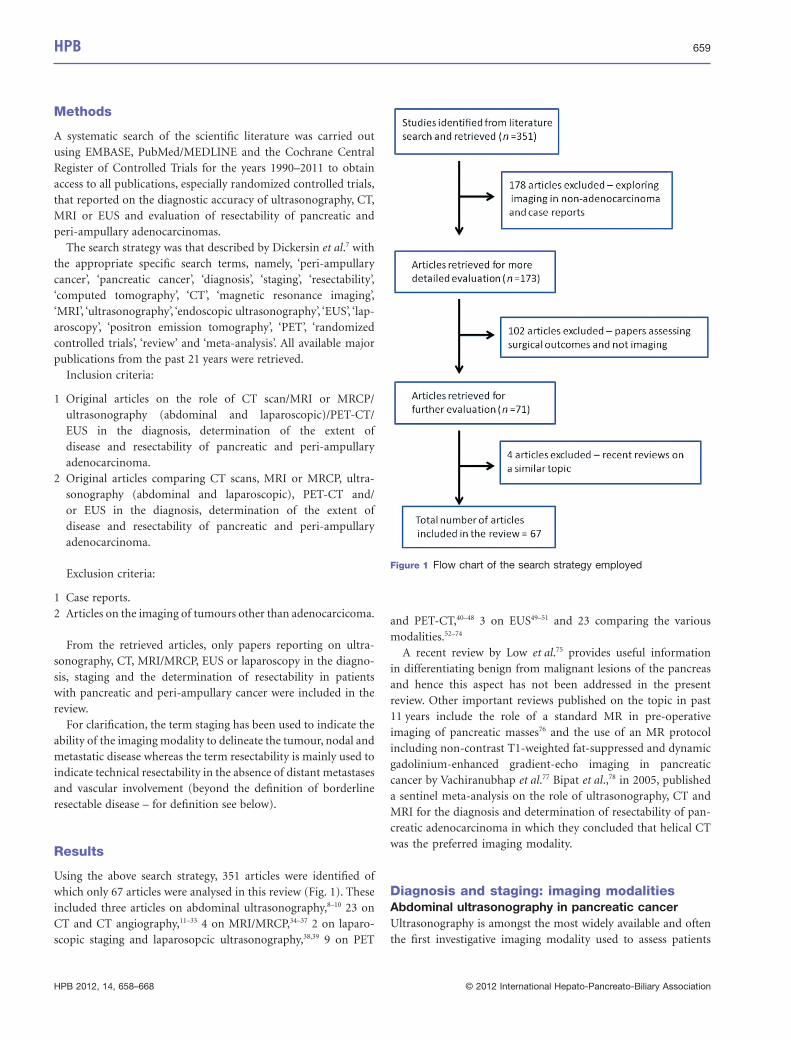
Figure 2 provides the authors’ algorithm to the pre-operativeevaluation of patients with pancreatic and peri-ampullary cancer.
It is pertinent to note that in spite of pre-operative imagingsuggesting the disease to be resectable, it is possible that the result-ant exploration may not lead to a complete (R0) resection.Borderline resectable tumours (defined above) constitute animportant class of such patients. While it has been shown thatsuch tumours which appear borderline resectable on pre-operative imaging may be amenable to a complete resection,29 thevalue of such resections in terms of overall survival need to bebetter defined. Offering these patients neoadjuvant chemo-radiotherapy has been suggested to be a viable option.21,30 In spiteof increasing surgical aggression and the availability of neoadju-vant treatment algorithms, certain features noted on pre-operative imaging have been linked to poor outcomes even in theevent of an apparently curative resection in non-metastatic pan-creatic cancer. Some authors have used techniques such as corre-lation of imaging with the surgical pathology86,87 to demonstratethis aspect. These include:
1 Features of local unresectability, including peri-pancreatic infil-tration and a tumour size >3 cm.88
2 Venous involvement – where the length of invasion is >3 cm.89
3 Arterial involvement on pre-operative imaging.90
With the increasing use of neoadjuvant chemo- and chemora-diotherapy protocols in patients with borderline pancreatictumours, further studies are needed to determine the imagingmodalities that can best guide surgical decision making afterneoadjuvant therapy especially when offering these patients theoption of an exploration with the intent of cure.
Conclusion
MDCT with angiography usually constitutes the first imagingmodality in suspected cancers of the pancreas. MRI and MRCPalternatively may be used in centres where these facilities arereadily available. The role of EUS appears complementary to theconventional CT and MRI/MRCP. It is of most benefit in theassessment of lesions not clearly detected on CT. PET-CT serves asa useful complementary investigation in patients with locallyadvanced disease to rule out metastases outside the abdomen.Staging laparoscopy and laparoscopic ultrasonography may helpin the re-staging of locally advanced/borderline resectable lesionstreated with neo-adjuvant therapy.
Conflicts of interest
None declared.
References
1. Cancer Facts and Figures. (2009) American Cancer Society. Available at
http://www.cancer.org/acs/groups/content/@nho/documents/document/
500809webpdf.pdf (last accessed 8 June 2012).
2. Cancer Research UK. Available at http://info.cancerresearchuk.org/
cancerstats/types/pancreas/incidence/ (last accessed 8 June 2012).
3. Chen KX, Wang PP, Zhang SW, Li LD, Lu FZ, Hao XS. (2003) Regional
variations in mortality rates of pancreatic cancer in China: results from
HPB 665
HPB 2012, 14, 658–668 © 2012 International Hepato-Pancreato-Biliary Association
1990–1992 national mortality survey. World J Gastroenterol 9:2557–
2560.
4. Seino T, Nakadaira H, Endoh K, Yamamoto M. (2008) Changes in pan-
creatic cancer mortality, period patterns, and birth cohort patterns in
Japan: analysis of mortality data in the period 1968–2002. Environ Health
Prev Med 13:234–242.
5. Paez D, Labonte M, Lenz H. (2012) Pancreatic Cancer: medical manage-
ment (novel chemotherapeutics). Gastroenterol Clin North Am 41:189–
209.
6. Shukla PJ, Barreto SG, Kulkarni A, Nagarajan G, Fingerhut A. (2010)
Vascular anomalies encountered during pancreatoduodenectomy: do
they influence outcomes? Ann Surg Oncol 17:186–193.
7. Dickersin K, Scherer R, Lefebvre C. (1994) Identifying relevant studies for
systematic reviews. BMJ 309:1286–1291.
8. Karlson BM, Ekbom A, Lindgren PG, Kallskog V, Rastad J. (1999)
Abdominal US for diagnosis of pancreatic tumor: prospective cohort
analysis. Radiology 213:107–111.
9. Koito K, Namieno T, Nagakawa T, Morita K. (1997) Inflammatory pancre-
atic masses: differentiation from ductal carcinomas with contrast-
enhanced sonography using carbon dioxide microbubbles. AJR Am J
Roentgenol 169:1263–1267.
10. Morrin MM, Kruskal JB, Raptopoulos V, Weisinger K, Farrell RJ, Steer ML
et al. (2001) State-of-the-art ultrasonography is as accurate as helical
computed tomography and computed tomographic angiography for
detecting unresectable periampullary cancer. J Ultrasound Med 20:481–
490.
11. Bluemke DA, Cameron JL, Hruban RH, Pitt HA, Siegelman SS, Soyer P
et al. (1995) Potentially resectable pancreatic adenocarcinoma: spiral CT
assessment with surgical and pathologic correlation. Radiology 197:381–
385.
12. Brennan DD, Zamboni GA, Raptopoulos VD, Kruskal JB. (2007) Compre-
hensive preoperative assessment of pancreatic adenocarcinoma with
64-section volumetric CT. Radiographics 27:1653–1666.
13. Bronstein YL, Loyer EM, Kaur H, Choi H, David C, DuBrow RA et al.
(2004) Detection of small pancreatic tumors with multiphasic helical CT.
AJR Am J Roentgenol 182:619–623.
14. Brugel M, Rummeny EJ, Dobritz M. (2004) Vascular invasion in
pancreatic cancer: value of multislice helical CT. Abdom Imaging 29:239–
245.
15. Diehl SJ, Lehmann KJ, Sadick M, Lachmann R, Georgi M. (1998) Pan-
creatic cancer: value of dual-phase helical CT in assessing resectability.
Radiology 206:373–378.
16. Fletcher JG, Wiersema MJ, Farrell MA, Fidler JL, Burgart LJ, Koyama T
et al. (2003) Pancreatic malignancy: value of arterial, pancreatic, and
hepatic phase imaging with multi-detector row CT. Radiology 229:81–
90.
17. Graf O, Boland GW, Warshaw AL, Fernandez-del-Castillo C, Hahn PF,
Mueller PR. (1997) Arterial versus portal venous helical CT for revealing
pancreatic adenocarcinoma: conspicuity of tumor and critical vascular
anatomy. AJR Am J Roentgenol 169:119–123.
18. Imbriaco M, Megibow AJ, Ragozzino A, Liuzzi R, Mainenti P, Bortone S
et al. (2005) Value of the single-phase technique in MDCT assessment of
pancreatic tumors. AJR Am J Roentgenol 184:1111–1117.
19. Kaneko OF, Lee DM, Wong J, Kadell BM, Reber HA, Lu DS et al. (2010)
Performance of multidetector computed tomographic angiography in
determining surgical resectability of pancreatic head adenocarcinoma.
J Comput Assist Tomogr 34:732–738.
20. Karmazanovsky G, Fedorov V, Kubyshkin V, Kotchatkov A. (2005) Pan-
creatic head cancer: accuracy of CT in determination of resectability.
Abdom Imaging 30:488–500.
21. Katz MH, Pisters PW, Evans DB, Sun CC, Lee JE, Fleming JB et al. (2008)
Borderline resectable pancreatic cancer: the importance of this emerging
stage of disease. J Am Coll Surg 206:833–846. discussion 46–8.
22. Kent TS, Raptopoulos V, Callery MP, Gautam S, Vollmer CM. (2010)
Escalating computed tomography angiogram (CTA) grade predicts unre-
sectability and margin status for pancreaticobiliary neoplasms. HPB
12:115–122.
23. Kim YE, Park MS, Hong HS, Kang CM, Choi JY, Lim JS et al. (2009)
Effects of neoadjuvant combined chemotherapy and radiation therapy on
the CT evaluation of resectability and staging in patients with pancreatic
head cancer. Radiology 250:758–765.
24. Kinney T. (2010) Evidence-based imaging of pancreatic malignancies.
Surg Clin North Am 90:235–249.
25. Lu DS, Reber HA, Krasny RM, Kadell BM, Sayre J. (1997) Local staging
of pancreatic cancer: criteria for unresectability of major vessels as
revealed by pancreatic-phase, thin-section helical CT. AJR Am J Roent-
genol 168:1439–1443.
26. McNulty NJ, Francis IR, Platt JF, Cohan RH, Korobkin M, Gebremariam
A. (2001) Multi–detector row helical CT of the pancreas: effect of
contrast-enhanced multiphasic imaging on enhancement of the pan-
creas, peripancreatic vasculature, and pancreatic adenocarcinoma.
Radiology 220:97–102.
27. O'Malley ME, Boland GW, Wood BJ, Fernandez-del Castillo C, Warshaw
AL, Mueller PR. (1999) Adenocarcinoma of the head of the pancreas:
determination of surgical unresectability with thin-section pancreatic-
phase helical CT. AJR Am J Roentgenol 173:1513–1518.
28. Prokesch RW, Chow LC, Beaulieu CF, Bammer R, Jeffrey RB, Jr. (2002)
Isoattenuating pancreatic adenocarcinoma at multi-detector row CT: sec-
ondary signs. Radiology 224:764–768.
29. Shrikhande SV, Arya S, Barreto SG, Ingle S, D'Souza MA, Hawaldar R
et al. (2011) Borderline resectable pancreatic tumors: is there a need for
further refinement of this stage? Hepatobiliary Pancreat Dis Int 10:319–
324.
30. Varadhachary GR, Tamm EP, Abbruzzese JL, Xiong HQ, Crane CH, Wang
H et al. (2006) Borderline resectable pancreatic cancer: definitions, man-
agement, and role of preoperative therapy. Ann Surg Oncol 13:1035–1046.
31. Varadhachary GR, Tamm EP, Crane C, Evans DB, Wolff RA. (2005) Bor-
derline resectable pancreatic cancer. Curr Treat Options Gastroenterol
8:377–384.
32. Zamboni GA, Kruskal JB, Vollmer CM, Baptista J, Callery MP, Raptopou-
los VD. (2007) Pancreatic adenocarcinoma: value of multidetector CT
angiography in preoperative evaluation. Radiology 245:770–778.
33. Zhao WY, Luo M, Sun YW, Xu Q, Chen W, Zhao G et al. (2009) Computed
tomography in diagnosing vascular invasion in pancreatic and periamp-
ullary cancers: a systematic review and meta-analysis. Hepatobiliary
Pancreat Dis Int 8:457–464.
34. Hanninen EL, Pech M, Jonas S, Ricke J, Thelen A, Langrehr J et al. (2005)
Magnetic resonance imaging including magnetic resonance cholangio-
pancreatography for tumor localization and therapy planning in malignant
hilar obstructions. Acta Radiol 46:462–470.
35. Hochwald SN, Rofsky NM, Dobryansky M, Shamamian P, Marcus SG.
(1999) Magnetic resonance imaging with magnetic resonance cholang-
iopancreatography accurately predicts resectability of pancreatic carci-
noma. J Gastrointest Surg 3:506–511.
666 HPB
HPB 2012, 14, 658–668 © 2012 International Hepato-Pancreato-Biliary Association
36. Megibow AJ, Zhou XH, Rotterdam H, Francis IR, Zerhouni EA, Balfe DM
et al. (1995) Pancreatic adenocarcinoma: CT versus MR imaging in the
evaluation of resectability–report of the Radiology Diagnostic Oncology
Group. Radiology 195:327–332.
37. Tapper EB, Martin D, Adsay NV, Kooby D, Kalb B, Sarmiento JM. (2010)
An MRI-driven practice: a new perspective on MRI for the evaluation of
adenocarcinoma of the head of the pancreas. J Gastrointest Surg
14:1292–1297.
38. Hariharan D, Constantinides VA, Froeling FE, Tekkis PP, Kocher HM.
(2010) The role of laparoscopy and laparoscopic ultrasound in the pre-
operative staging of pancreatico-biliary cancers–a meta-analysis. Eur J
Surg Oncol 36:941–948.
39. Shoup M, Winston C, Brennan MF, Bassman D, Conlon KC. (2004) Is
there a role for staging laparoscopy in patients with locally advanced,
unresectable pancreatic adenocarcinoma? J Gastrointest Surg 8:1068–
1071.
40. Buck AC, Schirrmeister HH, Guhlmann CA, Diederichs CG, Shen C,
Buchmann I et al. (2001) Ki-67 immunostaining in pancreatic cancer and
chronic active pancreatitis: does in vivo FDG uptake correlate with pro-
liferative activity? J Nucl Med 42:721–725.
41. Diederichs CG, Staib L, Vogel J, Glasbrenner B, Glatting G, Brambs HJ
et al. (2000) Values and limitations of 18F-fluorodeoxyglucose-positron-
emission tomography with preoperative evaluation of patients with pan-
creatic masses. Pancreas 20:109–116.
42. Gambhir SS, Czernin J, Schwimmer J, Silverman DH, Coleman RE,
Phelps ME. (2001) A tabulated summary of the FDG PET literature. J Nucl
Med 42:1S–93S.
43. Imdahl A, Nitzsche E, Krautmann F, Hogerle S, Boos S, Einert A et al.
(1999) Evaluation of positron emission tomography with 2-[18F]fluoro-2-
deoxy-D-glucose for the differentiation of chronic pancreatitis and pan-
creatic cancer. Br J Surg 86:194–199.
44. Kauhanen SP, Komar G, Seppanen MP, Dean KI, Minn HR, Kajander SA
et al. (2009) A prospective diagnostic accuracy study of 18F-
fluorodeoxyglucose positron emission tomography/computed tomogra-
phy, multidetector row computed tomography, and magnetic resonance
imaging in primary diagnosis and staging of pancreatic cancer. Ann Surg
250:957–963.
45. Lee TY, Kim MH, Park do H, Seo DW, Lee SK, Kim JS et al. (2009) Utility
of 18F-FDG PET/CT for differentiation of autoimmune pancreatitis with
atypical pancreatic imaging findings from pancreatic cancer. AJR Am J
Roentgenol 193:343–348.
46. Ozaki Y, Oguchi K, Hamano H, Arakura N, Muraki T, Kiyosawa K et al.
(2008) Differentiation of autoimmune pancreatitis from suspected pan-
creatic cancer by fluorine-18 fluorodeoxyglucose positron emission
tomography. J Gastroenterol 43:144–151.
47. Pakzad F, Groves AM, Ell PJ. (2006) The role of positron emission tomog-
raphy in the management of pancreatic cancer. Semin Nucl Med 36:248–
256.
48. van Kouwen MC, Jansen JB, van Goor H, de Castro S, Oyen WJ, Drenth
JP. (2005) FDG-PET is able to detect pancreatic carcinoma in chronic
pancreatitis. Eur J Nucl Med Mol Imaging 32:399–404.
49. Fisher L, Segarajasingam DS, Stewart C, Deboer WB, Yusoff IF. (2009)
Endoscopic ultrasound guided fine needle aspiration of solid pancreatic
lesions: performance and outcomes. J Gastroenterol Hepatol 24:90–
96.
50. Owens DJ, Savides TJ. (2010) Endoscopic ultrasound staging and novel
therapeutics for pancreatic cancer. Surg Oncol Clin N Am 19:255–266.
51. Tadic M, Kujundzic M, Stoos-Veic T, Kaic G, Vukelic-Markovic M. (2008)
Role of repeated endoscopic ultrasound-guided fine needle aspiration in
small solid pancreatic masses with previous indeterminate and negative
cytological findings. Dig Dis. 26:377–382.
52. Kitano M, Kudo M, Maekawa K, Suetomi Y, Sakamoto H, Fukuta N et al.
(2004) Dynamic imaging of pancreatic diseases by contrast enhanced
coded phase inversion harmonic ultrasonography. Gut 53:854–859.
53. Fusari M, Maurea S, Imbriaco M, Mollica C, Avitabile G, Soscia F et al.
(2010) Comparison between multislice CT and MR imaging in the diag-
nostic evaluation of patients with pancreatic masses. Radiol Med
115:453–466.
54. Cannon ME, Carpenter SL, Elta GH, Nostrant TT, Kochman ML, Ginsberg
GG et al. (1999) EUS compared with CT, magnetic resonance imaging,
and angiography and the influence of biliary stenting on staging accuracy
of ampullary neoplasms. Gastrointest Endosc 50:27–33.
55. Chen CH, Yang CC, Yeh YH, Chou DA, Nien CK. (2009) Reappraisal of
endosonography of ampullary tumors: correlation with transabdominal
sonography, CT, and MRI. J Clin Ultrasound 37:18–25.
56. Howard TJ, Chin AC, Streib EW, Kopecky KK, Wiebke EA. (1997) Value of
helical computed tomography, angiography, and endoscopic ultrasound
in determining resectability of periampullary carcinoma. Am J Surg
174:237–241.
57. Midwinter MJ, Beveridge CJ, Wilsdon JB, Bennett MK, Baudouin CJ,
Charnley RM. (1999) Correlation between spiral computed tomography,
endoscopic ultrasonography and findings at operation in pancreatic and
ampullary tumours. Br J Surg 86:189–193.
58. Rivadeneira DE, Pochapin M, Grobmyer SR, Lieberman MD, Christos PJ,
Jacobson I et al. (2003) Comparison of linear array endoscopic ultra-
sound and helical computed tomography for the staging of periampullary
malignancies. Ann Surg Oncol 10:890–897.
59. Rosch T, Lorenz R, Braig C, Feuerbach S, Siewert JR, Schusdziarra V
et al. (1991) Endoscopic ultrasound in pancreatic tumor diagnosis. Gas-
trointest Endosc 37:347–352.
60. Shoup M, Hodul P, Aranha GV, Choe D, Olson M, Leya J et al. (2000)
Defining a role for endoscopic ultrasound in staging periampullary
tumors. Am J Surg 179:453–456.
61. Soriano A, Castells A, Ayuso C, Ayuso JR, de Caralt MT, Gines MA et al.
(2004) Preoperative staging and tumor resectability assessment of pan-
creatic cancer: prospective study comparing endoscopic ultrasonogra-
phy, helical computed tomography, magnetic resonance imaging, and
angiography. Am J Gastroenterol 99:492–501.
62. DeWitt J, Devereaux B, Chriswell M, McGreevy K, Howard T, Imperiale TF
et al. (2004) Comparison of endoscopic ultrasonography and multidetec-
tor computed tomography for detecting and staging pancreatic cancer.
Ann Intern Med 141:753–763.
63. Legmann P, Vignaux O, Dousset B, Baraza AJ, Palazzo L, Dumontier I
et al. (1998) Pancreatic tumors: comparison of dual-phase helical CT and
endoscopic sonography. AJR Am J Roentgenol 170:1315–1322.
64. Trede M, Rumstadt B, Wendl K, Gaa J, Tesdal K, Lehmann KJ et al. (1997)
Ultrafast magnetic resonance imaging improves the staging of pancreatic
tumors. Ann Surg 226:393–405. discussion -7.
65. Lee JK, Kim AY, Kim PN, Lee MG, Ha HK. (2010) Prediction of vascular
involvement and resectability by multidetector-row CT versus MR
imaging with MR angiography in patients who underwent surgery for
resection of pancreatic ductal adenocarcinoma. Eur J Radiol 73:310–316.
66. Park HS, Lee JM, Choi HK, Hong SH, Han JK, Choi BI. (2009) Preopera-
tive evaluation of pancreatic cancer: comparison of gadolinium-
HPB 667
HPB 2012, 14, 658–668 © 2012 International Hepato-Pancreato-Biliary Association
enhanced dynamic MRI with MR cholangiopancreatography versus
MDCT. J Magn Reson Imaging 30:586–595.
67. Casneuf V, Delrue L, Kelles A, Van Damme N, Van Huysse J, Berrevoet F
et al. (2007) Is combined 18F-fluorodeoxyglucose-positron emission
tomography/computed tomography superior to positron emission
tomography or computed tomography alone for diagnosis, staging and
restaging of pancreatic lesions? Acta Gastroenterol Belg 70:331–338.
68. Farma JM, Santillan AA, Melis M, Walters J, Belinc D, Chen DT et al.
(2008) PET/CT fusion scan enhances CT staging in patients with pancre-
atic neoplasms. Ann Surg Oncol 15:2465–2471.
69. Tang S, Huang G, Liu J, Liu T, Treven L, Song S et al. (2011) Usefulness
of (18)F-FDG PET, combined FDG-PET/CT and EUS in diagnosing
primary pancreatic carcinoma: a meta-analysis. Eur J Radiol 78:142–
150.
70. Sperti C, Bissoli S, Pasquali C, Frison L, Liessi G, Chierichetti F et al.
(2007) 18-fluorodeoxyglucose positron emission tomography enhances
computed tomography diagnosis of malignant intraductal papillary muci-
nous neoplasms of the pancreas. Ann Surg 246:932–937. discussion 7–9.
71. Artifon EL, Couto D, Jr, Sakai P, da Silveira EB. (2009) Prospective
evaluation of EUS versus CT scan for staging of ampullary cancer.
Gastrointest Endosc 70:290–296.
72. Agarwal B, Abu-Hamda E, Molke KL, Correa AM, Ho L. (2004) Endo-
scopic ultrasound-guided fine needle aspiration and multidetector spiral
CT in the diagnosis of pancreatic cancer. Am J Gastroenterol 99:844–
850.
73. Miller FH, Rini NJ, Keppke AL. (2006) MRI of adenocarcinoma of the
pancreas. AJR Am J Roentgenol 187:W365–W374.
74. Donahue TR, Isacoff WH, Hines OJ, Tomlinson JS, Farrell JJ, Bhat YM
et al. (2011) Downstaging chemotherapy and alteration in the classic
computed tomography/magnetic resonance imaging signs of vascular
involvement in patients with pancreaticobiliary malignant tumors: influ-
ence on patient selection for surgery. Arch Surg 146:836–843.
75. Low G, Panu A, Millo N, Leen E. (2011) Multimodality imaging of neo-
plastic and nonneoplastic solid lesions of the pancreas. Radiographics
31:993–1015.
76. Martin DR, Semelka RC. (2000) MR imaging of pancreatic masses. Magn
Reson Imaging Clin N Am 8:787–812.
77. Vachiranubhap B, Kim YH, Balci NC, Semelka RC. (2009) Magnetic
resonance imaging of adenocarcinoma of the pancreas. Top Magn Reson
Imaging 20:3–9.
78. Bipat S, Phoa SS, van Delden OM, Bossuyt PM, Gouma DJ, Lameris JS
et al. (2005) Ultrasonography, computed tomography and magnetic
resonance imaging for diagnosis and determining resectability of pancre-
atic adenocarcinoma: a meta-analysis. J Comput Assist Tomogr 29:438–
445.
79. Khan I, Conlon K. (2009) Pancreatic adenocarcinoma. In: Garden O,
ed. Hepatobiliary and Pancreatic Surgery, 4th edn. Oxford: Elsevier,
pp. 283–298.
80. Iglesias Garcia J, Larino Noia J, Dominguez Munoz JE. (2009) Endo-
scopic ultrasound in the diagnosis and staging of pancreatic cancer. Rev
Esp Enferm Dig 101:631–638.
81. Barreto S, Shukla P, Shrikhande S. (2007) Periampullary carcinoma. In:
Shrikhande S, Friess H, Buechler M, eds. Surgery of Pancreatic Tumors.
New Delhi: BI publications, pp. 206–215.
82. Goldberg J, Rosenblat J, Khatri G, Schwender B, Kaushik N, Katz D et al.
(2010) Complementary roles of CT and endoscopic ultrasound in evalu-
ating a pancreatic mass. AJR Am J Roentgenol 194:984–992.
83. Mayo SC, Austin DF, Sheppard BC, Mori M, Shipley DK, Billingsley KG.
(2009) Evolving preoperative evaluation of patients with pancreatic
cancer: does laparoscopy have a role in the current era? J Am Coll Surg
208:87–95.
84. Enestvedt CK, Mayo SC, Diggs BS, Mori M, Austin DA, Shipley DK et al.
(2008) Diagnostic laparoscopy for patients with potentially resectable
pancreatic adenocarcinoma: is it cost-effective in the current era?
J Gastrointest Surg 12:1177–1184.
85. Nieveen van Dijkum EJ, Romijn MG, Terwee CB, de Wit LT, van der
Meulen JH, Lameris HS et al. (2003) Laparoscopic staging and subse-
quent palliation in patients with peripancreatic carcinoma. Ann Surg
237:66–73.
86. Aziz AM, Said T, Poovathumkadavil A, Almulla A. (2010) Using multide-
tector CT in predicting resectability of pancreatic head tumors: surgical
and pathologic correlation. J Egypt Natl Canc Inst 22:233–239.
87. Olivie D, Lepanto L, Billiard JS, Audet P, Lavallee JM. (2007) Predicting
resectability of pancreatic head cancer with multi-detector CT. Surgical
and pathologic correlation. JOP 8:753–758.
88. Phoa SS, Tilleman EH, van Delden OM, Bossuyt PM, Gouma DJ, Lameris
JS. (2005) Value of CT criteria in predicting survival in patients with
potentially resectable pancreatic head carcinoma. J Surg Oncol 91:33–40.
89. Kaneoka Y, Yamaguchi A, Isogai M. (2009) Portal or superior mesenteric
vein resection for pancreatic head adenocarcinoma: prognostic value of
the length of venous resection. Surgery 145:417–425.
90. Shrikhande SV, Barreto SG. (2010) Extended pancreatic resections and
lymphadenectomy: an appraisal of the current evidence. World J Gas-
trointest Surg 2:39–46.
668 HPB
HPB 2012, 14, 658–668 © 2012 International Hepato-Pancreato-Biliary Association


