Embed Size (px)
Citation preview

A pH-stabilizing role of voltage-gated protonchannels in IgE-mediated activation ofhuman basophilsBoris Musset*†, Deri Morgan*†, Vladimir V. Cherny*, Donald W. MacGlashan, Jr.‡, Larry L. Thomas§,Eduardo Rios*, and Thomas E. DeCoursey*¶
Departments of *Molecular Biophysics and Physiology and §Immunology/Microbiology, Rush University Medical Center, Chicago, IL 60612; and ‡JohnsHopkins Asthma and Allergy Center, The Johns Hopkins University School of Medicine, Baltimore, MD 21224
Edited by Michael D. Cahalan, University of California, Irvine, CA, and accepted by the Editorial Board May 14, 2008 (received for review January 28, 2008)
Eosinophils and other phagocytes use NADPH oxidase to killbacteria. Proton channels in human eosinophils and neutrophils arethought to sustain NADPH oxidase activity, and their opening isgreatly enhanced by a variety of NADPH oxidase activators, in-cluding phorbol myristate acetate (PMA). In nonphagocytic cellsthat lack NADPH oxidase, no clear effect of PMA on protonchannels has been reported. The basophil is a granulocyte that isdevelopmentally closely related to the eosinophil but neverthelessdoes not express NADPH oxidase. Thus, one might expect thatstimulating basophils with PMA would not affect proton currents.However, stimulation of human basophils in perforated-patchconfiguration with PMA, N-formyl-methionyl-leucyl-phenylala-nine, or anti-IgE greatly enhanced proton currents, the lattersuggesting involvement of proton channels during activation ofbasophils by allergens through their highly expressed IgE receptor(Fc�RI). The anti-IgE-stimulated response occurred in a fraction ofcells that varied among donors and was less profound than that toPMA. PKC inhibition reversed the activation of proton channels,and the proton channel response to anti-IgE or PMA persisted inCa2�-free solutions. Zn2� at concentrations that inhibit protoncurrent inhibited histamine release elicited by PMA or anti-IgE.Studied with confocal microscopy by using SNARF-AM and theshifted excitation and emission ratioing of fluorescence approach,anti-IgE produced acidification that was exacerbated in the pres-ence of 100 �M Zn2�. Evidently, proton channels are active inbasophils during IgE-mediated responses and prevent excessiveacidification, which may account for their role in histamine release.
asthma � histamine � patch-clamp � HNVCN1 � allergy
The IgE-mediated activation of basophils is a hallmark ofallergic reactions. Binding of allergen to allergen-specific IgE
on basophils results in cross-linking of high-affinity receptors(Fc�RI), thereby activating a signaling pathway that results in therelease of histamine and other mediators. Basophils are amongseveral cell types that express voltage-gated proton channels (1).Proton channels are opened by depolarization and/or cytoplas-mic acidification and seem to be designed for rapid, efficient acidextrusion from cells (2). In phagocytes, they are thought toenable sustained NADPH oxidase activity by compensating forthe electrogenic activity of the oxidase (3–5). Stimuli thatactivate NADPH oxidase in human eosinophils and neutrophilsand in murine osteoclasts greatly enhance the opening of protonchannels in these cells studied in perforated-patch configuration(6–10), largely via PKC phosphorylation (11). In contrast,phorbol myristate acetate (PMA) has no clear effect on protoncurrents in alveolar epithelial cells that lack NADPH oxidase (6).The function of proton channels in basophils is unknown, butmust differ from that of phagocytes, because basophils lackNADPH oxidase (12). Here, we show that proton channels inbasophils respond vigorously to agents that elicit histaminerelease. Furthermore, histamine release stimulated by anti-IgEor PMA was inhibited by Zn2� at concentrations that inhibit
proton currents, consistent with the idea that proton channelactivity is linked to basophil activation. Studies of pHi using thefluorescent pH sensitive dye SNARF-AM suggest that protonchannels limit acidification during basophil responses.
ResultsProton Currents in Human Basophils Respond to PMA. Human ba-sophils voltage-clamped in the perforated-patch configurationexhibit voltage-gated proton currents (Fig. 1A) that resemblethose in human eosinophils (7), both in properties and inamplitude. In basophils at symmetrical pH 7.0 (50 mM NH4
� onboth sides of the membrane clamps pHi near pHo), the protonconductance, gH, activates during depolarizing voltage pulsesabove �20 mV, and activates more rapidly at more positivevoltages. The net, leak-subtracted H� current (IH) at �80 mVwas 30 � 18 pA (mean � SD) in 21 cells. In similar measure-ments, IH in human eosinophils was 48 pA (7).
The PKC activator PMA is a potent and effective stimulus ofhistamine release from basophils (13). Nearly every basophil stim-ulated with PMA exhibited a profound enhancement of protoncurrents. After addition of PMA to the bath solution, IH during atest pulse increased progressively over several minutes (Fig. 1B).The same family of pulses in PMA elicited larger currents (Fig. 1C)that activated more rapidly at each voltage. H� current–voltagerelationships (Fig. 1F) reveal that the voltage at which the H�
current was first activated, Vthreshold, was shifted �20 mV morenegative after stimulation with PMA. The chord conductance (gH)calculated from the H� current (Fig. 1G) was also shifted �20 mVby PMA, and the maximum H� conductance (gH,max) was doubledin this cell. The impression that PMA stimulation increased theopening rate of proton channels is confirmed in Fig. 1H. Theactivation time constant, �act, was decreased (activation becamefaster) by �3-fold at all voltages. Average responses of protoncurrents in basophils and neutrophils to PMA are summarized inTable 1. Consistent with the lack of NADPH oxidase (12), no hintof electron current was seen.
The enhanced gating mode, or ‘‘activation,’’ of phagocyteproton channels is largely attributable to phosphorylation byPKC of either the channel itself or a closely related protein (11).Fig. 1D shows that the PKC inhibitor GF109203X (GFX)
Author contributions: B.M., D.M., V.V.C., D.W.M., L.L.T., E.R., and T.E.D. designed research;B.M., D.M., V.V.C., and D.W.M. performed research; B.M., D.M., V.V.C., D.W.M., L.L.T., E.R.,and T.E.D. analyzed data; and D.W.M., L.L.T., and T.E.D. wrote the paper.
The authors declare no conflict of interest.
This article is a PNAS Direct Submission. M.D.C. is a guest editor invited by the EditorialBoard.
†B.M., and D.M. contributed equally to this work.
¶To whom correspondence should be addressed. E-mail: [email protected].
This article contains supporting information online at www.pnas.org/cgi/content/full/0800886105/DCSupplemental.
© 2008 by The National Academy of Sciences of the USA
11020–11025 � PNAS � August 5, 2008 � vol. 105 � no. 31 www.pnas.org�cgi�doi�10.1073�pnas.0800886105

reversed the increase in IH produced by PMA in basophils. GFXalso reversed the shift in the IH–V relationship (Fig. 1F), theincrease in gH,max (Fig. 1G), and the change in �act (Fig. 1H). Inthis cell, GFX seemed to reverse all effects of PMA. In contrast,GFX reversed the effects of PMA on proton currents in eosin-ophils only partially (11).
Proton Currents in Human Basophils Respond to Anti-IgE. The char-acteristic response of human basophils occurs when allergensbind to IgE, resulting in cross-linking of high-affinity receptors(Fc�RI) that activates a signaling pathway that results in therelease of histamine and other mediators. Cross-linking IgE withgoat anti-human IgE antibody (anti-IgE) elicited a distinct
proton channel response (Fig. 2) in a fraction of cells. More thanhalf of the cells from the most responsive donors responded toanti-IgE, but most cells from certain other donors failed torespond. Five cells each from three donors failed to respondto anti-IgE but responded subsequently to PMA. In studies ofhighly purified (�98%) basophils (in Methods), 60% of cells(6/10) responded to anti-IgE, confirming that some nonrespond-ing cells were basophils. Donor-to-donor variability is typicallyobserved in histamine release by basophils in response toanti-IgE (14, 15).
Fig. 1. Protoncurrents inhumanbasophils respondvigorously toPMAandGFX.(A, C, and E) Families of currents in a basophil before stimulation (A), afterexposure to 60 nM PMA for 4 min (C), and after addition of 2.5 �M GFX (E). Ineach, the membrane was held at �20 mV, and 8-s pulses were applied in 10-mVincrements every 30 s up to �80 mV. (B) Superimposed currents during thetransition to PMA, with 4-s pulses to �60 mV applied every 15 s. (D) Test currentsafteradditionofGFX. (F) I–V relationshipsfromthefamilies inA,C,andE. (G)gH–Vrelationships from the families in A, C, and E were calculated by using Vrev
measured in each condition and the steady-state IH obtained by extrapolatingexponential fits. (H) Voltage dependence of the activation time constant (�act)obtained by fitting the currents in A, C, and E to a rising exponential (after adelay). Calibration bars apply to A–E. The bath included Ca2�.
Table 1. Effects of anti-IgE and PMA on proton currents in human basophils compared withPMA effects in human neutrophils
Parameter Basophil with anti-IgE Basophil with PMA Neutrophil with PMA*
gH,max 2.29 � 0.16 (31) 2.86 � 0.21 (40) 1.9(anti-IgE/control) (PMA/control) (PMA/control)
�act at Vtest 2.17 � 0.19 (28) 5.04 � 0.56 (52) 3.7(control/anti-IgE) (control/PMA) (control/PMA)
�tail at Vhold 1.20 � 0.04 (33) 1.32 � 0.07 (64) 5.5(anti-IgE/control) (PMA/control) (PMA/control)
�Vthreshold, mV �11.0 � 0.9 (33) �19.0 � 1.0 (61) �38.8(anti-IgE � control) (PMA � control) (PMA � control)
Parameters [mean � SEM (n)] were measured when the effects of PMA (60–160 nM) or anti-IgE (usually 0.5–1.0�g/ml) peaked or approached steady state, usually within a few minutes. Only cells that clearly responded areincluded. Responses are given as ratios where 1.0 means no response, except for Vthreshold, for which 0 mV meansno response. The gH,max values were calculated from the largest pulses applied. Gating kinetics, �act and �tail,respectively, were obtained from exponential fits to currents during test pulses or upon repolarization to theholding potential (Vhold). Data for Ca2� or Ca2�-free bath are combined because no parameter differed signifi-cantly for either stimulus. All PMA effects except on �tail are significantly greater (P � 0.05) than those for anti-IgE.*Values from a previous study (6).
Fig. 2. Proton currents in human basophils are enhanced by anti-IgE stim-ulation in a GFX-sensitive manner. (A–C) Currents in response to the sameapplied family of pulses before stimulation (A), after stimulation by 0.8 �g/mlanti-IgE (B), and in the presence of 3 �M GFX (C). (D) The time course of theseresponses. Anti-IgE was added to the bath incrementally, and then the bathstirred. The bath was Ca2� free. (E–H) Time course of anti-IgE responses in fourother basophils. Horizontal arrows indicate zero current. In each, test pulsesto �40 mV (E–G) or �60 mV (H) were applied every 30 s, and at the arrow, 0.6�g/ml anti-IgE was applied to the bath followed by stirring, except for G,where 6.6 �g/ml was applied. In H, ‘‘wash’’ indicates a bath exchange with thesame solution as a control; in this experiment anti-IgE was applied by completebath exchange. The bath included 2 mM Ca2�, except H, which was Ca2�-free.
Musset et al. PNAS � August 5, 2008 � vol. 105 � no. 31 � 11021
PHYS
IOLO
GY
Anti-IgE responses in Fig. 2 D–H illustrate the variety of timecourses observed. Depolarizing pulses were applied every 30 s toelicit proton currents. Typically, there was a delay of 1 min toseveral minutes before the onset of a response. IH then beganto increase. In some cells, IH peaked and immediately began todecline (Fig. 2 E and G), although not to its original amplitude.In other cells, IH remained elevated for prolonged periods. Theseresponses were observed after stimulation with anti-IgE atconcentrations ranging from 0.2 to 10 �g/ml. At higher concen-trations, the response seemed more rapid and transient, butthere were exceptions. Most measurements were performed byusing 0.5–1.0 �g/ml anti-IgE.
In the experiment in Fig. 2D, anti-IgE was added incremen-tally, with a profound response occurring at 0.8 �g/ml. In familiesof currents recorded before and after the addition of anti-IgE(Fig. 2 A and B), the H� current was larger and activated fasterat each voltage after anti-IgE. In Table 1, the parameter valuesduring the peak of the anti-IgE response are given. In cellsresponding to anti-IgE, gH,max doubled, and channel opening wasfaster (smaller �act), but closing kinetics (�tail) was not affected.The proton conductance–voltage (gH–V) relationship character-ized as Vthreshold was shifted �11 mV by anti-IgE and �20 mVby PMA. Proton channel responses to anti-IgE and PMA inhuman basophils were qualitatively identical, but the anti-IgEresponses were consistently smaller.
In basophils that exhibited a sustained increase in protoncurrent in response to anti-IgE, GFX reversed the anti-IgEeffects distinctly but not completely (Fig. 2 C and D). In cells thatresponded to anti-IgE with a transient increase in IH (e.g., Fig.2G), GFX added after the response had subsided but remainedabove the initial level, further reduced the current (data notshown).
Roughly half the cells studied did not respond to anti-IgE butdid respond to a subsequent application of PMA, confirming thatthey had not entered whole-cell configuration due to spontane-ous patch rupture, which abolishes the PMA response in eosin-ophils (16). Addition of PMA to cells that had already respondedto anti-IgE invariably produced a greater response that (com-pared with their initial state) was indistinguishable from theresponse of cells stimulated only with PMA (Table 1 and notshown). GFX reversed most of the combined effects of anti-IgEand PMA (data not shown).
fMLF Also Activates Proton Channels. Stimulation of basophils with10 �M N-formyl-methionyl-leucyl-phenylalanine (fMLF), anotheragonist of histamine release (15), enhanced proton currents in 10of 23 cells studied (data not shown). The response to fMLFresembled that to anti-IgE and was augmented by subsequentaddition of PMA (n � 8). GFX partially reversed the effect of fMLFalone (n � 1) or fMLF and PMA together (n � 4).
Proton Channel Responses Do Not Require Ca2� Influx. Basophilproton channels responded to PMA or anti-IgE whether or notCa2� was present in the bath. Considering only cells from tworesponsive donors, 14 of 22 cells (64%) responded to anti-IgEwith �0.5 mM free Ca2� (1.5 mM CaCl2 and 1 mM EGTA) inthe bath, and 13 of 20 cells (65%) responded to anti-IgE in aCa2�-free (EGTA-containing) bath solution. The presence orabsence of Ca2� in the bath did not detectably affect themagnitude of any proton channel response to PMA or anti-IgE[supporting information Tables S1 and S2]. Proton channels inbasophils exposed to 20 �M 1,2-bis(2-aminophenoxy)ethane-N,N,N�,N�-tetraacetic acid, tetraacetoxymethyl ester (BAPTA-AM) in Ca2�-free solutions responded to stimulation withanti-IgE (n � 3) or PMA (n � 2), suggesting that elevated [Ca2�]iis not required for a response.
Proton Channel Response Precedes Degranulation. If histamine stor-age granules expressed proton channels, their fusion with theplasma membrane might increase gH owing to channel insertion.However, the capacitance of a sample of basophils did notchange significantly after proton channel responses to anti-IgE(mean � SE increase 10 � 9%, n � 16) or PMA (mean decrease6 � 11%, n � 10). Histamine release is slow, especially withPMA as a stimulus (17) and likely did not occur during theproton channel response.
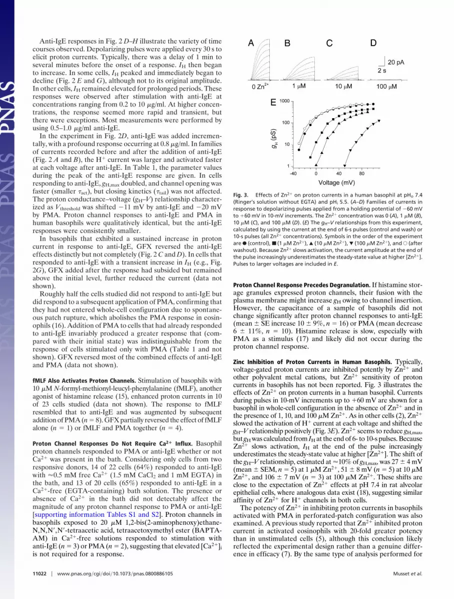
Zinc Inhibition of Proton Currents in Human Basophils. Typically,voltage-gated proton currents are inhibited potently by Zn2� andother polyvalent metal cations, but Zn2� sensitivity of protoncurrents in basophils has not been reported. Fig. 3 illustrates theeffects of Zn2� on proton currents in a human basophil. Currentsduring pulses in 10-mV increments up to �60 mV are shown for abasophil in whole-cell configuration in the absence of Zn2� and inthe presence of 1, 10, and 100 �M Zn2�. As in other cells (2), Zn2�
slowed the activation of H� current at each voltage and shifted thegH–V relationship positively (Fig. 3E). Zn2� seems to reduce gH,max,but gH was calculated from IH at the end of 6- to 10-s pulses. BecauseZn2� slows activation, IH at the end of the pulse increasinglyunderestimates the steady-state value at higher [Zn2�]. The shift ofthe gH–V relationship, estimated at �10% of gH,max, was 27 � 4 mV(mean � SEM, n � 5) at 1 �M Zn2�, 51 � 8 mV (n � 5) at 10 �MZn2�, and 106 � 7 mV (n � 3) at 100 �M Zn2�. These shifts areclose to the expectation of Zn2� effects at pH 7.4 in rat alveolarepithelial cells, where analogous data exist (18), suggesting similaraffinity of Zn2� for H� channels in both cells.
The potency of Zn2� in inhibiting proton currents in basophilsactivated with PMA in perforated-patch configuration was alsoexamined. A previous study reported that Zn2� inhibited protoncurrent in activated eosinophils with 20-fold greater potencythan in unstimulated cells (5), although this conclusion likelyreflected the experimental design rather than a genuine differ-ence in efficacy (7). By the same type of analysis performed for
Fig. 3. Effects of Zn2� on proton currents in a human basophil at pHo 7.4(Ringer’s solution without EGTA) and pHi 5.5. (A–D) Families of currents inresponse to depolarizing pulses applied from a holding potential of �60 mVto �60 mV in 10-mV increments. The Zn2� concentration was 0 (A), 1 �M (B),10 �M (C), and 100 �M (D). (E) The gH–V relationships from this experiment,calculated by using the current at the end of 6-s pulses (control and wash) or10-s pulses (all Zn2� concentrations). Symbols in the order of the experimentare F (control), ■ (1 �M Zn2�), Œ (10 �M Zn2�), � (100 �M Zn2�), and E (afterwashout). Because Zn2� slows activation, the current amplitude at the end ofthe pulse increasingly underestimates the steady-state value at higher [Zn2�].Pulses to larger voltages are included in E.
11022 � www.pnas.org�cgi�doi�10.1073�pnas.0800886105 Musset et al.
whole-cell studies, the Zn2� sensitivity was similar in basophilsstudied in perforated-patch configuration after PMA stimula-tion. The average shift of the gH–V relationship was 25 � 6 mV(n � 4) at 1 �M Zn2�, 45 � 4 mV (n � 5) at 10 �M Zn2�, and67 � 8 mV (n � 3) at 100 �M Zn2�. The slightly lower efficacyin these measurements may reflect the lower pHo of 7.0compared with pHo 7.4 for the whole-cells studies. Competi-tion between H� and Zn2� reduces the apparent potency ofZn2� at lower pHo (18). Thus, the affinity of externally appliedZn2� for proton channels in basophils is similar before or afterstimulation.
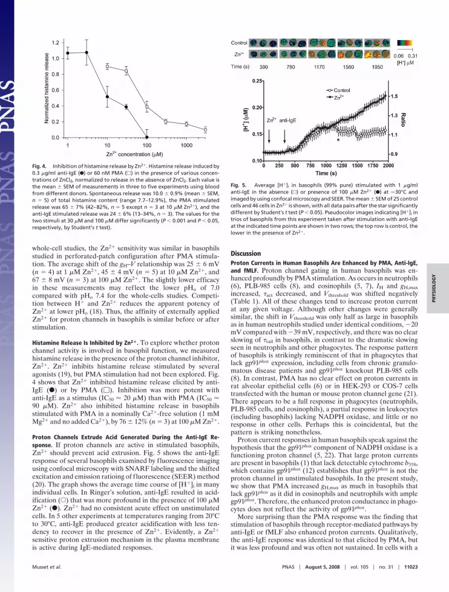
Histamine Release Is Inhibited by Zn2�. To explore whether protonchannel activity is involved in basophil function, we measuredhistamine release in the presence of the proton channel inhibitor,Zn2�. Zn2� inhibits histamine release stimulated by severalagonists (19), but PMA stimulation had not been explored. Fig.4 shows that Zn2� inhibited histamine release elicited by anti-IgE (F) or by PMA (�). Inhibition was more potent withanti-IgE as a stimulus (IC50 � 20 �M) than with PMA (IC50 �90 �M). Zn2� also inhibited histamine release in basophilsstimulated with PMA in a nominally Ca2�-free solution (1 mMMg2� and no added Ca2�), by 76 � 12% (n � 3) at 100 �M Zn2�.
Proton Channels Extrude Acid Generated During the Anti-IgE Re-sponse. If proton channels are active in stimulated basophils,Zn2� should prevent acid extrusion. Fig. 5 shows the anti-IgEresponse of several basophils examined by fluorescence imagingusing confocal microscopy with SNARF labeling and the shiftedexcitation and emission ratioing of fluorescence (SEER) method(20). The graph shows the average time course of [H�]i in manyindividual cells. In Ringer’s solution, anti-IgE resulted in acid-ification (E) that was more profound in the presence of 100 �MZn2� (F). Zn2� had no consistent acute effect on unstimulatedcells. In 5 other experiments at temperatures ranging from 20°Cto 30°C, anti-IgE produced greater acidification with less ten-dency to recover in the presence of Zn2�. Evidently, a Zn2�
sensitive proton extrusion mechanism in the plasma membraneis active during IgE-mediated responses.
DiscussionProton Currents in Human Basophils Are Enhanced by PMA, Anti-IgE,and fMLF. Proton channel gating in human basophils was en-hanced profoundly by PMA stimulation. As occurs in neutrophils(6), PLB-985 cells (8), and eosinophils (5, 7), IH and gH,maxincreased, �act decreased, and Vthreshold was shifted negatively(Table 1). All of these changes tend to increase proton currentat any given voltage. Although other changes were generallysimilar, the shift in Vthreshold was only half as large in basophilsas in human neutrophils studied under identical conditions, �20mV compared with �39 mV, respectively, and there was no clearslowing of �tail in basophils, in contrast to the dramatic slowingseen in neutrophils and other phagocytes. The response patternof basophils is strikingly reminiscent of that in phagocytes thatlack gp91phox expression, including cells from chronic granulo-matous disease patients and gp91phox knockout PLB-985 cells(8). In contrast, PMA has no clear effect on proton currents inrat alveolar epithelial cells (6) or in HEK-293 or COS-7 cellstransfected with the human or mouse proton channel gene (21).There appears to be a full response in phagocytes (neutrophils,PLB-985 cells, and eosinophils), a partial response in leukocytes(including basophils) lacking NADPH oxidase, and little or noresponse in other cells. Perhaps this is coincidental, but thepattern is striking nonetheless.
Proton current responses in human basophils speak against thehypothesis that the gp91phox component of NADPH oxidase is afunctioning proton channel (5, 22). That large proton currentsare present in basophils (1) that lack detectable cytochrome b558,which contains gp91phox (12) establishes that gp91phox is not theproton channel in unstimulated basophils. In the present study,we show that PMA increased gH,max as much in basophils thatlack gp91phox as it did in eosinophils and neutrophils with amplegp91phox. Therefore, the enhanced proton conductance in phago-cytes does not reflect the activity of gp91phox.
More surprising than the PMA response was the finding thatstimulation of basophils through receptor-mediated pathways byanti-IgE or fMLF also enhanced proton currents. Qualitatively,the anti-IgE response was identical to that elicited by PMA, butit was less profound and was often not sustained. In cells with a
Fig. 4. Inhibition of histamine release by Zn2�. Histamine release induced by0.3 �g/ml anti-IgE (F) or 60 nM PMA (�) in the presence of various concen-trations of ZnCl2, normalized to release in the absence of ZnCl2. Each value isthe mean � SEM of measurements in three to five experiments using bloodfrom different donors. Spontaneous release was 10.0 � 0.9% (mean � SEM,n � 5) of total histamine content (range 7.7–12.9%), the PMA stimulatedrelease was 65 � 7% (42–82%, n � 5 except n � 3 at 10 �M Zn2�), and theanti-IgE stimulated release was 24 � 6% (13–34%, n � 3). The values for thetwo stimuli at 30 �M and 100 �M differ significantly (P � 0.001 and P � 0.05,respectively, by Student’s t test).
Fig. 5. Average [H�]i in basophils (99% pure) stimulated with 1 �g/mlanti-IgE in the absence (E) or presence of 100 �M Zn2� (F) at �30°C andimaged by using confocal microscopy and SEER. The mean � SEM of 25 controlcells and 46 cells in Zn2� is shown, with all data pairs after the star significantlydifferent by Student’s t test (P � 0.05). Pseudocolor images indicating [H�]i intrios of basophils from this experiment taken after stimulation with anti-IgEat the indicated time points are shown in two rows; the top row is control, thelower in the presence of Zn2�.
Musset et al. PNAS � August 5, 2008 � vol. 105 � no. 31 � 11023
PHYS
IOLO
GY

clear anti-IgE response, subsequent addition of PMA invariablyproduced an augmented response. There are striking similaritiesbetween the stimulation of proton channels and histaminerelease from basophils. There was marked variability amongdonors in the responsiveness of their basophils to anti-IgE, withproton channel response rates ranging from 0 to 69%. Nearly allcells from all donors responded to PMA. Donor-to-donor vari-ability in histamine release in response to anti-IgE (14, 15)mirrors the variability in proton channel responsiveness. In thegeneral population, the distribution of responsiveness of ba-sophils to activation by anti-IgE is broad, ranging 100-fold, butbasophils respond well to PMA even in individuals whosebasophil response to anti-IgE is not detectable (14). In addition,the proton channel response occurred after a delay reminiscentof the distinct delay that precedes the increase in [Ca2�]i (23, 24)and the release of histamine in basophils stimulated with anti-IgE (17) or ragweed pollen antigen (25).
Pathways That Activate Histamine Release and Proton Channels inBasophils Are Not Identical and Vary According to Agonist. Histaminerelease from basophils elicited by PMA is inhibited by theselective PKC inhibitor GFX. In contrast, anti-IgE stimulatedhistamine release is insensitive to GFX, and inhibition by severalother PKC inhibitors is attributable to their effects on otherkinases (15, 26). PKC activity has been implicated in bothactivating and de-activating signaling in both rat basophilicleukemia cells and human basophils. Therefore, inhibitors of thebroader family of PKC isozymes might produce a weak effect onsecretion, due to competing inhibition of both activating andde-activating mechanisms. GFX reversed the activation of pro-ton channels by PMA or anti-IgE, supporting a role for phos-phorylation in proton channel activation by either agonist. Theanti-IgE response was only partially reversed, suggesting involve-ment of an additional pathway. The lack of capacitance changesduring proton channel responses together with previous studiesof the time course of histamine release (17) indicate that protonchannel enhancement likely precedes degranulation.
The proton channel inhibitors Zn2� and La3� inhibit hista-mine release from basophils stimulated with anti-IgE or A23187(19, 27), raising the possibility that proton channels are involved.However, both metal cations also inhibit calcium release acti-vated calcium (CRAC) currents (28) and Na�/Ca2� exchange(29). Hypothetically, either transporter, if present in basophils,might mediate the Ca2� influx that is required for histaminerelease in response to anti-IgE (30). Although CRAC currentshave not been recorded in basophils, they exist in humanneutrophils (31), and CRACM1 protein is expressed in basophils(D.W.M., unpublished data). However, Zn2� inhibits histaminerelease from rat basophilic leukemia cells more potently than itprevents Ca2� influx, suggesting that it acts by a differentmechanism (32). Surprisingly, Ca2� influx stimulated by anti-IgEin human basophils was not inhibited by up to 300 �M Zn2�
(D.W.M., unpublished data). This result suggests that in humanbasophils, CRAC channels either have low Zn2� sensitivity, orthey do not mediate the anti-IgE induced Ca2� influx requiredfor histamine release.
To isolate proton channel involvement in histamine release,we examined the Zn2� sensitivity of histamine release inducedby PMA, which occurs without Ca2� influx (13) or [Ca2�]iincreases (26, 33); in fact, PMA inhibits CRAC currents (34).The proton channel responses to either PMA or anti-IgEoccurred in Ca2�-free solutions and were indistinguishable fromresponses in Ca2�-containing solutions and hence are indepen-dent of Ca2� influx. That histamine release stimulated by PMAwas inhibited by Zn2� (Fig. 4) suggests that proton channelactivity may be required. The inhibition by Zn2� seemed to bemore potent for anti-IgE (IC50 � 20 �M) than for PMA-stimulated (IC50 � 90 �M) responses, consistent with a previous
study in which histamine release elicited by anti-IgE and fMLFwas inhibited by Zn2�, with IC50 10 �M and 40 �M, respectively(19). The variable sensitivity to Zn2� was attributed to eachagonist activating histamine release by a different mechanism.Because the anti-IgE response requires Ca2� influx, but theresponses to fMLF and PMA do not (30, 33), the anti-IgEresponse might normally involve a combination of proton chan-nels and CRAC channels (or whatever mediates Ca2� influx),whereas the PMA and fMLF responses may reflect Zn2� actingexclusively on proton channels.
The Zn2� data suggest that proton channels are required forhistamine release. Proton channels might facilitate histaminerelease by compensating charge, as they do in phagocytes (3, 4),or by modulating pH. Outward H� current could compensate forelectrogenic Ca2� inf lux, which is required for anti-IgE-stimulated histamine release. However, during the PMA re-sponse, which does not involve Ca2� influx, proton channelsmust act by a different mechanism. Histamine release consumesmetabolic energy (13, 25, 27) and thus likely generates cytoplas-mic acid. Consistent with this interpretation, the results in Fig.5 showed acidification after anti-IgE stimulation, which has notbeen reported previously. Importantly, Zn2� unmasked occultproton efflux during the response, thereby indicating that protonchannels are most likely responsible. Other plasma membraneproton extrusion mechanisms (Na�/H�-antiport and H�-ATPase) are insensitive to Zn2� (35, 36). Together, these resultsindicate that voltage-gated proton channels in basophils mayhelp keep pHi in an optimal range for histamine secretion.
MethodsIsolation of Human Basophils. Basophil-containing mononuclear cell fractionswere isolated by density gradient centrifugation from freshly drawn venousblood of healthy adult donors according to a protocol approved by theInstitutional Review Board of Rush University Medical Center. Basophils wereenriched by negative selection using immunomagnetic beads (Basophil Isola-tion Kit; Miltenyi Biotec, Auburn, CA) and the miniMacs magnetic cell sepa-ration system (Miltenyi Biotec) according to the manufacturer’s instructions;cells enriched by this method are 59–88% basophils (37). For some experi-ments basophils were further purified by countercurrent elutriation andtwo-step Percoll density gradient followed by negative selection (24), result-ing in 98–99% purity by Alcian blue staining. The basophils were suspendedin PBS with 2 mM EDTA and 0.5% BSA. Freshly isolated cells and cells main-tained for up to 2 days at 4°C were used for electrophysiologic measurements.
Electrophysiology. Standard whole-cell or perforated-patch recording wasperformed as described previously (16). Spherical, nonadherent cells 7–8 �min diameter (1.68 � 0.49 pF, mean � SD, n � 147) were selected. Seals wereformed with Ringer’s solution (in mM: 160 NaCl, 4.5 KCl, 2 CaCl2, 1 MgCl2, 5Hepes, pH 7.4) in the bath, and the potential zeroed after the pipette was incontact with the cell. For perforated-patch recording, the pipette and bathsolutions contained 110 mM tetramethylammonium methanesulfonate, 50mM NH4
� in the form of 25 mM (NH4)2SO4, 2 mM MgCl2, 10 mM BES buffer, and1 mM EGTA and were titrated to pH 7.0 with tetramethylammonium hydrox-ide. A Ca2�-containing bath solution was identical, but with 1.5 mM CaCl2added. The NH4
� in bath and pipette solutions ‘‘clamps’’ pHi near pHo (6, 38).The pipette solution included �500 �g/ml solubilized amphotericin B (�45%purity; Sigma); the pipette tip was dipped briefly into amphotericin-freesolution. For whole-cell recording, bath and pipette solutions contained100–200 mM buffer, 1–2 mM CaCl2 or MgCl2 (pipette solutions were Ca-free),1–2 mM EGTA, and TMAMeSO3 to adjust the osmolality to �300 mOsm,titrated with tetramethylammonium hydroxide or methanesulfonate. Noliquid junction potential or leak correction has been applied. Experimentswere performed at room temperature (20–25°C). Bioactive substances (PMA,anti-IgE, or GFX) were introduced by complete bath changes or were addeddirectly to the bath with subsequent stirring. Nominal concentrations werecalculated according to approximate bath volume, and stirring may not havealways been complete; thus, stated concentrations are somewhat approxi-mate. Basophils were stimulated with affinity purified goat anti-human IgE(Immunology Consultants Laboratory).
Histamine Release. Basophil-containing mononuclear cell fractions were iso-lated from venous blood of healthy adult donors as described above. The cells
11024 � www.pnas.org�cgi�doi�10.1073�pnas.0800886105 Musset et al.

were suspended in Ringer containing 5 mM glucose, and 0.003% BSA was addeddirectly to each sample before incubation. Contaminating metals in Milliporepurified water had been removedby using Chelex 100 Resin (Bio-Rad Laborato-ries) before adding salts and glucose. The cells (20,000–45,000 basophils asdetermined by Alcian blue staining) were incubated with the indicated concen-trations of anti-IgE or PMA alone and in the presence of the indicated concen-trations of ZnCl2 for 45–60 min at 37°C in round-bottom polystyrene tubes. Totalincubation volume was 1 ml. Reactions were stopped by ice. After centrifugation,histamine content in the supernatants was measured by an automated fluoro-metric assay (39). Spontaneous histamine release was measured by using cellsincubated in the absence of stimulus. Total histamine content was measured insupernatants of cells lysed with 1.4% perchloric acid. Results are expressed ashistamine released above the spontaneous value.
Dynamic Imaging of the Cytosolic pH of Individual Basophils by SEER Imaging.Human basophils were allowed to adhere to glass coverslips, incubated with10 �M 5-(and 6-)carboxy SNARF-1 in HBSS for 15–20 min at room temperature,and washed. SEER imaging (20) was performed by simultaneously acquiring 2confocal images: F1, excited at 514 nm and emitted at 500–604 nm and F2
(excited at 594 nm and emitted at 620–715 nm). The ratio F1/F2 monitors [H�]with a dynamic range of �150.
ACKNOWLEDGMENTS. We thank Dr. Tatiana Iastrebova for excellent techni-cal assistance. This work was supported by the Schmidtmann Foundation(B.M.); National Institutes of Health Heart, Lung and Blood Institute ResearchGrant HL61437 (to T.D.); and by Philip Morris, USA and Philip Morris Interna-tional (T.D.).
1. Cherny VV, Thomas LL, DeCoursey TE (2001) Voltage-gated proton currents in humanbasophils. Biol Membr 18:458–465.
2. DeCoursey TE (2003) Voltage-gated proton channels and other proton transfer path-ways. Physiol Rev 83:475–579.
3. Henderson LM, Chappell JB, Jones OTG (1987) The superoxide-generating NADPHoxidase of human neutrophils is electrogenic and associated with an H� channel.Biochem J 246:325–329.
4. DeCoursey TE, Morgan D, Cherny VV (2003) The voltage dependence of NADPH oxidasereveals why phagocytes need proton channels. Nature 422:531–534.
5. Banfi B, et al. (1999) A novel H� conductance in eosinophils: Unique characteristics andabsence in chronic granulomatous disease. J Exp Med 190:183–194.
6. DeCoursey TE, Cherny VV, Zhou W, Thomas LL (2000) Simultaneous activation ofNADPH oxidase-related proton and electron currents in human neutrophils. Proc NatlAcad Sci USA 97:6885–6889.
7. DeCoursey TE, Cherny VV, DeCoursey AG, Xu W, Thomas LL (2001) Interactions be-tween NADPH oxidase-related proton and electron currents in human eosinophils.J Physiol 535:767–781.
8. DeCoursey TE, Cherny VV, Morgan D, Katz BZ, Dinauer MC (2001) The gp91phox
component of NADPH oxidase is not the voltage-gated proton channel in phagocytes,but it helps. J Biol Chem 276:36063–36066.
9. Cherny VV, Henderson LM, Xu W, Thomas LL, DeCoursey TE (2001) Activation of NADPHoxidase-related proton and electron currents in human eosinophils by arachidonicacid. J Physiol 535:783–794.
10. Mori H, et al. (2003) Regulatory mechanisms and physiological relevance of a voltage-gated H� channel in murine osteoclasts: Phorbol myristate acetate induces cell acidosisand the channel activation. J Bone Miner Res 18:2069–2076.
11. Morgan D, et al. (2007) Sustained activation of proton channels and NADPH oxidase inhuman eosinophils and murine granulocytes requires PKC but not cPLA2� activity.J Physiol 579:327–344.
12. de Boer M, Roos D (1986) Metabolic comparison between basophils and other leuko-cytes from human blood. J Immunol 136:3447–3454.
13. Schleimer RP, Gillespie E, Lichtenstein LM (1981) Release of histamine from humanleukocytes stimulated with the tumor-promoting phorbol diesters. I. Characterizationof the response. J Immunol 126:570–574.
14. Nguyen KL, Gillis S, MacGlashan DW, Jr (1990) A comparative study of releasing andnonreleasing human basophils: Nonreleasing basophils lack an early component of thesignal transduction pathway that follows IgE cross-linking. J Allergy Clin Immunol85:1020–1029.
15. Knol EF, Koenderman L, Mul FP, Verhoeven AJ, Roos D (1990) Differential mechanismsin the stimulus-secretion coupling in human basophils: Evidence for a protein-kinase-C-dependent and a protein-kinase-C-independent route. Agents Actions 30:49–52.
16. Morgan D, et al. (2003) Temperature dependence of NADPH oxidase in humaneosinophils. J Physiol 550:447–458.
17. Knol EF, Koenderman L, Mul FP, Verhoeven AJ, Roos D (1991) Differential activation ofhuman basophils by anti-IgE and formyl-methionyl-leucyl-phenylalanine. Indicationsfor protein kinase C-dependent and -independent activation pathways. Eur J Immunol21:881–885.
18. Cherny VV, DeCoursey TE (1999) pH-dependent inhibition of voltage-gated H� cur-rents in rat alveolar epithelial cells by Zn2� and other divalent cations. J Gen Physiol114:819–838.
19. Marone G, Findlay SR, Lichtenstein LM (1981) Modulation of histamine release fromhuman basophils in vitro by physiological concentrations of zinc. J Pharmacol Exp Ther217:292–298.
20. Launikonis BS, Zhou J, Royer L, Shannon TR, Brum G, Rıos E (2005) Confocal imaging of[Ca2�] in cellular organelles by SEER, shifted excitation and emission ratioing offluorescence. J Physiol 567:523–543.
21. Musset B, et al. (2008) Detailed comparison of expressed and native voltage-gatedproton channel currents. J Physiol 586:2477–2486.
22. Henderson LM, Banting G, Chappell JB (1995) The arachidonate-activatable, NADPHoxidase-associated H� channel. Evidence that gp91-phox functions as an essential partof the channel. J Biol Chem 270:5909–5916.
23. MacGlashan DW, Jr, Gleich GJ, Thomas LL (1997) Increases in cytosolic Ca2� accompanybasophil activation by eosinophil granule major basic protein. Immunol Lett 58:37–42.
24. MacGlashan D, Jr, Lavens-Phillips S (2001) Characteristics of the free cytosolic calciumtimelag following IgE-mediated stimulation of human basophils: Significance for thenonreleasing basophil phenotype. J Leukoc Biol 69:224–232.
25. Lichtenstein LM, Osler AG (1964) Studies on the mechanisms of hypersensitivity phe-nomena. IX. Histamine release from human leukocytes by ragweed pollen antigen. JExp Med 120:507–530.
26. Miura K, MacGlashan DW, Jr (1998) Expression of protein kinase C isozymes in humanbasophils: Regulation by physiological and nonphysiological stimuli. Blood 92:1206–1218.
27. Lichtenstein LM (1975) The mechanism of basophil histamine release induced byantigen and by the calcium ionophore A23187. J Immunol 114:1692–1699.
28. Zhang L, McCloskey MA (1995) Immunoglobulin E receptor-activated calcium conduc-tance in rat mast cells. J Physiol 483:59–66.
29. Simchowitz L, Foy MA, Cragoe EJ (1990) A role for Na�/Ca2� exchange in the gener-ation of superoxide radicals by human neutrophils. J Biol Chem 265:13449–13456.
30. MacGlashan D, Jr, Botana LM (1993) Biphasic Ca2� responses in human basophils.Evidence that the initial transient elevation associated with the mobilization ofintracellular calcium is an insufficient signal for degranulation. J Immunol 150:980–991.
31. Demaurex N, Monod A, Lew DP, Krause K-H (1994) Characterization of receptor-mediated and store-regulated Ca2� influx in human neutrophils. Biochem J 297:595–601.
32. Beaven MA, et al. (1984) The mechanism of the calcium signal and correlation withhistamine release in 2H3 cells. J Biol Chem 259:7129–7136.
33. Warner JA, MacGlashan DW, Jr (1990) Signal transduction events in human basophils.A comparative study of the role of protein kinase C in basophils activated by anti-IgEantibody and formyl-methionyl-leucyl-phenylalanine. J Immunol 145:1897–1905.
34. Parekh AB, Penner R (1995) Depletion-activated calcium current is inhibited by proteinkinase in RBL-2H3 cells. Proc Natl Acad Sci USA 92:7907–7911.
35. Demaurex N, Orlowski J, Brisseau G, Woodside M, Grinstein S (1995) The mammalianNa�/H� antiporters NHE-1, NHE-2, and NHE-3 are electroneutral and voltage indepen-dent, but can couple to an H� conductance. J Gen Physiol 106:85–111.
36. Sakai H, et al. (2006) pH dependence and inhibition by extracellular calcium of protoncurrents via plasmalemmal vacuolar-type H�-ATPase in murine osteoclasts. J Physiol576:417–425.
37. Plager DA, et al. (1999) A novel and highly divergent homolog of human eosinophilgranule major basic protein. J Biol Chem 274:14464–14473.
38. Grinstein S, Romanek R, Rotstein OD (1994) Method for manipulation of cytosolic pHin cells clamped in the whole cell or perforated-patch configurations. Am J Physiol267:C1152–C1159.
39. Siraganian RP (1974) An automated continuous-flow system for the extraction andfluorometric analysis of histamine. Anal Biochem 57:383–394.
Musset et al. PNAS � August 5, 2008 � vol. 105 � no. 31 � 11025
PHYS
IOLO
GY


