Embed Size (px)
Citation preview

10.1128/JVI.74.18.8635-8647.2000.
2000, 74(18):8635. DOI:J. Virol. Nicholas MuzyczkaAgbandje-McKenna, Thomas Ferkol, Terence Flotte and Pei Wu, Wu Xiao, Thomas Conlon, Jeffrey Hughes, Mavis Vectors with Altered TropismCapsid Gene and Construction of AAV2Adeno-Associated Virus Type 2 (AAV2) Mutational Analysis of the
http://jvi.asm.org/content/74/18/8635Updated information and services can be found at:
These include:
REFERENCEShttp://jvi.asm.org/content/74/18/8635#ref-list-1at:
This article cites 50 articles, 29 of which can be accessed free
CONTENT ALERTS more»articles cite this article),
Receive: RSS Feeds, eTOCs, free email alerts (when new
http://journals.asm.org/site/misc/reprints.xhtmlInformation about commercial reprint orders: http://journals.asm.org/site/subscriptions/To subscribe to to another ASM Journal go to:
on Novem
ber 16, 2014 by guesthttp://jvi.asm
.org/D
ownloaded from
on N
ovember 16, 2014 by guest
http://jvi.asm.org/
Dow
nloaded from
JOURNAL OF VIROLOGY,0022-538X/00/$04.0010
Sept. 2000, p. 8635–8647 Vol. 74, No. 18
Copyright © 2000, American Society for Microbiology. All Rights Reserved.
Mutational Analysis of the Adeno-Associated Virus Type 2 (AAV2)Capsid Gene and Construction of AAV2 Vectors
with Altered TropismPEI WU,1,2 WU XIAO,2,3 THOMAS CONLON,2,4 JEFFREY HUGHES,2,3
MAVIS AGBANDJE-MCKENNA,5,6,7 THOMAS FERKOL,8 TERENCE FLOTTE,1,2,4
AND NICHOLAS MUZYCZKA1,2,6*
Department of Molecular Genetics and Microbiology,1 Department of Pediatrics,4 Department of MolecularPharmaceutics,3 Department of Biochemistry,5 Powell Gene Therapy Center,2 UF Brain Institute,6
and Center for Structural Biology,7 University of Florida, Gainesville, Florida 32610-0266,and Division of Pediatric Pulmonology, Rainbow Babies and Children’s Hospital,
Cleveland, Ohio 44106-60068
Received 1 March 2000/Accepted 8 June 2000
Adeno-associated virus type 2 (AAV2) has proven to be a valuable vector for gene therapy. Characterizationof the functional domains of the AAV capsid proteins can facilitate our understanding of viral tissue tropism,immunoreactivity, viral entry, and DNA packaging, all of which are important issues for generating improvedvectors. To obtain a comprehensive genetic map of the AAV capsid gene, we have constructed 93 mutants at 59different positions in the AAV capsid gene by site-directed mutagenesis. Several types of mutants were studied,including epitope tag or ligand insertion mutants, alanine scanning mutants, and epitope substitution mu-tants. Analysis of these mutants revealed eight separate phenotypes. Infectious titers of the mutants revealedfour classes. Class 1 mutants were viable, class 2 mutants were partially defective, class 3 mutants weretemperature sensitive, and class 4 mutants were noninfectious. Further analysis revealed some of the defectsin the class 2, 3, and 4 mutants. Among the class 4 mutants, a subset completely abolished capsid formation.These mutants were located predominantly, but not exclusively, in what are likely to be b-barrel structures inthe capsid protein VP3. Two of these mutants were insertions at the N and C termini of VP3, suggesting thatboth ends of VP3 play a role that is important for capsid assembly or stability. Several class 2 and 3 mutantsproduced capsids that were unstable during purification of viral particles. One mutant, R432A, made onlyempty capsids, presumably due to a defect in packaging viral DNA. Additionally, five mutants were defectivein heparan binding, a step that is believed to be essential for viral entry. These were distributed into two aminoacid clusters in what is likely to be a cell surface loop in the capsid protein VP3. The first cluster spannedamino acids 509 to 522; the second was between amino acids 561 and 591. In addition to the heparan bindingclusters, hemagglutinin epitope tag insertions identified several other regions that were on the surface of thecapsid. These included insertions at amino acids 1, 34, 138, 266, 447, 591, and 664. Positions 1 and 138 werethe N termini of VP1 and VP2, respectively; position 34 was exclusively in VP1; the remaining surface positionswere located in putative loop regions of VP3. The remaining mutants, most of them partially defective, werepresumably defective in steps of viral entry that were not tested in the preliminary screening, includingintracellular trafficking, viral uncoating, or coreceptor binding. Finally, in vitro experiments showed thatinsertion of the serpin receptor ligand in the N-terminal regions of VP1 or VP2 can change the tropism of AAV.Our results provide information on AAV capsid functional domains and are useful for future design of AAVvectors for targeting of specific tissues.
Adeno-associated virus type 2 (AAV2) belongs to the humanparvovirus family, which requires a helper virus for productivereplication (5, 7, 8). The nonenveloped capsid adopts an icosahe-dral structure with a diameter of approximately 20 nm. Packagedwithin the capsid is a single-stranded DNA genome of 4.7 kb thatcontains two large open reading frames (ORFs), rep and cap (35).Three structural proteins, designated VP1, VP2, and VP3, areencoded in the cap ORF and made from the p40 promoter by useof alternative splicing and alternative start codons. The threeproteins share the same ORF and end at the same stop codon.The C-terminal regions common to all three capsid proteins foldinto a b-barrel structure that is present in several viruses (31).
Their molecular masses are 87, 73, and 62 kDa, and their relativeabundances within the capsid are approximately 5, 5, and 90%,respectively (26). Recently, AAV has attracted a significantamount of interest as a vector for gene therapy (6, 26). It has anumber of unique advantages that are potentially useful for genetherapy applications, including the ability to infect nondividingcells, a lack of pathogenicity, and the ability to establish long-termgene expression.
Early genetic studies on deletion mutants of AAV revealedthat capsid proteins were required for accumulation of single-stranded DNA and production of infectious particles (19, 38).Mutations in the C-terminal region common to all three pro-teins also abolished virion formation and failed to accumulatesingle-stranded DNA (32). VP1 was thought to be importantfor virus infectivity or stability because mutations in the N-terminal region unique to VP1 produced DNA-containing par-ticles with significantly reduced infectivity (19, 38). In vitro
* Corresponding author. Mailing address: Department of MolecularGenetics and Microbiology, P.O. Box 100266, College of Medicine,University of Florida, Gainesville, FL 32610. Phone: (352) 392-5913.Fax: (352) 392-5914. E-mail: [email protected].
8635
on Novem
ber 16, 2014 by guesthttp://jvi.asm
.org/D
ownloaded from
assembly studies (33) and capsid initiation codon mutagenesisstudies (25) suggested that both VP2 and VP3 were requiredfor capsid formation and production of infectious particles,and either VP1 or VP2 was required for nuclear localization ofVP3. Recently, Hoque et al. (19b) have shown that the VP2N-terminal residues 29 to 34 are sufficient for nuclear translo-cation and suggested that the major function of VP2 is totranslocate VP3 into the nucleus. A recent insertional muta-tion study on AAV capsid protein revealed that mutations inthe capsid gene could affect AAV capsid assembly and infec-tion (30). Since the crystal structure of AAV was still unavail-able, the functional domains of the AAV capsid proteins weremostly predicted based on information derived from otherrelated autonomous parvoviruses, canine parvovirus (CPV),feline panleukopenia virus, and B19, whose crystal structureswere available (1, 2, 40, 41). Sequence comparison of AAV tothese viruses revealed a few conserved functional domains (9,10), but the exact functions of these domains were not clear.
While certain groups of cells cannot be transduced by AAV(22, 27), AAV can transduce a wide variety of tissues, includingbrain, muscle, liver, lung, vascular endothelial, and hematopoi-etic cells (12–14, 16, 21, 45, 48). Recently, Summerford andSamulski (37) reported that heparan sulfate proteoglycan isthe primary cellular receptor for AAV, and their group furtherrevealed that the binding site lies within VP3 (30). In addition,human fibroblast growth factor receptor 1 and avb5 integrinwere identified as coreceptors for AAV (28, 36). Attempts toalter the AAV capsid also have been made in order to expandthe tropism of AAV. Yang et al. (47) showed improved infec-tivity of hematopoietic progenitor cells by generating a chi-meric recombinant AAV (rAAV) having the single-chain an-tibody against human CD34 protein. Girod et al. (15) showedthat insertion of the L14 epitope into the capsid coding regioncan expand the tropism of this virus to cells nonpermissive forAAV infection that bear the L14 receptor. However, in bothcases the normal AAV tropism was not disrupted. Ideally, forthe purpose of retargeting, the normal AAV receptor bindingwould need to be modified so that rAAV infects only targetsbearing the receptors for the engineered epitope.
In this study, we used site-directed mutagenesis to mutatethe capsid ORF. Initially, 48 alanine scanning mutations weremade in which two to five charged amino acids in the AAVcapsid ORF were mutated to alanine residues by site-directedmutagenesis. We reasoned that since the mutations were anaverage of 15 to 20 amino acids (aa) apart and spanned thewhole capsid gene, some of them would inevitably fall in ornear the functional domains of AAV capsid. In addition, over40 substitution and insertion mutations were made in a searchfor regions that could tolerate insertions for the purpose ofretargeting AAV vectors. By analyzing these mutants, we ob-tained a preliminary functional map of the AAV capsid pro-tein. Our results identified critical regions within the capsidthat were potentially responsible for receptor binding, DNApackaging, capsid formation, and infectivity. In addition, weidentified sites that were suitable for epitope insertions thatmight be useful for targeted gene delivery.
MATERIALS AND METHODS
Cell culture. Low-passage-number (passages 27 to 38) HFK 293 cells (17) andHeLa cells were grown in Dulbecco’s modified Eagle’s medium supplementedwith 10% fetal calf serum, penicillin (100 U/ml), and streptomycin (100 U/ml) at37°C and 5% CO2. IB3 cells were propagated as described elsewhere (34).
Construction of AAV capsid mutant plasmids. Plasmid pIM45 (previouslycalled pIM29-45 [23]) was used as the template for all mutant constructions.Mutagenesis was achieved by using the Stratagene site-directed mutagenesis kitaccording to the supplier’s manual. For each mutant, we designed two PCRprimers which contained the sequence of alanine substitution or insertion plus a
unique endonuclease restriction site flanked by 15 to 20 homologous bp on eachside of the substitution or insertion. The restriction site was designed to facilitatesubsequent DNA sequencing of the mutants and for potential insertion of tags orforeign epitopes. The PCR products were digested with endonuclease DpnI toeliminate the parental plasmid template and were propagated in Escherichia coliXL-Blue (Stratagene). Miniprep DNAs were extracted from ampicillin-resistantcolonies and were screened by restriction endonuclease digestion. Positive cloneswere sequenced in the capsid ORF region. The capsid ORF was then subclonedback into the pIM45 backbone with SmaI and SphI to eliminate backgroundmutations. The same mutagenesis strategy was used for peptide substitution andinsertion mutant constructions.
Production of rAAV particles. To produce rAAV with mutant capsid proteins,we transfected 293 cells with three plasmids: (i) pIM45, which supplied eitherwild-type (wt) or mutant capsid proteins (23); (ii) pXX6, which contained theadenovirus (Ad) helper genes (46); and (iii) pTRUF5, which contains the greenfluorescent protein (gfp) gene driven by the cytomegalovirus (CMV) promoterand flanked by the AAV terminal repeats (22). In some experiments, pTRUF5was substituted with CBA-AT, a recombinant AAV plasmid that contains thehuman a1-antitrypsin (hAAT) gene under the control of the CMV–b-actinpromoter. The plasmids were mixed at a 1:1:1 molar ratio. Plasmid DNAs usedfor transfection were purified by the QIAGEN Maxi-prep kit according to thesupplier’s manual.
The transfections were carried out as follows. 293 cells were split 1:2 the daybefore the transfection so that they could reach 75% confluency the next day.Ten 15-cm-diameter plates were transfected at 37°C, using calcium phosphate asdescribed elsewhere (51), and incubated at 37°C. Forty-eight hours after trans-fection, cells were harvested by centrifugation at 1,140 3 g for 10 min, the pelletswere resuspended in 10 ml of lysis buffer (0.15 M NaCl, 50 mM Tris-HCl [pH8.5]), and viruses were released by freezing and thawing three times. The cruderAAV lysates were treated with Benzonase (pure grade; Nycomed Pharma A/S)at a final concentration of 50 U/ml at 37°C for 30 min. The crude lysates wereclarified by centrifugation at 3,700 3 g for 20 min, and the supernatant wassubjected to further purification by iodixanol step gradient and heparan sulfateaffinity purification as previously described (51).
To determine whether any of the mutants were temperature sensitive, thetransfections were done in six-well dishes as duplicates at 39.5 and 32°C. Viruseswere resuspended in 250 ml of lysis buffer. All crude rAAV preparations werestored at 280°C until their titers were determined.
Gel electrophoresis, immunoblotting, and immunoprecipitation. Crude orpurified rAAV samples were analyzed on sodium dodecyl sulfate (SDS)–10%polyacrylamide gels. The samples were mixed with sample buffer and boiled at100°C for 5 min before loading. For immunoblotting, the proteins were trans-ferred to a Nitro-bond membrane at 4°C, and the membrane was probed withmonoclonal antibody (MAb) B1, directed against the capsid proteins (43). Thecapsid bands were visualized by peroxidase-coupled secondary antibodies usingECL (enhanced chemiluminescence detection) (Amersham) as suggested by thesupplier.
For immunoprecipitation, heparan column-purified rAAV samples were di-luted in 10 volumes of NETN buffer (0.1 M NaCl, 1 mM EDTA, 20 mM Tris-HCl[pH 7.5], 0.5% Nonidet P-40) and incubated overnight at 4°C in the presence ofa MAb to the hemagglutinin (HA) epitope conjugated to Sepharose beads(BAbCo). For a negative control, MAb AU1-conjugated beads (BAbCo) wereused. AU1 is a commonly used epitope, DTYRYI. After incubation, the sampleswere centrifuged for 5 min at 17,600 3 g at 4°C. The beads were washed threetimes with 1 ml of NETN for 10 min at room temperature and resuspended inprotein loading buffer. After centrifugation, the supernatant was precipitatedwith 15% trichloroacetic acid on ice for 1 h and centrifuged for 45 min at 4°C,and the pellet was resuspended in loading buffer. The samples then were boiledin sample buffer and analyzed by Western blotting with MAb B1 as describedabove.
Virus titers. The infectious titers of rAAV-containing wt and mutant capsidswere measured at two temperatures, 39.5 and 32°C, for the alanine scanningmutants and at 37°C for all other mutants by using the fluorescent cell assay,which detects expression of the gfp gene. This was done essentially as describedpreviously by Zolotukhin et al. (51). Briefly, 293 cells were seeded in a 96-welldish the day before infection so that they would reach about 75% confluence thenext day. Serial dilutions of wt and mutant rAAV-GFP crude preparations wereadded to the cells in the presence of Ad5 at a multiplicity of infection (MOI) of10. The cells and viruses were incubated at 37°C (or 32° and 39.5°C) for 48 h, andthe titers were determined by counting the number of green cells with thefluorescence microscope. For each mutant, the infections were done twice andthe average was taken. For mutants that contained a packaged CBA-AT gene,infectivity was measured by the infectious center assay on 293 cells as previouslydescribed (51) and by enzyme-linked immunosorbent assay (ELISA) measure-ment of hAAT secreted into culture media from infected cells as describedelsewhere (34).
To determine the rAAV physical particle titer, we used the A20 ELISA kit(American Research Bioproducts). The crude rAAV stocks were serially dilutedand incubated with the A20 kit plate. The readings that fell into the linear rangewere taken, and the titers were read off the standard according to the manufac-turer’s instructions. The A20 antibody detects both full and empty particles (44).
To determine the titer of rAAV physical particles that were full (i.e., contained
8636 WU ET AL. J. VIROL.
on Novem
ber 16, 2014 by guesthttp://jvi.asm
.org/D
ownloaded from
DNA), we used the quantitative competitive PCR (QC-PCR) assay as describedpreviously (51). The crude rAAV stocks (100 ml) were digested first with DNaseI to eliminate contaminating unpackaged DNA in 50 mM Tris-HCl (pH 7.5)–10mM MgCl2 for 1 h at 37°C and then incubated with proteinase K (Boehringer)in 10 mM Tris HCl (pH 8.0)–10 mM EDTA–1% SDS for 1 h at 37°C. Viral DNAwas extracted twice in phenol-chloroform and once with chloroform and thenprecipitated by ethanol in the presence of glycogen (10%). The DNA was washedwith ethanol, dried, and dissolved in 100 ml of H2O, and 1 ml of the viral DNAwas used for QC-PCR. Serial dilutions of the internal standard plasmid DNAwith a deletion of GFP were included in the reaction, and the PCR products wereseparated by 2% agarose gel electrophoresis. The densities of the target andcompetitor bands in each lane were measured using ZERO-Dscan image analysissystem software (version 1.0; Scanalytics) to determine the DNA concentrationof the virus stock.
Heparan column binding assay. The ability of mutants to bind to heparansulfate was tested essentially as previously described (51). Crude rAAV prepa-rations containing wt or mutant capsids were first subjected to iodixanol gradientpurification. The 40% layer was then collected and loaded onto a 1-ml preequili-brated heparan column at room temperature (immobilized on cross-linked 4%beaded agarose; Sigma H-6508). The flowthrough fraction, wash (3 columnvolumes), and 1 M NaCl eluate were collected, and equivalent amounts of eachsample were mixed with SDS sample buffer and electrophoresed on SDS-poly-acrylamide gels. The yield of capsid proteins in each fraction was monitored withMAb B1 by Western blotting and ECL detection.
EM. Electron microscopy (EM) was done in the ICBR EM lab of the Uni-versity of Florida. Iodixanol gradient and heparan column-purified wt or mutantGFP-rAAVs were desalted and concentrated by using a Centricon 10 filter
(Amicon). About a 5-ml drop of the virus sample was spotted onto carbon-coatedgrids and left for 1 min at room temperature. Excess fluid was drawn off, and thesample was washed three times with phosphate-buffered saline; 5 ml of 1% uranylacetate was added for 10 s, and the grid was dried at room temperature for 10min before viewing under EM.
RESULTSGeneration of AAV capsid mutations. We began our studies
by using alanine scanning site-directed mutagenesis in thehope that some of the mutants would be temperature sensitive(11). The mutants were constructed in the noninfectious AAVplasmid, pIM45, which contains all of the AAV DNA sequenceexcept the AAV terminal repeats. There are approximately 60charged clusters in the AAV capsid gene. Some of the clustersare overlapping; in those cases, only one cluster was chosen.For the initial round of mutagenesis, 48 sites, named mut1to mut48, were targeted. These were spaced approximatelyequally over the capsid gene, with 12 mutants exclusively inVP1, 5 in VP2, and the rest in VP3 (Fig. 1). With the excep-tions noted below, in each cluster, all charged amino acidswere converted to alanine. The mutations were created so thatthey also contained a restriction site at the site of mutation to
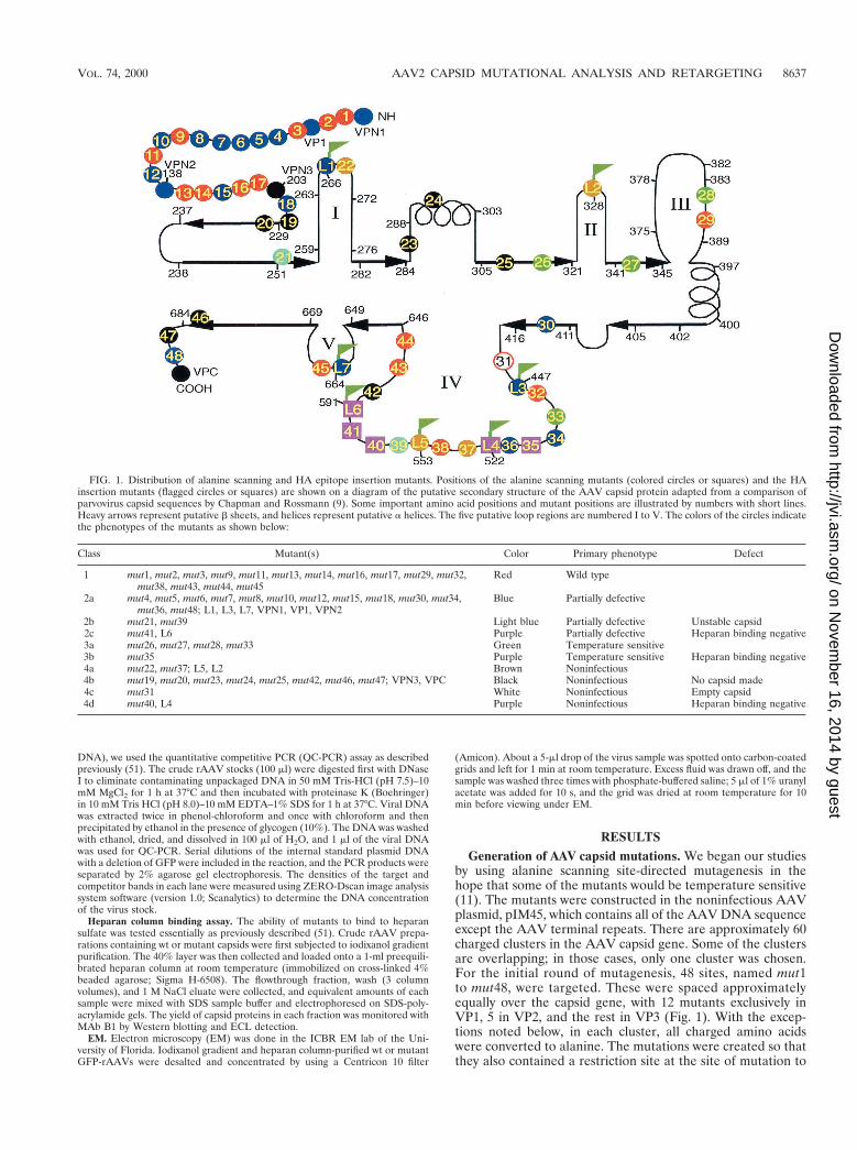
FIG. 1. Distribution of alanine scanning and HA epitope insertion mutants. Positions of the alanine scanning mutants (colored circles or squares) and the HAinsertion mutants (flagged circles or squares) are shown on a diagram of the putative secondary structure of the AAV capsid protein adapted from a comparison ofparvovirus capsid sequences by Chapman and Rossmann (9). Some important amino acid positions and mutant positions are illustrated by numbers with short lines.Heavy arrows represent putative b sheets, and helices represent putative a helices. The five putative loop regions are numbered I to V. The colors of the circles indicatethe phenotypes of the mutants as shown below:
Class Mutant(s) Color Primary phenotype Defect
1 mut1, mut2, mut3, mut9, mut11, mut13, mut14, mut16, mut17, mut29, mut32,mut38, mut43, mut44, mut45
Red Wild type
2a mut4, mut5, mut6, mut7, mut8, mut10, mut12, mut15, mut18, mut30, mut34,mut36, mut48; L1, L3, L7, VPN1, VP1, VPN2
Blue Partially defective
2b mut21, mut39 Light blue Partially defective Unstable capsid2c mut41, L6 Purple Partially defective Heparan binding negative3a mut26, mut27, mut28, mut33 Green Temperature sensitive3b mut35 Purple Temperature sensitive Heparan binding negative4a mut22, mut37; L5, L2 Brown Noninfectious4b mut19, mut20, mut23, mut24, mut25, mut42, mut46, mut47; VPN3, VPC Black Noninfectious No capsid made4c mut31 White Noninfectious Empty capsid4d mut40, L4 Purple Noninfectious Heparan binding negative
VOL. 74, 2000 AAV2 CAPSID MUTATIONAL ANALYSIS AND RETARGETING 8637
on Novem
ber 16, 2014 by guesthttp://jvi.asm
.org/D
ownloaded from
facilitate confirmation of the mutant sequence and subsequentinsertion of foreign epitopes (Table 1). In addition, after se-quence comparison of AAV serotypes 1 to 6, several otherpositions were targeted. mut28 and mut35 were made at posi-tions where extra amino acids were found in AAV4 by se-quence comparison with AAV2. mut32 was made by replacingTTT with AAA since TTT was not conserved among otherAAV serotypes at aa 454. Finally, in mut29 and mut31, onlyone Arg residue was changed to Ala, and in mut45 and mut48,only one Lys was changed to Ala. The positions of the alaninescanning mutants and the specific amino acid substitutions aresummarized in Table 1 and Fig. 1.
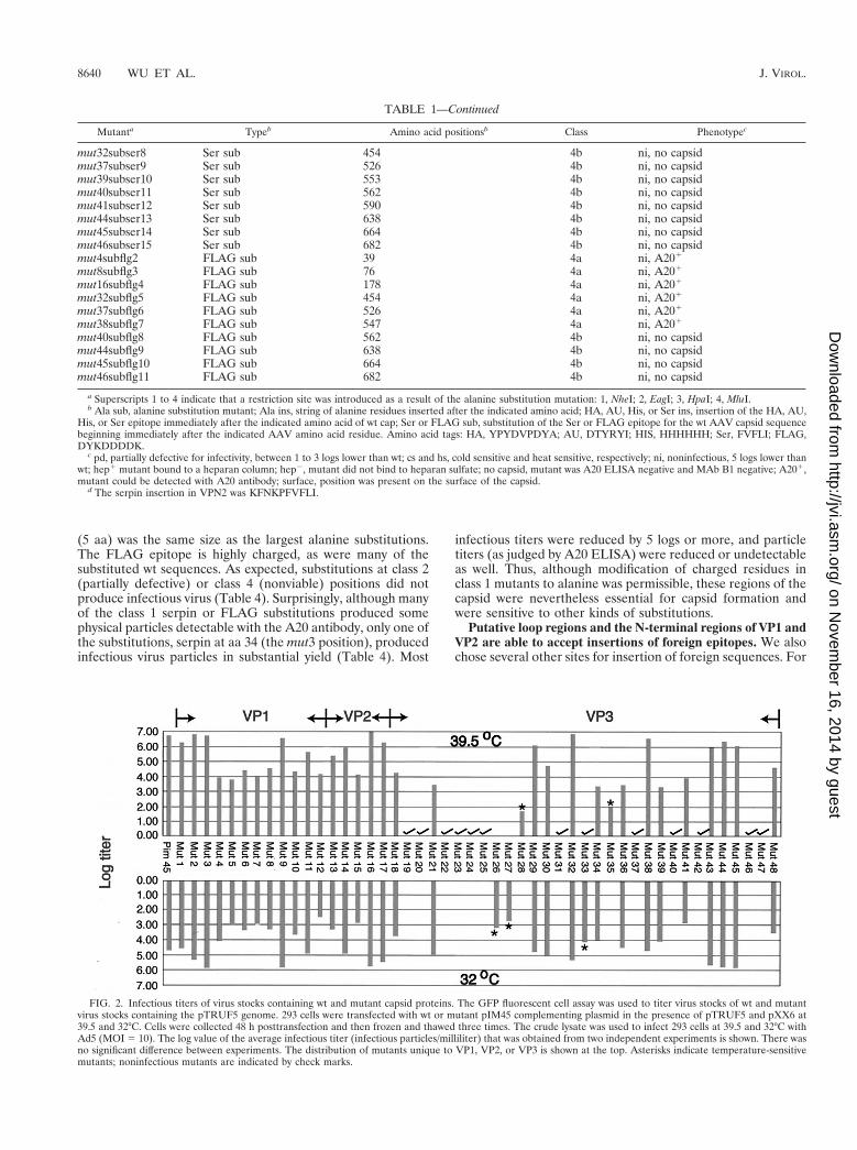
Infectious titer assays reveal four general classes of mu-tants. To determine the effect of each mutation on viral infec-tivity, we used either wt pIM45 or a mutant pIM45 plasmid tocomplement the growth of pTRUF5. pTRUF5 is a recombi-nant AAV plasmid that contains the gfp gene under the controlof a CMV enhancer-promoter (22). The resulting recombinantTRUF5 virus contained either wt or mutant capsid proteinsand could be titered for infectivity by counting green fluores-cent cells in the presence of an Ad5 coinfection. We had shownpreviously that the fluorescent cell assay produced titers withintwo- to threefold of those obtained with a conventional infec-tious center assay (51). Initially, each mutant was grown andtitered at either 39.5 or 32°C to determine if any of the mutantswere temperature sensitive. The experiments were done twice,and there was no significant variation in titer. On the basis ofthese titers, the mutants could be grouped into four classes(Fig. 2; Table 1). Class 1 contained mutants that have an in-fectious titer similar to the wt titer (less than 1 log difference;for example, mut1 and mut2). Class 2 contained partially de-fective mutants with infectious titers 2 to 3 logs lower than thewt titer (for example, mut4 and mut5). Class 3 contained tem-perature-sensitive mutants; three of these (mut26, mut27, andmut33) were heat sensitive, and two (mut28 and mut35) werecold sensitive. Class 4 consisted of 12 noninfectious mutants,whose titers were more than 5 logs lower than the wt titer.
Noninfectious (class 4) mutants and temperature-sensitive(class 3) mutants were defective in packaging DNA or in form-ing stable virus particles. To determine the probable causesfor the different defective mutants, we focused first on class 3and 4 mutants. For convenience, we ignored the fact that thetemperature-sensitive mutants had low infectivity when grownat the partially restrictive temperature of 37°C (data notshown), and viral preparations for all class 3 and 4 mutantswere made at 37°C. To determine if these mutants were ableto make capsids, we used the A20 ELISA. The A20 antibodyrecognizes only intact AAV particles (43) and is useful fordetermining the physical particle titer irrespective of whetherthe capsids contain DNA (18). Eight of sixteen mutants thatwere tested were negative by ELISA reading (Table 2), indi-cating that they were unable to make capsids or that the cap-sids were unstable even in crude lysate preparations. All ofthese were class 4 (noninfectious) mutants and were classifiedas class 4b (Table 1; Fig. 1).
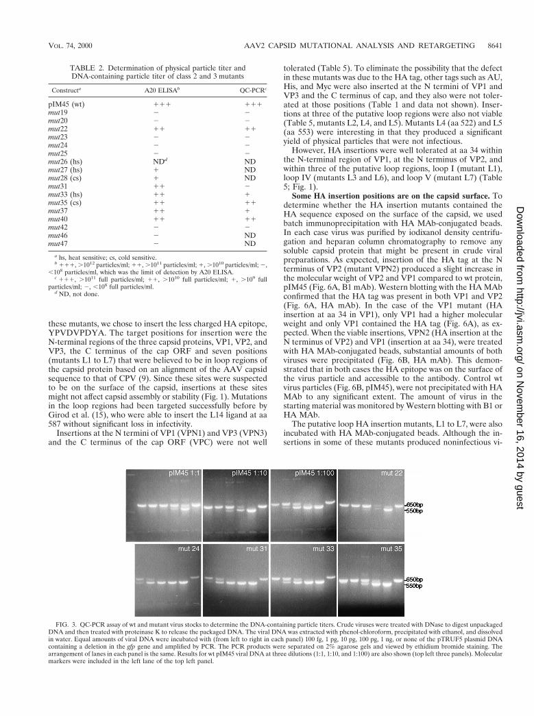
QC-PCR assays also were performed on most of the class 3and 4 mutants. The QC-PCR assay measures the titer of AAVparticles that contain DNase-resistant rAAV genomes (Fig. 3).We have shown previously that it provides physical particletiters that are equivalent to those obtained by dot blot assaybut has better sensitivity at low particle titers (51). As ex-pected, mutants that were negative for the synthesis of AAVparticles by A20 ELISA were also negative by QC-PCR assay(Table 2; Fig. 3). Most of the remaining mutants, which werepositive for A20 particles, were also positive for packaged viralDNA in the QC-PCR assay (Fig. 3; Table 2). This group of
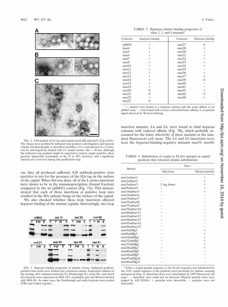
noninfectious mutants (mut22 and mut37) were called class 4a(Table 1; Fig. 1). Their defect was not in packaging but ratherin the binding, internalization, or uncoating steps of the viralentry process. One A20-positive mutant (mut31) was an excep-tion in that it was A20 positive but DNA negative by QC-PCRassay. This meant that mut31 formed intact virus particles thatwere empty. To confirm this, mut31 was examined by EM (Fig.4), and it did indeed make empty particles. In contrast, thepartially defective class 2 mutant, mut4, produced particlessimilar to wt particles. mut31 was assigned to class 4c (Fig. 1;Table 1).
Some mutants are defective for binding the viral receptor.One potential cause for the reduced infectivity of class 2, 3, or4 mutants might be that they were unable to bind the viral cellsurface receptor, the first step of the infectious cycle. Heparansulfate proteoglycan has been identified as the primary cellsurface receptor for AAV (37). To test whether these mutantscould bind heparan, we developed a heparan column bindingassay (Materials and Methods). Iodixanol-purified wt or mu-tant rAAVs were passed through a heparan agarose column,and the AAV capsid proteins in the starting material and thebound (eluate) and unbound (flowthrough and wash) fractionswere monitored by Western blotting using MAb B1, whichrecognizes all three capsid proteins (Fig. 5; Table 3). As expect-ed, wt AAV had a high affinity for the heparan column, sincelittle capsid protein was detected in the flowthrough and washfractions, and most of the capsid protein was detected in theeluate. The same was true of most of the mutants tested (Fig.5; Table 3). Two mutants, however, mut35 and mut41, boundpoorly to heparan (Fig. 5). A third mutant, mut40, which is lo-cated about 20 aa away from mut41, also bound with reducedaffinity (Fig. 5). This suggested that the primary defect in thesemutants was their inability to bind to heparan sulfate proteo-glycan. We classified mut35 as class 3b (temperature sensitiveand heparan binding negative), mut41 as class 2c (partially de-fective and heparan binding negative), and mut40 as class 4d(noninfectious and heparan binding negative) (Fig. 1; and Ta-ble 1).
Three class 4b mutants, mut20, mut25, and mut46, could notbe detected by Western analysis (Table 3). This was consistentwith the fact that they made no capsid that was detectable withthe A20 antibody (Table 2). Additionally, mut27, a tempera-ture-sensitive mutant, and two class 2 mutants, mut21 andmut39, did not give any Western signal with MAb B1 (Fig. 5;Table 3). The heat-sensitive mutant, mut27, was presumablyunstable at the nonpermissive temperature used for growingthis virus. mut21 and mut39 were partially defective when as-sayed in crude extracts (Fig. 2). The fact that they could not bedetected by capsid antibody after iodixanol centrifugation sug-gests that these capsids were also unstable during purification.These mutants were assigned to class 2b on the basis of theircapsid instability (Table 1; Fig. 1). The rest of the mutants inclass 2 that bind to heparan were classified as class 2a, partiallydefective, and heparan binding positive (Tables 1 and 3; Fig.1). The nature of their defect was not clear but presumably wasdue to some step in the infectious process that occurs afterviral attachment to the cell surface.
Regions tolerating alanine substitutions do not tolerateother kinds of substitutions. We wanted to determine whetherthe class 1 mutants defined positions in the capsid genes thatwere truly nonessential for capsid function. To test this, weconstructed a series of mutants in which either the serpin re-ceptor ligand, FVFLI (50), or the FLAG antibody epitope, DYKDDDDKYK, was substituted for capsid sequences at manyof the class 1 mutant positions (Table 4). A number of class 2and class 4 mutants were tried as well. The serpin substitution
8638 WU ET AL. J. VIROL.
on Novem
ber 16, 2014 by guesthttp://jvi.asm
.org/D
ownloaded from
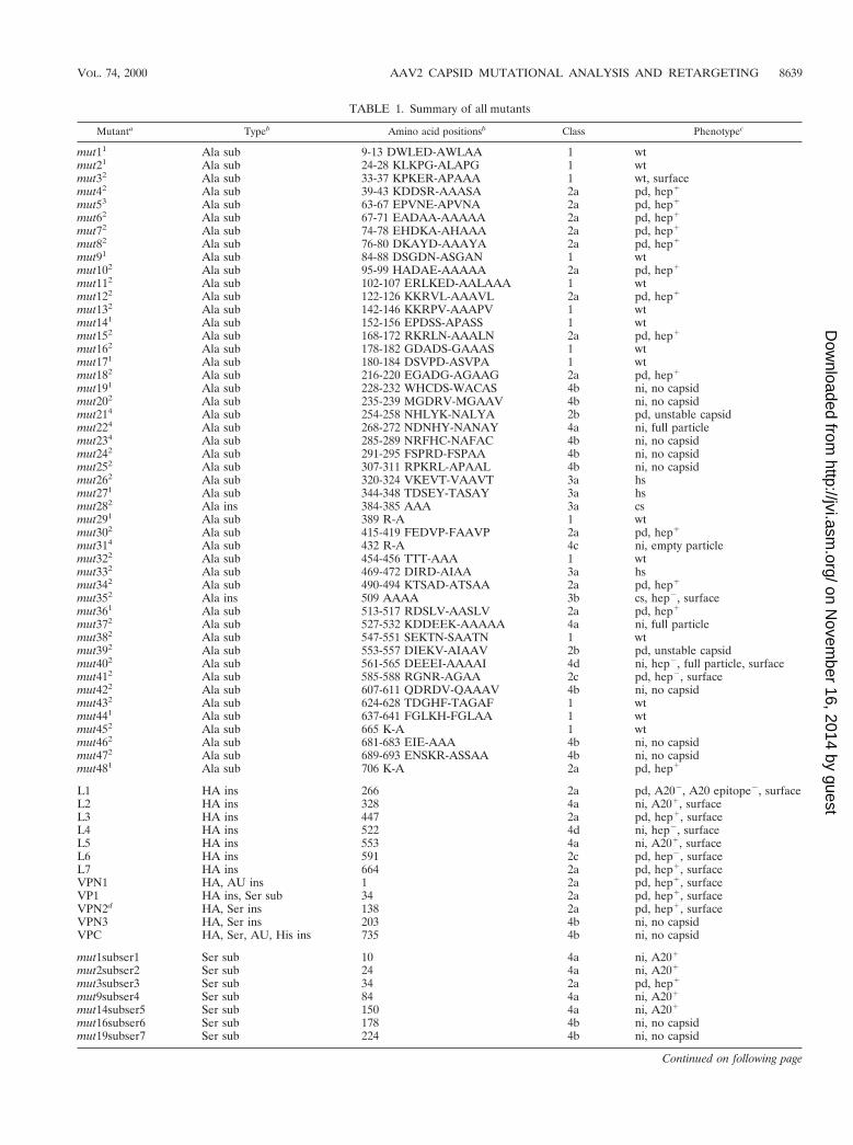
TABLE 1. Summary of all mutants
Mutanta Typeb Amino acid positionsb Class Phenotypec
mut11 Ala sub 9-13 DWLED-AWLAA 1 wtmut21 Ala sub 24-28 KLKPG-ALAPG 1 wtmut32 Ala sub 33-37 KPKER-APAAA 1 wt, surfacemut42 Ala sub 39-43 KDDSR-AAASA 2a pd, hep1
mut53 Ala sub 63-67 EPVNE-APVNA 2a pd, hep1
mut62 Ala sub 67-71 EADAA-AAAAA 2a pd, hep1
mut72 Ala sub 74-78 EHDKA-AHAAA 2a pd, hep1
mut82 Ala sub 76-80 DKAYD-AAAYA 2a pd, hep1
mut91 Ala sub 84-88 DSGDN-ASGAN 1 wtmut102 Ala sub 95-99 HADAE-AAAAA 2a pd, hep1
mut112 Ala sub 102-107 ERLKED-AALAAA 1 wtmut122 Ala sub 122-126 KKRVL-AAAVL 2a pd, hep1
mut132 Ala sub 142-146 KKRPV-AAAPV 1 wtmut141 Ala sub 152-156 EPDSS-APASS 1 wtmut152 Ala sub 168-172 RKRLN-AAALN 2a pd, hep1
mut162 Ala sub 178-182 GDADS-GAAAS 1 wtmut171 Ala sub 180-184 DSVPD-ASVPA 1 wtmut182 Ala sub 216-220 EGADG-AGAAG 2a pd, hep1
mut191 Ala sub 228-232 WHCDS-WACAS 4b ni, no capsidmut202 Ala sub 235-239 MGDRV-MGAAV 4b ni, no capsidmut214 Ala sub 254-258 NHLYK-NALYA 2b pd, unstable capsidmut224 Ala sub 268-272 NDNHY-NANAY 4a ni, full particlemut234 Ala sub 285-289 NRFHC-NAFAC 4b ni, no capsidmut242 Ala sub 291-295 FSPRD-FSPAA 4b ni, no capsidmut252 Ala sub 307-311 RPKRL-APAAL 4b ni, no capsidmut262 Ala sub 320-324 VKEVT-VAAVT 3a hsmut271 Ala sub 344-348 TDSEY-TASAY 3a hsmut282 Ala ins 384-385 AAA 3a csmut291 Ala sub 389 R-A 1 wtmut302 Ala sub 415-419 FEDVP-FAAVP 2a pd, hep1
mut314 Ala sub 432 R-A 4c ni, empty particlemut322 Ala sub 454-456 TTT-AAA 1 wtmut332 Ala sub 469-472 DIRD-AIAA 3a hsmut342 Ala sub 490-494 KTSAD-ATSAA 2a pd, hep1
mut352 Ala ins 509 AAAA 3b cs, hep2, surfacemut361 Ala sub 513-517 RDSLV-AASLV 2a pd, hep1
mut372 Ala sub 527-532 KDDEEK-AAAAA 4a ni, full particlemut382 Ala sub 547-551 SEKTN-SAATN 1 wtmut392 Ala sub 553-557 DIEKV-AIAAV 2b pd, unstable capsidmut402 Ala sub 561-565 DEEEI-AAAAI 4d ni, hep2, full particle, surfacemut412 Ala sub 585-588 RGNR-AGAA 2c pd, hep2, surfacemut422 Ala sub 607-611 QDRDV-QAAAV 4b ni, no capsidmut432 Ala sub 624-628 TDGHF-TAGAF 1 wtmut441 Ala sub 637-641 FGLKH-FGLAA 1 wtmut452 Ala sub 665 K-A 1 wtmut462 Ala sub 681-683 EIE-AAA 4b ni, no capsidmut472 Ala sub 689-693 ENSKR-ASSAA 4b ni, no capsidmut481 Ala sub 706 K-A 2a pd, hep1
L1 HA ins 266 2a pd, A202, A20 epitope2, surfaceL2 HA ins 328 4a ni, A201, surfaceL3 HA ins 447 2a pd, hep1, surfaceL4 HA ins 522 4d ni, hep2, surfaceL5 HA ins 553 4a ni, A201, surfaceL6 HA ins 591 2c pd, hep2, surfaceL7 HA ins 664 2a pd, hep1, surfaceVPN1 HA, AU ins 1 2a pd, hep1, surfaceVP1 HA ins, Ser sub 34 2a pd, hep1, surfaceVPN2d HA, Ser ins 138 2a pd, hep1, surfaceVPN3 HA, Ser ins 203 4b ni, no capsidVPC HA, Ser, AU, His ins 735 4b ni, no capsid
mut1subser1 Ser sub 10 4a ni, A201
mut2subser2 Ser sub 24 4a ni, A201
mut3subser3 Ser sub 34 2a pd, hep1
mut9subser4 Ser sub 84 4a ni, A201
mut14subser5 Ser sub 150 4a ni, A201
mut16subser6 Ser sub 178 4b ni, no capsidmut19subser7 Ser sub 224 4b ni, no capsid
Continued on following page
VOL. 74, 2000 AAV2 CAPSID MUTATIONAL ANALYSIS AND RETARGETING 8639
on Novem
ber 16, 2014 by guesthttp://jvi.asm
.org/D
ownloaded from
(5 aa) was the same size as the largest alanine substitutions.The FLAG epitope is highly charged, as were many of thesubstituted wt sequences. As expected, substitutions at class 2(partially defective) or class 4 (nonviable) positions did notproduce infectious virus (Table 4). Surprisingly, although manyof the class 1 serpin or FLAG substitutions produced somephysical particles detectable with the A20 antibody, only one ofthe substitutions, serpin at aa 34 (the mut3 position), producedinfectious virus particles in substantial yield (Table 4). Most
infectious titers were reduced by 5 logs or more, and particletiters (as judged by A20 ELISA) were reduced or undetectableas well. Thus, although modification of charged residues inclass 1 mutants to alanine was permissible, these regions of thecapsid were nevertheless essential for capsid formation andwere sensitive to other kinds of substitutions.
Putative loop regions and the N-terminal regions of VP1 andVP2 are able to accept insertions of foreign epitopes. We alsochose several other sites for insertion of foreign sequences. For
TABLE 1—Continued
Mutanta Typeb Amino acid positionsb Class Phenotypec
mut32subser8 Ser sub 454 4b ni, no capsidmut37subser9 Ser sub 526 4b ni, no capsidmut39subser10 Ser sub 553 4b ni, no capsidmut40subser11 Ser sub 562 4b ni, no capsidmut41subser12 Ser sub 590 4b ni, no capsidmut44subser13 Ser sub 638 4b ni, no capsidmut45subser14 Ser sub 664 4b ni, no capsidmut46subser15 Ser sub 682 4b ni, no capsidmut4subflg2 FLAG sub 39 4a ni, A201
mut8subflg3 FLAG sub 76 4a ni, A201
mut16subflg4 FLAG sub 178 4a ni, A201
mut32subflg5 FLAG sub 454 4a ni, A201
mut37subflg6 FLAG sub 526 4a ni, A201
mut38subflg7 FLAG sub 547 4a ni, A201
mut40subflg8 FLAG sub 562 4b ni, no capsidmut44subflg9 FLAG sub 638 4b ni, no capsidmut45subflg10 FLAG sub 664 4b ni, no capsidmut46subflg11 FLAG sub 682 4b ni, no capsid
a Superscripts 1 to 4 indicate that a restriction site was introduced as a result of the alanine substitution mutation: 1, NheI; 2, EagI; 3, HpaI; 4, MluI.b Ala sub, alanine substitution mutant; Ala ins, string of alanine residues inserted after the indicated amino acid; HA, AU, His, or Ser ins, insertion of the HA, AU,
His, or Ser epitope immediately after the indicated amino acid of wt cap; Ser or FLAG sub, substitution of the Ser or FLAG epitope for the wt AAV capsid sequencebeginning immediately after the indicated AAV amino acid residue. Amino acid tags: HA, YPYDVPDYA; AU, DTYRYI; HIS, HHHHHH; Ser, FVFLI; FLAG,DYKDDDDK.
c pd, partially defective for infectivity, between 1 to 3 logs lower than wt; cs and hs, cold sensitive and heat sensitive, respectively; ni, noninfectious, 5 logs lower thanwt; hep1 mutant bound to a heparan column; hep2, mutant did not bind to heparan sulfate; no capsid, mutant was A20 ELISA negative and MAb B1 negative; A201,mutant could be detected with A20 antibody; surface, position was present on the surface of the capsid.
d The serpin insertion in VPN2 was KFNKPFVFLI.
FIG. 2. Infectious titers of virus stocks containing wt and mutant capsid proteins. The GFP fluorescent cell assay was used to titer virus stocks of wt and mutantvirus stocks containing the pTRUF5 genome. 293 cells were transfected with wt or mutant pIM45 complementing plasmid in the presence of pTRUF5 and pXX6 at39.5 and 32°C. Cells were collected 48 h posttransfection and then frozen and thawed three times. The crude lysate was used to infect 293 cells at 39.5 and 32°C withAd5 (MOI 5 10). The log value of the average infectious titer (infectious particles/milliliter) that was obtained from two independent experiments is shown. There wasno significant difference between experiments. The distribution of mutants unique to VP1, VP2, or VP3 is shown at the top. Asterisks indicate temperature-sensitivemutants; noninfectious mutants are indicated by check marks.
8640 WU ET AL. J. VIROL.
on Novem
ber 16, 2014 by guesthttp://jvi.asm
.org/D
ownloaded from
these mutants, we chose to insert the less charged HA epitope,YPVDVPDYA. The target positions for insertion were theN-terminal regions of the three capsid proteins, VP1, VP2, andVP3, the C terminus of the cap ORF and seven positions(mutants L1 to L7) that were believed to be in loop regions ofthe capsid protein based on an alignment of the AAV capsidsequence to that of CPV (9). Since these sites were suspectedto be on the surface of the capsid, insertions at these sitesmight not affect capsid assembly or stability (Fig. 1). Mutationsin the loop regions had been targeted successfully before byGirod et al. (15), who were able to insert the L14 ligand at aa587 without significant loss in infectivity.
Insertions at the N termini of VP1 (VPN1) and VP3 (VPN3)and the C terminus of the cap ORF (VPC) were not well
tolerated (Table 5). To eliminate the possibility that the defectin these mutants was due to the HA tag, other tags such as AU,His, and Myc were also inserted at the N termini of VP1 andVP3 and the C terminus of cap, and they also were not toler-ated at those positions (Table 1 and data not shown). Inser-tions at three of the putative loop regions were also not viable(Table 5, mutants L2, L4, and L5). Mutants L4 (aa 522) and L5(aa 553) were interesting in that they produced a significantyield of physical particles that were not infectious.
However, HA insertions were well tolerated at aa 34 withinthe N-terminal region of VP1, at the N terminus of VP2, andwithin three of the putative loop regions, loop I (mutant L1),loop IV (mutants L3 and L6), and loop V (mutant L7) (Table5; Fig. 1).
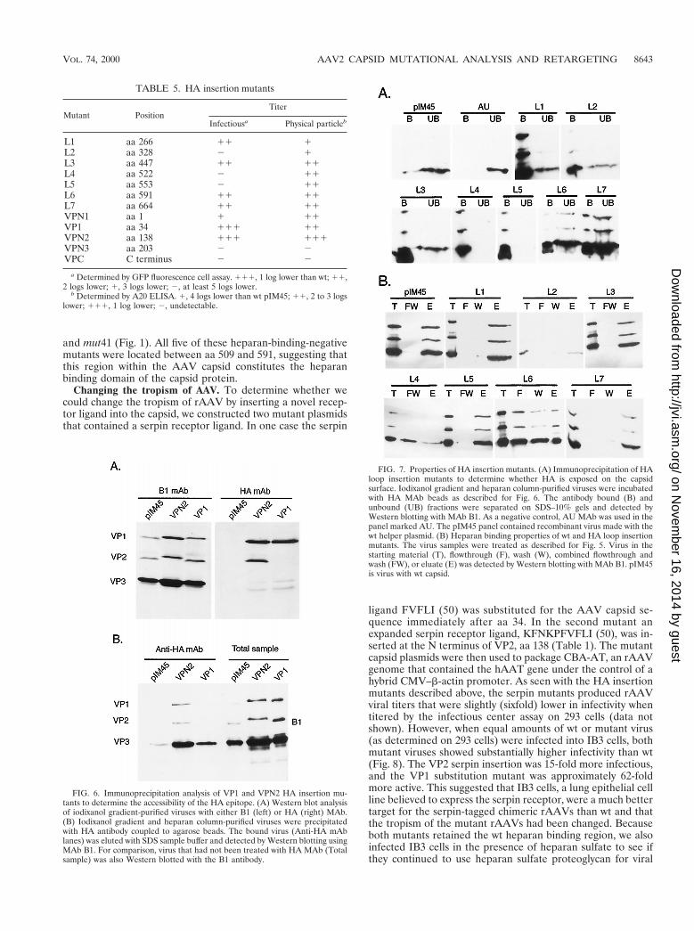
Some HA insertion positions are on the capsid surface. Todetermine whether the HA insertion mutants contained theHA sequence exposed on the surface of the capsid, we usedbatch immunoprecipitation with HA MAb-conjugated beads.In each case virus was purified by iodixanol density centrifu-gation and heparan column chromatography to remove anysoluble capsid protein that might be present in crude viralpreparations. As expected, insertion of the HA tag at the Nterminus of VP2 (mutant VPN2) produced a slight increase inthe molecular weight of VP2 and VP1 compared to wt protein,pIM45 (Fig. 6A, B1 mAb). Western blotting with the HA MAbconfirmed that the HA tag was present in both VP1 and VP2(Fig. 6A, HA mAb). In the case of the VP1 mutant (HAinsertion at aa 34 in VP1), only VP1 had a higher molecularweight and only VP1 contained the HA tag (Fig. 6A), as ex-pected. When the viable insertions, VPN2 (HA insertion at theN terminus of VP2) and VP1 (insertion at aa 34), were treatedwith HA MAb-conjugated beads, substantial amounts of bothviruses were precipitated (Fig. 6B, HA mAb). This demon-strated that in both cases the HA epitope was on the surface ofthe virus particle and accessible to the antibody. Control wtvirus particles (Fig. 6B, pIM45), were not precipitated with HAMAb to any significant extent. The amount of virus in thestarting material was monitored by Western blotting with B1 orHA MAb.
The putative loop HA insertion mutants, L1 to L7, were alsoincubated with HA MAb-conjugated beads. Although the in-sertions in some of these mutants produced noninfectious vi-
TABLE 2. Determination of physical particle titer andDNA-containing particle titer of class 2 and 3 mutants
Constructa A20 ELISAb QC-PCRc
pIM45 (wt) 111 111mut19 2 2mut20 2 2mut22 11 11mut23 2 2mut24 2 2mut25 2 2mut26 (hs) NDd NDmut27 (hs) 1 NDmut28 (cs) 1 NDmut31 11 2mut33 (hs) 11 1mut35 (cs) 11 11mut37 11 1mut40 11 11mut42 2 2mut46 2 NDmut47 2 ND
a hs, heat sensitive; cs, cold sensitive.b 111, .1012 particles/ml; 11, .1011 particles/ml; 1, .1010 particles/ml; 2,
,108 particles/ml, which was the limit of detection by A20 ELISA.c 111, .1011 full particles/ml; 11, .1010 full particles/ml; 1, .109 full
particles/ml; 2, ,108 full particles/ml.d ND, not done.
FIG. 3. QC-PCR assay of wt and mutant virus stocks to determine the DNA-containing particle titers. Crude viruses were treated with DNase to digest unpackagedDNA and then treated with proteinase K to release the packaged DNA. The viral DNA was extracted with phenol-chloroform, precipitated with ethanol, and dissolvedin water. Equal amounts of viral DNA were incubated with (from left to right in each panel) 100 fg, 1 pg, 10 pg, 100 pg, 1 ng, or none of the pTRUF5 plasmid DNAcontaining a deletion in the gfp gene and amplified by PCR. The PCR products were separated on 2% agarose gels and viewed by ethidium bromide staining. Thearrangement of lanes in each panel is the same. Results for wt pIM45 viral DNA at three dilutions (1:1, 1:10, and 1:100) are also shown (top left three panels). Molecularmarkers were included in the left lane of the top left panel.
VOL. 74, 2000 AAV2 CAPSID MUTATIONAL ANALYSIS AND RETARGETING 8641
on Novem
ber 16, 2014 by guesthttp://jvi.asm
.org/D
ownloaded from
rus, they all produced sufficient A20 antibody-positive virusparticles to test for the presence of the HA tag on the surfaceof the capsid. When this was done, all of the L-series insertionswere shown to be in the immunoprecipitate (bound fraction)compared to the wt (pIM45) control (Fig. 7A). This demon-strated that each of these insertions at putative loop sitesresulted in the HA epitope being on the surface of the capsid.
We also checked whether these loop insertions affectedheparan binding of the mutant capsids. Interestingly, two loop
insertion mutants, L4 and L6, were found to bind heparancolumns with reduced affinity (Fig. 7B), which probably ac-counted for the lower infectivity of these mutants in the stan-dard fluorescent cell assay. The L4 and L6 insertions werenear the heparan-binding-negative mutants mut35, mut40,FIG. 4. EM analysis of wt (A) and mutant (mut4 [B] and mut31 [C]) rAAVs.
The viruses were purified by iodixanol step gradient centrifugation and heparancolumn chromatography as described elsewhere (51), concentrated in a Centri-con 10, and negatively stained with 1% uranyl acetate. Bar 5 40 nm. Althoughthe iodixanol step gradient might be expected to remove empty particles, theseparticles apparently accumulate at the 25 to 40% interface, and a significantfraction are recovered during this purification step.
FIG. 5. Heparan binding properties of mutant viruses. Iodixanol gradient-purified virus stocks were loaded onto a heparan column. Equivalent volumes ofthe starting, 40% iodixanol material (T), flowthrough (F), wash (W), and eluted(E) fractions were separated on SDS–10% acrylamide gels and Western blottedwith MAb B1. In some cases, the flowthrough and wash fractions were pooled(FW) and loaded together.
TABLE 3. Heparan column binding properties ofclass 2, 3, and 4 mutantsa
Construct Heparan binding Construct Heparan binding
pIM45 1 mut27 0mut4 1 mut28 1mut5 1 mut30 1mut6 1 mut31 1mut7 1 mut32 1mut8 1 mut33 1mut10 1 mut34 1mut11 1 mut35 2mut12 1 mut36 1mut13 1 mut37 1mut14 1 mut39 0mut15 1 mut40 2mut18 1 mut41 2mut20 0 mut43 1mut21 0 mut46 0mut22 1 mut48 1mut25 0
a 1, mutant virus bound to a heparan column with the same affinity as wtpIM45 virus; 2, virus bound with at least a threefold-lower affinity; 0, no proteinsignal detected by Western blotting.
TABLE 4. Substitution of serpin or FLAG epitopes at capsidpositions that tolerated alanine substitutions
MutantTitera
Infectious Physical particle
mut1subser1 2 1mut2subser2 2 1mut3subser3 1 log lower 1mut9subser4 2 1mut14subser5 2 1mut16subser6 2 2mut19subser7 2 2mut32subser8 2 2mut37subser9 2 2mut39subser10 2 2mut40subser11 2 2mut41subser12 2 2mut44subser13 2 2mut45subser14 2 2mut46subser15 2 2mut4subflg2 2 1mut8subflg3 2 1mut16subflg4 2 1mut32subflg5 2 1mut37subflg6 2 1mut38subflg7 2 1mut40subflg8 2 2mut44subflg9 2 2mut45subflg10 2 2mut46subflg11 2 2
a Either a serpin peptide sequence or the FLAG sequence was substituted forthe AAV capsid sequence at the positions used previously for alanine scanningmutagenesis (Fig. 2). Infectious titers were determined by GFP fluorescent cellassay. 2, infectious virus could not be detected. Physical particle titers werejudged by A20 ELISA. 1, particles were detectable; 2, particles were notdetectable.
8642 WU ET AL. J. VIROL.
on Novem
ber 16, 2014 by guesthttp://jvi.asm
.org/D
ownloaded from
and mut41 (Fig. 1). All five of these heparan-binding-negativemutants were located between aa 509 and 591, suggesting thatthis region within the AAV capsid constitutes the heparanbinding domain of the capsid protein.
Changing the tropism of AAV. To determine whether wecould change the tropism of rAAV by inserting a novel recep-tor ligand into the capsid, we constructed two mutant plasmidsthat contained a serpin receptor ligand. In one case the serpin
ligand FVFLI (50) was substituted for the AAV capsid se-quence immediately after aa 34. In the second mutant anexpanded serpin receptor ligand, KFNKPFVFLI (50), was in-serted at the N terminus of VP2, aa 138 (Table 1). The mutantcapsid plasmids were then used to package CBA-AT, an rAAVgenome that contained the hAAT gene under the control of ahybrid CMV–b-actin promoter. As seen with the HA insertionmutants described above, the serpin mutants produced rAAVviral titers that were slightly (sixfold) lower in infectivity whentitered by the infectious center assay on 293 cells (data notshown). However, when equal amounts of wt or mutant virus(as determined on 293 cells) were infected into IB3 cells, bothmutant viruses showed substantially higher infectivity than wt(Fig. 8). The VP2 serpin insertion was 15-fold more infectious,and the VP1 substitution mutant was approximately 62-foldmore active. This suggested that IB3 cells, a lung epithelial cellline believed to express the serpin receptor, were a much bettertarget for the serpin-tagged chimeric rAAVs than wt and thatthe tropism of the mutant rAAVs had been changed. Becauseboth mutants retained the wt heparan binding region, we alsoinfected IB3 cells in the presence of heparan sulfate to see ifthey continued to use heparan sulfate proteoglycan for viral
TABLE 5. HA insertion mutants
Mutant PositionTiter
Infectiousa Physical particleb
L1 aa 266 11 1L2 aa 328 2 1L3 aa 447 11 11L4 aa 522 2 11L5 aa 553 2 11L6 aa 591 11 11L7 aa 664 11 11VPN1 aa 1 1 11VP1 aa 34 111 11VPN2 aa 138 111 111VPN3 aa 203 2 2VPC C terminus 2 2
a Determined by GFP fluorescence cell assay. 111, 1 log lower than wt; 11,2 logs lower; 1, 3 logs lower; 2, at least 5 logs lower.
b Determined by A20 ELISA. 1, 4 logs lower than wt pIM45; 11, 2 to 3 logslower; 111, 1 log lower; 2, undetectable.
FIG. 6. Immunoprecipitation analysis of VP1 and VPN2 HA insertion mu-tants to determine the accessibility of the HA epitope. (A) Western blot analysisof iodixanol gradient-purified viruses with either B1 (left) or HA (right) MAb.(B) Iodixanol gradient and heparan column-purified viruses were precipitatedwith HA antibody coupled to agarose beads. The bound virus (Anti-HA mAblanes) was eluted with SDS sample buffer and detected by Western blotting usingMAb B1. For comparison, virus that had not been treated with HA MAb (Totalsample) was also Western blotted with the B1 antibody.
FIG. 7. Properties of HA insertion mutants. (A) Immunoprecipitation of HAloop insertion mutants to determine whether HA is exposed on the capsidsurface. Iodixanol gradient and heparan column-purified viruses were incubatedwith HA MAb beads as described for Fig. 6. The antibody bound (B) andunbound (UB) fractions were separated on SDS–10% gels and detected byWestern blotting with MAb B1. As a negative control, AU MAb was used in thepanel marked AU. The pIM45 panel contained recombinant virus made with thewt helper plasmid. (B) Heparan binding properties of wt and HA loop insertionmutants. The virus samples were treated as described for Fig. 5. Virus in thestarting material (T), flowthrough (F), wash (W), combined flowthrough andwash (FW), or eluate (E) was detected by Western blotting with MAb B1. pIM45is virus with wt capsid.
VOL. 74, 2000 AAV2 CAPSID MUTATIONAL ANALYSIS AND RETARGETING 8643
on Novem
ber 16, 2014 by guesthttp://jvi.asm
.org/D
ownloaded from
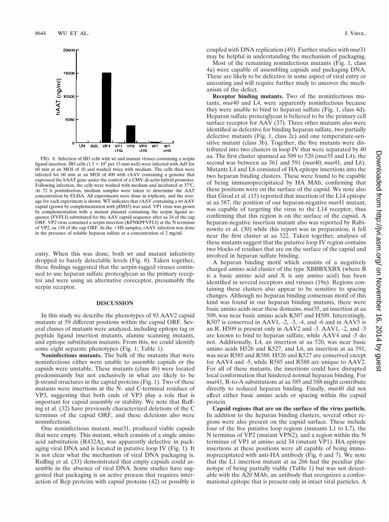
entry. When this was done, both wt and mutant infectivitydropped to barely detectable levels (Fig. 8). Taken together,these findings suggested that the serpin-tagged viruses contin-ued to use heparan sulfate proteoglycan as the primary recep-tor and were using an alternative coreceptor, presumably theserpin receptor.
DISCUSSION
In this study we describe the phenotypes of 93 AAV2 capsidmutants at 59 different positions within the capsid ORF. Sev-eral classes of mutants were analyzed, including epitope tag orpeptide ligand insertion mutants, alanine scanning mutants,and epitope substitution mutants. From this, we could identifysome eight separate phenotypes (Fig. 1; Table 1).
Noninfectious mutants. The bulk of the mutants that werenoninfectious either were unable to assemble capsids or thecapsids were unstable. These mutants (class 4b) were locatedpredominantly but not exclusively in what are likely to beb-strand structures in the capsid proteins (Fig. 1). Two of thesemutants were insertions at the N- and C-terminal residues ofVP3, suggesting that both ends of VP3 play a role that isimportant for capsid assembly or stability. We note that Ruff-ing et al. (32) have previously characterized deletions of the Cterminus of the capsid ORF, and these deletions also werenoninfectious.
One noninfectious mutant, mut31, produced viable capsidsthat were empty. This mutant, which consists of a single aminoacid substitution (R432A), was apparently defective in pack-aging viral DNA and is located in putative loop IV (Fig. 1). Itis not clear what the mechanism of viral DNA packaging is.Ruffing et al. (33) demonstrated that empty capsids could as-semble in the absence of viral DNA. Some studies have sug-gested that packaging is an active process that requires inter-action of Rep proteins with capsid proteins (42) or possibly is
coupled with DNA replication (49). Further studies with mut31may be helpful in understanding the mechanism of packaging.
Most of the remaining noninfectious mutants (Fig. 1, class4a) were capable of assembling capsids and packaging DNA.These are likely to be defective in some aspect of viral entry oruncoating and will require further study to uncover the mech-anism of the defect.
Receptor binding mutants. Two of the noninfectious mu-tants, mut40 and L4, were apparently noninfectious becausethey were unable to bind to heparan sulfate (Fig. 1, class 4d).Heparan sulfate proteoglycan is believed to be the primary cellsurface receptor for AAV (37). Three other mutants also wereidentified as defective for binding heparan sulfate, two partiallydefective mutants (Fig. 1, class 2c) and one temperature-sen-sitive mutant (class 3b). Together, the five mutants were dis-tributed into two clusters in loop IV that were separated by 40aa. The first cluster spanned aa 509 to 520 (mut35 and L4); thesecond was between aa 561 and 591 (mut40, mut41, and L6).Mutants L4 and L6 consisted of HA epitope insertions into thetwo heparan binding clusters. These were found to be capableof being immunoprecipitated by HA MAb, confirming thatthese positions were on the surface of the capsid. We note alsothat Girod et al. (15) reported that insertion of the L14 epitopeat aa 587, the position of our heparan-negative mut41 mutant,was capable of targeting the virus to the L14 receptor, thusconfirming that this region is on the surface of the capsid. Aheparan-negative insertion mutant also was reported by Rabi-nowitz et al. (30) while this report was in preparation; it fellnear the first cluster at aa 522. Taken together, analyses ofthese mutants suggest that the putative loop IV region containstwo blocks of residues that are on the surface of the capsid andinvolved in heparan sulfate binding.
A heparan binding motif which consists of a negativelycharged amino acid cluster of the type XBBBXXBX (where Bis a basic amino acid and X is any amino acid) has beenidentified in several receptors and viruses (19a). Regions con-taining these clusters also appear to be sensitive to spacingchanges. Although no heparan binding consensus motif of thiskind was found in our heparan binding mutants, there werebasic amino acids near these domains. mut35, an insertion at aa509, was near basic amino acids K507 and H509. Interestingly,K507 is conserved in AAV1, -2, -3, -4, and -6 and in AAV5 isan R. H509 is present only in AAV2 and -3. AAV1, -2, and -3are known to bind to heparan sulfate, while AAV4 and -5 donot. Additionally, L4, an insertion at aa 520, was near basicamino acids H526 and K527, and L6, an insertion at aa 591,was near R585 and R588. H526 and K527 are conserved exceptfor AAV4 and -5, while R585 and R588 are unique to AAV2.For all of these mutants, the insertions could have disruptedlocal conformation that hindered normal heparan binding. Formut41, R-to-A substitutions at aa 585 and 588 might contributedirectly to reduced heparan binding. Finally, mut40 did notaffect either basic amino acids or spacing within the capsidprotein.
Capsid regions that are on the surface of the virus particle.In addition to the heparan binding clusters, several other re-gions were also present on the capsid surface. These includefour of the five putative loop regions (mutants L1 to L7), theN terminus of VP2 (mutant VPN2), and a region within the Nterminus of VP1 at amino acid 34 (mutant VP1). HA epitopeinsertions at these positions were all capable of being immu-noprecipitated with anti-HA antibody (Fig. 6 and 7). We notethat the L1 insertion mutant at aa 266 had the peculiar phe-notype of being partially viable (Table 1) but was not detect-able with the A20 MAb, an antibody that recognizes a confor-mational epitope that is present only in intact viral particles. A
FIG. 8. Infection of IB3 cells with wt and mutant viruses containing a serpinligand insertion. IB3 cells (1.5 3 105 per 15-mm well) were infected with Ad5 for60 min at an MOI of 10 and washed twice with medium. The cells then wereinfected for 60 min at an MOI of 400 with rAAV containing a genome thatexpressed the hAAT gene under the control of a CMV–b-actin hybrid promoter.Following infection, the cells were washed with medium and incubated at 37°C.At 72 h postinfection, medium samples were taken to determine the AATconcentration by ELISA. All experiments were done in triplicate, and the aver-age for each experiment is shown. WT indicates that rAAV containing a wt AAVcapsid (grown by complementation with pIM45) was used. VP1 virus was grownby complementation with a mutant plasmid containing the serpin ligand se-quence (FVFLI) substituted for the AAV capsid sequence after aa 34 of the capORF. VP2 virus contained a serpin insertion (KFNKPFVFLI) at the N terminusof VP2, aa 138 of the cap ORF. In the 1HS samples, rAAV infection was donein the presence of soluble heparan sulfate at a concentration of 2 mg/ml.
8644 WU ET AL. J. VIROL.
on Novem
ber 16, 2014 by guesthttp://jvi.asm
.org/D
ownloaded from
nearby capsid-forming mutant made by Girod et al. (15) at aa261 was also negative for A20 antibody binding. This suggeststhat at least part of the epitope for the A20 MAb consists ofamino acids between 261 and 266 and confirms that this regionis on the surface of the intact particle.
Of the positions identified as being on the surface of thecapsid, we found six that potentially are capable of acceptingforeign epitope or ligand insertions for retargeting the viralcapsid to alternative receptors. These are the N-terminal re-gion of VP1 (near aa 34), the N terminus of VP2 (aa 138), theloop I region (aa 266), the loop IV region (near aa 447 and591), and the loop V region (aa 664). All of these locations
were capable of tolerating an HA (or serpin) insertion andproduced recombinant virus titers that were within 1 to 2 logsof the wt value. Furthermore, HA epitope insertions at thesepositions were capable of being immunoprecipitated with an-ti-HA antibody (Fig. 6 and 7). Two of these positions, whentested with a serpin ligand insertion or substitution, producedvirus that was much more infectious on IB3 cells than wt virus.Curiously, both serpin mutants were still inhibited by solubleheparan sulfate, suggesting that heparan sulfate proteoglycanwas still the primary receptor for these mutants and that theserpin receptor was being used as an alternative coreceptor. Itis conceivable that one or both of these capsid positions is
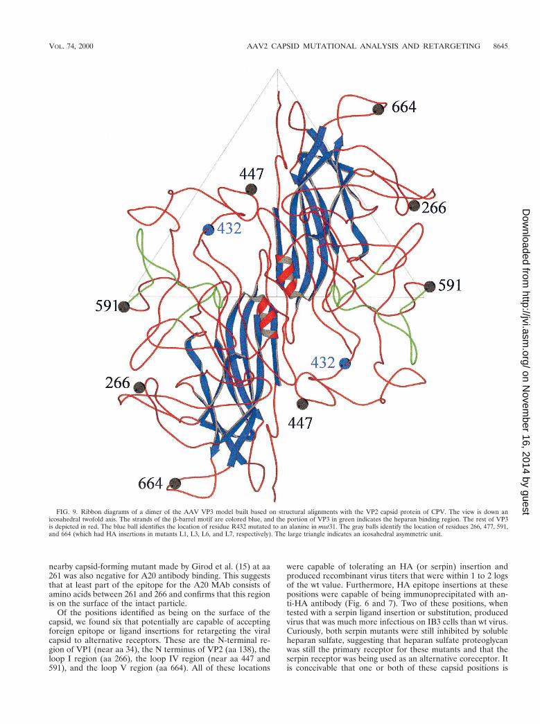
FIG. 9. Ribbon diagrams of a dimer of the AAV VP3 model built based on structural alignments with the VP2 capsid protein of CPV. The view is down anicosahedral twofold axis. The strands of the b-barrel motif are colored blue, and the portion of VP3 in green indicates the heparan binding region. The rest of VP3is depicted in red. The blue ball identifies the location of residue R432 mutated to an alanine in mut31. The gray balls identify the location of residues 266, 477, 591,and 664 (which had HA insertions in mutants L1, L3, L6, and L7, respectively). The large triangle indicates an icosahedral asymmetric unit.
VOL. 74, 2000 AAV2 CAPSID MUTATIONAL ANALYSIS AND RETARGETING 8645
on Novem
ber 16, 2014 by guesthttp://jvi.asm
.org/D
ownloaded from
involved in binding to one or both of the proteins that normallyact as coreceptors for wt virus, fibroblast growth factor (28), orintegrin avb5 (36). This would explain their partial defect on293 cells and the recovery of infectivity on IB3 cells. Furtherstudies will be needed to test this possibility.
Mutants with unstable capsids and temperature-sensitivephenotypes. Three mutants, mut21, mut27, and mut39, werefound to have capsids that were unstable when purifiedthrough an iodixanol gradient. Iodixanol is an iso-osmotic gra-dient purification method that appears to be gentler than CsClcentrifugation (51). Thus, these mutants appear to be partic-ularly sensitive to capsid denaturation. mut21 and mut27 are inputative b sheets, and mut39 is in loop IV. It is worth notingthat Rabinowitz et al. (30) also isolated an unstable capsidmutant at aa 247 that is near the mut21 position, aa 254. mut27is also one of five temperature-sensitive mutants isolated dur-ing this study. The temperature-sensitive mutants and the un-stable capsid mutants should prove useful in future studies foridentifying steps in the capsid assembly or the infection process.
Viable and partially defective mutants. The two largestclasses of mutants isolated were either wt (class 1) or partiallydefective (class 2a) with no identifiable defect (Fig. 1). Bothclass 1 and class 2a mutants were distributed either in the VP1and VP2 unique regions or in the predicted loop regions of thecapsid protein. We naively assumed that class 1 mutant posi-tions, which produced viable capsids after substitution of twoto five alanine residues, were regions that were nonessentialfor capsid assembly or stability and therefore should accom-modate other kinds of substitutions. However, when serpin orFLAG epitopes were substituted at many of these sites, mostof the mutants were nonviable, with the exception of aa 34 inVP1. Indeed, many of these viruses were negative for capsidassembly and should also be useful for identifying possibleintermediates in capsid assembly.
Ruffing et al. (33) showed previously that VP1 and VP2 butnot VP3 contained nuclear localization signals (NLS), andthree putative NLS are located in the VP1/VP2 region at aa121 to 125, 141 to 145, and 167 to 171. Hoque et al. (19b) haveshown that aa 167 to 172 were sufficient to target VP2 to thenucleus, although their experiments did not rule out possibleredundancy with the other two putative NLS sequences. Allthree of these putative signals were targeted with alanine scan-ning mutants (mut12, mut13, and mut15) in our study. Two ofthese mutants, mut12 and mut15, were partially defective, andthe inactivation of an NLS may be the reason for their pheno-type (19b, 33). We note that mut15 should have eliminated theNLS identified by Hoque and colleagues. The fact that mut15was only partially defective suggests that there may be an al-ternative, redundant NLS sequences that are used by the cap-sid proteins. The third mutant (mut13) was classified as viable,but it also showed a lower than wt titer (Fig. 1).
Molecular computer graphics construction of an AAV modeland structural localization of mutant residues. Because theAAV crystal structure is not available, the atomic coordinatesof CPV VP2 (PDB accession no. 4DPV) were interactivelymutated using the program O (20) to generate a homology-based model of the AAV capsid, using modifications of thealignments of the AAV major capsid protein (VP3) with theVP2 capsid protein of CPV (9, 15). The mutations were fol-lowed by refinement constrained with standard geometry in theO database. The model provided a means for preliminarystructural identification of the heparan receptor attachmentsites in the surface depression (dimple) near the twofold ico-sahedral axes of the capsid, surface loop regions which cantolerate foreign peptide sequence insertions, and a possibleexplanation for the phenotype of mut31 (Fig. 9).
The topographic location of the putative heparan bindingregion is consistent with regions that have been suggested asbeing involved in host cellular factor(s) recognition and impli-cated in tissue tropism and in vivo pathogenicity for otherparvoviruses (3, 4, 24, 39). It is of interest that the putativeheparan binding site is adjacent to a region of the AAV capsidthat contains a peptide insert when the AAV VP3 sequence iscompared to that of CPV VP2 and the VP2 of most of theother autonomous parvovirus sequences (9). Also a similarinsertion of peptide sequences compared to CPV (althoughnot in a homologous region of the VP2 to that observed inAAV) is present in the capsid of Aleutian mink disease par-vovirus and minute virus of mice, proximal to residues in thedimple depression which are implicated in tissue tropism (24).Thus, these insertions may be capsid surface adaptations thatenable the capsids to recognize different receptors during in-fection. In the case of AAV, its dimple peptide insertion, whichis absent in the other parvoviruses, may enable it to recognizeheparan sulfate, which has not been implicated in cellularinfectivity by any other parvovirus.
The model also clearly shows that regions of the capsidwhich tolerated the insertions of the HA epitope (i.e., at res-idues 266, 447, 591, and 664) are on the surface loops presentbetween the b strands of the b-barrel motif (Fig. 9). Theb-barrel motif forms the core contiguous shell of parvoviruscapsids, while the surface loops make up the surface decora-tions, dictating the strain-specific biological properties of themembers. The observation that these surface regions can tol-erate foreign peptide insertion is an indication that they arenot involved in the interactions that govern capsid assembly.
Finally, the model provides a possible explanation for theobservation that mut31 (R432A) is able to form only emptyparticles. In the unassembled VP3 monomer, the side chain ofR432, points toward the interior of the capsid and would mostlikely be in contact with DNA. If recognition and encapsida-tion of AAV DNA precede final capsid assembly and involveoligomeric intermediates, then R432 contacts with DNA maybe essential for initiating capsid assembly around a nascentDNA strand.
In summary, we have reported a preliminary analysis of mu-tants at 59 positions within the AAV2 capsid ORF. We haveidentified regions in the capsid proteins that affect infectivity,capsid formation, capsid stability, DNA packaging, and recep-tor binding. These mutants should be valuable for defining thefunctional domains of AAV capsid proteins and for dissectingthe molecular mechanism of viral entry. Additionally, we havedefined a number of regions in the capsid gene at which foreignligands can be inserted and have demonstrated that insertionof a foreign receptor ligand at some of these positions canchange the tropism of the virus. This is the first step in thedevelopment of the next generation of AAV vectors, which canbe targeted to specific cellular receptors or tissues.
ACKNOWLEDGMENTS
We thank J. Kleinschmidt for kindly providing MAbs A20 and B1.We also thank R. J. Samulski for providing plasmid pXX6. We ac-knowledge the Vector Core Laboratory at the Powell Gene TherapyCenter, University of Florida Medical School, for technical assistanceon rAAV production. We thank Corrine Abernathy, Daniel Lackner,and Eric Kolbrener for help on this project.
This work was supported by grants PO1 HL59412, PO1 HL51811,and PO1 NS36302 from the National Institutes of Health.
REFERENCES
1. Agbandje, M., S. Kajigaya, R. McKenna, N. S. Young, and M. G. Rossmann.1994. The structure of human parvovirus B19 at 8 Å resolution. Virology 203:106–115.
8646 WU ET AL. J. VIROL.
on Novem
ber 16, 2014 by guesthttp://jvi.asm
.org/D
ownloaded from
2. Agbandje, M., R. McKenna, M. G. Rossmann, M. L. Strassheim, and C. R.Parrish. 1993. Structure determination of feline panleukopenia virus emptyparticles. Proteins 16:155–171.
3. Agbandje-McKenna, M., A. L. Llamas-Saiz, F. Wang, P. Tattersall, andM. G. Rossmann. 1998. Functional implications of the structure of themurine parvovirus, minute virus of mice. Structure 6:1369–1381.
4. Barbis, D. P., S. F. Chang, and C. R. Parrish. 1992. Mutations adjacent tothe dimple of the canine parvovirus capsid structure affect sialic acid binding.Virology 191:301–308.
5. Berns, K. I., and R. A. Bohenzky. 1987. Adeno-associated viruses: an update.Adv. Virus Res. 32:243–306.
6. Berns, K. I., and C. Giraud. 1995. Adenovirus and adeno-associated virus asvectors for gene therapy. Ann. N. Y. Acad. Sci. 772:95–104.
7. Buller, R. M., J. E. Janik, E. D. Sebring, and J. A. Rose. 1981. Herpessimplex virus types 1 and 2 completely help adenovirus-associated virusreplication. J. Virol. 40:241–247.
8. Casto, B. C., J. A. Armstrong, R. W. Atchison, and W. M. Hammon. 1967.Studies on the relationship between adeno-associated virus type 1 (AAV-1)and adenoviruses. II. Inhibition of adenovirus plaques by AAV; its natureand specificity. Virology 33:452–458.
9. Chapman, M. S., and M. G. Rossmann. 1993. Structure, sequence, andfunction correlations among parvoviruses. Virology 194:491–508.
10. Chiorini, J. A., L. Yang, Y. Liu, B. Safer, and R. M. Kotin. 1997. Cloning ofadeno-associated virus type 4 (AAV4) and generation of recombinant AAV4particles. J. Virol. 71:6823–6833.
11. Cunningham, B. C., and J. A. Wells. 1989. High resolution epitope mappingof hGH-receptor interactions by alanine-scanning mutagenesis. Science 244:1081–1085.
12. Fisher, K. J., G. P. Gao, M. D. Weitzman, R. DeMatteo, J. F. Burda, andJ. M. Wilson. 1996. Transduction with recombinant adeno-associated virusfor gene therapy is limited by leading-strand synthesis. J. Virol. 70:520–532.
13. Fisher-Adams, G., K. K. Wong, Jr., G. Podsakoff, S. J. Forman, and S.Chatterjee. 1996. Integration of adeno-associated virus vectors in CD341human hematopoietic progenitor cells after transduction. Blood 88:492–504.
14. Flotte, T. R., S. A. Afione, C. Conrad, S. A. McGrath, R. Solow, H. Oka, P. L.Zeitlin, W. B. Guggino, and B. J. Carter. 1993. Stable in vivo expression ofthe cystic fibrosis transmembrane conductance regulator with an adeno-associated virus vector. Proc. Natl. Acad. Sci. USA 90:10613–10617.
15. Girod, A., M. Ried, C. Wobus, H. Lahm, K. Leike, J. Kleinschmidt, G.Deleage, and M. Hallek. 1999. Genetic capsid modifications allow efficientre-targeting of adeno-associated virus type 2. Nat. Med. 5:1438.
16. Gnatenko, D., T. E. Arnold, S. Zolotukhin, G. J. Nuovo, N. Muzyczka, andW. F. Bahou. 1997. Characterization of recombinant adeno-associated vi-rus-2 as a vehicle for gene delivery and expression into vascular cells. J. In-vestig. Med. 45:87–98.
17. Graham, F. L., J. Smiley, W. C. Russell, and R. Nairn. 1977. Characteristicsof a human cell line transformed by DNA from human adenovirus type 5.J. Gen. Virol. 36:59–74.
18. Grimm, D., A. Kern, M. Pawlita, F. Ferrari, R. Samulski, and J. Klein-schmidt. 1999. Titration of AAV-2 particles via a novel capsid ELISA:packaging of genomes can limit production of recombinant AAV-2. GeneTher. 6:1322–1330.
19. Hermonat, P. L., M. A. Labow, R. Wright, K. I. Berns, and N. Muzyczka.1984. Genetics of adeno-associated virus: isolation and preliminary charac-terization of adeno-associated virus type 2 mutants. J. Virol. 51:329–339.
19a.Hileman, R. E., J. R. Fromm, J. M. Weiler, and R. J. Linhardt. 1998.Glycosaminoglycan-protein interactions: definition of consensus sites in gly-cosaminoglycan binding proteins. Bioessays 2:156–167.
19b.Hoque, M., K. Ishizu, A. Matsumoto, S. I. Han, F. Arisaka, M. Takayama,K. Suzuki, K. Kato, T. Kanda, H. Watanabe, and H. Handa. 1999. Nucleartransport of the major capsid protein is essential for adeno-associated viruscapsid formation. J. Virol. 73:7912–7915.
20. Jones, T. A., J. Y. Zou, S. W. Cowan, and Kjeldgaard. 1991. Improvedmethods for binding protein models in electron density maps and the loca-tion of errors in these models. Acta Crystallogr. A 47:110–119.
21. Kaplitt, M. G., P. Leone, R. J. Samulski, X. Xiao, D. W. Pfaff, K. L. O’Malley,and M. J. During. 1994. Long-term gene expression and phenotypic correc-tion using adeno-associated virus vectors in the mammalian brain. Nat.Genet. 8:148–154.
22. Klein, R. L., E. M. Meyer, A. L. Peel, S. Zolotukhin, C. Meyers, N. Muzyczka,and M. A. King. 1998. Neuron-specific transduction in the rat septohip-pocampal or nigrostriatal pathway by recombinant adeno-associated virusvectors. Exp. Neurol. 150(2):183–194.
23. McCarty, D. M., M. Christensen, and N. Muzyczka. 1991. Sequences re-quired for coordinate induction of adeno-associated virus p19 and p40 pro-moters by Rep protein. J. Virol. 65:2936–2945.
24. McKenna, R., N. H. Olson, P. R. Chipman, T. S. Baker, T. F. Booth,J. Christensen, B. Aasted, J. M. Fox, M. E. Bloom, J. B. Wolfinbarger, andM. Agbandje-McKenna. 1999. Three-dimensional structure of Aleutian minkdisease parvovirus: implications for disease pathogenicity. J. Virol. 73:6882–6891.
25. Muralidhar, S., S. P. Becerra, and J. A. Rose. 1994. Site-directed mutagen-esis of adeno-associated virus type 2 structural protein initiation codons:
effects on regulation of synthesis and biological activity. J. Virol. 68:170–176.26. Muzyczka, N. 1992. Use of adeno-associated virus as a general transduction
vector for mammalian cells. Curr. Top. Microbiol. Immunol. 158:97–129.27. Ponnazhagan, S., P. Mukherjee, X. S. Wang, K. Qing, D. M. Kube, C. Mah,
C. Kurpad, M. C. Yoder, E. F. Srour, and A. Srivastava. 1997. Adeno-associatedvirus type 2-mediated transduction in primary human bone marrow-derivedCD341 hematopoietic progenitor cells: donor variation and correlation of trans-gene expression with cellular differentiation. J. Virol. 71:8262–8267.
28. Qing, K., C. Mah, J. Hansen, S. Zhou, V. Dwarki, and A. Srivastava. 1999.Human fibroblast growth factor receptor 1 is a co-receptor for infection byadeno-associated virus 2. Nat. Med. 5:71–77.
29. Rabinowitz, J. E., and J. Samulski. 1998. Adeno-associated virus expressionsystems for gene transfer. Curr. Opin. Biotechnol. 9:470–475.
30. Rabinowitz, J. E., W. Xiao, and R. J. Samulski. 1999. Insertional mutagen-esis of AAV2 capsid and the production of recombinant virus. Virology 265:274–285.
31. Rossmann, M. G. 1989. The canyon hypothesis. Hiding the host cell receptorattachment site on a viral surface from immune surveillance. J. Biol. Chem.264:14587–14590.
32. Ruffing, M., H. Heid, and J. A. Kleinschmidt. 1994. Mutations in the carboxyterminus of adeno-associated virus 2 capsid proteins affect viral infectivity:lack of an RGD integrin-binding motif. J. Gen. Virol. 75:3385–3392.
33. Ruffing, M., H. Zentgraf, and J. A. Kleinschmidt. 1992. Assembly of viruslikeparticles by recombinant structural proteins of adeno-associated virus type 2in insect cells. J. Virol. 66:6922–6930.
34. Song, S., M. Morgan, T. Ellis, A. Poirier, K. Chesnut, J. Wang, M. Brantly,N. Muzyczka, B. J. Byrne, M. Atkinson, and T. R. Flotte. 1998. Sustainedsecretion of human alpha-1-antitrypsin from murine muscle transduced withadeno-associated virus vectors. Proc. Natl. Acad. Sci. USA 95:14384–14388.
35. Srivastava, A., E. W. Lusby, and K. I. Berns. 1983. Nucleotide sequence andorganization of the adeno-associated virus 2 genome. J. Virol. 45:555–564.
36. Summerford, C., J. S. Bartlett, and R. J. Samulski. 1999. AlphaVbeta5 integrin:a co-receptor for adeno-associated virus type 2 infection. Nat. Med. 5:78–82.
37. Summerford, C., and R. J. Samulski. 1998. Membrane-associated heparansulfate proteoglycan is a receptor for adeno-associated virus type 2 virions.J. Virol. 72:1438–1445.
38. Tratschin, J. D., I. L. Miller, and B. J. Carter. 1984. Genetic analysis ofadeno-associated virus: properties of deletion mutants constructed in vitro andevidence for an adeno-associated virus replication function. J. Virol. 51:611–619.
39. Tresnan, D. B., L. Southard, W. Weichert, J. Y. Sgro, and C. R. Parrish.1995. Analysis of the cell and erythrocyte binding activities of the dimple andcanyon regions of the canine parvovirus capsid. Virology 211:123–132.
40. Tsao, J., M. S. Chapman, M. Agbandje, W. Keller, K. Smith, H. Wu, M. Luo,T. J. Smith, M. G. Rossmann, R. W. Compans, et al. 1991. The three-dimensional structure of canine parvovirus and its functional implications.Science 251:1456–1464.
41. Tsao, J., M. S. Chapman, H. Wu, M. Agbandje, W. Keller, and M. G.Rossmann. 1992. Structure determination of monoclinic canine parvovirus.Acta Crystallogr. B 48:75–88.
42. Weger, S., M. Wendland, J. A. Kleinschmidt, and R. Heilbronn. 1999. Theadeno-associated virus type 2 regulatory proteins Rep78 and Rep68 interactwith the transcriptional coactivator PC4. J. Virol. 73:260–269.
43. Wistuba, A., A. Kern, S. Weger, D. Grimm, and J. A. Kleinschmidt. 1997.Subcellular compartmentalization of adeno-associated virus type 2 assembly.J. Virol. 71:1341–1352.
44. Wistuba, A., S. Weger, A. Kern, and J. A. Kleinschmidt. 1995. Intermediatesof adeno-associated virus type 2 assembly: identification of soluble com-plexes containing Rep and Cap proteins. J. Virol. 69:5311–5319.
45. Xiao, X., J. Li, and R. J. Samulski. 1996. Efficient long-term gene transferinto muscle tissue of immunocompetent mice by adeno-associated virusvector. J. Virol. 70:8098–8108.
46. Xiao, X., J. Li, and R. J. Samulski. 1998. Production of high-titer recombi-nant adeno-associated virus vectors in the absence of helper adenovirus.J. Virol. 72:2224–2232.
47. Yang, Q., M. Mamounas, G. Yu, S. Kennedy, B. Leaker, J. Merson, F.Wong-Staal, M. Yu, and J. R. Barber. 1998. Development of novel cellsurface CD34-targeted recombinant adeno-associated virus vectors for genetherapy. Hum. Gene Ther. 9:1929–1937.
48. Zhou, S. Z., S. Cooper, L. Y. Kang, L. Ruggieri, S. Heimfeld, A. Srivastava,and H. E. Broxmeyer. 1994. Adeno-associated virus 2-mediated high effi-ciency gene transfer into immature and mature subsets of hematopoieticprogenitor cells in human umbilical cord blood. J. Exp. Med. 179:1867–1875.
49. Zhou, X., and N. Muzyczka. 1998. In vitro packaging of adeno-associatedvirus DNA. J. Virol. 72:3241–3247.
50. Ziady, A. G., J. C. Perales, T. Ferkol, T. Gerken, H. Beegen, D. H. Perlmut-ter, and P. B. Davis. 1997. Gene transfer into hepatoma cell lines via theserpin enzyme complex receptor. Am. J. Physiol. 273(2 Pt. 1):G545–G552.
51. Zolotukhin, S., B. J. Byrne, E. Mason, I. Zolotukhin, M. Potter, K. Chesnut,C. Summerford, R. J. Samulski, and N. Muzyczka. 1999. Recombinantadeno-associated virus purification using novel methods improves infectioustiter and yield. Gene Ther. 6:973–985.
VOL. 74, 2000 AAV2 CAPSID MUTATIONAL ANALYSIS AND RETARGETING 8647
on Novem
ber 16, 2014 by guesthttp://jvi.asm
.org/D
ownloaded from


