Embed Size (px)
Citation preview

Neuropeptides xxx (2012) xxx–xxx
Contents lists available at SciVerse ScienceDirect
Neuropeptides
journal homepage: www.elsevier .com/locate /npep
News and Reviews
Nesfatin-1, a unique regulatory neuropeptide of the brain
Artur Pałasz a,⇑, Marek Krzystanek b, John Worthington c, Beata Czajkowska c, Karol Kostro d,Ryszard Wiaderkiewicz a, Grzegorz Bajor d
a Department of Histology, Medical University of Silesia, Medyków Street 18, 40-752 Katowice, Polandb Department and Clinic of Psychiatry and Psychotherapy, Medical University of Silesia, Ziolowa Street 45/47, 40-635 Katowice, Polandc Immunology Group, Faculty of Life Sciences, University of Manchester, AV Hill Building, Oxford Road, Manchester M13 9PT, UKd Department of Anatomy, Medical University of Silesia, Medyków Street 18, 40-752 Katowice, Poland
a r t i c l e i n f o
Article history:Received 19 July 2011Accepted 19 December 2011Available online xxxx
Keywords:Nesfatin-1NUCB2NeuropeptidesHypothalamusAnorexia
0143-4179/$ - see front matter � 2011 Elsevier Ltd. Adoi:10.1016/j.npep.2011.12.002
⇑ Corresponding author.E-mail address: [email protected] (A. Pałasz).
Please cite this article in press as: Pałasz, A.,j.npep.2011.12.002
a b s t r a c t
Nesfatin-1, a newly discovered NUCB2-derived satiety neuropeptide is expressed in several neurons offorebrain, hindbrain, brainstem and spinal cord. This novel anorexigenic substance seems to play animportant role in hypothalamic pathways regulating food intake and energy homeostasis. Nesfatin-1immunoreactive cells are detectable in arcuate (ARC), paraventricular (PVN) and supraoptic nuclei(SON), where the peptide is colocalized with POMC/CART, NPY, oxytocin and vasopressin. The nesfatin-1 molecule interacts with a G-protein coupled receptor and its cytophysiological effect depends on inhib-itory hyperpolarization of NPY/AgRP neurons in ARC and melanocortin signaling in PVN. Administrationof nesfatin-1 significantly inhibits consumatory behavior and decreases weight gain in experimental ani-mals. These recent findings suggest the evidence for nesfatin-1 involvement in other important brainfunctions such as reproduction, sleep, cognition and anxiety- or stress-related responses. The neuropro-tective and antiapoptotic properties of nesfatin-1 were also reported. From the clinical viewpoint itshould be noteworthy, that the serum concentration of nesfatin-1 may be a sensitive marker of epilepticseizures. However, the details of nesfatin-1 physiology ought to be clarified, and it may be consideredsuitable in the future, as a potential drug in the pharmacotherapy of obesity, especially in patients treatedwith antipsychotics and antidepressants. On the other hand, some putative nesfatin-1 antagonists mayimprove eating disorders.
� 2011 Elsevier Ltd. All rights reserved.
Contents
1. Introduction . . . . . . . . . . . . . . . . . . . . . . . . . . . . . . . . . . . . . . . . . . . . . . . . . . . . . . . . . . . . . . . . . . . . . . . . . . . . . . . . . . . . . . . . . . . . . . . . . . . . . . . . . . 00
1.1. Overview . . . . . . . . . . . . . . . . . . . . . . . . . . . . . . . . . . . . . . . . . . . . . . . . . . . . . . . . . . . . . . . . . . . . . . . . . . . . . . . . . . . . . . . . . . . . . . . . . . . . . . . 001.2. Molecular structure. . . . . . . . . . . . . . . . . . . . . . . . . . . . . . . . . . . . . . . . . . . . . . . . . . . . . . . . . . . . . . . . . . . . . . . . . . . . . . . . . . . . . . . . . . . . . . . 001.3. Distribution . . . . . . . . . . . . . . . . . . . . . . . . . . . . . . . . . . . . . . . . . . . . . . . . . . . . . . . . . . . . . . . . . . . . . . . . . . . . . . . . . . . . . . . . . . . . . . . . . . . . . 002. Anorexigenic activity of nesfatin-1 at the level of hypothalamus. . . . . . . . . . . . . . . . . . . . . . . . . . . . . . . . . . . . . . . . . . . . . . . . . . . . . . . . . . . . . . . . 00
2.1. Mode of action in the arcuate nucleus. . . . . . . . . . . . . . . . . . . . . . . . . . . . . . . . . . . . . . . . . . . . . . . . . . . . . . . . . . . . . . . . . . . . . . . . . . . . . . . . 002.2. Interactions with oxytocin, CRF and melanocortin pathway . . . . . . . . . . . . . . . . . . . . . . . . . . . . . . . . . . . . . . . . . . . . . . . . . . . . . . . . . . . . . . 003. Involvement of nesfatin-1 in the regulation of reproductive processes . . . . . . . . . . . . . . . . . . . . . . . . . . . . . . . . . . . . . . . . . . . . . . . . . . . . . . . . . . . 004. Neuropsychiatric aspects of nesfatin-1 physiological actions . . . . . . . . . . . . . . . . . . . . . . . . . . . . . . . . . . . . . . . . . . . . . . . . . . . . . . . . . . . . . . . . . . . 00
4.1. Stress responses and depression . . . . . . . . . . . . . . . . . . . . . . . . . . . . . . . . . . . . . . . . . . . . . . . . . . . . . . . . . . . . . . . . . . . . . . . . . . . . . . . . . . . . 004.2. Eating disorders. . . . . . . . . . . . . . . . . . . . . . . . . . . . . . . . . . . . . . . . . . . . . . . . . . . . . . . . . . . . . . . . . . . . . . . . . . . . . . . . . . . . . . . . . . . . . . . . . . 004.3. Epilepsy . . . . . . . . . . . . . . . . . . . . . . . . . . . . . . . . . . . . . . . . . . . . . . . . . . . . . . . . . . . . . . . . . . . . . . . . . . . . . . . . . . . . . . . . . . . . . . . . . . . . . . . . 00
5. Concluding remarks . . . . . . . . . . . . . . . . . . . . . . . . . . . . . . . . . . . . . . . . . . . . . . . . . . . . . . . . . . . . . . . . . . . . . . . . . . . . . . . . . . . . . . . . . . . . . . . . . . . . 00Acknowledgement . . . . . . . . . . . . . . . . . . . . . . . . . . . . . . . . . . . . . . . . . . . . . . . . . . . . . . . . . . . . . . . . . . . . . . . . . . . . . . . . . . . . . . . . . . . . . . . . . . . . . 00References . . . . . . . . . . . . . . . . . . . . . . . . . . . . . . . . . . . . . . . . . . . . . . . . . . . . . . . . . . . . . . . . . . . . . . . . . . . . . . . . . . . . . . . . . . . . . . . . . . . . . . . . . . . 00
ll rights reserved.
et al. Nesfatin-1, a unique regulatory neuropeptide of the brain. Neuropeptides (2012), doi:10.1016/

2 A. Pałasz et al. / Neuropeptides xxx (2012) xxx–xxx
1. Introduction
1.1. Overview
In recent years, thanks to a dynamic development of molecularbiology, a number of new regulatory neuropeptides have beenidentified and described. A great majority of them have uniquecharacteristics, a wide multidirectional spectrum of physiologicalactivity and act at the level of many neuronal pathways. In thiscontext, a recently discovered and still relatively unknownnesfatin-1 (NEFA/NUCB2-encoded satiety and fat-influencing pro-tein) appears to be a particularly interesting substance. The firstinformation regarding its existence was provided in 2006 by Ohet al., as result of studies on lung carcinoma cell lines, revealingthe expression of leptin receptors following activation by thePPAR-c ligand - troglitazone. Presently, research studies on nesfa-tin-1 and its neurophysiological properties are still in their initialphase.
Nesfatin-1 is a potent anorexigenic factor inducing satiety andstrongly inhibiting food and water intake (Stengel and Tache,2010; Stengel et al., 2010a; Pałasz et al., 2010; Shimizu et al.,2009). Upon direct injection to a lateral ventricle of the rat brain,it causes a dose dependent depression of consumatory behavior.Continuous infusion to the III-rd ventricle results in a significantreduction of body mass and in a decreased amount of white adi-pose tissue. An intraperitoneal injection of nesfatin-1 induces a3-hour suppression of food intake in mice. Also, its subcutaneousadministration induces the identical effect, and this anorexigenicaction can be maintained for 14 h. Repeated intraperitoneal doseshave substantially inhibited the increase of body mass, over a6-day period (Shimizu et al., 2009). It should be underlined thatthe peripheral nesfatin-1 doses required to suppress food intakeare approximately 1000-fold higher than those effective in theCNS (Stengel et al., 2010a).
Serum levels of nesfatin-1 are substantially decreased in the stateof starvation, while refeeding leads to normalization. Nesfatin-1penetrates the blood–brain barrier (Pan et al., 2007) that may poten-tially create an opportunity for its putative therapeutic use. Itappears that after reaching the hypothalamic centers, nesfatin-1may inhibit appetite and food intake. It has recently been noted thatin humans, the CSF/plasma nesfatin-1 ratio is negatively correlatedwith body mass index (BMI) and body mass, possibly suggesting thatnesfatin-1 is a protein-bound neuropeptide. A hypothesis has beenproposed that, dependent on body mass changes, efficiency ofnesfatin-1 uptake by the CSF can be caused by saturation of its trans-porters (Tan et al., 2011). Recently, studies clearly suggesting theparticipation of nesfatin-1 in brain actions diverse from energy bal-ance regulation have been published. Nesfatin-1 appears to be aneuropeptide that plays a significant role in reproductive processes,stress responses, and pathology of mental and neurological disor-ders (Könczöl et al., 2010; Ogiso et al., 2011; Aydin et al., 2011, Sten-gel and Tache, 2011). These completely innovative aspects ofnesfatin-1 physiology are more extensively presented in subsec-tions of this review.
With regard to the anorectic properties of nesfatin-1, the recentpresumption that it can display some characteristics of neuropro-tective factors seems particularly intriguing. At the present time,the only source of these suggestions is the study conducted byÖzsavci et al. (2011), who examined the influence of nesfatin-1,administered intraperitoneally, on the profile of oxidative stressmarkers and the permeability of blood–brain barrier in rats withsubarachnoid hemorrhage (SAH). The authors have noted thatthe levels of SAH-induced oxidative brain injury markers and plas-ma levels of pro-inflammatory cytokines: TNF-a, IL-1b, and IL-6 aswell as proapoptotic protein caspase-3 were significantly
Please cite this article in press as: Pałasz, A., et al. Nesfatin-1, a unique rej.npep.2011.12.002
decreased in animals exposed to the actions of nesfatin-1. More-over, these authors have reported that nesfatin-1 reduced the,SAH-dependent, histological structural changes of basilar arteriesby inhibiting neutrophil infiltration. This research allows us to de-duce that nesfatin-1 can play the role of anti-inflammatory andantiapoptotic factor in the central nervous system. However, asof today, it is still too early to conclude that nesfatin-1 representsa compound which plays the key role in the neuroprotective mech-anisms of the brain. There is no doubt that this remarkably inter-esting issue requires further experimental studies.
1.2. Molecular structure
Formation of the 82-amino acid molecule nesfatin-1 is the effectof posttranslational cleavage of prohormone NEFA/nucleobindin-2(NUCB2), performed by the specific convertases PC3/1 and PC2 (Gar-cia-Galiano et al., 2010, Stengel and Tache, 2010). NUCB2, a polypep-tide composed of 396 amino acids (aa), preceded by a 24-aa signalpeptide is located both on the plasma membrane and in the neuro-plasm. This precursor protein consists of the following domains:N-terminal signal peptide, Leu/Ile rich region, DNA-binding domain,nuclear targeting signal, two Ca2+-EF-hand motifs and leucine zipperdomain (Stengel et al., 2010a). Nesfatin-1 has a substantial, above85% homology, of its sequences between humans and mammals,and even the lower vertebrates (Gonzales et al., 2010). The nesfa-tin-1 molecule is composed of three domains: N-terminal (N23),middle part (M30) and C-terminal (C29). The M30 active coreappears to play the key role in the induction of physiological effectsof this peptide, and especially in its anorectic responses (Fig1. A).Another effect of the NUCB2 proteolytic processing is a productionof its inactive derivatives: nesfatin 2 and 3, spanning residues85–163 and 166–396, respectively (Oh et al., 2006).
1.3. Distribution
In the rat hypothalamus, neurons localized in the arcuate nu-cleus (ARC), paraventricular nucleus (PVN), supraoptic nucleus(SON), dorsomedial (DMH), lateral hypothalamus (LHA), and zonaincerta are characterized by expression of nesfatin-1 (Stengel etal., 2010a; Shimizu et al., 2009). It is assumed that the anorexigenicaction of nesfatin-1 is performed mostly in the first three key reg-ulatory hypothalamic centers. The nesfatin-1 immunopositive neu-rons are also present in the brain stem, including serotoninergiccells – of the raphe pallidus (RPa), raphe obscurus (ROb), and cho-linergic cells – of the nucleus accessorius of oculomotor nerve (nu-cleus Westphal-Edinger; EW), and nucleus dorsalis of vagus nerve(Foo et al., 2008). Since some sympathetic axons originate from theraphe nuclei and terminate in brown adipose cells, it is suggestedthat nesfatin-1 takes part in the regulation of thermogenesis(Brailoiu et al., 2007). It is also possible that nesfatin-1, which is re-leased from the synaptic endings’ of the vagus nerve, can have animpact on secretory and motor activity of the gastrointestinal tract,and may regulate the course of digestive functions (Stengel et al.,2010a; Goebel et al., 2009). On the other hand, it has recently beendemonstrated that pretreatment with capsaicin (to block auto-nomic C fibers) abolished the food intake reduction caused byperipheral nesfatin-1 injection. These findings indicate a putativerole of vagal afferents in peripheral nesfatin-1 signaling to thebrain centers (Shimizu et al., 2009). As further evidence to this, ithas been demonstrated that nesfatin-1 activates cell bodies inthe nodose ganglion in vitro. This assembly of neurons containsthe perikarya of vagal nerve fibers projecting to the nucleus ofthe solitary tract (Iwasaki et al., 2009). The nesfatin-1 contributionto the physiological functions of the oculomotor nerve has notbeen clearly explained (Goebel et al., 2009). In addition, perikarya
gulatory neuropeptide of the brain. Neuropeptides (2012), doi:10.1016/

Fig. 1. The primary structure of nesfatin-1 and its prohormone NUCB2. (A) SP, signal peptide. Nesfatin-1 interaction with a putative G-coupled receptor leads to opening ofthe calcium channels (L and P/Q) or/and to activating of the adenylate cyclase pathway and protein kinase A in the hypothalamic neurons. The neuropeptide may also inhibitthe NPY/AgRP cells directly, causing their hyperpolarization, through the ATP-dependent potassium channels Kir6.2. (B).
A. Pałasz et al. / Neuropeptides xxx (2012) xxx–xxx 3
of the piriform, insular and cingulate cortex, endopiriform nucleus,lateral septum, anterior cortical and central amygdaloid nucleus,bed nucleus of stria terminalis and interstitial nucleus of the pos-terior limb of anterior commissure, are also characterized by nesf-atin-1 immunoreactivity. The neuropeptide was also identified inother structures of brain stem, in nucleus of the solitary tract(NST),in dorsal raphe nucleus, raphe magnus nucleus, gigantocellu-lar reticular nucleus, lateral parabrachial nucleus, nucleus ambigu-ous, central gray (nucleus O), and even in the Purkyne’s cells ofcerebellar cortex. Moreover, the presence of nesfatin-1 was de-tected in sympathetic and parasympathetic preganglionic neurons,originating from thoracic, lumbar and sacral segments of the spinalcord (Goebel et al., 2009). One of the achievements of new exper-imental research was the discovery of nesfatin-1 in those regionsof animal brain in which it was not previously identified. Althoughthe distribution of nesfatin-1 in the mice brain is in general verysimilar to the one observed in rat, many other previously unknownareas in which this peptide is encountered including: anteriorolfactory nucleus (AOL), accumbens nucleus (Ac), caudate puta-men, piramidal cell layer of the hippocampus, dorsal and dorsolat-eral tegmental nuclei, nucleus of the facial nerve, area postrema,inferior olivary nucleus, and a newly identified region, called inter-mediate dorsomedial hypothalamus (IDMH) were found in thisspecies (Goebel-Stengel et al., 2011). Distribution of nesfatin-1 inperikarya of autonomic brain centers, preganglionic visceralmotoneurons of spinal cord, and forebrain nuclei involved in painperception and in cognitive processes, seems to confirm apresumption that the physiological role of this neuropeptide isfar beyond the sphere of food-intake regulation. In particular,it participates in vasomotor, neurosecretory, and emotionalfunctions.
Although, the present article emphasizes the nesfatin-1 activityin some brain structures, the fact that the expression of this pep-tide has also been revealed in cells of some other organs cannot
Please cite this article in press as: Pałasz, A., et al. Nesfatin-1, a unique rej.npep.2011.12.002
be neglected. In particular, nesfatin-1 has been identified in gastricglands, in submucosal layer of the duodenum (Zhang et al., 2010),and in B cells of the pancreatic islets (Foo et al., 2010). It should bementioned, that in the gastric glands, the majority of nesfatin-1immunopositive X/A-like endocrine cells are characterized bycoexpression of ghrelin, an orexigenic hormone (Stengel et al.,2010), and only a small number of these cells reveals the presenceof somatostatin (Maejima et al., 2009; Stengel et al., 2009). Thenewest reports suggest that in vitro, nesfatin-1 augments the glu-cose-induced insulin secretion through the activation of L-type cal-cium channels in the B cells of mice (Nakata et al., 2011 Gonzalezet al., 2011). In turn, an elevated glucose level increases signifi-cantly the nesfatin-1 release by pancreatic endocrine cells (Foo etal., 2010). It has been reported that the administration of nesfa-tin-1 reduces a blood glucose level in mice with a hyperglycemicgenetic profile (Su et al., 2010). Expression of NUCB2 and nesfa-tin-1 has also been revealed in human and murine adipocytes. Itis noteworthy that this expression is particularly evident in thesubcutaneous fat tissue cells (Ramanjaneya et al., 2010). Recently,some preliminary reports regarding identification of the nesfatin-1immunopositive cells in testis and ovaries of rat have appeared inthe literature. (Garcia-Galiano et al., 2010a).
2. Anorexigenic activity of nesfatin-1 at the level ofhypothalamus
2.1. Mode of action in the arcuate nucleus
The arcuate nucleus (ARC), which is excluded from the blood–brain barrier, is one of the hypothalamic structures participatingin energy balance regulation. Its neurons are characterized by a par-ticular sensitivity to peripheral hormonal signals such as circulatingblood molecules of adipokines, ghrelin, insulin, cholecystokinin
gulatory neuropeptide of the brain. Neuropeptides (2012), doi:10.1016/

Fig. 2. Schematic representation of nesfatin-1 neuroanatomical distribution in the rat hypothalamus. (A) ARC, arcuate nucleus; DMH, dorsomedial hypothalmus; LHA, lateralhypothalamus; PVN, paraventricular nucleus; SON, supraoptic nucleus; ZI, zona incerta. Rat brain coronal sections according to Paxinos G., Watson C.; Rat Brain Atlas (2007);�2.04 mm (A1) and �2.52 mm (A2) from bregma. The highest level of nesfatin-1 immunoreactivity was detected at the LHA, PVN, SON (red areas) and fainter reaction wasobserved at the ZI, ARC and DMH (blue areas). Coexpression of nesfatin-1 immunoreactivity with other hypothalamic peptides (B). The graph shows the mean percentage ofneurons positive for other transmitters colocalized with nesfatin-1. a-MSH, Melanocyte stimulating hormone a; CRF, corticotropin releasing factor; CART, cocaine-amphetamine regulated transcript; GHRH, growth hormone-releasing hormone; NT, neurotensin; OX oxytocin; TRH thyreotropin releasing hormone; VP, vasopressin. (Forinterpretation of the references to colour in this figure legend, the reader is referred to the web version of this article.)
4 A. Pałasz et al. / Neuropeptides xxx (2012) xxx–xxx
(CCK), peptide YY (PYY), oxyntomodulin, and some other com-pounds. A structure of ARC consists of two functionally antagonisticgroups of neurons. The first group is composed of cells, which syn-thesize orexigenic factors – neuropeptide Y (NPY) and Agouti-re-lated protein (AgRP). The second group contains neuronsproducing pro-opiomelanocortin (POMC), which has active deriva-tives, including a-MSH and cocaine-amphetamine regulated tran-script (CART), characterized by anorexigenic properties. It wasoriginally suggested that only the POMC/CART neurons of the rathypothalamus are characterized by nesfatin-1 expression, and thatin the NPY/AgRP cells this peptide has not been identified. However,some later studies have found that the relatively limited populationof NPY cells (approximately 20%) reveals immunoreactivity of nesf-atin-1 that to some degree complicates an explanation of the somesignaling events in ARC, related to this peptide (Inhoff et al., 2010).A more detailed analysis of coexpression of the nesfatin ARC (Fig. 2.)profile has revealed that above 60% of ARC cells synthesize POMCand CART, 46% – NPY, 29% – growth hormone-releasing hormone(GHRH), and 14% – neurotensin (Stengel et al., 2010a). Nesfatin-1,which is released from the POMC/CART neurons, directly inhibitsthe NPY/AgRP neurons, causing their hyperpolarization, through
Please cite this article in press as: Pałasz, A., et al. Nesfatin-1, a unique rej.npep.2011.12.002
the ATP-dependent potassium channels Kir6.2. This signaling mech-anism can be confirmed by an observation that glibenclamide, anantagonist of the same type of K+ channels, relieves this peptide’sphysiological effect. There are some suggestions that the hyperpo-larization of orexigenic ARC neurons can be a key phenomenon inanorexia, induced by nesfatin-1 (Price and Samson, 2008). How-ever, according to other reports, nesfatin-1 contributes to an in-crease of Ca2+ ion concentration in neuroplasma of the targetcells. This can be reversed by using pertussis toxin, clearly suggest-ing that this neuropeptide is also a ligand of the metabotropicreceptor, connected with Gi/o. protein (Fig 1B). It has been foundthat the receptors activation leads to opening of the L and P/Q-typecalcium channels, since the use of selective inhibitors of these chan-nels such as verapamil and x-conotoxin results in removal of thenesfatin-1 dependent influx of Ca2+ ions (Brailoiu et al., 2007).
Interestingly enough, the nesfatin-1 induced elevation of Ca2+
ion concentration is also reduced by KT5720, the specific inhibitorof protein kinase A, suggesting a contribution of this cAMP –dependent enzyme in the signal transduction process. This is a veryunusual phenomenon, since the effect of Gi protein activation is exdefinition the inhibition of adenyl cyclase, causing a decrease of the
gulatory neuropeptide of the brain. Neuropeptides (2012), doi:10.1016/

A. Pałasz et al. / Neuropeptides xxx (2012) xxx–xxx 5
cAMP concentration, and in consequence, inhibition of protein ki-nase A. One possible explanation of this surprising issue is the sug-gestion that nesfatin-1 is able to activate some different signalingpathways, influencing cross-binding of the receptor with varioustypes of G protein, initially by activating Gi, and then, by stimulat-ing Gs (Brailoiu et al., 2007). Unfortunately, the nesfatin-1 receptorhas not been cloned yet. This makes conducting of the detailedpharmacological studies impossible. It also causes some seriousdifficulties in searching for molecules which can be potential mod-ulators. It should be underlined that nesfatin-1 is not released frompresynaptic axons, but it is secreted by dendrites, and acts as a lo-cal paracrine or/and autocrine factor (Yosten and Samson, 2010).Nesfatin-1 may also be involved in deacylation of ghrelin, adminis-tered peripherally, which relieves ghrelin’s orexigenic actionamong rats. These actions have been revealed in experimental ratstudies, in which the nesfatin-1 neurons of the arcuate nucleuswere activated through simultaneous administration of ghrelinand deacylated ghrelin. Probably, the deacylated ghrelin can inhi-bit the ghrelin sensitive NPY/AgRP neurons by acting through thenesfatin-1 releasing cells (Inhoff et al., 2008). It is worth noticingthat presently, there is no experimental evidence suggesting anycontribution of leptin to the nesfatin-1 anorexigenic mechanismsof action. The blockage of nesfatin-1-dependent signaling doesnot diminish leptin’s physiological effects. In turn, leptin by itselfdoes not appear to be a modulator of the NUCB2 and nesfatin-1expression in hypothalamic nuclei (Oh et al., 2006; Garcia-Galianoet al., 2010). It has been known that the feeding suppression byleptin occurs via the POMC and CART neurons’ activation, but ithas not been reported that the nesfatin-1 is capable of stimulationof this group of cells. This observation may confirm that nesfatin-1has its own mode of action, which is leptin-independent (Shimizuet al., 2009). Nevertheless, there is some basis to consider that theanorexigenic action of nesfatin-1 was made possible through itsinfluence on the hypothalamic melanocortin system. The key ele-ments of this signaling pathway are the POMC neurons, whichare a source of a-MSH, as well as PVN and SON cells, rich in MC3
and MC4 receptors (discussed in another subsection) (Oh et al.,2006; Yosten and Samson, 2010).
A great majority (above 75%) of nesfatin neurons located in thetuberal part of the hypothalamus reveals coexpression with melaninconcentrating hormone (MCH). Therefore, one may suppose thatnesfatin-1 can be involved in a number of other MCH-dependenthypothalamic functions such as regulation of autonomic processes,stress response, mood, cognitive functions and sleep (Fort et al.,2008).
2.2. Interactions with oxytocin, CRF and melanocortin pathway
Expression of nesfatin-1 is also manifested by numerousoxytocinergic and vasopressinergic perikarya of the paraventricu-lar nucleus (PVN) and supraoptic nucleus (SON). Concentration ofnesfatin-1 and the level of expression of NUCB2 in these nuclei de-creases after 24 days of starvation. Refeeding results in a repeatedactivation of those cells, and is expressed by elevation of c-Fos lev-els (Kohno et al., 2008). Relatively large (on average 43%) group ofnesfatin PVN neurons reveals oxytocin expression, but above 40%of oxytocinergic neurons synthesize nesfatin-1 (Stengel et al.,2010a). This cytophysiological correlation allows us to suspect thatthere are some subtle functional associations between these twoactive hypothalamic peptides. This is confirmed by an observationthat the action of ornithine vasotocin (OVT), an antagonist of theoxytocinergic receptor, relieves the anorexigenic effects of nesfa-tin-1 in rats. Nesfatin-1 is a factor that significantly stimulates oxy-tocin secretion by magno- and parvocellular neurons (PVN) in rat.However, it has not been found that it causes the elevation of oxy-tocin concentration in serum (Yosten and Samson, 2010). On the
Please cite this article in press as: Pałasz, A., et al. Nesfatin-1, a unique rej.npep.2011.12.002
other hand, an increase of c-Fos expression in parvocellular neu-rons (PVN) has been noted.
There are some reports that the axons of parvocellular oxytocin-ergic neurons (PVN) arrive to the nucleus of solitary tract (NST), anintegration area of central and peripheral pathways of food intakecontrol, where they exert their inhibitory effect (Blevins et al.,2004; Sabatier, 2006). Above 50% of PVN nesfatin cells synthesizesvasopressin, and about 23% of them thyreotropin releasing hor-mone (TRH). Functions of this cell population as yet have not beenexplained (Stengel et al., 2010a). A small (on average 19%) subpop-ulation of nesfatin PVN neurons display coexpression of CRF, a fac-tor, which inhibits food intake in stressful circumstances, and thus,is suspected to play an important role in the pathogenesis of eatingdisorders (Stengel et al., 2010; Connan et al., 2007). Also, this groupof cells has been considered to participate in generating stronglyanorexigenic effects. Satiety, caused by intracerebroventricular(i.v.) administration of nesfatin-1 is relieved by administration ofthe CRF2 receptor antagonist – astressin2-B (Yosten and Samson,2010). It has also been noted that the injection of a-MSH to therat cerebral ventricles increases the expression of NUCB2 mRNA,a protein precursor of nesfatin-1 in the PVN neurons. This suggeststhat the cells which synthesize this peptide, act through the mela-nocortin receptors (Maejima et al., 2009) Although the mecha-nisms of these actions are still unknown, the veracity of theproposed hypothesis is supported by the fact that the change ofNUCB2 expression levels had not been reported after prior use ofSHU9119, a selective antagonist of melanocortin MC3 and MC4
receptors (Brailoiu et al., 2007). The melanocortin MC4 receptorin PVN plays a crucial role in the regulation of eating process,and therefore, one may speculate that the nesfatin-1 neurons, dis-playing coexpression of oxytocin, vasopressin, and CRF are theeffectors in melanocortin signalization pathway (Kishi et al.,2003; Yosten and Samson, 2009). It has also been reported thatupon refeeding c-Fos expression in abdominal neurons of parvocel-lular PVN layer depends on the melanocortin signaling (Singru etal., 2007). One probable explanation may be that the initiation offeeding, after a period of starvation, affects the POMC neurons,which induce c-Fos expression in nesfatin-1 hypothalamicneurons. In addition, neuromedin U (NMU) can be regulated bynesfatin-1, because after its i.v. injection, the increase of c-Fosexpression occurs in PVN and SON (Ozaki et al., 2002). It is worthnoticing that one of the factors that activates nesfatin PVN andNTS neurons is an anorectic intestinal hormone cholecystokininCCK-8S (Noetzel et al., 2009). A potential participation ofnesfatin-1 in the gut-hypothalamus regulatory axis is supportedby recent reports that abdominal surgery activates nesfatin-1hypothalamic neurons (SON and PVN) of the rat (Stengel et al.,2010b). In contrast, an intraperitoneal injection of this peptide re-duces the stomach emptying in rats (Stengel et al., 2009). In turn,an i.v. administration of nesfatin-1 inhibits gastroduodenal motil-ity in mice (Atsuchi et al., 2010).
3. Involvement of nesfatin-1 in the regulation of reproductiveprocesses
A multilevel regulatory activity of autonomic centers and neu-ronal pathways is a noteworthy characteristic of many hypotha-lamic structures. The same groups of neurons are simultaneouslyinvolved in the performance of many regulatory functions. Theaforementioned neurons of the hypothalamic nuclei: ARC, PVN,SON and LHA are responsible for maintenance of energetic andosmotic homeostasis. They are also involved in the central regula-tion of reproductive processes, including sexual maturation andmating behavior. Expression of nesfatin-1 in these nuclei suggestsits contribution to these physiological activities. The newest
gulatory neuropeptide of the brain. Neuropeptides (2012), doi:10.1016/
6 A. Pałasz et al. / Neuropeptides xxx (2012) xxx–xxx
research studies seem to confirm these presumptions, and havefound that nesfatin-1 expression in the rat hypothalamic nuclei in-creases significantly during the course of sexual maturation, andthen achieves its peak between the 20-th and 35-th day of life.At that time, a 3-fold increase in the total amount of neuropeptideprotein in PVN, SON and LHA occurs (Garcia-Galiano et al., 2010). Itshould be emphasized that this effect can be greatly disturbed bynegative energy balance, caused by a short-term starvation ornutritional deficiency. It has also been noted that the intracerebro-ventricular administration of nesfatin-1 induces a significant,above 2-fold, increase in the LH concentration in the serum of fe-male rats, being fed in a standard way. In contrast, the short-termstarvation causes a dramatic increase of the gonadotropin re-sponses to nesfatin-1. A 9-fold increase in the LH level, within15 min after an injection has been reported (Garcia-Galiano et al.,2010). During puberty, a functional blockage of nesfatin-1 in hypo-thalamus delays an appearance of external sexual signs (e.g., vag-inal opening), causes a decrease of the ovarian mass, and areduction of the LH serum concentration. Thus, some valid pre-sumptions exist that nesfatin-1 can also participate in the regula-tion of gonadotropin secretion in adulthood. Despite somepreliminary studies, which have not revealed any statistically sig-nificant differences in the gonadotropin levels among adult femalerats exposed to the central action of nesfatin-1 (Garcia-Galiano etal., 2010), some reports have appeared, suggesting that the highdoses of this neuropeptide can modulate the LH and FSH concen-trations in male adult rats (Tadross et al., 2010). Although fullexplanation of this problem requires further studies, even at thepresent stage of knowledge, nesfatin-1 can be considered as oneof the regulatory factors of the hypothalamic-pituitary-axis. Somesignaling processes, in which it is involved, make an importantcontribution to the gonadotropin secretion during puberty. Itshould also be underlined that these processes are downregulatedby distorted energy homeostasis.
4. Neuropsychiatric aspects of nesfatin-1 physiological actions
4.1. Stress responses and depression
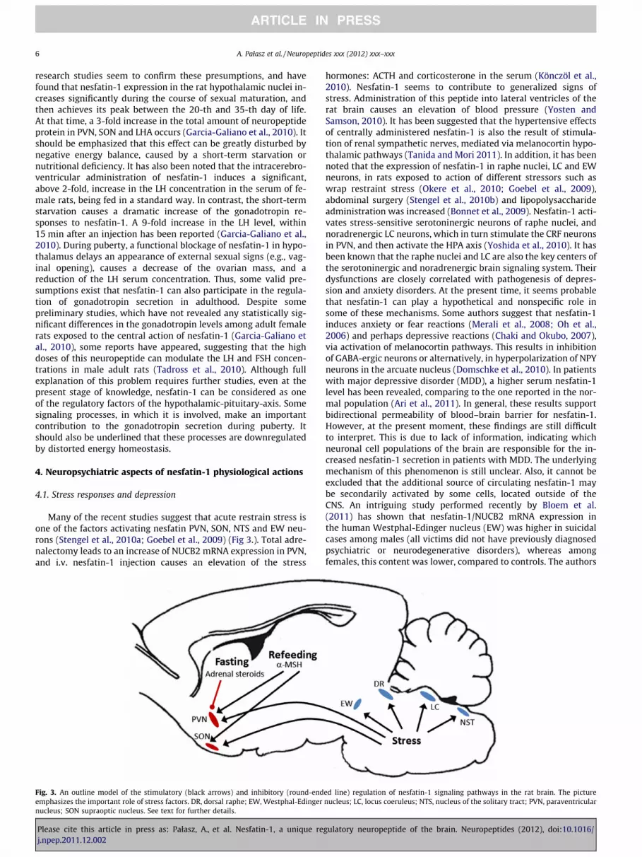
Many of the recent studies suggest that acute restrain stress isone of the factors activating nesfatin PVN, SON, NTS and EW neu-rons (Stengel et al., 2010a; Goebel et al., 2009) (Fig 3.). Total adre-nalectomy leads to an increase of NUCB2 mRNA expression in PVN,and i.v. nesfatin-1 injection causes an elevation of the stress
Fig. 3. An outline model of the stimulatory (black arrows) and inhibitory (round-endemphasizes the important role of stress factors. DR, dorsal raphe; EW, Westphal-Edingernucleus; SON supraoptic nucleus. See text for further details.
Please cite this article in press as: Pałasz, A., et al. Nesfatin-1, a unique rej.npep.2011.12.002
hormones: ACTH and corticosterone in the serum (Könczöl et al.,2010). Nesfatin-1 seems to contribute to generalized signs ofstress. Administration of this peptide into lateral ventricles of therat brain causes an elevation of blood pressure (Yosten andSamson, 2010). It has been suggested that the hypertensive effectsof centrally administered nesfatin-1 is also the result of stimula-tion of renal sympathetic nerves, mediated via melanocortin hypo-thalamic pathways (Tanida and Mori 2011). In addition, it has beennoted that the expression of nesfatin-1 in raphe nuclei, LC and EWneurons, in rats exposed to action of different stressors such aswrap restraint stress (Okere et al., 2010; Goebel et al., 2009),abdominal surgery (Stengel et al., 2010b) and lipopolysaccharideadministration was increased (Bonnet et al., 2009). Nesfatin-1 acti-vates stress-sensitive serotoninergic neurons of raphe nuclei, andnoradrenergic LC neurons, which in turn stimulate the CRF neuronsin PVN, and then activate the HPA axis (Yoshida et al., 2010). It hasbeen known that the raphe nuclei and LC are also the key centers ofthe serotoninergic and noradrenergic brain signaling system. Theirdysfunctions are closely correlated with pathogenesis of depres-sion and anxiety disorders. At the present time, it seems probablethat nesfatin-1 can play a hypothetical and nonspecific role insome of these mechanisms. Some authors suggest that nesfatin-1induces anxiety or fear reactions (Merali et al., 2008; Oh et al.,2006) and perhaps depressive reactions (Chaki and Okubo, 2007),via activation of melanocortin pathways. This results in inhibitionof GABA-ergic neurons or alternatively, in hyperpolarization of NPYneurons in the arcuate nucleus (Domschke et al., 2010). In patientswith major depressive disorder (MDD), a higher serum nesfatin-1level has been revealed, comparing to the one reported in the nor-mal population (Ari et al., 2011). In general, these results supportbidirectional permeability of blood–brain barrier for nesfatin-1.However, at the present moment, these findings are still difficultto interpret. This is due to lack of information, indicating whichneuronal cell populations of the brain are responsible for the in-creased nesfatin-1 secretion in patients with MDD. The underlyingmechanism of this phenomenon is still unclear. Also, it cannot beexcluded that the additional source of circulating nesfatin-1 maybe secondarily activated by some cells, located outside of theCNS. An intriguing study performed recently by Bloem et al.(2011) has shown that nesfatin-1/NUCB2 mRNA expression inthe human Westphal-Edinger nucleus (EW) was higher in suicidalcases among males (all victims did not have previously diagnosedpsychiatric or neurodegenerative disorders), whereas amongfemales, this content was lower, compared to controls. The authors
ed line) regulation of nesfatin-1 signaling pathways in the rat brain. The picturenucleus; LC, locus coeruleus; NTS, nucleus of the solitary tract; PVN, paraventricular
gulatory neuropeptide of the brain. Neuropeptides (2012), doi:10.1016/

A. Pałasz et al. / Neuropeptides xxx (2012) xxx–xxx 7
have also found nesfatin-1/NUCB2 and CART colocalization in theEW, and they were the first to show sex-related changes in theneuropeptides levels in the brain stem of suicide victims versuscontrols. These findings may suggest the existence of nesfatin-CART interplay in the midbrain. However, due to lack of otherevidence on this topic, they are still difficult to interpret. Undoubt-edly, the potential role of nesfatin-1 signaling in the pathogenesisof depressive-like behavior requires further study.
4.2. Eating disorders
Due to some anorexigenic properties of nesfatin-1, it seems jus-tified to conduct further research studies analyzing its potentialrole in pathogenesis of psychogenic eating disorders. Recently, ithas been noted that plasma nesfatin-1 levels in patients sufferingfrom restricting-type anorexia nervosa (AN-R) were significantlylower, compared with healthy persons. This may indicate a nega-tive correlation with ghrelin and des-acyl ghrelin levels. In con-trast, a positive correlation between nesfatin-1 levels and BMIwas demonstrated (Ogiso et al., 2011). An opposite phenomenonwas displayed in healthy men, with normal body mass index, inwhom the fasting nesfatin-1 concentration negatively correlatedwith their BMI (Tsuchiya et al., 2010). This observation was similarto the one reported in rats (Stengel et al., 2009). However, there isstill no convincing evidence that the low nesfatin-1 level underliesanxiety disorders, often accompanying AN-R. On the other hand, itcannot be excluded that during periods of extreme starvation, eventhe decreased nesfatin-1 level may reduce anxiety or fear, andstimulate food-intake. Unfortunately, a very limited number of re-search papers on this topic does not allow us to precisely interpretthese phenomena. It appears that some valuable data, contributingto this discussion, could be provided by brain imaging studies (PET,SPECT), using radiolabelled nesfatin-1.
4.3. Epilepsy
The initial interesting reports regarding correlations betweennesfatin-1 levels in body fluids and a course of epilepsy were pub-lished by Aydin et al. (2009). It was noted that the primary general-ized epilepsy (PGE) and secondary generalized seizures (SGE)significantly elevated nesfatin-1 levels in the patients’ plasmaand saliva. During treatment, there was a decrease of this neuro-peptide concentration, but nevertheless, it did not reach the levelwhich is typical for healthy individuals. The newest studies, con-ducted by the same authors support the previous data. They alsoindicate that the plasma nesfatin-1 level is in the highest rangeduring the 5 min after onset of the PGE and SGE attack. After that,it decreases significantly, and then, after 48 h, it decreases toapproximately 50% of its original level (Aydin et al., 2011). How-ever, the exact mechanisms of these changes have not been clearlyexplained. Some hypotheses have been formulated that the ele-vated nesfatin-1 level can induce some excitotoxic effects thatmay disturb certain activities of cortical and subcortical neuronalcircuitries. This may result in onset of epileptic symptoms.
It has also been found that the plasma nesfatin-1 level was sig-nificantly elevated in rats with epileptic seizures, induced by kainicacid. This suggests that the plasma level of this neuropeptide mayillustrate a profile of its fluctuation in hypothalamus. (Liu et al.,2011) Although this issue certainly requires further study, at thepresent time, nesfatin-1 can be considered as a valuable and prom-ising marker of epileptic episodes, being potentially useful in aneurological diagnostic work-up.
5. Concluding remarks
Nesfatin-1 is a neuropeptide characterized by a wide spectrum ofactivity in the CNS. There is some evidence to consider nesfatin-1 as
Please cite this article in press as: Pałasz, A., et al. Nesfatin-1, a unique rej.npep.2011.12.002
a promising factor which can be useful for clinical application. It canbe potentially helpful in the therapy of metabolic disorders, andobesity of various origins. Elevated plasma nesfatin-1 levels in pa-tients suffering from primary generalized epileptic seizures, sug-gests its potential use as a biomarker in epilepsy diagnostic work-up (Aydin et al., 2009). Increase in body mass and BMI among psy-chiatric patients taking antipsychotic, antidepressive, and otherpsychotropic medications represents a huge clinical problem. Inmany of these obese patients, a substantial reduction of sensitivityto leptin or some other adipokines may occur, in various hypotha-lamic centers (Heymsfield et al., 1999). It seems that in these cases,the use of nesfatin-1 can cause a substantial clinical improvement.Based on experimental studies, an intraperitoneal administrationof nesfatin-1 has significantly reduced food intake in leptin-resis-tant animals. In addition, nesfatin-1 has been found to reduce anelevated body mass of experimental animals. A possible relation-ship between nesfatin-1 action and appetite regulation in patientssuffering from chronic kidney disease (CKD) has also been cau-tiously postulated (Saldanha et al., 2011).
One possible way of administration of this agent is a subcutane-ous injection. However, a particularly interesting and beneficialtherapeutic view seems to be its intranasal administration. It hasbeen effective in some experimental animal models (Shimizu etal., 2009). Although the results of many experimental studies areencouraging, any possible clinical applications of nesfatin-1 still re-main in the area of speculation. Further research on this topic cer-tainly merits attention. In the mean time, answers to severalquestions, concerning pharmacokinetics, potential adverse drugreactions, tolerability profile and interactions with other medica-tions are required. Another potential research direction consistsof searching for selective nesfatin-1 antagonists that perhaps cancontribute to opening of a new chapter in therapy of eating disor-ders such as anorexia nervosa. However it should be emphasizedthat the nesfatin-1 receptor so far remains uncloned. Therefore,the further progress of pharmacological studies in this field is stillvery limited.
Acknowledgement
The authors would particularly like to acknowledge the contri-bution of Kate Rygiel, MD to the preparation of this article.
References
Ari, M., Ozturk, O.H., Bez, Y., Oktar, S., Erduran, D., 2011. High plasma nesfatin-1level in patients with major depressive disorder. Prog. Neuropsychopharmacol.Biol. Psychiatry 35, 497–500.
Atsuchi, K., Asakawa, A., Ushikai, M., Ataka, K., Tsai, M., Koyama, K., Sato, Y., Kato, I.,Fujimiya, M., Inui, A., 2010. Centrally administered nesfatin-1 inhibits feedingbehaviour and gastroduodenal motility in mice. NeuroReport 21, 1008–1011.
Aydin, S., Dag, E., Ozkan, Y., Arslan, O., Koc, G., Bek, S., Kirbas, S., Kasikci, T., Abasli, D.,Gokcil, Z., Odabasi, Z., Catak, Z., 2011. Time-dependent changes in the serumlevels of prolactin, nesfatin-1 and ghrelin as a marker of epileptic attacks inyoung male patients. Peptides 32, 1276–1280.
Aydin, S., Dag, E., Ozkan, Y., Erman, F., Dagli, A.F., Kilic, N., Sahin, I., Karatas, F.,Yoldas, T., Barim, A.O., Kendir, Y., 2009. Nesfatin-1 and ghrelin levels in serumand saliva of epileptic patients: hormonal changes can have a major effect onseizure disorders. Mol. Cell. Biochem. 328, 49–56.
Blevins, J.E., Schwartz, M.W., Baskin, D.G., 2004. Evidence that paraventricularnucleus oxytocin neurons link hypothalamic leptin action to caudal brain stemnuclei controlling meal size. Am. J. Physiol. Regul. Integr. Comp. Physiol. 287,R87–96.
Bloem, B., Xu, L., Morava, E., Faludi, G., Palkovits, M., Roubos, E.W., Kozicz, T., 2011. Sex-specific differences in the dynamics of cocaine and amphetamine-regulatedtranscript and nesfatin-1 expressions in the midbrain of depressed suicide victimsvs. controls. Neuropharmacology (doi:10.1016/j.neuropharm.2011.07.023).
Bonnet, M.S., Pecchi, E., Trouslard, J., Jean, A., Dallaporta, M., Troadec, J.D., 2009.Central nesfatin-1 expressing neurons are sensitive to peripheral imflammatorystimulus. J Neuroinflammation 6, 27.
Brailoiu, G.C., Dun, S.L., Brailoiu, E., Inan, S., Yang, J., Chang, J.K., Dun, N.J., 2007.Nesfatin-1: distribution and interaction with a G protein-coupled receptor inthe rat brain. Endocrinology 148, 5088–5094.
gulatory neuropeptide of the brain. Neuropeptides (2012), doi:10.1016/

8 A. Pałasz et al. / Neuropeptides xxx (2012) xxx–xxx
Chaki, S., Okubo, T., 2007. Melanocortin-4 receptor antagonists for the treatment ofdepression and anxiety disorders. Curr. Top. Med. Chem. 7, 1145–1151.
Connan, F., Lightman, S.L., Landau, S., Wheeler, M., Treasure, J., Campbell, I.C., 2007.An investigation of hypothalamic-pituitary-adrenal axis hyperactivity inanorexia nervosa: the role of CRH and AVP. J. Psychiatr. Res. 41, 131–143.
Domschke, K., Danniowski, U., Hohoff, C., Ohrmann, P., Bauer, J., Kugel, H., Zwanger,P., Heindel, W., Deckert, J., Arolt, V., Suslow, T., Baune, B.T., 2010. NeuropeptideY (NPY) gene: Impact on emotional processing and treatment response inanxious depression. Eur. Neuropsychopharmacol. 20, 301–309.
Foo, K.S., Brismar, H., Broberger, C., 2008. Distribution and neuropeptide coexistenceof nucleobindin-2 mRNA/nesfatin-like immunoreactivity in the rat CNS.Neuroscience 156, 563–579.
Foo, K.S., Brauner, H., Ostenson, C.G., Broberger, C., 2010. Nucleobindin-2/nesfatin inthe endocrine pancreas: distribution and relationship to glycaemic state. J.Endocrinol. 204, 255–263.
Fort, P., Salvert, D., Hanriot, L., Jego, S., Shimizu, H., Hashimoto, K., Mori, M., Luppi,P.H., 2008. The satiety molecule nesfatin-1 is co-expressed with melaninconcentrating hormone in tuberal hypothalamic neurons of the rat.Neuroscience 155, 174–181.
Garcia-Galiano, D., Navarro, V.M., Roa, J., Ruiz-Pino, F., Sanchez-Garrido, M.A.,Pineda, R., Castellano, J.M., Romero, M., Aguilar, E., Gaytan, F., Dieguez, C.,Pinilla, L., Tena-Sempere, M., 2010a. The anorexigenic neuropeptide, nesfatin-1,is indispensable for normal puberty onset in the female rat. J. Neurosci. 30,7783–7792.
Garcia-Galiano, D., Navarro, V.M., Gaytan, F., Tena-Sempere, M., 2010b. Expandingroles of NUCB2/nesfatin-1 in neuroendocrine regulation. J. Mol. Endocrinol. 45,281–290.
Goebel, M., Stengel, A., Wang, L., Lambrecht, N.W., Taché, Y., 2009. Nesfatin-1immunoreactivity in rat brain and spinal cord autonomic nuclei. Neurosci. Lett.452, 241–246.
Goebel-Stengel, M., Wang, L., Stengel, A., Tache, Y., 2011. Localization of nesfatin-1neurons in the mouse brain and functional implication. Brain Res. 1396, 20–34.
Gonzalez, R., Reingold, B.K., Gao, X., Gaidhu, M.P., Tsushima, R.G., Uniappan, S., 2011.Nesfatin-1 exerts a direct, glucose-dependent insulinotropic action on mouseislet b- and MIN6 cells. J. Endocrinol. 208, R9–R16.
Gonzalez, R., Kerbel, B., Chun, A., Uniappan, S., 2010. Molecular, cellular andphysiological evidences for anorexigenic actions of nesfatin-1 in goldfish. PLoSONE 5, e15201.
Heymsfield, S.B., Greenberg, A.S., Fujioka, K., Dixon, R.M., Kushner, R., Hunt, T.,Lubina, J.A., Patane, J., Self, B., Hunt, P., McCamish, M., 1999. Recombinant leptinfor weight loss in obese and lean adults: a randomized, controlled, dose-escalation trial. JAMA 282, 1568–1575.
Inhoff, T., Stengel, A., Peter, L., Goebel, M., Taché, Y., Bannert, N., Wiedenmann, B.,Klapp, B.F., Mönnikes, H., Kobelt, P., 2010. Novel insight in distribution ofnesfatin-1 and phospho-mTOR in the arcuate nucleus of the hypothalamus ofrats. Peptides 31, 257–262.
Inhoff, T., Mönnikes, H., Noetzel, S., Stengel, A., Goebel, M., Dinh, Q.T., Riedl, A.,Bannert, N., Wisser, A.S., Wiedenmann, B., Klapp, B.F., Taché, Y., Kobelt, P., 2008.Desacyl ghrelin inhibits the orexigenic effect of peripherally injected ghrelin inrats. Peptides 29, 2159–2168.
Iwasaki, Y., Nakabayashi, H., Kakei, M., Shimizu, H., Mori, M., Yada, T., 2009.Nesfatin-1 evokes Ca2+ signaling in isolated vagal afferent neurons via Ca2+influx through N-type channels. Biochem. Biophys. Res. Commun. 390, 958–962.
Liu, Z., Wang, F., Li, Z.Z., Qi, J.H., Xu, W.Z., Zhang, P.S., Sun, T., 2011. Expression ofneuropeptides ghrelin and nesfatin-1 in kainic acid kindling rats. Zhonghua. Yi.Xue. Za. Zhi. 91, 496–500.
Kishi, T., Aschkenasi, C.J., Lee, C.E., Mountjoy, K.G., Saper, C.B., Elmquist, J.K., 2003.Expression of melanocortin 4 receptor mRNA in the central nervous system ofthe rat. J. Comp. Neurol. 457, 213–235.
Kohno, D., Nakata, M., Maejima, Y., Shimizu, H., Sedbazar, U., Yoshida, N., Dezaki, K.,Onaka, T., Mori, M., Yada, T., 2008. Nesfatin-1 neurons in paraventricular andsupraoptic nuclei of the rat hypothalamus coexpress oxytocin and vasopressinand are activated by refeeding. Endocrinology 149, 1295–1301.
Könczöl, K., Bodnar, I., Zelena, D., Pinter, O., Papp, R.S., Palkovits, M., Nagy, G.M.,Toth, Z.E., 2010. Nesfatin-1/NUCB2 may participate in the activation of thehypothalamic-pituitary-adrenal axis in rats. Neurochem. Int. 53, 189–197.
Maejima, Y., Sedbazar, U., Suyama, S., Kohno, D., Onaka, T., Takano, E., Yoshida, N.,Koike, M., Uchiyama, Y., Fujiwara, K., Yashiro, T., Horvath, T.L., Dietrich, M.O.,Tanaka, S., Dezaki, K., Oh-I, S., Hashimoto, K., Shimizu, H., Nakata, M., Mori, M.,Yada, T., 2009. Nesfatin-1-regulated oxytocinergic signaling in theparaventricular nucleus causes anorexia through a leptin-independentmelanocortin pathway. Cell Metab. 10, 355–365.
Merali, Z., Cayer, C., Kent, P., Anisman, H., 2008. Nesfatin-1 increases anxiety- andfear-related behaviors in the rat. Psychopharmacology (Berl) 201, 115–123.
Nakata, M., Manaka, K., Yamamoto, S., Mori, M., Yada, T., 2011. Nesfatin-1 enhancesglucose-induced insulin secretion by promoting Ca2+ influx through L-typechannels in mouse b-cells. Endocr. J. 58, 305–313.
Noetzel, S., Stengel, A., Inhoff, T., Goebel, M., Wisser, A.S., Bannert, N., Wiedenmann,B., Klapp, B.F., Tache, Y., Mönnikes, H., Kobelt, P., 2009. CCK-8S activates c-Fos ina dose-dependent manner in nesfatin-1 immunoreactive neurons in theparaventricular nucleus of the hypothalamus and in the nucleus of thesolitary tract in the brain stem. Regul. Pept. 157, 84–91.
Oh, I.S., Shimizu, H., Satoh, T., Okada, S., Adachi, S., Inoue, K., Eguchi, H., Yamamoto,M., Imaki, T., Hashimoto, K., Tsuchiya, T., Monden, T., Horiguchi, K., Yamada, M.,
Please cite this article in press as: Pałasz, A., et al. Nesfatin-1, a unique rej.npep.2011.12.002
Mori, . Identification of nesfatin-1 as a satiety molecule in the hypothalamus.Nature 443, 709–712.
Ogiso, K., Asakawa, A., Amitani, H., Nakahara, T., Ushikai, M., Haruta, I., Koyama, K.-I., Amitani, M., Harada, T., Yasuhara, D., Inui, A., 2011. Plasma nesfatin-1concentrations in restricting-type anorexia nervosa. Peptides 32, 150–153.
Okere, B., Xu, L., Roubos, E.W., Sonetti, D., Kozicz, T., 2010. Restraint stress alters thesecretory activity of neurons co-expressing urocortin-1, cocaine- andamphetamine-regulated transcript peptide and nesfatin-1 in the mouseEdinger-Westphal nucleus. Brain Res. 1317, 92–99.
Ozaki, Y., Onaka, T., Nakazato, M., Saito, J., Kanemoto, K., Matsumoto, T., Ueta, Y.,2002. Centrally administered neuromedin U activates neurosecretion andinduction of c-fos messenger ribonucleic acid in the paraventricular andsupraoptic nuclei of rat. Endocrinology 143, 4320–4329.
Özsavci, D., Ersahin, M., Sener, A., Özakpinar, Ö.B., Toklu, H.Z., Akakin, D., Sener, G.,Yegen, B.C., 2011. The novel function of nesfatin-1 as an anti-inflammatory andantiapoptotic peptide in subarachnoid hemorrhage-induced oxidative braindamage in rats. Neurosurgery 68, 1699–1708.
Pałasz, A., Bryzek, A., Krzystanek, M., Wiaderkiewicz, R., 2010. Anoreksygennaaktywnosc nesfatyny-1 w jadrach podwzgórza i jej potencjalne implikacjekliniczne/Anorexigenic activity of nesfatin-1 in hypothalamic nuclei and itspotential clinical implications. Farmakoter. Psychiatr. Neurol. 26, 39–43.
Pan, W., Hsuchou, H., Kastin, A.J., 2007. Nesfatin-1 crosses the blood-brain barrierwithout saturation. Peptides 28, 2223–2228.
Price, C.J., Samson, W.K., Ferguson, A.V., 2008. Nesfatin-1 inhibits NPY neurons inthe arcuate nucleus. Brain Res. 1230, 99–106.
Ramanjaneya, M., Chen, J., Brown, J.E., Tripathi, G., Hallschmid, M., Patel, S., Kern, W.,Hillhouse, E.W., Lehnert, H., Tan, B.K., Randeve, H.S., 2010. Identification ofnesfatin-1 in human and murine adipose tissue: a novel depot-specificadipokine with increased levels in obesity. Endocrinology 151, 3169–3180.
Sabatier, N., 2006. Alpha-Melanocyte-stimulating hormone and oxytocin: a peptidesignalling cascade in the hypothalamus. J. Neuroendocrinol. 18, 703–710.
Saldanha, J.F., Carrero, J.J., Mafra, D., 2011. The possible role of nesfatin-1 onappetite regulation in hemodialysis patients. Med. Hypotheses (doi:10.1016/j.mehy.2011.07.006).
Stengel, A., Goebel, M., Wang, L., Rivier, J., Kobelt, P., Mönnikes, H., Lambrecht, N.W.,Taché, Y., 2009. Central nesfatin-1 reduces dark-phase food intake and gastricemptying in rats: differential role of corticotropin-releasing factor2 receptor.Endocrinology 150, 4911–4919.
Stengel, A., Goebel, M., Wang, L., Tache, Y., 2010a. Ghrelin, des-acyl ghrelin andnesfatin-1 in gastric X/A-like cells: role as regulators of food intake and bodyweight. Peptides 31, 357–369.
Stengel, A., Goebel, M., Tache, Y., 2010b. Nesfatin-1: a novel inhibitory regulator offood intake and body weight. Obesity Rev. 12, 261–271.
Stengel, A., Goebel, M., Wang, L., Tache, Y., 2010c. Abdominal surgery activatesnesfatin-1 immunoreactive brain nuclei in rats. Peptides 31, 263–270.
Stengel, A., Tache, Y., 2010. Nesfatin-1: role as a possible new potent regulator offood intake. Regul. Pept. 9, 18–23.
Stengel, A., Tache, Y., 2011. Minireview: Nesfatin-1 an emerging new player in thebrain-gut, endocrine and metabolic axis. Endocrinology (doi:10.1210/en.2011-1500).
Shimizu, H., Oh, I.S., Okada, S., Mori, M., 2009. Nesfatin-1: an overview and futureclinical application. Endocr. J. 56, 537–543.
Singru, P.S., Sánchez, E., Fekete, C., Lechan, R.M., 2007. Importance of melanocortinsignaling in refeeding-induced neuronal activation and satiety. Endocrinology148, 638–646.
Su, Y., Zhang, J., Tang, Y., Bi, F., Liu, J.N., 2010. The novel function of nesfatin-1: anti-hyperglycemia. Biochem. Biophys. Res. Commun. 391, 1039–1042.
Tadross, J.A., Patterson, M., Wynne, K.J., Patel, S., Suzuki, K., Ghatei, M.A., Bloom, S.R.,2010. Nesfatin supresses feeding and stimulates the hypothalamo-pituitary-gonadal axis. Endocr. J. 57, S355–S648.
Tan, B.K., Hallschmid, M., Kern, W., Lehnert, H., Randeva, H.S., 2011. Decreasedcerebrospinal fluid/plasma ratio of the novel satiety molecule, nesfatin-1/NUCB-2 in obese humans: evidence of nesfatin-1/NUCB-2 resistance andimplications for obesity treatment. J. Clin. Endocrinol. Metab. 96, E669–E673.
Tanida, M., Mori, M., 2011. Nesfatin-1 stimulates renal sympathetic nerve activity inrats. NeuroReport 22, 309–312.
Tsuchiya, T., Shimizu, H., Yamada, M., Osaki, A., Oh, I.S., Ariyama, Y., Takahashi, H.,Okada, S., Hashimoto, K., Satoh, T., Kojima, M., Mori, M., 2010. Fastingconcentrations of nesfatin-1 are negatively correlated with body mass indexin non-obese males. Clin. Endocrinol. 73, 484–490.
Yoshida, N., Maejima, Y., Sedbazar, U., Ando, A., Kurita, H., Damdindorj, B., Takano,E., Gantulga, D., Iwasaki, Y., Kurashina, T., Onaka, T., Dezaki, K., Nakata, M., Mori,M., Yada, T., 2010. Stressor-responsive central nesfatin-1 activatescorticotropin-releasing hormone, noradrenaline and serotonin neurons andevokes hypothalamic-pituitary-adrenal axis. Aging 2, 775–784.
Yosten, G.L.C., Samson, W.K., 2009. Nesfatin-1 exerts cardiovascular actions inbrain: possible interaction with the central melanocortin system. Am. J. Physiol.Regul. Integr. Comp. Physiol. 297, R1330–R1336.
Yosten, G.L.C., Samson, W.K., 2010. The anorexigenic and hypertensive effects ofnesfatin-1 are reversed by pretreatment with an oxytocin receptor antagonist.Am. J. Physiol. Regul. Integr. Comp. Physiol. 298, R1642–R1647.
Zhang, A.Q., Li, X.L., Jiang, C.Y., Lin, L., Shi, R.H., Chen, J.D., Oomura, Y., 2010.Expression of nesfatin-1/NUCB2 in rodent digestive system. World J.Gastroenterol. 16, 1735–1741.
gulatory neuropeptide of the brain. Neuropeptides (2012), doi:10.1016/


