Embed Size (px)
Citation preview

NEURAL IMPLEMENTATIONS OF EXPERTISEEDITED BY : Merim Bilalić, Robert Langner, Guillermo Campitelli,
Luca Turella and Wolfgang GroddPUBLISHED IN : Frontiers in Human Neuroscience

1 | Neural Implementations of ExpertiseFrontiers in Human Neuroscience
Frontiers Copyright Statement
© Copyright 2007-2015 Frontiers Media SA. All rights reserved.
All content included on this site, such as text, graphics, logos, button
icons, images, video/audio clips, downloads, data compilations and
software, is the property of or is licensed to Frontiers Media SA
(“Frontiers”) or its licensees and/or subcontractors. The copyright in the
text of individual articles is the property of their respective authors, subject to
a license granted to Frontiers.
The compilation of articles constituting this e-book, wherever published,
as well as the compilation of all other content on this site, is the exclusive
property of Frontiers. For the conditions for downloading and
copying of e-books from Frontiers’ website, please see the Terms for
Website Use. If purchasing Frontiers e-books from other websites
or sources, the conditions of the website concerned apply.
Images and graphics not forming part of user-contributed materials may
not be downloaded or copied without permission.
Individual articles may be downloaded and reproduced in accordance
with the principles of the CC-BY licence subject to any copyright or
other notices. They may not be re-sold as an e-book.
As author or other contributor you grant a CC-BY licence to others to
reproduce your articles, including any graphics and third-party materials
supplied by you, in accordance with the Conditions for Website Use and
subject to any copyright notices which you include in connection with your
articles and materials.
All copyright, and all rights therein, are protected by national and
international copyright laws.
The above represents a summary only. For the full conditions see the
Conditions for Authors and the Conditions for Website Use.
ISSN 1664-8714 ISBN 978-2-88919-688-3
DOI 10.3389/978-2-88919-688-3
About Frontiers
Frontiers is more than just an open-access publisher of scholarly articles: it is a pioneering approach to the world of academia, radically improving the way scholarly research is managed. The grand vision of Frontiers is a world where all people have an equal opportunity to seek, share and generate knowledge. Frontiers provides immediate and permanent online open access to all its publications, but this alone is not enough to realize our grand goals.
Frontiers Journal Series
The Frontiers Journal Series is a multi-tier and interdisciplinary set of open-access, online journals, promising a paradigm shift from the current review, selection and dissemination processes in academic publishing. All Frontiers journals are driven by researchers for researchers; therefore, they constitute a service to the scholarly community. At the same time, the Frontiers Journal Series operates on a revolutionary invention, the tiered publishing system, initially addressing specific communities of scholars, and gradually climbing up to broader public understanding, thus serving the interests of the lay society, too.
Dedication to Quality
Each Frontiers article is a landmark of the highest quality, thanks to genuinely collaborative interactions between authors and review editors, who include some of the world’s best academicians. Research must be certified by peers before entering a stream of knowledge that may eventually reach the public - and shape society; therefore, Frontiers only applies the most rigorous and unbiased reviews. Frontiers revolutionizes research publishing by freely delivering the most outstanding research, evaluated with no bias from both the academic and social point of view.By applying the most advanced information technologies, Frontiers is catapulting scholarly publishing into a new generation.
What are Frontiers Research Topics?
Frontiers Research Topics are very popular trademarks of the Frontiers Journals Series: they are collections of at least ten articles, all centered on a particular subject. With their unique mix of varied contributions from Original Research to Review Articles, Frontiers Research Topics unify the most influential researchers, the latest key findings and historical advances in a hot research area! Find out more on how to host your own Frontiers Research Topic or contribute to one as an author by contacting the Frontiers Editorial Office: [email protected]
December 2015

2 | Neural Implementations of ExpertiseFrontiers in Human Neuroscience
When we think about expertise, we usually consider people who master tasks at a level not reachable by most other people. Although we rarely realise it, however, most humans are experts in many aspects of everyday life. This expertise enables us to find our way through a complex environment that is our life. For instance, we can instantly recognise multiple objects and rela-tions between them to form a meaningful unit, such as an office. Thus, research on expertise is not only important to investigate the cognitive and neural processes within an “elite” group, but it is also a powerful tool to understand how everyone can acquire complex skills.
The goal of this RESEARCH TOPIC is to shed further light on the common and distinct neural mechanisms that implement various kinds of expertise. We broadly define expertise as skill in any perceptual, cognitive, social or motor domain, with the common core being optimised information processing due to knowledge acquired from repeated experiences. Thus, we are interested in the full range of mental processes modulated or modified by expertise, from “simple” object or pattern recognition to complex decision making or problem solving in a particular domain. These domains can range from everyday or occupational expertise to sports and rather artificial domains such as board games. In all cases, the aim should be to elucidate how the brain implements these sometimes incredible feats. We are particularly interested in connecting cognitive theories about expertise and expertise-related performance differences with models and data on the neural implementation of expertise. We welcome original research contributions using the full range of behavioural neuroscience methods, as well as theoretical, methodological or historical reviews, and opinion papers focusing on any of the above-mentioned aspects.
Citation: Bilalic, M., Langner, R., Campitelli, C., Turella, L., Grodd, W., eds. (2015). Neural Implementations of Expertise. Lausanne: Frontiers Media. doi: 10.3389/978-2-88919-688-3
NEURAL IMPLEMENTATIONS OF EXPERTISE
Topic Editors: Merim Bilalić, Alpen Adria University Klagenfurt, AustriaRobert Langner, Heinrich Heine University Düsseldorf, GermanyGuillermo Campitelli, Edith Cowan University, AustraliaLuca Turella, University of Trento, ItalyWolfgang Grodd, University Hospital Aachen, Germany
December 2015
3 | Neural Implementations of ExpertiseFrontiers in Human Neuroscience
Table of Contents
05 Editorial: Neural implementation of expertiseMerim Bilalic, Robert Langner, Guillermo Campitelli, Luca Turella and Wolfgang Grodd
08 Expertise and processing distorted structure in chessJames C. Bartlett, Amy L. Boggan and Daniel C. Krawczyk
19 Exploring the brains of Baduk (Go) experts: gray matter morphometry, resting-state functional connectivity, and graph theoretical analysisWi Hoon Jung, Sung Nyun Kim, Tae Young Lee, Joon Hwan Jang, Chi-Hoon Choi, Do-Hyung Kang and Jun Soo Kwon
36 Brain regions concerned with the identification of deceptive soccer moves by higher-skilled and lower-skilled playersMichael J. Wright, Daniel T. Bishop, Robin C. Jackson and Bruce Abernethy
51 Expertise in action observation: recent neuroimaging findings and future perspectivesLuca Turella, Moritz F. Wurm, Raffaele Tucciarelli and Angelika Lingnau
56 Reorganization and plastic changes of the human brain associated with skill learning and expertiseYongmin Chang
63 Experts bodies, experts minds: How physical and mental training shape the brainUrsula Debarnot, Marco Sperduti, Franck Di Rienzo and Aymeric Guillot
80 Corrigendum: Experts bodies, experts minds: how physical and mental training shape the brainUrsula Debarnot, Marco Sperduti, Franck Di Rienzo and Aymeric Guillot
81 Melodic multi-feature paradigm reveals auditory profiles in music-sound encodingMari Tervaniemi, Minna Huotilainen and Elvira Brattico
91 Music practice is associated with development of working memory during childhood and adolescenceSissela Bergman Nutley, Fahimeh Darki and Torkel Klingberg
100 Neural implementation of musical expertise and cognitive transfers: could they be promising in the framework of normal cognitive aging?Baptiste Fauvel, Mathilde Groussard, Francis Eustache, Béatrice Desgranges and Hervé Platel
107 Neurophysiological constraints on the eye-mind linkErik D. Reichle and Eyal M. Reingold
December 2015
4 | Neural Implementations of ExpertiseFrontiers in Human Neuroscience
113 Assimilation of L2 vowels to L1 phonemes governs L2 learning in adulthood: a behavioral and ERP studyMirko Grimaldi, Bianca Sisinni, Barbara Gili Fivela, Sara Invitto, Donatella Resta, Paavo Alku and Elvira Brattico
127 Training of ultra-fast speech comprehension induces functional reorganization of the central-visual system in late-blind humansSusanne Dietrich, Ingo Hertrich and Hermann Ackermann
142 Neural correlates of verbal creativity: differences in resting-state functional connectivity associated with expertise in creative writingMartin Lotze, Katharina Erhard, Nicola Neumann, Simon B. Eickhoff and Robert Langner
150 Do neural correlates of face expertise vary with task demands? Event-related potential correlates of own- and other-race face inversionHolger Wiese
164 Stimulus familiarity modulates functional connectivity of the perirhinal cortex and anterior hippocampus during visual discrimination of faces and objectsVictoria C. McLelland, David Chan, Susanne Ferber and Morgan D. Barense
180 Beyond perceptual expertise: revisiting the neural substrates of expert object recognitionAssaf Harel, Dwight Kravitz and Chris I. Baker
192 Holding a stick at both ends: on faces and expertiseAssaf Harel, Dwight J. Kravitz and Chris I. Baker
194 Interaction between perceptual and cognitive processing well acknowledged in perceptual expertise researchAlan C.-N. Wong and Yetta K. Wong
197 Expertise paradigms for investigating the neural substrates of stable memoriesGuillermo Campitelli and Craig Speelman
201 Sparse distributed memory: understanding the speed and robustness of expert memoryMarcelo S. Brogliato, Daniel M. Chada and Alexandre Linhares
212 Functional cerebral reorganization: a signature of expertise? Reexamining Guida, Gobet, Tardieu, and Nicolas’ (2012) two-stage frameworkAlessandro Guida, Fernand Gobet and Serge Nicolas
217 Task decomposition: a framework for comparing diverse training models in human brain plasticity studiesEmily B. J. Coffey and Sibylle C. Herholz
223 The neural circuitry of expertise: perceptual learning and social cognitionMichael Harré
December 2015

EDITORIALpublished: 30 September 2015
doi: 10.3389/fnhum.2015.00545
Frontiers in Human Neuroscience | www.frontiersin.org September 2015 | Volume 9 | Article 545
Edited and reviewed by:
Srikantan S. Nagarajan,
University of California, San Francisco,
USA
*Correspondence:
Merim Bilalic
Received: 09 June 2015
Accepted: 17 September 2015
Published: 30 September 2015
Citation:
Bilalic M, Langner R, Campitelli G,
Turella L and Grodd W (2015) Editorial:
Neural implementation of expertise.
Front. Hum. Neurosci. 9:545.
doi: 10.3389/fnhum.2015.00545
Editorial: Neural implementation ofexpertise
Merim Bilalic 1*, Robert Langner 2, Guillermo Campitelli 3, Luca Turella 4 and
Wolfgang Grodd 5
1Department of Cognitive Psychology, Alps Adria University Klagenfurt, Klagenfurt, Austria, 2Clinical Neuroscience and
Medical Psychology, Heinrich Heine University Düsseldorf, Düsseldorf, Germany, 3 School of Psychology and Social Science,
Edith Cowan University, Perth, WA, Australia, 4Center for Mind/Brain Sciences, University of Trento, Trento, Italy,5Department of Magnetic Resonance, Max Planck Institute for Biological Cybernetics, Tuebingen, Germany
Keywords: expertise, neurosciences, sports, music, chess, go, language, perception
How the brain enables humans to reach an outstanding level of performance typical of expertiseis of great interest to cognitive neuroscience, as demonstrated by the number and diversity of thearticles in this Research Topic (RT). The RT presents a collection of 23 articles written by 80 authorson traditional expertise topics such as sport, board games, and music, but also on the expertiseaspects of everyday skills, such as language and the perception of faces and objects. Just as thetopics in the RT are diverse, so are the neuroimaging techniques employed and the article formats.Here we will briefly summarize the articles published in the RT.
Board Games
The traditional expertise domain of board games has been covered in the RT by two articles,both employing the expertise approach of pitting experts against novices (Bilalic et al., 2010, 2012,2014) but employing differing neuroimaging techniques. Bartlett et al. (2013) employed fMRI todemonstrate that chess experts engage the fronto-parietal network when they try to find a logicalpattern in a “constellation” of randomly placed chess pieces. Jung et al. (2013) found structuraldifferences as well as differences in brain networks between Baduk (Korean name for the boardgame Go) experts and novices, which point out the importance of visuospatial processing inproblem solving and decision making of board-game experts.
Sport
Wright et al. (2013) extended the research on anticipation of action in sport by showing thatthe neural basis for deception involves, besides the well-known action observation network, thestructures responsible for social cognition and affection. Turella et al. (2013) review other recentstudies on the anticipation of action in sport and connect them with the mirror neurons in animalresearch. The review by Chang (2014) deals with motor domains such as sports and music andthe structural and functional changes associated with expertise. Debarnot et al. (2014) go a stepfurther in their review and contrast the neural changes during skill acquisition with those in mentaltraining techniques such motor imagery and mediation.
Music
Music has been one of the most often investigated domains in expertise because its complexityand richness enable researchers to tackle diverse topics. The variety of themes in the domainof music is also evident in this RT. Tervaniemi et al. (2014), for example, pitted expert
5|

Bilalic et al. Editorial: Neural implementation of expertise
musicians against novices in a novel paradigm to investigatememory and attentional processes with EEG. On the otherhand, Bergman Nutley et al. (2014) used the music domainto investigate longitudinal effects on cognitive processes suchas working memory, speed of processing, and reasoning, whileFauvel et al. (2013) apply the promising findings of transfer andneural plasticity associated with musical practice to cognitiveaging in their review.
Language
Unlike the previous articles, which deal with specialized expertisedomains, a number of contributions highlight the fact thateven the everyday skills we often take for granted representimpressive feats of human expertise. One group of articles dealswith language, which is one such everyday skill. Reichle andReingold (2013) review the electrophysiological evidence of thelink between eye movements and the mind during reading.The learning of a second language based on its similarity toone’s native language was investigated by Grimaldi et al. (2014),while Dietrich et al. (2013) demonstrated the neural changesassociated with the process of learning to comprehend speechthat was several times faster than normal speech. Finally, Lotzeet al. (2014) demonstrate by means of resting-state fMRI thatpeople who write highly creatively have increased functionalconnectivity between the task-related brain regions in the righthemisphere but reduced interhemispheric connectivity.
Perception
Similarly, a couple of articles deal with perception of own-raceand other-race faces (Wiese, 2013) as well as with perception offamiliar faces and objects and the functional connectivity withinthe medial temporal lobe (McLelland et al., 2014). The role ofthe fusiform face area (FFA) in expertise has been a bone of
contention between Harel et al. (2013, 2014), on the one hand,and Wong and Wong (2014), on the other.
Theoretical and Simulation Work
Finally, a number of articles provide either new theoreticalideas or revisions of already established theories. Campitelli
and Speelman (2013) highlight the advantages of using theexpertise paradigm in investigating memory, while Brogliatoet al. (2014) expand the Sparse Distributed Memory (SDM)model to incorporate the effects of practice on memory retrieval.Guida et al. (2013) extend their two-stage framework of skillacquisition (Guida et al., 2012) by arguing for the functionalcerebral reorganization (FCR) as being the neural signature ofexpertise. The way one structures training studies is consideredby Coffey and Herholz (2013), who suggest a new approachfor characterizing and deconstructing the task requirements intraining studies. Finally, Harré (2013) demonstrates the parallelsbetween two seemingly unrelated fields, perceptual expertise andsocial cognition.
Conclusion
It is clear that we cannot do justice to all submissions inthis brief editorial. We hope, however, that our brief summarydemonstrates the diversity in topics and methods employed inresearch on human expertise and also, indirectly, the growinginterest in the field of expertise. It should become evident thatresearch on expertise is not only relevant for understandingexceptional human performance but also for understanding howmind and brain work more generally. We are grateful to allauthors for their contribution and hope that the RT, with its broadand deep coverage, will provide a useful reference for the readerinterested in expertise and, particularly, current approaches to itsneural implementation.
References
Bartlett, J., Boggan, A. L., and Krawczyk, D. C. (2013). Expertise and
processing distorted structure in chess. Front. Hum. Neurosci. 7:825. doi:
10.3389/fnhum.2013.00825
Bergman Nutley, S., Darki, F., and Klingberg, T. (2014). Music practice
is associated with development of working memory during childhood
and adolescence. Front. Hum. Neurosci. 7:926. doi: 10.3389/fnhum.2013.
00926
Bilalic, M., Grottenthaler, T., Nägele, T., and Lindig, T. (2014). The faces in
radiological images: fusiform face area supports radiological expertise. Cereb.
Cortex doi: 10.1093/cercor/bhu272. [Epub ahead of print]. Available online at:
http://cercor.oxfordjournals.org/content/early/2014/12/01/cercor.bhu272
Bilalic, M., Langner, R., Erb, M., and Grodd, W. (2010). Mechanisms and neural
basis of object and pattern recognition: a study with chess experts. J. Exp.
Psychol. Gen. 139, 728–742. doi: 10.1037/a0020756
Bilalic, M., Turella, L., Campitelli, G., Erb, M., and Grodd, W. (2012). Expertise
modulates the neural basis of context dependent recognition of objects and
their relations. Hum. Brain Mapp. 33, 2728–2740. doi: 10.1002/hbm.21396
Brogliato,M. S., Chada, D.M., and Linhares, A. (2014). Sparse distributedmemory:
understanding the speed and robustness of expert memory. Front. Hum.
Neurosci. 8:222. doi: 10.3389/fnhum.2014.00222
Campitelli, G., and Speelman, C. (2013). Expertise paradigms for investigating
the neural substrates of stable memories. Front. Hum. Neurosci. 7:740. doi:
10.3389/fnhum.2013.00740
Chang, Y. (2014). Reorganization and plastic changes of the human brain
associated with skill learning and expertise. Front. Hum. Neurosci. 8:35. doi:
10.3389/fnhum.2014.00035
Coffey, E. B. J., and Herholz, S. C. (2013). Task decomposition: a framework for
comparing diverse training models in human brain plasticity studies. Front.
Hum. Neurosci. 7:640. doi: 10.3389/fnhum.2013.00640
Debarnot, U., Sperduti, M., Di Rienzo, F., and Guillot, A. (2014). Experts bodies,
experts minds: how physical and mental training shape the brain. Front. Hum.
Neurosci. 8:280. doi: 10.3389/fnhum.2014.00280
Dietrich, S., Hertrich, I., and Ackermann, H. (2013). Training of ultra-fast
speech comprehension induces functional reorganization of the central-
visual system in late-blind humans. Front. Hum. Neurosci. 7:701. doi:
10.3389/fnhum.2013.00701
Fauvel, B., Groussard, M., Eustache, F., Desgranges, B., and Platel, H. (2013).
Neural implementation of musical expertise and cognitive transfers: could
they be promising in the framework of normal cognitive aging? Front. Hum.
Neurosci. 7:693. doi: 10.3389/fnhum.2013.00693
Grimaldi, M., Sisinni, B., Gili Fivela, B., Invitto, S., Resta, D., Alku, P., et al.
(2014). Assimilation of L2 vowels to L1 phonemes governs L2 learning in
Frontiers in Human Neuroscience | www.frontiersin.org September 2015 | Volume 9 | Article 545 6|

Bilalic et al. Editorial: Neural implementation of expertise
adulthood: a behavioral and ERP study. Front. Hum. Neurosci. 8:279. doi:
10.3389/fnhum.2014.00279
Guida, A., Gobet, F., and Nicolas, S. (2013). Functional cerebral reorganization:
a signature of expertise? Reexamining Guida, Gobet, Tardieu, and
Nicolas’ (2012) two-stage framework. Front. Hum. Neurosci. 7:590. doi:
10.3389/fnhum.2013.00590
Guida, A., Gobet, F., Tardieu, H., and Nicolas, S. (2012). How chunks, long-term
working memory and templates offer a cognitive explanation for neuroimaging
data on expertise acquisition: a two-stage framework. Brain Cogn. 79, 221–244.
doi: 10.1016/j.bandc.2012.01.010
Harel, A., Kravitz, D., and Baker, C. I. (2013). Beyond perceptual expertise:
revisiting the neural substrates of expert object recognition. Front. Hum.
Neurosci. 7:885. doi: 10.3389/fnhum.2013.00885
Harel, A., Kravitz, D. J., and Baker, C. I. (2014). Holding a stick at
both ends: on faces and expertise. Front. Hum. Neurosci. 8:442. doi:
10.3389/fnhum.2014.00442
Harré, M. (2013). The neural circuitry of expertise: perceptual learning and social
cognition. Front. Hum. Neurosci. 7:852. doi: 10.3389/fnhum.2013.00852
Jung, W. H., Kim, S. N., Lee, T. Y., Jang, J. H., Choi, C.-H., Kang, D.-H., et al.
(2013). Exploring the brains of Baduk (Go) experts: gray matter morphometry,
resting-state functional connectivity, and graph theoretical analysis. Front.
Hum. Neurosci. 7:633. doi: 10.3389/fnhum.2013.00633
Lotze, M., Erhard, K., Neumann, N., Eickhoff, S. B., and Langner, R. (2014).
Neural correlates of verbal creativity: differences in resting-state functional
connectivity associated with expertise in creative writing. Front. Hum. Neurosci.
8:516. doi: 10.3389/fnhum.2014.00516
McLelland, V. C., Chan, D., Ferber, S., and Barense, M. D. (2014). Stimulus
familiarity modulates functional connectivity of the perirhinal cortex and
anterior hippocampus during visual discrimination of faces and objects. Front.
Hum. Neurosci. 8:117. doi: 10.3389/fnhum.2014.00117
Reichle, E. D., and Reingold, E. M. (2013). Neurophysiological constraints on the
eye-mind link. Front. Hum. Neurosci. 7:361. doi: 10.3389/fnhum.2013.00361
Tervaniemi, M., Huotilainen, M., and Brattico, E. (2014). Melodic multi-feature
paradigm reveals auditory profiles in music-sound encoding. Front. Hum.
Neurosci. 8:496. doi: 10.3389/fnhum.2014.00496
Turella, L., Wurm, M. F., Tucciarelli, R., and Lingnau, A. (2013). Expertise
in action observation: recent neuroimaging findings and future perspectives.
Front. Hum. Neurosci. 7:637. doi: 10.3389/fnhum.2013.00637
Wiese, H. (2013). Do neural correlates of face expertise vary with task demands?
Event-related potential correlates of own- and other-race face inversion. Front.
Hum. Neurosci. 7:898. doi: 10.3389/fnhum.2013.00898
Wong, A. C.-N., and Wong, Y. K. (2014). Interaction between perceptual and
cognitive processing well acknowledged in perceptual expertise research. Front.
Hum. Neurosci. 8:308. doi: 10.3389/fnhum.2014.00308
Wright, M. J., Bishop, D. T., Jackson, R. C., and Abernethy, B. (2013). Brain
regions concerned with the identification of deceptive soccer moves by
higher-skilled and lower-skilled players. Front. Hum. Neurosci. 7:851. doi:
10.3389/fnhum.2013.00851
Conflict of Interest Statement: The authors declare that the research was
conducted in the absence of any commercial or financial relationships that could
be construed as a potential conflict of interest.
Copyright © 2015 Bilalic, Langner, Campitelli, Turella and Grodd. This is an open-
access article distributed under the terms of the Creative Commons Attribution
License (CC BY). The use, distribution or reproduction in other forums is permitted,
provided the original author(s) or licensor are credited and that the original
publication in this journal is cited, in accordance with accepted academic practice.
No use, distribution or reproduction is permitted which does not comply with these
terms.
Frontiers in Human Neuroscience | www.frontiersin.org September 2015 | Volume 9 | Article 545 7|

ORIGINAL RESEARCH ARTICLEpublished: 03 December 2013
doi: 10.3389/fnhum.2013.00825
Expertise and processing distorted structure in chessJames C. Bartlett1*, Amy L. Boggan2 and Daniel C. Krawczyk1,3
1 Program in Cognition and Neuroscience, School of Behavioral and Brain Sciences, The University of Texas at Dallas, Richardson, TX, USA2 Department of Psychology, Young Harris College, Young Harris, GA, USA3 Department of Psychiatry, The University of Texas Southwestern Medical Center, Dallas, TX, USA
Edited by:Merim Bilalic, University of TübingenClinic, Germany
Reviewed by:Guillermo Campitelli, Edith CowanUniversity, AustraliaFernand Gobet, University ofLiverpool, UK
*Correspondence:James C. Bartlett, School ofBehavioral and Brain Sciences, TheUniversity of Texas at Dallas, 800 WCampbell Rd., Richardson, TX75083-0688, USAe-mail: [email protected]
A classic finding in research on human expertise and knowledge is that of enhancedmemory for stimuli in a domain of expertise as compared to either stimuli outside thatdomain, or within-domain stimuli that have been degraded or distorted in some way.However, we do not understand how experts process degradation or distortion of stimuliwithin the expert domain (e.g., a face with the eyes, nose, and mouth in the wrongpositions, or a chessboard with pieces placed randomly). Focusing on the domain of chess,we present new fMRI evidence that when experts view such distorted/within-domainstimuli, they engage an active search for structure—a kind of exploratory chunking—thatinvolves a component of a prefrontal-parietal network linked to consciousness, attentionand working memory.
Keywords: chess, chunking, consciousness, expertise, meaning, prefrontal-parietal network, structure
INTRODUCTIONA useful strategy for addressing a complex cognitive process is topresent people with stimuli that engage that process, but whichwill also disrupt or interfere with it, creating errors or difficul-ties in its execution. A classic example is Bartlett’s (1932) famousstudy of memory for an English translation of a North American(Inuit) folk tale called “The War of the Ghosts.” To his Englishparticipants, the story was strange with bizarre details and weirdturns of events, and yet it was, quite clearly, a story. The well-known finding was that participants’ reproductions of the storywere distorted in a way that made them more coherent and plau-sible than the original story was, a phenomenon Bartlett called“rationalization” and which he attributed to “effort after mean-ing.” According to Bartlett (and many others since), rationaliza-tion and effort after meaning cannot be studied using meaninglessstimuli, such as lists of nonsense syllables, because the relevantprocess—effort after meaning—will not be activated with suchmaterials. At the same time, the cognitive effects of effort aftermeaning may be hard to discern with materials that are easy tointerpret and readily assimilated with a person’s prior knowl-edge. The effortful component of effort after meaning might beminimized in such cases.
Much more recently, Bor and colleagues (Bor and Owen, 2007;Bor, 2012; Bor and Seth, 2012) have proposed a conception ofhuman consciousness that emphasizes the importance of frontaland parietal neural networks in the active search for patterns orchunks in stimulus displays, a process akin to Bartlett’s conceptof “effort after meaning.” A core observation comes from work-ing memory tasks in which participants are able to improve theirmemory for a sequence of numbers by detecting that the sequencefollows a rule or is a repetition of a previously studied sequence,allowing chunking on that basis. Chunking of such sequencesis associated with extensive activation of a prefrontal-parietalnetwork, as detected by fMRI. In considering these studies, itis important to keep in mind the distinction between active,
strategic chunking and the identification of overlearned stimu-lus patterns such as familiar words and acronyms (e.g., dog, FBI,see Gobet et al., 2001, for an elaborated theoretical discussion).In Bor’s theory, it is only active, strategic chunking that engagesthe prefrontal-parietal network. Thus, as chunking of stimuli in agiven domain becomes automatized through practice—as mightbe the case in domains of expertise—the role of the prefrontal-parietal network will be decreased. For the sake of clarity, we willrefer to such automatized chunking as “pattern recognition.”
The present paper is focused on stimuli that differ rather dras-tically from sequences of numbers, but which are well suitedfor the study of chunking and pattern recognition as a func-tion of expertise. Specifically, we examine working memory forchessboard displays by master-level chess players, as well as, forcomparison, less skilled players and novices at chess. Our find-ings suggest that chess masters engage at least one component ofthe prefrontal-parietal network in the service of chunking chess-board displays. However, they do this more with “scrambled”displays—boards on which the pieces are placed randomly—asopposed to normal displays that represent possible chess gamepositions. We argue that our findings are in line with the viewthat the prefrontal-parietal network is involved with strategic,non-automatized chunking, as opposed to the automatized pat-tern recognition that occurs with chess experts viewing normalchessboard displays.
The basis for our argument is perhaps the best known find-ing from over 60 years of research on expertise: Chess experts aremuch better at recalling normal displays than randomized dis-plays and, with the former, their recall is much greater than thatof novices or less skilled players (Chase and Simon, 1973). Theresult has been attributed to knowledge structures in long-termmemory that allow experts to encode a normal chessboard as arelatively small number of patterns or groups, each including sev-eral pieces and their relative positions on the board. Novices lacksuch knowledge structures, and therefore, are unable to perform
Frontiers in Human Neuroscience www.frontiersin.org December 2013 | Volume 7 | Article 825 |
HUMAN NEUROSCIENCE
8

Bartlett et al. Processing distorted structure in chess
this type of grouping. Less skilled players have fewer such struc-tures, and/or less elaborated structures, as compared to moreskilled players. Therefore, less skilled players encode chessboardsless effectively than more skilled players do, encoding fewer andsmaller patterns (see, e.g., Gobet and Simon, 1996a,b).
The pattern recognition account of normal/random chess-board recall is virtually unchallenged in the expertise literature,and we accept it for purposes of the present research. However,little is known about what occurs when expert players encounterrandom chess displays. Gobet and Simon (1996a) marshaled evi-dence that recall of random chessboards is positively correlatedwith chess expertise, albeit more weakly than the recall of nor-mal chessboards. This finding suggests that experts perform somedegree of pattern recognition, even with random boards. Indeed,this finding (and others) was predicted by a computer modelthat was trained in identifying patterns of pieces in positionsfrom master-level chess games (Gobet and Simon, 1996b). Asthe degree of simulated training increased, the model recognizedmore patterns in random boards.
Yet the processes that differentiate more and less skilled play-ers when encoding random boards are not fully understood. Thecomputer simulations of Gobet and Simon (1996b) give strongsupport to one hypothesis: Because chess experts have a huge database of chess-piece configurations in long-term memory, they aremore likely than less skilled players to recognize meaningful pat-terns that occur by chance in random games. However, Gobet andSimon noted that some of the chunks encoded by chess players arenot meaningful in chess, citing the example of an expert noticingthat three white pawns formed a diagonal from a1. Based on suchobservations, they concluded that “.. chessplayers may use specialstrategies to recall pieces on a board that is almost bare of familiarpatterns” (p. 501). Similarly, Gobet and Simon (1996a) suggestedthat stronger and weaker players might differ in “the possessionof strategies for coping with uncommon positions” (p. 161).
The present study addressed an idea that links the special-strategies hypothesis to the prefrontal-parietal network as con-ceptualized by Bor and Owen (2007). We propose that experts’processing of random chess displays differs from their processingof normal displays not only quantitatively (involving fewer and/orsmaller patterns or groups), but qualitatively as well, engaging theprefrontal-parietal network in an active search for chunks. Thechunks in question may include patterns of pieces identical orsimilar to what might occur in real chess games, as well illegal,strange, or highly unlikely patterns that are, nonetheless, encod-able based on knowledge of chess (e.g., three white pawns on adiagonal from a1). The key claim is that experts engage this active,knowledge-based search process more than less skilled players do.
Our thinking departed from two recent studies comparingneural processing of chessboard displays—as well as faces andother stimuli—by experts and novices at chess (Bilalic et al.,2011; Krawczyk et al., 2011). Both studies were focused on thefusiform face area (FFA) in the ventral temporal cortex, due to itsimportance in the processing of faces. Further, both addressed thequestion of whether the FFA is better characterized as being face-specific—responding more to faces than non-faces—or expertisespecific—responding to faces as well as other objects with whichobservers have high expertise. Using a standard working memory
task (one-back), Bilalic et al. (2011, Experiment 1) reported thatFFA activity was substantially greater for faces than chessboards,whether shown in standard upright orientation or upside-down.However, they also found that FFA activity in response to chess-boards was greater among expert players than among novices atchess. Using a similar one-back working memory task, Krawczyket al. (2011) also observed substantially higher FFA activation forfaces than chessboards, though they found no evidence that FFAactivation in response to chessboards was greater among expertsthan novices. Despite this discrepancy, the two studies convergedin another respect: Both showed that FFA activation was as strongif not stronger with random chessboards than normal boards.In fact, in five of six conditions across Experiments 2 and 3 ofthe Bilalic et al. study, there was a statistically reliable interactivepattern such that experts showed stronger FFA activation withrandom boards than normal boards, while novices showed no dif-ference. The normal-random comparison in the Krawczyk et al.study produced only non-significant trends in FFA activation,possibly due to the limited sample size.
The experiment reported here is the same as that reportedby Krawczyk et al. (2011), with the addition of: (a) five newmaster-level chess experts, bringing the total expert sample toan n of 11, and (b) five midlevel players. According to theirinternational Elo ratings (Elo, 1986), our master-level experts(M = 2469) had greater expertise than the Bilalic et al. experts(M = 2117). Our midlevel players had lower expertise, yet theywere active players with national Elo ratings (M = 1501), andwere substantially more skilled than our novice participants (n =6), all of whom had played chess but did not do so regularly.Our goal was to further assess the strength of our prior observa-tions, and, more importantly, to determine if experts’ processingrandom boards produces not only an FFA response, but also acti-vation of the prefrontal-parietal network previously describedby Bor and Owen (2007). Our guiding hypothesis was thatplayers with greater expertise would engage an active chunkingstrategy with the random chessboards to maximize their perfor-mance on the working memory task, and that their use of thisstrategy would involve activation in the prefrontal-parietal net-work, possibly extending to FFA regions due to top-down controleffects (Corbetta and Shulman, 2002). Others have reported thatprefrontal-parietal activation associated with working memorytypically decreases in novices with practice up to a certain pointat which functional reorganization occurs with greater expertise(Guida et al., 2012). Once an expert has achieved functional reor-ganization, he or she is more likely to access long term memoryrepresentations in the domain of expertise. Our expert playerswould likely fit this profile, showing reduced prefrontal-parietalactivation during working memory for normal chess displays.Notwithstanding, an active chunking hypothesis predicts theywill show increased prefrontal-parietal activation during workingmemory for random displays.
We planned to test our hypothesis with a whole-brain analysisas well as with more focused region-of-interest (ROI) compar-isons based on coordinates for seven prefrontal-parietal regionsprovided by Bor and Owen (2007). These regions consisted of theanterior cingulate cortex (ACC) (Duncan and Owen, 2000), theleft and right inferior parietal sulcus (IPS) (Duncan, 2006), the
Frontiers in Human Neuroscience www.frontiersin.org December 2013 | Volume 7 | Article 825 | 9

Bartlett et al. Processing distorted structure in chess
left and right ventrolateral prefrontal cortex (VLPFC) (Bor andOwen, 2007), and the left and right dorsolateral prefrontal cor-tex (DLPFC) (Bor et al., 2003, 2004). We also collected subjectivereports of participants’ chunking activity at the end of the experi-mental session. One goal was to determine whether any incrementin activation for random boards as compared to normal boardsextends throughout the entire prefrontal-parietal network, or isrestricted to one or two components. A second goal was to testour assumption that such activation increments reflect an activesearch for chunks. To the extent that they do, these activationincrements should show correlations with subjective reports ofchunking.
In addition to the prefrontal-parietal ROI analyses, we alsoconducted ROI analyses for the left and right FFA. The goalhere was to determine whether activation increments for randomboards in the FFA (Bilalic et al., 2011) are linked to activationincrements in the prefrontal-parietal network.
One final ROI analysis was aimed at linking our active-chunking hypothesis with accumulating evidence that highlyexpert individuals show activation in medial-temporal brainregions in working memory tasks with objects of expertise (seeCampitelli et al., 2007 and Guida et al., 2012). The dominantexplanation for these medial-temporal activations is that well-formed stimuli in a domain of expertise contain many famil-iar patterns that activate representations in long-term memory,allowing long-term memory to support performance in workingmemory tasks. Because normal chessboards contain more famil-iar patterns than randomized boards, expertise-related medial-temporal activations should be largely restricted to normalchessboards. Hence, while expertise-related prefrontal-parietalactivations should be stronger with random boards than normalboards, expertise-related medial-temporal activations might bestronger with normal boards than random boards. We chose fourmedial-temporal ROIs—the left and right hippocampus and theleft and right parahippocampal gyrus—to asses this possibility.
MATERIALS AND METHODSSUBJECTSSubjects were 22 healthy, right-handed, male volunteers. Elevensubjects were chess experts recruited from the UT Dallas ChessProgram, age 19–28 (M = 23 years). These subjects rankedwithin the top one percent of active tournament players (threeGrandmasters; eight International Masters) at the time of theexperiment. Subject expertise was substantiated by their com-petitive ratings (Elo range 2353–2570; M = 2469), their yearsplaying chess (M = 16 years), and their tournament activity(M = 13 per year). Six of the remaining subjects were healthymales who were chess novices age 21–27 (M = 25 years). Thesesubjects reported that they rarely played chess and had neverparticipated in chess tournaments. Lastly, we included five play-ers (age range 19–40, M = 24) who had some tournamentexperience in chess and were competitively rated (Elo range= 1332–1634; M = 1495), but did not approach the skill-levelof the experts. Given the strong differences in expertise levelbetween the experts and the other two groups (novices andindividuals with some experience), we collapsed the non-expertgroups forming a larger group of 11 individuals termed “less
experienced players.” Notably there was no significant differencein behavioral accuracy on the chessboard conditions betweenthe true novices and the individuals with some chess experi-ence. This experimental protocol received approval from theInstitutional Review Boards of The University of Texas at Dallasand UT Southwestern Medical Center. All subjects providedinformed consent in accordance with the 1964 Declaration ofHelsinki.
MRI PERCEPTION TASKWe used a one-back task previously described by Krawczyk et al.(2011). In the task we presented blocks of visual items and sub-jects judged whether each item was a repeated image or newimage. Stimuli consisted of sets of images of chess boards fromnormal games, randomly positioned chess boards that could notoccur in normal games, faces, everyday objects (from Geusebroeket al., 2005), and outdoor scenes (Figure 1). Images were pre-sented in five runs of 8 blocks with 12 images per block, 2 s perimage, 500 ms inter-stimulus-interval (ISI). Stimulus exposuretimes and ISIs were set to ensure that novices could reasonablyperform the task given the degree of visual complexity presentin chess board stimuli. Images were presented offset from centerto the right or left in an alternating sequence to avoid appar-ent motion effects that occur in the chess conditions betweennon-matching stimuli that occur in sequence. One or two imagesrepeated per block, and subjects were instructed to press both but-tons (one in each hand) when a repeat was detected. Each blockcontained one type of image (e.g., faces) or was a fixation blocklasting 30 s. Block order was presented in a pseudo-randomizedmanner.
FIGURE 1 | Task figure showing each of the different condition blocks.Images repeated twice in each stimulus block.
Frontiers in Human Neuroscience www.frontiersin.org December 2013 | Volume 7 | Article 825 | 10

Bartlett et al. Processing distorted structure in chess
POST-SESSION QUESTIONNAIREAfter the imaging task participants completed a questionnaire onwhich they rated the difficulty they experienced normal chess-boards, random chessboards, faces, scenes, and objects on a 1 to 7scale. They also estimated the average number of groupings theyperceived with normal and random boards, the average numberof chess pieces per grouping, and the average total number ofpieces they tracked.
FUNCTIONAL MRI ACQUISITION AND ANALYSISImages were acquired using a 3T Philips Achieva MRI scannerrunning a gradient echoplanar sequence (TR = 2000 ms, TE =28 ms, flip angle = 20◦) sensitive to BOLD contrast. Each volumeconsisted of tilted axial slices (3 mm thick, 0.5 mm slice gap) thatprovided whole brain coverage. Anatomical T1-weighted imageswere acquired in the following space: TR = 2100 ms, TE = 10,slice thickness = 4 mm with no gap at a 90◦ flip angle. Headmotion was limited using foam head padding.
FMRI block design analyses were carried out using multipleregression. Preprocessing was conducted using SPM5 (WellcomeTrust Center for Neuroimaging, www.fil.ion.ucl.ac.uk/spm).Echoplanar Images (EPIs) were realigned to the first volumeof acquisition and then smoothed (8 mm 3D Gaussian ker-nel). Separate regressors were used to model each block-type.Each regressor was convolved with a canonical hemody-namic response function (HRF) used to model blood oxy-gen level dependent (BOLD) responses to trial blocks. At-statistic was generated for each voxel, and a subsequent map(an SPM) was created. Linear contrasts were used to testthe relative activation associated with conditions of interest.Resulting contrast maps reflected the differences in activationbetween the conditions at each voxel location. Significant vox-els met both a whole brain threshold of p < 0.001, and aminimum Familywise Error-corrected cluster size threshold com-puted using the SPM data structure for each relevant con-trast map (requiring minimum cluster sizes ranging from 60to 100 contiguous voxels). Contrasts between normal chess-boards minus random chessboards and for random chessboardsminus normal chessboards were used for each of the subjectsindependently. We then performed a second-level analysis of thegroup activation for these contrasts. Finally, we performed abetween groups analysis contrasting the experts and less experi-enced players on both the normal-minus-random contrast andrandom-minus-normal contrast in order to localize areas inwhich the effects of chessboard organization differed betweengroups.
RESULTSBEHAVIORAL RESULTSExperts performed with significantly greater accuracy(M = 98%) than less skilled players (M = 84%) for normalchessboard stimuli, t(19) = 3.09, p < 0.01. There were no groupdifferences and high accuracy with random chessboards (experts:M = 89%, less skilled: M = 86%), faces (experts: M = 96%,less skilled: M = 93%), objects (experts: M = 95%, lessskilled: M = 88%), and scenes (experts: M = 82%, less skilled:M = 86%).
The expert and less skilled groups varied on several question-naire dimensions reported after the task, further establishing thatthe two groups differed in their processing of chessboard stim-uli (evaluated with independent samples t-tests). Note that datawere incomplete for some comparisons due to some participantsnot answering some questions. Looking first at the difficulty rat-ings, less skilled players reported significantly greater difficulty(n = 11, M = 4.27, 1–7 scale) with tracking normal chess stimulithan experts (n = 11, M = 2.05), t(20) = 3.82, p < 0.001, but notwith random chess stimuli (M’s = 4.18 and 4.52, respectively).There were no significant differences in reported difficulty forcomparisons of experts and less skilled players on faces, scenes, orobjects. We note that nine of the eleven experts reported that therandom chessboards were more difficult to perceive than the nor-mal chessboards, with the two remaining experts reporting thatthey were equally difficult. Only three of the eleven less skilledplayers reported that the random boards were more difficult toperceive than the normal boards with all others reporting equaldifficulty.
With normal chessboards, experts reported tracking a greaternumber of pieces (n = 9, M = 21.22) than did less skilled par-ticipants (n = 11, M = 6.73), t(18) = 4.75, p < 0.0001, as wasexpected given the difference in expertise. With random boardsas well, experts reported tracking more pieces (n = 9, M = 10.28)than less skilled participants (n = 11, M = 5.00), though the dif-ference was smaller and only marginally significant, t(18) = 1.80,p = 0.08.
Many participants reported seeing groupings of pieces (i.e.,chunks) within chessboards. Experts reported more pieces pergrouping (n = 8, M = 9.69) than less skilled players (n = 10,M = 2.75), t(16) = 2.15, p < 0.04. Looking at normal and ran-dom chessboards separately, there was also a marginally signifi-cant difference with experts reporting more groupings of piecesthan did less skilled participants in normal chessboards (n = 8,10, M’s = 2.94 and 1.45, respectively, t(16) = 1.81, p = 0.08), butnot in random chessboards (M’s = 1.80 and 1.00).
NEUROIMAGING RESULTSOur initial comparisons were conducted independently on eachgroup (experts and less skilled players). A normal—randomcontrast (i.e., the normal-minus-random-chess difference)showed activation in the bilateral insula among the experts(Figure 2A), but it did not show any significant regions ofactivation among the less skilled players. The reverse, random—normal, comparison resulted in extensive activation in theexperts within the left inferior parietal lobe, the left middlefrontal gyrus, the lingual gyrus of the occipital lobe bilater-ally, the left cuneus, and the right temporal lobe (Figure 3A).By contrast, the less skilled players showed activation onlywithin the bilateral fusiform gyrus for the random—normalcomparison (Figure 3B, and refer to Table A1 in the Appendixfor activation coordinates and cluster sizes). Note that therandom—normal contrast showed extensive activation inthe expert group but not the less skilled group within pari-etal and frontal cortical regions that overlap with regions ofthe prefrontal-parietal network identified by Bor and Owen(2007).
Frontiers in Human Neuroscience www.frontiersin.org December 2013 | Volume 7 | Article 825 | 11

Bartlett et al. Processing distorted structure in chess
FIGURE 2 | Task-related activation comparing groups. (A) Chessexperts showed bilateral activation of the insula for the comparison ofreal chess perception minus randomly scrambled chess. (B) Chessexperts differed from less skilled players in showing more activationin posterior cingulate and right superior temporal cortex whenprocessing normal chess as compared to random chess. (C) Chessexperts differed from less skilled players in showing more activationwithin the left parietal cortex when processing randomly scrambledchess as compared to normal chess.
FIGURE 3 | Brain activation relevant to random chess. (A) Regions ofthe bilateral occipital and parietal cortex were active for experts whencontrasting randomly scrambled chess minus real chess. In addition the leftDLPFC was active along with a left predominance in parietal activation. (B)Less skilled players showed bilateral occipital activation with active clustersextending into the inferior parietal cortex when contrasting randomlyscrambled minus real chess.
As a conservative test of the group differences, we com-pared the experts to the less skilled players with respect to boththe normal—random difference (Figure 2B) and the random—normal difference (Figure 2C). The first of these comparisonsidentified two brain regions—the bilateral posterior cingulateand the right middle and superior temporal gyri—as those inwhich the normal—random difference was reliably greater inthe expert group than in the less skilled group. The underly-ing pattern was that, with normal chessboards, experts exceededless skilled players in mean percent signal change in both areas(M’s = 0.42 and 0.09, respectively, for the posterior-cingulatearea, and −0.02 and −0.13, respectively, for the combined righttemporal areas). These differences were absent or reversed withrandom chessboards (M’s = 0.10 and 0.11, respectively, for theposterior cingulate area and = −0.19 and −0.17 for the lateraltemporal areas).
The second comparison identified a single brain region—theleft inferior parietal cortex—as one in which random—normaldifference was reliably greater in the expert group (Figure 2C).This area overlaps with the left inferior parietal region (the IPS)in Bor’s frontal-parietal network. The next section examines theactivation pattern in that region well as other frontal-parietalregions.
PREFRONTAL-PARIETAL REGION-OF-INTEREST RESULTSThe left inferior parietal region identified by our between-group analysis of the random—normal contrast is only oneof seven areas that Bor and Owen (2007) linked to theprefrontal-parietal network. We sought to determine whetherone or more of the remaining six areas might show a similareffect among experts—as compared to less skilled players—ifexamined with more sensitive ROI-based analyses. We createdregions of interest (ROIs) using the coordinates and diame-ters specified by Bor and Owen (2007) based on prior reportsfrom Duncan and Owen (2000), Bor et al. (2003, 2004),and Duncan (2006) using MarsBaR software (http://marsbar.sourceforge.net/). All ROIs were spherical with diameters of10 mm and centers specified as follows in MNI coordinates:ACC (1, 33, 23), left DLPFC (−42, 33, 11), right DLPFC(39, 36, 13), left VLPFC (−43, 22, −6), right VLPFC (41,22, −5), left IPS (−38, −47, 45), and right IPS (41, −47, 43).All ROI center coordinates were obtained by converting theTalairach coordinates reported in Bor and Owen (2007) to MNIusing the Signed Differential Mapping software (http://www.
sdmproject.com/utilities/?show=Coordinates). These regions areshown in Figure 4. ROI data were extracted and converted topercent-signal-change for each participant using MarsBar soft-ware (sourceforge.net/projects/marsbar, Brett et al., 2002). Weextracted percent signal change for all ROIs for the chessboardand random chessboard conditions. We statistically evaluatedthe ROI data first by conducting 2 (group) × 2 (stimulus cat-egory) × 7 (area) ANOVA, which supported two interactions,an area by group interaction, F(6, 120) = 3.09, p = 0.01, and anarea × stimulus interaction, F(6, 120) = 4.89, p = 0.0002. We fol-lowed this with separate group × stimulus ANOVAs for each ofthe seven areas.
Frontiers in Human Neuroscience www.frontiersin.org December 2013 | Volume 7 | Article 825 | 12

Bartlett et al. Processing distorted structure in chess
FIGURE 4 | Regions of interest in the prefrontal-parietal network asdefined by Bor and Owen (2007). Included are four regions: left and rightDLPFC, left and right VLPFC, left and right IPS, and the ACC.
The ANOVAs of the DLPFC and VLPFC ROIs yielded no sig-nificant effects. However, the VLPFC areas showed significantcorrelations that are described in the subsequent correlationalanalyses section.
In the ACC ROI, we observed a significant main effect ofgroup, F(1, 20) = 10.77, p = 0.004, in which less skilled partici-pants showed greater activation (M percent signal change = 0.51)than experts (M = −0.12). This group effect was approximatelyequally strong with normal chess and random chess, as shown inFigure 5 (lower panel).
The IPS showed a different pattern, bilaterally differentiatingthe groups by stimulus category activation levels. The left IPSregion showed a significant effect of category with random chess-boards resulting in greater activation (M = 0.91) than normalboards (M = 0.67), F(1, 20) = 31.32, p = 0.0001. It also showeda group by stimulus interaction, F(1, 20) = 6.54, p = 0.02, suchthat the increased activation for random boards over normalboards was stronger among experts (M’s = 0.99 and 0.65, respec-tively), than among less skilled players, (M’s = 0.82 and 0.69respectively), as shown in Figure 5 (upper panel). A similar butless robust pattern was observed in the right IPS, where therewas a significant main effect of stimulus category, with randomboards yielding greater average activation (M = 0.66) than nor-mal chessboards (M = 0.55), F(1, 20) = 9.06, p = 0.007, with amarginal group by stimulus interaction present, F(1, 20) = 3.88,p = 0.06 (Figure 5, middle panel).
As the left IPS ROI overlaps with the region in which wepreviously found a reliable group difference in random-minus-normal activation levels (Figure 2C), the ROI-based analysis ofthis region confirms our prior observation that experts more thanless skilled players show increased activation to random boardsthan normal boards. The analysis of the right IPS suggests thatthe group difference is largely if not completely bilateral. Finally,the ACC analysis shows that this interactive pattern obtained inthe IPS regions does not extend to the entire prefrontal-parietalnetwork, and that, moreover, at least one region within thisnetwork—the ACC—shows a strikingly different pattern.
The significant group x stimulus category interaction in leftparietal cortex—and, by a less stringent criterion, right parietalcortex—suggests that parts of the prefrontal-parietal networkmay be relevant for the processing of random boards by experts.
FIGURE 5 | Regions of interest in experts and less skilledplayers for randomly scrambled and normal chess. (A) The leftIPS showed greater activation for scrambled over normal chess anda group by category interaction. (B) The right IPS showed greateractivation for scrambled chess over chess and a marginallysignificant interaction. (C) In the ACC, there was a significant maineffect of group in which less skilled participants showed greateractivation than experts.
However, the theoretical significance of this pattern dependson whether experts’ left parietal activation for random boardsexceeds their left parietal activation not only for normal boards,but also for nonchess stimuli. We therefore, examined IPS acti-vations in response to faces, scenes, and objects, in addition torandom chessboards. The left IPS showed significant effects of
Frontiers in Human Neuroscience www.frontiersin.org December 2013 | Volume 7 | Article 825 | 13

Bartlett et al. Processing distorted structure in chess
both category, F(3, 60) = 123.59, p < 0.001, and group (expertsgreater than novices), F(1, 20) = 4.59, p < 0.05. Similar resultswere observed for the right IPS, with significant effects of cat-egory, F(3, 60) = 63.43, p < 0.001, and group (experts greaterthan novices), F(1, 20) = 69.02, p < 0.001. To establish whetherthe IPS activation to random boards was different than othercategories in the experts, we conducted additional post-hoc com-parisons (Bonferroni corrected p < 0.05). For the left IPS inexperts, post-hoc comparisons revealed that random chess acti-vation (M = 0.99) was higher than faces (M = 0.25), scenes(M = 0.29), and objects (M = 0.40). Similarly for the right IPSin experts post-hoc comparisons also showed that random chessactivation (M = 0.71) was higher than faces (M = 0.13), scenes(M = 0.21), and objects (M = 0.20).
FUSIFORM FACE AREA REGION-OF-INTEREST RESULTSTo evaluate effects of stimulus and group in the FFA, we cre-ated left and right FFA ROIs independently for each partici-pant by defining maximally active voxels within the fusiformgyrus based on a contrast of face activation minus scene andobject activation. Note that this ROI does not violate inde-pendence, as the defining contrast did not include either ofthe conditions to be evaluated in the ROI data (normal andrandom chess). We then conducted 2 (group) × 2 (normalchess, random chess) ANOVAs like those we performed on theprefrontal-parietal ROIs. As shown in Figure 6, the left FFAshowed a significant effect of stimulus category, F(1, 19) = 11.75,p < 0.01, with the FFA response to random chess (M = 0.73)being higher than that for normal chess (M = 0.54). Similarresults were observed for the right FFA, F(1, 19) = 6.49, p < 0.05,with higher activation for random chess (M = 1.13) than normalchess (M = 0.96). Neither ANOVA supported effects involvingexpertise.
MEDIAL-TEMPORAL REGION-OF-INTEREST RESULTSTo examine expertise and stimulus effects in medial temporalregions linked to long-term memory, we evaluated hippocam-pus and parahippocampal gyrus ROIs bilaterally. These ROIswere created by using the anatomically defined ROI from theWFU PickAtlas tool (Maldjian et al., 2003, 2004). A set of fourANOVAs supported no significant differences within the rightor left hippocampus, but did support a significant interaction ofstimulus category and expertise, F(1, 20) = 6.75, p < 0.02, in theleft parahippocampal ROI. As shown in Figure 6, left parahip-pocampal activation to normal chess for experts (M = 0.27) washigher than that for less experienced players (M = 0.06), whileboth groups exhibited very similar activation levels to randomchess (experts M = 0.18, less experienced players M = 0.16).A similar, marginally significant, interaction emerged in rightparahippocampal gyrus, F(1, 20) = 3.96, p < 0.06, with the acti-vation to normal chess for experts (M = 0.34) being higher thanthat for less experienced players (M = 0.17), and both groupssimilar for random chess (experts M = 0.25, less experiencedplayers M = 0.25). These interactions are in line with priorevidence for activation increases in memory-related, medial-temporal regions with experts processing well-formed objectsof expertise (Campitelli et al., 2007; Guida et al., 2012). Notealso that the previously reported whole brain normal—random
FIGURE 6 | Activation from Regions of Interest within the FFA andParahippocampal gyrus. (A) Left FFA activation showed a significanteffect of stimulus category with the response to randomly scrambled chessbeing higher than that for real chess. (B) Similar results were observed forthe right FFA with higher activation for random chess than real chess. (C)An interaction in the left parahippocampal gyrus with activation to normalchess for experts being higher than that for less experienced players. Bothgroups showed similar activation to randomly scrambled chess. (D) Amarginally significant interaction in right parahippocampal gyrus with theactivation to normal chess for experts being higher than that for lessexperienced players.
Frontiers in Human Neuroscience www.frontiersin.org December 2013 | Volume 7 | Article 825 | 14

Bartlett et al. Processing distorted structure in chess
contrast (Figure 2B) supported similar patterns in the posteriorcingulate and right middle and superior temporal brain regions.
CORRELATIONAL ANALYSESOf interest was whether activation differences between randomchessboards and normal chessboards in the seven prefrontal-parietal ROIs might be correlated with (a) self-reportedchunking-related activity, and (b) activation differences in theFFA and parahippocampal gyri. We computed a mean percentsignal change difference score from each ROI for each par-ticipant by subtracting mean percent signal change from thenormal chess condition from that of the random chess condi-tion. We then computed inter-correlations (Pearson correlationcoefficients, r) between these ROI difference scores for the fiveprefrontal-parietal areas and the following behavioral measures:mean number of groupings of pieces for random chess trials,mean number of pieces per grouping for random chess tri-als, and mean overall estimate of number of pieces tracked forrandom chess trials (the latter measure was strongly correlatedwith the product of the first two, r = 0.87). Total-pieces-trackedwas reliably correlated with activation differences in both left IPS(r = 0.48, p < 0.05) and right IPS (r = 0.51, p < 0.05, df = 20in all cases).
We next computed correlations between random—normaldifferences in the five prefrontal-parietal ROIs with random—normal differences in the FFA and parahippocampal gyri (df =19 for correlations involving FFA, 20 for all others). Activationdifferences in the ACC were reliably correlated with activa-tion differences in both left and right FFA and in both leftand right parahippocampal gyrus (r’s = 0.54, 0.45, 0.46 and0.52, respectively, all p’s < 0.05). Similarly, activation dif-ferences in the left VLPFC were correlated with activationdifferences the left FFA and left and right parahippocampalgyrus (r’s = 0.54, 0.59 and 0.52, respectively, p’s < 0.05).Finally, activation differences in the right VLPFC were corre-lated with activation differences in the left FFA (r = 0.49, p <
0.05). The correlations involving the ACC and VLPFC regionsshould not be viewed as independent, as activation differencesin ACC were reliably correlated with activation differences inthe left and right VLPFC (r’s = 0.60 and 0.48, respectively,p < 0.05), though activation differences in the left and rightVLPFC were only weakly (and non-significantly) intercorrelated(r = 0.34). We note that activation differences in the left andright FFA and in the left and right parahippocampal gyri werestrongly intercorrelated (r’s = 0.73 and 0.95, respectively, p’s <
0.01).
DISCUSSIONThe main purpose of this study was to test the hypothesisthat when experts encounter stimuli that fall within their skill-domain, but that are distorted in a manner which makes themimpossible or in violation of rules in that domain, these expertsengage an active search for structure akin to Bartlett’s (1932) con-ception of effort after meaning. We framed this hypothesis inlight of Bor and Seth’s (2012) more recent theory linking con-scious awareness to an active search for units or chunks, a processakin to effort after meaning. Bor’s (2012) theory links this active
chunking process to neural activity in prefrontal and lateral pari-etal brain regions, and this theory is supported by recent fMRIstudies showing that neural activation in these brain regions—known to be increased in a range of working memory andattentional tasks—are particularly increased in tasks involvingactive chunking of information.
Our focus here was on how extreme expertise might be relatedto this prefrontal-parietal chunking network in the domain ofchess. Our starting point was the surprising observation that ran-domly scrambled chessboard displays, which violate the rules ofchess and are known to disrupt expert performance, evoke asmuch if not more neural activation than do normal, meaningfuldisplays in a ventral visual processing region linked to expertise,the FFA (Bilalic et al., 2011; Krawczyk et al., 2011). Consideringother evidence that the prefrontal-parietal network exerts top-down control over ventral visual cortex activity (Tomita et al.,1999), including the FFA (Chadick and Gazzaley, 2011), wehypothesized that experts’ processing of random displays, ascompared to normal displays, should be linked to activation inprefrontal and lateral parietal brain regions, as well as in the FFA.
We tested this prediction with 11 master level chess experts and11 less skilled players, all of whom performed a simple workingmemory task with normal chessboards, random chessboards, andthree other types of stimuli using a blocked design. A whole-brainanalysis identified a left IPS region that was reliably more activewith random boards than normal boards in our expert group.This effect was not observed among our less skilled players, and,moreover, the random—normal difference in this region was sig-nificantly greater in the expert group than in the less skilled group(Figure 2). This observation was extended in a ROI analysis basedon Bor and Owen’s (2007) coordinates for seven subregions ofthe prefrontal-parietal network. In one of these regions, the leftIPS, we found an interaction such that experts more than lessskilled players showed increased activation for random boardsover normal boards, confirming our finding from the whole-brain analysis (Figure 5, panel A). Activation in the right IPSshowed a similar pattern, though it was less robust statistically(Figure 5, panel B).
The IPS data are in line with the hypothesis that expertchess players performing a working memory task respondto random boards by engaging an active search for novelchunks involving prefrontal-parietal network. In further sup-port of this hypothesis, IPS activation among expert sub-jects not only was higher for random boards than normalboards; it also was higher for random boards than for threetypes of nonchess stimuli (faces, objects, and scenes). Finally,we observed correlations between random—normal activationdifferences in the left and right IPS and self-reports of thenumber of pieces tracked from random boards (a measurestrongly correlated with product of the reported number ofgroups and the average size of groups). These two findingsstrengthen the case that IPS activation is functionally linked toan active search for chunks, in line with the active chunkinghypothesis.
It may seem odd to propose that experts engaged in morechunking activity with randomly scrambled boards than nor-mal boards, as the former are undoubtedly difficult for them to
Frontiers in Human Neuroscience www.frontiersin.org December 2013 | Volume 7 | Article 825 | 15

Bartlett et al. Processing distorted structure in chess
encode and might reasonably be expected to support the detec-tion of fewer and/or smaller patterns or groups. Indeed, ourpost-test questionnaire data support this proposition. However,our proposal is more intuitive by a dual-mode view (Bor, 2012,pp. 152–156), a view that distinguishes the active search forand discovery of chunks from a more automatic process ofidentifying previously learned patterns. It is the former pro-cess that we suggest is supported by the IPS, and that is morestrongly engaged in expert processing of random boards thannormal boards. Of course, a change in the behavioral taskfrom our simple working memory test to one more demand-ing of chess expertise might increase experts’ active search forchunks in normal chess displays. The conditions in which expertsengage an active search for structure have only begun to beexplored.
Although an active chunking hypothesis is consistent ourdata from the IPS brain regions, it was not supported for fiveother components of the prefrontal-parietal network identifiedby Bor and Owen (2007). In none of these five regions didwe find support (in the form of a group x stimulus categoryinteraction) for experts showing increased activation for ran-dom boards than normal boards relative to less skilled play-ers. In fact, in one of these regions—the ACC—we found astrikingly different pattern: In that region associated with detec-tion of conflict and executive function (Botvinick et al., 1999,2001; Botvinick, 2007), experts showed reliably less activationthan less skilled persons with both normal and random boards(Figure 5, panel C). We did not predict this pattern, but it isconsistent with much evidence for reduced activation in pre-frontal brain regions as a function of expertise with the stim-uli being processed (Hill and Schneider, 2006). The fact thatour ACC data support this effect with random boards as wellas normal boards might be viewed as contradicting this gen-eral expertise-deactivation relationship. However, Bilalic et al.(2010) have provided evidence that chess expertise entails objectlevel processing (identifying individual chess pieces) as well aspattern-level processing (processing chess configurations). Thus,one possible resolution is that our ACC data reflect the effectsof expertise on object level processing of chessboard pieces,which might proceed similarly with normal boards and randomboards.
Another interesting difference between the ACC area and theIPS regions concerned correlations with the ventro-temporal FFAregions. We expected to observe correlations between random—normal differences in FFA activation and random—normal dif-ferences in prefrontal-parietal regions. Indeed, activation differ-ences in the left FFA were reliably correlated with activationdifferences in the ACC, and in the left and right VLPFC areas.However, activation differences in the FFA were not reliably cor-related with activation differences in the IPS areas. The pattern ispuzzling, as the ACC region did not show an overall random—normal difference while the FFA and IPS regions did. Further,only the IPS regions supported the critical prediction of anincreased random—normal difference in experts as compared toless skilled players.
There is much to learn. However, thinking more broadly, thestriking difference between the IPS and ACC areas in the pattern
of group and randomization effects underscores the impor-tance of characterizing the functions of different prefrontal-parietal regions as they relate to expertise. It is possible thatonly some of these regions contribute to the active search forchunks, or that, alternatively, all contribute to active chunk-ing, but in different ways. One plausible and readily testablehypothesis is that our simple, 1-back working memory taskdid not evoke a full-fledged active chunking strategy, but onlyone component of that strategy. There is evidence that the IPSregion may be involved in the conjoint encoding or “binding”of features “intrinsic” to a stimulus (e.g., the color and spa-tial location of a word, Uncapher et al., 2006). In the caseof chess displays, binding of color, spatial location and shapemight constitute one component of an active chunking strat-egy, one that is engaged in simple, time-limited tasks, suchas the 1-back task used here. In more complex and tempo-rally extended tasks, other components of an active chunkingstrategy might become engaged, and expertise-related random—normal differences might emerge in other prefrontal-parietalbrain regions.
A subsidiary goal of this study was to extend prior evi-dence bearing on the hypothesis that well-formed stimuli in adomain of expertise contain many familiar patterns that acti-vate representations in long-term memory, allowing long-termmemory to support performance in working memory tasks(Campitelli et al., 2007; Guida et al., 2012). An ROI analysisof the left and right parahippocampal regions supported thishypothesis: In both of these regions, known to be involvedin recollection and memory for the spatial contexts of objects(Eichenbaum et al., 2007), chess expertise was linked to increasedactivation with normal boards, but not with random boards(see Figure 6). Our whole brain normal—random contrast sup-ported a similar interactive pattern in the posterior cingulateand the middle and superior right temporal gyri (Figure 2B).Using a threat-detection task, Bilalic et al. (2012) observed asimilar interactive pattern in both the parahippocampal and ante-rior cingulate regions, though in their case expertise effect waslarger with normal than random boards, though it appearedto be present with both. More research is needed to bettercharacterize the processes linked to these regions in expertchess processing, as there are several viable candidates includ-ing episodic memory retrieval, pattern recognition, visuospatialsemantic memory retrieval, and self-referential/default networkprocessing (discussions in Krawczyk et al., 2011 and Bilalic et al.,2012). For the present, the data from this and several otherrecent studies are consistent with the use of long-term mem-ory in working memory tasks as one aspect of expertise. At thesame time, the present study suggests that an active chunkingstrategy—or some component of that strategy—is engaged byexperts when processing distorted structure in their domain ofexpertise.
ACKNOWLEDGMENTSThe authors wish to thank James Stallings and the University ofTexas at Dallas Chess Program for assisting with the study. DanielC. Krawczyk received financial support from a UT Dallas CatalystGrant.
Frontiers in Human Neuroscience www.frontiersin.org December 2013 | Volume 7 | Article 825 | 16
Bartlett et al. Processing distorted structure in chess
REFERENCESBartlett, F. C. (1932). Remembering: A Study in Experimental and Social Psychology.
New York, NY: Macmillan.Bilalic, M., Langner, R., Erb, M., and Grodd, W. (2010). Mechanisms and neu-
ral basis of object and pattern recognition: a study with chess experts. J. Exp.Psychol. Gen. 139, 728–742. doi: 10.1037/a0020756
Bilalic, M., Langner, R., Ulrich, R., and Grodd, W. (2011). Many faces of expertise:fusiform face area in chess experts and novices. J. Neurosci. 31, 10206–10214.doi: 10.1523/JNEUROSCI.5727-10.2011
Bilalic, M., Turella, L., Campitelli, G., Erb, M., and Grodd, W. (2012). Expertisemodulates the neural basis of context dependent recognition of objectsand their relations. Hum. Brain Mapp. 33, 2728–2740. doi: 10.1002/hbm.21396
Bor, D. (2012). The Ravenous Brain: How the New Science of Consciousness ExplainsOur Insatiable Search for Meaning. New York, NY: Basic Books.
Bor, D., Cumming, N., Scott, C. E., and Owen, A. M. (2004). Prefrontal corticalinvolvement in verbal encoding strategies. Eur. J. Neurosci. 19, 3365–3370. doi:10.1111/j.1460-9568.2004.03438.x
Bor, D., Duncan, J., Wiseman, R. J., and Owen, A. M. (2003). Encoding strate-gies dissociate prefrontal activity from working memory demand. Neuron 37,361–367. doi: 10.1016/S0896-6273(02)01171-6
Bor, D., and Owen, A. M. (2007). A common prefrontal-parietal network formnemonic and mathematical recoding strategies within working memory.Cereb. Cortex 17, 778–786. doi: 10.1093/cercor/bhk035
Bor, D., and Seth, A. K. (2012). Consciousness and the prefrontal parietal network:insights from attention, working memory, and chunking. Front. Psychol. 3:63.doi: 10.3389/fpsyg.2012.00063
Botvinick, M. M. (2007). Conflict monitoring and decision making: reconcilingtwo perspectives on anterior cingulate function. Cogn. Affect. Behav. Neurosci. 7,356–366. doi: 10.3758/CABN.7.4.356
Botvinick, M. M., Braver, T., Barch, D., Carter, C., and Cohen, J. (2001).Conflict monitoring and cognitive control. Psychol. Rev. 108, 624–652. doi:10.1037/0033-295X.108.3.624
Botvinick, M. M., Nystrom, L., Fissell, K., Carter, C., and Cohen, J. (1999). Conflictmonitoring vs. selection-for-action in anterior cingulate cortex. Nature 402,179–181. doi: 10.1038/46035
Brett, M., Anton, J., Valabregue, R., and Poline, J. (2002). “Region of interest anal-ysis using SPM toolbox,” in Proceedings of the 8th International Conference onFunctional Mapping of the Human Brain (Sendai). Available on CD-ROM inNeuroimage, 16.
Campitelli, G., Gobet, F., Head, K., Buckley, M., and Parker, A. (2007). Brain local-ization of memory chunks in chessplayers. Int. J. Neurosci. 117, 1641–1659. doi:10.1080/00207450601041955
Chadick, J. Z., and Gazzaley, A. (2011). Differential coupling of visual cortex withdefault or frontal-parietal network based on goals. Nat. Neurosci. 14, 830–832.doi: 10.1038/nn.2823
Chase, W. G., and Simon, H. A. (1973). Perception in chess. Cogn. Psychol. 4, 55–81.doi: 10.1016/0010-0285(73)90004-2
Corbetta, M., and Shulman, G. L. (2002). Control of goal-directed andstimulus-driven attention in the brain. Nat. Rev. Neurosci. 3, 201–215. doi:10.1038/nrn755
Duncan, J. (2006). EPS Mid-career Award 2004: brain mechanisms of attention. Q.J. Exp. Psychol. 59, 2–27. doi: 10.1080/17470210500260674
Duncan, J., and Owen, A. M. (2000). Common regions of the human frontallobe recruited by diverse cognitive demands. Trends Neurosci. 23, 475–483. doi:10.1016/S0166-2236(00)01633-7
Eichenbaum, H., Yonelinas, A. W., and Ranganath, C. (2007). The medial tem-poral lobe and recognition memory. Annu. Rev. Neurosci. 30, 123–152. doi:10.1146/annurev.neuro.30.051606.094328
Elo, A. E. (1986). The Rating of Chessplayers, Past and Present, 2nd Edn. New York,NY: Arco Chess.
Geusebroek, J. M., Burghouts, G. J., and Smeulders, A. W. M. (2005). TheAmsterdam library of object images. Int. J. Comput. Vis. 61, 103–112. doi:10.1023/B:VISI.0000042993.50813.60
Gobet, F., Lane, P. C. R., Croker, S., Cheng, P. C.-H., Jones, G. O., Oliver, L., et al.(2001). Chunking mechanisms in human learning. Trends Cogn. Sci. 6, 236–243.doi: 10.1016/S1364-6613(00)01662-4
Gobet, F., and Simon, H. A. (1996a). Recall of rapidly presented randomchess positions is a function of skill. Psychon. Bull. Rev. 3, 159–163. doi:10.3758/BF03212414
Gobet, F., and Simon, H. A. (1996b). Recall of random and distorted chess posi-tions: Implications for the theory of expertise. Mem. Cognit. 24, 493–503. doi:10.3758/BF03200937
Guida, A., Gobet, F., Tardieu, H., and Nicolas, S. (2012). How chunks, long-termworking memory and templates offer a cognitive explanation for neuroimagingdata on expertise acquisition: a two-stage framework. Brain Cogn. 79, 221–244.doi: 10.1016/j.bandc.2012.01.010
Hill, N. M., and Schneider, W. (2006). “Brain changes in the develop-ment of expertise: Neuroanatomical and neurophysiological evidenceabout skill-based adaptations,” in The Cambridge Handbook of Expertiseand Expert Performance, eds K. A. Ericsson, N. Charness, P. Feltovich,and R. Hoffman (New York, NY: Cambridge University Press),655–684.
Krawczyk, D. C., Boggan, A. L., McLelland, M. M., and Bartlett, J. C. (2011). Brainorganization of perception in chess experts. Neurosci. Lett. 499, 64–69. doi:10.1016/j.neulet.2011.05.033
Maldjian, J. A., Laurienti, P. J., and Burdette, J. H. (2004). Precentral gyrus discrep-ancy in electronic versions of the Talairach Atlas. Neuroimage 21, 450–455. doi:10.1016/j.neuroimage.2003.09.032
Maldjian, J. A., Laurienti, P. J., Burdette, J. B., and Kraft, R. A. (2003). An automatedmethod for neuroanatomic and cytoarchitectonic atlas-based interrogationof fMRI data sets. Neuroimage 19, 1233–1239. doi: 10.1016/S1053-8119(03)00169-1
Tomita, H., Ohbayashi, M., Nakahara, K., Hasegawa, I., and Miyashita, Y. (1999).Top-down signal from prefrontal cortex in executive control of memoryretrieval. Nature 401, 699–703. doi: 10.1038/44372
Uncapher, M. R., Otten, L. J., and Rugg, M. D. (2006). Episodic endoing is morethan the sum of its parts: an fMRI investigation of multifeatural contextualencoding. Neuron 52, 547–556. doi: 10.1016/j.neuron.2006.08.011
Conflict of Interest Statement: The authors declare that the research was con-ducted in the absence of any commercial or financial relationships that could beconstrued as a potential conflict of interest.
Received: 08 August 2013; paper pending published: 07 September 2013; accepted: 14November 2013; published online: 03 December 2013.Citation: Bartlett JC, Boggan AL and Krawczyk DC (2013) Expertise and process-ing distorted structure in chess. Front. Hum. Neurosci. 7:825. doi: 10.3389/fnhum.2013.00825This article was submitted to the journal Frontiers in Human Neuroscience.Copyright © 2013 Bartlett, Boggan and Krawczyk. This is an open-access arti-cle distributed under the terms of the Creative Commons Attribution License(CC BY). The use, distribution or reproduction in other forums is permitted, pro-vided the original author(s) or licensor are credited and that the original pub-lication in this journal is cited, in accordance with accepted academic practice.No use, distribution or reproduction is permitted which does not comply withthese terms.
Frontiers in Human Neuroscience www.frontiersin.org December 2013 | Volume 7 | Article 825 | 17

Bartlett et al. Processing distorted structure in chess
APPENDIX
Table A1 | Independent whole-brain comparisons for Expert and LessExperienced player groups.
Anatomicalregion
Laterality Voxels Peak coordinatesx, y, z (MNI)
EXPERTS
Chess minus random chess
Insula L 127 −40, −14, 14
−42, −24, 20
Insula R 91 40, −10, 2
40, −20, 4
44, −16, 18
EXPERTS
Random chess minus chess
Inferior parietal L 1151 −30, −58, 42
−26, −54, 36
−40, −54, 46
Middle frontalgyrus
L 213 −44, 38, 26−34, 44, 34
−42, 26, 22
Lingual gyrus(Occipital)
R 527 20, −82, −1624, −76, −12
20, −94, −6
Cuneus(Occipital)
L 114 −28, −68, 6−16, −74, 10
Lingual gyrus(Occipital)
L 1439 −16, −82, −4−26, −76, −12
−38, −84, −6
Temporal R 118 32, −70, 26
22, −76, 18
34, −68, 36
LESS EXPERIENCED PLAYERS
Random chess minus chess
Fusiform gyrus Bilateral 3595 −30, −78, −18
32, −42, −24
−38, −70, −14
EXPERT > LESS EXPERIENCED PLAYERS
Chess minus random chess
Posteriorcingulate
Bilateral 22 10, −52, 837 −8, −54, 10
2, −60, 22
Middle temporalgyrus
R 14 46, −64, 2010 54, −66, 22
40 46, −56, 18
EXPERT > LESS EXPERIENCED PLAYERS
Random chess minus chess
Inferior parietallobule
L 291 −36, −56, 40−30, −62, 46
−44, −46, 40
Frontiers in Human Neuroscience www.frontiersin.org December 2013 | Volume 7 | Article 825 | 18

ORIGINAL RESEARCH ARTICLEpublished: 02 October 2013
doi: 10.3389/fnhum.2013.00633
Exploring the brains of Baduk (Go) experts: gray mattermorphometry, resting-state functional connectivity, andgraph theoretical analysisWi Hoon Jung1, Sung Nyun Kim2, Tae Young Lee2, Joon Hwan Jang2, Chi-Hoon Choi3,Do-Hyung Kang2 and Jun Soo Kwon1,2,4*1 Department of Psychiatry, Clinical Cognitive Neuroscience Center, SNU-MRC, Seoul, South Korea2 Department of Psychiatry, Seoul National University College of Medicine, Seoul, South Korea3 Department of Diagnostic Radiology, National Medical Center, Seoul, South Korea4 Brain and Cognitive Sciences-WCU Program, College of Natural Sciences, Seoul National University, Seoul, South Korea
Edited by:Merim Bilalic, University Tübingen,University Clinic, Germany
Reviewed by:Robert Langner, Heinrich HeineUniversity Düsseldorf, GermanyXiaochen Hu, University ofTübingen, Germany
*Correspondence:Jun Soo Kwon, Department ofPsychiatry & Behavioral Sciences,Seoul National University College ofMedicine, 101 Daehak-no,Chongno-gu, Seoul 110-744,South Koreae-mail: [email protected]
One major characteristic of experts is intuitive judgment, which is an automatic processwhereby patterns stored in memory through long-term training are recognized. Indeed,long-term training may influence brain structure and function. A recent study revealedthat chess experts at rest showed differences in structure and functional connectivity(FC) in the head of caudate, which is associated with rapid best next-move generation.However, less is known about the structure and function of the brains of Baduk experts(BEs) compared with those of experts in other strategy games. Therefore, we performedvoxel-based morphometry (VBM) and FC analyses in BEs to investigate structural braindifferences and to clarify the influence of these differences on functional interactions. Wealso conducted graph theoretical analysis (GTA) to explore the topological organizationof whole-brain functional networks. Compared to novices, BEs exhibited decreased andincreased gray matter volume (GMV) in the amygdala and nucleus accumbens (NA),respectively. We also found increased FC between the amygdala and medial orbitofrontalcortex (mOFC) and decreased FC between the NA and medial prefrontal cortex (mPFC).Further GTA revealed differences in measures of the integration of the network andin the regional nodal characteristics of various brain regions activated during Baduk.This study provides evidence for structural and functional differences as well as alteredtopological organization of the whole-brain functional networks in BEs. Our findings alsooffer novel suggestions about the cognitive mechanisms behind Baduk expertise, whichinvolves intuitive decision-making mediated by somatic marker circuitry and visuospatialprocessing.
Keywords: amygdala, Baduk , head of caudate, intuitive judgment, resting-state functional connectivity, somaticmarker hypothesis, voxel-based morphometry
INTRODUCTIONBoard games such as chess have been studied by researchers froma variety of fields, such as economics (Levitt et al., 2011), com-puter science (Bouzy and Cazenave, 2001; Cai et al., 2010), andcognitive science (de Groot, 1965; Chase and Simon, 1973),because of the similarity between board games and real life interms of the need to engage in decision-making and adaptivebehavior to achieve specific goals under changing environmen-tal conditions. Cognitive science, in particular, has used boardgames to study cognitive expertise, as playing involves diversecognitive functions such as attention, working memory, visuospa-tial processing, and decision-making (Chase and Simon, 1973;Gobet and Charness, 2006). Board-game players with the high-est level of skill, known as grand masters, are considered cognitiveexperts who develop the knowledge structures used in problemsolving in a given domain through long periods of deliberatepractice (Chase and Simon, 1973). Using these knowledge struc-tures, called chunks, templates, or schemas (Chase and Simon,
1973; Gobet and Charness, 2006), experts can rapidly match thepatterns they have learned and make faster and better decisions.Such chunk-driven unconscious automatic cognitive processesare often referred to as intuition, which is defined as the recog-nition of patterns or structures stored in long-term memory(Chase and Simon, 1973), and a number of researchers haveproposed accounts of the mechanisms underlying intuitive judg-ment (Hodgkinson et al., 2009; Minavand chal et al., 2013),such as the following: dual-process theory, naturalistic decision-making (NDM), and somatic marker hypothesis (SMH). Forexample, the recognition-primed decision (RPD) model withinthe NDM approach focuses on the success of expert intuition(de Groot, 1965; Klein, 1998, 2008), as opposed to the heuristic-and-biases approach which adopts a skeptical attitude towardexpert judgment (Kahneman and Klein, 2009). This shows howexperts can make extremely rapid and favorable decisions bycombining two processes: (i) an intuitive (automatic) processinvolving pattern matching based on past experience and (ii)
Frontiers in Human Neuroscience www.frontiersin.org October 2013 | Volume 7 | Article 633 |
HUMAN NEUROSCIENCE
19

Jung et al. Brain differences in Baduk experts
a deliberative (conscious) process involving mental simulation(or analysis) to imagine how a course of action will play out(Klein, 1993; Kahneman and Klein, 2009). The SMH emphasizesthe influence of emotion-based signals (somatic states) emergingfrom the body, such as gut feelings on intuitive decision-making(Damasio, 1996; Dunn et al., 2006). Despite previous extensivestudies on the mechanism behind intuitive expertise in boardgames, its neural basis remained largely enigmatic until the lasttwo decades (Nichelli et al., 1994). Recent brain imaging studiesduring board-game play have resulted in renewed interest in theneural basis of cognitive expertise and have revealed brain regionsassociated with object recognition, such as the lateral occipitalcomplex, occipitotemporal junction, (Bilalic et al., 2011a,b) andthe fusiform cortex (FFC) (Bilalic et al., 2011c), with patternrecognition, such as the collateral sulcus (CoS) and retrosplenialcortex (RSC) (Bilalic et al., 2010, 2011b), with recognition of rela-tions between objects, such as the supramarginal gyrus (SMG)(Bilalic et al., 2011a,b), and with intuitive best next-move gener-ation during chess play, such as the head of the caudate (HOC)(Wan et al., 2011, 2012). However, most neuroimaging studieswith board-game experts have involved chess, even though Badukdiffers fundamentally from chess in terms of the mental strategiesinvolved.
Baduk, as it is known in Korean (Go in Japanese and Weiqi inChinese), is a popular board game in East Asia; it is played ona square board consisting of a pattern of 19 by 19 crossed lines.Whereas chess pieces have specific identities and functions, allBaduk pieces (called stones) have the same value and function.Rules of the game are very simple (http://english.Baduk.or.kr);two players, one playing with black stones and the other play-ing with white ones, alternately place a stone to capture as largean area as possible on the board by surrounding the opponent’sstones. Despite its simple rules, Baduk is characterized by greatercombinatorial complexity than chess due to the tremendous sizeof its game tree; the average branching factor (i.e., the num-ber of move choices available per turn) is approximately 200 inBaduk, whereas it is about 35 in chess (Keene and Levy, 1991).Additionally, unlike most other strategy games, Baduk cannotbe won by a computer program, whereas computerized chessprograms can beat even the world’s best human player (Bouzyand Cazenave, 2001). Although chess and Baduk share commoncognitive and affective processes, such as memory, attention, per-ception, and emotional regulation, the two games nonethelessdiffer in the following important ways. Given its larger gametree and heavy dependence on spatial positioning rather thanon selecting pieces according to their roles, knowledge and pat-tern recognition with respect to spatial positioning may be moreimportant in Baduk than in other strategy games (Gobet et al.,2004). Recent neuroimaging studies on Baduk experts (BEs) havedemonstrated increased activity in the occipitotemporal and pari-etal cortices, areas associated with visuospatial processing, suchas integration of local features (Kourtzi et al., 2003) and spa-tial attention (Fink et al., 1996) respectively, while performingBaduk tasks (Chen et al., 2003; Ouchi et al., 2005). In additionto cognitive competences such as spatial processing, researchershave recently emphasized emotional processing in competitiveboard-game (Grabner et al., 2007) because based on evidence for
the SMH (Bechara et al., 1994; Blakemore and Robbins, 2012),our performance (i.e., decision-making) is strongly affected byemotions. Thus, since board-game players experience a vari-ety of emotions while playing, an imbalance in the emotionscan cause mistakes (DeGroot and Broekens, 2003). Accumulatedevidence from neuroimaging and lesion studies implicates theamygdala (AMY), striatum, and orbitofrontal cortex in emotionalprocessing (Phillips et al., 2003a,b), and suggests the ventro-medial prefrontal cortex (vmPFC), AMY, somatosensory cortex,and insula as regions of brain circuitry involved in the SMH(Damasio, 1996; Dunn et al., 2006). Particularly, the vmPFC isthought to play a role in generating somatic markers (Damasio,1996). Taken together, BEs may show differences in morphologyand/or function in brain regions associated with spatial process-ing and emotion-based decision-making. However, until recently,there have not been studies investigating whether such specificdifferences exist in the brains of long-term trained BEs.
Many neuroimaging studies about the learning- and practice-based superior performance of experts have provided evidencefor cross-sectional differences and longitudinal changes in brainstructure and function, known as neuroplasticity, in brain areasunderlying specific skills. Such brain areas include the occipito–temporal cortex, which is associated with complex visual motionsin jugglers (Draganski et al., 2004), the hippocampus, whichis associated with spatial learning and memory in taxi drivers(Maguire et al., 2000; Spiers and Maguire, 2006), and the medialprefrontal cortex (mPFC)/medial orbitofrontal cortex (mOFC),which is associated with emotion regulation and self-referentialprocessing in meditation experts (Jang et al., 2011; Kang et al.,2013). In particular, recent studies have revealed that, comparedto novices, chess experts demonstrate morphological differencesin the HOC and its influence on functional circuits, showinga decrease in gray matter volume (GMV) and an increase infunctional connectivity (FC) in this region during resting-state(Duan et al., 2012). However, whether such brain differences arespecific to chess experts or extend to experts in other strategygames remains unclear. To address this issue, we used voxel-basedmorphometry (VBM) and resting-state functional connectivity(RSFC) analysis to compare BEs and novices in terms of GMVand to examine the effects of these morphological differences onfunctional brain connectivity at rest.
RSFC analysis based on resting-state functional magneticresonance imaging (rs-fMRI) reveals spontaneous or intrinsicfunctional connections of the brain, which are reflected in the cor-relation pattern of low-frequency blood-oxygen-level-dependent(BOLD) fluctuations between small regions of interest and allother brain regions (Fox et al., 2005). Recently, this approachhas been extensively used in conjunction with graph theory toinvestigate the topological organization of brain networks (Wanget al., 2010). Graph theoretical analysis (GTA) of rs-fMRI enablesvisualization of the overall connectivity pattern across all brainregions and provides quantitative measurement of complex pat-terns of organization across a network, such as small-worldness,which measures global network connection efficiency. Using thisapproach, recent studies have reported differences in topologicalorganization of the whole-brain functional network between per-sonality dimensions of extraversion and neuroticism (Gao et al.,
Frontiers in Human Neuroscience www.frontiersin.org October 2013 | Volume 7 | Article 633 | 20

Jung et al. Brain differences in Baduk experts
2013), as well as between various brain diseases that involve cog-nitive impairments, such as Alzheimer’s disease (Supekar et al.,2008) and schizophrenia (Lynall et al., 2010), and healthy con-trols. However, the topological organization of the whole-brainfunctional network in cognitive experts is yet to be elucidated.
We hypothesized that BEs would exhibit morphological differ-ences in brain regions underlying expertise in Baduk, particularlythe occipitotemporal and parietal areas associated with visuospa-tial processing and spatial attention respectively, as well as thesomatic marker circuitry involved in emotion-based decision-making, and that these morphological differences may be associ-ated with alterations in the functional circuits of these regions. Wealso predicted that the topological organization of their whole-brain functional network would be altered in the service ofachieving the most efficient network for playing Baduk. To testour hypotheses, we employed VBM and RSFC, and further ana-lyzed the topological properties of the intrinsic brain connectivitynetwork using a graph theoretical approach. We expect that thisstudy will provide evidence for structural and functional braindifferences in BEs, as well as offer additional insight into thenature of the varied and complex cognitive mechanisms thatenable superior performance by BEs.
MATERIALS AND METHODSPARTICIPANTSSeventeen BEs who had been training for 12.47 ± 1.50years were recruited from the Korea Baduk Association(http://english.Baduk.or.kr/). BEs experts were statisticallymatched for age, sex, and education level to 16 novices whoknew the rules for playing Baduk. All subjects were right handedand had no history of neurological or psychiatric problems.The demographic characteristics of each group are presented inTable 1. All procedures performed in this study were approvedby the Institutional Review Board of Seoul National UniversityHospital.
DATA ACQUISITIONAll image data were acquired using a 1.5-T scanner (SiemensAvanto, Germany). High-resolution anatomical images of thewhole brain were acquired with T1-weighted 3-D magnetization-prepared rapid-acquisition gradient-echo (MPRAGE) sequence[repetition time (TR)/echo time (TE) = 1160/4.76 ms, flipangle = 15◦, field of view (FOV) = 230 mm, matrix size = 256 ×256]. rs-fMRI data were obtained via a gradient echo-planar
Table 1 | Demographic characteristics of experts and novices.
Experts (n = 17) Novices (n = 16) p-value
Sex (male/female)† 14/3 12/4 0.606
Age (years)‡ 17.177 (1.131) 16.938 (1.124) 0.547
Education (years)‡ 9.647 (2.805) 10.688 (1.302) 0.186
IQ‡ 93.118 (10.093) 100.750 (12.503) 0.062
Training duration (years) 12.471 (1.505) – –
IQ, intelligence quotient. Values are presented as mean (standard deviation). †χ2
test was used. ‡Independent t-test was used.
imaging pulse sequence (TR/TE = 2340/52 ms, flip angle = 90◦,FOV = 220 mm, voxel size = 3.44 × 3.44 × 5 mm3), duringwhich subjects were instructed to relax with their eyes closedwithout falling asleep. rs-fMRI scans were part of fMRI ses-sions, during which participants performed working memorytasks. Resting-state runs were performed for 4.68-min (120 vol-umes) prior to administration of the working memory tasks.Other image parameters (task-related fMRI and DTI) that arenot related to the present study are not described herein. Basedon visual inspection, a neuroradiologist (CHC) judged all scansto be excellent, without obvious motion artifacts, signal loss, orgross pathology.
VOXEL-BASED MORPHOMETRY ANALYSIST1 data were processed using VBM8 toolbox (http://dbm.neuro.uni-jena.de/vbm.html) implemented in SPM8 (http://www.fil.ion.ucl.ac.uk/spm), with default parameters incorpo-rating the DARTEL toolbox to produce a high-dimensionalnormalization protocol (Ashburner, 2007). Images were cor-rected for bias-field inhomogeneities, tissue-classified into graymatter (GM), white matter (WM), and cerebrospinal fluid(CSF) based on unified segmentation from SPM8, and spatiallynormalized to the MNI space using linear (12-parameter affine)and non-linear transformations (warping). The nonlinear trans-formation parameters were calculated via the DARTEL algorithm(Ashburner, 2007) with an existing standard template in VBM8.The warped GM segments were modified to compensate forvolume changes during spatial normalization by multiplyingthe intensity value in each voxel by the Jacobian determinants(modulated GMVs). Finally, the resulting GM images weresmoothed with an 8-mm full-width at half maximum (FWHM)isotropic Gaussian kernel. Voxel-wise comparisons of GMV inthe two groups were performed using two-sample t-tests. Totalintracranial volume (TIV) was modeled as a covariate of nointerest. TIV was calculated by summing the raw volumes of GM,WM, and CSF, in which each tissue volume was automaticallygenerated as a text file for each subject (∗_seg8.txt) in VBM8processing. The statistical significance of group differences wasset at p < 0.05 using AlphaSim correction (with a combinationof a threshold of p < 0.005 and a minimum cluster size of 340voxels) (Cox, 1996). Based on previous research (Duan et al.,2012), a looser p-threshold was chosen (p < 0.005 and expectedvoxels per cluster k > 133) to detect the presence of groupdifferences in the HOC. To investigate associations betweenthe GMV of BEs and training duration, we employed SPM8 toperform voxel-wise correlation analysis between these two valuesusing a multiple regression model.
FUNCTIONAL CONNECTIVITY ANALYSISrs-fMRI data preprocessing was performed using SPM8 and RESTV1.7 toolkit (http://www.restfmri.net/; Song et al., 2011). Thepreprocessing procedures for rs-fMRI data were performed asfollows. After discarding the first four volumes to allow for sta-bilization of the BOLD signal, each subject’s rs-fMRI data were(i) corrected for slice-timing differences, (ii) realigned to theirfirst scan to correct for movement, (iii) spatially normalized tothe MNI echo-planar imaging template in SPM8 (voxels were
Frontiers in Human Neuroscience www.frontiersin.org October 2013 | Volume 7 | Article 633 | 21

Jung et al. Brain differences in Baduk experts
resampled to 3 × 3 × 3 mm3), (iv) spatially smoothed with a6-mm FWHM Gaussian kernel, (v) removed of the linear trendof time courses, (vi) temporally band-pass filtered (0.01–0.08 Hz),and (vii) conducted regression of nuisance signals (head-motionprofiles, global signal, WM, and CSF) to correct for physiologi-cal noises. Regions showing significant group differences in GMVaccording to the VBM results were defined as seed regions forsubsequent FC analysis [i.e., right AMY, right and left nucleusaccumbens (NA); Figure 1A]. FC maps were produced by extract-ing the time series averaged across voxels within each seed regionand then computing the Pearson’s correlation between that timeseries and those from all other brain voxels. Finally, correlationcoefficients for each voxel were converted into a normal distribu-tion by Fischer’s z transform (Fox et al., 2005). For each group,individual z-value maps were analyzed with a random-effectone-sample t-test to identify voxels with a significant positivecorrelation to the seed time series, which correlations thresholdat p < 0.001, uncorrected, and a topological false-discovery-rate(FDR) correction threshold at p < 0.05 for multiple comparisons(Figure 2A; Chumbley et al., 2010). For between-group compar-isons, two-sample t-tests were used to compare z-value mapsbetween experts and novices using AlphaSim correction with sig-nificance set at p < 0.05 (with a combination of a threshold ofp < 0.005 and a minimum cluster size of 13 voxels for each maskmap) (Table 2). This analysis was restricted to the voxels showingsignificant positive correlation maps for either experts or novicesby using an explicit mask from the combined sets of the results ofthe one-sample t-tests (p < 0.05, topological FDR corrected) ofthe two groups.
NETWORK CONSTRUCTION AND ANALYSISIn this study, brain networks were composed of nodes rep-resenting brain regions and edges representing interregionalRSFC. To define network nodes, the Harvard–Oxford atlas
(HOA) was employed to divide the whole brain, excludingthe brainstem, into 110 (55 for each hemisphere) cortical andsubcortical regions-of-interest (ROIs) (Table 3). To define thenetwork edges, we calculated the Pearson correlations betweenpairs of ROIs. Correlation matrices were thresholded into binarynetworks, applying network sparsity (S) (the ratio of the numberof existing edges divided by the maximum number of possibleedges in a network). The sparsity threshold was normalized sothat each group network had the same number of nodes andedges, allowing investigation of the relative network efficiency ofeach group (Achard and Bullmore, 2007). Given the absence of agold standard for selecting a single threshold, based on previousstudies (Wang et al., 2010; Tian et al., 2011), a continuous rangeof 0.10 ≤ S ≤ 0.42 with an interval of 0.01 was employed tothreshold the correlation matrices into a set of binary matrices.This range of sparsity allows prominent small-world propertiesof brain networks to be observed (Watts and Strogatz, 1998); thatis, the small-worldness of the thresholded networks was largerthan 1.1 for all participants (Zhang et al., 2011; Gao et al., 2013).We calculated both global and regional network measures ofbrain networks at each sparsity threshold (Figures 3A,B, 4A,B).The global measures included (i) small-world parameters(clustering coefficient CP, characteristic path length LP, andsmall-worldness σ) and (ii) network efficiency (local effi-ciency Eloc and global efficiency Eglob). The regional measuresincluded three nodal centrality metrics: degree, efficiency, andbetweenness (Rubinov and Sporns, 2010; Tian et al., 2011).In this study, we calculated all these metrics using GRETNAv1.0 (https://www.nitrc.org/projects/gretna/), which is a graph-theoretical network analysis toolkit, with PSOM (PipelineSystem for Octave and Matlab, (http://code.google.com/p/psom)and MatlabBGL package (http://www.cs.purdue.edu/homes/dgleich/packages/matlab_bgl/). Mathematical explanations foreach network metric are provided in the following sub-sections.
FIGURE 1 | Brain regions showing significant group differences in graymatter volume and showing a correlation with Baduk training years.Relative to novices, Baduk experts showed significantly increased graymatter volume in the bilateral caudate, particularly the nucleus accumbens,
and significantly decreased gray matter volume in the right amygdala(panel A). Experts showed significantly negative correlations between graymatter volume in the medial orbitofrontal cortex (mOFC) adjacent to thegyrus rectus and their training years (panels B,C).
Frontiers in Human Neuroscience www.frontiersin.org October 2013 | Volume 7 | Article 633 | 22

Jung et al. Brain differences in Baduk experts
FIGURE 2 | Results from within- and between-group analyses ofresting-state functional connectivity. These figures provide significantlypositive correlation maps for the right amygdala, right nucleus accumbens,and left nucleus accumbens as seed regions in experts (the left column) and
novices (the right column) (panel A). Experts displayed significantly increasedfunctional connectivity between the right amygdala and left medialorbitofrontal cortex, and significantly decreased connectivity between theright nucleus accumbens and right medial prefrontal cortex (panel B).
Table 2 | Regions showing significant group differences in gray matter volume and functional connectivity.
Regions MNI coordinates Experts (n = 17)† Novices (n = 16)† t-value z-value
VOXEL-BASED MORPHOMETRY RESULTS
Experts > Novices
Right nucleus accumbens 9, 21, −6 0.428 (0.027) 0.382 (0.038) 3.47 3.16
Left nucleus accumbens −11, 20, −8 0.503 (0.030) 0.453 (0.041) 3.45 3.14
Experts < Novices
Right amygdala 21, 2, −24 0.541 (0.055) 0.573 (0.031) −3.53 3.20
Negative correlation between gray matter volumes and training duration
Right medial orbitofrontal cortex‡ 3, 20, −17 0.604 (0.061) – 5.26 3.84
RESTING-STATE FUNCTIONAL CONNECTIVITY RESULTS
Experts > Novices
Right amygdala seed
Left medial orbitofrontal cortex −9, 39, −21 0.276 (0.136) 0.078 (0.137) 3.68 3.32
Experts < Novices
Right nucleus accumbens seed
Right medial prefrontal cortex 12, 51, −6 0.102 (0.120) 0.321 (0.178) −3.95 3.53
†Values are presented as mean beta values (standard deviation) for each group; mean beta values for each subject were extracted from each region using MarsBaR
toolbox for SPM (http://marsbar.sourceforge.net/).‡We performed a correlation analysis between beta value extracted from the region of and training duration of each subject using SPSS (p < 0.001, r = −0.802).
Global network parametersTo characterize the global topological organization of whole-brain functional network, we considered five network met-rics: clustering coefficient (CP), characteristic path length (LP),
small-worldness (σ), global efficiency (Eglob), and local effi-ciency (Eloc). CP indicates how well neighbors of a node iare connected (i.e., local interconnectivity of a network). LP
is the shortest path length (i.e., number of edges) required to
Frontiers in Human Neuroscience www.frontiersin.org October 2013 | Volume 7 | Article 633 | 23

Jung et al. Brain differences in Baduk experts
Table 3 | Anatomic regions-of-interest included in the network analysis†.
Regions Abbreviation Classification‡ ROI index
Left hemisphere Right hemisphere
Frontal pole FP frontal 1 2
Insular cortex Insula frontal 3 4
Superior frontal gyrus SFG frontal 5 6
Middle frontal gyrus MFG frontal 7 8
Inferior frontal gyrus, pars triangularis IFG_PTR frontal 9 10
Inferior frontal gyrus, pars opercularis IFG_POP frontal 11 12
Precentral gyrus PrCG frontal 13 14
Temporal pole TP temporal 15 16
Superior temporal gyrus, anterior division aSTG temporal 17 18
Superior temporal gyrus, posterior division pSTG temporal 19 20
Middle temporal gyrus, anterior division aMTG temporal 21 22
Middle temporal gyrus, posterior division pMTG temporal 23 24
Middle temporal gyrus, temporooccipital part MTG_TOpart temporal 25 26
Inferior temporal gyrus, anterior division aITG temporal 27 28
Inferior temporal gyrus, posterior division pITG temporal 29 30
Inferior temporal gyrus, temporooccipital part ITG_TOpart temporal 31 32
Postcentral gyrus PoCG parietal 33 34
Superior parietal lobule SPL parietal 35 36
Supramarginal gyrus, anterior division aSMG parietal 37 38
Supramarginal gyrus, posterior division pSMG parietal 39 40
Angular gyrus AG parietal 41 42
Lateral occipital cortex, superior division sLO occipital 43 44
Lateral occipital cortex, inferior division iLO occipital 45 46
Intracalcarine cortex IntraCALC occipital 47 48
Frontal medial cortex FmC frontal 49 50
Supplementary motor cortex SMC frontal 51 52
Subcallosal cortex SubCC frontal 53 54
Paracingulate gyrus ParaCG frontal 55 56
Cingulate gyrus, anterior division ACG frontal 57 58
Cingulate gyrus, posterior division PCG parietal 59 60
Precuneous cortex PrCN parietal 61 62
Cuneal cortex CN occipital 63 64
Frontal orbital cortex OFC frontal 65 66
Parahippocampal gyrus, anterior division aPHG temporal 67 68
Parahippocampal gyrus, posterior division pPHG temporal 69 70
Lingual gyrus LG occipital 71 72
Temporal fusiform cortex, anterior division aTFC temporal 73 74
Temporal fusiform cortex, posterior division pTFC temporal 75 76
Temporal occipital fusiform cortex TOF temporal 77 78
Occipital fusiform gyrus OF occipital 79 80
Frontal operculum cortex FO frontal 81 82
Central opercular cortex CO frontal 83 84
Parietal operculum cortex PO parietal 85 86
Planum polare PP temporal 87 88
Heschl’s gyrus HG temporal 89 90
Planum temporale PT temporal 91 92
Supracalcarine cortex SupraCALC occipital 93 94
Occipital pole OP occipital 95 96
(Continued)
Frontiers in Human Neuroscience www.frontiersin.org October 2013 | Volume 7 | Article 633 | 24
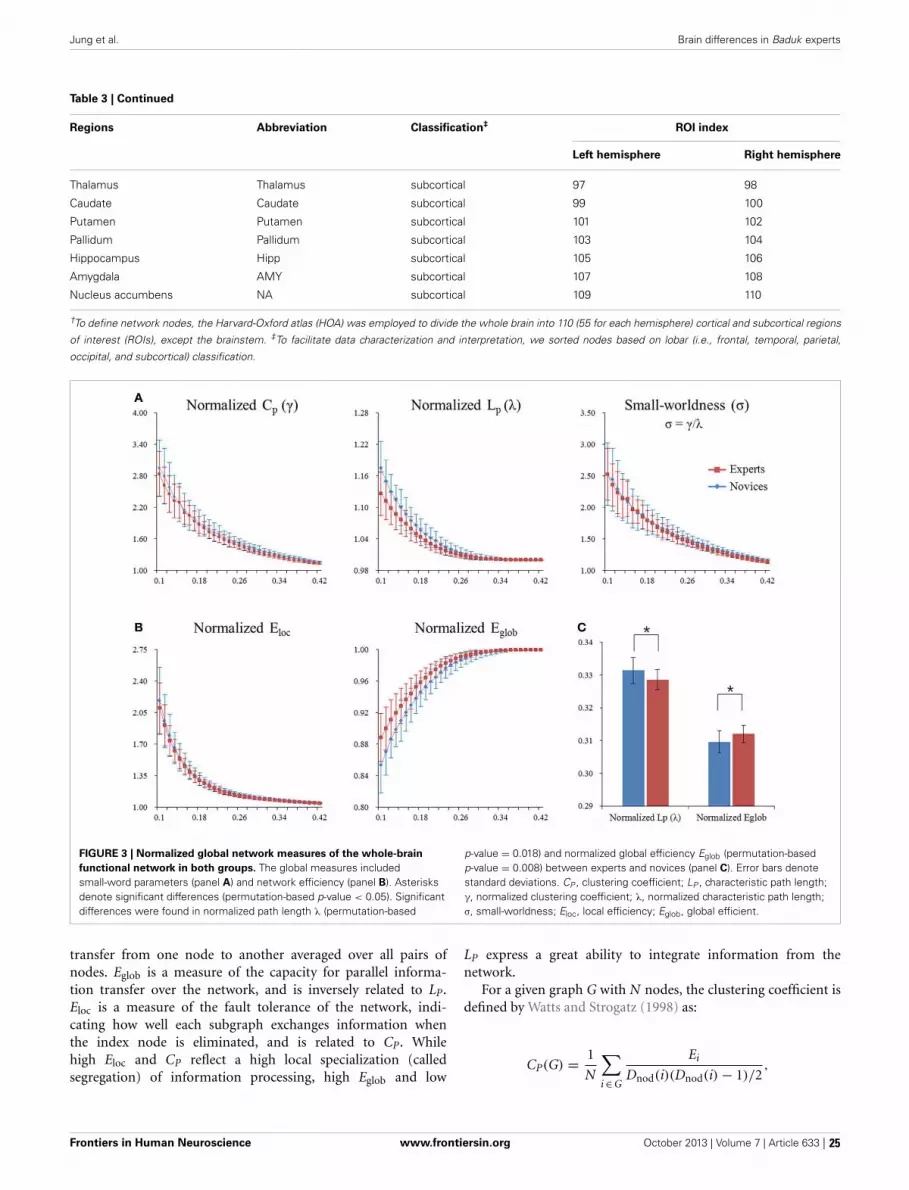
Jung et al. Brain differences in Baduk experts
Table 3 | Continued
Regions Abbreviation Classification‡ ROI index
Left hemisphere Right hemisphere
Thalamus Thalamus subcortical 97 98
Caudate Caudate subcortical 99 100
Putamen Putamen subcortical 101 102
Pallidum Pallidum subcortical 103 104
Hippocampus Hipp subcortical 105 106
Amygdala AMY subcortical 107 108
Nucleus accumbens NA subcortical 109 110
†To define network nodes, the Harvard-Oxford atlas (HOA) was employed to divide the whole brain into 110 (55 for each hemisphere) cortical and subcortical regions
of interest (ROIs), except the brainstem. ‡To facilitate data characterization and interpretation, we sorted nodes based on lobar (i.e., frontal, temporal, parietal,
occipital, and subcortical) classification.
FIGURE 3 | Normalized global network measures of the whole-brainfunctional network in both groups. The global measures includedsmall-word parameters (panel A) and network efficiency (panel B). Asterisksdenote significant differences (permutation-based p-value < 0.05). Significantdifferences were found in normalized path length λ (permutation-based
p-value = 0.018) and normalized global efficiency Eglob (permutation-basedp-value = 0.008) between experts and novices (panel C). Error bars denotestandard deviations. CP , clustering coefficient; LP , characteristic path length;γ, normalized clustering coefficient; λ, normalized characteristic path length;σ, small-worldness; Eloc, local efficiency; Eglob, global efficient.
transfer from one node to another averaged over all pairs ofnodes. Eglob is a measure of the capacity for parallel informa-tion transfer over the network, and is inversely related to LP.Eloc is a measure of the fault tolerance of the network, indi-cating how well each subgraph exchanges information whenthe index node is eliminated, and is related to CP. Whilehigh Eloc and CP reflect a high local specialization (calledsegregation) of information processing, high Eglob and low
LP express a great ability to integrate information from thenetwork.
For a given graph G with N nodes, the clustering coefficient isdefined by Watts and Strogatz (1998) as:
CP(G) = 1
N
∑i ∈ G
Ei
Dnod(i)(Dnod(i) − 1)/2,
Frontiers in Human Neuroscience www.frontiersin.org October 2013 | Volume 7 | Article 633 | 25

Jung et al. Brain differences in Baduk experts
FIGURE 4 | Regional network measures (i.e., nodal degree, nodalefficiency, and nodal betweenness) for experts and novices. (Panel A)shows values of each nodal metric over a range of thresholds in each group.(Panel B) shows mean values for each nodal metric across a range of thresholds
in each group, which were superimposed on an inflated standard brain usingBrainNet Viewer (http://www.nitrc.org/projects/bnv/). (Panel C) shows a bar plotof the AUC values of each nodal metric (red, frontal areas; green, temporalareas; blue, parietal areas; sky, occipital areas; purple, subcortical areas).
Frontiers in Human Neuroscience www.frontiersin.org October 2013 | Volume 7 | Article 633 | 26
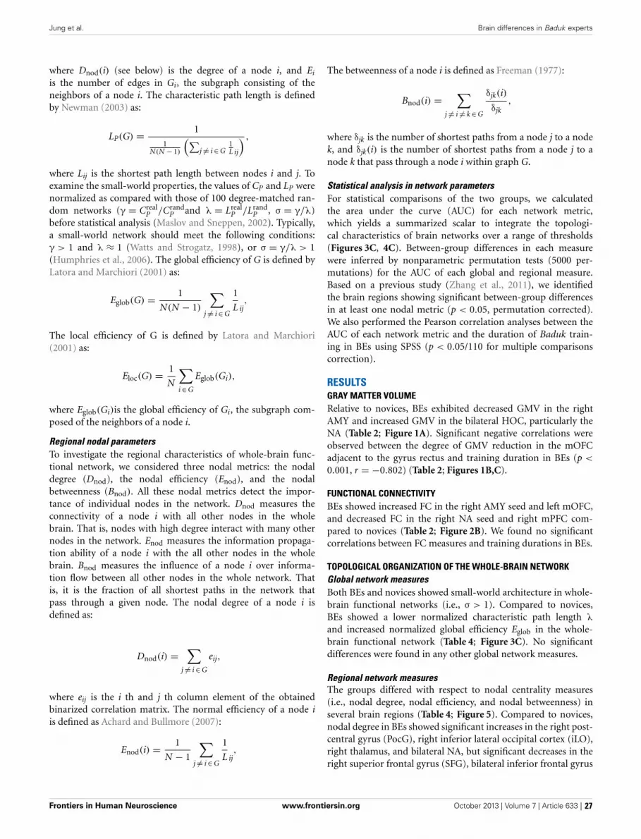
Jung et al. Brain differences in Baduk experts
where Dnod(i) (see below) is the degree of a node i, and Ei
is the number of edges in Gi, the subgraph consisting of theneighbors of a node i. The characteristic path length is definedby Newman (2003) as:
LP(G) = 1
1N(N − 1)
(∑j �= i ∈ G
1L ij
) ,
where Lij is the shortest path length between nodes i and j. Toexamine the small-world properties, the values of CP and LP werenormalized as compared with those of 100 degree-matched ran-dom networks (γ = Creal
P /CrandP and λ = Lreal
P /LrandP , σ = γ/λ)
before statistical analysis (Maslov and Sneppen, 2002). Typically,a small-world network should meet the following conditions:γ > 1 and λ ≈ 1 (Watts and Strogatz, 1998), or σ = γ/λ > 1(Humphries et al., 2006). The global efficiency of G is defined byLatora and Marchiori (2001) as:
Eglob(G) = 1
N(N − 1)
∑j �= i ∈ G
1
L ij,
The local efficiency of G is defined by Latora and Marchiori(2001) as:
Eloc(G) = 1
N
∑i ∈ G
Eglob(Gi),
where Eglob(Gi)is the global efficiency of Gi, the subgraph com-posed of the neighbors of a node i.
Regional nodal parametersTo investigate the regional characteristics of whole-brain func-tional network, we considered three nodal metrics: the nodaldegree (Dnod), the nodal efficiency (Enod), and the nodalbetweenness (Bnod). All these nodal metrics detect the impor-tance of individual nodes in the network. Dnod measures theconnectivity of a node i with all other nodes in the wholebrain. That is, nodes with high degree interact with many othernodes in the network. Enod measures the information propaga-tion ability of a node i with the all other nodes in the wholebrain. Bnod measures the influence of a node i over informa-tion flow between all other nodes in the whole network. Thatis, it is the fraction of all shortest paths in the network thatpass through a given node. The nodal degree of a node i isdefined as:
Dnod(i) =∑
j �= i ∈ G
eij,
where eij is the i th and j th column element of the obtainedbinarized correlation matrix. The normal efficiency of a node iis defined as Achard and Bullmore (2007):
Enod(i) = 1
N − 1
∑j �= i ∈ G
1
L ij,
The betweenness of a node i is defined as Freeman (1977):
Bnod(i) =∑
j �= i �= k ∈ G
δjk(i)
δjk,
where δjk is the number of shortest paths from a node j to a nodek, and δjk(i) is the number of shortest paths from a node j to anode k that pass through a node i within graph G.
Statistical analysis in network parametersFor statistical comparisons of the two groups, we calculatedthe area under the curve (AUC) for each network metric,which yields a summarized scalar to integrate the topologi-cal characteristics of brain networks over a range of thresholds(Figures 3C, 4C). Between-group differences in each measurewere inferred by nonparametric permutation tests (5000 per-mutations) for the AUC of each global and regional measure.Based on a previous study (Zhang et al., 2011), we identifiedthe brain regions showing significant between-group differencesin at least one nodal metric (p < 0.05, permutation corrected).We also performed the Pearson correlation analyses between theAUC of each network metric and the duration of Baduk train-ing in BEs using SPSS (p < 0.05/110 for multiple comparisonscorrection).
RESULTSGRAY MATTER VOLUMERelative to novices, BEs exhibited decreased GMV in the rightAMY and increased GMV in the bilateral HOC, particularly theNA (Table 2; Figure 1A). Significant negative correlations wereobserved between the degree of GMV reduction in the mOFCadjacent to the gyrus rectus and training duration in BEs (p <
0.001, r = −0.802) (Table 2; Figures 1B,C).
FUNCTIONAL CONNECTIVITYBEs showed increased FC in the right AMY seed and left mOFC,and decreased FC in the right NA seed and right mPFC com-pared to novices (Table 2; Figure 2B). We found no significantcorrelations between FC measures and training durations in BEs.
TOPOLOGICAL ORGANIZATION OF THE WHOLE-BRAIN NETWORKGlobal network measuresBoth BEs and novices showed small-world architecture in whole-brain functional networks (i.e., σ > 1). Compared to novices,BEs showed a lower normalized characteristic path length λ
and increased normalized global efficiency Eglob in the whole-brain functional network (Table 4; Figure 3C). No significantdifferences were found in any other global network measures.
Regional network measuresThe groups differed with respect to nodal centrality measures(i.e., nodal degree, nodal efficiency, and nodal betweenness) inseveral brain regions (Table 4; Figure 5). Compared to novices,nodal degree in BEs showed significant increases in the right post-central gyrus (PocG), right inferior lateral occipital cortex (iLO),right thalamus, and bilateral NA, but significant decreases in theright superior frontal gyrus (SFG), bilateral inferior frontal gyrus
Frontiers in Human Neuroscience www.frontiersin.org October 2013 | Volume 7 | Article 633 | 27
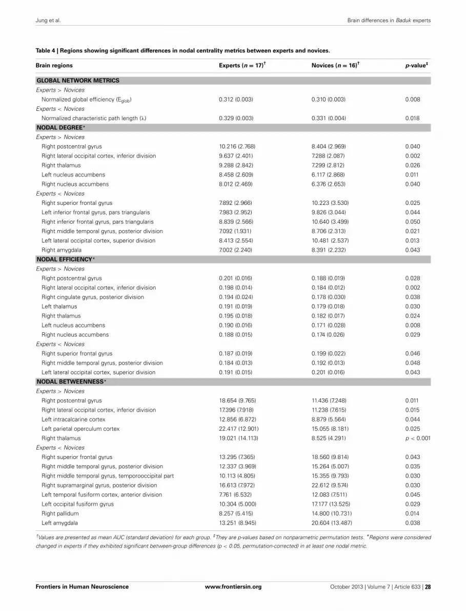
Jung et al. Brain differences in Baduk experts
Table 4 | Regions showing significant differences in nodal centrality metrics between experts and novices.
Brain regions Experts (n = 17)† Novices (n = 16)† p-value‡
GLOBAL NETWORK METRICS
Experts > Novices
Normalized global efficiency (Eglob) 0.312 (0.003) 0.310 (0.003) 0.008
Experts < Novices
Normalized characteristic path length (λ) 0.329 (0.003) 0.331 (0.004) 0.018
NODAL DEGREE*
Experts > Novices
Right postcentral gyrus 10.216 (2.768) 8.404 (2.969) 0.040
Right lateral occipital cortex, inferior division 9.637 (2.401) 7.288 (2.087) 0.002
Right thalamus 9.288 (2.842) 7.299 (2.812) 0.026
Left nucleus accumbens 8.458 (2.609) 6.117 (2.868) 0.011
Right nucleus accumbens 8.012 (2.469) 6.376 (2.653) 0.040
Experts < Novices
Right superior frontal gyrus 7.892 (2.966) 10.223 (3.530) 0.025
Left inferior frontal gyrus, pars triangularis 7.983 (2.952) 9.826 (3.044) 0.044
Right inferior frontal gyrus, pars triangularis 8.839 (2.566) 10.640 (3.499) 0.050
Right middle temporal gyrus, posterior division 7.092 (1.931) 8.706 (2.313) 0.021
Left lateral occipital cortex, superior division 8.413 (2.554) 10.481 (2.537) 0.013
Right amygdala 7.002 (2.240) 8.391 (2.232) 0.043
NODAL EFFICIENCY*
Experts > Novices
Right postcentral gyrus 0.201 (0.016) 0.188 (0.019) 0.028
Right lateral occipital cortex, inferior division 0.198 (0.014) 0.184 (0.012) 0.002
Right cingulate gyrus, posterior division 0.194 (0.024) 0.178 (0.030) 0.038
Left thalamus 0.191 (0.019) 0.179 (0.018) 0.030
Right thalamus 0.195 (0.018) 0.182 (0.017) 0.024
Left nucleus accumbens 0.190 (0.016) 0.171 (0.028) 0.008
Right nucleus accumbens 0.188 (0.015) 0.174 (0.026) 0.029
Experts < Novices
Right superior frontal gyrus 0.187 (0.019) 0.199 (0.022) 0.046
Right middle temporal gyrus, posterior division 0.184 (0.013) 0.192 (0.013) 0.048
Left lateral occipital cortex, superior division 0.191 (0.015) 0.201 (0.016) 0.043
NODAL BETWEENNESS*
Experts > Novices
Right postcentral gyrus 18.654 (9.765) 11.436 (7.248) 0.011
Right lateral occipital cortex, inferior division 17.396 (7.918) 11.238 (7.615) 0.015
Left intracalcarine cortex 12.856 (6.872) 8.879 (5.564) 0.044
Left parietal operculum cortex 22.417 (12.901) 15.055 (8.181) 0.025
Right thalamus 19.021 (14.113) 8.525 (4.291) p < 0.001
Experts < Novices
Right superior frontal gyrus 13.295 (7.365) 18.560 (9.814) 0.043
Right middle temporal gyrus, posterior division 12.337 (3.969) 15.264 (5.007) 0.035
Right middle temporal gyrus, temporooccipital part 10.113 (4.805) 15.355 (9.793) 0.030
Right supramarginal gyrus, posterior division 16.613 (7.972) 22.612 (9.574) 0.030
Left temporal fusiform cortex, anterior division 7.761 (6.532) 12.083 (7.511) 0.045
Left occipital fusiform gyrus 10.304 (5.000) 17.177 (13.525) 0.029
Right pallidum 8.257 (5.415) 14.800 (10.731) 0.014
Left amygdala 13.251 (8.945) 20.604 (13.487) 0.038
†Values are presented as mean AUC (standard deviation) for each group. ‡They are p-values based on nonparametric permutation tests. *Regions were considered
changed in experts if they exhibited significant between-group differences (p < 0.05, permutation-corrected) in at least one nodal metric.
Frontiers in Human Neuroscience www.frontiersin.org October 2013 | Volume 7 | Article 633 | 28

Jung et al. Brain differences in Baduk experts
FIGURE 5 | Brain regions showing significant differences in each nodalmetric between experts and novices. Regions with significant groupdifferences, including nodal degree (panel A), nodal efficiency (panel B), andnodal betweenness (panel C), were rendered using BrainNet Viewer(http://www.nitrc.org/projects/bnv/). See Table 4 for detailed information. R,right; L, left; Bi, bilateral; PoCG, postcentral gyrus; SFG, superior frontalgyurs; IFG, inferior frontal gyrus; pMTG, posterior division of middletemporal gyrus; sLO, superior division of lateral occipital cortex; iLO,interior division of lateral occipital cortex; THAL, thalamus; AMY, amygdala;NA, nucleus accumbens; PCG, posterior cingulate gyrus; pSMG, posteriordivision of supramarginal gyrus; OF, occipital fusiform gyrus; aTFC, anteriordivision of temporal fusiform cortex; PALL, pallidum; PO, parietaloperculum cortex; TO, temporooccipital part of middle temporal gyrus;intraCALC, intracalcarine cortex.
(IFG), right posterior middle temporal gyrus (pMTG), left supe-rior lateral occipital cortex (sLO), and right AMY (Figure 5A).In comparison to novices, nodal efficiency in BEs was signif-icantly increased in the right PocG, right iLO, right posteriorcingulate gyrus (PCG), bilateral thalamus, and bilateral NA, butsignificantly decreased in the right SFG, right pMTG, and leftsLO (Figure 5B). Compared to novices, nodal betweenness in BEswas significantly higher for the right PocG, right iLO, left intra-calcarine cortex, left parietal operculum cortex (PO), and rightthalamus, while significantly lower for the right SFG, right pMTG,temporooccipital part of right middle temporal gyrus (TO), rightposterior supramarginal gyrus (pSMG), left anterior temporalfusiform cortex (aTFC), left occipital fusiform gyrus (OF), rightpallidum, and left AMY (Figure 5C).
Nodal network metrics in several regions were correlated withtraining duration of BEs (p < 0.05; Figure 6), although therewere no correlations between brain regions showing significantgroup difference in nodal network metrics and training dura-tions of BEs. The duration of training in BEs was positivelycorrelated with nodal degree in the left SPL, but negatively cor-related with nodal degree in the left CN and left pTFC. Nodalefficiency in the bilateral SPL, right anterior supramarginal gyrus(aSMG), and right pSMG was positively correlated with train-ing duration. Training duration in BEs was positively correlatedwith nodal betweenness in the left SPL, but negatively corre-lated with nodal betweenness in the right CN, left pMTG, andright PO. Figure 6 shows plots of the correlation between nodalmetrics and training duration in BEs. However, all of these cor-relations did not withstand correction for multiple comparisons(p < 0.05/110).
DISCUSSIONTo our knowledge, this is the first study to combine structuralMRI and rs-fMRI to investigate the morphological differencesin the brain of BEs, the effect of such morphological differencesto functional circuits at rest, and the topological organizationof the whole-brain functional network in board-game experts,who are treated as excellent examples of cognitive expertise.Four main findings emerged from this study. First, relative tonovices, BEs showed increased GMV in the bilateral NA andreduced GMV in the right AMY. Additionally, the GMV inthe mOFC was correlated with training duration. Second, BEsshowed higher FC between the right AMY and left mOFC, anddecreased FC between the right NA and right mPFC. Third,BEs showed increased global efficiency and decreased charac-teristic path length, implying an increase in the global integra-tion of the whole-brain functional network. Fourth, BEs exhib-ited differences in the nodal centrality metrics of many brainregions related to the diverse cognitive functions utilized in Badukgames.
INTUITIVE EXPERTISE IN BOARD-GAME EXPERTS AND ITSIMPLICATION ON THE PRESENT FINDINGSBoard games have historically been the primary focus of researchon expertise (de Groot, 1965; Chase and Simon, 1973; Kahnemanand Klein, 2009). Specifically, interest in how board-game expertsmake rapid and effective decisions and in the neural correlatesassociated with these intuitive judgments has been increasing inrecent years (Duan et al., 2012; Wan et al., 2012). Such exper-tise or unconscious processing and linked brain changes are theproducts of prolonged experience within a domain and can-not be obtained through shortcuts. As it was previously stated,researchers have studied intuitive expertise in board-game expertsusing the NDM approach, particularly the RPD model (de Groot,1965; Klein, 1998, 2008; Kahneman and Klein, 2009). To describehow experts can make extremely rapid and good decisions,the RPD model combines intuitive pattern matching processesbased on past experience and deliberative mental simulationprocesses (Klein, 2008; Kahneman and Klein, 2009). These twoprocesses correspond to System 1 and System 2, respectively,in dual-process accounts of cognition (Hodgkinson et al., 2009;
Frontiers in Human Neuroscience www.frontiersin.org October 2013 | Volume 7 | Article 633 | 29

Jung et al. Brain differences in Baduk experts
FIGURE 6 | Plots of the correlations between each nodal metric andtraining duration of experts. The value of regional nodal measures(red, nodal degree; green, nodal efficiency; blue, nodal betweenness)
for each BEs is shown on the x-axes and duration of Baduk training (inyears) on the y -axes. All these correlations did not survive multiplecomparisons correction.
Kahneman and Klein, 2009). Interestingly, in this study, brainregions showing significant group difference in GMV, the NAand AMY, correspond to theories of the psychological and neu-robiological mechanisms underlying intuitive judgment. That is,these regions belong to the X-system (System 1), which supportsreflexive cognitive processing in social cognition (Lieberman,2007) and these regions, as expected, also belong to the somaticmarker circuitry based on the SMH. Compared with novices, BEsshowed reduced GMV in the AMY and increased GMV in theNA. This finding is very interesting because of the inverse effect(increase/decrease) on the GMV of these two regions. Previousstudies have reported specific correlations between regional cor-tical thickness and each component of cognition (Westlye et al.,2011), as well as a mixed pattern of increases and decreasesin regional cortical thickness in experts (Kang et al., 2013).For example, meditation experts showed a thicker cortex in themOFC and temporal pole, areas associated with emotional pro-cessing, and a thinner cortex in the parietal areas and PCG, areasassociated with attention and self-perception when comparedwith novices (Kang et al., 2013). In addition, recent studies haveargued for the nonlinearity of training-induced GMV changes,showing an initial increase followed by a decrease in regionalGMV (Boyke et al., 2008; Driemeyer et al., 2008), and suggestedthat these changes are affected by training length and intensity(Takeuchi et al., 2011). Based on such previous studies, a pos-sible explanation for the inverse effect of the AMY and NA onthe GMV is that each component involved in Baduk expertiseaffects a different part of the brain in a distinctive way (e.g.,increase/decrease on GMV), or that specific processing related tohow the amygdala shows reduced GMV may be more stronglyinvolved in BEs.
Contrary to our expectations, BEs and novices did notshow any differences in the GMV of the occipitotemporal andparietal areas associated with visuospatial processing and spatialattention. However, we found significant group differences interms of regional nodal properties of the functional brain net-work in these regions, particularly in the iLO, pSMG, TO, andpMTG, as well as in the NA and AMY, showing morpho-logical differences. Previous studies have reported that com-pared with novices, long-term chess experts (over 10 yearsof training) have morphological and functional differences inthe HOA, showing decreased GMV and increased FC in brainregions known as the default-mode network (DMN) duringresting-state in the region (Duan et al., 2012), whereas indi-viduals who trained for 15 weeks showed difference only inthe activity of anatomically identified HOA but not in mor-phological differences in the region between before and aftertraining, exhibiting increased activity during chess play (Wanet al., 2012). Based on previous studies and our present find-ings, it is, thus, conceivable that changes in brain structure,as well as those in brain function, through long-term train-ing may be required to become experts (i.e., to reach a pro-fessional level), and that BEs may primarily be involved inintuitive decision-making, which is associated with the regionsshowing both morphological and FC differences, and secon-darily in visuospatial processing, which is associated with theregions showing group differences only in the functional brainnetwork.
STRUCTURAL AND FUNCTIONAL DIFFERENCES IN THE AMYGDALAThe observed decrease in GMV of the AMY following train-ing is consistent with results from previous studies that involved
Frontiers in Human Neuroscience www.frontiersin.org October 2013 | Volume 7 | Article 633 | 30

Jung et al. Brain differences in Baduk experts
cognitive, motor, or mental training (Boyke et al., 2008; Takeuchiet al., 2011; Kang et al., 2013), suggesting that a possiblemechanism underlying such structural changes is the use-dependent selective elimination of synapses (Huttenlocher andDabholkar, 1997). The AMY is involved in configural/holisticvisual processing for both faces and emotional facial expressions(Sato et al., 2011); it enables people to master facial affective pro-cessing. More specifically, the right AMY is more relevant to theunconscious (Morris et al., 1998) and rapid (Wright et al., 2001)processing of facial expressions than the left AMY. Beyond its rolein emotional and facial processing, the AMY, in addition to theVMPFC, has also been proposed as one of key structures in theSMH and decision-making, as measured by the Iowa gamblingtask (Bechara et al., 2005), a decision-making task that requiresimplicit learning and executive function abilities. Additionally,animal studies have found that the AMY may participate in therecognition of visual patterns based on past experiences throughdirect thalamo–amygdala projection (LeDoux et al., 1989), andthat lesions to this area may increase impulsivity in decision-making (Winstanley et al., 2004). Human patients with AMYdamage have shown decreased decision-making performanceunder conditions of ambiguity and risk (Brand et al., 2007).Thus, GMV reduction in the AMY of BEs may be related tointuitive decision-making that is based on feelings (a somaticmarker) rather than on reasoning and/or the enhanced cogni-tive functioning (e.g., holistic visual processing) achieved throughlong-term Baduk training. The results of our RSFC and corre-lation analyses support this interpretation by showing increasedFC between the AMY and mOFC in BEs and a correlationbetween the GMV in the mOFC and training duration. Previousstudies have demonstrated that interactions between the AMYand mOFC are crucial for goal-directed behavior (Holland andGallagher, 2004) and emotional regulation (Banks et al., 2007).Lee et al. (2010) recently reported differences in the WM tract,the uncinate fasciculus, connecting these two regions in BEs. Asmentioned above, the mOFC/vmPFC is an important area thatgenerates somatic markers based on the SMH, and correlationbetween the GMV in the region and training duration suggeststhat morphological difference in the region may be contributed byBaduk training rather than pre-existing differences before train-ing. Contrary to this interpretation, an alternative one of thepresent finding is that this opposing pattern (reduced GMV andincreased FC in the amygdala) may reflect a compensatory neu-ral mechanism pre-existing in people who later become BEs. Thatis, the observed pattern may be an endophenotype that enablesimproved somatic-marker-based (automatic) decision-making,and thus favorably influences (i.e., predicts) the development ofBaduk expertise.
STRUCTURAL AND FUNCTIONAL DIFFERENCES IN THE STRIATUMIn contrast to the GMV in the AMY, the GMV in the NAwas increased in BEs compared to novices. This is espe-cially intriguing, as a recent study with chess experts usingthe same method reported a GMV decrease in the dor-sal part of HOC, which is known to be involved in thequick generation of the best next move during chess play-ing (Wan et al., 2011), while we found a GMV increase in
the ventral part of the HOC, the NA (Duan et al., 2012).Whereas the dorsal striatum is associated with sensorimotorexperiences or reward-outcomes processing, the ventral stria-tum is associated with emotional and motivational experi-ences or reward-anticipation processing. Consistent with thepresent finding, Boyke et al. (2008) found increased GMVin the NA after juggling training. They suggested that learn-ing to juggle may stimulate an increase in the size of thisregion due to its role as an interface between the limbicand motor systems rather than any role in motor control(Mogenson et al., 1980).
We also found decreased FC between the NA and mPFCin BEs compared with novices. Such connections are seenin the anterior part of the DMN (Di Martino et al., 2008;Duan et al., 2012; Jung et al., 2013), which is thought tobe involved in self-referential processing and theory of mind(ToM) (Murdaugh et al., 2012; Reniers et al., 2012). ToM refersto the ability to infer the thoughts or intentions of others.Given the hyperconnectivity within the DMN of individualsat high-risk for psychosis (Shim et al., 2010), who also suf-fer from impaired ToM (Chung et al., 2008), a reverse pat-tern (decreased FC) in BEs may be due to their capacity toinfer the opponent’s intention while playing Baduk. It is alsoconceivable that the strength of this connection may reflect achange in their sensitivity to feedback, given that these couplingsare thought to alter expectations in the face of negative feed-back (van den Bos et al., 2012). However, these ideas are justspeculations, and additional neuroimaging studies with Baduktasks are necessary to identify the physiological mechanismsthat underlie these brain differences and to clarify the relation-ship between their cognitive functions and brain structure andfunction.
TOPOLOGICAL ALTERATION IN THE WHOLE-BRAIN FUNCTIONALNETWORKThe GTA results reveal increased global efficiency and decreasedcharacteristic path length in Bes compared with novices, implyingan increase in the global integration of the whole-brain network.This can be reflective of effective integrity and rapid informa-tion propagation between and among the remote regions of thebrain involved in the cognitive processing required for Badukplay in BEs (Wang et al., 2010). Thus, this finding reflects a dif-ference in the functional aspect of the whole-brain circuitry inthe service of achieving the most efficient network for playingBaduk.
We also found the following differences between the twogroups in the regional nodal characteristics of many brainregions: increased nodal centrality metrics in nine regions,namely the right PocG, right iLO, right PCG, left intracal-carine cortex, left PO, bilateral NA, and bilateral thalamus, anddecreased nodal centrality metrics in 12 regions, namely the rightSFG, right pMTG, right TO of MTG, right pSMG, right pal-lidum, left sLO, left aTFC, left OF, bilateral IFG, and bilateralAMY. Interestingly, whereas the brain regions showing increasednodal centralities in BEs were involved primarily in implicit pro-cessing, such as somatic sensation (PocG and thalamus), visualexpertise (iLO), and affective/motivational processing (NA), the
Frontiers in Human Neuroscience www.frontiersin.org October 2013 | Volume 7 | Article 633 | 31

Jung et al. Brain differences in Baduk experts
brain regions showing decreased nodal centralities were relatedto higher-order cognitive functions such as executive function(SFG, IFG, and sLO), semantic memory processing (pMTG andpSMG), and visual perception (TO and OF). It is speculatedthat brain regions showing differences in nodal centralities areimportant contributors in Baduk expertise, and may facilitateefficient exchange of information. In this context, our findingsare consistent with the RPD model mentioned above (Klein,2008) Therefore, Baduk is thought to involve diverse cogni-tive functions with respect to both automatic and deliberativeprocesses.
Previous neuroimaging studies that focused on Badukreported enhanced activation in the occipitotemporal andparietal cortices, during these games (Chen et al., 2003;Ouchi et al., 2005), as well as learning-induced differ-ences in activity in fronto–parietal and visual cortices (Itohet al., 2008), which corresponds to our findings of differ-ences in the PocG and iLO. Correlation analyses betweeneach nodal metric and training duration of BEs showedpositive correlations between these two values in the pari-etal areas, particularly more extensive in the right than lefthemisphere, although it did not remain significant after cor-rection for multiple comparison. The right SPL in termsof Baduk play, may contribute to spatial working memory(Ungerleider et al., 1998) and/or spatial attention (Fink et al.,1996).
Chess requires the recognition of the identity and functionof each piece (i.e., chess-specific object and function recogni-tion, which is known to be associated with the occipito-parieto-temporal junction, OTJ; Bilalic et al., 2011a,b), whereas thisis not needed in Baduk. This difference as a confounding fac-tor makes it difficult to compare or interpret the results of thepresent study with those of previous neuroimaging studies onchess experts. However, both Baduk and chess involve patternrecognition with respect to the spatial positioning of stones orpieces, which is an essential component in improving both games.Recently, Bilalic et al. (2010, 2011b) demonstrated that the CoSand RSC, part of the parahippocampal place area, play an impor-tant role in chess-specific pattern recognition. Intriguingly, wefound increased nodal centralities in the iLO, which correspondsto the OTJ associated with chess-specific object and functionrecognition in studies by Bilalic et al. (2011a,b), in BEs com-pared to novices but not any differences in the Cos and RSCbetween the two groups. This discrepancy in results betweenthe present study with BEs and previous studies with chessexperts may stem from differences between the basic aspect ofpattern recognition in these board games; pattern recognitionin Baduk is only based on the shapes the stones take, whilethat in chess is based on object and function recognition ofchess pieces and their ability to rapidly access the informationof potential moves or move sequences for each piece. Thus, dif-ferences in the iLO may be linked to the recognition of shapesand integration of local features (Kourtzi et al., 2003) duringBaduk play.
Taken together, these GTA results provide such insight intothe topological organization of the functional brain networks ofBEs: increased functional integration across global brain regions
and increased nodal centralities in regions associated with spatialattention and somatic sensation.
LIMITATIONSThe present study has some limitations. First, it is difficultto accurately and quantitatively assess the skill level of BEs.Although we used training duration (in years) as a proxy forskill level in BEs, the validity of this proxy can be challenged.Skill levels in BEs may be independent of training duration,resulting in similar skill levels for any given pair of short- andlong-trained participants. Previous studies have described thatthe lack of any significant correlation between brain imagingdata and the amount/intensity of training in experts may resultfrom the inaccuracy of the proxy chosen in determining theactual extent/intensity of the individual training (Luders et al.,2011; Kang et al., 2013). Second, as a result of the cross-sectionalnature of this study, our findings do not allow any unambiguousdefinitive causality. Therefore, it is unclear whether the brain dif-ferences we found result from acquiring Baduk expertise throughprolonged training, or if they simply reflect pre-existing differ-ences in brain structure and function that predict later expertise.Longitudinal studies will help to clarify this issue. Finally, thepresent findings with respect to FC and GTA are based on rs-fMRI data of BEs rather than on brain activity during gameperformance. Thus, further functional imaging studies are nec-essary to investigate the topological properties and FC withinthe functional brain network while the individuals actually playBaduk.
CONCLUSIONSThe current study demonstrates differences in the structure andthe functional circuits of the AMY and NA in BEs; comparedwith novices, experts showed decreased GMV and increased FCwith the mOFC in the AMY as well as increased GMV anddecreased FC with the mPFC in the NA. As interfaces betweenthe cognitive and affective components of the limbic cortico–striatal loop, the AMY and NA are involved in implicit processingand goal-directed adaptive behavior under changing environ-mental conditions. In particular, the AMY is critical for emo-tional and holistic visual processing, as well as emotion-baseddecision-making. Based on our hypothesis that long-term Baduktraining would influence the structure and functional circuitsof regions associated with the cognitive mechanisms underlyingBaduk expertise, our findings suggest that intuitive decision-making, which is mediated by somatic marker circuitry suchas the AMY and NA, is a key cognitive component of Badukplay. The current study also provides new evidence for differ-ences in the topological organization of the whole-brain net-work of BEs, showing increased global integration and alteredregional nodal centralities in the regions related to visuospatialprocessing.
ACKNOWLEDGMENTSThis study was supported by a grant of the Baduk (Go)Research Project, Korea Baduk Association, Republic of Korea(KBA11). We thank the Baduk experts for participating, the KoreaBaduk Association (http://english.Baduk.or.kr/) for supporting
Frontiers in Human Neuroscience www.frontiersin.org October 2013 | Volume 7 | Article 633 | 32

Jung et al. Brain differences in Baduk experts
this study, Yong He, Jin-hui Wang, Xin-di Wang, and Ming-ruiXia for their development of the GRETNA toolbox, Sang-yoonJamie Jung for the continuing encouragement, and Jiyoon Seolfor the English revision of the revised manuscript. Furthermore,
we are especially grateful to a number of individuals who pro-vided valuable contributions to the study, including Ji Yeon Han,Bon-mi Gu, Ji-Young Park, and the clinical and nursing staff ofthe Clinical Cognitive Neuroscience Center.
REFERENCESAchard, S., and Bullmore, E. (2007).
Efficiency and cost of econom-ical brain functional networks.PLoS Comput. Biol. 3:e17. doi:10.1371/journal.pcbi.0030017
Ashburner, J. (2007). A fast diffeo-morphic image registration algo-rithm. Neuroimage 38, 95–113. doi:10.1016/j.neuroimage.2007.07.007
Banks, S. J., Eddy, K. T., Angstadt, M.,Nathan, P. J., and Phan, K. L. (2007).Amygdala-frontal connectivity dur-ing emotion regulation. Soc. Cogn.Affect. Neurosci. 2, 303–312. doi:10.1093/scan/nsm029
Bechara, A., Damasio, A. R., Damasio,H., and Anderson, S. W. (1994).Insensitivity to future consequencesfollowing damage to human pre-frontal cortex. Cognition 50, 7–15.doi: 10.1016/0010-0277(94)90018-3
Bechara, A., Damasio, H., Tranel,D., and Damasio, A. R. (2005).The Iowa Gambling Task andthe somatic marker hypothesis:some questions and answers.Trends Cogn. Sci. 9, 159–162. doi:10.1016/j.tics.2005.02.002
Bilalic, M., Kiesel, A., Pohl, C., Erb,M., and Grodd, W. (2011a). It takestwo-skilled recognition of objectsengages lateral areas in both hemi-spheres. PLoS ONE 6:e16202. doi:10.1371/journal.pone.0016202
Bilalic, M., Turella, L., Campitelli, G.,Erb, M., and Grodd, W. (2011b).Expertise modulates the neu-ral basis of context dependentrecognition of objects and theirrelations. Hum. Brain Mapp.33, 2728–2740. doi: 10.1002/hbm.21396
Bilalic, M., Langner, R., Ulrich, R.,and Grodd, W. (2011c). Manyfaces of expertise: fusiform facearea in chess experts and novices.J. Neurosci. 31, 10206–10214. doi:10.1523/JNEUROSCI.5727-10.2011
Bilalic, M., Langner, R., Erb, M., andGrodd, W. (2010). Mechanismsand neural basis of object andpattern recognition: a study withchess experts. J. Exp. Psychol.Gen. 139, 728–742. doi: 10.1037/a0020756
Blakemore, S. J., and Robbins, T.W. (2012). Decision-making in theadolescent brain. Nat. Neurosci. 15,1184–1191. doi: 10.1038/nn.3177
Bouzy, B., and Cazenave, T. (2001).Computer Go: an AI-oriented sur-vey. Artif. Intell. 132, 39–103. doi:10.1016/S0004-3702(01)00127-8
Boyke, J., Driemeyer, J., Gaser, C.,Buchel, C., and May, A. (2008).Training-induced brain struc-ture changes in the elderly.J. Neurosci. 28, 7031–7035. doi:10.1523/JNEUROSCI.0742-08.2008
Brand, M., Grabenhorst, F., Starcke,K., Vandekerckhove, M. M.,and Markowitsch, H. J. (2007).Role of the amygdala in deci-sions under ambiguity anddecisions under risk: evidencefrom patients with Urbach-Wiethe disease. Neuropsychologia45, 1305–1317. doi: 10.1016/j.neuropsychologia.2006.09.021
Cai, X., Venayagamoorthy, G. K.,and Wunsch, D. C. 2nd. (2010).Evolutionary swarm neural net-work game engine for Capture Go.Neural Netw. 23, 295–305. doi:10.1016/j.neunet.2009.11.001
Chase, W. G., and Simon, H. A.(1973). Perception in chess.Cogn. Psychol. 4, 55–81. doi:10.1016/0010-0285(73)90004-2
Chen, X., Zhang, D., Zhang, X., Li, Z.,Meng, X., He, S., et al. (2003). Afunctional MRI study of high-levelcognition. II. The game of Go. BrainRes. Cogn. Brain Res. 16, 32–37. doi:10.1016/S0926-6410(02)00206-9
Chumbley, J., Worsley, K., Flandin,G., and Friston, K. (2010).Topological FDR for neuroimaging.Neuroimage 49, 3057–3064. doi:10.1016/j.neuroimage.2009.10.090
Chung, Y. S., Kang, D. H., Shin,N. Y., Yoo, S. Y., and Kwon, J.S. (2008). Deficit of theory ofmind in individuals at ultra-high-risk for schizophrenia.Schizophr. Res. 99, 111–118. doi:10.1016/j.schres.2007.11.012
Cox, R. W. (1996). AFNI: Softwarefor analysis and visualizationof functional magnetic reso-nance neuroimages. Comput.Biomed. Res. 29, 162–173. doi:10.1006/cbmr.1996.0014
Damasio, A. R. (1996). The somaticmarker hypothesis and the possi-ble functions of the prefrontal cor-tex. Philos. Trans. R. Soc. Lond.B Biol. Sci. 351, 1413–1420. doi:10.1098/rstb.1996.0125
de Groot, A. D. (1965). Thoughtand Choice in Chess. The Hague:Mouton Publishers.
DeGroot, D., and Broekens, J. (2003).“Using negative emotions to impairgame play,” in Proceedings of the15th Belgian-Dutch Conference onArtificial Intelligence, (Nijmegen),32–40.
Di Martino, A., Scheres, A., Margulies,D. S., Kelly, A. M., Uddin, L.Q., Shehzad, Z., et al. (2008).Functional connectivity of humanstriatum: a resting state FMRI study.Cereb. Cortex 18, 2735–2747. doi:10.1093/cercor/bhn041
Draganski, B., Gaser, C., Busch, V.,Schuierer, G., Bogdahn, U., andMay, A. (2004). Neuroplasticity:changes in grey matter induced bytraining. Nature 427, 311–312. doi:10.1038/427311a
Driemeyer, J., Boyke, J., Gaser,C., Buchel, C., and May,A. (2008). Changes in graymatter induced by learning-revisited. PLoS ONE 3:e2669. doi:10.1371/journal.pone.0002669
Duan, X., He, S., Liao, W., Liang,D., Qiu, L., Wei, L., et al.(2012). Reduced caudate vol-ume and enhanced striatal-DMNintegration in chess experts.Neuroimage 60, 1280–1286. doi:10.1016/j.neuroimage.2012.01.047
Dunn, B. D., Dalgleish, T., andLawrence, A. D. (2006). Thesomatic marker hypothesis: acritical evaluation. Neurosci.Biobehav. Rev. 30, 239–271. doi:10.1016/j.neubiorev.2005.07.001
Fink, G. R., Halligan, P. W., Marshall,J. C., Frith, C. D., Frackowiak, R.S., and Dolan, R. J. (1996). Wherein the brain does visual atten-tion select the forest and the trees?Nature 382, 626–628. doi: 10.1038/382626a0
Fox, M. D., Snyder, A. Z., Vincent,J. L., Corbetta, M., Van Essen, D.C., and Raichle, M. E. (2005). Thehuman brain is intrinsically orga-nized into dynamic, anticorrelatedfunctional networks. Proc. Natl.Acad. Sci. U.S.A. 102, 9673–9678.doi: 10.1073/pnas.0504136102
Freeman, L. C. (1977). Set of measuresof centrality based on between-ness. Sociometry 40, 35–41. doi:10.2307/3033543
Gao, Q., Xu, Q., Duan, X., Liao,W., Ding, J., Zhang, Z., et al.(2013). Extraversion and neuroti-cism relate to topological proper-ties of resting-state brain networks.Front. Hum. Neurosci. 7:257. doi:10.3389/fnhum.2013.00257
Gobet, F., and Charness, N. (2006).Expertise in Chess. The CambridgeHandbook of Expertise and ExpertPerformance. Cambridge, MA:Cambridge University Press. doi:10.1017/CBO9780511816796.030
Gobet, F., de Voogt, A. J., andRetschitzki, J. (2004). Moves inMind: the Psychology of BoardGames. New York, NY: PsychologyPress.
Grabner, R. H., Stern, E., andNeubauer, A. C. (2007). Individualdifferences in chess expertise: apsychometric investigation. ActaPsychol. (Amst.) 124, 398–420. doi:10.1016/j.actpsy.2006.07.008
Hodgkinson, G. P., Sadler-Smith,E., Burke, L.A., Claxton, G., andSparrow, P. R. (2009). Intuitionin organizations: implicationsfor strategic management. LongRange Plan. 42, 277–297. doi:10.1016/j.lrp.2009.05.003
Holland, P. C., and Gallagher, M.(2004). Amygdala-frontal interac-tions and reward expectancy. Curr.Opin. Neurobiol. 14, 148–155. doi:10.1016/j.conb.2004.03.007
Humphries, M. D., Gurney, K., andPrescott, T. J. (2006). The brainstemreticular formation is a small-world, not scale-free, network.Proc. Biol. Sci. 273, 503–511. doi:10.1098/rspb.2005.3354
Huttenlocher, P. R., and Dabholkar,A. S. (1997). Regional differencesin synaptogenesis in human cere-bral cortex. J. Comp. Neurol. 387,167–178. doi: 10.1002/(SICI)1096-9861(19971020)387:2<167::AID-CNE1>3.0.CO;2-Z
Itoh, K., Kitamura, H., Fujii, Y.,and Nakada, T. (2008). Neural sub-strates for visual pattern recognitionlearning in Igo. Brain Res. 1227,162–173. doi: 10.1016/j.brainres.2008.06.080
Jang, J. H., Jung, W. H., Kang, D. H.,Byun, M. S., Kwon, S. J., Choi,C. H., et al. (2011). Increaseddefault mode network connec-tivity associated with meditation.
Frontiers in Human Neuroscience www.frontiersin.org October 2013 | Volume 7 | Article 633 | 33

Jung et al. Brain differences in Baduk experts
Neurosci. Lett. 487, 358–362. doi:10.1016/j.neulet.2010.10.056
Jung, W. H., Kang, D. H., Kim, E. T.,Shin, K. S., Jang, J. H., and Kwon, J.S. (2013). Abnormal corticostriatal-limbic functional connectivity inobsessive-compulsive disorder dur-ing reward processing and resting-state. Neuroimage Clin. 3, 27–38.doi: 10.1016/j.nicl.2013.06.013
Kahneman, D., and Klein, G. (2009).Conditions for intuitive expertise: afailure to disagree. Am. Psychol. 64,515–526. doi: 10.1037/a0016755
Kang, D. H., Jo, H. J., Jung, W. H., Kim,S. H., Jung, Y. H., Choi, C. H., et al.(2013). The effect of meditation onbrain structure: cortical thicknessmapping and diffusion tensor imag-ing. Soc. Cogn. Affect. Neurosci. 8,27–33. doi: 10.1093/scan/nss056
Keene, R., and Levy, D. (1991). How toBeat your Chess Computer (BatsfordChess Library). London: Henry Holtand Company Publisher.
Klein, G. (2008). Naturalistic deci-sion making. Hum. Factors 50,456–460. doi: 10.1518/001872008X288385
Klein, G. A. (1993). “A recognition-primed decision (RPD) modelof rapid decision making,” inDecision Making in Action: Modelsand Methods, eds G. A. Klein, J.Orasanu, R. Calderwood, and C.E. Zsambok (Norwood, NJ: AblexPublishing Corporation).
Klein, G. A. (1998). Sources of Power:How People Make Decisions.Cambridge, MA: MIT Press.
Kourtzi, Z., Tolias, A. S., Altmann, C. F.,Augath, M., and Logothetis, N. K.(2003). Integration of local featuresinto global shapes: monkey andhuman FMRI studies. Neuron 37,333–346. doi: 10.1016/S0896-6273(02)01174-1
Latora, V., and Marchiori, M.(2001). Efficient behavior ofsmall-world networks. Phys.Rev. Lett. 87, 198701. doi:10.1103/PhysRevLett.87.198701
LeDoux, J. E., Romanski, L., andXagoraris, A. (1989). Indelibilityof subcortical emotional memories.J. Cogn. Neurosci. 1, 238–243. doi:10.1162/jocn.1989.1.3.238
Lee, B., Park, J. Y., Jung, W. H., Kim,H. S., Oh, J. S., Choi, C. H., et al.(2010). White matter neuroplasticchanges in long-term trained play-ers of the game of “Baduk” (GO): avoxel-based diffusion-tensor imag-ing study. Neuroimage 52, 9–19.doi: 10.1016/j.neuroimage.2010.04.014
Levitt, S. D., List, J. A., and Sadoff,S. E. (2011). Checkmate: explor-ing backward induction among
chess players. Am. Econ. Rev. 101,975–990. doi: 10.1257/aer.101.2.975
Lieberman, M. D. (2007). “The X-and C-systems: the neural basis ofautomatic and controlled social cog-nition,” in Fundamentals of SocialNeuroscience, eds E. Harmon-Johnsand P. Winkelman (New York, NY:Guilford), 290–315.
Luders, E., Clark, K., Narr, K. L., andToga, A. W. (2011). Enhancedbrain connectivity in long-term meditation practitioners.Neuroimage 57, 1308–1316. doi:10.1016/j.neuroimage.2011.05.075
Lynall, M. E., Bassett, D. S., Kerwin,R., McKenna, P. J., Kitzbichler,M., Muller, U., et al. (2010).Functional connectivity andbrain networks in schizophrenia.J. Neurosci. 130, 9477–9487. doi:10.1523/JNEUROSCI.0333-10.2010
Maguire, E. A., Gadian, D. G.,Johnsrude, I. S., Good, C. D.,Ashburner, J., Frackowiak, R. S.,et al. (2000). Navigation-relatedstructural change in the hip-pocampi of taxi drivers. Proc. Natl.Acad. Sci. U.S.A. 97, 4398–4403.doi: 10.1073/pnas.070039597
Maslov, S., and Sneppen, K. (2002).Specificity and stability in topol-ogy of protein networks. Science296, 910–913. doi: 10.1126/science.1065103
Minavand chal, E., MohammadipourPamsari, M., and MohammadipourPamsari, M. (2013). Toward anintegrated model of intuitivedecision making. Researcher 5,1–10.
Mogenson, G. J., Jones, D. L., andYim, C. Y. (1980). From motivationto action: functional interfacebetween the limbic system and themotor system. Prog. Neurobiol. 14,69–97. doi: 10.1016/0301-0082(80)90018-0
Morris, J. S., Ohman, A., and Dolan,R. J. (1998). Conscious and uncon-scious emotional learning in thehuman amygdala. Nature 393,467–470. doi: 10.1038/30976
Murdaugh, D. L., Shinkareva, S.V., Deshpande, H. R., Wang, J.,Pennick, M. R., and Kana, R.K. (2012). Differential deacti-vation during mentalizing andclassification of autism based ondefault mode network connec-tivity. PLoS ONE 7:e50064. doi:10.1371/journal.pone.0050064
Newman, M. E. J. (2003). The struc-ture and function of complex net-works. SIAM Rev. 45, 167–256. doi:10.1137/S003614450342480
Nichelli, P., Grafman, J., Pietrini, P.,Always, D., Carton, J. C., and
Miletich, R. (1994). Brain activity inchess playing. Nature 369, 191. doi:10.1038/369191a0
Ouchi, Y., Kanno, T., Yoshikawa,E., Futatsubashi, M., Okada,H., Torizuka, T., et al. (2005).Neural substrates in judgmentprocess while playing go: acomparison of amateurs withprofessionals. Brain Res. Cogn.Brain Res. 23, 164–170. doi:10.1016/j.cogbrainres.2004.10.004
Phillips, M. L., Drevets, W. C., Rauch,S. L., and Lane, R. (2003a).Neurobiology of emotion per-ception I: The neural basis ofnormal emotion perception.Biol. Psychiatry 54, 504–514. doi:10.1016/S0006-3223(03)00168-9
Phillips, M. L., Drevets, W. C.,Rauch, S. L., and Lane, R.(2003b). Neurobiology of emo-tion perception II: Implicationsfor major psychiatric disorders.Biol. Psychiatry 54, 515–528. doi:10.1016/S0006-3223(03)00171-9
Reniers, R. L., Corcoran, R., Völlm,B. A., Mashru, A., Howard, R.,and Liddle, P. F. (2012). Moraldecision-making, ToM, empathyand the default mode network.Biol. Psychol. 90, 202–210. doi:10.1016/j.biopsycho.2012.03.009
Rubinov, M., and Sporns, O.(2010). Complex networkmeasures of brain connectiv-ity: uses and interpretations.Neuroimage 52, 1059–1069. doi:10.1016/j.neuroimage.2009.10.003
Sato, W., Kochiyama, T., andYoshikawa, S. (2011). The inversioneffect for neutral and emotionalfacial expressions on amygdalaactivity. Brain Res. 1378, 84–90. doi:10.1016/j.brainres.2010.12.082
Shim, G., Oh, J. S., Jung, W. H.,Jang, J. H., Choi, C. H., Kim, E.,et al. (2010). Altered resting-stateconnectivity in subjects at ultra-high risk for psychosis: an fMRIstudy. Behav. Brain Funct. 6, 58. doi:10.1186/1744-9081-6-58
Song, X. W., Dong, Z. Y., Long, X.Y., Li, S. F., Zuo, X. N., Zhu, C.Z., et al. (2011). REST: a toolkitfor resting-state functional mag-netic resonance imaging data pro-cessing. PLoS ONE 6:e25031. doi:10.1371/journal.pone.0025031
Spiers, H. J., and Maguire, E. A.(2006). Thoughts, behaviour,and brain dynamics duringnavigation in the real world.Neuroimage 31, 1826–1840. doi:10.1016/j.neuroimage.2006.01.037
Supekar, K., Menon, V., Rubin, D.,Musen, M., and Greicius, M. D.(2008). Network analysis of intrin-sic functional brain connectivity in
Alzheimer’s disease. PLoS Comput.Biol. 4:e1000100. doi: 10.1371/jour-nal.pcbi.1000100
Takeuchi, H., Taki, Y., Sassa, Y.,Hashizume, H., Sekiguchi, A.,Fukushima, A., et al. (2011).Working memory training usingmental calculation impacts regionalgray matter of the frontal and pari-etal regions. PLoS ONE 6:e23175.doi: 10.1371/journal.pone.0023175
Tian, L., Wang, J., Yan, C., andHe, Y. (2011). Hemisphere- andgender-related differences insmall-world brain networks: aresting-state functional MRI study.Neuroimage 54, 191–202. doi:10.1016/j.neuroimage.2010.07.066
Ungerleider, L. G., Courtney, S. M.,and Haxby, J. V. (1998). A neuralsystem for human visual work-ing memory. Proc. Natl. Acad.Sci. U.S.A. 95, 883–890. doi:10.1073/pnas.95.3.883
van den Bos, W., Cohen, M. X.,Kahnt, T., and Crone, E. A.(2012). Striatum-medial pre-frontal cortex connectivity predictsdevelopmental changes in reinforce-ment learning. Cereb. Cortex 22,1247–1255. doi: 10.1093/cercor/bhr198
Wan, X., Nakatani, H., Ueno, K.,Asamizuya, T., Cheng, K., andTanaka, K. (2011). The neuralbasis of intuitive best next-move generation in board gameexperts. Science 331, 341–346. doi:10.1126/science.1194732
Wan, X., Takano, D., Asamizuya, T.,Suzuki, C., Ueno, K., Cheng, K.,et al. (2012). Developing intu-ition: neural correlates of cognitive-skill learning in caudate nucleus.J. Neurosci. 32, 17492–17501. doi:10.1523/JNEUROSCI.2312-12.2012
Wang, J., Zuo, X., and He, Y. (2010).Graph-based network analysisof resting-state functional MRI.Front. Syst. Neurosci. 4:16. doi:10.3389/fnsys.2010.00016
Watts, D. J., and Strogatz, S. H.(1998). Collective dynamics of‘small-world’ networks. Nature 393,440–442. doi: 10.1038/30918
Westlye, L. T., Grydeland, H., Walhovd,K. B., and Fjell, A. M. (2011).Associations between regionalcortical thickness and atten-tional networks as measuredby the attention network test.Cereb. Cortex 21, 345–356. doi:10.1093/cercor/bhq101
Winstanley, C. A., Theobald,D. E., Cardinal, R. N., andRobbins, T. W. (2004).Contrasting roles of basolateralamygdala and orbitofrontalcortex in impulsive choice.
Frontiers in Human Neuroscience www.frontiersin.org October 2013 | Volume 7 | Article 633 | 34

Jung et al. Brain differences in Baduk experts
J. Neurosci. 24, 4718–4722. doi:10.1523/JNEUROSCI.5606-03.2004
Wright, C. I., Fischer, H., Whalen, P.J., McInerney, S. C., Shin, L. M.,and Rauch, S. L. (2001). Differentialprefrontal cortex and amygdalahabituation to repeatedly presentedemotional stimuli. Neuroreport 12,379–383. doi: 10.1097/00001756-200102120-00039
Zhang, J., Wang, J., Wu, Q., Kuang,W., Huang, X., He, Y., et al. (2011).Disrupted brain connectivity
networks in drug-naive, first-episode major depressivedisorder. Biol. Psychiatry 70,334–342. doi: 10.1016/j.biopsych.2011.05.018
Conflict of Interest Statement: Theauthors declare that the researchwas conducted in the absence of anycommercial or financial relationshipsthat could be construed as a potentialconflict of interest.
Received: 12 June 2013; accepted: 12September 2013; published online: 02October 2013.Citation: Jung WH, Kim SN, Lee TY,Jang JH, Choi C-H, Kang D-H andKwon JS (2013) Exploring the brains ofBaduk (Go) experts: gray matter mor-phometry, resting-state functional con-nectivity, and graph theoretical analy-sis. Front. Hum. Neurosci. 7:633. doi:10.3389/fnhum.2013.00633This article was submitted to the journalFrontiers in Human Neuroscience.
Copyright © 2013 Jung, Kim, Lee, Jang,Choi, Kang and Kwon. This is an open-access article distributed under the termsof the Creative Commons AttributionLicense (CC BY). The use, distribution orreproduction in other forums is permit-ted, provided the original author(s) orlicensor are credited and that the origi-nal publication in this journal is cited, inaccordance with accepted academic prac-tice. No use, distribution or reproductionis permitted which does not comply withthese terms.
Frontiers in Human Neuroscience www.frontiersin.org October 2013 | Volume 7 | Article 633 | 35

ORIGINAL RESEARCH ARTICLEpublished: 17 December 2013
doi: 10.3389/fnhum.2013.00851
Brain regions concerned with the identification of deceptivesoccer moves by higher-skilled and lower-skilled playersMichael J. Wright1*, Daniel T. Bishop1,2, Robin C. Jackson2 and Bruce Abernethy3,4
1 Department of Psychology, Centre for Cognition and Neuroimaging, Brunel University, Uxbridge, Middlesex, UK2 School of Sport Sciences and Education, Centre for Sports Medicine and Human Performance, Brunel University, Uxbridge, Middlesex, UK3 Faculty of Health Sciences, University of Queensland, Brisbane, QLD, Australia4 Institute of Human Performance, University of Hong Kong, Pokfulam, Hong Kong, China
Edited by:Merim Bilalic, University Tübingen,University Clinic, Germany
Reviewed by:Yongmin Chang, KyungpookNational University, South KoreaRouwen Canal-Bruland, VUUniversity Amsterdam, Netherlands
*Correspondence:Michael J. Wright, Department ofPsychology, Centre for Cognitionand Neuroimaging, BrunelUniversity, Kingston Lane, Uxbridge,Middlesex, UB8 3PH, UKe-mail: [email protected]
Expert soccer players are able to utilize their opponents’ early body kinematics topredict the direction in which the opponent will move. We have previously demonstratedenhanced fMRI activation in experts in the motor components of an action observationnetwork (AON) during sports anticipation tasks. Soccer players often need to preventopponents from successfully predicting their line of attack, and consequently may tryto deceive them; for example, by performing a step-over. We examined how AONactivations and expertise effects are modified by the presence of deception. Three groupsof participants; higher-skilled males, lower-skilled males, and lower-skilled females, viewedvideo clips in point-light format, from a defender’s perspective, of a player approachingand turning with the ball. The observer’s task in the scanner was to determine whetherthe move was normal or deceptive (involving a step-over), while whole-brain functionalimages were acquired. In a second counterbalanced block with identical stimuli thetask was to predict the direction of the ball. Activations of AON for identification ofdeception overlapped with activations from the direction identification task. Higher-skilledplayers showed significantly greater activation than lower-skilled players in a subset ofAON areas; and lower-skilled males in turn showed greater activation than lower-skilledfemales, but females showed more activation in visual cortex. Activation was greater fordeception identification than for direction identification in dorsolateral prefrontal cortex,medial frontal cortex, anterior insula, cingulate gyrus, and premotor cortex. Conversely,greater activation for direction than deception identification was found in anterior cingulatecortex and caudate nucleus. Results are consistent with the view that explicit identificationof deceptive moves entails cognitive effort and also activates limbic structures associatedwith social cognition and affective responses.
Keywords: fMRI, action observation, deception, expertise, soccer, football, mirror neuron system, sport
INTRODUCTIONExpert players in interceptive sports such as soccer react undergreat time pressure and therefore, need to predict the actionsof their opponents and the direction of play (Reilly et al., 2000;Abernethy et al., 2001; Savelsbergh et al., 2005; Williams et al.,2011). Studies have shown that superior analysis of body kinemat-ics underpins much anticipation skill in sport. In the temporalocclusion paradigm, action is cut off at various time intervalsrelative to a crucial event (such as point of direction changein soccer), and the observer judges the direction of the shot.Results consistently show that experts are able to detect the pre-dictive information with greater accuracy and earlier than novices(Abernethy and Russell, 1984, 1987; Abernethy et al., 2008). Thenature of the predictive information has also been identified usingtechniques such as spatial occlusion, in which different parts ofthe opponent’s body are systematically masked (Muller et al.,2006; Jackson and Mogan, 2007). The reductive approach toidentifying the minimum visual information sufficient to sup-port expert anticipation has been taken further with the use of
point-light video stimuli. Comparisons of performance based onball trajectory alone and studies using point-light stimuli indicatethe pre-eminence of body kinematics as a cue to future action(Abernethy et al., 2001, 2008; Huys et al., 2008).
A general conclusion from this research is that experts are bet-ter than novices at detecting predictive cues in opponents’ bodykinematics, and this gives them an advantage in speed and accu-racy. Precisely for this reason, skilled players also need to developstrategies to reduce the predictability of their own actions. Theeffectiveness with which deceptive moves can thwart anticipa-tion has been established in soccer (Dicks et al., 2011; Smeetonand Williams, 2012; Bishop et al., 2013); rugby football (Jacksonet al., 2006; Brault et al., 2012; Mori and Shimada, 2013); bas-ketball (Sebanz and Shiffrar, 2009; Kunde et al., 2011); handball(Cañal-Bruland and Schmidt, 2009; Cañal-Bruland et al., 2010),and tennis (Rowe et al., 2009). In these studies it was found thatexperts are more accurate than novices in predicting the outcomeof deceptive moves, and that the expert-novice difference tends tobe greater for deceptive than for normal moves.
Frontiers in Human Neuroscience www.frontiersin.org December 2013 | Volume 7 | Article 851 |
HUMAN NEUROSCIENCE
36

Wright et al. Identification of direction and deception in soccer moves
Neuroimaging studies have provided some insights into theneural structures that mediate anticipation skills. A substantialliterature has developed around functional imaging studies ofcortical networks that mediate the perception of, and the produc-tion of responses to, others’ actions. Molenberghs et al. (2012)conducted a meta-analysis of 125 fMRI studies of the human“mirror neuron system” (MNS) and identified a core networkof brain areas including inferior frontal gyrus, dorsal and ven-tral premotor cortex, and inferior and superior parietal lobule,that were activated in studies involving the observation and/orproduction of actions. Most fMRI experiments on the observa-tion of actions do not include direct evidence for the presenceof mirror neurons, so we refer in the present paper to an actionobservation network (AON: Grafton, 2009) rather than MNS.The AON does nevertheless include the structures identified byMolenberghs et al. (2012) as core elements of the MNS.
Research has demonstrated the importance of the AON insport, including structures traditionally interpreted as havingmotor functions. Wright and Jackson (2007) measured corticalfMRI activation in predicting the direction of a tennis serve fromtemporally-occluded video clips. Relative to a passive, action-observation control condition, action prediction activated theanterior components of the AON, particularly the dorsal andventral premotor cortex. Aglioti et al. (2008) found that obser-vation of basketball shots increased the strength of motor-evokedpotentials elicited by transcranial magnetic stimulation, and thatexperts showed a time-specific motor activation for missed shots,indicating a close and specific interaction between perceptualand motor systems that is dependent on experiential learning.Wright et al. (2010) found stronger activation for expert bad-minton players while predicting the direction of badminton shotsin components of the AON, specifically, medial frontal cortex,inferior frontal gyrus, anterior insula, and superior parietal lob-ule. Wright et al. (2011) showed that low-resolution point-lightbadminton video effectively supported judgments of the directionof a shot, and elicited a corresponding full pattern of fMRI activa-tions in these areas including expertise effects, thus, indicating thesufficiency of body kinematics as input to the AON. Bishop et al.(2013) studied neural correlates of direction prediction in soccer,with temporally-occluded video stimuli that included deceptivemoves, and with randomized presentation that maximized uncer-tainty. High-skilled observers showed stronger responses thanintermediates and novices not only in cortical AON structuresbut also in subcortical structures, including cerebellum, lentiformnucleus, and thalamus, that have been implicated in responseselection (Yarrow et al., 2009).
Correct direction prediction in a situation where an oppo-nent can use deceptive moves, for example when an oncomingrugby player executes a side-step, may involve attending to “hon-est” movement cues and ignoring “deceptive” movement signals(Brault et al., 2012). This is a complex skill that entails morethan simply being able to recognize a normal or a deceptivemove: the correct implication of that move in terms of outcome(future direction of play) must also be perceived or compre-hended. This is perhaps the reason that highly-skilled playersoften take longer to react than novices in the presence of decep-tion, and achieve greater accuracy as a result (Brault et al., 2012;
Mori and Shimada, 2013). In some studies, experts are foundto be significantly disadvantaged by deception, notwithstandingthey may be less disadvantaged than novices (Brault et al., 2012;Bishop et al., 2013; Mori and Shimada, 2013). Possible reasons forthis include an increased cognitive load, perceptual uncertainty,or misdirection of attention in the presence of deception.
The purpose of the present study was to analyze the neural andbehavioral responses of lower-skilled and higher-skilled players tothe task of identifying soccer moves as normal or deceptive, andby comparison, measuring the neural and behavioral response toidentifying future direction of play in an identical (normal plusdeceptive) stimulus set. Most studies of deceptive moves in sporthave used identification of future direction of play as a mea-sure (Jackson et al., 2006; Rowe et al., 2009; Dicks et al., 2011;Kunde et al., 2011; Brault et al., 2012; Smeeton and Williams,2012; Bishop et al., 2013; Mori and Shimada, 2013). A smallernumber have measured deception identification (Cañal-Brulandand Schmidt, 2009; Sebanz and Shiffrar, 2009; Cañal-Brulandet al., 2010). These tasks are not equivalent. Firstly, as Cañal-Bruland and Williams (2010) found, the kinematic informationused when predicting the direction of a shot differs from thatused when discriminating between two different movement pat-terns. Secondly, the consequences of the judgment are different.Direction identification requires a directional or spatial judgmentwith implications for the direction of an interceptive movement.Equally, deception identification implies a more analytical judg-ment of an observed action as having some goal or intent, butwithout specifying direction. It was therefore, hypothesized thatboth behavioral performance and cortical patterns of activationfor direction identification and deception identification may dif-fer, and that there would be differences in the activation oftask-related regions, as identified by fMRI, in lower-skilled andhigher-skilled players. In view of the research reviewed aboveshowing the sufficiency of body kinematics in sport action pre-diction tasks, and in order to eliminate irrelevant stimulation bybackground stimuli, physical appearance and clothing of actors,we utilized point-light stimuli for the tasks.
MATERIALS AND METHODSEXPERIMENTAL DESIGN AND PROCEDUREIn a block-design, fMRI study, participants in the scanner viewed2-s video sequences of an opposing soccer player dribbling theball toward the viewer, and pressed a button to indicate whichdirection the player would turn; that is, the leftmost button fora turn to the observer’s left, and the rightmost button for a turnto the observer’s right. There was an interstimulus interval of 2-sduring which a gray screen at mean luminance was present, andinstructions were to respond as accurately as possible during theinterstimulus interval. There were five video clips in each block.Exactly half of the sequences of each type were based on deceptivemoves (step-over) and half on normal moves, both for directionprediction and for control conditions. The type of move (nor-mal or deceptive) was randomized within blocks. In addition tofMRI data, button press responses were recorded and analyzed foraccuracy.
A second session of the experiment utilized exactly the samestimulus material and block design but required a different action
Frontiers in Human Neuroscience www.frontiersin.org December 2013 | Volume 7 | Article 851 | 37

Wright et al. Identification of direction and deception in soccer moves
identification task: instead of predicting which direction theplayer would turn, the observer had to indicate by a button presswhether a move was normal or deceptive. The order of sessionswas counterbalanced across participants.
For both action identification tasks, we used a control block:a single static frame at the start of the point-light footballer’srun was used, and it was slowly magnified (zoomed) over 2-s tomatch the apparent motion of the footballer toward the observer.However, as it was derived from a static frame, there was nobiological motion: that is, there was no relative motion betweenthe dots representing the movements of the footballer’s limbsand trunk. We therefore, refer to this as a non-biological motion(NBM) control. The required response for this type of video wassimply to press a middle button. Mean accuracy on this task was99.9%. A further type of block required participants to respondto an altered dot in the point-light footballer video (98.5% cor-rect): but further analyses of the responses to this condition arenot within the scope of the present paper.
Before each block of 5 trials, a 5-s instruction screen appearedspecifying the task for the subsequent block. Blocks were pre-sented in a fixed pseudorandom sequence. Altogether there wereeight repetitions of the three types of block: (1) soccer directionidentification with 0 ms occlusion; (2) soccer direction identi-fication with −160 ms occlusion; (3) NBM control. The totalduration including instruction screens and blank intervals was18 min.
The same control task and stimuli were used in the deceptionidentification session as in the direction identification session.Thus, the only difference in the material for the two versionsof the experiment was in the on-screen instructions. The threetypes of block were thus, (1) soccer deception identification with0 ms occlusion; (2) soccer deception identification with −160 msocclusion; (3) NBM control. Participants undertook both ver-sions of the experiment, and the order was counterbalancedbetween experiments. The experiment as a whole comprised two18-min sessions, plus an anatomical scan lasting 5 min.
After completing the pre-scan screening and informed consentprocedures, participants were instructed in the nature of the taskand shown examples of the stimuli. They were asked whether theywere familiar with the step-over as a deceptive move, and if not, abrief verbal explanation was given.
PARTICIPANTSThe participants were 17 higher-skilled male soccer players (meanage 22.6, SD 4.0, range 19–33 years), 17 lower-skilled male soccer
players (mean age 22.1, SD 3.7, range 19–31 years). Additionally17 females (mean age 20.1, SD 1.1, range 19–23 years) wereincluded as a group with minimal soccer experience. Participantswere recruited by advertising on University notice-boards andwebsites and by word of mouth and were offered £20 in expensesto recompense for their time and inconvenience. All partici-pants gave their written informed consent as part of a protocolapproved by the Brunel University Department of PsychologyEthics Committee. Procedures for fMRI were conducted accord-ing to the Rules of Operation of the Combined UniversitiesBrain Imaging Centre. All participants completed a question-naire giving brief demographic details and providing informationon their soccer experience and expertise. Higher-skilled playerswere defined as those playing currently or within the last yearin a league with regular fixtures and for a named club whoseprovenance could be checked on the internet. They were drawnfrom local leagues and University teams and did not include eliteor professional players. Lower-skilled players were nonplayers orrecreational players, but included some with previous experi-ence (more than 1 year previous) of playing competitively forlocal sports clubs or school teams. All but one participant in thelower-skilled male group had played soccer in childhood. Table 1compares the samples according to age and soccer experience.Higher- and lower-skilled males differed significantly on a Mann-Whitney U test in the highest level of competition achieved;U = 29, p < 0.0005, the number of hours per week in training;U = 50, p < 0.005; and the number of matches watched (liveor on television or other media) per month, U = 105, p < 0.05.They did not differ significantly in age, in the age at whichthey started playing, or in the skill level of other sports played.The lower-skilled females differed significantly from the lower-skilled males in the number of years playing, U = 82, p < 0.005;competitive level, U = 116, p < 0.05; hours per week training,U = 111, p < 0.05 and matches watched per month, U = 70,p < 0.005. They did not differ significantly in the level of othersports played. From the point of view of the research hypothe-ses, the female lower-skilled group provides a baseline with a lowlevel of soccer experience: the possible influence of gender will beaddressed in the Discussion.
STIMULIAll experiments utilized 2-s point-light video clips of three juniorinternational male soccer players dribbling the ball toward a videocamera (NV GS400; Panasonic Corporation, Secaucus, NJ) placedat a distance of 11.5 m from the start of the player’s run, in an
Table 1 | Comparison of the soccer experience of the participant groups.
Yearsplaying
Median competitive Hours training Matches watched Median level of main
level per week per month other sport played
Higher-skilled males M = 13.6SD = 4.2
Local league M = 4.5SD = 3.4
M = 8.5SD = 5.3
Recreational
Lower-skilled males M = 8.9SD = 7.6
None M = 1.5SD = 2.5
M = 5.1SD = 4.5
Recreational
Lower-skilled females M = 0.5SD = 1.4
None M = 0.1SD = 0.3
M = 0.6SD = 0.9
Recreational
Frontiers in Human Neuroscience www.frontiersin.org December 2013 | Volume 7 | Article 851 | 38

Wright et al. Identification of direction and deception in soccer moves
indoor sports hall. The actors ran toward the camera, then at apredetermined point, moved obliquely to the left or right as theywould in evading a defending player’s interception. They per-formed a deceptive maneuver known as a step-over in 50% ofruns immediately prior to a direction change. The color video wasedited (Pinnacle Studio Pro v 11.0, Pinnacle Systems, CA) frameby frame to produce sparse binary (black/white) point-light rep-resentations consisting of 15 small disc markers on principal bodyjoints and extremities. The ball was represented in each frame by awhite disc. There was no representation of surface texture, depth,orientation, or color, either that of the player or that of the back-ground. To generate different levels of temporal occlusion, thevideo was truncated at various time points relative to the passingof the floor marker (0 ms). Two occlusion levels were used (−160and 0 ms).
ACQUISITION AND ANALYSIS OF fMRI DATAFunctional and structural images were acquired on aMAGNETOM Trio 3T MRI scanner (Siemens Medical Solutions;Bracknell, UK) using Siemens’ parallel imaging technology (iPat),which was deployed with a generalized auto calibrating partiallyparallel acquisitions (GRAPPA) acceleration factor of two, via aSiemens eight-channel array head coil. For each functional run,an ultra-fast echo planar gradient-echo imaging sequence sensi-tive to blood-oxygen-level dependent (BOLD) contrast was usedto acquire 41 transverse slices (3 mm thickness) per TR (3000 ms,TE 31 ms, flip angle = 90◦). For each version of the experiment,360 volumes were acquired in a 192 × 192 mm field of viewwith a matrix size of 64 × 64 mm, giving an in-plane spatialresolution of 3 mm (generating 3 mm3 voxels). Anatomical datawere collected in the same orientation and plane as the functionaldata to enable localization, using an MP-RAGE T1-weightedsequence, in which 176 one-mm slices alternated with a 0.5 mmgap. The structural sequence incorporated 1830 ms TR, 4.43 msTE, FoV 256 mm and a GRAPPA acceleration factor of two.
Data acquisition and preprocessing
both sessions were spatially realigned by initially aligning thefirst images of each session, and then aligning the images withineach session to the first image, to moderate the effects of partici-pants’ head motion. Images were normalized using the SPM8 EPItemplate to account for anatomical variability, and to facilitatereporting of activation sites in the Montreal Neurological Institute(MNI) standard space. Finally, data were smoothed using aGaussian kernel of 6 mm full-width half-maximum (FWHM) toincrease the signal-to-noise ratio according to the matched filtertheorem. The selected design matrix convolved the experimen-tal design with a hemodynamic response function to model thehemodynamic lag behind the neuronal response. This model wasestimated using proportional scaling over the session to removeglobal effects, and with a high pass filter of 128 s.
Statistical analysisIndividual level whole-brain fMRI t-contrasts were computedbetween experimental and control conditions as follows: (1) 0 ms
occlusion vs. NBM control, (2) −160 ms occlusion vs. NBM con-trol. This analysis was repeated for both direction identificationand deception identification. Second-level, group data were ana-lyzed using the SPM8 full factorial ANOVAs procedure. TwoANOVAs were conducted: the first, within-group ANOVA wasbased on the first level contrast between the experimental tasksand the NBM control and it was carried out twice, once for eachgroup. The purpose of this analysis was to identify the within-group patterns of activation for the two tasks, deception anddirection identification. Between-group differences were analyzedin the main (3 × 2 × 2) mixed ANOVA; the three factors wereexpertise (higher-skilled males, lower-skilled males, lower-skilledfemales), task (deception identification, direction identification),and occlusion (0, −160 ms). The input data to the ANOVA modelwere the first-level t-contrasts for 0 ms and for −160 ms occlusionvs. NBM control, for both the deception and direction identifica-tion tasks. Family-wise error (FWE) correction was used for allwhole brain data. Identification of the location of peaks and clus-ters and assignment of Brodmann area (BA) labels was carriedout in MNI space using WFU-Pickatlas (Maldjian et al., 2003).Accuracy of behavioral responses in the scanner was also analyzedstatistically: details are given below.
RESULTSBEHAVIORAL RESULTSIdentification accuracyThe percentage of correct responses was measured for both direc-tion identification and deception identification in the scanner,and a mixed ANOVA was conducted with identification task(deception, direction), trial type (normal, deceptive), and occlu-sion (0, −160 ms) as within-participant variables and group(higher-skilled male, lower-skilled male, lower-skilled female) as abetween-participant variable. There was a significant main effectof trial type, F(1, 48) = 63.5, p < 0.0005, η2
p = 0.59; with themean accuracy higher for normal, M = 75.8% than for decep-tive trials, M = 53.6%. There was also a significant main effectof occlusion, F(1, 48) = 64.5, p < 0.0005, η2
p = 0.59, with higheraccuracy for late occlusion (M = 72.4%) than for early occlusion(M = 57.6%). There was also a significant main effect of group,F(2, 48) = 10.4, p < 0.005, η2
p = 0.32. Tukey’s HSD showed thathigher-skilled males (M = 72.7) differed significantly in over-all accuracy from lower-skilled males (M = 64.5%, p < 0.05)and lower-skilled females (M = 57.9%, p < 0.001). Lower-skilledmales and females did not differ significantly from one another.It was also expected that higher-skilled participants would be rel-atively superior in their response to deceptive stimuli, and thiswas confirmed; the interaction of expertise and trial type was sig-nificant, F(2, 48) = 3.9, p =< 0.05, η2
p = 0.15. These results arebroadly consistent with previous work on expertise and antic-ipation skill. Additionally, normal and deceptive stimuli weredifferentially affected by occlusion, thus, for trial type x occlusion,F(1, 48) = 25.7, p < 0.0005, η2
p = 0.39.A novel aspect of the design was the comparison of two
different identification tasks. Overall, the two tasks had a simi-lar level of difficulty: for deception identification (M = 65.7%)and for direction identification (M = 64.3%) and the over-all difference in accuracy was not significant. However, there
Frontiers in Human Neuroscience www.frontiersin.org December 2013 | Volume 7 | Article 851 |
fMRI data were analyzed using the batch processing utilities ofSPM8 (http://www.fil.ion.ucl.ac.uk/spm/). Functional images for
39

Wright et al. Identification of direction and deception in soccer moves
was a significant two-way interaction between identificationtask (deception, direction) and trial type (normal, deceptive),F(1, 48) = 28.7, p < 0.0005, η2
p = 0.39. As shown in Figure 1, onnormal moves, accuracy was significantly higher for directionidentification, but on deceptive moves, accuracy was significantlyhigher for deception identification. There was also a significantthree-way interaction between identification task, trial type andocclusion, F(1, 48) = 13.2, p < 0.005, η2
p = 0.22, such that thetask difference on deceptive trials was greater on late-occludedthan early-occluded blocks. These interactions are of particularinterest and to aid their interpretation, the mean scores are shownin Figure 1.
Figure 1 shows that for normal trial stimuli, mean accuracyof identification was relatively high (66–86%), both for early andlate occlusion. For deceptive trial stimuli, the results were morecomplex. Planned comparisons (within-participants t-tests) werecarried out for all comparisons of deception identification anddirection identification, and the significant results are shown inFigure 1. Thus, for late occluded stimuli, participants performedwith 76% accuracy in identifying deceptive stimuli on decep-tive trials, but on judging direction with the same stimuli theiraccuracy (54%) was not significantly better than chance on aone-sample t-test.
Signal detection theory analysisBecause the percentage correct accuracy values may be affectedby response bias, signal detection theory (SDT: Green and Swets,1966) was applied. This method has been used previously toanalyze the identification of normal vs. deceptive movements byCañal-Bruland and Schmidt (2009). SDT calculates two variables,d-prime (d′: perceptual sensitivity), and beta (β: likelihood ratioor response bias). The d′ is a measure of the difference betweenthe signal and noise distributions, calculated as d′ = z(H) - z(F),where H is “hits” or correct identifications, and F is false pos-itives, expressed in terms of their common standard deviation(z-units) (Macmillan and Creelman, 2005). Thus, for deception
FIGURE 1 | Mean percentage accuracy on normal and deceptive trialsin scanner sessions where the task was to identify of the type of move(normal or deceptive) and in sessions where the task was to identifythe direction of play (left or right). Error bars are ±1 s.e.m. Differencebetween deception identification and direction identification (bracketedbars) is significant at ∗∗p < 0.005.
identification, H was taken to be the proportion of correct identi-fications of normal moves, and F was taken to be the proportionof deceptive moves incorrectly identified as normal. For directionidentification, H was taken to be the proportion of correct identi-fications of direction on normal moves, and F was taken to be theproportion of incorrect identifications of direction on deceptivemoves.
For deception identification, d′ was significantly greater forlate-occluded stimuli (M = 1.46) than for early-occluded stimuli(M = 0.59). There was also a significant main effect of exper-tise. Post-hoc contrasts (Tukey) showed that the higher-skilledmales were more sensitive to the difference between normal anddeceptive moves than the lower-skilled males, and lower-skilledfemales.
For direction identification, d′ was significantly greater forstimuli occluded at 0 ms (M = 1.07) than for stimuli occludedat −160 ms (M = 0.39). There was also a significant main effectof expertise and post-hoc contrasts (Tukey) showed that thehigher-skilled males were more sensitive to direction than thelower-skilled males or females, taking into account the falseidentifications of direction on deceptive moves. Although rawaccuracy scores on normal moves were higher for directionidentification (Figure 1), overall sensitivity to direction (d′) waslower than for deception identification, because of the incorrectresponses on deceptive moves (Figure 2).
The criterion position c is the midpoint of the normalizedhits and false positives, c = −1/2[z(H) + z(F)]. A more gener-ally accepted measure of response bias is the likelihood ratio (β)which takes sensitivity (d′) into account, and is calculated as ecd′
,where cd′ = −1/2 [z(H)2− z(F)2] (Macmillan and Creelman,2005; Cañal-Bruland and Schmidt, 2009). A neutral criterionis c = 0, or β = 1. For deception identification, if an observerwere biased toward identifying moves as normal, then this wouldincrease both the hits and the false positives, and give β < 1, thatis, a liberal criterion. If the observer were biased toward identi-fying moves as deceptive, it would decrease both the hits and thefalse positives and give β > 1, that is, a conservative approach toidentifying a normal shot.
For deception identification ANOVA showed a significantmain effect of occlusion (Table 2), with larger β indicating aconservative criterion for late-occluded (M = 1.7) but not forearly-occluded stimuli (M = 0.39). There was also a significantmain effect of expertise. Post-hoc comparison showed that higher-skilled males set their criterion significantly further toward decep-tion, relative to lower-skilled females. The interaction betweenexpertise and occlusion was also significant with the expertisedifference appearing on late-occluded stimuli (Figure 2).
For direction identification the interpretation of β is a lit-tle more complex, as it represents a perceptual bias rather thana response bias. A value of β < 1 implies incorrect identifica-tion of direction on deceptive moves (designated false positives),without a corresponding increase in errors on normal moves(designated misses), that is, a tendency to analyze the directionof all moves as if they were normal. This would arise if deceptivecues that are incongruent with direction resemble normal cuesthat are congruent with direction. A value of β > 1, conversely,would represent a tendency to err on normal moves but not
Frontiers in Human Neuroscience www.frontiersin.org December 2013 | Volume 7 | Article 851 | 40
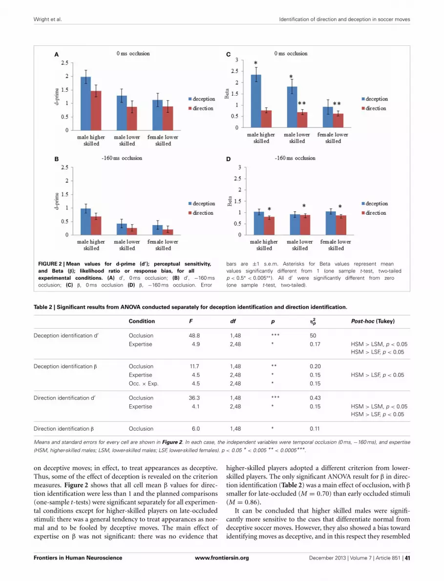
Wright et al. Identification of direction and deception in soccer moves
FIGURE 2 | Mean values for d-prime (d′); perceptual sensitivity,and Beta (β); likelihood ratio or response bias, for allexperimental conditions. (A) d′, 0 ms occlusion; (B) d′, −160 msocclusion; (C) β, 0 ms occlusion (D) β, −160 ms occlusion. Error
bars are ±1 s.e.m. Asterisks for Beta values represent meanvalues significantly different from 1 (one sample t-test, two-tailedp < 0.5∗ < 0.005∗∗). All d′ were significantly different from zero(one sample t-test, two-tailed).
Table 2 | Significant results from ANOVA conducted separately for deception identification and direction identification.
Condition F df p η2p Post-hoc (Tukey)
Deception identification d′ Occlusion 48.8 1,48 *** 50
Expertise 4.9 2,48 * 0.17 HSM > LSM, p < 0.05HSM > LSF, p < 0.05
Deception identification β Occlusion 11.7 1,48 ** 0.20
Expertise 4.5 2,48 * 0.15 HSM > LSF, p < 0.05
Occ. × Exp. 4.5 2,48 * 0.15
Direction identification d′ Occlusion 36.3 1,48 *** 0.43
Expertise 4.1 2,48 * 0.15 HSM > LSM, p < 0.05HSM > LSF, p < 0.05
Direction identification β Occlusion 6.0 1,48 * 0.11
Means and standard errors for every cell are shown in Figure 2. In each case, the independent variables were temporal occlusion (0 ms, −160 ms), and expertise
(HSM, higher-skilled males; LSM, lower-skilled males; LSF, lower-skilled females). p < 0.05 * < 0.005 ** < 0.0005***.
on deceptive moves; in effect, to treat appearances as deceptive.Thus, some of the effect of deception is revealed on the criterionmeasures. Figure 2 shows that all cell mean β values for direc-tion identification were less than 1 and the planned comparisons(one-sample t-tests) were significant separately for all experimen-tal conditions except for higher-skilled players on late-occludedstimuli: there was a general tendency to treat appearances as nor-mal and to be fooled by deceptive moves. The main effect ofexpertise on β was not significant: there was no evidence that
higher-skilled players adopted a different criterion from lower-skilled players. The only significant ANOVA result for β in direc-tion identification (Table 2) was a main effect of occlusion, with β
smaller for late-occluded (M = 0.70) than early occluded stimuli(M = 0.86).
It can be concluded that higher skilled males were signifi-cantly more sensitive to the cues that differentiate normal fromdeceptive soccer moves. However, they also showed a bias towardidentifying moves as deceptive, and in this respect they resembled
Frontiers in Human Neuroscience www.frontiersin.org December 2013 | Volume 7 | Article 851 | 41

Wright et al. Identification of direction and deception in soccer moves
the skilled handball goalkeepers in the study of Cañal-Brulandand Schmidt (2009). Results also showed that higher-skilledmales were significantly more sensitive overall than lower-skilledobservers to directional cues. However, observers on averageadopted a liberal criterion for direction identification (Figure 2),which is a significant tendency to treat deceptive moves likenormal moves for the purposes of identifying direction, in otherwords, to be fooled by the deceptive stimuli.
WHOLE-BRAIN ANALYSIS OF fMRI DATAWithin-groups analysisFigures 3–5 show group data for the activations due to the first-level contrast between the two soccer identification conditionsand the NBM control trials, superimposed on horizontal sectionsof a normalized brain anatomy.
The data were entered into separate 2 × 2 ANOVAs, using thefactorial design options of SPM8, One ANOVA for each partic-ipant group. Figures 3–5 show the responses in the deceptiontask colored cyan (light blue), and the responses in the direc-tion task colored magenta (pink). Overlapping activations areshown in a mixed color (purple). Both sets of data are basedon first-level t-contrasts measured relative to NBM control. Thesecond level data are displayed with a very conservative statis-tical threshold (p < 0.0005, FWE corrected) for both occlusionlevels combined. Figure 2 shows results for higher-skilled males,Figure 3 for lower-skilled males, and Figure 4 for lower-skilled
females. For all participant groups, the main anatomical areasshowing strong activations were similar for deception identifica-tion and direction identification and included regions identifiedas part of a human AON specifically the intraparietal sulcus(BA40) and premotor cortex (BA6). The supplementary motorarea in medial frontal cortex (BA6) was also consistently activated,along with the adjoining anterior cingulate cortex (ACC; BA32).Consistent activations in the anterior insula (BA13) were alsopresent. The numerical data corresponding to Figures 3–5 areavailable in Tables 3–5.
Areas showing stronger activation to deception than direc-tion identification are shown in darker blue; and areas showingstronger activation to direction than deception identification areshown in red. These are displayed with a liberal statistical crite-rion (p < 0.001 uncorrected, minimum cluster size = 5); some ofthese clusters coincide with the principal task-sensitive areas butsome do not. A further analysis of task differences will be given inthe next section of the Results.
Analysis of differences between identification tasks and expertisegroupsTo establish the significance of differences in between exper-tise groups and tasks, fMRI data were combined in a facto-rial ANOVA. There were three expertise groups (higher-skilledmales, lower-skilled males, and lower-skilled females), two lev-els of task (deception identification, direction identification), and
FIGURE 3 | Higher-skilled males. Second-level fMRI activations(p < 0.005, FWE corrected, 25 voxels minimum cluster size) to deceptionidentification (cyan) and direction identification (magenta) in point-lightsoccer video clips, relative to stimulus-matched non-biological motion(NBM) controls. Overlapping areas responding to both identification tasksappear purple. Activations above threshold (blobs) are displayed inco-registration with an individual normalized structural brain image andsampled in horizontal sections 10 mm apart from z = 60 to z = −20. In
darker blue areas, activation to deception identification exceeds activationto direction identification; and in red areas, activation to directionidentification exceeds activation to deception identification (at p < 0.001uncorrected). Key: a: premotor, BA6; b: parietal, BA40; c: medial frontal,BA6; d: anterior cingulate, BA32; e: posterior cingulate, BA23; f:dorsolateral prefrontal, BA46; g: caudate nucleus; h: superior temporalgyrus, BA37; i: anterior insula/frontal operculum, BA13/45; j: cerebellum;k: superior parietal lobule, BA7.
Frontiers in Human Neuroscience www.frontiersin.org December 2013 | Volume 7 | Article 851 | 42
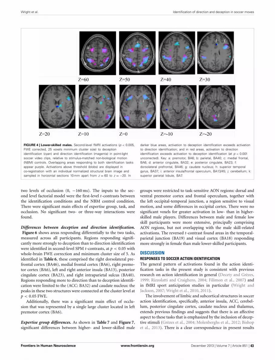
Wright et al. Identification of direction and deception in soccer moves
FIGURE 4 | Lower-skilled males. Second-level fMRI activations (p < 0.005,FWE corrected, 25 voxels minimum cluster size) to deceptionidentification (cyan) and direction identification (magenta) in point-lightsoccer video clips, relative to stimulus-matched non-biological motion(NBM) controls. Overlapping areas responding to both identification tasksappear purple. Activations above threshold (blobs) are displayed inco-registration with an individual normalized structural brain image andsampled in horizontal sections 10 mm apart from z = 60 to z = −20. In
darker blue areas, activation to deception identification exceeds activationto direction identification; and in red areas, activation to directionidentification exceeds activation to deception identification (at p < 0.001uncorrected). Key: a: premotor, BA6; b: parietal, BA40; c: medial frontal,BA6; d: anterior cingulate, BA32; e: posterior cingulate, BA23; f:dorsolateral prefrontal, BA46; g: caudate nucleus; h: superior temporalgyrus, BA37; i: anterior insula/frontal operculum, BA13/45; j: cerebellum; k:superior parietal lobule, BA7.
two levels of occlusion (0, −160 ms). The inputs to the sec-ond level factorial model were the first-level t-contrasts betweenthe identification conditions and the NBM control condition.There were significant main effects of expertise group, task, andocclusion. No significant two- or three-way interactions werefound.
Differences between deception and direction identification.Figure 6 shows areas responding differentially to the two tasks,
measured across all participants. Regions responding signifi-cantly more strongly to deception than to direction identificationwere identified in second-level SPM t-contrasts, at p < 0.05 withwhole-brain FWE correction and minimum cluster size of 5. Asidentified in Table 6, these comprised the right dorsolateral pre-frontal cortex (BA46), medial frontal cortex (BA6), right premo-tor cortex (BA6), left and right anterior insula (BA13), posteriorcingulate cortex (BA23), and right intraparietal sulcus (BA40).Regions responding more to direction than to deception identifi-cation were limited to the (ACC: BA32) and caudate nucleus: thepeaks in these two structures were connected at the cluster level atp < 0.05 FWE.
Additionally, there was a significant main effect of occlu-sion that was represented by a single large cluster located in leftpremotor cortex (BA6).
Expertise group differences. As shown in Table 7 and Figure 7,significant differences between higher- and lower-skilled male
groups were restricted to task-sensitive AON regions: dorsal andventral premotor cortex and frontal operculum, together withthe left occipital-temporal junction, a region sensitive to visualmotion, and some differences in occipital cortex. There were nosignificant voxels for greater activation in low- than in higher-skilled male players. Differences between male and female lowskill participants were more extensive, principally comprisingAON regions, but not overlapping with the male skill-relatedactivations. The reversed t-contrast found areas in the temporal-parietal junction (BA19) and visual cortex (BA18) respondingmore strongly in female than male lower-skilled participants.
DISCUSSIONRESPONSES TO SOCCER ACTION IDENTIFICATIONThe general pattern of activations found in the action identi-fication tasks in the present study is consistent with previousresearch on action identification in general (Decety and Grèzes,1999; Rizzolatti and Craighero, 2004; Filimon et al., 2007) andin fMRI sport anticipation studies in particular (Wright andJackson, 2007; Wright et al., 2010, 2011).
The involvement of limbic and subcortical structures in socceraction identification, specifically, anterior insula, ACC, cerebel-lum, posterior cingulate cortex, caudate nucleus and thalamus,extends previous findings and suggests that there is an affectiveaspect to these tasks that is emphasized by the inclusion of decep-tive stimuli (Grèzes et al., 2004; Molenberghs et al., 2012; Bishopet al., 2013). There is a clear correspondence in present results
Frontiers in Human Neuroscience www.frontiersin.org December 2013 | Volume 7 | Article 851 | 43
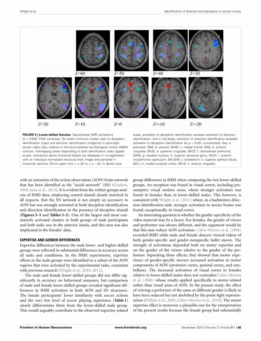
Wright et al. Identification of direction and deception in soccer moves
FIGURE 5 | Lower-skilled females. Second-level fMRI activations(p < 0.005, FWE corrected, 25 voxels minimum cluster size) to deceptionidentification (cyan) and direction identification (magenta) in point-lightsoccer video clips, relative to stimulus-matched non-biological motion (NBM)controls. Overlapping areas responding to both identification tasks appearpurple. Activations above threshold (blobs) are displayed in co-registrationwith an individual normalized structural brain image and sampled inhorizontal sections 10 mm apart from z = 60 to z = −20. In darker blue
areas, activation to deception identification exceeds activation to directionidentification; and in red areas, activation to direction identification exceedsactivation to deception identification (at p < 0.001 uncorrected). Key: a:premotor, BA6; b: parietal, BA40; c: medial frontal, BA6; d: anteriorcingulate, BA32; e: posterior cingulate, BA23; f: dorsolateral prefrontal,BA46; g: caudate nucleus; h: superior temporal gyrus, BA37; i: anteriorinsula/frontal operculum, BA13/45; j: cerebellum; k: superior parietal lobule,BA7; m: medial occipital cortex, BA18; n: anterior cingulate.
with an extension of the action observation (AON) brain networkthat has been identified as the “social network” (SN) (Grafton,2009; Juan et al., 2013). It is evident from the within-groups anal-ysis of fMRI data, employing control stimuli closely matched inall respects, that the SN network is not simply an accessory toAON but was strongly activated in both deception identificationand direction identification in the presence of deceptive stimuli(Figures 3–5 and Tables 3–5). One of the largest and most con-sistently activated clusters in both groups of male participantsand both tasks was in the anterior insula, and this area was alsoimplicated in the females’ data.
EXPERTISE AND GENDER DIFFERENCESExpertise differences between the male lower- and higher-skilledgroups were reflected in substantial differences in accuracy acrossall tasks and conditions. In the fMRI experiments, expertiseeffects in the male groups were identified in a subset of the AONregions that were activated by the experimental tasks, consistentwith previous research (Wright et al., 2010, 2011).
The male and female lower-skilled groups did not differ sig-nificantly in accuracy on behavioral measures, but comparisonof male and female lower-skilled groups revealed significant dif-ferences in fMRI activation in both AON and SN structures.The female participants’ lower familiarity with soccer actionsand the very low level of soccer playing experience (Table 1)clearly differentiates them from the lower-skilled male group.This would arguably contribute to the observed expertise-related
group differences in fMRI when comparing the two lower-skilledgroups. An exception was found in visual cortex, including pre-sumptive visual motion areas, where stronger activation wasfound in females than in lower-skilled males. This however, isconsistent with Wright et al. (2011) where, in a badminton direc-tion identification task, stronger activation in novice brains wasfound, exceptionally, in visual cortex.
An interesting question is whether the gender-specificity of thevideo material may be a factor. For females, the gender of viewerand performer was always different, and the argument would bethat this may reduce AON activation. Calvo-Merino et al. (2006)recorded fMRI while male and female dancers viewed videos ofboth gender-specific and gender-nonspecific ballet moves. Thestrength of activations depended both on motor expertise andon the gender of the viewer relative to the gender of the per-former. Separating these effects; they showed that motor expe-rience of gender-specific movers increased activation in motorcomponents of AON (premotor cortex, parietal cortex, and cere-bellum). The increased activation of visual cortex in femalesrelative to lower-skilled males does not contradict Calvo-Merinoet al. (2006) whose results applied specifically to motor-relatedrather than visual areas of AON. In the present study, the effectof viewing a performer of the same or different gender is likely tohave been reduced but not abolished by the point-light represen-tation (Pollick et al., 2005; Calvo-Merino et al., 2010). The motorexpertise effect is moreover a plausible one for the interpretationof the present results because the female group had substantially
Frontiers in Human Neuroscience www.frontiersin.org December 2013 | Volume 7 | Article 851 | 44

Wright et al. Identification of direction and deception in soccer moves
Table 3 | Locations of significant clusters as shown in Figure 3.
Higher-skilled males
Deception Direction
BA x y z Peak t Cluster BA x y z Peak t Cluster
L insula 13 −33 17 4 13.1 13 −30 17 4 9.8 108
R insula 13 33 20 −2 15.1 13 30 23 −2 11.3 96
medial frontal 6,8 3 17 49 11.6 306 6,8 3 17 49 8.2
anterior cingulate 32 6 23 34 10.7 32 12 17 40 8.3
32 9 17 40 9.9
L premotor 6 −24 −7 55 16.6 856 6 −21 −7 55 19.4 557
6 −39 −1 37 11.0
R premotor 6 30 −7 55 15.2 1760 6 30 −7 55 15.4 1646
6 42 5 40 12.8
L caudate −15 −7 19 7.7 −15 8 7 7.3
L thalamus VAN −12 −1 4 8.7 100 VAN −12 −1 4 7.7
VLN −12 −19 16 8.0
R thalamus LPN 15 −22 16 8.3 40
L parietal 40 −39 −46 58 13.0 957 40 −33 −46 55 11.6 688
40 −30 −49 55 40 −21 −49 55 11.4
40 −39 −43 43 40 −24 −58 55 10.4
R parietal 40 36 −43 49 15.6 1404 40 33 −43 49 13.5
40 45 −37 58 12.8 40 39 −34 55 12.3
40 36 −52 43 10.4
L fusiform 20 −42 −58 −23 11.3
R fusiform 20 45 −52 20 8.7
R mid temporal 21 54 −49 7 9.9
L temporal 19,39 −48 −70 13 16.1 355 19,39 −48 −70 13 18.6 321
39 −48 −58 10 11.6 39 −45 −61 10 12.1
R temporal 37,39 48 −61 7 13.5 589 37 51 −64 4 13.9 459
39 45 −58 10 12.0
L cerebellum 6 L −33 −58 −29 12.2 6 L −27 −55 −29 11.2 242
6 L −12 −73 −26 13.95 589 6 L −9 −70 −29 11.1
R cerebellum 6 R 6 −73 −26 8.0
R occipital 18 30 −91 7 7.9 39 18 30 −91 7 9.7 62
18 24 −91 −2 7.8 18 24 91 −2 9.1
VAN, ventral anterior nucleus; VLN, ventral lateral nucleus; LPN, lateral posterior nucleus.
less motor experience of soccer moves than the lower-skilled malegroup.
Together with previous work, our results suggest that increas-ing familiarity with observed actions as well as motor experienceof those actions is associated with increasing expertise and resultsin a shift in brain activation away from visual brain areas andtoward AON motor areas and SN areas.
There are some general limitations of current fMRI researchinto action observation in general and sporting expertise in par-ticular (Mann et al., 2013). The whole-body sensory-motor cou-pling, affordance-rich environment, and powerful contextual cuesin soccer field play greatly exceed what is available to an immobileviewer of videos in a scanner. It is a challenge for future researchto study the neural basis of sporting expertise in more dynamicand interactive scenarios. Despite this limitation, research to datehas shown a consistent relationship between anticipatory behav-ioral responses to sports video and expertise in open-skill sports,
and this extends to the use of point-light video stimuli (Abernethyet al., 2001, 2008; Huys et al., 2008). Moreover the present behav-ioral results recorded in the scanner (section Signal DetectionTheory Analysis) have revealed expertise effects both in sensitiv-ity (d′) and in response strategy (β). We would therefore, arguethat the present fMRI results reflect the brain’s processing of theminimum visual information sufficient to support an anticipa-tory response, and that our methods provide sufficient sensitivityto detect and localize expertise effects in the brain.
TASK-RELATED DIFFERENCES IN fMRIThere were no significant interactions between the task (direc-tion identification or deception identification) and the threeparticipant groups, either in the behavioral or in the fMRIdata, but there were significant task-related differences in fMRIactivations overall. Although AON and SN were activatedstrongly in both deception and direction identification, there
Frontiers in Human Neuroscience www.frontiersin.org December 2013 | Volume 7 | Article 851 | 45

Wright et al. Identification of direction and deception in soccer moves
Table 4 | Locations of significant clusters as shown in Figure 4.
Lower-skilled males
Deception Direction
BA x y z Peak t Cluster BA x y z Peak t Cluster
L insula 13 −33 17 −2 10.8 118 13 −33 17 −2 10.1 78
R insula 13 30 23 4 12.6 806
Medial frontal 6 0 14 49 12.9 187 6 0 14 49 11.4 95
Anterior cingulate 32 6 23 31 6.7
L premotor 6 −24 −4 58 11.2 99 6 −24 −4 58 17.2 296
6 −36 −4 55 9.3 6 −33 −4 55 11.4
R premotor 6 33 −4 55 11.0 6 33 −4 55 14.0 756
9 45 5 31 12.5 6 24 −7 67 13.9
6,9 48 8 34 12.0
L parietal 40 −33 −52 58 11.6 434 40 −33 −52 58 14.3 757
40 −54 −40 37 11.1 40 −30 −43 46 12.7
40 −33 −43 46 9.9
R parietal 40 39 −49 40 12.4 40 36 −49 52 12.3 655
40 30 −49 40 11.0 469 39 −37 43 12.0
40 63 −34 31 7.7 27
R temporal 37,21 54 −58 4 14.5 359
L temporal 39 −27 −58 −29 8.5 38
39 −42 −61 −29 7.0
R temporal 37 54 −58 4 12.5 39 45 −67 7 13.0 231
37 48 −67 −5 11.6 37 45 −64 −5 12.1
39 39 −67 13 7.2
L sup parietal 7 −12 −73 49 8.0 32 7 −24 −52 64 11.1
7 −9 −67 58
R sup parietal 7 12 −73 55 10.9 7 12 −73 −23 10.7
Post. cingulate 23 −6 −55 −20 6.9
Medial occipital 18 −9 −76 −23 8.6 66
31 −18 −58 −23 6.7
Cerebellum 6 L −9 −73 −23 9.4 166
6 L −27 −55 −29 7.4
6 L −6 −55 −23 7.3
were also significant differences in the two conditions (sectionsWithin-Groups Analysis and Differences Between Deception andDirection Identification). The SN network is engaged particu-larly when participants are required to make inferences aboutthe intentions of other people’s behavior (Juan et al., 2013). Thiswas explicitly the case in the deception identification condition,and in comparison with the direction identification condition,where participants were not required to identify deception, therewas significantly greater activation in left and right insula andposterior cingulate, which are part of SN. It must also be fullyrecognized that despite the simplified and abstract nature of thepoint-light stimuli, they were universally understood as meaning-ful in a specific social context (the game of soccer).
The direct comparison of deception identification with direc-tion identification in the present results provides further insightsinto specialization within the AON/SN network. First of all, therewas significantly greater activation of ACC and caudate nucleusin the direction identification task compared with the deception
identification task. The role of anterior cingulate has been estab-lished in response conflict and suppression of incorrect responsetendencies (Carter and van Veen, 2007); and the caudate has beenimplicated in the learning of associations between stimuli andresponse tendencies (Melcher et al., 2013). This interpretationis consistent with both previous and present results; for exam-ple, Bishop et al. (2013) found ACC activity in higher-skilledplayers in the presence of deception, at a very early occlusionlevel, −160 ms. Bishop et al. (2013) also proposed that enhancedcaudate activation in experts when predicting direction at veryearly occlusion (−160 ms), prior to an oncoming opponent’schange of direction, indicated the learning of response contin-gencies. In the present study, awareness that an automatic leftor right response tendency may need to be corrected, accordingto whether the move appears normal or deceptive, would occuronly in the direction identification task. Thus, the greater cau-date activation when predicting direction, may arise because theclose mapping of leftward and rightward movements to left or
Frontiers in Human Neuroscience www.frontiersin.org December 2013 | Volume 7 | Article 851 | 46
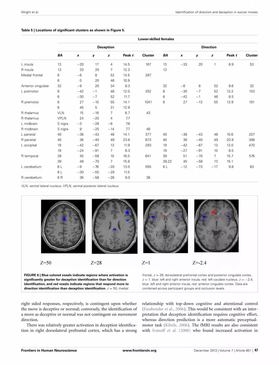
Wright et al. Identification of direction and deception in soccer moves
Table 5 | Locations of significant clusters as shown in Figure 5.
Lower-skilled females
Deception Direction
BA x y z Peak t Cluster BA x y z Peak t Cluster
L insula 13 −20 17 4 14.5 167 13 −33 20 1 8.9 53
R insula 13 33 20 7 12.3 13
Medial frontal 6 −6 8 52 14.5 287
6 0 20 46 10.9
Anterior cingulate 32 −9 20 34 8.3 32 −6 8 52 9.6 32
L premotor 6 −42 −1 46 12.0 252 6 −30 −7 52 13.2 153
6 −30 −7 52 11.7 6 −42 −1 46 8.5
R premotor 6 27 −10 55 14.1 1041 6 27 −13 55 13.9 191
6 45 5 31 12.9
R thalamus VLN 15 −16 7 6.7 43
R thalamus VPLN 24 −25 4 7.7
L midbrain S nigra −3 −28 −8 7.6
R midbrain S nigra 9 −25 −14 7.7 48
L parietal 40 −36 −43 46 14.1 377 40 −36 −43 46 10.6 207
R parietal 40 36 −40 49 23.6 673 40 36 −40 49 20.9 398
L occipital 19 −42 −67 13 11.9 293 19 −42 −67 13 13.0 470
19 −24 −91 7 8.3 19 −27 −91 10 8.5
R temporal 39 45 −58 10 16.5 641 39 51 −70 7 15.7 576
39 48 −70 7 15.8 39,22 45 −58 13 15.1
L cerebellum 6 L −9 −76 −20 13.0 555 6 L −12 −73 −17 9.8 83
6 L −30 −55 −29 11.5
R cerebellum 6 R 36 −58 −26 9.0 36
VLN, ventral lateral nucleus; VPLN, ventral posterior lateral nucleus.
FIGURE 6 | Blue colored voxels indicate regions where activation issignificantly greater for deception identification than for directionidentification, and red voxels indicate regions that respond more todirection identification than deception identification. z = 50; medial
frontal, z = 28; dorsolateral prefrontal cortex and posterior cingulate cortex,z = 1; blue: left and right anterior insula, red: left caudate nucleus. z = −2.4;blue: left and right anterior insula; red: anterior cingulate cortex. Data arecombined across participant groups and occlusion levels.
right sided responses, respectively, is contingent upon whetherthe move is deceptive or normal; conversely, the identification ofa move as deceptive or normal was not contingent on movementdirection.
There was relatively greater activation in deception identifica-tion in right dorsolateral prefrontal cortex, which has a strong
relationship with top-down cognitive and attentional control(Fassbender et al., 2006). This would be consistent with an inter-pretation that deception identification requires cognitive effort,whereas direction prediction is a more automatic perceptual-motor task (Kibele, 2006). The fMRI results are also consistentwith Ivanoff et al. (2008) who found increased activation in
Frontiers in Human Neuroscience www.frontiersin.org December 2013 | Volume 7 | Article 851 | 47

Wright et al. Identification of direction and deception in soccer moves
Table 6 | Peak activations at p < 0.05 FWE corrected.
BA Cluster Coordinates Peak t
DECEPTION > DIRECTION
Medial frontal 6 36 6, 11, 49 6.3
R dorsolateral prefrontal 46 62 45, 29, 25 5.8
L insula 13 21 −42, 14, 1 5.6
R insula 13 53 33, 20, 10 5.3
13 45, 14, −5 4.9
13 36, 17, 1 4.7
R premotor 6 42 42, −1, 31 5.3
Posterior cingulate 24 8 −3, −31, 28 5.0
R parietal 40 5 60, −37, 31 4.9
DIRECTION > DECEPTION
Anterior cingulate 32 53 −12, 20, −2 5.9
Caudate nucleus 53 −3, 20, 1 5.0
LATE > EARLY OCCLUSION
L premotor 6 151 −33, −22, 70 6.0
pre-SMA (medial frontal cortex, BA6) associated with criterion(β) effects in a motion coherence task.
The behavioral data identified the strong influence of the trialtype (deceptive vs. normal) on accuracy, and found significantinteractions with task type. It may be possible in future to con-duct a finer-grained analysis of fMRI responses to normal anddeceptive moves using single-trial blocks (Bishop et al., 2013) andmulti-voxel pattern analysis (Norman et al., 2006) in order tostudy how normal and deceptive moves are classified, and howthis classification interacts with other variables such as temporalocclusion and task type.
It is likely that for both higher- and lower-skilled players,deception identification is a less practiced skill, requiring greatercognitive effort, and that conversely, the ability to react to thetrajectory of someone’s body actions is to some extent based ongeneral as well as sport-specific experience, and therefore, likelyto have become somewhat automatic. Thus, for late-occludedsequences, the direction of a normal move was determined atsignificantly higher accuracy than identification of this move asnormal, suggesting that if the valid direction cues can be picked
Table 7 | Expertise group differences at p < 0.05 FWE corrected.
BA Coordinates Cluster Peak t Coordinates Cluster Peak t
Higher-skilled males > Lower-skilled males Lower-skilled males > Lower-skilled females
L premotor 6 −27, −7, 46 40 7.3 −15, −1, 73 48 6.0
9 −42, 2, 28 10 5.0 −21, −4, 61 48 5.7
R premotor 6 30, −1, 67 6 5.3
6 27, −10, 55 5 4.8 18, −7, 61 16 5.5
R prefrontal 10 33, 41, 13 21 5.8
R frontal operculum 44 39, 8, 22 5 5.0
R frontal operculum 45 39, 20, 16 10 5.0
L anterior cingulate 32 −21, 44, −2 80 5.1
R anterior cingulate 32 15, 47, 4 36 5.2
L Parietal 40 −60, −34, 31 37 5.3
L superior parietal lobule 7 −12, −73, 43 54 5.3
R superior parietal lobule 7 15, −76, 46 14 6.1
L ventrolateral temporal 21 66, −19, −14 17 5.1
L temporal occipital 19 −54, −67, 13 13 5.8 −27, −76, 22 27 6.1
37 −45, −67, −5 43 5.8
L occipital 18 −24, −94, 1 17 5.5
R occipital 18 27, −94, 7 6 5.0
L caudate nucleus −21, 26, −2 80) 5.8
R caudate nucleus 18, 26, −2 36 5.6
Cerebellum anterior lobe −21, −58, −29 6 5.0
Lower-skilled females > Lower-skilled males
L temporal 39 36, −64, 25 23 5.4
R temporal 39 45, −65, 16 70 6.2
L occipital 19 −15, −97, 19 31 5.8
18 −21, −91, −2 12 5.2
R occipital 18 15, −85, 16 18 5.8
18 24, −94, 7 5 5.1
Frontiers in Human Neuroscience www.frontiersin.org December 2013 | Volume 7 | Article 851 | 48

Wright et al. Identification of direction and deception in soccer moves
FIGURE 7 | Activation differences between higher- and lower-skilledmales (red) and between lower-skilled males and females(green) in soccer action identification tasks at p < 0.05 FWEcorrected, minimum cluster size = 5 voxels. Data are combined
across task types (deception identification and direction identification)and occlusion levels (0 ms, −160 ms). Second-level group analysis isbased on first-level contrasts between identification tasks and NBMcontrols.
up, they readily prime the appropriate response. However, analy-sis of the sensitivity (d′) which takes into account the proportionof incorrect responses to deceptive moves, showed similar butslightly lower overall sensitivity for direction identification com-pared with deception identification, which would be consistentwith the similar global strength of fMRI activations seen acrosstasks.
Likewise, early-occluded deceptive moves gave rise to signifi-cantly worse than chance direction identification because lower-skilled players especially were not simply responding randomlybut were misdirected by the deceptive cues. This was borne out byanalysis of likelihood ratio (β). In the direction identification task,observers adopted a liberal criterion, that is, one which increasesboth hits and false positives, and this was interpreted as a directresponse to directional cues—veridical cues in the case of normalmoves and false cues in the case of deceptive moves. Conversely,in the deception detection task, male observers tended to adopta conservative criterion on late-occluded stimuli, which meansthat they were biased toward judging such moves as deceptive,and this inflated their correct detections of deceptive moves andreduced their correct detections of normal moves. Their over-all accuracy remained higher than that of the two lower-skilledgroups, as revealed by the d′ measure. The ability to identify amove as deceptive however, does not guarantee that its true direc-tion can be identified. Experts are known in some circumstancesto delay their responses (Brault et al., 2012; Mori and Shimada,2013), perhaps so that they can inhibit and correct their initial
automatic reactions. This dissociation between performance ondeception and identification tasks is consistent with the differinginvolvement of the components of the AON and SN observed inthe brain imaging data.
ACKNOWLEDGMENTSSupported by the Research Grants Council of the HongKong Special Administrative Region, China. Project No. HKU7400/05H.
REFERENCESAbernethy, B., Gill, D., Parks, S. L., and Packer, S. T. (2001).
Expertise and the perception of kinematic and situationalprobability information. Perception 30, 233–252. doi: 10.1068/p2872
Abernethy, B., and Russell, D. G. (1984). Advance cue utilisation by skilled cricketbatsmen. Austr. J. Sci. Med. Sport 16, 2–10.
Abernethy, B., Zawi, K., and Jackson, R. (2008). Expertise and attune-ment to kinematic constraints. Perception 37, 931–948. doi: 10.1068/p5340
Abernethy, B., and Russell, D. G. (1987). Expert-novice differences in an appliedselective attention task. J. Sport Psychol. 9, 326–245.
Aglioti, S. M., Cesari, P., Romani, M. and Urgesi, C. (2008). Action anticipation andmotor resonance in elite basketball players. Nat. Neurosci. 11, 1109–1116. doi:10.1038/nn.2182
Bishop, D. T., Wright, M. J., Jackson, R. C., and Abernethy, B. (2013). Neural basisof anticipation skill in soccer: an fMRI study. J. Sport Exerc. Psychol. 35, 98–109.
Brault, S., Bideau, B., Kulpa, R., and Craig, C. M. (2012). Detecting deceptionin movement: the case of the side-step in rugby. PLoS ONE 7:e37494. doi:10.1371/journal.pone.0037494
Frontiers in Human Neuroscience www.frontiersin.org December 2013 | Volume 7 | Article 851 | 49

Wright et al. Identification of direction and deception in soccer moves
Calvo-Merino, B., Ehrenberg, S., Leung, D., and Haggard, P. (2010). Experts seeit all: configural effects in action observation. Psychol. Res. Psych. Forsch. 74,400–406. doi: 10.1007/s00426-009-0262-y
Calvo-Merino, B., Grèzes, J., Glaser, D. E., Passingham, R. E., and Haggard, P.(2006). Seeing or doing? Influence of visual and motor familiarity in actionobservation. Curr. Biol. 16, 1905–1910. doi: 10.1016/j.cub.2006.07.065
Cañal-Bruland, R., and Schmidt, M. (2009). Response bias in judging deceptivemovements. Acta Psychol. 130, 235–240. doi: 10.1016/j.actpsy.2008.12.009
Cañal-Bruland, R., van der Kamp, J., and van Kesteren, J. (2010). An examinationof motor and perceptual contributions to the recognition of deception fromothers’ actions. Hum. Mov. Sci. 29, 94–102. doi: 10.1016/j.humov.2009.10.001
Cañal-Bruland, R., and Williams, A. M. (2010). Recognizing and predicting move-ment effects: identifying critical movement features. Exp. Psychol. 57, 320–326.doi: 10.1027/1618-3169/a000038
Carter, C. S., and van Veen, C. (2007). Anterior cingulate cortex and conflict detec-tion: an update of theory and data. Cogn. Affect. Neurosci. 7, 367–379. doi:10.3758/CABN.7.4.367
Decety, J., and Grèzes, J. (1999). Neural mechanisms subserving the percep-tion of human actions. Trends Cogn. Sci. 3, 172–178. doi: 10.1016/S1364-6613(99)01312-1
Dicks, M., Uehara, L., and Lima, C. (2011). Deception, individual differences andpenalty kicks: implications for Goalkeeping in association football. Int. J. SportsSci. Coaching 6, 515–521. doi: 10.1260/1747-9541.6.4.515
Fassbender, C., Simoes-Franklin, C., Murphy, K., Hester, R., Meaney, J., Roberton,I. H., et al. (2006). The role of a right fronto-parietal network in cognitivecontrol: common activations for “Cues-to-Attend” and response inhibition.J. Psychophysiol. 20, 286–296. doi: 10.1027/0269-8803.20.4.286
Filimon, F., Nelson, J. D. Hagler, D. J., and Sereno, M. I. (2007). Human corti-cal representations for reaching: mirror neurons for execution, observation andimagery, Neuroimage 37, 1315–1328. doi: 10.1016/j.neuroimage.2007.06.008
Grafton, S. T. (2009). Embodied cognition and the simulation of action to under-stand others. Ann. N.Y. Acad. Sci. 1156, 97–117. doi: 10.1111/j.1749-6632.2009.04425.x
Green, D. M., and Swets, J. A. (1966). Signal detection theory and psychophysics. NewYork, NY; London: Wiley.
Grèzes, J., Frith, C., and Passingham, R. E. (2004). Brain mechanisms forinferring deceit in the actions of others. J. Neurosci. 24, 5500–5505 doi:10.1523/JNEUROSCI.0219-04.2004
Huys, R., Smeeton, N. J., Hodges, N. J., Beek, P. J., and Williams, A. M. (2008).On the dynamic information underlying visual anticipation skill. Percept.Psychophys. 70, 1217–1234. doi: 10.3758/PP.70.7.1217
Ivanoff, J., Branning, P., and Marois, R. (2008). fMRI evidence for a dual processaccount of the speed-accuracy tradeoff in decision-making. PLoS ONE 3:e2635.doi: 10.1371/journal.pone.0002635
Jackson, R. C., and Mogan, P. (2007). Advance visual information, awareness, andanticipation skill. J. Motor Behav. 39, 341–351. doi: 10.3200/JMBR.39.5.341-352
Jackson, R. C., Warren, S., and Abernethy, B. (2006). Anticipation skill andsusceptibility to deceptive movement. Acta Psychol. 123, 355–371. doi:10.1016/j.actpsy.2006.02.002
Juan, E., Frum, C., Bianchi-Demicheli, F., Wang, Y.-W., Lewis, J. W., and Cacioppo,S. (2013). Beyond human intentions and emotions. Front. Hum. Neurosci. 7:99.doi: 10.3389/fnhum.2013.00099
Kibele, A. (2006). Non-consciously controlled decision making for fast motorreactions in sports –a priming approach for motor responses to non-consciously perceived movement features. Psychol. Sport Exerc. 7, 591–610. doi:10.1016/j.psychsport.2006.05.001
Kunde, W., Skirde, S., and Weigelt, M. (2011). Trust my face: cognitive factors ofhead fakes in sports. J. Exp. Psychol. Appl. 17, 110–127. doi: 10.1037/a0023756
Macmillan, N., and Creelman, C. (2005). Detection Theory: A User’s Guide. 2ndEdn. Mawah, NJ: Lawrence Erlbaum Associates.
Maldjian, J. A., Laurienti, P. J., Kraft, R. A., and Burdette, J. H. (2003). Anautomated method for neuroanatomic and cytoarchitectonic atlas-based inter-rogation of fmri data sets. Neuroimage 19, 1233–1239. doi: 10.1016/S1053-8119(03)00169-1
Mann, D., Dicks, M., Cañal-Bruland, R., and van der Kamp, J. (2013).Neurophysiological studies may provide a misleading picture of howperceptual-motor interactions are coordinated. i-Perception 4, 78–80. doi:10.1068/i0569ic
Melcher, T., Winter, D., Hommel, B., Pfister, R., Dechent, P., and Gruber, O.(2013). The neural substrate of the ideomotor principle revisited: evidencefor asymmetries in action-effect learning. Neuroscience 231, 13–27. doi:10.1016/j.neuroscience.2012.11.035
Molenberghs, P., Cunnington, R., and Mattingley, J. B. (2012). Brain regions withmirror properties: a meta-analysis of 125 human fMRI studies. Neurosci. Behav.Rev. 36, 341–349. doi: 10.1016/j.neubiorev.2011.07.004
Mori, S., and Shimada, T. (2013). Expert anticipation from deceptive action. Atten.Percept. Psychol. 75, 751–770. doi: 10.3758/s13414-013-0435-z
Muller, S., Abernethy, B., and Farrow, D. (2006). How do world-class cricket bats-men anticipate a bowler’s intention? Q. J. Exp. Psychol. 59, 2162–2186. doi:10.1080/02643290600576595
Norman, K. A., Polyn, S. M., Detre, G. J., and Haxby, J. V. (2006). Beyondmind-reading: multi-voxel pattern analysis of fMRI data. Trends Cogn. Sci. 10,424–430. doi: 10.1016/j.tics.2006.07.005
Pollick, F. E., Kay, J. W., Heim, K., and Stringer, R. (2005). Gender recognition frompoint-light walkers. J. Exp. Psychol. Hum. Percept. Perform. 31, 1247–1265. doi:10.1037/0096-1523.31.6.1247
Reilly, T., Williams, A. M., Nevill, A., and Franks, A. (2000). A multidisciplinaryapproach to talent identification in soccer. J. Sports Sci. 18, 695–702. doi:10.1080/02640410050120078
Rizzolatti, G., and Craighero, L. (2004). The mirror–neuron system. Annu. Rev.Neurosci. 27, 169–192. doi: 10.1146/annurev.neuro.27.070203.144230
Rowe, R., Poulter, D. M., and McKenna, F. P. (2009). The effect of disguise on noviceand expert tennis players’ anticipation ability. J. Appl. Sport Psychol. 21, 178–185.doi: 10.1080/10413200902785811
Savelsbergh, G. J. P., Van der Kamp, J., Williams, A. M., and Ward, P.(2005). Anticipation and visual search behaviour in expert soccer goalkeepers.Ergonomics 48, 1686–1697. doi: 10.1080/00140130500101346
Sebanz, N., and Shiffrar, M. (2009). Detecting deception in a bluffing body: the roleof expertise. Psychon. Bull. Rev. 16, 170–175. doi: 10.3758/PBR.16.1.170
Smeeton, N. J., and Williams, A. M. (2012). The role of movement exaggeration inthe anticipation of deceptive soccer penalty kicks. Br. J. Psychol. 103, 539–555.doi: 10.1111/j.2044-8295.2011.02092.x
Williams, A. M., Ford, P. R., Eccles, D. W., and Ward, P. (2011). Perceptual-cognitiveexpertise in sport and its acquisition: implications for applied cognitive psychol-ogy. Appl. Cogn. Psychol. 25, 432–442. doi: 10.1002/acp.1710
Wright, M. J., Bishop, D. T., Jackson, R. C., and Abernethy, B. (2010). FunctionalMRI reveals expert-novice differences in brain activation during sport-relatedanticipation. Neuroreport 21, 94–98. doi: 10.1097/WNR.0b013e328333dff2
Wright, M. J., Bishop, D. T., Jackson, R. C., and Abernethy, B. (2011). CorticalfMRI activation to opponents’ body kinematics in sport-related anticipation:expert-novice differences with normal and point-light video. Neurosci. Lett. 500,216–221. doi: 10.1016/j.neulet.2011.06.045
Wright, M. J., and Jackson, R. C. (2007). Brain regions concerned with percep-tual skills in tennis: an fMRI study. Int. J. Psychophysiol. 63, 214–220. doi:10.1016/j.ijpsycho.2006.03.018
Yarrow, K., Brown, P., and Krakauer, J. W. (2009). Inside the brain of an eliteathlete: the neural processes that support high achievement in sports. Nat. Rev.Neurosci. 10, 585–596. doi: 10.1038/nrn2672
Conflict of Interest Statement: The authors declare that the research was con-ducted in the absence of any commercial or financial relationships that could beconstrued as a potential conflict of interest.
Received: 29 August 2013; accepted: 21 November 2013; published online: 17 December2013.Citation: Wright MJ, Bishop DT, Jackson RC and Abernethy B (2013) Brain regionsconcerned with the identification of deceptive soccer moves by higher-skilled and lower-skilled players. Front. Hum. Neurosci. 7:851. doi: 10.3389/fnhum.2013.00851This article was submitted to the journal Frontiers in Human Neuroscience.Copyright © 2013 Wright, Bishop, Jackson and Abernethy. This is an open-accessarticle distributed under the terms of the Creative Commons Attribution License(CC BY). The use, distribution or reproduction in other forums is permitted, pro-vided the original author(s) or licensor are credited and that the original publi-cation in this journal is cited, in accordance with accepted academic practice. Nouse, distribution or reproduction is permitted which does not comply with theseterms.
Frontiers in Human Neuroscience www.frontiersin.org December 2013 | Volume 7 | Article 851 | 50

OPINION ARTICLEpublished: 16 October 2013
doi: 10.3389/fnhum.2013.00637
Expertise in action observation: recent neuroimagingfindings and future perspectivesLuca Turella1*, Moritz F. Wurm1, Raffaele Tucciarelli 1 and Angelika Lingnau1,2
1 Center for Mind/Brain Sciences (CIMeC), University of Trento, Trento, Italy2 Department of Cognitive Sciences, University of Trento, Trento, Italy*Correspondence: [email protected]; [email protected]
Edited by:Robert Langner, Heinrich Heine University Düsseldorf, Germany
Reviewed by:Svenja Caspers, Research Centre Juelich, Germany
Keywords: fMRI, expertise, action observation network, mentalizing system
INTRODUCTIONIn everyday life, we continuously inter-act with other individuals. Understandingactions of other people, i.e., the ability todistinguish between different actions, suchas passing over vs. threatening someonewith a knife, has been crucial for the sur-vival of our species and is a fundamentalcapability for our social interactions.
Neuroimaging studies investigated theneural substrates subtending action per-ception using a variety of techniques,ranging from univariate analysis of fMRIdata (Brass et al., 2007; Gazzola et al.,2007; De Lange et al., 2008; Gazzola andKeysers, 2009; Turella et al., 2009a, 2012;Wurm et al., 2011; Wurm and Schubotz,2012; Wurm et al., 2012; Lingnau andPetris, 2013), to fMRI repetition sup-pression (Dinstein et al., 2007; Chonget al., 2008; Lingnau et al., 2009; Kilneret al., 2009) and multivoxel patternanalysis (MVPA; Dinstein et al., 2008a;Oosterhof et al., 2010, 2012). These stud-ies reported the consistent recruitment ofa number of regions, generally assumedas pertaining to two different networks,typically referred to as the action obser-vation network (AON) and the mental-izing system (Figure 1A). Both networkshave been advocated to be involved inaction understanding (Brass et al., 2007;De Lange et al., 2008; Van Overwalle,2009; Van Overwalle and Baetens, 2009;Wurm et al., 2011), but their preciseroles and their causal involvement arestrongly debated (Dinstein et al., 2008b;Mahon and Caramazza, 2008; Hickok,2009; Turella et al., 2009b; Rizzolatti andSinigaglia, 2010).
In homology with monkey neuro-physiological studies, three regions have
been proposed to form the human AON(Rizzolatti and Craighero, 2004; Rizzolattiand Sinigaglia, 2010; see Figure 1A). This“core” AON was defined as comprising(i) the ventral premotor cortex (PMV)together with the posterior part of theinferior frontal gyrus (pIFG), (ii) the ante-rior inferior parietal lobule (aIPL) and(iii) the superior temporal sulcus (STS).Human neuroimaging studies suggestedthe recruitment of several additional areasthat were incorporated in an “extended”version of the AON (Gazzola and Keysers,2009; Caspers et al., 2010; see Figure 1A).
The mentalizing system has been identi-fied in human neuroimaging studies inves-tigating social cognition tasks, such asintention and beliefs attribution about theself or others, while observing action-related stimuli (Van Overwalle, 2009). Theregions consistently assigned to this net-work are the medial prefrontal cortex(mPFC) and the temporo-parietal junc-tion (TPJ) (Figure 1A), and less oftenalso the precuneus and the posterior cin-gulate cortex (Amodio and Frith, 2006;Brass et al., 2007; De Lange et al., 2008;Van Overwalle, 2009; Van Overwalle andBaetens, 2009).
The first description of the involvementof sensorimotor regions during actionperception started with the discovery ofmirror neurons in the ventral premotorcortex in macaque monkeys (Di Pellegrinoet al., 1992). These visuomotor neuronsresponded both while the monkey exe-cuted or observed similar actions andwere later described also within the mon-key inferior parietal lobule (Fogassi et al.,2005). Note that both regions also containneurons with motor-only and visual-onlyproperties (Gallese et al., 1996, 2002).
Following their discovery, motortheories of action understanding proposedthat mirror neurons might provide thebasis for a matching mechanism betweenwhat we observe and what we can performallowing the understanding of observedactions in motoric terms (Rizzolatti et al.,2001). Even if this hypothesis is stronglydebated (Jacob and Jeannerod, 2005;Mahon and Caramazza, 2008; Hickok,2009, 2013), a similar homologue mech-anism has been proposed to exist inthe human AON (Rizzolatti et al., 2001;Rizzolatti and Craighero, 2004; Rizzolattiand Sinigaglia, 2010).
In this brief overview, we will firstdescribe previous fMRI studies that inves-tigated how motor experience affects acti-vation within the AON, and to whichdegree these studies allow drawing con-clusions about the role of this network inaction understanding. As the majority ofthe studies investigated only the AON andgiven the limited scope of this Opinion, wewill focus on this network, even if our con-siderations might also hold true for otherareas. We will then try to delineate howfuture studies might exploit motor exper-tise as a tool for gaining insights into theneural basis of action understanding.
RECENT NEUROIMAGING FINDINGSON MOTOR EXPERTISE IN ACTIONOBSERVATIONFollowing motor theories of actionunderstanding, changes in motor reper-toire should modify the brain responsewithin the AON while observing thesenewly acquired actions. Starting fromthis assumption, most studies on exper-tise investigated how the acquisitionof a skilled action, such as sport or
Frontiers in Human Neuroscience www.frontiersin.org October 2013 | Volume 7 | Article 637 |
HUMAN NEUROSCIENCE
51
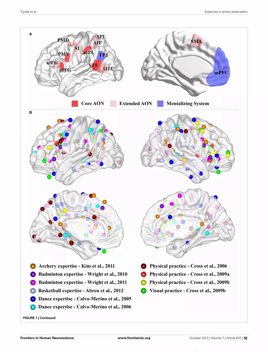
Turella et al. Expertise in action observation
FIGURE 1 | Continued
Frontiers in Human Neuroscience www.frontiersin.org October 2013 | Volume 7 | Article 637 | 52

Turella et al. Expertise in action observation
FIGURE 1 | (A) Schematic representation of the core AON, extended AONand mentalizing system. Three-dimensional representation of lateral andmedial brain surface. The regions assigned to the “core” AON, “extended”AON and the “mentalizing” system are depicted. In red, the core AON ispresented comprising: the PMV/pIFG complex, the aIPL and the STS. In palered, the “extended” AON is presented comprising: the anterior part of theinferior frontal gyrus, (aIFG), the dorsal premotor cortex (PMD), thesupplementary motor area (SMA), the superior parietal lobule (SPL), theanterior intraparietal sulcus (AIP), the somatosensory cortex (S1) and theoccipito-temporal cortex (OTC), including also STS. The mentalizing system(blue) is assumed to consist of the medial prefrontal cortex (mPFC) and thetemporo-parietal junction (TPJ). Note that the extension of these networks isnot representative of their real dimension or functional significance.(B) Expertise effects. Three-dimensional representation of lateral and medial
brain surface with location of peaks for the comparisons of interestsuperimposed. For Kim et al. (2011), we considered the comparison betweenthe two groups (Table 2). For Wright et al. (2010), we considered results fromROI analysis (Table 2). For Wright et al. (2011), we plotted results for normalvideo (Table 2). For Abreu et al. (2012), we used the peak of the significantcluster within the temporal lobe in the group comparison (page 1649 of themanuscript). For Calvo-Merino et al. (2005), we plotted the reportedinteraction (see Table 1). For Calvo-Merino et al. (2006), we plotted the resultsfrom Table S2. For Cross et al. (2006), we considered the main effect of thecontrast of interest (Table 2). For Cross et al. (2009a), we reported thecontrast for physical training (Danced > Untrained) (Table 1). For Cross et al.(2009b), we considered the physical training results (Table 1) and theobservational training results (Table 1). We excluded the peaks located withinthe cerebellum.
dance moves, affects AON activity whileobserving the same movement.
Most of the contributions investigat-ing motor expertise while observing sportactions are limited to one or few studieswithin the same domain, such as archery(Kim et al., 2011), badminton (Wrightet al., 2010, 2011) or basketball (Abreuet al., 2012). Typically, these studies com-pare the blood-oxygen-level-dependent(BOLD) response between experts andnovices. Although these studies consid-ered different tasks and comparisons ofexperimental conditions, they seem tosuggest a stronger activation for expertsin comparison to novices not limitedto the AON but recruiting also otherbrain regions (see Figure 1B). However,an interpretation of these results is dif-ficult as, in addition to extensive prac-tice of the observed movements, expertsalso have a strong visual familiarity withthe observed stimuli which might affectthe BOLD effect within the very sameregions.
Beside these sparse investigations ondifferent sport actions, a more system-atic investigation involved the effect ofdance expertise on activity within theAON (Calvo-Merino et al., 2005, 2006;Cross et al., 2006, 2009a,b, 2012, 2013;Pilgramm et al., 2010). Calvo-Merinoet al. (2005) measured the BOLD effectof ballet dancers, capoeira dancers, andnon-dancers watching two different typesof dance movements (ballet or capoeiramoves). They found a stronger recruit-ment of several regions within the AON(bilateral PMD, bilateral SPL and AIP, leftPMV and left STS) in ballet and capoeiradancers for the observation of the trainedin comparison to the untrained dancestyle, whereas they found no difference
between the two dance styles in the non-dancers.
Calvo-Merino et al. argued that theactivation for the trained in compari-son to the untrained dance style wasdue to simulation of those actions thatwere within the motor repertoire of thedancer. Alternatively, as pointed out above,dancers’ strong visual familiarity with theobserved stimuli might affect the mea-sured difference in BOLD effect.
In a follow up study, Calvo-Merinoet al. (2006) investigated this issueby trying to disentangle the differentcontributions of visual familiarity andmotor practice on the BOLD effect withinthe AON of expert dancers. They exploitedthe fact that some ballet movements aregender-specific while others are com-monly performed by both male and femaledancers. Calvo-Merino et al. (2006) foundthat activity within several regions ofthe AON (left PMD, bilateral AIP) washigher when observing actions withinthe observer’s motor repertoire. However,visual familiarity might have played a rolealso in this study since dancers mighthave gathered more visual experience withthose movements that are part of theirown motor repertoire.
Another series of studies by Crosset al. (2006, 2009a,b) explored howactivity related to action observation ismodified after the acquisition of motor(physical practice) and/or visual expe-rience (visual practice) with specificdance actions. These authors demon-strated stronger activity within AONregions during the observation and imag-ination of observed actions which werepreviously trained physically in com-parison to actions that were not (Crosset al., 2006). In a subsequent study
Cross et al. (2009a,b) showed that bothprevious physical and visual practiceof dance sequences modulates activitywithin the AON while observing dancemovements.
Figure 1B shows the peaks of activa-tions for the different motor expertisestudies. It is evident that there seems to bea consistent recruitment of premotor andparietal nodes of the AON for observingtrained with respect to untrained moves,but, at the same time, there is also awidespread recruitment of other brainregions.
These studies suggest an effect ofmotor expertise on AON activation whileperceiving an action, but it is difficultto assess the involvement of the AONin action understanding as none of thesestudies adopted a task directly investigat-ing this process in a quantitative manner.Action understanding is intended here asthe distinction between different actionsirrespective of the properties (e.g., kine-matics, goal, environmental cues, etc.)adopted to achieve such discrimination.We will elaborate on this point in the finalsection.
FUTURE PERSPECTIVE: USING MOTOREXPERTISE TO STUDY ACTIONUNDERSTANDINGIn this section, we discuss possible waysof testing the proposed role of the AONin action understanding. If the ability tounderstand actions depends on sensory-motor representations of these actions,then an experience-based modification(either impairment or improvement) ofthese representations should lead to a cor-responding measurable modification inthe ability to understand these actions,as in tasks involving action recognition.
Frontiers in Human Neuroscience www.frontiersin.org October 2013 | Volume 7 | Article 637 | 53

Turella et al. Expertise in action observation
Crucially, it is also necessary to dis-count the possible role of regions out-side this network (e.g., the mentalizingsystem).
Motor expertise might serve as an inter-esting tool to test the involvement ofareas within and outside the AON inaction understanding. However, one ofthe problems to overcome is making surethat the learned movements were not pre-viously experienced by the participants.As most everyday actions are physicallyor visually experienced during normaldevelopment, the new acquisition of com-plex movements, such as sport and dancemoves, allows to more easily control forpossible confounds related to previousexposure or practice of the studied move-ments. Another problem to face is that per-formance might be close to ceiling in tasksusing natural stimuli (videos or pictures ofactions), making it difficult to find a mod-ulation of performance as a function ofmotor experience. One possibility to over-come this issue could be to use point-lightdisplay (Johansson, 1973). This stimula-tion recruits part of the AON (Saygin et al.,2004; Wright et al., 2011), and its per-ception has been shown to be affected bymotor expertise (Casile and Giese, 2006).Furthermore, the adoption of point-lightdisplay might mitigate visual familiarityconfounds as they do not resemble a “nat-ural” stimulation, and they can be easilymanipulated in order to disrupt the per-ceived movement simply by adding noise.A recent study (Lingnau and Petris, 2013)adopted this approach and observed thatthe ability to understand actions decreasedwith increasing noise level.
This approach could be adoptedto investigate differences in actionunderstanding, using point-light displaywith different level of noise, within thesame individual on trained and untrainedstimuli after different types of practice (asin Cross et al., 2006, 2009a,b). In additionto the possible effects of visual and physicalpractice, a motor-only training could beintroduced where physical practice mightbe performed blindfolded in order to elim-inate potential visual confounds (as inCasile and Giese, 2006). These differenttypes of training might affect commonor different parts of the brain duringaction understanding. Crucially, motorlearning without visual feedback alone
could determine a modification in actionunderstanding performance and a relatedfunctional modification within or outsideregions of the AON. This could demon-strate that motor learning alone mighthave an effect on visual recognition oftrained actions, avoiding interpretationalconfounds induced by a concomitantvisual learning.
We have highlighted motor expertiseas an interesting experimental manipula-tion to comprehend the role of the AONin action understanding. Further, thesestudies will profit strongly from the adop-tion of new MVPA decoding techniques(Kriegeskorte and Bandettini, 2007) asthey allow a more fine-grained distinction(e.g., between different types of observedor executed actions, see also Oosterhofet al., 2013) that are not possible toreveal with univariate methods. This couldbe especially useful to assess decodingaccuracy modifications between differentactions (e.g., move A vs. move B) based onthe type of training (trained vs. untrained)with different levels of noise. Further,changes in decoding accuracy between dif-ferent actions before and after trainingmight be also informative regarding theregions affected by the different types oftraining (physical, visual or motor-only).
To conclude, this Opinion focused ondescribing neuroimaging investigations onaction perception/understanding, whichare correlational in nature. It is not pos-sible to define a causal link betweensuch results and concomitant behavioralchanges. However, these studies mightprovide interesting starting points forfuture studies using TMS in healthy par-ticipants or voxel-based lesion-symptommapping in brain damaged patients.
ACKNOWLEDGMENTSThis work was supported by a CARITROgrant of the Fondazione Cassa diRisparmio e Rovereto to AngelikaLingnau, and by the Provincia Autonomadi Trento.
REFERENCESAbreu, A. M., Macaluso, E., Azevedo, R. T., Cesari,
P., Urgesi, C., and Aglioti, S. M. (2012). Actionanticipation beyond the action observation net-work: a functional magnetic resonance imag-ing study in expert basketball players. Eur. J.Neurosci. 35, 1646–1654. doi: 10.1111/j.1460-9568.2012.08104.x
Amodio, D. M., and Frith, C. D. (2006). Meetingof minds: the medial frontal cortex and socialcognition. Nat. Rev. Neurosci. 7, 268–277. doi:10.1038/nrn1884
Brass, M., Schmitt, R. M., Spengler, S., andGergely, G. (2007). Investigating action under-standing: inferential processes versus actionsimulation. Curr. Biol. 17, 2117–2121. doi:10.1016/j.cub.2007.11.057
Calvo-Merino, B., Glaser, D. E., Grèzes, J.,Passingham, R. E., and Haggard, P. (2005).Action observation and acquired motor skills: anFMRI study with expert dancers. Cereb. Cortex 15,1243–1249. doi: 10.1093/cercor/bhi007
Calvo-Merino, B., Grèzes, J., Glaser, D. E.,Passingham, R. E., and Haggard, P. (2006).Seeing or doing. influence of visual and motorfamiliarity in action observation. Curr. Biol. 16,2277. doi: 10.1016/j.cub.2006.10.065
Casile, A., and Giese, M. A. (2006). Nonvisualmotor training influences biological motionperception. Curr. Biol. 16, 69–74. doi:10.1016/j.cub.2005.10.071
Caspers, S., Zilles, K., Laird, A. R., and Eickhoff, S. B.(2010). ALE meta-analysis of action observationand imitation in the human brain. Neuroimage50, 1148–1167. doi: 10.1016/j.neuroimage.2009.12.112
Chong, T. T.-J., Cunnington, R., Williams, M. A.,Kanwisher, N., and Mattingley, J. B. (2008). fMRIadaptation reveals mirror neurons in human infe-rior parietal cortex. Curr. Biol. 18, 1576–1580. doi:10.1016/j.cub.2008.08.068
Cross, E. S., Hamilton, A. F. D. C., and Grafton, S.T. (2006). Building a motor simulation de novo:observation of dance by dancers. Neuroimage31, 1257–1267. doi: 10.1016/j.neuroimage.2006.01.033
Cross, E. S., Hamilton, A. F. D. C., Kraemer, D. J.M., Kelley, W. M., and Grafton, S. T. (2009a).Dissociable substrates for body motion and phys-ical experience in the human action observationnetwork. Eur. J. Neurosci. 30, 1383–1392. doi:10.1111/j.1460-9568.2009.06941.x
Cross, E. S., Kraemer, D. J. M., Hamilton, A. F. D.C., Kelley, W. M., and Grafton, S. T. (2009b).Sensitivity of the action observation network tophysical and observational learning. Cereb. Cortex19, 315–326. doi: 10.1093/cercor/bhn083
Cross, E. S., Liepelt, R., de C Hamilton, A. F.,Parkinson, J., Ramsey, R., Stadler, W., et al.(2012). Robotic movement preferentially engagesthe action observation network. Hum. Brain Mapp.33, 2238–2254. doi: 10.1002/hbm.21361
Cross, E. S., Stadler, W., Parkinson, J., Schütz-Bosbach, S., and Prinz, W. (2013). The influenceof visual training on predicting complex actionsequences. Hum. Brain Mapp. 34, 467–486. doi:10.1002/hbm.21450
De Lange, F. P., Spronk, M., Willems, R. M., Toni,I., and Bekkering, H. (2008). Complementary sys-tems for understanding action intentions. Curr.Biol. 18, 454–457. doi: 10.1016/j.cub.2008.02.057
Di Pellegrino, G., Di Fadiga, L., Fogassi, L., Gallese,V., and Rizzolatti, G. (1992). Understanding motorevents: a neurophysiological study. Exp. Brain Res.91, 176–180. doi: 10.1007/BF00230027
Dinstein, I., Hasson, U., Rubin, N., and Heeger, D.J. (2007). Brain areas selective for both observed
Frontiers in Human Neuroscience www.frontiersin.org October 2013 | Volume 7 | Article 637 | 54

Turella et al. Expertise in action observation
and executed movements. J. Neurophysiol. 98,1415–1427. doi: 10.1152/jn.00238.2007
Dinstein, I., Gardner, J. L., Jazayeri, M., and Heeger, D.J. (2008a). Executed and observed movements havedifferent distributed representations in humanaIPS. J. Neurosci. 28, 11231–11239. doi: 10.1523/JNEUROSCI.3585-08.2008
Dinstein, I., Thomas, C., Behrmann, M., and Heeger,D. (2008b). A mirror up to nature. Curr. Biol. 18,13–18. doi: 10.1016/j.cub.2007.11.004
Fogassi, L., Ferrari, P. F., Gesierich, B., Rozzi, S.,Chersi, F., and Rizzolatti, G. (2005). Parietal lobe:from action organization to intention under-standing. Science 308, 662–667. doi: 10.1126/sci-ence.1106138
Gallese, V., Fadiga, L., Fogassi, L., and Rizzolatti,G. (1996). Action recognition in the premotorcortex. Brain 119, 593–609. doi: 10.1093/brain/119.2.593
Gallese, V., Fadiga, L., Fogassi, L., and Rizzolatti,G. (2002). “Action representation and the infe-rior parietal lobule,” in Common Mechanisms inPerception and Action Attention and Performance,Vol. XIX, eds W. Prinz and B. Hommel (Oxford:Oxford University Press), 247–266.
Gazzola, V., and Keysers, C. (2009). The obser-vation and execution of actions share motorand somatosensory voxels in all tested subjects:single-subject analyses of unsmoothed fMRI data.Cereb. Cortex 19, 1239–1255. doi: 10.1093/cercor/bhn181
Gazzola, V., Rizzolatti, G., Wicker, B., and Keysers,C. (2007). The anthropomorphic brain: the mir-ror neuron system responds to human androbotic actions. Neuroimage 35, 1674–1684. doi:10.1016/j.neuroimage.2007.02.003
Hickok, G. (2009). Eight problems for the mirror neu-ron theory of action understanding in monkeysand humans. J. Cogn. Neurosci. 21, 1229–1243. doi:10.1162/jocn.2009.21189
Hickok, G. (2013). Do mirror neurons subserve actionunderstanding. Neurosci. Lett. 540, 56–58. doi:10.1016/j.neulet.2012.11.001
Jacob, P., and Jeannerod, M. (2005). The motor theoryof social cognition: a critique. Trends Cogn. Sci. 9,21–25. doi: 10.1016/j.tics.2004.11.003
Johansson, G. (1973). Visual perception of biologi-cal motion and a model for its analysis. Percept.Psychophys. 14, 201–211. doi: 10.3758/BF03212378
Kilner, J. M., Neal, A., Weiskopf, N., Friston, K. J.,and Frith, C. D. (2009). Evidence of mirror neu-rons in human inferior frontal gyrus. J. Neurosci.29, 10153–10159. doi: 10.1523/JNEUROSCI.2668-09.2009
Kim, Y.-T., Seo, J.-H., Song, H.-J., Yoo, D.-S., Lee, H.J., Lee, J., et al. (2011). Neural correlates relatedto action observation in expert archers. Behav.Brain Res. 223, 342–347. doi: 10.1016/j.bbr.2011.04.053
Kriegeskorte, N., and Bandettini, P. (2007). Analyzingfor information, not activation, to exploithigh-resolution fMRI. Neuroimage 38, 649–662.doi: 10.1016/j.neuroimage.2007.02.022
Lingnau, A., Gesierich, B., and Caramazza, A.(2009). Asymmetric fMRI adaptation reveals noevidence for mirror neurons in humans. Proc.Natl. Acad. Sci. U.S.A. 106, 9925–9930. doi:10.1073/pnas.0902262106
Lingnau, A., and Petris, S. (2013). Action understand-ing within and outside the motor system: the roleof task difficulty. Cereb. Cortex 23, 1342–1350. doi:10.1093/cercor/bhs112
Mahon, B. Z., and Caramazza, A. (2008). A crit-ical look at the embodied cognition hypoth-esis and a new proposal for grounding con-ceptual content. J. Physiol. 102, 59–70. doi:10.1016/j.jphysparis.2008.03.004
Oosterhof, N. N., Tipper, S. P., and Downing,P. E. (2012). Viewpoint (in)dependence ofaction representations: an MVPA study.J. Cogn. Neurosci. 24, 975–989. doi: 10.1162/jocn_a_00195
Oosterhof, N. N., Tipper, S. P., and Downing, P.E. (2013). Crossmodal and action-specific: neu-roimaging the human mirror neuron system.Trends Cogn. Sci. (Regul. Ed.) 17, 311–318. doi:10.1016/j.tics.2013.04.012
Oosterhof, N. N., Wiggett, A. J., Diedrichsen,J., Tipper, S. P., and Downing, P. E. (2010).Surface-based information mapping revealscrossmodal vision-action representations inhuman parietal and occipitotemporal cortex.J. Neurophysiol. 104, 1077–1089. doi: 10.1152/jn.00326.2010
Van Overwalle, F. (2009). Social cognition and thebrain: a meta-analysis. Hum. Brain Mapp. 30,829–858. doi: 10.1002/hbm.20547
Van Overwalle, F., and Baetens, K. (2009).Understanding others’ actions and goalsby mirror and mentalizing systems: a meta-analysis. Neuroimage 48, 564–584. doi: 10.1016/j.neuroimage.2009.06.009
Pilgramm, S., Lorey, B., Stark, R., Munzert, J., Vaitl,D., and Zentgraf, K. (2010). Differential activationof the lateral premotor cortex during action obser-vation. BMC Neurosci. 11:89. doi: 10.1186/1471-2202-11-89
Rizzolatti, G., Fogassi, L., and Gallese, V. (2001).Neurophysiological mechanisms underlying theunderstanding and imitation of action. Nat. Rev.Neurosci. 2, 1–10. doi: 10.1038/35090060
Rizzolatti, G., and Craighero, L. (2004). The mirror-neuron system. Annu. Rev. Neurosci. 27, 169–192.doi: 10.1146/annurev.neuro.27.070203.144230
Rizzolatti, G., and Sinigaglia, C. (2010). Thefunctional role of the parieto-frontal mirrorcircuit: interpretations and misinterpretations.Nat. Rev. Neurosci. 11, 264–274. doi: 10.1038/nrn2805
Saygin, A. P., Wilson, S. M., Hagler, D. J., Bates,E., and Sereno, M. I. (2004). Point-light bio-logical motion perception activates human pre-motor cortex. J. Neurosci. 24, 6181–6188. doi:10.1523/JNEUROSCI.0504-04.2004
Turella, L., Erb, M., Grodd, W., and Castiello,U. (2009a). Visual features of an observed
agent do not modulate human brain activ-ity during action observation. Neuroimage46, 844–853. doi: 10.1016/j.neuroimage.2009.03.002
Turella, L., Pierno, A. C., Tubaldi, F., and Castiello,U. (2009b). Mirror neurons in humans: consistingor confounding evidence. Brain Lang. 108, 10–21.doi: 10.1016/j.bandl.2007.11.002
Turella, L., Tubaldi, F., Erb, M., Grodd, W., andCastiello, U. (2012). Object presence mod-ulates activity within the somatosensorycomponent of the action observation network.Cereb. Cortex 22, 668–679. doi: 10.1093/cercor/bhr140
Wright, M. J., Bishop, D. T., Jackson, R. C.,and Abernethy, B. (2010). Functional MRIreveals expert-novice differences duringsport-related anticipation. Neuroreport21, 94–98. doi: 10.1097/WNR.0b013e328333dff2
Wright, M. J., Bishop, D. T., Jackson, R. C.,and Abernethy, B. (2011). Cortical fMRI acti-vation to opponents’ body kinematics in sport-related anticipation: expert-novice differenceswith normal and point-light video. Neurosci.Lett. 500, 216–221. doi: 10.1016/j.neulet.2011.06.045
Wurm, M. F., Cramon, D. Y., and Schubotz, R.I. (2012). The context-object-manipulation triad:cross talk during action perception revealed byfMRI. J. Cogn. Neurosci. 24, 1548–1559. doi:10.1162/jocn_a_00232
Wurm, M. F., von Cramon, D. Y., and Schubotz,R. I. (2011). Do we mind other minds when wemind other minds’ actions. A functional magneticresonance imaging study. Hum. Brain Mapp. 32,2141–2150. doi: 10.1002/hbm.21176
Wurm, M. F., and Schubotz, R. I. (2012). Squeezinglemons in the bathroom: contextual informationmodulates action recognition. Neuroimage 59,1551–1559. doi: 10.1016/j.neuroimage.2011.08.038
Received: 30 July 2013; accepted: 13 September 2013;published online: 16 October 2013.Citation: Turella L, Wurm MF, Tucciarelli R andLingnau A (2013) Expertise in action observation:recent neuroimaging findings and future perspectives.Front. Hum. Neurosci. 7:637. doi: 10.3389/fnhum.2013.00637This article was submitted to the journal Frontiers inHuman Neuroscience.Copyright © 2013 Turella, Wurm, Tucciarelli andLingnau. This is an open-access article distributed underthe terms of the Creative Commons Attribution License(CC BY). The use, distribution or reproduction in otherforums is permitted, provided the original author(s)or licensor are credited and that the original pub-lication in this journal is cited, in accordance withaccepted academic practice. No use, distribution orreproduction is permitted which does not comply withthese terms.
Frontiers in Human Neuroscience www.frontiersin.org October 2013 | Volume 7 | Article 637 | 55

MINI REVIEW ARTICLEpublished: 04 February 2014
doi: 10.3389/fnhum.2014.00035
Reorganization and plastic changes of the human brainassociated with skill learning and expertiseYongmin Chang*
Department of Molecular Medicine and BK21 Plus KNU Biomedical Convergence Program, Kyungpook National University School of Medicine, Daegu, South Korea
Edited by:Merim Bilalic, University Tübingen,Germany
Reviewed by:Marika Berchicci, University ofRome “Foro Italico”, ItalyAna M. Abreu, Technical Universityof Lisbon, Portugal
*Correspondence:Yongmin Chang, Department ofMolecular Medicine and Radiology,Kyungpook National University andHospital, 50 Samduk-Dong 2Ga,Chung-Gu, Daegu 700-721,South Koreae-mail: [email protected]
Novel experience and learning new skills are known as modulators of brain function.Advances in non-invasive brain imaging have provided new insight into structural andfunctional reorganization associated with skill learning and expertise. Especially, significantimaging evidences come from the domains of sports and music. Data from in vivo imagingstudies in sports and music have provided vital information on plausible neural substratescontributing to brain reorganization underlying skill acquisition in humans. This mini reviewwill attempt to take a narrow snapshot of imaging findings demonstrating functional andstructural plasticity that mediate skill learning and expertise while identifying convergingareas of interest and possible avenues for future research.
Keywords: expertise, plasticity, reorganization, neuro imaging, skill learning
INTRODUCTIONNeuroplasticity, which refers to the brain’s ability to changeits structure and function, is not an occasional state of thebrain, but rather the normal ongoing state of the human brainthroughout the life span (Zilles, 1992; Pascual-Leone et al., 2005;Kempermann, 2006; Jancke, 2009). Plastic changes in the humanbrain lead to brain reorganization that might be demonstrable atthe level of behavior, anatomy, and function and down to the cel-lular and even molecular levels (Kolb and Whishaw, 1998; Kellyand Garavan, 2005; Kleim et al., 2006).
Intentional practice in sports and music has been shown tocontribute to acquisition of expertise (Schlaug, 2001; Baker et al.,2003; Hutchinson et al., 2003; Lotze et al., 2003; Calvo-Merinoet al., 2005; Ericsson, 2005; Cross et al., 2006; Hung et al., 2007;Nielsen and Cohen, 2008). Acquisition of expertise is accom-panied by structural and functional changes of the brain andthe advent of brain imaging methods has bolstered the study ofthese changes in the human brain. Understanding of the neu-ral mechanisms underpinning expertise may provide a basis fordetermining what types of practice or training are most likelyto be beneficial for performance enhancement. This knowledgemay also provide a clue as to why some people show improve-ment at different rates than others or reach much higher levels ofachievement. Thus, the study of plastic changes associated withskill learning and expertise in the human brain is one of the mostchallenging areas of current neuroscience research.
This mini review provides a summary of the in vivo imagingevidence of longitudinal and cross-sectional studies on structuraland functional plasticity of the human brain in skill learning andexpertise with emphasis on sports and music. In the literature, across-sectional approach has been most widely used, and manyinteresting findings have been reported. However, one of the crit-icisms of cross-sectional studies is that the differences in brainorganization are possibly correlational, and, thus, caution should
be used in order not to draw overly strong causal inferences fromthe cross-sectional data. The concept of plasticity can involvemany levels of organization involving molecular, neuronal, orchemical events, and these molecular views of neuroplasticity arebeyond the scope of this mini review.
STRUCTURAL NEUROPLASTICITY IN SKILL LEARNING ANDEXPERTISECROSS-SECTIONAL STUDIESCross-sectional imaging studies have demonstrated structuralchanges of the human brain as a result of experience and learn-ing in sports and music (Amunts et al., 1997; Gaser and Schlaug,2003; Bangert and Schlaug, 2006; Jacini et al., 2009; Jäncke et al.,2009; Park et al., 2009; Hänggi et al., 2010; Wan and Schlaug,2010; Wei et al., 2011; Di Paola et al., 2013). For example, Jaciniet al. (2009) reported that elite judo players had significantlyhigher gray matter volume in the frontal lobe, related to motorplanning and execution and in regions of the prefrontal cortex,related to working memory and cognitive processes, comparedto control subjects. Training induced enlargement in gray matterstructure was not limited to brain regions associated with motorplanning and execution. When compared to age-matched con-trol subjects, world-class mountain climbers showed significantlylarger vermian lobule volumes, possibly associated with highlydexterous hand movements and eye-hand coordination in detec-tion of and correction of visuomotor errors (Di Paola et al., 2013).In the music domain, with measurement of the length of the pos-terior wall of the precentral gyrus as an estimate of the size ofthe hand motor area, Amunts et al. (1997) identified substantialstructural differences in the hand motor area between profes-sional musicians and non-musicians: in general, the hand motorarea was larger in professional musicians than in non-musicians.More importantly, the authors also found that the measures ofhand motor area on both hemispheres showed correlation with
Frontiers in Human Neuroscience www.frontiersin.org February 2014 | Volume 8 | Article 35 |
HUMAN NEUROSCIENCE
56

Chang Plastic changes of brain associated with expertise
the age of commencement of musical training, implying that ear-lier musical training results in a stronger impact on structuralchanges in the hand motor area.
In a study using the voxel-based morphometry (VBM) tech-nique, it was found that skilled golfers (professional and lowhandicap golfers) had larger gray matter volumes in a fronto-parietal network, including premotor and parietal areas (Jänckeet al., 2009). Using the VBM approach, Gaser and Schlaug (2003)reported that professional keyboard players showed differences ingray matter volume in motor, auditory, and visual-spatial brainregions when compared with a matched group of amateur play-ers and non-keyboard players. While the majority of studies onstructural neuroplasticity have reported increased gray matterdensity or volume in expert brains, few studies have reported onthe inverse relationship, that is, decreased gray matter volume(Draganski et al., 2006; Hänggi et al., 2010). The several possi-ble reasons for discrepant findings were suggested (Hänggi et al.,2010).
A handful of studies have investigated differences in white mat-ter structure between experts and non-experts, using diffusiontensor imaging (DTI); however, the results have been inconsis-tent. Using DTI, Jäncke et al. (2009) demonstrated decreasedwhite matter volume and fractional anisotropy (FA) values inseveral brain structures, including the corticospinal tract (CST),in skilled golfers, compared with less-skilled golfers. Additionalevidence for decreased white matter volume and FA values wasreported in a study of professional ballet dancers (Hänggi et al.,2010). Contrary to decreased FA values in white matter structures,a very recent study on professional gymnasts showed increased FAvalues in the bilateral CST in elite gymnasts, possibly in responseto long-term gymnastic training as compared to the control sub-jects (Wang et al., 2013). Inconsistent results have also beenreported in the music domain. Imfeld et al. (2009) reported sig-nificantly lower FA values in both the left and the right CST inprofessional musicians compared to non-musicians. However, inanother study, pianists who practiced frequently showed higherFA values (Han et al., 2009). Therefore, it appears that acquisitionof further evidence will be necessary in order to make a conclu-sion with regard to whether specific structural changes in whitematter can be induced by extensive training.
LONGITUDINAL STUDIESTo date, only a small number of longitudinal studies have investi-gated structural brain reorganization as a result of experience andlearning. Draganski et al. (2004) investigated the training effect ofjuggling in inexperienced young jugglers. After a 3-month train-ing period, subjects in the training group showed changes in graymatter density in the intraparietal sulcus and the midtemporalarea of visual cortex. The intraparietal sulcus is involved in trans-forming retinotopic into body centered information necessaryto visually control movements. The midtemporal area of visualcortex is a highly specialized brain area for analyzing visual move-ment information. Of particular interest, the authors also foundthat after another 3 months without juggling practice, the increasein gray matter density following practice had diminished in allsubjects in juggling practice, indicating that structural plasticity isreversible. In a recent study of 60-year old elderly individuals who
were able to learn juggling, gray matter changes related to skillacquisition were observed in the midtemporal area of visual cor-tex similar to that found in young subjects, suggesting that age isnot in itself a limiting factor for structural brain plasticity drivenby skill learning (Boyke et al., 2008). In a more recent longitudinalstudy using VBM, in golf novices between the ages of 40 and 60years, 40 h of golf training showed an association with gray matterincreases in a task-relevant cortical network encompassing senso-rimotor regions and areas belonging to the dorsal stream (Bezzolaet al., 2011). More importantly, in that study, a strong positiverelationship was observed between the increase in gray matter andtraining intensity in the parieto-occipital junction (POJ), a criticalstructure of the dorsal stream. A recent review provided evidenceof a close association of the POJ with visuomotor processes, par-ticularly in the on-line control and on-line correction of visuallyguided arm movements (Kravitz et al., 2011). For musical train-ing, Hyde et al. (2009) found that 6-year-old children receivinginstrumental musical training for 15 months showed structuralchange in brain areas such as the precentral gyrus, which is knownto be involved in control of playing a musical instrument. Mostof these brain areas are part of the cortical motor system; how-ever, structural changes in the auditory system, such as the Heschlgyrus and the corpus callosum, were also observed. These struc-tural changes in the brain showed correlation with performanceon various auditory and motor tasks. In addition, in the musicdomain, the evidence suggests that training-induced plasticity inmusicians appears to be most prominent in those who engagedin practice early in childhood (for a review, see Wan and Schlaug,2010).
FUNCTIONAL NEUROPLASTICITY IN SKILL LEARNING ANDEXPERTISECROSS-SECTIONAL STUDIESIn motor function, a common finding is the functional enlarge-ment or focused activation of the motor area involved in con-trol of that particular skill (Krings et al., 2000; Pearce et al.,2000; Lotze et al., 2003; Haslinger et al., 2004; Meister et al.,2005; Bangert and Schlaug, 2006). For example, Pearce et al.(2000) reported that the cortical representation of the handused for playing is larger in professional racquet ball playersas compared with novices. In music, one study demonstrateda differential brain adaptation depending on instrument played(Bangert and Schlaug, 2006). More specifically, keyboard playershad the left motor area more pronounced as they predomi-nantly use the right hand. In contrast, string players had theright motor area pronounced as the left hand is crucially engagedwhile playing.
Recent neuroimaging studies have attempted to elucidate theneural activity during action observation in expert brain (Calvo-Merino et al., 2006; Pilgramm et al., 2010; Kim et al., 2011).For example, Calvo-Merino et al. (2006) demonstrated the neuralbases of motor influences on action observation in expert balletdancers. They have shown an effect of motor expertise on neuralactivation within the ventral premotor area and also stronger acti-vation in the inferior parietal and cerebellar regions when observ-ing dance videos, suggesting that the action observation networkis more extended than previously suggested (Di Pellegrino et al.,
Frontiers in Human Neuroscience www.frontiersin.org February 2014 | Volume 8 | Article 35 | 57

Chang Plastic changes of brain associated with expertise
Table 1 | Imaging evidences for structural and functional plasticity in sports and music.
Study Skill Design Method Main findings
STRUCTURAL PLASTICITY
Jacini et al., 2009 Sports Cross-sectional VBM Judo players showed larger GM volume in frontal and prefrontalcortex
Jäncke et al., 2009 Sports Cross-sectional VBM, DTI Golfers showed Larger GM volumes in premotor and parietalcortices; smaller FA along the internal and external capsule andthe parietal operculum
Di Paola et al., 2013 Sports Cross-sectional VBM Mountain climbers showed significantly larger vermian lobulevolumes
Draganski et al., 2004 Sports Longitudinal VBM Three months’ practice-induced GM expansion in mid-temporalarea and posterior intraparietal sulcus, followed by a decreasedto baseline levels after 3 months with no practice
Bezzola et al., 2011 Sports Longitudinal VBM Forty hours of golf training showed an association with graymatter increases in a task-relevant cortical network
Amunts et al., 1997 Music Cross-sectional MRI Hand motor area was larger in professional musicians than innon-musicians
Gaser and Schlaug, 2003 Music Cross-sectional VBM GM volume differences in sensorimotor cortex, premotorcortex, and cerebellum
Han et al., 2009 Music Cross-sectional VBM, DTI Musician showed higher GM density in sensorimotor cortexand cerebellum; higher FA in internal capsule
Hyde et al., 2009 Music Longitudinal DBM Fifteen months of musical training in early childhood showedstructural change in brain areas which are known to be involvedin control of playing a musical instrument
FUNCTIONAL PLASTICITY
Pearce et al., 2000 Sports Cross-sectional TMS Cortical representation of the hand used for playing is larger inprofessional racquet ball players
Milton et al., 2007 Sports Cross-sectional fMRI Elite athletes showed neural efficiency in the cortical processesduring the specific challenge in which they are highly practiced
Sekiguchi et al., 2011 Sports Cross-sectional fMRI Elite rugby players differ in visuospatial abilities directly tied totheir domain of expertise
Doyon et al., 2002 Sports Longitudinal fMRI Shift of activation from the cerebellar cortex to the dentatenucleus during early learning, and from a cerebellar–cortical to astriatal–cortical network with extended practice
Cross et al., 2009 Sports Longitudinal fMRI Emergence of action resonance processes in the human brainbased on 5 day observational learning of dance sequencewithout physical practice
Lotze et al., 2003 Music Cross-sectional EMG Professional violinists showed focused cerebral activations inthe motor network as compared to amateur violinists during theimagination of violin-playing movements
Oechslin et al., 2013 Music Cross-sectional fMRI Levels of musical expertise stepwise modulate higher orderbrain functioning
Bangert and Altenmüller,2003
Music Longitudinal EEG Auditory-sensorimotor co-activity occurred within only 20 minand the effect was enhanced after 5-week training, contributingelements of both perception and action to the mentalrepresentation of the instrument
Herdener et al., 2010 Music Longitudinal fMRI Following the aural skills training, hippocampal responses totemporal novelty in sounds were increased
MRI, magnetic resonance imaging; VBM, voxel-based morphometry; GM, gray matter; DTI, diffusion tensor imaging; FA, fractional anisotropy; DBM, Deformation
based morphometry; TMS, transcranial magnetic stimulation; fMRI, functional MRI; EMG, Electromyography; EEG, electroencephalography.
1992). For motor planning in expertise, an fMRI study usingmotor imagery task, which refers to the mental rehearsal of motoracts, demonstrated that the task-related neural networks of expertgolfers are focused and efficiently organized, whereas novices havedifficulty filtering out irrelevant information (Milton et al., 2007).This finding is consistent with the notion of relative economy
(neural efficiency) in the cortical processes of elite athletes duringthe specific challenge in which they are highly practiced. Similarfinding was also observed in professional musicians. Lotze et al.(2003) reported that professional violinists showed focused cere-bral activations in the contralateral primary sensorimotor cortex,the bilateral superior parietal lobes, and the ipsilateral anterior
Frontiers in Human Neuroscience www.frontiersin.org February 2014 | Volume 8 | Article 35 | 58

Chang Plastic changes of brain associated with expertise
FIGURE 1 | Advances in neuro imaging technique have provided newinsights into the neuroplastic changes underlying skill learning andexpertise at both structural and functional levels. At structural level, amain finding is increased gray matter volume or density of brain areas
associated with skill learning. In functional reorganization, functional imagingevidence has shown that functional neuroplasticity occurs not only in themotor domain but also in cognitive and perceptual domains associated withimproved performances.
cerebellar hemisphere as compared to amateur violinists duringthe imagination of violin-playing movements.
As for the visuospatial abilities in sport, evidences seem tosuggest that experts differ in visuospatial abilities directly tiedto their domain of expertise. For example, one study reportedthat expert athletes did not differ in their visuospatial capac-ity than novices as measured on the general visuospatial test(Furley and Memmert, 2010). However, a recent study usingfMRI reported quantitative differences in brain activation duringvisuospatial processing between elite rugby players and novices,indicating the possible existence of a strategy (a bird’s eye view)regarding visuospatial cognitive processing for elite rugby play-ers that differs from that of novices (Sekiguchi et al., 2011). Morerecently, Seo et al. (2012) investigated possible difference in cogni-tive strategy between archery experts and novices in visuospatialworking memory processing. According to their results, archeryexperts have increased activation in cortical regions importantfor visuospatial attention and working memory, suggesting thatdegree of expertise may modulate higher order brain function-ing. Taken together, these studies therefore demonstrated thatthe differences in visuospatial abilities are pronounced in specificdomain but those differences did not transfer outside the domainto general visuospatial ability. The possible modulation on func-tion of working memory and attention by expertise was alsorecently demonstrated in music training. In their multilevel cross-sectional study, Oechslin et al. (2013) found evidence for stepwisemodulation of brain responses according to level of music exper-tise in a fronto-temporal network hosting functions of workingmemory and attention.
LONGITUDINAL STUDIESFor motor skill acquisition, previous studies using fMRI demon-strated that learning of sequential finger movements initially leads
to a functional expansion in the primary motor cortex (M1)and this change in M1 follows more dynamic, rapid changes inthe cerebellum, striatum, and other motor-related cortical areas,suggesting an experience-dependent shift of activation from acerebellar–cortical to a striatal–cortical network with extendedpractice (Karni et al., 1995; Doyon et al., 2002). In addition,repetition of movements has been suggested to result in motormemories in the primary motor cortex and probably other cor-tical areas that encode the kinematic details of the practicedmovements (Classen et al., 1998; Butefisch et al., 2000; Stefanet al., 2005; Cross et al., 2009). Of particular interest, previ-ous studies have demonstrated that motor memory can also beencoded by action observation and this form of action obser-vation can enhance the effects of motor training on memoryencoding, possibly through modulation of intracortical excitatorymechanisms (Stefan et al., 2005; Celnik et al., 2006).
Formation of multisensory connection during motor learn-ing has often been reported in music. In a longitudinal EEGstudy (Bangert and Altenmüller, 2003), beginning pianists, whohad never played an instrument before, were trained on acomputer piano over a period of 5 weeks. They listened toshort piano melodies, and, after a brief pause, they were thenrequired to replay the melodies using their right hand. After 5weeks of practice, listening to piano tunes produced additionalactivity in the sensorimotor regions and in turn, playing on akeyboard produced additional activity in the auditory regions.Therefore, this study nicely demonstrates how dynamic brainadaptations accompany these multisensorimotor learning pro-cesses. In another longitudinal study using fMRI (Herdener et al.,2010), the neural responses of musical students in acoustic nov-elty detection were compared before and after two semesters ofintensive aural skills training. Following the training period, hip-pocampal responses to temporal novelty in sounds were increased
Frontiers in Human Neuroscience www.frontiersin.org February 2014 | Volume 8 | Article 35 | 59
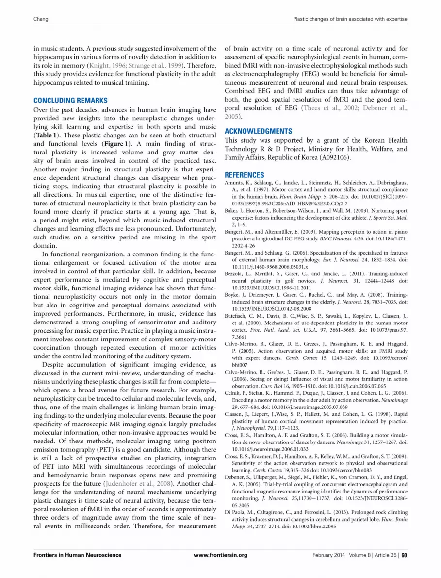
Chang Plastic changes of brain associated with expertise
in music students. A previous study suggested involvement of thehippocampus in various forms of novelty detection in addition toits role in memory (Knight, 1996; Strange et al., 1999). Therefore,this study provides evidence for functional plasticity in the adulthippocampus related to musical training.
CONCLUDING REMARKSOver the past decades, advances in human brain imaging haveprovided new insights into the neuroplastic changes under-lying skill learning and expertise in both sports and music(Table 1). These plastic changes can be seen at both structuraland functional levels (Figure 1). A main finding of struc-tural plasticity is increased volume and gray matter den-sity of brain areas involved in control of the practiced task.Another major finding in structural plasticity is that experi-ence dependent structural changes can disappear when prac-ticing stops, indicating that structural plasticity is possible inall directions. In musical expertise, one of the distinctive fea-tures of structural neuroplasticity is that brain plasticity can befound more clearly if practice starts at a young age. That is,a period might exist, beyond which music-induced structuralchanges and learning effects are less pronounced. Unfortunately,such studies on a sensitive period are missing in the sportdomain.
In functional reorganization, a common finding is the func-tional enlargement or focused activation of the motor areainvolved in control of that particular skill. In addition, becauseexpert performance is mediated by cognitive and perceptualmotor skills, functional imaging evidence has shown that func-tional neuroplasticity occurs not only in the motor domainbut also in cognitive and perceptual domains associated withimproved performances. Furthermore, in music, evidence hasdemonstrated a strong coupling of sensorimotor and auditoryprocessing for music expertise. Practice in playing a music instru-ment involves constant improvement of complex sensory-motorcoordination through repeated execution of motor activitiesunder the controlled monitoring of the auditory system.
Despite accumulation of significant imaging evidence, asdiscussed in the current mini-review, understanding of mecha-nisms underlying these plastic changes is still far from complete—which opens a broad avenue for future research. For example,neuroplasticity can be traced to cellular and molecular levels, and,thus, one of the main challenges is linking human brain imag-ing findings to the underlying molecular events. Because the poorspecificity of macroscopic MR imaging signals largely precludesmolecular information, other non-invasive approaches would beneeded. Of these methods, molecular imaging using positronemission tomography (PET) is a good candidate. Although thereis still a lack of prospective studies on plasticity, integrationof PET into MRI with simultaneous recordings of molecularand hemodynamic brain responses opens new and promisingprospects for the future (Judenhofer et al., 2008). Another chal-lenge for the understanding of neural mechanisms underlyingplastic changes is time scale of neural activity, because the tem-poral resolution of fMRI in the order of seconds is approximatelythree orders of magnitude away from the time scale of neu-ral events in milliseconds order. Therefore, for measurement
of brain activity on a time scale of neuronal activity and forassessment of specific neurophysiological events in human, com-bined fMRI with non-invasive electrophysiological methods suchas electroencephalography (EEG) would be beneficial for simul-taneous measurement of neuronal and neural brain responses.Combined EEG and fMRI studies can thus take advantage ofboth, the good spatial resolution of fMRI and the good tem-poral resolution of EEG (Thees et al., 2002; Debener et al.,2005).
ACKNOWLEDGMENTSThis study was supported by a grant of the Korean HealthTechnology R & D Project, Ministry for Health, Welfare, andFamily Affairs, Republic of Korea (A092106).
REFERENCESAmunts, K., Schlaug, G., Jancke, L., Steinmetz, H., Schleicher, A., Dabringhaus,
A., et al. (1997). Motor cortex and hand motor skills: structural compliancein the human brain. Hum. Brain Mapp. 5, 206–215. doi: 10.1002/(SICI)1097-0193(1997)5:3%3C206::AID-HBM5%3E3.0.CO;2-7
Baker, J., Horton, S., Robertson-Wilson, J., and Wall, M. (2003). Nurturing sportexpertise: factors influencing the development of elite athlete. J. Sports Sci. Med.2, 1–9.
Bangert, M., and Altenmüller, E. (2003). Mapping perception to action in pianopractice: a longitudinal DC-EEG study. BMC Neurosci. 4:26. doi: 10.1186/1471-2202-4-26
Bangert, M., and Schlaug, G. (2006). Specialization of the specialized in featuresof external human brain morphology. Eur. J. Neurosci. 24, 1832–1834. doi:10.1111/j.1460-9568.2006.05031.x
Bezzola, L., Merillat, S., Gaser, C., and Jancke, L. (2011). Training-inducedneural plasticity in golf novices. J. Neurosci. 31, 12444–12448 doi:10.1523/JNEUROSCI.1996-11.2011
Boyke, J., Driemeyer, J., Gaser, C., Buchel, C., and May, A. (2008). Training-induced brain structure changes in the elderly. J. Neurosci. 28, 7031–7035. doi:10.1523/JNEUROSCI.0742-08.2008
Butefisch, C. M., Davis, B. C.,Wise, S. P., Sawaki, L., Kopylev, L., Classen, J.,et al. (2000). Mechanisms of use-dependent plasticity in the human motorcortex. Proc. Natl. Acad. Sci. U.S.A. 97, 3661–3665. doi: 10.1073/pnas.97.7.3661
Calvo-Merino, B., Glaser, D. E., Grezes, J., Passingham, R. E. and Haggard,P. (2005). Action observation and acquired motor skills: an FMRI studywith expert dancers. Cereb. Cortex 15, 1243–1249. doi: 10.1093/cercor/bhi007
Calvo-Merino, B., Gre‘zes, J., Glaser, D. E., Passingham, R. E., and Haggard, P.(2006). Seeing or doing? Influence of visual and motor familiarity in actionobservation. Curr. Biol 16, 1905–1910. doi: 10.1016/j.cub.2006.07.065
Celnik, P., Stefan, K., Hummel, F., Duque, J., Classen, J. and Cohen, L. G. (2006).Encoding a motor memory in the older adult by action observation. Neuroimage29, 677–684. doi: 10.1016/j.neuroimage.2005.07.039
Classen, J., Liepert, J.,Wise, S. P., Hallett, M. and Cohen, L. G. (1998). Rapidplasticity of human cortical movement representation induced by practice.J. Neurophysiol. 79,1117–1123.
Cross, E. S., Hamilton, A. F. and Grafton, S. T. (2006). Building a motor simula-tion de novo: observation of dance by dancers. Neuroimage 31, 1257–1267. doi:10.1016/j.neuroimage.2006.01.033
Cross, E. S., Kraemer, D. J., Hamilton, A. F., Kelley, W. M., and Grafton, S. T. (2009).Sensitivity of the action observation network to physical and observationallearning. Cereb. Cortex 19,315–326 doi: 10.1093/cercor/bhn083
Debener, S., Ullsperger, M., Siegel, M., Fiehler, K., von Cramon, D. Y., and Engel,A. K. (2005). Trial-by-trial coupling of concurrent electroencephalogram andfunctional magnetic resonance imaging identifies the dynamics of performancemonitoring. J. Neurosci. 25,11730−11737. doi: 10.1523/JNEUROSCI.3286-05.2005
Di Paola, M., Caltagirone, C., and Petrosini, L. (2013). Prolonged rock climbingactivity induces structural changes in cerebellum and parietal lobe. Hum. BrainMapp. 34, 2707–2714. doi: 10.1002/hbm.22095
Frontiers in Human Neuroscience www.frontiersin.org February 2014 | Volume 8 | Article 35 | 60

Chang Plastic changes of brain associated with expertise
Di Pellegrino, G., Fadiga, L., and Gallese, V. (1992). Understanding motor events:a neurophysiological study. Exp. Brain Res. 91, 176–180. doi: 10.1007/BF00230027
Doyon, J., Song, A. W., Karni, A., Lalonde, F., Adams, M. M., and Ungerleider,L. G. (2002). Experience-dependent changes in cerebellar contributions tomotor sequence learning. Proc. Natl. Acad. Sci. U.S.A. 99,1017–1022. doi:10.1073/pnas.022615199
Draganski, B., Gaser, C., Busch, V., Schuierer, G., Bogdahn, U., and May, A.(2004). Neuroplasticity: changes in grey matter induced by training. Nature 427,311–312. doi: 10.1038/427311a
Draganski, B., Gaser, C., Kempermann, G., Kuhn, H. G., Winkler, J., Buchel, C.,et al. (2006). Temporal and spatial dynamics of brain structure changes duringextensive learning. J. Neurosci. 26,6314–6317. doi: 10.1523/JNEUROSCI.4628-05.2006
Ericsson, K. A. (2005). Recent advances in expertise research. Appl. Cogn. Psychol.19, 233–241. doi: 10.1002/acp.1111
Furley, P., and Memmert, D. (2010). Differences in spatial working mem-ory as a function of team sports expertise: the Corsi Block-tapping taskin sport psychological assessment. Percept. Mot. Skills110, 801–808. doi:10.2466/pms.110.3.801-808
Gaser, C., and Schlaug, G. (2003). Brain structures differ between musicians andnonmusicians. J. Neurosci. 23, 9240–9245.
Hänggi, J., Koeneke, S., Bezzola, L., and Jäncke, L. (2010). Structural neuroplasticityin the sensorimotor network of professional female ballet dancers. Hum. BrainMapp. 31, 1196–1206. doi: 10.1002/hbm.20928
Han, Y., Yang, H., Lv, Y. T., Zhu, C. Z., He, Y., Tang, H. H., et al. (2009).Gray matter density and white matter integrity in pianists’ brain: a com-bined structural and diffusion tensor MRI study. Neurosci. Lett. 459, 3–6. doi:10.1016/j.neulet.2008.07.056
Haslinger, B., Erhard, P., Altenmuller, E., Hennenlotter, A., Schwaiger, M., Grafinvon Einsiedel, H., et al. (2004). Reduced recruitment of motor associationareas during bimanual coordination in concert pianists. Hum. Brain Mapp. 22,206–215. doi: 10.1002/hbm.20028
Herdener, M., Esposito, F., di Salle, F., Boller, C., Hilti, C. C., Habermeyer, B., et al.(2010). Musical training induces functional plasticity in human hippocampus.J. Neurosci. 30, 1377–84. doi: 10.1523/JNEUROSCI.4513-09.2010
Hung, T., Haufler, A., Lo, L., Mayer-Kress, G., and Hatfield, B. (2007). Visuomotorexpertise and dimensional complexity of cerebral cortical activity. Med. Sci.Sports Exerc. 40, 752–759. doi: 10.1249/MSS.0b013e318162c49d
Hutchinson, S., Lee, L. H., Gaab, N., and Schlaug, G. (2003). Cerebellar volume ofmusicians. Cereb. Cortex 13, 943–949. doi: 10.1093/cercor/13.9.943
Hyde, K. L., Lerch, J., Norton, A., Forgeard, M., Winner, E., Evans, A. C., et al.(2009). Musical training shapes structural brain development. J. Neurosci. 29,3019–3025. doi: 10.1523/JNEUROSCI.5118-08.2009
Imfeld, A., Oechslin, M. S., Meyer, M., Loenneker, T., and Jancke, L. (2009).White matter plasticity in the corticospinal tract of musicians: A diffusion ten-sor imaging study. Neuroimage 46, 600–607. doi: 10.1016/j.neuroimage.2009.02.025
Jacini, W. F., Cannonieri, G. C., Fernandes, P. T., Bonilha, L., Cendes, F., and Li,L. M. (2009). Can exercise shape your brain? Cortical differences associatedwith judo practice. J. Sci. Med. Sport 12, 688–690. doi: 10.1016/j.jsams.2008.11.004
Jancke, L. (2009). The plastic human brain. Restor. Neurol. Neurosci. 27, 521–538.doi: 10.3233/RNN-2009-0519
Jäncke, L., Koeneke, S., Hoppe, A., Rominger, C., and Hänggi, J. (2009). Thearchitecture of the golfer’s brain. PLoS ONE 4:e4785. doi: 10.1371/jour-nal.pone.0004785
Judenhofer, M. S., Wehrl, H. F., Newport, D. F., Catana, C., Siegel, S. B., Becker,M., et al. (2008). Simultaneous PET-MRI: a new approach for functional andmorphological imaging. Nat. Med. 14, 459–465. doi: 10.1038/nm1700
Karni, A., Meyer, G., Jezzard, P., Adams, M. M., Turner, R. and Ungerleider, L. G.(1995). Functional MRI evidence for adult motor cortex plasticity during motorskill learning. Nature 377, 155–158. doi: 10.1038/377155a0
Kelly, A. M. C., and Garavan, H. (2005). Human functional neuroimaging ofbrain changes associated with practice. Cereb. Cortex 15, 1089–1102. doi:10.1093/cercor/bhi005
Kempermann, G. (2006). “Adult neurogenesis,” in In Lifespan Development and theBrain eds P. B. Baltes, P. A. Reuter-Lorenz, and F. Rösler (Cambridge UniversityPress), 82–107. doi: 10.1017/CBO9780511499722.006
Kim, Y. T., Seo, J. H., Song, H. J., Yoo, D. S., Lee, H. J., Lee, J., et al. (2011). Neuralcorrelates related to action observation in expert archers. Behav. Brain Res. 223,342–347. doi: 10.1016/j.bbr.2011.04.053
Kleim, J. A., Chan, S., Pringle, E., Schallert, K., Procaccio, V., Jimenez, R., et al.(2006). BDNF val66met polymorphism is associated with modified experience-dependent plasticity in human motor cortex. Nat. Neurosci. 9, 735–737. doi:10.1038/nn1699
Knight, R. (1996). Contribution of human hippocampal region to novelty detec-tion. Nature 383, 256–259. doi: 10.1038/383256a0
Kolb, B., and Whishaw, I. Q. (1998). Brain plasticity and behavior. Annu. Rev.Psychol. 49, 43–64. doi: 10.1146/annurev.psych.49.1.43
Kravitz, D. J., Saleem, K. S., Baker, C. I., and Mishkin, M. (2011). A new neuralframework for visuospatial processing. Nat. Rev. Neurosci. 12, 217–230. doi:10.1038/nrn3008
Krings, T., Topper, R., Foltys, H., Erberich, S., Sparing, R., Willmes, K., et al. (2000).Cortical activation patterns during complex motor tasks in piano players andcontrol subjects. a functional magnetic resonance imaging study. Neurosci. Lett.278, 189–193. doi: 10.1016/S0304-3940(99)00930-1
Lotze, M., Scheler, G., Tan, H. R., Braun, C., and Birbaumer, N. (2003).The musician’s brain: Functional imaging of amateurs and profession-als during performance and imagery. Neuroimage 20, 1817–1829. doi:10.1016/j.neuroimage.2003.07.018
Meister, I., Krings, T., Foltys, H., Boroojerdi, B., Muller, M., Topper, R., et al.(2005). Effects of long-term practice and task complexity in musicians andnonmusicians performing simple and complex motor tasks: implications forcortical motor organization. Hum. Brain Mapp. 25, 345–352. doi: 10.1002/hbm.20112
Milton, J., Solodkin, A., Hluštík, P., and Small, S. L. (2007). The mind ofexpert motor performance is cool and focused. Neuroimage 35, 804–813. doi:10.1016/j.neuroimage.2007.01.003
Nielsen, J. B., and Cohen, L. G. (2008). The Olympic brain. Does corticospinal plas-ticity play a role in acquisition of skills required for high performance sports? J.Physiol. 586, 65–70. doi: 10.1113/jphysiol.2007.142661
Oechslin, M. S., Van De Ville, D., Lazeyras, F., Hauert, C. A., and James, C. E.(2013). Degree of musical expertise modulates higher order brain functioning.Cereb. Cortex 23, 2213–2224. doi: 10.1093/cercor/bhs206
Park, I. S., Lee, K. J., Han, J. W., Lee, N. J., Lee, W. T., Park, K. A., et al. (2009).Experience-dependent plasticity of cerebellar vermis in basketball players.Cerebellum 8, 334–339. doi: 10.1007/s12311-009-0100-1
Pascual-Leone, A., Amedi, A., Fregni, F., and Merabet, L. B. (2005). Theplastic human brain cortex. Annu. Rev. Neurosci. 28, 377–401. doi:10.1146/annurev.neuro.27.070203.144216
Pearce, A. J., Thickbroom, G. W., Byrnes, M. L., and Mastaglia, F. L. (2000).Functional reorganisation of the corticomotor projection to the hand inskilled racquet players. Exp. Brain Res. 130, 238–243. doi: 10.1007/s002219900236
Pilgramm, S., Lorey, B., Stark, R., Munzert, J., Vaitl, D., and Zentgraf, K. (2010).Differential activation of the lateral premotor cortex during action observation.BMC Neurosci. 11:89. doi: 10.1186/1471-2202-11-89
Schlaug, G. (2001). The brain of musicians. A model for functional and struc-tural adaptation. Ann. N.Y. Acad. Sci. 930, 281–299. doi: 10.1111/j.1749-6632.2001.tb05739.x
Sekiguchi, A., Yokoyama, S., Kasahara, S., Yomogida, Y., Takeuchi, H., Ogawa,T., et al. (2011). Neural bases of a specific strategy for visuospatial pro-cessing in rugby players. Med. Sci. Sports Exerc. 43, 1857–1862. doi:10.1249/MSS.0b013e31821920f3
Seo, J., Kim, Y. T., Song, H. J., Lee, H. J., Lee, J., Jung, T. D., et al. (2012). Strongeractivation and deactivation in archery experts for differential cognitive strategyin visuospatial working memory processing. Behav. Brain Res. 229,185–193. doi:10.1016/j.bbr.2012.01.019
Stefan, K., Cohen, L. G., Duque, J., Mazzocchio, R., Celnik, P., Sawaki,L., et al. (2005). Formation of a motor memory by action obser-vation. J. Neurosci. 25, 9339–9346. doi: 10.1523/JNEUROSCI.2282-05.2005
Strange, B. A., Fletcher, P. C., Henson, R. N., Friston, K. J., and Dolan, R. J. (1999).Segregating the functions of human hippocampus. Proc. Natl. Acad. Sci. U.S.A.96, 4034–4039. doi: 10.1073/pnas.96.7.4034
Thees, S., Blankenburg, F., Taskin, B., Curio, G., and Villringer, A. (2002). Dipolesource localization and fMRI of simultaneously recorded data applied to
Frontiers in Human Neuroscience www.frontiersin.org February 2014 | Volume 8 | Article 35 | 61

Chang Plastic changes of brain associated with expertise
somatosensory categorization. Neuroimage 18, 707–719. doi: 10.1016/S1053-8119(02)00054-X
Wan, C. Y., and Schlaug, G. (2010). Music making as a tool for promot-ing brain plasticity across the life span. Neuroscientist 16, 566–577. doi:10.1177/1073858410377805
Wang, B., Fan, Y., Lu, M., Li, S., Song, Z., Peng, X., et al. (2013).Brain anatomical networks in world class gymnasts: A DTI tractog-raphy study. Neuroimage 65, 476–487. doi: 10.1016/j.neuroimage.2012.10.007
Wei, G., Zhang, Y., Jiang, T., and Luo, J. (2011). Increased cortical thickness insports experts: a comparison of diving players with the controls. PLoS ONE6:e17112. doi: 10.1371/journal.pone.0017112
Zilles, K. (1992). Neuronal plasticity as an adaptive property of the cen-tral nervous system. Ann. Anat. 174, 383–391. doi: 10.1016/S0940-9602(11)80255-4
Conflict of Interest Statement: The author declares that the research was con-ducted in the absence of any commercial or financial relationships that could beconstrued as a potential conflict of interest.
Received: 18 July 2013; accepted: 17 January 2014; published online: 04 February 2014.Citation: Chang Y (2014) Reorganization and plastic changes of the human brain asso-ciated with skill learning and expertise. Front. Hum. Neurosci. 8:35. doi: 10.3389/fnhum.2014.00035This article was submitted to the journal Frontiers in Human Neuroscience.Copyright © 2014 Chang. This is an open-access article distributed under the termsof the Creative Commons Attribution License (CC BY). The use, distribution or repro-duction in other forums is permitted, provided the original author(s) or licensor arecredited and that the original publication in this journal is cited, in accordance withaccepted academic practice. No use, distribution or reproduction is permitted whichdoes not comply with these terms.
Frontiers in Human Neuroscience www.frontiersin.org February 2014 | Volume 8 | Article 35 | 62

REVIEW ARTICLEpublished: 07 May 2014
doi: 10.3389/fnhum.2014.00280
Experts bodies, experts minds: How physical and mentaltraining shape the brainUrsula Debarnot 1,2 , Marco Sperduti 3,4 , Franck Di Rienzo 2 and Aymeric Guillot 2,5*1 Département des Neurosciences Fondamentales, Centre Médical Universitaire, Université de Genève, Genève, Suisse2 Centre de Recherche et d’Innovation sur le Sport, Université Claude Bernard Lyon 1, Université de Lyon, Villeurbanne Cedex, Lyon, France3 Centre de Psychiatrie et Neurosciences (Inserm UMR S894), Université Paris Descartes, Paris, France4 Laboratoire Mémoire et Cognition, Institut de Psychologie, Boulogne-Billancourt, France5 Institut Universitaire de France, Paris, France
Edited by:Merim Bilalic, Alpen-Adria-UniversitätKlagenfurt, Austria
Reviewed by:Luca Turella, University of Trento, ItalyAlessandro Guida, University ofRennes 2, France
*Correspondence:Aymeric Guillot, Centre de Rechercheet d’Innovation sur le Sport, EA 647,Université Claude Bernard Lyon 1,Université de Lyon, 27-29 boulevarddu 11 Novembre 1918, 69622Villeurbanne Cedex, Lyon, Francee-mail: [email protected]
Skill learning is the improvement in perceptual, cognitive, or motor performance followingpractice. Expert performance levels can be achieved with well-organized knowledge, usingsophisticated and specific mental representations and cognitive processing, applyingautomatic sequences quickly and efficiently, being able to deal with large amounts of infor-mation, and many other challenging task demands and situations that otherwise paralyzethe performance of novices. The neural reorganizations that occur with expertise reflectthe optimization of the neurocognitive resources to deal with the complex computationalload needed to achieve peak performance. As such, capitalizing on neuronal plasticity,brain modifications take place over time-practice and during the consolidation process.One major challenge is to investigate the neural substrates and cognitive mechanismsengaged in expertise, and to define “expertise” from its neural and cognitive underpinnings.Recent insights showed that many brain structures are recruited during task performance,but only activity in regions related to domain-specific knowledge distinguishes expertsfrom novices. The present review focuses on three expertise domains placed across amotor to mental gradient of skill learning: sequential motor skill, mental simulation of themovement (motor imagery), and meditation as a paradigmatic example of “pure” mentaltraining. We first describe results on each specific domain from the initial skill acquisitionto expert performance, including recent results on the corresponding underlying neuralmechanisms. We then discuss differences and similarities between these domains withthe aim to identify the highlights of the neurocognitive processes underpinning expertise,and conclude with suggestions for future research.
Keywords: expertise, motor skill, motor imagery, meditation, motor consolidation, neural networks
Brain plasticity refers to the putative changes in neural orga-nization that accounts for the diverse forms of short-lasting orenduring behavioral modifiability. There is considerable evidencethat neuronal plasticity is not an occasional state, but rather allowsthe human brain to adapt to environmental pressure, physiologicchanges, and experiences (Johansson, 2004). Interestingly, changesin the input of any neural system, or in the trafficking of its effer-ent connections, lead to reorganizations that are visible at the levelof behavior, anatomy, and physiology, encompassing cellular andmolecular levels (Pascual-Leone et al., 2005). Currently, the chal-lenge is to explore in greater details the processes of neuroplasticityand how to modulate them to achieve the best behavioral outcome.Typically, the human brain undergoes constant changes triggeredby environmental stimulation or resulting from intrinsic remod-eling activity (Johansson, 2011). Therefore, brain is the source ofbehavior, but in turn is modified by the behavior itself.
Most of the prior studies that aimed to investigate the neu-ral substrates underlying expertise compared the skill level ofnovices and experts, which gives a snapshot of two endpointson the skill level continuum. So far, a growing number ofinvestigations rather test subjects at different occasions to assess
the gain of expertise throughout training period, hence the“novicestate” can be compared with the “expert state,” or additionallyin between (Guida et al., 2012). Using both approaches enablesto infer the neural reorganization that contributes to reach thehighest level of performance, which is impacted and reinforcedthrough the consolidation processes (Diekelmann et al., 2011).This set of processes takes place automatically, without aware-ness, and allows the conversion of the initial unstable memoryrepresentation into a more stable and effective form, availablefor continued reactivation and recall over extended periods oftime (Stickgold and Walker, 2007). Interestingly, processes of con-solidation over time can also facilitate behavior, often throughoffline memory reorganization (Stickgold and Walker, 2005).Indeed, continued plasticity over time is crucial whenever newlyacquired information are integrated with old memories (Abra-ham and Robins, 2005). Given this consolidation effect, someinvestigations include follow-up tests (for example, Draganskiet al., 2004; Scholz et al., 2009). For all of these memory pro-cesses, sleep has been shown to play a critical role in meetingthe demands of the organism (Walker and Stickgold, 2010; Raschand Born, 2013). At the functional level, the consolidation refers
Frontiers in Human Neuroscience www.frontiersin.org May 2014 | Volume 8 | Article 280 | 63

Debarnot et al. Neural mechanisms of expertise
to processes in which reverberating activity in newly encodedrepresentations stimulate a redistribution of the neuronal rep-resentations to other neuronal circuitries for long-term storage(Dudai, 2004).
On the basis of several thousand years of education, along withmore recent laboratory research on learning and skill acquisition,a number of conditions for optimal learning and improvementof performance have been uncovered. The most common con-dition for optimal learning and improvement of performanceconcerns the extended periods of deliberate practice (Ericssonet al., 1993). Four hours of physical/mental deliberate practicedaily for approximately 10 years purportedly leads, for exam-ple, to expertise in chess, music composition, art, sport, andscience (the 10-year or 10,000-h rule; Ericsson, 2008). One ofthe major interests for modern neuroscience is to investigatethe plastic changes that occur in brain structures when peo-ple participate in such intensive motor and/or mental training.Nowadays, skill acquisition and training in various domains, frommotor function to higher order cognitive skills, were shown toelicit substantial changes in brain anatomy (Jäncke, 2009). Pre-vious studies including cross-sectional and longitudinal designsshowed that skill acquisition and practice can induce changesin the functional properties of specific brain regions involvedin the corresponding task (Buschkuehl et al., 2012; Guida et al.,2012). Recent investigations further demonstrated that not onlybrain activity, but also gray and white matter structures changeas a consequence of skill learning (Draganski et al., 2004; Scholzet al., 2009). For instance, recent neuroimaging studies focusingon the effects of motor or mental (meditation) training, last-ing several weeks in previously untrained participants, showedplastic changes in specific white matter regions (Scholz et al.,2009; Tang et al., 2012). Overall, technological and methodologicaladvances in neuroimaging and non-invasive human brain stim-ulation provided insights into the neuroplastic mechanisms thatunderlie skill expertise. More generally, cross-sectional paradigms,where highly skilled participants were compared with less-skilledpersons, showed a strong link between skill acquisition and neu-ronal plasticity at both cortical and subcortical levels over time,engaging different spatially distributed and interconnected brainregions.
A fundamental ability of the human brain is to form andretrieve memories enabling the individual to adapt its behaviorto the demands of an ever-changing environment, and appropri-ately select and improve the behaviors of its given repertoire. Thedistinction between expertise based on declarative (knowing that)and procedural knowledge (knowing how) is directly related to thereal-world domains of expertise. Some domains are mostly char-acterized by one type of knowledge, although experts may haveboth and often need to rely on one type of knowledge rather thanthe other (Dror, 2011). Practically, the most appropriate type ofexpert knowledge depends on the situational demands and cogni-tive mechanisms involved in operationalizing this knowledge (e.g.,Beilock et al., 2004; Dror, 2011). Such kind of adaptation is a reli-able marker of expertise. Interestingly, it has been shown that thereare both costs and benefits to expertise, so that the inflexibility ofexperts might make them unable to adapt to new task demands, atleast in some occasions (Bilalic et al., 2008a,b) Typically, experts
may fail to develop successful patterns of thought by using aless familiar, albeit more appropriate solution, when available.Bilalic et al. (2008a,b, 2010a), however, demonstrated that individ-uals with the highest degree of expertise remained more flexibleand more likely to find the optimal solution by memory retrievaland/or by search.
The primary aim of the present paper is to review studiesshowing how learning and experience induce structural and func-tional brain plasticity that supports expertise, following a motorto mental gradient of skill (Figure 1). We not only consider thestructural and functional organization that the brain shapes alongthe course of gain in expertise, but also the brain’s potential toreorganize its functional organization by modifying its structurein response to practice. Meeting this challenge, this review inte-grates available results from several neuroimaging approaches inhuman populations and animal models. For the first time, weaddress these issues in exploring three domains of expertise whichare usually disconnected (motor skill, mental simulation of move-ment, and meditation). We first report and underline separatelythe neuroanatomic correlates of expertise following a motor tomental gradient, and then attempt to identify and link the com-mon patterns of changes in the brain subserving expert-levelof performance within each domain. Practically, using such atransversal approach, this review aims at providing new insightsabout how brain plasticity occurs and supports the expert level ofperformance.
WHEN MOTOR SKILLS EXPERTISE SUBSERVES BRAINOPTIMIZATIONRecent years have seen a significant growing of knowledge on theneuroplasticity that underpins motor skill learning from its acqui-sition to the expert level of performance (for review, see Patel et al.,2013). Basically, compared to novices, highly trained individu-als exhibit a number of differences including a reduction in thevariability of repeated movements (Milton et al., 2004), in muscleactivation (Lay et al., 2002), and a decrease in the overall volumeof brain activation together with a relative increased intensity ofactivation in specific brain regions necessary for the execution ofthe task (Jäncke et al., 2000; Munte et al., 2002; Lotze et al., 2003).
FIGURE 1 | Practice modes and consolidation processes leading toexpertise.
Frontiers in Human Neuroscience www.frontiersin.org May 2014 | Volume 8 | Article 280 | 64

Debarnot et al. Neural mechanisms of expertise
In our attempt to understand such robust expertise-dependentdifferences, we first report in this section the main data interestinghow motor skills are acquired and enhanced through the consoli-dation process. Then, we review recent findings showing changesin the pattern of brain activity as well as modifications in the neuralstructures associated with expertise.
THE NEUROCOGNITIVE BASIS OF MOTOR SKILL LEARNINGMotor skill acquisition refers to the process by which a movementis performed effortlessly through repeated practice and interac-tions with the environment (Willingham, 1998). Accurate motorperformance is essential to almost everything we do, from typingto driving, or playing sports. In cognitive psychology, theoret-ical descriptions of changes in skilled performance were shownto move from cognitive to automatic processing (Fitts, 1964).The key concept is the increasing automaticity: controlled pro-cesses are attention demanding, conscious and inefficient, whereasautomatic processes are rapid, smooth, effortless, require littleattentional capacity, and are difficult to be consciously disrupted(Shiffrin and Schneider, 1977). Two experimental paradigms areused to investigate the cognitive processes and the neural substratesmediating our capacity to learn behaviors: the first measuresthe incremental acquisition of movements into a well-executedbehavior (motor sequence learning), whereas the second testsour capacity to compensate for environmental changes (motoradaptation). Doyon and Benali (2005) proposed an integratedview of the functional plasticity that such a motor memory traceundergoes in each case. This model suggests that depending uponthe nature of the cognitive processes required during learning,both motor sequence and motor adaptation tasks recruit simi-lar cerebral structures early in the learning phase, including thestriatum, cerebellum, and motor cortical regions, in addition toprefrontal, parietal and limbic areas. A shift of motor represen-tation from the associative to the sensorimotor striatal territorycan be seen during sequence learning, whereas additional rep-resentation of the skill can be observed in the cerebellar nucleiafter practice on motor adaptation tasks. When consolidationhas occurred, the participant has achieved asymptotic perfor-mance. However, the neural representation of a new motor skillis believed to be distributed in a network of structures involvingthe cortico-striatal and/or cortico-cerebellar circuits dependingon the type of skill acquired. At this stage, the model suggests thatmotor adaptation rather involves the cerebellum for the reten-tion and future executions of the acquired skill. By contrast, areverse pattern of functional plasticity occurs in motor sequencelearning, where the cerebellum is no longer essential, and theconsolidation of the skill involves representational changes in thestriatum and associated motor cortical regions. New insights intothe neuroplastic mechanisms that underlie motor skill learningcorroborate that skill acquisition is subserved by multiple mech-anisms that operate across different temporal scales (Dayan andCohen, 2011).
Practically, it is not automaticity per se that is indicative ofhigh proficiency, but rather the level of skill at which automatic-ity is attained. Although the border between the automaticityand the expertise concepts beg for clarification, one may con-sider that most people fail to develop beyond a hobbyist level
of performance as they settle into automaticity at a given levelof skill that they find enjoyable, rather than continuing toimprove skills (Ericsson, 2007). Hence, automaticity is morea false ceiling than a measure of excellence. Previous stud-ies typically defined skill acquisition in terms of reduction inthe speed of movement execution or reaction times, increasein accuracy, or decrease in movement variability. Yet, suchmeasurements are often interdependent, in that faster move-ments can be performed at the cost of reduced accuracy andvice versa, a phenomenon which has been often referred to thespeed-accuracy trade-off (Fitts, 1954). One solution to this issueis through assessment of changes in speed-accuracy trade-offfunctions, i.e., to defy the speed–accuracy tradeoff for a giventask (Krakauer, 2009; Krakauer and Mazzoni, 2011). In otherwords, a skilled tennis player can serve both faster and moreaccurately than a novice. Thus, sporting skill at the level ofmotor execution can be considered as acquiring a new speed–accuracy trade-off relationships for each sub-task of the motorsequence.
FUNCTIONAL PLASTICITY CHARACTERIZING NOVICES AND EXPERTSThe existing longitudinal studies in healthy participants attachedgreat importance to controlled practice situations by keepingtraining parameters as much constant as possible (e.g., trainingduration per day, overall training duration, training schedule,strategies, etc.). Movement automatization reflects high level ofmotor skill performance and has been associated with increasedactivation in the primary motor cortex (M1), primary somatosen-sory cortex, supplementary motor area (SMA), and putamen,as well as decreased activation in the lobule VI of the cerebel-lum (Floyer-Lea and Matthews, 2005; Lehericy et al., 2005). Thus,training-related automaticity decreases in the fronto-parietal anddorsal attention networks, hence suggesting that progress fromacquisition to automatization stages of motor skill learning is char-acterized by concomitant reduced demands on externally focusedattention and executive function (Kelly and Garavan, 2005). Suchpattern of activation is particularly elicited for motor sequencelearning, but one would hypothesize that forms of motor andvisuomotor learning which are more cognitive or associative innature (Parsons et al., 2005) may recruit slightly different cerebralnetworks undergoing other patterns of plasticity with learning.For example, the preparatory period has been most extensivelystudied in athletes where it is called the “pre-shot routine.” Con-sistency and reproducibility of pre-shot routines are suggestedto be among the most important differences that distinguishexperts from novices in sport such as Golf (Feltz and Landers,1983), compared to some motor skills such as goal kicking inrugby (Jackson, 2003) where no association between temporalconsistency of the pre-shot routine and performance has beenobserved. In a recent study, Milton et al. (2007) found differ-ent functional activations during the pre-shot routine in expertand novice golfers. Especially, the posterior cingulate cortex, theamygdala–forebrain complex, and the basal ganglia were activeonly in novices, whereas experts yielded activation primarily inthe superior parietal lobule, the dorsal lateral premotor area,and the occipital cortex. These results suggest that the dispar-ity between the quality of the performance of novice and expert
Frontiers in Human Neuroscience www.frontiersin.org May 2014 | Volume 8 | Article 280 | 65

Debarnot et al. Neural mechanisms of expertise
golfers lies at the level of the functional organization of neural net-works during motor planning. More generally, Patel et al. (2013)demonstrated that spatially distributed cortical networks and sub-cortical striatal regions may serve as neural markers of practiceinterventions.
The expertise stage of motor skill learning, both in humansand animals, consistently reported either increased or decreasedM1 activation depending on both the time interval and taskcomplexity. On one hand, performing an explicit sequence of fin-ger movements over several weeks showed a progressive increaseof activity in M1 (Karni, 1995; Hlustik et al., 2004; Floyer-Leaand Matthews, 2005), hence reflecting recruitment of additionalM1 units into the local network that represents the acquiredsequence of movements (Ungerleider et al., 2002). Learning amotor sequence over several days was also accompanied by anincrease in the size of motor maps and cortico-motoneuronalexcitability of the body parts involved in the task (Pascual-Leoneet al., 1995). Such plastic changes in M1 function linked with slowstage of motor skill learning are well established in animal mod-els as well. For example, functional reorganization of movementrepresentations in M1 has been documented in squirrel monkeys(Nudo and Milliken, 1996) and rodents (Kleim et al., 2004). Itwas found that an expansion in movement representations withtraining, detectable only after substantial practice periods, paral-leled behavioral gains. Such findings challenged theories of neuralefficiency proposing that optimized neural processing is associ-ated with reduced activity in M1 (Jäncke et al., 2000; Krings et al.,2000; Haslinger et al., 2004). Especially, an increased activity ofthe motor network was reported, including M1, cerebellum, pre-motor cortex, basal ganglia, pre-SMA, and SMA in the initialacquisition of motor skills, with significant attenuation of activityfollowing consolidation of the motor skill (Steele and Penhune,2010). Recently, Picard et al. (2013) examined the consequenceof practice-dependent motor learning on the metabolic and neu-ral activity in M1 of monkeys who had extensive training (∼1–6years) on sequential movement tasks. They found that practic-ing a skilled movement and the development of expertise leadto lower M1 metabolic activity, without a concomitant reductionin neuron activity. In other term, they showed that less synapticactivity was required to generate a given amount of neuronal activ-ity. The authors suggested that this gain in M1 efficiency mightresult from a number of factors such as more effective synapses,greater synchrony in inputs and more finely tuned inputs. Theyconcluded that low activation in M1 elicited during extended prac-tice might be a reflection of plastic mechanisms involved in thedevelopment of expertise. Although there is no clear consensusabout M1 activation during the gain of expertise, it is more likelythat time interval might be one of the main factors that play arole in brain modifications. This issue has been partially raisedby Hotermans et al. (2008) who investigated the role of M1 inthe different phases of motor memory consolidation by apply-ing repetitive transcranial magnetic stimulation (rTMS) over M1just after a training session involving a sequential finger-tappingtask. They showed that interfering with M1 attenuated the earlypost-training performance without any detrimental consequenceson the long-term behavioral improvement tested 4 or 24 h after.These results support that M1 is causally involved early after the
acquisition of a new motor-skill but is no longer mandatory fol-lowing a consolidation-period. Overall, increased activity in M1might correspond to the integration of the new sensory informa-tion during the short period of training of a new skill, but whenthe new learned skill becomes automatic, M1 activation is lessimportant. Table 1 summarizes the human studies reviewed in thissection with the brain areas showing early–late and expert-relatedfunctional plasticity.
An alternative explanation for the modulation of M1 activityhas been suggested by Landau and D’Esposito (2006) who reportedthat optimized motor system is capable of greater flexibility andadaptability, depending on the nature of the task demands. Usinga complex sequential finger task, they found increased motor acti-vations in pianists than non-pianists, and further argued thatthe former may yield decreased activation when they were lesschallenged. Further research is therefore needed to determine themodulation of M1 activation in experts and novices using taskcharacteristics that vary across several degrees of difficulty eitherearly or late during the learning process.
EXPERIENCE-DEPENDENT STRUCTURAL CHANGES IN THE HUMANBRAINBesides to the functional reorganization of the brain motornetworks, current neuroimaging studies suggest that physicalpractice is also reflected in macroscopic changes in motor-related structures (Draganski and May, 2008). Practically, highergray matter volume in auditory, sensorimotor, and premotorcortex, as well as in the cerebellum, was reported in musi-cians compared to non-musicians (Gaser and Schlaug, 2003).Related findings have been reported in many other specifictasks, including typing (Cannonieri et al., 2007), basketball(Park et al., 2009, 2011), or golf performance (Jäncke et al.,2009; Bezzola et al., 2011). Although these findings tend tofollow the law “more skill, more gray matter,” there is newevidence that training and ensuing expertise may induce localdecrease of cortical volume (Hänggi et al., 2010; Granert et al.,2011). Controversial findings were also reported on training-related plasticity in white matter microstructure in healthy adults,although fewer studies dealing with this issue have been pub-lished.
On the one side, numerous investigations reported a strongassociation between specialized skill and structural changes in par-ticular brain structures (Maguire et al., 2000; Hutchinson et al.,2003; Mechelli et al., 2004; Park et al., 2009). In a longitudinalstudy, Draganski et al. (2004) used a complex visuo-motor jug-gling task where perception and anticipation of moving targetsdetermined the planning of the subsequent motor action. Youngvolunteers were scanned before and after 3 months of a dailytraining period. The results showed a transient bilateral expan-sion in gray matter in the mid-temporal area (hMT/V5) and theleft inferior parietal sulcus. The authors concluded that juggling,and consequently the perception and spatial anticipation of mov-ing objects, is a stronger stimulus for structural plasticity in visualareas than in motor areas. Similarly, cross-sectional neuroimag-ing studies showed experience-dependent structural plasticity inthe cerebellum following training of complex motor skills (Parket al., 2009; Scholz et al., 2009). For example, Park et al. (2012)
Frontiers in Human Neuroscience www.frontiersin.org May 2014 | Volume 8 | Article 280 | 66

Debarnot et al. Neural mechanisms of expertise
Table 1 | Functional plasticity through longitudinal and cross-sectional approaches: focus on the motor network.
Study Task/method Design Main outcome
Longitudinal
Normal adults
Floyer-Lea and Matthews (2005) Tracking sequential task/fMRI Single training session vs.
15 min/session 5
weekdays/3 weeks
Early: ↘ activity DLPC, anterior cingulate, posterior
parietal, M1 and cerebellar cortex
Long-term: ↗ activity left S1, M1 and right putamen
Steele and Penhune (2010) FTT/fMRI 5 weekdays Early: ↗ cerebellum, PMC, basal ganglia, pre-SMA
and SMA
Long-term: ↘ in these regions
Karni (1995) FTT/fMRI 10–20 min daily session/3
weeks
Greater activation in M1 was significantly compared
to the extent of activation evoked by an untrained
sequence
Hlustik et al. (2004) FTT/fMRI 15 min daily session/3
weeks
Gradual expansion of M1 and S1 correlating with
performance
Hotermans et al. (2008) FTT/rTMS rTMS immediately before
testing at 30 min, 4 or 24 h
after practice
Early acquisition M1 supports performance, but is no
longer mandatory following consolidation
Parsons et al. (2005) FTT/fMRI 5 weekdays Early: ↗ activation in motor areas and ant/post CMA
Long-term: ↘ activation cerebellum, motor areas,
striatum, fronto-parietal cortices
Pascual-Leone et al. (1995) FTT/TMS 2 h daily training vs.
no-training control 5 days
Following training: hand motor areas enlarged while
activation ↘Cross-sectional
Pianists vs. novices
Haslinger et al. (2004) FTT/fMRI Pianists recruited an extensive motor network but
with a lesser degree of activation than novices
Jäncke et al. (2000) FTT/fMRI Less activation in M1, SMA, pre-SMA, and CMA in
pianists
Krings et al. (2000) FTT/fMRI Pianists showed less activation in M1, SMA, and
PMC
FTT, finger tapping task; rTMS, repetitive transcranial magnetic stimulation; fMRI, functional magnetic resonance imagery; ↗, increase; ↘, decrease; M1, primarymotor cortex; S1, primary somatosensory cortex; DLPC, dorsolateral prefrontal cortex; SMA, supplementary motor area; pre-SMA, pre-supplementary motor area;PMC, premotor cortex; CMA, cingulate motor areas.
found greater right- than left cerebellar volume asymmetry andrelatively larger volumes of right hemisphere and vermis lob-ules VI–VII (declive, folium, and tuber) in short-track speedskating players compared to matched controls. This finding sug-gests that the specialized abilities of balance and coordinationare associated with structural plasticity of the right hemisphereof cerebellum and vermis VI–VII, these regions playing a crit-ical role in balance and coordination. Cannonieri et al. (2007)further found that the volume of brain areas correspondingto the motor skill increased proportionally to the duration ofthe training period. Together, these findings demonstrate thatpractice modulates brain anatomy specifically associated withpractice demands, albeit opposite structural neural correlatesreflecting stepwise increases in expertise have also been found.For instance, Hänggi et al. (2010) found differences in structural
characteristics within the sensorimotor neural network betweenprofessional women ballet dancers and novices. Especially, theyreported decreased gray matter volumes in the left premotorcortex, the SMA, the putamen, and the superior frontal gyrusanterior to the premotor cortex. More recently, James et al. (2014)reported changes in gray matter as a function of musical train-ing intensity in three groups of young adults (non-musicians,amateurs, and professionals). Surprisingly, they observed a pro-gressive increase of gray matter density with respect to the levelof expertise in several regions involved in higher-order cognitiveprocessing (e.g., right fusiform gyrus activated for visual pat-tern recognition), whereas an opposite pattern of results werefound in sensorimotor areas. To summarize, the type of densitychanges (increase or decrease) and the localization of struc-tural plasticity in gray matter may be related to different factors
Frontiers in Human Neuroscience www.frontiersin.org May 2014 | Volume 8 | Article 280 | 67

Debarnot et al. Neural mechanisms of expertise
such as the nature of the motor-skill, duration and stage ofpractice.
Nowadays, investigations are designed to determine the timescales of gray matter changes from novices to experts (Taubertet al., 2010; Dayan and Cohen, 2011). So far, the process of graymatter adaptation has been observed as early as following 7 days ofpractice (Driemeyer et al., 2008) and as late as after 6 weeks (Scholzet al., 2009), hence demonstrating a relatively fast and durablestructural gray matter plasticity. Although interpretation of suchstriking results are premature, it has been proposed that processesoccurring both at the synapse level and larger scales (e.g., glialhypertrophy), may play a contributory role (Draganski and May,2008).
Recent longitudinal neuroimaging studies further focused onthe effects of motor training lasting several days or weeks in pre-viously untrained participants, and showed specific structuralplasticity in white matter regions (Scholz et al., 2009; Taubertet al., 2010). Scholz et al. (2009) reported experience-inducedchanges in white matter architecture following a short periodof practice. Practically, it was found that 6 weeks of jugglingpractice protracted an increased fractional anisotropy in a regionof white matter underlying the intraparietal sulcus. Interest-ingly, Della-Maggiore et al. (2009) further demonstrated that thespeed in a visuomotor adaptation task might be partially deter-mined by the variation of fractional anisotropy in the posteriorcerebellum and superior cerebellar peduncle. Together, thesefindings show that rates of motor skill practice might correlatewith higher values of fractional anisotropy at a local level trig-gered by the nature of the motor task. Cross-sectional studies,primarily in highly trained musicians, also examined white mat-ter correlates of skilled behavior (Schmithorst and Wilke, 2002;Bengtsson et al., 2005; Han et al., 2009). Bengtsson et al. (2005)found a correlation between fractional anisotropy in the pos-terior limb of the internal capsule, which contains descendingcorticospinal fibers from the primary sensorimotor and premo-tor cortices, with number of practice hours during childhood inskilled musicians. These results demonstrate that training dur-ing a critical developmental period may induce local structuralplasticity. Rüber et al. (2013) further reported higher fractionalanisotropy values in musicians than non-musicians in descend-ing motor tracts, with differences when practice was a fine fingerunimanual or bimanual motor skill. The matter tracts were mod-ified to reflect specific motor demands, unimanual motor skillprimarily eliciting structural remodeling of right hemispherictracts, and bimanual motor skill leading to bilateral structuraltract remodeling. Accordingly, Roberts et al. (2012) compared thebehavior and brain structure of healthy controls with a groupof karate black belts, an expert group who are able to performrapid, complex movements that require years of training. Asexpected, experts were more able than novices to coordinate thetiming of inter-segmental joint velocities, and data revealed sig-nificant group differences in the microstructure of white matterin the superior cerebellar peduncles and M1, these brain regionsparticipating to the voluntary control of the movement. Over-all, as experts demonstrate optimal behavior on specific tasks,differences in white matter structure relative to novices mightreflect a “fine-tuning” of the connectivity between specific brain
regions. Nevertheless, contrasting patterns of results shows lowerwhite matter values in experts than in non-experts. In orderto disentangle the unresolved question of whether sensorimo-tor training leads to increased or decreased white matter density,Imfeld et al. (2009) investigated the corticospinal tract of musi-cians using different methods of analysis (fiber tractography,voxelwise analysis, region of interest analysis, and detailed slice-wise analysis of diffusion parameters). Data clearly demonstratedthat sensorimotor training leads to decreased white matter den-sity. This finding is supported by Hänggi et al. (2010) who foundreduced white matter volume in dancers vs. non-dancers. Furtherinvestigations are required to understand the cellular mecha-nisms underlying learning-dependent changes in white mattermicrostructure.
THE NEURAL CORRELATES OF MOTOR IMAGERYAcross a motor to mental gradient of skill learning, there is nowcompelling evidence that motor imagery contributes to enhancemotor skill learning and motor performance (for reviews, see Feltzand Landers, 1983; Driskell et al., 1994; Guillot and Collet, 2008;Schuster et al., 2011). Motor imagery is a dynamic state duringwhich one simulates an action mentally without any concomi-tant body movement. Since the last three decades, the adventof functional brain mapping studies has allowed researchers toinvestigate the neural correlates of motor imagery and to under-stand in greater details the neural underpinnings of expertise inimagery.
THE EFFECT OF EXPERTISE LEVELIn previous sections, we reviewed a handful of experimentalstudies providing clear evidence that individuals differ in theirability to perform voluntary motor acts, and that advancedmotor learning is associated with functional brain plasticity (forreviews, see Doyon and Benali, 2005; Doyon, 2008; Johans-son, 2011). Practically, neuroimaging studies showed that theneural networks activated by the execution of the motor task dif-fered as a function of the individual expertise level. Althoughthere are few studies looking at this issue in motor imagery,similar observations were reported in the neural activationsbetween novices and experts. For instance, by comparing theneural substrate of judgment processing in amateurs and pro-fessionals “Go players,” Ouchi et al. (2005) found that in thecheckmate-decision problems, the precuneus and cerebellumwere activated in the professionals, while the premotor andparieto-occipital cortices were extensively activated in the ama-teurs. Their results support that the precuneus and the cere-bellum play a crucial role in processing of accurate judgmentby visual imagery. Likewise, brain imaging studies investigatingobject and pattern identification, such as pieces on chess andtheir respective prospective functions (e.g., imagining movinga piece to capture another), revealed that expertise modu-lates the activity of several regions. Accordingly, Bilalic et al.(2010a,b, 2011) showed that chess-specific object recognitionwas accompanied by bilateral activation of the occipito-temporaljunction, while chess-specific pattern recognition was relatedto bilateral activations in the middle part of the collateralsulci. More generally, Bilalic et al. (2012) provided evidence
Frontiers in Human Neuroscience www.frontiersin.org May 2014 | Volume 8 | Article 280 | 68

Debarnot et al. Neural mechanisms of expertise
that experts not only engage the same regions as novices, butalso recruit additional regions including bilateral activation ofthe retrosplenial cortex, the collateral sulcus, and the temporo-parietal region (see also Rennig et al., 2013), hence suggestingthat the pattern of activation moves from frontal parts at thebeginning of the process to posterior parts responsible forretrieval of domain specific knowledge around the final expertisestage.
Several other studies were designed to investigate the neu-ral networks mediating the phenomenological experience ofimagining music (Halpern and Zatorre, 1999; Halpern et al.,2004; Zatorre et al., 2007; for reviews, see Zatorre and Halpern,2005; Lotze, 2013). A nice study by Lotze et al. (2003) com-pared the patterns of brain activation during auditory imageryin experienced and novice musicians, with the formers report-ing high vividness and frequent use of imagery. Interest-ingly, they showed that experienced musicians overall recruitedfewer cerebral areas, while amateurs manifested a widely dis-tributed activation map. In the professional group, however,more activation was observed in regions assuming motor func-tions including the SMA, the superior premotor cortex, andthe cerebellum, as well as in the superior parietal lobule. Bymeans of magnetoencephalography, Herholz et al. (2008) alsocompared musicians and non-musicians during imagery of amusical task. An early pre-attentive brain response (imagerymismatch negative response) to unexpected continuations ofimagined melodies was observed only in musicians, hencereflecting the neuroplasticity due to intense training for musicprocessing.
Similar experiments were conducted in the field of motorimagery per se by comparing the neural networks mediatingthe imagery experience in novices and elite athletes (e.g., Mil-ton et al., 2008). Especially, Ross et al. (2003) compared thebrain activations of six participants during motor imagery of agolf swing. They found an inverse relationship between brainactivity and skill level, i.e., decreased activations occurred withincreased golf skill level, especially in the SMA and cerebel-lum. Also, imagined golf swing elicited little activation of basalganglia and cingulate gyri across all skill levels (see also Mil-ton et al., 2007). Wei and Luo (2010) compared the patternof cerebral activations in professional divers and novices dur-ing imagery of both professional (diving task) and simple (basicgymnastics task) motor skills. Elite athletes yielded greater acti-vation in the parahippocampus during imagery of professionalskills and a more focused activity of the prefrontal regionsin both tasks. In a comparable study in archers, Chang et al.(2011) reported peaks of activation in premotor and SMA,in the inferior frontal region, as well as in basal ganglia andcerebellum, in novices. In contrast, elite archers involved pre-dominantly the SMA, hence confirming a more focused patternof activity following intense training. The between-group analy-sis revealed that novices exhibited significantly higher activationin M1, premotor area, inferior parietal cortex, basal ganglia, andcerebellum.
Overall, these data strongly support the existence of dis-tinct neural mechanisms of motor expertise during imagery,as a function of the individual skill level. Interestingly,
dynamic changes resulting from intense practice of a givenmotor task tend to support a reduction of the general cor-tical activation during motor imagery, with a more refinedand circumscribed pattern of activity in trained participants(Figure 2).
Apart differences in terms of motor expertise, there is nowample evidence that the expertise in the use of imagery, whichcommonly refers to the individual imagery ability, also widelyvaries across individuals. It is therefore possible distinguishinggood from poor imagers. Yet, very few experimental studiesinvestigated the functional neuroanatomical correlates of imageryability/expertise (Figure 3). Guillot et al. (2008) compared thepattern of cerebral activations in 13 skilled and 15 unskilledimagers, during both physical execution and imagery of a fin-ger movement sequence. As expected, both groups manifestedsimilar peaks of activation in many cerebral regions (inferiorand superior parietal lobules, as well as motor-related regionsincluding the lateral and medial premotor cortex, the cerebel-lum and putamen). Inter-group comparisons revealed, however,that good imagers activated more the parietal and ventrolateralpremotor regions, known as playing a crucial role in the forma-tion of the mental images. In contrast, poor imagers recruitedthe cerebellum, the orbito-frontal and posterior cingulate cor-tices. With reference to the motor sequence learning literature(Doyon and Ungerleider, 2002; Doyon and Benali, 2005), thesefindings strongly support that the neural networks mediatingexpertise in motor imagery are not identical in high and low-skilled individuals. Findings also suggested that compared to poorimagers, good imagers have a more efficient recruitment of move-ment engrams. In a recent study, van der Meulen et al. (2014)investigated the effect of imagery ability/expertise on the neuralcorrelates of gait control. They confirmed that both good andpoor imagers groups activated a network of similar brain acti-vations. Good imagers, however, showed greater activity in themotor-related areas including the left M1, right thalamus andbilateral cerebellum, as well as the left prefrontal cortex contribut-ing to higher order gait control. A greater activation was alsofound in the right SMA. Differences in the experimental designsas well as the criteria for determining the individual imagery abil-ity may explain the differences between the two latter studiesin terms of brain activations. Despite this, these studies pro-vide a better understanding of the neural networks underlyingimagery ability/expertise and highlight the importance of assess-ing the ability of participants to generate accurate mental imagesin order to adjust and individualize the content of mental practiceprograms.
BRAIN PLASTICITY FOLLOWING MOTOR IMAGERY PRACTICEWe provided evidence that the neural networks underlying motorimagery in novices and elite athletes are not totally overlappingbut selectively depend upon the individual level of motor expertiseand imagery ability. Based on this assumption, one may postulatethat the pattern of cerebral activation recorded during imageryin poor imagers and/or novices might improve and evolve closeto that observed in good imagers and/or experts with practice.This might suggest that the expected changes in subcortical andcortical activations during motor imagery would reflect those
Frontiers in Human Neuroscience www.frontiersin.org May 2014 | Volume 8 | Article 280 | 69

Debarnot et al. Neural mechanisms of expertise
FIGURE 2 | Influence of the level of motor expertise on the functionalnetworks underlying motor imagery. 1 – frontal cortex (BA10), 2 –pre-motor cortex, 3 – SMA, 4 – M1, 5 – primary somatosensory cortex, 6 –inferior parietal cortex, 7 – precuneus, 8 – superior temporal lobe, 9 –posterior cingulate cortex, 10 – anterior cingulate cortex, 11 –
parahippocampal gyrus (BA36), 12 – hippocampus, 13 – insula/inferior frontalcortex, 14 – striatum, 15 – putamen, 16 – cerebellum (lobules IV/V region).Data were extracted from Lafleur et al. (2002), Lacourse et al. (2005), Miltonet al. (2007), Olsson et al. (2008), Wei and Luo (2010), Chang et al. (2011),Baeck et al. (2012), Bezzola et al. (2012).
elicited by the process of actual motor learning. In a pioneer-ing series of experimental studies, Lacourse et al. (2004, 2005)reported functional cerebral and cerebellar sensorimotor plastic-ity following either physical or motor imagery practice. In linewith such hypothesis, Lafleur et al. (2002) and Jackson et al. (2003)demonstrated that the functional plasticity occurring during theincremental acquisition of a motor sequence was also reflectedduring motor imagery. In other words, the patterns of dynamicchanges in cerebral activity were significantly different when com-paring both early and more advanced learning phases of imaginedsequential foot movements. In particular, Lafleur et al. (2002)observed significant differences medially in the rostral portion ofthe anterior cingulate and orbito-frontal cortices, as well as in thestriatum, bilaterally. Jackson et al. (2003) also explored the func-tional cerebral reorganization following motor imagery learningof a similar task. They confirmed the robustness of Lafleur et al.’s(2002) findings by a regression analysis, which showed a positivecorrelation between the increase in cerebral blood flow within theright medial orbito-frontal cortex and the individual performanceenhancement. Recently, Baeck et al. (2012) confirmed the neuro-plasticity elicited by motor imagery practice, while Bezzola et al.(2012) demonstrated that performing intensive physical traininginfluences subsequent motor imagery of the corresponding task.
CONCLUSIONThe results reviewed in this section support the existence of dis-tinct neural mechanisms for expertise in imagery. Accordingly, the
neural networks mediating the imagery experience in individualswith poor imagery ability are not totally similar to those observedin high imagers. As well, comparisons between expert athletesand novices demonstrated different patterns of brain activationduring motor imagery of the corresponding task. At this stage,one cannot totally rule out that the modulation in brain activitymight also arise from the visual familiarity by experts of the move-ment to be imagined. Accordingly, higher activation might thusarise by the fact that it’s easier to imagine an action either becauseexperts can perform the action or because they see/experiencethis action extensively in daily life. Such comparisons betweenexpert and non-expert athletes, or between good and poorimagers, thus suffer from a major limit related to the familiaritywith the movement to imagine, regardless the motor experienceper se.
An interesting result supporting the functional equivalencebetween motor imagery and motor performance is that the func-tional plasticity that occurs during mental practice was found toclosely mimic that observed after physical practice of the samemotor skill. Imagery training may thus result in dynamic plasticchanges so that the neural networks mediating imagery prac-tice in poor imagers become closer to those observed in goodimagers. Along these lines, real-time image analyses in func-tional magnetic resonance imaging (fMRI) studies may provideto participants some objective information related to the vivid-ness of their imagery content. This method might be particularlyuseful during the imagery learning process, with regards to the
Frontiers in Human Neuroscience www.frontiersin.org May 2014 | Volume 8 | Article 280 | 70

Debarnot et al. Neural mechanisms of expertise
FIGURE 3 | Influence of motor imagery ability on the functionalnetworks underlying motor imagery. 1 – precuneus, 2 – superior parietalcortex, 3 – inferior parietal cortex, 4 – fusiform gyrus, 5 – lateral premotorcortex, 6 – frontal cortex, 7 – dorsal premotor cortex, 8 – anterior cingulate
cortex, 9 – post-central gyrus, 10 – posterior cingulate cortex, 11 – thalamus,12 – cerebellar cortex, 13 – cerebellar lobule IV, 14 – cerebellar lobules IV/V.Data were extracted from Guillot et al. (2008), Lorey et al. (2009), Hasenkampet al. (2012).
potential modification of the mental images, when the pattern ofactivation during mental simulation is not the one expected. Inother words, the participant may directly modulate his/her levelof motor imagery expertise.
A significant illustration of the strength of this methodology inthe field of motor imagery was offered by de Charms et al. (2004)during imagery of a manual action task. In this study, participantsreceived feedback about the activation level in the somatomotorcortex with a simple virtual reality interface. The results showedthat they enhanced the level of activation driven by motor imageryin the somatomotor cortex through the course of training. More-over, the activation of this region after imagery training was asrobust as that recorded during actual practice. Yoo et al. (2008)later showed that real time fMRI might help individuals to learnhow to increase region-specific cortical activity associated with amotor imagery task. Practically, the level of increased activation inmotor areas was consolidated after the 2-week self-practice period.More recently, Xie et al. (2011) supported the effectiveness of deliv-ering neurofeedback during motor imagery using real-time fMRI,and further provided evidence that the SMA was controllable byparticipants. These data strongly support that real-time fMRI isa valuable technique to investigate whether participants are ableto use a cognitive strategy to control a target brain region in real-time, and that motor imagery can reflect plastic changes of neuralcorrelates associated with intensive training (Baeck et al., 2012).
MENTAL TRAINING: MEDITATION AS A PARADIGMATICEXAMPLEIn this section, we discuss recent findings on the cognitive pro-cesses and the neural underpinning supporting a pure mental
task, i.e., meditation. First, we briefly introduce the concept ofmeditation and give some definition broadly diffused in the neu-roscientific community, then we review findings on the cognitiveenhancement linked to different levels of meditative expertise.Finally, we expose recent findings on the brain structural andfunctional changes associated with long-term meditation prac-tice and the neuronal underpinning of meditation. In the last twosections, we mainly take into account data from functional andstructural neuroimaging studies without considering results fromthe large body of researches using electroencephalography (weaddress readers to some recent extensive reviews of this field, e.g.,Cahn and Polich, 2006; Travis and Shear, 2010).
MEDITATION: BASIC CONCEPTSMeditation is commonly used in the literature as an umbrella termthat covers different practices ranging from yoga, tai-chi, tran-scendental meditation and different techniques derived from theBuddhist tradition. Here we will mainly focus on the latter, sincethey have received great attention in the last years, also due to theirclinical application (Rubia, 2009), and since they focus on thetraining of well-defined cognitive processes. Indeed, a commonfeature of these practices is the voluntary control of the atten-tional focus. A distinction between focused attention (FA) andopen monitoring (OM) techniques has been proposed (Lutz et al.,2008). FA practices are based on the concentration of attention ona particular external, corporal, or mental object while ignoring allirrelevant stimuli. At the opposite, OM techniques try to enlargethe attentional focus to all incoming sensations, emotions, andthoughts from moment to moment without focusing on any ofthem with a non-judgmental attitude (Lutz et al., 2008).
Frontiers in Human Neuroscience www.frontiersin.org May 2014 | Volume 8 | Article 280 | 71

Debarnot et al. Neural mechanisms of expertise
FA is thought to not only train sustained attention but alsoto develop three attentional skills: the monitoring and vigilance todistracting stimuli beyond the intended focus of attention, the dis-engagement of attention from distracting stimuli once the mindhas wondered, and the redirection of FA on the intended object.OM meditation involves more monitoring processes of one’s ownphenomenological experience and is thought to develop awarenessand non-reactive meta-cognitive monitoring (Lutz et al., 2008;Slagter et al., 2011).
Expertise in other domains (e.g., sport, music) is defined asthe achievement of better performance compared to novices inthe particular activity that is trained. Following this definition itis not clear what could be an objective measure of expertise inmeditation, since there are no clear assessments of “meditativeperformance,” and the quality of meditation is at best measuredby subjective introspective reports. For this reason, the only con-sensual measurable parameter of expertise is the extent of practicereported in years or hours per week of practice. Nevertheless, anindirect quantitative measure of expertise could be the improve-ment of cognitive functions that are supposed to be trained duringmeditation. Thus, in the following section, we start presentingbehavioral results that have highlighted cognitive improvementsin expert meditators.
COGNITIVE ENHANCEMENT INDUCED BY MEDITATIONMeditation training has been recently shown to improve differentcognitive abilities. One of the most investigated domains, not sur-prisingly, is that of attentional improvement after either long- orshort-term meditative practice.
Following the distinction of attentional processes proposed byPosner and Petersen (1990) in three subdivisions comprising alert-ing (the ability to reach and maintain a vigilance state), orienting(the capacity of focusing attention on a subset of stimuli) andconflict resolution or executive attention (the ability to resolveconflict or allocate limited resources between competing stim-uli), different researchers have employed the Attentional NetworkTask (Fan et al., 2002) to investigate the impact of meditationon each subcomponent. Results on expert mindfulness medi-tators showed better performance in the orienting componentand a trend to better executive attention (van den Hurk et al.,2010). These findings are consistent with other studies report-ing enhanced sustained attention in expert meditators (Valentineand Sweet, 1999; Pagnoni and Cekic, 2007; Josefsson and Broberg,2010).
In another study, Jha et al. (2007) compared performance on asimilar task between a group of expert meditators, a group that fol-lowed a 5-week mindfulness training, and a control group. Theyfurther compared the performance between the three groups atbaseline (T1) and after a 1-month meditative retreat for the med-itators group, the 5-week training for the mindfulness group, andno treatment for the control group (T2). Their findings showedthat at T1, experts had better performance in conflict monitoringcompared to the other two groups, while at T2 the mindful-ness group showed enhanced orienting and the retreat groupperformed better in the alerting component. In the same vein,Tang et al. (2007) reported that a short 5-days meditation trainingenhanced performance in conflict monitoring.
Other results in accordance with a benefit of meditation on theexecutive component of attention come from a study investigatingthe attentional blink effect. This effect consists on the fact that iftwo target stimuli (t1 and t2) are presented in rapid succession,normally the t2 is not detected (Raymond et al., 1992; Ward et al.,1996). Slagter et al. (2007) reported that after 3 months the inten-sive meditative practice the attentional blink was reduced as theresult of reduced neurocognitive resources allocated to t1, as evi-denced by a reduced electrophysiological brain potential (the P3b)normally associated with attentional resource allocation. Theseresults, using a similar paradigm, were also replicated in a sampleof more expert meditators (van Leeuwen et al., 2009).
Taken together, these findings suggest that the more affectedcomponent of the attentional network would be the executiveone, even if some studies reported better performance on the ori-enting or the alerting component (van den Hurk et al., 2010). Itshould be noted, however, that participants in this study had amuch longer meditation experience (mean 14.5 years) than in theothers. This could suggest that the executive system is the firstattentional component to benefit from meditation training andthat longer practice is needed to achieve improvement in the othersystems.
Beyond attentional performance, preliminary findings haveshown that meditation could have a beneficial effect on othercognitive functions. For example, some studies reported betterperformance in participants assigned to a mindfulness meditationgroup on several executive functions such as verbal fluency (Wenk-Sormaz, 2005; Heeren et al., 2009; Zeidan et al., 2010), cognitiveinhibition (Heeren et al., 2009) and working memory (Chamberset al., 2008; Zeidan et al., 2010; Mrazek et al., 2013).
Moreover, it has been shown that short-term meditationtraining can have a positive effect on autobiographical memoryspecificity in formerly depressed patients (Williams et al., 2000)and also in non-clinical population (Heeren et al., 2009), but theseeffects are largely mediated by enhanced performance in executivefunctions (Heeren et al., 2009).
STATE-DEPENDENT FUNCTIONAL ACTIVATION DURING MEDITATIONIn this section, we mainly focus on the ongoing brain activityduring the practice of meditation and in particular on the evo-lution of this activity with the extent of practice. Before startingreviewing existing data, we want to underline that since medita-tion is a long-lasting state, in which different parallel cognitiveprocesses coexist, that could be achieved with a variable lapseof time, it is a phenomenon that is difficultly suitable for cur-rent neuroimaging investigation. Indeed, the temporal course forentering the meditation state is unknown and variable across par-ticipants. One interesting solution has been recently applied byHasenkamp et al. (2012). In their study, they asked participantswho performed FA meditation to press a button as soon as theyrealize their attention was wandering. Using this stratagem theauthors were able to dissociate different aspects of the medita-tive process: mind wandering, awareness of mind wandering,shift of attention and FA. Moreover, in trying to adapt medita-tion task to neuroimaging protocol, most of the previous studiesused blocked designs with short meditation period (i.e., from30 s to several minutes); this methodology may not reflect the
Frontiers in Human Neuroscience www.frontiersin.org May 2014 | Volume 8 | Article 280 | 72

Debarnot et al. Neural mechanisms of expertise
complexity of meditative processes. Thus, results from functionalneuroimaging studies on meditation should be taken with caution.Nevertheless, several activated regions were constantly reportedacross different studies and meditative techniques as pointed outby two recent meta-analyses (Sperduti et al., 2012; Tomasino et al.,2013).
Different studies have reported that meditation is supportedby a large set of brain areas encompassing lateral and medialfrontal regions, comprising the anterior cingulate cortex (ACC),parietal structures, the insula, and medial temporal structuressuch as the hippocampus and the parahippocampal formation,and the basal ganglia (Brefczynski-Lewis et al., 2007; Hölzel et al.,2007; Bærentsen et al., 2010; Engström et al., 2010; Manna et al.,2010; Wang et al., 2011; Sperduti et al., 2012; Tomasino et al.,2013). Frontal and parietal regions encompassing the ACC arethought to reflect cognitive control and attentional monitoringduring meditation, while insula is known to be involved in inte-roceptive awareness. On the contrary, the role of hippocampusduring meditation is still a matter of debate and could reflect mem-ory consolidation, emotional regulation, or spontaneous thoughtsmonitoring (Engström et al., 2010; Sperduti et al., 2012).
Nevertheless, establishing a unitary neural correlate of the com-plex task of meditation is not without problems. Indeed, differentstudies have shown that the neural underpinning of meditationcould differ depending on the specific meditative technique thatis investigated and the expertise of the participants. For example,Manna et al. (2010) directly compared FA and OM meditationin the same participants, and reported that OM more stronglyactivated lateral prefrontal regions. In the same vein, Wang et al.(2011) showed that a breath-based meditation (defined by theauthors as a non-FA technique), compared to mantra repeti-tion (a form of FA meditation), activated to a greater extentlimbic structures (hippocampus, parahippocampus, and amyg-dala), insula and lateral frontal areas, while mantra repetition wasmore associated with activations in the precentral gyrus, pari-etal cortex, and medial frontal gyrus. Moreover, the engagementof certain structures, above all frontal regions, could vary withthe individual expertise. Indeed, Brefczynski-Lewis et al. (2007)reported an inverted U-shaped relation between frontal activityand meditators’ expertise. In other words, the more trained per-sons showed a less frontal activity, suggesting a disengagement ofattentional control with training. These results are in line with arecent meta-analysis by Sperduti et al. (2012) showing that pool-ing together studies on expert meditators belonging to differentmeditative techniques did not report frontal activity, but com-mon activation in the basal ganglia, medial prefrontal cortex, andparahippocampus. These data suggest that some brain regions,in particular those involved in cognitive control, are necessary atthe initial and intermediary levels of expertise, while with prac-tice meditation may be a highly automatic and effortless process(Lutz et al., 2008). Moreover, while the neurocognitive underpin-nings of different meditative practices would differ in novices, itcould eventually converge when expertise is attained, as suggestedby Newberg and Iversen (2003, p. 283): “Phenomenological anal-ysis suggests that the end results of many practices of meditationare similar, although these results might be described using differentcharacteristics depending on the culture and individual. Therefore,
it seems reasonable that while the initial neurophysiological activa-tion occurring during any given practice may differ, there shouldeventually be a convergence.”
TRAIT STABLE MODIFICATION OF BRAIN ORGANIZATION ASSOCIATEDWITH LONG-TERM MEDITATIONOne of the first studies investigating long-term effect of medi-tation on brain morphology is that of Lazar et al. (2005). Theauthors reported greater cortical thickness in meditators than con-trols in the prefrontal cortex, the insula, and the somatosensorycortex. Moreover, they found a positive correlation between corti-cal thickness and expertise in the occipito-temporal visual cortex.Increase in gray matter concentration in the insula was further con-firmed by Hölzel et al. (2008), who additionally reported greatergray matter concentration in the right hippocampus and left infe-rior temporal gyrus. In this latter region, the amount of graymatter concentration positively correlated with that of meditationtraining. Hippocampal alteration in expert meditators has alsobeen reported in several recent studies (Luders et al., 2009, 2013).Another structure that has been found to be altered in meditatorsis the putamen (Pagnoni and Cekic, 2007). Indeed, while in agingthis region normally shows a decrease in gray matter concentra-tion, this was not the case for a group of expert meditators. Thisresult, together with findings suggesting a protective effect of long-term meditation on the cognitive decline in aging (Prakash et al.,2012), opens the interesting perspective of employing meditationas a neurocognitive training technique in elderly.
There are also some studies reporting white matter changesinduced by meditation. Luders et al. (2011) observed widespreadincreased of fractional anisotropy, a measure of fiber tracksintegrity through the brain in expert meditators compared to con-trols. Similar results have been reported in a recent study by Kanget al. (2013). Moreover, two interesting studies on novice med-itators showed that only 11 h of meditation training increasedfractional anisotropy in the corona radiata (Tang et al., 2010,2012), a tract connecting the ACC with other cortical struc-tures. Increased fractional anisotropy could be interpreted asan enhanced connectivity between large scale networks. Studiesreporting altered resting state functional connectivity in medita-tors are compatible with this interpretation. Indeed, several studiesreported both increased connectivity within the default mode net-work (Brewer et al., 2011; Jang et al., 2011) and the attentionalnetwork (Hasenkamp and Barsalou, 2012), and between these twonetworks (Brewer et al., 2011; Hasenkamp and Barsalou, 2012;Taylor et al., 2013).
These findings confirm that long-, but also short-term med-itative practice, could lead to stable alterations at the structurallevel in gray and white matter, and in a functional rewiring oflarge scale networks. Regions that are active during meditation arethe most affected by these structural changes, and in some cases,expertise has been reported to correlate with the magnitude ofmorphological changes.
DISCUSSIONIn the previous sections, we reviewed findings showing that med-itation is subserved by a widespread network of brain regionsinvolved in different processes such as cognitive control, attention,
Frontiers in Human Neuroscience www.frontiersin.org May 2014 | Volume 8 | Article 280 | 73

Debarnot et al. Neural mechanisms of expertise
and interoceptive awareness. Moreover, the engagement of someof these structures seems dependent on the degree of expertise,with frontal regions being possibly involved at initial stages ofpractice, while at more advanced stages, when meditation practiceeventually becomes“effortless,” basal ganglia seem to play a centralrole. The continuous training in meditation, possibly resulting onthe repeated activation of this set of regions, produces long-termstructural and functional changes at the local level, but also in longrange brain connectivity.
At the behavioral level, meditation has been shown to producenot only an enhancement in attentional performance that is sup-posed to be directly trained during practice, but also to improvedifferent cognitive processes, such as executive functions, workingmemory, and long-term autobiographical memory. These find-ings are in line with the proposal of Slagter et al. (2011) statingthat meditation could promote “process-specific learning” that isa kind of learning which is not confined to the enhancement ofperformance in the trained task, but could transfer to other tasksand domains.
Since most of the evidence about the relationship between med-itative practice and cognitive, structural, and functional brainalteration comes from cross-sectional studies, conclusion aboutthe causal role should be taken with caution. Nevertheless, thecorrelations often reported between the degree of expertise andthe amount of performance or neural modifications, together withsome recent longitudinal studies on short-term meditative train-ing, seem to corroborate this interpretation. Further efforts shouldbe done to carry out longitudinal studies that could shed lighton the progressive cognitive and brain reorganization induced bydifferent levels of meditative expertise.
GENERAL CONCLUSIONSTechnological and methodological advances in neuroimaging andnon-invasive brain stimulation in humans, along with novel find-ings stemming from animal studies, provide new insights intothe neuroplastic mechanisms underlying expert level of perfor-mance, and suggest that multiple mechanisms operate acrossdifferent temporal scales from skill acquisition to expertise. Inthis review, we attempted to identify such typical changes follow-ing a motor to mental gradient of skills, which affects neuronalprocesses.
We have seen, across each domain of expertise, that thedegree of functional brain plasticity reflecting an increased levelof expertise was task-dependent and followed extensive prac-tice. Enhanced behavioral and cognitive performances involvedynamic shifts in the strength of pre-existing neuronal con-nections, including changes in task-related cortico-cortical andcortico-subcortical coherence. In the motor domain, expertsusually yield a reduced pattern of brain activity in the cortico-cerebellum pathway, despite a more focused activation in thestriatum (caudate nucleus/putamen), suggesting that this struc-ture may be critical for long-term retention of well-learned motorsequences. Likewise, in the cognitive domain, Dahlin et al. (2009)demonstrated positive behavioral effects of training on workingmemory which are associated by a decrease in cortical areas typi-cally related to working memory along with fewer peaks of activityin regions participating to attentional control processes (e.g.,
fronto-parietal regions) and increases in the striatum. Together,these data suggest that following a motor to mental gradient,there is lower involvement of control-related cortical areas (e.g.,prefrontal cortex) and an increase in the recruitment of the stria-tum along with practice. Accordingly, we also underlined in thisreview the role of basal ganglia across the motor to mental gra-dient. Yet, basal ganglia are not restricted to motoric aspects ofbehavior, but rather involved most areas of cognitive and emo-tional functioning (Seger, 2006), which is consistent with theiranatomical connections with all areas of the cortex (Alexanderet al., 1986). Briefly, basal ganglia interact with the cortex throughindependent processing loops in which the cortex projects to thestriatum, the striatum to the pallidum, the pallidum to the tha-lamus, and from there back to the cortex. These processing loopshave functions that complement those of the cortical areas theyinteract with. Neuroimaging studies in humans have undoubt-edly demonstrated that basal ganglia play a critical role in theplanning, learning, and the execution of a new motor skill, aswell as the long-lasting representation of the skilled behavior(Doyon et al., 2009). In the same vein, and in addition to com-mon activations in brain areas associated with generation andmaintenance of mental images in the working memory, basalganglia showed increased activity after intensive motor imagerypractice, and therefore demonstrate its involvement in the exper-tise level of performance and reinforcement learning (Baeck et al.,2012). Finally, the contribution of basal ganglia in meditationhas also been reported, albeit in a different way, in experiencedmeditators. Indeed, meditative state starts by activation of frontalregions that would successively activate, in cascade, differentcortico-thalamic-limbic-basal ganglia loops that would maintainthe meditative state. Ritskes et al. (2003) further demonstrated thatwhen experienced meditators switched from normal conscious-ness to meditative state, increased activation in the prefrontalcortex and basal ganglia occured along with decreased activa-tion in the superior occipital gyrus and anterior cingulate. Ithas been hypothesized that increased activations may be asso-ciated with the gating of cortical–subcortical interactions thatleads to an overall decrease in readiness for action (Kjaer et al.,2002).
As seen in this review, complex motor skills that involve proce-dural learning, as well as purely mental skills, result in measurablechanges in brain structure. Recently, studies using MR-based mor-phometry provided new insight to our understanding of brainplasticity related to the expertise level. Accordingly, clear modi-fications of the gray and white matter volumes are related to theskill level, which in turn depends on training characteristics. More-over, those structural changes have been shown to correlate withbehavioral improvements (for motor practice) and attentional andemotional regulation (for meditation). This latter finding mightserve as a neural body of evidence for the common idea that longerpractice makes perfection. However, it remains unknown whenmorphometric changes can first be detected and how long changeslast.
One of the main characteristic that entails neural functionalplasticity across a motor to mental gradient of expertise is thatexperts achieve higher levels of performance with less cogni-tive effort. Especially, using a quantitative ALE meta-analytic
Frontiers in Human Neuroscience www.frontiersin.org May 2014 | Volume 8 | Article 280 | 74

Debarnot et al. Neural mechanisms of expertise
method, Patel et al. (2013) demonstrated common reduced acti-vation for both motor and cognitive training in regions closelyoverlapping with the fronto-parietal control and dorsal attentionnetworks. Indeed, it remains difficult and time-consuming to con-trol multiple tasks concurrently (Miller, 1956; Just et al., 2001;Rubinstein et al., 2001), so that any strategy that minimizes thedemands placed on intentional control is of great benefit. Forinstance, the functional activation observed in premotor areas orM1 during motor performance is reduced or more focused inprofessional musicians compared to amateurs or non-musicians(Hund-Georgiadis and von Cramon, 1999; Meister et al., 2005).More generally, the reduced activation in highly skilled perform-ers is often taken as evidence for “increased efficiency of the motorsystem”and the need for a smaller number of active neurons to per-form a given set of movements (Krings et al., 2000; Haslinger et al.,2004), albeit others researchers argued that lower levels of activa-tion result from reduced attention or task difficulty (Floyer-Leaand Matthews, 2004; Landau and D’Esposito, 2006). As suggestedby Picard et al. (2013), low activation is not always a sign of lowneuronal activity, but it may rather be a reflection of plastic mecha-nisms involved as expertise emerges. Connectivity studies providea definitive answer to this point by examining whether the reduc-tion in activity of certain areas is followed by increased activity ofother brain regions when experts perform skilled and non-skilledtasks.
FUTURE PERSPECTIVESIn this review, we attempted to provide an overview, althoughnot exhaustive, of structural and functional brain plasticity thatoccurs while practicing at an expert level of performance. It isworth acknowledging that the emphasis given on each specific,albeit substantial, domain of research following a motor to mentalgradient, imposes limits given that each constitutes a currently veryactive area of research in cognitive neuroscience. Nevertheless,and despite these limitations, the main purpose of this reviewwas to select and consider the most relevant insights in the threedomains of expertise and to identify the common patterns of brainchanges.
As a postulate, changes in the functional or structural networksmay be expected to occur after long-term intensive skill train-ing. Many studies exploited those changes through cross-sectionaldesigns to reveal group differences underpinning expert skills ofperformance. However, the main issue using this approach is thedifference in individual anatomical networks before training thatcould allow for predisposition in practicing a specific skill. A solu-tion to this problem comes from longitudinal studies, which aretime-consuming and often nearly impossible if training occursover long time periods. Thus, in the future, the emphasis shouldbe the use of multimodal imaging approaches (e.g., DTI and high-field MRI) to provide conjoint analyses of changes in activitywithin specific regions and patterns of connectivity between dif-ferent regions. Further work is still needed to better discriminatethe specific composition of training tasks that influence exper-tise and how/when this is reflected in functional and structuralplasticity.
Although further research is needed to understand and iden-tify the functional and structural changes protracted by expertise,
the current challenge to modulate neural plasticity for optimaland long-term behavioral gains is now possible. The use ofreal-time fMRI specifically opens a space to examine functionalchanges associated with the acquisition of a motor or cogni-tive skill, since participants have been educated to gain somefamiliarity with the neuroanatomy before neurofeedback ses-sions (Sulzer et al., 2013). Given that brain-activation patternsand behavior are assumed to be closely linked, it seems likelythat learned control over brain activation would lead to changesin cognition and behavior (Fetz, 2007). A growing research isactually attempting to develop new types of task paradigms thatmight benefit most from neurofeedback fMRI. This neuroimagingtechnology has already demonstrated performance-enhancementapplications, such as boosting memory by increasing activationin memory-related brain areas (Zhang et al., 2013), or improv-ing motor imagery accuracy (de Charms et al., 2004). Likewise,non-invasive brain stimulations such as transcranial magneticor direct current stimulation (TMS/tDCS) induce lasting effectsthat can be used to explore the mechanisms of cortical plas-ticity in the intact human cortex and determine therapeuticpotentials for behavioral and cognitive improvement. For exam-ple, Marshall et al. (2004) showed that the application of tDCSover frontal areas during slow-wave sleep contributed to improvedeclarative memory consolidation. Thus, both approaches canbe applied to explore and enhance brain plasticity in the contextof expertise, and future research should certainly investigate thesynergist effects with sleep homeostatic functions. Enthusiasticfindings have already demonstrated that rTMS applied concomi-tantly to daily motor training during 4 weeks might contributeto improve motor performance, along with corresponding neuralplasticity in motor sequence learning networks (Narayana et al.,2014).
REFERENCESAbraham, W. C., and Robins, A. (2005). Memory retention – the synap-
tic stability versus plasticity dilemma. Trends Neurosci. 28, 73–78. doi:10.1016/j.tins.2004.12.003
Alexander, G. E., DeLong, M. R., and Strick, P. L. (1986). Parallel orga-nization of functionally segregated circuits linking basal ganglia and cor-tex. Annu. Rev. Neurosci. 9, 357–381. doi: 10.1146/annurev.ne.09.030186.002041
Baeck, J. S., Kim, Y. T., Seo, J. H., Ryeom, H. K., Lee, J., Choi, S. M., et al.(2012). Brain activation patterns of motor imagery reflect plastic changes asso-ciated with intensive shooting training. Behav. Brain Res. 234, 26–32. doi:10.1016/j.bbr.2012.06.001
Bærentsen, K. B., Stødkilde-Jørgensen, H., Sommerlund, B., Hartmann, T.,Damsgaard-Madsen, J., Fosnæs, M., et al. (2010). An investigation of brainprocesses supporting meditation. Cogn. Process. 11, 57–84. doi: 10.1007/s10339-009-0342-3
Beilock, S. L., Bertenthal, B. I., McCoy, A. M., and Carr, T. H. (2004). Haste doesnot always make waste: expertise, direction of attention, and speed versus accu-racy in performing sensorimotor skills. Psychon. Bull. Rev. 11, 373–379. doi:10.3758/BF03196585
Bengtsson, S. L., Nagy, Z., Skare, S., Forsman, L., Forssberg, H., and Ullén, F.(2005). Extensive piano practicing has regionally specific effects on white matterdevelopment. Nat. Neurosci. 8, 1148–1150. doi: 10.1038/nn1516
Bezzola, L., Merillat, S., Gaser, C., and Jancke, L. (2011). Training-induced neural plasticity in golf novices. J. Neurosci. 31, 12444–12448. doi:10.1523/JNEUROSCI.1996-11.2011
Bezzola, L., Mérillat, S., and Jäncke, L. (2012). The effect of leisure activity golfpractice on motor imagery: an fMRI study in middle adulthood. Front. Hum.Neurosci. 29:67. doi: 10.3389/fnhum.2012.00067
Frontiers in Human Neuroscience www.frontiersin.org May 2014 | Volume 8 | Article 280 | 75

Debarnot et al. Neural mechanisms of expertise
Bilalic, M., Kiesel, A., Pohl, C., Erb, M., and Grodd, W. (2011). It takes two-skilledrecognition of objects engages lateral areas in both hemispheres. PLoS ONE6:e16202. doi: 10.1371/journal.pone.0016202
Bilalic, M., Langner, R., Erb, M., and Grodd, W. (2010a). Mechanisms and neuralbasis of object and pattern recognition: a study with chess experts. J. Exp. Psychol.Gen. 139, 748–752. doi: 10.1037/a0020756
Bilalic, M., McLeod, P., and Gobet, F. (2010b). The mechanism of the Einstellung(set) effect: a pervasive source of cognitive bias. Curr. Dir. Psychol. Sci. 19, 111–115. doi: 10.1177/0963721410363571
Bilalic, M., McLeod, P., and Gobet, F. (2008a). Inflexibility of experts – reality ormyth? Quantifying the Einstellung effect in chess masters. Cogn. Psychol. 56,73–102. doi: 10.1016/j.cogpsych.2007.02.001
Bilalic, M., McLeod, P., and Gobet, F. (2008b). Why good thoughts block better ones:the mechanism of the pernicious Einstellung (set) effect. Cognition 108, 652–661.doi: 10.1016/j.cognition.2008.05.005
Bilalic, M., Turella, L., Campitelli, G., Erb, M., and Grodd, W. (2012). Exper-tise modulates the neural basis of context dependent recognition of objectsand their relations. Hum. Brain Mapp. 33, 2728–2740. doi: 10.1002/hbm.21396
Brefczynski-Lewis, J. A., Lutz, A., Schaefer, H. S., Levinson, D. B., and Davidson,R. J. (2007). Neural correlates of attentional expertise in long-term medi-tation practitioners. Proc. Natl. Acad. Sci. U.S.A. 104, 11483–11488. doi:10.1073/pnas.0606552104
Brewer, J. A., Worhunsky, P. D., Gray, J. R., Tang, Y. Y., Weber, J., and Kober,H. (2011). Meditation experience is associated with differences in default modenetwork activity and connectivity. Proc. Natl. Acad. Sci. U.S.A. 108, 20254–20259.doi: 10.1073/pnas.1112029108
Buschkuehl, M., Jaeggi, S. M., and Jonides, J. (2012). Neuronal effects fol-lowing working memory training. Dev. Cogn. Neurosci. 15, 167–179. doi:10.1016/j.dcn.2011.10.001
Cahn, B. R., and Polich, J. (2006). Meditation states and traits: EEG, ERP, and neu-roimaging studies. Psychol. Bull. 132, 180–211. doi: 10.1037/0033-2909.132.2.180
Cannonieri, G. C., Bonilha, L., Fernandes, P. T., Cendes, F., and Li, L. M. (2007). Prac-tice and perfect: length of training and structural brain changes in experiencedtypists. Neuroreport 18, 1063–1066. doi: 10.1097/WNR.0b013e3281a030e5
Chambers, R., Lo, B. C. Y., and Allen, N. B. (2008). The impact of intensive mind-fulness training on attentional control, cognitive style and affect. Cogn. Ther. Res.32, 303–322. doi: 10.1007/s10608-007-9119-0
Chang, Y., Lee, J. J., Seo, J. H., Song, H. J., Kim, Y. T., Lee, H. J., et al. (2011). Neuralcorrelates of motor imagery for elite archers. NMR Biomed. 24, 366–372. doi:10.1002/nbm.1600
Dahlin, E., Bäckman, L., Neely, A. S., and Nyberg, L. (2009). Training of the executivecomponent of working memory: subcortical areas mediate transfer effects. Restor.Neurol. Neurosci. 27, 405–419. doi: 10.3233/RNN-2009-0492
Dayan, E., and Cohen, L. G. (2011). Neuroplasticity subserving motor skill learning.Neuron 72, 443–454. doi: 10.1016/j.neuron.2011.10.008
de Charms, R. C., Christoff, K., Glover, G. H., Pauly, J. M., Whitfield, S., andGabrieli, J. D. (2004). Learned regulation of spatially localized brain activationusing real-time fMRI. Neuroimage 21, 436–443. doi: 10.1016/j.neuroimage.2003.08.041
Della-Maggiore, V., Scholz, J., Johansen-Berg, H., and Paus, T. (2009). The rate ofvisuomotor adaptation correlates with cerebellar white-matter microstructure.Hum. Brain Mapp. 30, 4048–4053. doi: 10.1002/hbm.20828
Diekelmann, S., Buchel, C., Born, J., and Rasch, B. (2011). Labile or stable: oppos-ing consequences for memory when reactivated during waking and sleep. Nat.Neurosci. 14, 381–386. doi: 10.1038/nn.2744
Doyon, J. (2008). Motor sequence learning and movement disorders. Curr. Opin.Neurol. 21, 478–483. doi: 10.1097/WCO.0b013e328304b6a3
Doyon, J., and Benali, H. (2005). Reorganization and plasticity in the adultbrain during learning of motor skills. Curr. Opin. Neurobiol. 15, 161–167. doi:10.1016/j.conb.2005.03.004
Doyon, J., Korman, M., Morin, A., Dostie, V., Hadj Tahar, A., Benali, H., et al.(2009). Contribution of night and day sleep vs. simple passage of time to theconsolidation of motor sequence and visuomotor adaptation learning. Exp. BrainRes. 195, 15–26. doi: 10.1007/s00221-009-1748-y
Doyon, J., and Ungerleider, L. (2002). “Functional anatomy of motor skill learning,”in Neuropsychology of Memory, eds L. Squire and D. Schacter (New York: GuilfordPress), 225–238.
Draganski, B., Gaser, C., Busch, V., Schuierer, G., Bogdahn, U., and May, A. (2004).Neuroplasticity: changes in grey matter induced by training. Nature 427, 311–312.doi: 10.1038/427311a
Draganski, B., and May, A. (2008). Training-induced structural changes in the adulthuman brain. Behav. Brain Res. 192, 137–142. doi: 10.1016/j.bbr.2008.02.015
Driemeyer, J., Boyke, J., Gaser, C., Buchel, C., and May, A. (2008). Changes ingray matter induced by learning-revisited. PLoS ONE 3:e2669. doi: 10.1371/jour-nal.pone.0002669
Driskell, J., Copper, C., and Moran, A. (1994). Does mental practice enhanceperformance? J. Appl. Psychol. 79, 481–492. doi: 10.1037/0021-9010.79.4.481
Dror, I. E. (2011). “The paradox of human expertise: why experts get it wrong,” inThe Paradoxical Brain, ed. N. Kapur (Cambridge: Cambridge University Press),177–188.
Dudai, Y. (2004). The neurobiology of consolidations, or, how stable is the engram?Annu. Rev. Psychol. 55, 51–86. doi: 10.1146/annurev.psych.55.090902.142050
Engström, M., Pihlsgård, J., Lundberg, P., and Söderfeldt, B. (2010). Functional mag-netic resonance imaging of hippocampal activation during silent mantra medi-tation. J. Altern. Complement. Med. 16, 1253–1258. doi: 10.1089/acm.2009.0706
Ericsson, K. A. (2007). Deliberate practice and the modifiability of body and mind:toward a science of the structure and acquisition of expert and elite performance.Int. J. Sport Psychol. 38, 4–34.
Ericsson, K. A. (2008). Deliberate practice and acquisition of expert performance:a general overview. Acad. Emerg. Med. 15, 988–994. doi: 10.1111/j.1553-2712.2008.00227.x
Ericsson, K. A., Krampe, R. T., and Tesch-Romer, C. (1993). The role of deliberatepractice in the acquisition of expert performance. Psychol. Rev. 100, 363–406. doi:10.1037/0033-295X.100.3.363
Fan, J., McCandliss, B. D., Sommer, T., Raz, A., and Posner, M. I. (2002). Testingthe efficiency and independence of attentional networks. J. Cogn. Neurosci. 14,340–347. doi: 10.1162/089892902317361886
Feltz, D. L., and Landers, D. M. (1983). The effects of mental practice on motor skilllearning and performance. A meta-analysis. J. Sport Psychol. 5, 25–27.
Fetz, E. E. (2007). Volitional control of neural activity: implications for brain-computer interfaces. J. Physiol. 579, 571–579. doi: 10.1113/jphysiol.2006.127142
Fitts, P. M. (1954). The information capacity of the human motor system incontrolling the amplitude of movement. J. Exp. Psychol. 47, 381–391. doi:10.1037/h0055392
Fitts, P. M. (1964). “Perceptual-motor skill learning,” in Categories of HumanLearning, ed. A. W. Melton (New York: Academic), 243–285.
Floyer-Lea, A., and Matthews, P. M. (2004). Changing brain networks for visuo-motor control with increased movement automaticity. J. Neurophysiol. 92,2405–2412. doi: 10.1152/jn.01092.2003
Floyer-Lea, A., and Matthews, P. M. (2005). Distinguishable brain activation net-works for short- and long-term motor skill learning. J. Neurophysiol. 94, 512–518.doi: 10.1152/jn.00717.2004
Gaser, C., and Schlaug, G. (2003). Brain structures differ between musicians andnon-musicians. J. Neurosci. 23, 9240–9245.
Granert, O., Peller, M., Jabusch, H. C., Altenmuller, E., and Siebner, H. R.(2011). Sensorimotor skills and focal dystonia are linked to putaminal grey-matter volume in pianists. J. Neurol. Neurosurg. Psychiatry 82, 1225–1231. doi:10.1136/jnnp.2011.245811
Guida, A., Gobet, F., Tardieu, H., and Nicolas, S. (2012). How chunks, long-termworking memory and templates offer a cognitive explanation for neuroimagingdata on expertise acquisition: a two-stage framework. Brain Cogn. 79, 221–244.doi: 10.1016/j.bandc.2012.01.010
Guillot, A., and Collet, C. (2008). Construction of the motor imagery integrativemodel in sport: a review and theoretical investigation of motor imagery use. Int.Rev. Sport Exerc. Psychol. 1, 32–44. doi: 10.1080/17509840701823139
Guillot, A., Collet, C., Nguyen, V. A., Malouin, F., Richards, C., and Doyon,J. (2008). Functional neuroanatomical networks associated with expertise inmotor imagery. Neuroimage 41, 1471–1483. doi: 10.1016/j.neuroimage.2008.03.042
Halpern, A. R., and Zatorre, R. J. (1999). When that tune run through your head:a PET investigation of auditory imagery for familiar melodies. Cereb. Cortex 9,697–704. doi: 10.1093/cercor/9.7.697
Halpern, A. R., Zatorre, R. J., Bouffard, M., and Johnson, J. A. (2004). Behavioral andneural correlates of perceived and imagined musical timbre. Neuropsychologia 42,1281–1292. doi: 10.1016/j.neuropsychologia.2003.12.017
Frontiers in Human Neuroscience www.frontiersin.org May 2014 | Volume 8 | Article 280 | 76

Debarnot et al. Neural mechanisms of expertise
Han, Y., Yang, H., Lv, Y. T., Zhu, C. Z., He, Y., Tang, H. H., et al. (2009).Gray matter density and white matter integrity in pianists’ brain: a com-bined structural and diffusion tensor MRI study. Neurosci. Lett. 459, 3–6. doi:10.1016/j.neulet.2008.07.056
Hänggi, J., Koeneke, S., Bezzola, L., and Jäncke, L. (2010). Structural neuroplasticityin the sensorimotor network of professional female ballet dancers. Hum. BrainMapp. 31, 1196–1206. doi: 10.1002/hbm.20928
Hasenkamp, W., and Barsalou, L. W. (2012). Effects of meditation experience onfunctional connectivity of distributed brain networks. Front. Hum. Neurosci. 6:38.doi: 10.3389/fnhum.2012.00038
Hasenkamp, W., Wilson-Mendenhall, C. D., Duncan, E., and Barsalou, L. W.(2012). Mind wandering and attention during focused meditation: a fine-grainedtemporal analysis of fluctuating cognitive states. Neuroimage 59, 750–760. doi:10.1016/j.neuroimage.2011.07.008
Haslinger, B., Erhard, P., Altenmuller, E., Hennenlotter, A., Schwaiger, M., Grafin vonEinsiedel, H., et al. (2004). Reduced recruitment of motor association areas duringbimanual coordination in concert pianists. Hum. Brain Mapp. 22, 206–215. doi:10.1002/hbm.20028
Heeren, A., Van Broeck, N., and Philippot, P. (2009). The effects of mindfulness onexecutive processes and autobiographical memory specificity. Behav. Res. Ther.47, 403–409. doi: 10.1016/j.brat.2009.01.017
Herholz, S. C., Lappe, C., Knief, A., and Pantev, C. (2008). Neural basis of musicalimagery and the effect of musical expertise. Eur. J. Neurosci. 28, 2352–2360. doi:10.1111/j.1460-9568.2008.06515.x
Hlustik, P., Solodkin, A., Noll, D. C., and Small, S. L. (2004). Cortical plasticityduring three-week motor skill learning. J. Clin. Neurophysiol. 21, 180–191. doi:10.1097/00004691-200405000-00006
Hölzel, B. K., Ott, U., Gard, T., Hempel, H., Weygandt, M., Morgen, K., et al.(2008). Investigation of mindfulness meditation practitioners with voxel-basedmorphometry. Soc. Cogn. Affect. Neurosci. 3, 55–61. doi: 10.1093/scan/nsm038
Hölzel, B. K., Ott, U., Hempel, H., Hackl, A., Wolf, K., Stark, R., et al. (2007).Differential engagement of anterior cingulate and adjacent medial frontal cor-tex in adept meditators and non-meditators. Neurosci. Lett. 421, 16–21. doi:10.1016/j.neulet.2007.04.074
Hotermans, C., Peigneux, P., de Noordhout, A. M., Moonen, G., and Maquet, P.(2008). Repetitive transcranial magnetic stimulation over the primary motor cor-tex disrupts early boost but not delayed gains in performance in motor sequencelearning. Eur. J. Neurosci. 28, 1216–1221. doi: 10.1111/j.1460-9568.2008.06421.x
Hund-Georgiadis, M., and von Cramon, D. Y. (1999). Motor-learning-relatedchanges in piano players and non-musicians revealed by functional magnetic-resonance signals. Exp. Brain Res. 125, 417–425. doi: 10.1007/s002210050698
Hutchinson, S., Lee, L. H. L., Gaab, N., and Schlaug, G. (2003). Cerebellar volumeof musicians. Cereb. Cortex 13, 943–949. doi: 10.1093/cercor/13.9.943
Imfeld, A., Oechslin, M. S., Meyer, M., Loenneker, T., and Jancke, L. (2009). Whitematter plasticity in the corticospinal tract of musicians: a diffusion tensor imagingstudy. Neuroimage 46, 600–607. doi: 10.1016/j.neuroimage.2009.02.025
Jackson, P. L., Lafleur, M. F., Malouin, F., Richards, C. L., and Doyon, J.(2003). Functional cerebral reorganization following motor sequence learningthrough mental practice with motor imagery. Neuroimage 16, 142–157. doi:10.1016/S1053-8119(03)00369-0
Jackson, R. C. (2003). Pre-performance routine consistency: temporal analysis ofgoal kicking in the Rugby Union World Cup. J. Sports Sci. 21, 803–814. doi:10.1080/0264041031000140301
James, C. E., Oechslin, M. S., Van De Ville, D., Hauert, C. A., Descloux, C., andLazeyras, F. (2014). Musical training intensity yields opposite effects on greymatter density in cognitive versus sensorimotor networks. Brain Struct. Funct.219, 353–366. doi: 10.1007/s00429-013-0504-z
Jäncke, L. (2009). The plastic human brain. Restor. Neurol. Neurosci. 27, 521–538.doi: 10.3233/RNN-2009-0519
Jäncke, L., Koeneke, S., Hoppe, A., Rominger, C., and Hanggi, J. (2009). The architec-ture of the golfer’s brain. PLoS ONE 4:e4785. doi: 10.1371/journal.pone.0004785
Jäncke, L., Shah, N. J., and Peters, M. (2000). Cortical activations in primaryand secondary motor areas for complex bimanual movements in profes-sional pianists. Cogn. Brain Res. 10, 177–183. doi: 10.1016/S0926-6410(00)00028-8
Jang, J. H., Jung, W. H., Kang, D. H., Byun, M. S., Kwon, S. J., Choi, C. H., et al.(2011). Increased default mode network connectivity associated with meditation.Neurosci. Lett. 487, 358–362. doi: 10.1016/j.neulet.2010.10.056
Jha, A. P., Krompinger, J., and Baime, M. J. (2007). Mindfulness training mod-ifies subsystems of attention. Cogn. Affect. Behav. Neurosci. 7, 109–119. doi:10.3758/CABN.7.2.109
Johansson, B. B. (2004). Brain plasticity in health and disease. Keio J. Med. 53,231–246. doi: 10.2302/kjm.53.231
Johansson, B. B. (2011). Current trends in stroke rehabilitation. A review withfocus on brain plasticity. Acta Neurol. Scand. 123, 147–159. doi: 10.1111/j.1600-0404.2010.01417.x
Josefsson, T., and Broberg, A. (2010). Meditators and non-meditators on sustainedand executive attentional performance. Ment. Health Relig. Cult. 14, 291–309.doi: 10.1080/13674670903578621
Just, M. A., Carpenter, P. A., Keller, T. A., Emery, L., Zajac, H., and Thulborn, K.R. (2001). Interdependence of nonoverlapping cortical systems in dual cognitivetasks. Neuroimage 14, 417–426. doi: 10.1006/nimg.2001.0826
Kang, D. H., Jo, H. J., Jung, W. H., Kim, S. H., Jung, Y. H., Choi, C. H., et al.(2013). The effect of meditation on brain structure: cortical thickness map-ping and diffusion tensor imaging. Soc. Cogn. Affect. Neurosci. 8, 27–33. doi:10.1093/scan/nss056
Karni, A. (1995). When practice makes perfect. Lancet 11, 345–395.Kelly, A. M., and Garavan, H. (2005). Human functional neuroimaging of
brain changes associated with practice. Cereb. Cortex 15, 1089–1102. doi:10.1093/cercor/bhi005
Kjaer, T. W., Nowak, M., and Lou, H. C. (2002). Reflective self-awareness andconscious states: PET evidence for a common midline parietofrontal core.Neuroimage 17, 1080–1086. doi: 10.1006/nimg.2002.1230
Kleim, J. A., Hogg, T. M.,VandenBerg, P. M., Cooper, N. R., Bruneau, R., and Remple,M. (2004). Cortical synaptogenesis and motor map reorganization occur duringlate, but not early, phase of motor skill learning. J. Neurosci. 24, 628–633. doi:10.1523/JNEUROSCI.3440-03.2004
Krakauer, J. W. (2009). Motor learning and consolidation: the case of visuomotorrotation. Adv. Exp. Med. Biol. 629, 405–421. doi: 10.1007/978-0-387-77064-2_21
Krakauer, J. W., and Mazzoni, P. (2011). Human sensorimotor learning:adaptation, skill, and beyond. Curr. Opin. Neurobiol. 21, 636–644. doi:10.1016/j.conb.2011.06.012
Krings, T., Topper, R., Foltys, H., Erberich, S., Sparing, R., Willmes, K., et al. (2000).Cortical activation patterns during complex motor tasks in piano players andcontrol subjects. A functional magnetic resonance imaging study. Neurosci. Lett.278, 189–193. doi: 10.1016/S0304-3940(99)00930-1
Lacourse, M. G., Orr, E. L., Cramer, S. C., and Cohen, M. J. (2005). Brain activa-tion during execution and motor imagery of novel and skilled sequential handmovements. Neuroimage 27, 505–519. doi: 10.1016/j.neuroimage.2005.04.025
Lacourse, M. G., Turner, J. A., Randolph-Orr, E., Schandler, S. L., and Cohen, M. J.(2004). Cerebral and cerebellar sensorimotor plasticity following motor imagery-based mental practice of a sequential movement. J. Rehabil. Res. Dev. 41, 505–524.doi: 10.1682/JRRD.2004.04.0505
Lafleur, M. F., Jackson, P. L., Malouin, F., Richards, C. L., Evans, A. C., and Doyon,J. (2002). Motor learning produces parallel dynamic functional changes duringthe execution and imagination of sequential foot movements. Neuroimage 20,1171–1180. doi: 10.1006/nimg.2001.1048
Landau, S. M., and D’Esposito, M. (2006). Sequence learning in pianists and non-pianists: an fMRI study of motor expertise. Cogn. Affect. Behav. Neurosci 6,246–259. doi: 10.3758/CABN.6.3.246
Lay, B. S., Sparrow, W. A., Hughes, K. M., and O’Dwyer, N. J. (2002). Practiceeffects on coordination and control, metabolic energy expenditure, and muscleactivation. Hum. Mov. Sci. 21, 807–830. doi: 10.1016/S0167-9457(02)00166-5
Lazar, S. W., Kerr, C. E., Wasserman, R. H., Gray, J. R., Greve, D. N., Treadway,M. T., et al. (2005). Meditation experience is associated with increased cor-tical thickness. Neuroreport 16, 1893–1897. doi: 10.1097/01.wnr.0000186598.66243.19
Lehericy, S., Benali, H., Van de Moortele, P. F., Pelegrini-Issac, M., Waechter, T.,Ugurbil, K., et al. (2005). Distinct basal ganglia territories are engaged in earlyand advanced motor sequence learning. Proc. Natl. Acad. Sci. U.S.A. 102, 12566–12571. doi: 10.1073/pnas.0502762102
Lorey, B., Bischoff, M., Pilgramm, S., Stark, R., Munzert, J., and Zentgraf, K. (2009).The embodied nature of motor imagery: the influence of posture and perspective.Exp. Brain Res. 194, 233–243. doi: 10.1007/s00221-008-1693-1
Lotze, M. (2013). Kinesthetic imagery of musical performance. Front. Hum.Neurosci. 7:280. doi: 10.3389/fnhum.2013.00280
Frontiers in Human Neuroscience www.frontiersin.org May 2014 | Volume 8 | Article 280 | 77

Debarnot et al. Neural mechanisms of expertise
Lotze, M., Scheler, G., Tan, H. R., Braun, C., and Birbaumer, N.(2003). The musician’s brain: functional imaging of amateurs and profes-sionals during performance and imagery. Neuroimage 20, 1817–1829. doi:10.1016/j.neuroimage.2003.07.018
Luders, E., Clark, K., Narr, K. L., and Toga, A. W. (2011). Enhanced brain con-nectivity in long-term meditation practitioners. Neuroimage 57, 1308–1316. doi:10.1016/j.neuroimage.2011.05.075
Luders, E., Thompson, P. M., Kurth, F., Hong, J. Y., Phillips, O. R., Wang,Y., et al. (2013). Global and regional alterations of hippocampal anatomy inlong-term meditation practitioners. Hum. Brain Mapp. 34, 3369–3375. doi:10.1002/hbm.22153
Luders, E., Toga, A. W., Lepore, N., and Gaser, C. (2009). The underlying anatomicalcorrelates of long-term meditation: larger hippocampal and frontal volumes ofgray matter. Neuroimage 45, 672–678. doi: 10.1016/j.neuroimage.2008.12.061
Lutz, A., Slagter, H. A., Dunne, J. D., and Davidson, R. (2008). Attention reg-ulation and monitoring in meditation. Trends Cogn. Sci. 12, 163–169. doi:10.1016/j.tics.2008.01.005
Maguire, E. A., Gadian, D. G., Johnsrude, I. S., Good, C. D., Ashburner, J.,Frackowiak, R. S., et al. (2000). Navigation-related structural change in thehippocampi of taxi drivers. Proc. Natl. Acad. Sci. U.S.A. 97, 4398–4403. doi:10.1073/pnas.070039597
Manna, A., Raffone, A., Perrucci, M. G., Nardo, D., Ferretti, A., Tartaro, A.,et al. (2010). Neural correlates of focused attention and cognitive monitoringin meditation. Brain Res. Bull. 82, 46–56. doi: 10.1016/j.brainresbull.2010.03.001
Marshall, L., Molle, M., Hallschmid, M., and Born, J. (2004). Transcranial directcurrent stimulation during sleep improves declarative memory. J. Neurosci. 24,9985–9992. doi: 10.1523/JNEUROSCI.2725-04.2004
Mechelli, A., Crinion, J. T., Noppeney, U., O’Doherty, J., Ashburner, J., Frackowiak,R. S. et al. (2004). Neurolinguistics: structural plasticity in the bilingual brain.Nature 431, 757. doi:10.1038/431757a
Meister, I., Krings, T., Foltys, H., Boroojerdi, B., Muller, M., Topper, R., et al. (2005).Effects of long-term practice and task complexity in musicians and nonmusiciansperforming simple and complex motor tasks: implications for cortical motororganization. Hum. Brain Mapp. 25, 345–352. doi: 10.1002/hbm.20112
Miller, G. A. (1956). The magical number seven, plus or minus two: some lim-its on our capacity for processing information. Psychol. Rev. 63, 81–97. doi:10.1037/h0043158
Milton, J., Solodkin, A., Hlustik, P., and Small, S. L. (2007). The mind ofexpert motor performance is cool and focused. Neuroimage 35, 804–813. doi:10.1016/j.neuroimage.2007.01.003
Milton, J. G., Small, S. S., and Solodkin, A. (2004). On the road to automatic:dynamic aspects in the development of expertise. J. Clin. Neurophysiol. 21, 134–143. doi: 10.1097/00004691-200405000-00002
Milton, J. G., Small, S. S., and Solodkin, A. (2008). Imaging motor imagery:methodological issues related to expertise. Methods 45, 336–341. doi:10.1016/j.ymeth.2008.05.002
Mrazek, M. D., Franklin, M. S., Phillips, D. T., Baird, B., and Schooler, J. W.(2013). Mindfulness training improves working memory capacity and GREperformance while reducing mind wandering. Psychol. Sci. 24, 776–781. doi:10.1177/0956797612459659
Munte, T. F., Altenmuller, E., and Jancke, L. (2002). The musician’s brain as a modelof neuroplasticity. Nat. Rev. Neurosci. 3, 473–478. doi: 10.1038/nrn843
Narayana, S., Zhang, W., Rogers, W., Strickland, C., Franklin, C., Lancaster,J. L., et al. (2014). Concurrent TMS to the primary motor cortex augmentsslow motor learning. Neuroimage 15, 971–984. doi: 10.1016/j.neuroimage.2013.07.024
Newberg, A. B., and Iversen, J. (2003). The neural basis of the complex mentaltask of meditation: neurotransmitter and neurochemical considerations. Med.Hypotheses 61, 282–291. doi: 10.1016/S0306-9877(03)00175-0
Nudo, R. J., and Milliken, G. W. (1996). Reorganization of movement representa-tions in primary motor cortex following focal ischemic infarcts in adult squirrelmonkeys. J. Neurophysiol. 75, 2144–2149.
Olsson, C. J., Jonsson, B., Larsson, A., and Nyberg, L. (2008). Motor representationsand practice affect brain systems underlying imagery: an FMRI study of internalimagery in novices and active high jumpers. Open Neuroimag. J. 2, 5–13. doi:10.2174/1874440000802010005
Ouchi, Y., Kanno, T., Yoshikawa, E., Futatsubashi, M., Okada, H., Torizuka,T., et al. (2005). Neural substrates in judgment process while playing go: a
comparison of amateurs with professionals. Cogn. Brain Res. 23, 164–170. doi:10.1016/j.cogbrainres.2004.10.004
Pagnoni, G., and Cekic, M. (2007). Age effects on gray matter volume and atten-tional performance in Zen meditation. Neurobiol. Aging 28, 1623–1627. doi:10.1016/j.neurobiolaging.2007.06.008
Park, I. S., Lee, K. J., Han, J. W., Lee, N. J., Lee, W. T., Park, K. A., et al.(2009). Experience-dependent plasticity of cerebellar vermis in basketball players.Cerebellum 8, 334–339. doi: 10.1007/s12311-009-0100-1
Park, I. S., Lee, K. J., Han, J. W., Lee, N. J., Lee, W. T., Park, K. A., et al. (2011).Basketball training increases striatum volume. Hum. Mov. Sci. 30, 56–62. doi:10.1016/j.humov.2010.09.001
Park, I. S., Lee, N. J., Kim, T. Y., Park, J. H., Won, Y. M., Jung, Y. J., et al. (2012).Volumetric analysis of cerebellum in short-track speed skating players. Cerebellum11, 925–930. doi: 10.1007/s12311-012-0366-6
Parsons, M. W., Harrington, D. L., and Rao, S. M. (2005). Distinct neural systemsunderlie learning visuomotor and spatial representations of motor skills. Hum.Brain Mapp. 24, 229–247. doi: 10.1002/hbm.20084
Pascual-Leone, A., Amedi, A., Fregni, F., and Merabet, L. B. (2005). Theplastic human brain cortex. Annu. Rev. Neurosci. 28, 377–401. doi:10.1146/annurev.neuro.27.070203.144216
Pascual-Leone, A., Nguyet, D., Cohen, L. G., Brasil-Neto, J. P., Cammarota, A., andHallett, M. (1995). Modulation of muscle responses evoked by transcranial mag-netic stimulation during the acquisition of new fine motor skills. J. Neurophysiol.74, 1037–1045.
Patel, R., Spreng, R. N., and Turner, G. R. (2013). Functional brain changes followingcognitive and motor skills training: a quantitative meta-analysis. Neurorehabil.Neural Repair 27, 187–199. doi: 10.1177/1545968312461718
Picard, N., Matsuzaka, Y., and Strick, P. L. (2013). Extended practice of a motor skillis associated with reduced metabolic activity in M1. Nat. Neurosci. 16, 1340–1347.doi: 10.1038/nn.3477
Posner, M. I., and Petersen, S. E. (1990). The attention system of the human brain.Annu. Rev. Neurosci. 13, 25–42. doi: 10.1146/annurev.ne.13.030190.000325
Prakash, R., Rastogi, P., Dubey, I., Abhishek, P., Chaudhury, S., and Small, B. J.(2012). Long-term concentrative meditation and cognitive performance amongolder adults. Neuropsychol. Dev. Cogn. B Aging Neuropsychol. Cogn. 19, 479–494.doi: 10.1080/13825585.2011.630932
Rasch, B., and Born, J. (2013). About sleep’s role in memory. Physiol. Rev. 93,681–766. doi: 10.1152/physrev.00032.2012
Raymond, J. E., Shapiro, K. L., and Arnell, K. M. (1992). Temporary suppressionof visual processing in an RSVP task: an attentional blink? J. Exp. Psychol. Hum.Percept. Perform. 18, 849–860. doi: 10.1037/0096-1523.18.3.849
Rennig, J., Bilalic, M., Huberle, E., Karnath, H. O., and Himmelbach, M.(2013). The temporo-parietal junction contributes to global gestalt perception-evidence from studies in chess experts. Front. Hum. Neurosci. 7:513. doi:10.3389/fnhum.2013.00513
Ritskes, R., Ritskes-Hoitinga, M., Stodkilde-Jorgensen, H., Baerentsen, K., andHartman, T. (2003). MRI scanning during Zen meditation: the picture ofenlightenment? Constructiv. Hum. Sci. 8, 85–90.
Roberts, R. E., Bain, P. G., Day, B. L., and Husain, M. (2012). Individual differ-ences in expert motor coordination associated with white matter microstruc-ture in the cerebellum. Cereb. Cortex 23, 2282–2292. doi: 10.1093/cercor/bhs219
Ross, J. S., Tkach, J., Ruggieri, P. M., Lieber, M., and Lapresto, E. (2003). Themind’s eye: functional MR imaging evaluation of golf motor imagery. AJNR Am.J. Neuroradiol. 24, 1036–1044.
Rüber, T., Lindenberg, R., and Schlaug, G. (2013). Differential adaptation ofdescending motor tracts in musicians. Cereb. Cortex. doi: 10.1093/cercor/bht331[Epub ahead of print].
Rubia, K. (2009). The neurobiology of meditation and its clinicaleffectiveness in psychiatric disorders. Biol. Psychol. 82, 1–11. doi:10.1016/j.biopsycho.2009.04.003
Rubinstein, J. S., Meyer, D. E., and Evans, J. E. (2001). Executive control of cognitiveprocesses in task switching. J. Exp. Psychol. Hum. Percept. Perform. 27, 763–797.doi: 10.1037/0096-1523.27.4.763
Schmithorst, V. J., and Wilke, M. (2002). Differences in white mat-ter architecture between musicians and non-musicians: a diffusion tensorimaging study. Neurosci. Lett. 321, 57–60. doi: 10.1016/S0304-3940(02)00054-X
Frontiers in Human Neuroscience www.frontiersin.org May 2014 | Volume 8 | Article 280 | 78

Debarnot et al. Neural mechanisms of expertise
Scholz, J., Klein, M. C., Behrens, T. E., and Johansen-Berg, H. (2009). Traininginduces changes in white-matter architecture. Nat. Neurosci. 12, 1370–1371. doi:10.1038/nn.2412
Schuster, C., Hilfiker, R., Amft, O., Scheidhauer, A., Andrews, B., Butler, J., et al.(2011). Best practice for motor imagery: a systematic literature review on motorimagery training elements in five different disciplines. BMC Med. 9:75. doi:10.1186/1741-7015-9-75
Seger, C. A. (2006). The basal ganglia in human learning. Neuroscientist 12, 285–290.doi: 10.1177/1073858405285632
Shiffrin, R. M., and Schneider, W. (1977). Controlled and automatic human infor-mation processing: II. Perceptual learning, automatic attending, and a generaltheory. Psychol. Rev. 84, 127–190. doi: 10.1037/0033-295X.84.2.127
Slagter, H. A., Davidson, R. J., and Lutz, A. (2011). Mental training as a tool in theneuroscientific study of brain and cognitive plasticity. Front. Hum. Neurosci. 5:17.doi: 10.3389/fnhum.2011.00017
Slagter, H. A., Lutz, A., Greischar, L. L., Francis, A. D., Nieuwenhuis, S., Davis, J. M.,et al. (2007). Mental training affects distribution of limited brain resources. PLoSBiol. 5:e138. doi: 10.1371/journal.pbio.0050138
Sperduti, M., Martinelli, P., and Piolino, P. (2012). A neurocognitive model of med-itation based on activation likelihood estimation (ALE) meta-analysis. Conscious.Cogn. 21, 269–276. doi: 10.1016/j.concog.2011.09.019
Steele, C. J., and Penhune, V. B. (2010). Specific increases within global decreases:a functional magnetic resonance imaging investigation of five days of motorsequence learning. J. Neurosci. 16, 8332–8341. doi: 10.1523/JNEUROSCI.5569-09.2010
Stickgold, R., and Walker, M. P. (2005). Memory consolidation and recon-solidation: what is the role of sleep? Trends Neurosci. 28, 408–415. doi:10.1016/j.tins.2005.06.004
Stickgold, R., and Walker, M. P. (2007). Sleep-dependent memory consolidationand reconsolidation. Sleep Med. 8, 331–343. doi: 10.1016/j.sleep.2007.03.011
Sulzer, J., Haller, S., Scharnowski, F., Weiskopf, N., Birbaumer, N., Blefari, M. L., et al.(2013). Real-time fMRI neurofeedback: progress and challenges. Neuroimage 76,386–399. doi: 10.1016/j.neuroimage.2013.03.033
Tang, Y. Y., Lu, Q., Fan, M., Yang, Y., and Posner, M. I. (2012). Mechanisms ofwhite matter changes induced by meditation. Proc. Natl. Acad. Sci. U.S.A. 109,10570–10574. doi: 10.1073/pnas.1207817109
Tang, Y. Y., Lu, Q., Geng, X., Stein, E. A., Yang, Y., and Posner, M. I. (2010). Short-term meditation induces white matter changes in the anterior cingulate. Proc.Natl. Acad. Sci. U.S.A. 31, 15649–15652. doi: 10.1073/pnas.1011043107
Tang, Y. Y., Ma, Y., Wang, J., Fan, Y., Feng, S., Lu, Q., et al. (2007). Short-termmeditation training improves attention and self-regulation. Proc. Natl. Acad. Sci.U.S.A. 104, 17152–17156. doi: 10.1073/pnas.0707678104
Taubert, M., Draganski, B., Anwander, A., Muller, K., Horstmann, A., Villringer,A., et al. (2010). Dynamic properties of human brain structure: learning-relatedchanges in cortical areas and associated fiber connections. J. Neurosci. 30, 11670–11677. doi: 10.1523/JNEUROSCI.2567-10.2010
Taylor, V. A., Daneault, V., Grant, J., Scavone, G., Breton, E., Roffe-Vidal, S., et al.(2013). Impact of meditation training on the default mode network during arestful state. Soc. Cogn. Affect. Neurosci. 8, 4–14. doi: 10.1093/scan/nsr087
Tomasino, B., Fregona, S., Skrap, M., and Fabbro, F. (2013). Meditation-relatedactivations are modulated by the practices needed to obtain it and by theexpertise: an ALE meta-analysis study. Front. Hum. Neurosci. 6:346. doi:10.3389/fnhum.2012.00346
Travis, F., and Shear, J. (2010). Focused attention, open monitoring andautomatic self-transcending: categories to organize meditations from Vedic,Buddhist and Chinese traditions. Conscious. Cogn. 19, 1110–1118. doi:10.1016/j.concog.2010.01.007
Ungerleider, L. G., Doyon, J., and Karni, A. (2002). Imaging brain plastic-ity during motor skill learning. Neurobiol. Learn. Mem. 78, 553–564. doi:10.1006/nlme.2002.4091
Valentine, E. R., and Sweet, P. L. G. (1999). Meditation and attention: a compar-ison of the effects of concentrative versus mindfulness meditation on sustainedattention. Ment. Health Relig. Cult. 2, 59–70. doi: 10.1080/13674679908406332
van den Hurk, P. A. M., Giommi, F., Gielen, S. C., Speckens, A. E. M., and Barendregt,H. P. (2010). Greater efficiency in attentional processing related to mindfulnessmeditation. Q. J. Exp. Psychol. 63, 1168–1180. doi: 10.1080/17470210903249365
van der Meulen, M., Allali, G., Rieger, S. W., Assal, F., and Vuilleumier, P. (2014). Theinfluence of individual motor imagery ability on cerebral recruitment during gaitimagery. Hum. Brain Mapp. 35, 455–470. doi: 10.1002/hbm.22192
van Leeuwen, S., Muller, N. G., and Melloni, L. (2009). Age effects on atten-tional blink performance in meditation. Conscious. Cogn. 18, 593–599. doi:10.1016/j.concog.2009.05.001
Walker, M. P., and Stickgold, R. (2010). Overnight alchemy: sleep-dependentmemory evolution. Nat. Rev. Neurosci. 11, 218. doi: 10.1038/nrn2762-c1
Wang, D. J., Rao, H., Korczykowski, M., Wintering, N., Pluta, J., Khalsa, D. S.,et al. (2011). Cerebral blood flow changes associated with different meditationpractices and perceived depth of meditation. Psychiatry Res. 191, 60–67. doi:10.1016/j.pscychresns.2010.09.011
Ward, R., Duncan, J., and Shapiro, K. (1996). The slow time-course of visualattention. Cogn. Psychol. 30, 79–109. doi: 10.1006/cogp.1996.0003
Wei, G., and Luo, J. (2010). Sport expert’s motor imagery: functional imaging ofprofessional motor skills and simple motor skills. Brain Res. 1341, 52–62. doi:10.1016/j.brainres.2009.08.014
Wenk-Sormaz, H. (2005). Meditation can reduce habitual responding. Altern. Ther.Health Med. 11, 42–58.
Williams, J. M., Teasdale, J. D., Segal, Z. V., and Soulsby, J. (2000). Mindfulness-basedcognitive therapy reduces overgeneral autobiographical memory in formerlydepressed patients. J. Abnorm. Psychol. 109, 150–155. doi: 10.1037/0021-843X.109.1.150
Willingham, D. B. (1998). A neuropsychological theory of motor skill learning.Psychol. Rev. 105, 558–584. doi: 10.1037/0033-295X.105.3.558
Xie, B. Q., Ma, X. Y., Yao, L., Long, Z. Y., and Zhao, X. J. (2011). “Real-time fMRIdata analysis using region of interest selection based on fast ICA,” in MedicalImaging: Biomedical Applications in Molecular, Structural, and Functional Imaging,eds J. B. Weaver and R. C. Molthen, 69651S (Bellingham: Spie-Int Soc OpticalEngineering), doi: 10.1117/12.877141
Yoo, S. S., Lee, J. H., O’Leary, H., Panych, L. P., and Jolesz, F. A. (2008). NeurofeedbackfMRI-mediated learning and consolidation of regional brain activation duringmotor imagery. Int. J. Imaging Syst. Technol. 18, 69–78. doi: 10.1002/ima.20139
Zatorre, R. J., Chen, J. L., and Penhune, V. B. (2007). When the brain playsmusic: auditory-motor interactions in music perception and production. Nat.Rev. Neurosci. 8, 547–558. doi: 10.1038/nrn2152
Zatorre, R. J., and Halpern, A. R. (2005). Mental concerts: musical imagery andauditory cortex. Neuron 47, 9–12. doi: 10.1016/j.neuron.2005.06.013
Zeidan, F., Johnson, S. K., Diamond, B. J., David, Z., and Goolkasian, P. (2010).Mindfulness meditation improves cognition: evidence of brief mental training.Conscious. Cogn. 19, 597–605. doi: 10.1016/j.concog.2010.03.014
Zhang, G., Yao, L., Zhang, H., Long, Z., and Zhao, X. (2013). Improvedworking memory performance through self-regulation of dorsal lateral pre-frontal cortex activation using real-time fMRI. PLoS ONE 8:e73735. doi:10.1371/journal.pone.0073735
Conflict of Interest Statement: The authors declare that the research was conductedin the absence of any commercial or financial relationships that could be construedas a potential conflict of interest.
Received: 01 November 2013; accepted: 15 April 2014; published online: 07 May 2014.Citation: Debarnot U, Sperduti M, Di Rienzo F and Guillot A (2014) Experts bod-ies, experts minds: How physical and mental training shape the brain. Front. Hum.Neurosci. 8:280. doi: 10.3389/fnhum.2014.00280This article was submitted to the journal Frontiers in Human Neuroscience.Copyright © 2014 Debarnot, Sperduti, Di Rienzo and Guillot. This is an open-access article distributed under the terms of the Creative Commons Attribution License(CC BY). The use, distribution or reproduction in other forums is permitted, providedthe original author(s) or licensor are credited and that the original publication in thisjournal is cited, in accordance with accepted academic practice. No use, distribution orreproduction is permitted which does not comply with these terms.
Frontiers in Human Neuroscience www.frontiersin.org May 2014 | Volume 8 | Article 280 | 79

GENERAL COMMENTARYpublished: 09 February 2015
doi: 10.3389/fnhum.2015.00047
Corrigendum: Experts bodies, experts minds: how physicaland mental training shape the brainUrsula Debarnot1, Marco Sperduti2,3, Franck Di Rienzo4 and Aymeric Guillot4*1 Département des Neurosciences Fondamentales, Centre Médical Universitaire, Université de Genève, Genève, Switzerland2 Centre de Psychiatrie et Neurosciences (InsermUMRS894), Université Paris Descartes, Paris, France3 Laboratoire Mémoire et Cognition, Institut de Psychologie, Boulogne Billancourt, France4 Centre de Recherche et d’Innovation sur le Sport, University Claude Bernard Lyon 1, Villeurbanne, France
*Correspondence: [email protected]
Edited by:John J. Foxe, Albert Einstein College of Medicine, USA
Reviewed by:John W. Krakauer, Johns Hopkins University, USA
Keywords: expertise, meditation, motor imagery, motor skills, motor consolidation, neural networks
A corrigendum on
Experts bodies, experts minds: howphysical and mental training shape thebrainby Debarnot, U., Sperduti, M., Di Rienzo,F., and Guillot, A. (2014). Front. Hum.Neurosci. 8:280. doi: 10.3389/fnhum.2014.00280
An important reference (Yarrow et al.,2009) has been mistakenly omitted fromthe published article.
The review uses the material of thearticle by Yarrow et al. in three parts,specifically in the section: The neurocog-nitive basis of motor skill learning. Thesentences were, unfortunately, used andcited verbatim without citing this impor-tant reference. The passages concerned arereproduced below.
“In cognitive psychology, theoreticaldescriptions of changes in skilled per-formance were shown to move fromcognitive to automatic processing (Fitts,1964). The key concept is the increas-ing automaticity: controlled processesare attention demanding, conscious andinefficient, whereas automatic processesare rapid, smooth, effortless, require lit-tle attentional capacity, and are difficultto be consciously disrupted (Shiffrin andSchneider, 1977).”
“Practically, it is not automaticity per sethat is indicative of high proficiency, butrather the level of skill at which auto-maticity is attained. Although the border
between the automaticity and the exper-tise concepts beg for clarification, onemay consider that most people fail todevelop beyond a hobbyist level of per-formance as they settle into automatic-ity at a given level of skill that theyfind enjoyable, rather than continuing toimprove skills (Ericsson, 2007). Hence,automaticity is more a false ceiling thana measure of excellence.”
“One solution to this issue is throughassessment of changes in speed-accuracytrade-off functions, i.e., to defy thespeed–accuracy tradeoff for a given task(Krakauer, 2009; Krakauer and Mazzoni,2011). In other words, a skilled tennisplayer can serve both faster and moreaccurately than a novice. Thus, sport-ing skill at the level of motor execu-tion can be considered as acquiring anew speed–accuracy trade-off relation-ships for each sub-task of the motorsequence.”
These sentences are lifted verbatim fromthe study by Yarrow and collaboratorsin their important review paper, whichshould have therefore been cited explic-itly. This is an individual error thatwe collectively take responsibility for.The authors apologize for their over-sight and the unintentional inconveniencecaused.
REFERENCESEricsson, K. A. (2007). Deliberate practice and the
modifiability of body and mind: toward a scienceof the structure and acquisition of expert and eliteperformance. Int. J. Sport Psychol. 38, 4–34.
Fitts, P. M. (1964).“Perceptual-motor skill learning,”inCategories of Human Learning, ed A. W. Melton(NewYork: Academic), 243–285.
Krakauer, J. W. (2009). Motor learning and consoli-dation: the case of visuomotor rotation. Adv. Exp.Med. Biol. 629, 405–421. doi: 10.1007/978-0-387-77064-2_21
Krakauer, J. W., and Mazzoni, P. (2011). Humansensorimotor learning: adaptation, skill, andbeyond. Curr. Opin. Neurobiol. 21, 636–644. doi:10.1016/j.conb.2011.06.012
Shiffrin, R. M., and Schneider, W. (1977). Controlledand automatic human information processing: II.Perceptual learning, automatic attending, and ageneral theory. Psychol. Rev. 84, 127–190. doi:10.1037/0033-295X.84.2.127
Yarrow, K., Brown, P., and Krakauer, J. W. (2009).Inside the brain of an elite athlete: the neuralprocesses that support high achievement insports. Nat. Rev. Neurosci. 10, 585–596. doi:10.1038/nrn2672.
Conflict of Interest Statement: The authors declarethat the research was conducted in the absence of anycommercial or financial relationships that could beconstrued as a potential conflict of interest.
Received: 10 August 2014; accepted: 17 January 2015;published online: 09 February 2015.Citation: Debarnot U, Sperduti M, Di Rienzo F andGuillot A (2015) Corrigendum: Experts bodies, expertsminds: how physical and mental training shape thebrain. Front. Hum. Neurosci. 9:47. doi: 10.3389/fnhum.2015.00047This article was submitted to the journal Frontiers inHuman Neuroscience.Copyright © 2015 Debarnot, Sperduti, Di Rienzo andGuillot. This is an open-access article distributed underthe terms of the Creative Commons Attribution License(CC BY). The use, distribution or reproduction in otherforums is permitted, provided the original author(s) orlicensor are credited and that the original publication inthis journal is cited, in accordance with accepted aca-demic practice. No use, distribution or reproduction ispermitted which does not comply with these terms.
Frontiers in Human Neuroscience www.frontiersin.org February 2015 | Volume 9 | Article 47 |
HUMAN NEUROSCIENCE
80
HUMAN NEUROSCIENCEORIGINAL RESEARCH ARTICLE
published: 07 July 2014doi: 10.3389/fnhum.2014.00496
Melodic multi-feature paradigm reveals auditory profiles inmusic-sound encodingMari Tervaniemi1*, Minna Huotilainen1,2 and Elvira Brattico1,3
1 Cognitive Brain Research Unit, Institute of Behavioural Sciences, University of Helsinki, Helsinki, Finland2 Finnish Institute of Occupational Health, Helsinki, Finland3 Brain and Mind Laboratory, Department of Biomedical Engineering and Computational Science (BECS), Aalto University School of Science, Espoo, Finland
Edited by:Merim Bilalic, Alpen AdriaUniversity Klagenfurt, Austria
Reviewed by:Lutz Jäncke, University of Zurich,SwitzerlandErich Schröger, University ofLeipzig, Germany
*Correspondence:Mari Tervaniemi, Cognitive BrainResearch Unit, Institute ofBehavioural Sciences, P.O. Box 9(Siltavuorenpenger 1 B), FIN-00014University of Helsinki, Helsinki,Finlande-mail: [email protected]
Musical expertise modulates preattentive neural sound discrimination. However, thisevidence up to great extent originates from paradigms using very simple stimulation. Herewe use a novel melody paradigm (revealing the auditory profile for six sound parametersin parallel) to compare memory-related mismatch negativity (MMN) and attention-relatedP3a responses recorded from non-musicians and Finnish Folk musicians. MMN emergedin both groups of participants for all sound changes (except for rhythmic changes innon-musicians). In Folk musicians, the MMN was enlarged for mistuned sounds whencompared with non-musicians. This is taken to reflect their familiarity with pitch informationwhich is in key position in Finnish folk music when compared with e.g., rhythmicinformation. The MMN was followed by P3a after timbre changes, rhythm changes,and melody transposition. The MMN and P3a topographies differentiated the groups forall sound changes. Thus, the melody paradigm offers a fast and cost-effective meansfor determining the auditory profile for music-sound encoding and also, importantly, forprobing the effects of musical expertise on it.
Keywords: musical expertise, auditory event-related potentials (ERPs), mismatch negativity (MMN), P3a, learning,memory, involuntary attention
INTRODUCTIONIntensive music making modulates brain function and structurequite dramatically. By comparing adult individuals with trainingin music with those individuals who lack personal experience inmusic making, various functional and anatomical differences havebeen observed (for reviews, see Jäncke, 2009; Tervaniemi, 2009,2012; Pantev and Herholz, 2011). In the auditory modality, thesefindings indicate enhanced functions in cortical (e.g., Koelschet al., 1999; Pantev et al., 2001; Fujioka et al., 2004; Tervaniemiet al., 2005; Kühnis et al., 2013) and subcortical (Musacchia et al.,2007; Wong et al., 2007; Lee et al., 2009; Strait et al., 2009) brainregions.
In the current context, several relevant findings have beenobtained by investigating how brain function can be adaptivelyadjusted to perceive and encode musical information even whenthe sounds are not in the focus of the selective attention of theparticipants. Such a setting is particularly well suited for studieson musicians since the differences in motivational backgroundor attentional abilities of the participants are less likely to con-taminate the observations (Kujala et al., 2007). More specifically,by recording the mismatch negativity (MMN) component ofthe auditory event-related potentials (ERPs), we can determinethe accuracy of the auditory cortex to map the regularities ofthe sound stream. The MMN is an index for the acoustical orcognitive incongruity between various relatively constant soundevents (Näätänen et al., 2001). If the incongruity between the
most plausible sound and the encountered one is large, then theMMN can be followed by a P3a response (Escera et al., 2000;Friedman et al., 2001). Traditionally, it is assumed to reflectthe involuntary attention shift towards the sound input. Morerecently, it has been suggested to reflect the multi-stage processof sound evaluation which, in turn, leads into attention shift(Schröger et al., 2013).
Originally, the MMN was first observed and extensively inves-tigated in an oddball paradigm, consisting of two sounds withdifferent acoustical features (Näätänen, 1992). In other words,within a sequence of frequent standard sounds (e.g., probabilityof occurrence 0.9) infrequent deviant sounds are presented (e.g.,with a probability of 0.1). They differ from the standard sounds,for instance, in their frequency or in duration. However, morerecently, researchers have taken special efforts to render the stimu-lus paradigms more ecologically valid to avoid artificially simpli-fied paradigms. A more ecologically valid stimulation paradigmin ERP research can, for instance, consist of short transposedmelodies with different contour or interval structure (Tervaniemiet al., 2001; Fujioka et al., 2004; Seppänen et al., 2007), oftransposed chords differing in their mode (major/minor) (Virtalaet al., 2011, 2012, 2013), or of melodies composed for the experi-ment (Brattico et al., 2006).
However, oddball paradigms were quite time-consuming. Forinstance, to record brain responses to out-of-key and out-of-scale stimuli subjects an EEG session of almost 1 h was required
Frontiers in Human Neuroscience www.frontiersin.org July 2014 | Volume 8 | Article 496 | 81

Tervaniemi et al. Auditory profile of musical expertise
(Brattico et al., 2006). Most recently, MMN has been studiedusing a so-called multi-feature paradigm in which deviant soundswith different acoustic deviances alternate with standard sounds,allowing recordings of several MMNs as fast as in 15 min(Näätänen et al., 2004; Pakarinen et al., 2007; Partanen et al.,2011; Vuust et al., 2011; Torppa et al., 2012). In that paradigm,each standard sound is followed by a different deviant, soundsequence being, for instance, Standard Deviant-Frequency Stan-dard Deviant-Intensity Standard Deviant-Duration etc. Thesesound sequences can consist of sinusoidal sound complexes(Näätänen et al., 2004; Pakarinen et al., 2007), phonemes(Partanen et al., 2011), or alternating piano tones (Vuust et al.,2011; Torppa et al., 2012).
During the past 15 years, the MMN and P3a responses havebeen found to differentiate musicians and non-musicians invarious acoustical and musical features such as pitch (Koelschet al., 1999; Brattico et al., 2001, 2009; Fujioka et al., 2004;Marie et al., 2012), spatial sound origin (Nager et al., 2003;Tervaniemi et al., 2006), sound omission (Rüsseler et al., 2001),speech sound features (Kühnis et al., 2013), rhythm (Vuust et al.,2005), and grouping (van Zuijen et al., 2004, 2005). In all thesecases, the MMN and/or P3a have been either larger and/or fasterin musicians than in non-musicians, this being considered as anindex of facilitated neural processing in musicians.
However, the majority of the studies on musical expertise aresuboptimal for two reasons. First, in these studies the soundstimulation has lacked ecological validity. To have a full controlupon the sounds and their time of occurrence, they have beenpresented one by one or as brief sound patterns. Spectrally,they have also been relatively simple (e.g., sinusoidal tones orchords composed of sinusoidal tones). Even the two multi-featureparadigms utilized to study music-related brain functions havenot improved much in terms of ecological validity due to theirrepetitive characteristics of the sound sequences. Thus, comparedto real music with its multitude of overlapping and temporallysuccessive sounds, the sound environment in the former ERPexperiments of music-related brain functions has been over-simplified. Furthermore, the experiments have also been quitelong and thus quite demanding to some groups of participants,particularly to children.
Second, in vast majority of the studies in the field of musicalexpertise, the participating musicians have had training in West-ern classical music (for exceptions, see Vuust et al., 2005, 2012;Tervaniemi et al., 2006; Brattico et al., 2013). Investigating brainbasis of musical expertise in classical musicians was an excellentstarting point for the pioneering studies on musical expertise andits brain basis. Musicians with classical orientation have relativelywell documented history in their training (e.g., amount of hoursspent in practicing) and most of them also have started theirplaying quite early in age (e.g., before the age of 7). Thus it hasbeen feasible and fruitful to start tracing the neuroplastic effects ofmusical expertise from classically trained musicians. One shouldnote, however, that in our current society, not all musical activitiesoccur in the formal training contexts of classical music. Neitherdo all musicians have a musical history since their childhood. Forinstance, many teenagers all over the world play in bands, some ofthem even ending up to work and earn their livings as musicians
and as other professionals in the music industry, without anyformal training.
In the current paper, we wish to meet these two challengesat once. Here we use a novel melodic multi-feature paradigm,introduced by Putkinen et al. (2014). It is fast in practice andhas better ecological validity than previous MMN paradigms(see below). Furthermore, because of the diversity of the currentmusical scene, we broaden the selection of the musicians underneuroscientific investigation. For us, of specific interest was toinvestigate folk musicians in Finland who, in most cases, havetheir first training in Western classical music but who later on turnto highly original forms of music making. In most cases, the musi-cal score is not very much utilized which makes the performanceinclude a different cognitive load within the auditory modality.Additionally Finnish folk musicians usually perform with manyinstruments, either within one instrumental family (e.g., differenttypes of flutes) or from several families (e.g., flutes, percussions,string instruments with and/or without bowing with divergenttuning systems). Thus, from the viewpoints of music perception,cognition, and performance, the demands set by their music arerather different from the classical orientation. As the first evi-dence about their advanced neurocognitive processes, we recentlyfound that the early right anterior negativity (ERAN; triggered bychord incongruities among Western cadences) (Koelsch, 2009) isboth quantitatively and qualitatively modulated in Finnish folkmusicians compared to previous data from musicians and non-musicians (Brattico et al., 2013). More specifically, ERAN wasgenerally larger in amplitude in folk musicians compared withnon-musicians. Importantly, folk musicians showed a subsequentP3a to the strongly-violating ending chord and their late ERANto mildly-violating ending tones was more prominent in folkmusicians than in non-musicians.
Here, we investigated the auditory profile of Finnish Folkmusicians using a novel melodic multi-feature paradigm lastingless than 20 min (see Figure 1). To upgrade the traditions ofthe ERP paradigms, it did not have any pauses even betweensubsequent melody presentations but instead the melody waspresented in a looped manner. As introduced below, it had severalkinds of deviant sounds in terms of pitch, timbre, harmony,and timing, ranging from low-level changes not affecting themelody content to high-level changes modulating the course ofthe successive melody. It was also transposed to 12 keys to preventfatigue, as well as to increase sound variation and hence ecologicalvalidity.
METHODSThe experiment was approved by the Ethical committee of theFaculty of Behavioural Sciences, University of Helsinki. All partic-ipants signed an informed consent form and the study was con-ducted according to the principles of the Declaration of Helsinki.
STIMULIThe stimuli consisted of brief melodies composed by the secondauthor. They are described in detail in Putkinen et al. (2014) andbriefly introduced in the following.
The sound parameters were selected on the basis of care-ful listening of different combinations of deviances followed by
Frontiers in Human Neuroscience www.frontiersin.org July 2014 | Volume 8 | Article 496 | 82

Tervaniemi et al. Auditory profile of musical expertise
FIGURE 1 | Stimulation paradigm. The melodies were presented whilethe participants were watching a silenced movie. The upward arrowsindicate the changes in tuning (mistuning), timbre and timing (low-levelchanges which did not modulate the melody). The downward arrowsindicate the changes in rhythm, melody, and key of the melody(transposition) (high-level changes which modulated the continuation ofthe melody). It was of specific interest to determine whether folkmusicians would differ from non-musicians in all of these musical features
or selectively in some of them (pitch/timbre/harmony/timing). In particular,we hypothesized that folk musicians would show superior automaticneural discrimination of low- and high-level features related to pitch, suchas mistuning, as a result of the emphasis given in Finnish folk music tothe melodic aspects over rhythmic ones (for details on Finnish folk music,see Saha, 1996; Asplund et al., 2006; Brattico et al., 2013). This wouldindicate that the specific features of each music genre can have theirimprints in the neural architecture of the musicians.
pilot EEG recordings. In the melodies, as the standard timbre,digital piano tones (McGill University Master Samples) wereused to form a short melody pattern that was in accordancewith Western tonal rules and was recursively repeated. Shortmelodies always started with a triad (300 ms), which was fol-lowed by four tones of varying length and an ending tone.There was a 50-ms gap between successive tones. The end-ing tone was 575 ms in duration. There was also a 125 msgap between each melody. So, one melody lasted for 2100 ms.The melody was presented in total for 15 min, in a loopedmanner.
Six different deviant events (changes) were included in themelodies. They are divided into low-level changes which do notchange the melody and into high-level changes which alter themelodic contour. One melody could contain several changes. Forillustration, see Figure 1.
Low-level changes
1. Mistuning (half of a semitone upwards, 3% of the fundamentalfrequency of the sound) could occur in the first, second orfourth tone within the melody in 14% of the melodies.
2. Timbre deviant (flute instead of a piano) could occur in thefirst, third or fourth tone of melodies (in 8% of the melodies).In addition, some timbre changes occurred in the ending tone(not analyzed here).
3. Timing delay (=100 ms silent gap) could occur in the first,second or third tone (in 8% of the melodies). In addition, sometiming delays occurred in the ending tone (not analyzed here).
High-level changes
1. Melody modulation was presented as a pitch change of thethird or fourth tone within the melody in 12% of them. Itslightly changed the prevailing melody and endured until anew melody modulation was introduced.
2. Rhythm modulation (=reversal of the duration of two sequen-tial tones) could occur in the second or third tone (in 7% ofthe melodies). There were thus two alternatives for rhythmmodulation, either long tone was replaced by a short one (toneshortening) or short tone was replaced by a long tone (tonelengthening).
3. Transposition (one semitone up or down) could occur in thefirst triad (in 16% of melodies). After chord transpositionthe following melodies retained the converted key until a newchord transposition was introduced.
All high-level changes became the repeated form of the melodyin the subsequent presentations in the so-called roving-standardfashion (Cowan et al., 1993). Thus, for example, after a mod-ulation of the melody, the following melodies were repeated inthe modulated form. In addition, all high-level changes weremusically plausible, i.e., the resulting melody was in key andconsonant, very similar to the other variants of the melody. Corre-spondingly, the rhythm modulations resulted in maintaining thebeat of the repeated melodies, and the melody modulations werefrom 1 to 3 semitones such that they resulted in the new tonesbelonging to the original key.
Frontiers in Human Neuroscience www.frontiersin.org July 2014 | Volume 8 | Article 496 | 83

Tervaniemi et al. Auditory profile of musical expertise
PARTICIPANTSIn total, there were 28 healthy adult participants involved in EEGrecordings. Two of them were excluded from the EEG analyses,one based on noisy EEG and another one based on unspecificmusical profile (this participant had some training in folk musicbut not advanced enough to justify being a member of that group,see below).
From the remaining 26 participants, 13 were active in learningand performing Finnish folk music. Their mean age was 26.7 yrs(range: 18–31 yrs, SD = 3.6), 10 of them were females. Currentlythey were either actively performing artists or students of theSibelius Academy (Finnish university for music performance).They had started to play, on average, at the age of 7.8 years (range:4–25 yrs, SD = 5.8 yrs). Their instrumental choices are listed inTable 1. Six of them named violin as the major instrument. Therest listed kantele (2; Finnish traditional instrument resemblingCeltic harp), vocals (2), accordion, guitar, and wind instrumentsas their major instrument. In total, they named 21 instruments astheir minor instruments. In this paper, these participants will becalled Folk musicians. From the remaining 13 participants, mosthad not been involved in music lessons outside the school at all(N = 10) while some had some extracurricular music activitiesfor 1–2 years prior to their puberty (N = 3). Their mean age was25.1 yrs (range: 20–35 yrs, SD = 4.1), 11 of them were females. Inthis paper, they will be called Non-musicians.
EXPERIMENTProcedureDuring the EEG recordings, the participants were sitting ina dimly lit EEG chamber in a comfortable chair. They wereinstructed to watch a silent nature documentary while stimuliwere presented via headphones. The EEG recording was precededby a 10 min session during which the participants were askedto listen to three self-selected music samples. These data will bereported elsewhere.
EEG recordingsThe recordings were conducted in an acoustically and electromag-netically shielded room (Euroshield Ltd., Finland) of the Instituteof Behavioural Sciences, University of Helsinki.
The EEG was recorded with BioSemi–system with a sam-pling rate of 4096 Hz by using a 64-electrode EEG-cap and sixadditional facial silver-chloride (Ag/AgCl) electrodes. They wereinserted on the mastoids, on the tip of the nose, under right eye(for EOG monitoring) and two on EMG-related (electromyogra-phy) sites on left cheek and over the left eyebrow. The mean valueof mastoid electrodes was used as a reference during the offlineanalyses. The EOG electrode was used to extract artifacts fromthe data due to eye blinks.
Hearing thresholds were determined by averaging five testsof just-audible sounds. A volume level of 60 dB HL over theindividually determined hearing threshold was used. The soundswere presented via headphones.
DATA ANALYSISIn the first step of preprocessing, data were referenced to thenose electrode, resampled to 256 Hz and, due to fast stimulation
Table 1 | Instrumental background of the Folk musicians.
Main instruments N of musicians Other instruments
Violin 6 PianoFolk wind kantele 2 HarmoniumVocals 2 MandolinAccordion 1 GuitarAccordion (2 rows) 1 VocalsGuitar 1 Percussions
KanteleJouhikkoKeyboardsNyckelharpaAccordion (2 rows)Estonian bagpipesDidgeridooViolaDrumsSaxophoneBanjoViolinBass guitarBouzoukiDouble bass
rate, filtered with 1-Hz high-pass cut-off. After this, data weredivided into epochs from −100 ms prestimulus to 600 ms post-stimulus and individual data blocks were merged together as onedataset. As recommended by Onton and Makeig (2006) beforeindependent component analysis (ICA), the removal of “non-stereotyped” or “paroxysmal” noise, associated with non-fixed(random) scalp projections, was obtained by rejecting epochsbased on voltage amplitudes. This voltage rejection was done witha threshold of ±300 µV in most participants, of ±340 µV withfour participants and of ±370 µV in one participant, dependingon the quality of the data in those different experimental sessions.This initial cleaning of the largest artifacts based on voltageamplitudes was conducted to improve the performance of ICAdecomposition, which is known to be optimal for separating onlycertain kinds of artifacts, and particularly those associated withfixed scalp projections. The final rejection of extra-encephalicartifacts was hence achieved with ICA, which was conducted withthe runica-algorithm of the EEGLab-software. The independentcomponents (IC’s) originating from eye blinks, ocular movementsand other muscle artifacts were removed manually from the dataof each individual subject. IC’s were identified based on theirtopographical location, frequency power spectrum, and temporalshape. After removing non-encephalic IC’s 25-Hz low-pass fil-tering was done and ±100 µV voltage rejection was applied toepochs to remove artifacts from ±100 to ±300 µV remainingin data. Finally, all the qualified data of each single subject wasseparately averaged according to the sound type (standard and theseveral different deviance types).
The ERP amplitudes were quantified by first determining thepeak latencies from the grand-average difference waves separatelyfor each sound change (deviant) as the largest peak between100–300 ms at Fz for MMN and between 300 and 400 msfor P3a. After the peak definition, the amplitude values at the
Frontiers in Human Neuroscience www.frontiersin.org July 2014 | Volume 8 | Article 496 | 84

Tervaniemi et al. Auditory profile of musical expertise
individual difference waveforms were integrated over a 40-mswindow centered at the ERP peak.
Statistical analysisThe group differences in the brain responses were testedwith separate three-way mixed-model ANOVAs with MMN/P3aamplitudes as the dependent variables. Group (Folk/Non-musicians) was used as a between-subject factor and Laterality(left/midline/right) and Frontality (frontal/central/posterior) aswithin-subject factors. Factor Frontality consisted of the ampli-tude values obtained at three lines of electrodes: Frontal (F3, Fz,F4), Central (C3, Cz, C4), Posterior (P3, Pz, P4). Factor Lateralityconsisted of amplitude values as obtained at the Left (F3, C3, P3),Middle (Fz, Cz, Pz), and Right (F4, C4, P4). Subsequently, pairedpost hoc comparisons were conducted using Least SignificantDifferent–test.
RESULTSUsing a novel melodic multi-feature paradigm with six soundchanges (deviants) embedded within it, we show here that despitethe complexity of the stimulation both Folk musicians and Non-musicians were able to neurally discriminate the changes from theregular melody continuation. This was evidenced by the presenceof the MMN in both groups of participants to all other deviantsexcept the rhythmic modulation in non-musicians (see Table 2).
LOW-LEVEL CHANGESMistuningAs indicated in Figure 2, MMN to mistuning was significantlylarger in Folk musicians than in Non-musicians (Group: F(1,24) =17.3, p < 0.0001). Moreover, it was frontally maximal and largestat the midline (Frontality; F(2,48) = 24.1, p < 0.0001; FrontalMMN > Posterior MMN, p < 0.0001; Laterality; F(2,48) = 5.1,
Table 2 | Two-tailed t-tests for verifying the significance of the MMNamplitudes against the zero baseline.
Mean SD T -value df P-value
Folk musicians
Mistuning −2.7 1.1 −9.2 12 > 0.001Timbre −3.2 1.6 −7.3 12 > 0.001Timing delay −1.0/−1.2 1.0/2.2 −3.6/−1.9 12 = 0.003/= 0.08Melody modulation −1.9 1.3 −5.3 12 > 0.001Rhythm shortening −1.3 1.8 −2.4 12 = 0.03Rhythm lengthening −1.7 1.5 −4.1 12 = 0.001Transposition −0.7 1.0 −2.6 12 = 0.03
Non-musicians
Mistuning −0.7 0.9 −2.6 12 = 0.03Timbre −2.6 2.0 −4.6 12 = 0.001Timing delay −0.7/−1.4 1.4/1.5 −1.79/−3.6 12 = 0.09/= 0.004Melody modulation −1.9 1.2 −5.9 12 < 0.001Rhythm shortening −0.2 1.3 −0.6 12 = 0.6Rhythm lengthening −0.7 1.3 −2.0 12 = 0.07Transposition −0.8 0.8 −3.4 12 = 0.005
For timing delay two tests were conducted since there were two distinct peaks
observable.
FIGURE 2 | Brain responses (ERP to deviant melody subtracted fromthe ERP evoked by the standard melody) in Folk musicians(continuous line) and Non-musicians (dashed line) to Mistuning,Timbre, and Timing delay. These changes were introduced in the melodybut they did not modulate the continuation of the melody.
p < 0.01; Midline MMN > Left MMN, p < 0.009, and Frontality× Laterality: F(4,96) = 4.0, p = 0.01, ε = 0.7). Additionally, itsdistribution was modulated by musical expertise (Frontality ×
Group; F(2,48) = 11.5, p < 0.0001), MMN being enhanced at thefrontocentral electrodes in Folk musicians (p< 0.0001).
Folk musicians were highly sensitive towards mistuning asindicated by a significantly larger MMN when compared to that ofNon-musicians. Additionally about half of the deviants initiatedfurther evaluation and attentional processing of the participants
Frontiers in Human Neuroscience www.frontiersin.org July 2014 | Volume 8 | Article 496 | 85

Tervaniemi et al. Auditory profile of musical expertise
Table 3 | Two-tailed t-tests for verifying the significance of the P3aamplitudes against the zero baseline.
Mean SD T -value df P-value
Folk musicians
Timbre 1.4 1.8 2.8 12 = 0.02Timing delay 1.8 1.5 4.2 12 = 0.001Melody modulation 0.3 1.0 1.0 12 = 0.4Rhythm lengthening 0.6 1.3 1.7 12 = 0.1Transposition 1.5 0.9 5.8 12 < 0.001
Non-musicians
Timbre 1.9 1.7 4.1 12 = 0.001Timing delay 3.0 1.5 7.2 12 < 0.001Melody modulation −0.3 1.1 −1.0 12 = 0.3Rhythm lengthening 0.2 1.0 0.8 12 = 0.4Transposition 0.6 0.8 2.7 12 = 0.02
T-tests were not conducted for mistuning and rhythm shortening since no P3a
peaks were visible from the grand-average curves.
as evidenced by the P3a elicitation (Table 3). These and otherresults are further specified below, first for low-level changes andthereafter for high-level changes.
TimbreMMN to timbre change was maximal at mid-central electrodes(Frontality; F(2,48) = 16.7, p < 0.0001, ε = 0.6; central MMN >frontal and posterior MMN, p < 0.006; Laterality: F(2,48) = 9.1,p < 0.001; Midline MMN > Left and Right MMN, p < 0.004).Additionally, its distribution was modulated by the musical exper-tise (Frontality × Laterality × Group, F(4,96) = 5.2, p = 0.001).According to post-hoc tests, in Folk musicians, the MMN over theleft hemisphere and midline electrodes was larger at frontal andcentral electrodes than at posterior ones (p < 0.04). Over theirright hemisphere, the MMN was larger at central than at posteriorelectrodes (p = 0.001). In contrast, in Non-musicians, the leftcentral MMN was larger than posterior MMN (p = 0.03), mid-central MMN was larger than the frontal and posterior MMN(p < 0.02 in both comparisons), and the right central MMN waslarger than the frontal MMN which, in turn, was larger than theposterior MMN (p< 0.02 in all comparisons).
P3a to timbre change was frontally maximal and largest at themidline (Frontality: F(2,48) = 19.9, p< 0.0001; frontal and centralP3a being larger than posterior P3a, p < 0.002 for all; Laterality:F(2,48) = 17.5, p < 0.0001; midline P3a being larger than both leftP3a and right P3a, p < 0.0001; Laterality × Frontality: F(4,96) =6.7, p < 0.0001). However, musical expertise did not significantlymodulate the P3a amplitude or its distribution above the scalp.
Timing delayTiming delay elicited two subsequent MMN responses. The firstone was frontocentrally maximal (Frontality; F(2,48) = 9.0, p =0.004, ε = 0.6, frontal and central MMN larger than posteriorMMN, p < 0.009 for all). Additionally, its distribution was mod-ulated by the musical expertise (Laterality × Group, F(2,48) = 6.5,p = 0.003; Frontality × Laterality × Group, F(4,96) = 4.5,p = 0.002), with a tendency for a larger MMN in Folk musicians
over Non-musicians at the left posterior electrode site (p =0.08). Moreover, in Folk musicians the MMN over the lefthemisphere was largest at frontal and central electrodes whereasit was minimal at the posterior site (p = 0.006). At midlineelectrodes their MMN was largest frontally and smallest pos-teriorly (p < 0.02). Over the right hemisphere there were noindications of frontality (p > 0.05). In Non-musicians the scalpdistribution followed a different pattern: they also showed theMMN over the left hemisphere which was largest frontally andsmallest posteriorly (p < 0.03). Over the midline electrodesthere were no indications of frontality and, finally, over theirright hemisphere the MMN was largest centrally, smaller at thefrontal electrodes and smallest at the posterior electrodes (p <0.05).
The second one was maximal at central electrodes (Frontality:F(2,48) = 4.5, p = 0.04, ε = 0.7; central MMN being largerthan the frontal MMN and posterior MMN, p = 0.001 for all;Laterality: F(2,48) = 4.7, p = 0.01; midline MMN being largerthan the left MMN, p = 0.005). Its distribution was modulatedby musical expertise (Frontality × Laterality × Group: F(4,96) =3.3, p = 0.01), resulting from the varying pattern of frontalitybetween Folk musicians and Non-musicians. Folk musicians hadcentrally and posteriorly larger MMN than their frontal MMNat left and midline sites (p < 0.05 for all). Moreover, theirMMN over the right hemisphere had no indication of frontality(p > 0.05). Non-musicians showed a broadly distributed MMNat left sites and a central MMN which was larger than theirfrontal MMN at midline sites (p = 0.01) and a central MMNwhich was larger than their posterior MMN at right sites (p =0.003).
The MMN to timing delay was followed by a P3a that wasmaximal at central electrodes (Frontality: F(2,48) = 22.3, p <
0.0001; central P3a was larger than frontal P3a which, in turn,was larger than Posterior P3a, p< 0.02 for all; Laterality: F(2,48) =22.4, p< 0.0001; midline P3a being larger than left and right P3a,p< 0.0001). Additionally, its scalp distribution was modulated bymusical expertise (Frontality × Laterality × Group: F(4,96) = 4.1,p = 0.004), due to the larger P3a in Non-musicians compared toFolk musicians over the right frontal region (p = 0.01) and leftposterior region (p< 0.05).
HIGH-LEVEL CHANGESMelody modulationMMN to melody modulation was maximal at mid-central elec-trodes (see Figure 3; Frontality: F(2,48) = 21.1, p < 0.0001; frontaland central MMN being larger than posterior MMN, p < 0.001;Laterality: F(2,48) = 16.4, p < 0.0001; midline MMN being largerthan left and right MMN, p < 0.0001). Additionally, its distri-bution was modulated by musical expertise (Laterality × Group:F(2,48) = 3.9, p = 0.03). This was caused by the MMN amplitudeswhich did not differ from each other between the left and rightsite electrodes but which were largest at the midline region (p< 0.05) for Folk musicians compared to other electrode sites.For Non-musicians, the MMN was larger at the left electrodescompared to the right ones (p = 0.02). Additionally, the leftand right MMNs were smaller than the MMN at the midline(p < 0.006 for both comparisons). Also an interaction between
Frontiers in Human Neuroscience www.frontiersin.org July 2014 | Volume 8 | Article 496 | 86

Tervaniemi et al. Auditory profile of musical expertise
FIGURE 3 | Brain responses to Rhythm modulation, Melodymodulation, and Transposition. These changes were introduced in themelody and they modulated its continuation.
Laterality and Frontality was observed (F(4,96) = 5.0, p = 0.001),deriving from larger MMN at the frontal and central electrodesthan at the posterior and lateral electrodes (p < 0.01 in allcomparisons).
Rhythm modulation: shortening of a long toneMMN to rhythm modulation of tone shortening was maximalat frontocentral midline electrodes (Frontality: F(2,48) = 7.5,p = 0.007, ε = 0.6; frontal and central MMN being larger thanposterior MMN, p < 0.02; Laterality: F(2,48) = 6.4, p = 0.004;midline MMN larger than left and right MMN, p< 0.002).
Rhythm modulation: lengthening of a short toneMMN to rhythm modulation of tone lengthening was fronto-centrally maximal (Frontality: F(2,48) = 4.0, p = 0.04, ε = 0.8;Frontal and Central MMN > Posterior MMN) and modulatedby musical expertise (Frontality × Group: F(2,48) = 7.4, p =0.002). This resulted from larger MMN above the frontal andcentral electrodes (vs. posterior ones) in Folk musicians only(p < 0.002 for both) and small uniformly distributed MMN inNon-musicians (p > 0.05 in all comparisons).
TranspositionIn both groups, MMN to transposition was maximal at thefrontal electrodes (Frontality; F(2,48) = 12.1, p < 0.0001, ε = 0.7;frontal and central MMN being larger than posterior MMN, p <0.001). P3a to transposition was maximal at frontocentral midlineelectrodes (Frontality: F(2,48) = 25.6, p < 0.0001, ε = 0.8; frontaland central P3a larger than posterior P3a, p < 0.0001; Laterality:F(2,48) = 3.4, p = 0.04; midline P3a larger than left and right P3a,p < 0.03). It was modulated by musical expertise (Frontality ×
Group: F(2,48) = 7.5, p = 0.001; Frontality × Laterality × Group:F(4,96) = 3.0, p = 0.02): In post-hoc analyses, Folk musiciansshowed a larger P3a overall at frontal electrodes (p = 0.008)and at the left central electrode (p = 0.03) when compared withNon-musicians.
DISCUSSIONIn the present study, we use the novel melodic multi-featureparadigm for investigating the brain basis of early musical soundencoding using the MMN and P3a components of the audi-tory ERPs. We composed a short melody with several changesembedded in it (Figure 1). “Low-level” changes in timbre, tun-ing, and timing interspersed the regular melody content, butwere not included in the continued melody. In contrast, “high-level” changes in melody contour, rhythm, and key modifiedthe continuation of the melody. The melody was presented ina looped fashion without any pauses between the subsequentmelodies. Half of our participants were professional musicianswith professional-level expertise in current Finnish folk music.According to the present results, the training in folk music isespecially reflected in the brain encoding of pitch, as evidencedby larger MMN for mistuning observed in Folk musicians thanin Non-musicians. Moreover, all changes evoked MMN or P3aresponses with different scalp distributions in Folk musiciansthan in Non-musicians implying that the brain dynamics can berelatively broadly modulated by the music activities. Importantly,we also show here that this novel melodic multi-feature paradigmprovides an excellent tool for revealing the auditory neurocogni-tive profile of musically non-trained adults.
To specify the results further, the MMN and P3a responseswere enhanced or generated with slightly different brain archi-tectures in Folk musicians and in non-musicians. These effectsof musical expertise were most obvious for pitch information interms of enhanced MMN specifically to mistuning in Folk musi-cians. This finding coincides and upgrades our previous resultswhich indicated that Folk musicians encode chord cadences in avery unique manner (Brattico et al., 2013): in addition to gener-ally enhanced early anterior negativity (ERAN) around 200 ms,
Frontiers in Human Neuroscience www.frontiersin.org July 2014 | Volume 8 | Article 496 | 87

Tervaniemi et al. Auditory profile of musical expertise
Folk musicians had subsequent late ERAN and P3a responsesas a function of the given inappropriate chord position. Morespecifically, Folk musicians showed a subsequent P3a only to thestrongly-violating ending chord and a late ERAN in response tothe milder chord violation at the fifth position (where it violatedless the expectations of the listeners). Since in Brattico et al. (2013)the ERAN was recorded in a semiattentive paradigm when theparticipants were instructed to detect timbre-deviant chords (butnot inappropriate chord functions), those results, however, leaveit open as to whether the auditory sensitivity of Folk musiciansexists already at the pre-attentive stage of processing. Even ifthe current study does not allow us to completely rule out theinvolvement of involuntary attentional functions either (espe-cially for deviances in timbre, timing, and transposition as theywere followed by P3a responses) particularly mistuned soundsseem to be encoded without the triggering of involuntary atten-tion. This further strengthens the conclusion about a parameter(pitch, harmony) specific enhancement of sound processing inFolk musicians.
Notably, pitch is a pivotal dimension in Finnish folk music.Similarly to folk music in other parts of the world, Finnish folkmusic stresses the importance of improvisation and variationof a motif by adding grace notes or various kinds of auxiliarynotes (Saha, 1996). These pitch-based features are partly a con-sequence of the independence of folk music from musical scores:most folk tunes are transmitted from generation to generationthrough performance practice and memory rather than writtenscores. Moreover, Finnish folk music is often monophonic orheterophonic, with contemporary variations of the same lines ofmusic played according to the idiomatic characteristics associatedwith the performance of a particular instrument (Asplund et al.,2006). For instance, music played by the kantele is repeated andvaried particularly in the melody and harmony dimensions (Saha,1996). Hence, our findings of pitch-specific enhancements ofERP responses contribute to the growing literature on auditoryneuroplasticity specific to the idiosyncratic practices of a musicalstyle (see below).
Previously, parameter-specific pre-attentive enhancement ofsound processing has been evidenced among classical musiciansfor timbre (trumpet vs. violin players) (Pantev et al., 2001) andspatial sound origin (conductors vs. pianists) (Nager et al., 2003).It was also shown for pitch (Koelsch et al., 1999) in violinplayers without being generalizable for other instrumentalists(Tervaniemi et al., 2005). Correspondingly, it was shown forspatial sound origin and intensity in rock musicians (Tervaniemiet al., 2006) and, among other sound features, also for rhythmicchanges and pitch glides in jazz musicians (Vuust et al., 2005,2012). Together with the current MMN data and previouslyintroduced ERAN evidence (Brattico et al., 2013), we are temptedto conclude that the sound parameters which are of most impor-tance in a given musical genre or which became most familiarduring the training history (e.g., timbre of one’s own instrument)become particularly sensitively processed during the course of themusicians’ training history. This line of reasoning is supported bythe current finding with regard to the rhythm MMN which, incontrast to pitch MMN, was enhanced in Folk musicians whencompared with non-musicians only when tones were lengthened
but not when they were shortened. In other words, rhythmicmodulation was not encoded by Folk musicians in great detail.This is not a surprise when keeping in mind the special char-acteristics of Finnish folk music as described above. In contrast,rhythm MMN did differentiate jazz musicians and non-musiciansin a pioneering MMNm study (Vuust et al., 2005). In jazz, veryfine-grained modulations in the timing of the performance carryhigh importance in expressivity of music.
Yet, without a large cross-sectional study systematically com-paring musicians from several genres (e.g., classical, jazz, rock,folk) using a same stimulation paradigm such a conclusion shouldbe considered only as a tentative one. By such a laborious pro-cedure one would obtain reliable information only about thecurrent functionality of the auditory encoding in these musicians.
It is noteworthy that even then the outcome of such a cross-sectional study conducted in adult musicians would not revealwhether the resulting auditory profile reflects the auditory encod-ing abilities as they were prior to commencement of play, ortheir maturation, and enhanced development due to training inmusic, or their combination. Therefore, there is an urgent needfor longitudinal projects on children who are either involved inmusical programs (musically active children) or in other trainingprograms of comparative intensity and content but without amusical aspect (control children). Only those can reveal the rela-tionship between the original auditory encoding accuracy and itsfurther development, for instance, in terms of sound parametersthat are unequally important in musical genres and instrumentfamilies. First of study along these lines was recently conductedby Putkinen et al. (2014). According to those results, the childrenactively training to play classical music displayed larger MMNsthan control children for the melody modulations by the age 13and for the rhythm modulations, timbre deviants and slightlymistuned tones already at the age of 11. At the onset of the studywhen children were 9 years old, there were no group differences.Thus, the current paradigm is also sensitive with regard to thedevelopment of the neural sound discrimination during musicaltraining.
Methodologically, the current paradigm provides anovel means for determining the integrity of the auditoryneurocognition simultaneously for six acoustical and musicalparameters. It thus updates the tradition initiated by themulti-feature paradigm, which used isolated sinusoidal orspeech-sound stimulation (Näätänen et al., 2004; Pakarinen et al.,2007; Partanen et al., 2011). Recently, the multi-feature paradigmwas also successfully extended into fast musical MMN paradigmusing instrumental sounds in a harmonic accompaniment setting(Vuust et al., 2011, 2012). The current paradigm is an upgrade ofthat since it includes both low- and high-level deviances, whicheither preserve or modify the melody. In parallel, one should notignore previous initiatives to use more ecologically valid music-like stimuli in MMN studies (see, e.g., Tervaniemi et al., 2001;Fujioka et al., 2004, 2005; van Zuijen et al., 2004; Brattico et al.,2006; Virtala et al., 2012, 2013). Even if those included a morelimited number of deviances than the more recent multi-featureparadigms, they were able to reveal novel information regardingpreattentive encoding of musically relevant sound-attributes bothin musically trained and in non-trained groups of participants.
Frontiers in Human Neuroscience www.frontiersin.org July 2014 | Volume 8 | Article 496 | 88

Tervaniemi et al. Auditory profile of musical expertise
Highly promisingly, we show here that despite the complexityof the melodic multi-feature paradigm, the MMN could also beobserved at the group level in non-musicians for all deviancesexcept rhythm modulation. Moreover, the paradigm has beensuccessfully used also in child recordings (Putkinen et al., 2014).Thus, in the future, auditory profiles of child and adult partic-ipants can be probed during the whole life span with variousmulti-features paradigms using sinusoidal, linguistic, and musicalstimulation entities approaching natural sound environments aspresent in everyday life.
ACKNOWLEDGMENTSWe thank Ms. Tiina Tupala, M.A. (Psych) for the data acquisitionand Mr. Hannu Loimo, M.Sc. (Tech.) for help with the dataanalyses. Technical assistance provided by Mr. Tommi Makkonen,M.Sc. (Tech.), is also warmly acknowledged.
REFERENCESAsplund, A., Hoppu, P., Laitinen, H., Leisiö, T., Saha, H., and Westerholm, S.
(2006). Suomen Musiikin Historia. Kansanmusiikki. Porvoo: WSOY.Brattico, E., Näätänen, R., and Tervaniemi, M. (2001). Context effects on pitch per-
ception in musicians and nonmusicians: evidence from event-related potentialrecordings. Music Percept. 19, 1–24. doi: 10.1525/mp.2001.19.2.199
Brattico, E., Pallesen, K. J., Varyagina, O., Bailey, C., Anourova, I., Järvenpää, M.,et al. (2009). Neural discrimination of nonprototypical chords in music expertsand laymen: an MEG study. J. Cogn. Neurosci. 21, 2230–2244. doi: 10.1162/jocn.2008.21144
Brattico, E., Tervaniemi, M., Näätänen, R., and Peretz, I. (2006). Musical scaleproperties are automatically processed in the human auditory cortex. Brain Res.1117, 162–174. doi: 10.1016/j.brainres.2006.08.023
Brattico, E., Tupala, T., Glerean, E., and Tervaniemi, M. (2013). Modulated brainprocessing of Western harmony in folk musicians. Psychophysiology 50, 653–663.doi: 10.1111/psyp.12049
Cowan, N., Winkler, I., Teder, W., and Näätänen, R. (1993). Memory prerequisitesof the mismatch negativity in the auditory event-related potential (ERP).J. Exp. Psychol. Learn. Mem. Cogn. 19, 909–921. doi: 10.1037//0278-7393.19.4.909
Escera, C., Alho, K., Schröger, E., and Winkler, I. (2000). Involuntary attention anddistractibility as evaluated with event-related potentials. Audiol. Neurootol. 5,151–166. doi: 10.1159/000013877
Friedman, D., Cycowicz, Y. M., and Gaeta, H. (2001). The novelty P3: an event-related brain potential (ERP) sign of the brain’s evaluation of novelty. Neurosci.Biobehav. Rev. 25, 355–373. doi: 10.1016/s0149-7634(01)00019-7
Fujioka, T., Trainor, L., Ross, B., Kakigi, R., and Pantev, C. (2004). Musical trainingenhances automatic encoding of melodic contour and interval structure. J. Cogn.Neurosci. 16, 1010–1021. doi: 10.1162/0898929041502706
Fujioka, T., Trainor, L. J., Ross, B., Kakigi, R., and Pantev, C. (2005). Automaticencoding of polyphonic melodies in musicians and nonmusicians. J. Cogn.Neurosci. 17, 1578–1592. doi: 10.1162/089892905774597263
Jäncke, L. (2009). The plastic human brain. Restor. Neurol. Neurosci. 27, 521–538.doi: 10.3233/RNN-2009-0519
Koelsch, S. (2009). Music-syntactic processing and auditory memory: similari-ties and differences between ERAN and MMN. Psychophysiology 46, 179–190.doi: 10.1111/j.1469-8986.2008.00752.x
Koelsch, S., Schröger, E., and Tervaniemi, M. (1999). Superior attentive and pre-attentive auditory processing in musicians. Neuroreport 10, 1309–1313. doi: 10.1097/00001756-199904260-00029
Kühnis, J., Elmer, S., Meyer, M., and Jäncke, L. (2013). The encoding of vowels andtemporal speech cues in the auditory cortex of professional musicians: an EEGstudy. Neuropsychologia 51, 1608–1618. doi: 10.1016/j.neuropsychologia.2013.04.007
Kujala, T., Tervaniemi, M., and Schröger, E. (2007). The mismatch negativity incognitive and clinical neuroscience: theoretical and methodological considera-tions. Biol. Psychol. 74, 1–19. doi: 10.1016/j.biopsycho.2006.06.001
Lee, K. M., Skoe, E., Kraus, N., and Ashley, R. (2009). Selective subcorticalenhancement of musical intervals in musicians. J. Neurosci. 29, 5832–5840.doi: 10.1523/jneurosci.6133-08.2009
Marie, C., Kujala, T., and Besson, M. (2012). Musical and linguistic expertiseinfluence pre-attentive and attentive processing of non-speech sounds. Cortex48, 447–457. doi: 10.1016/j.cortex.2010.11.006
Musacchia, G., Sams, M., Skoe, E., and Kraus, N. (2007). Musicians have enhancedsubcortical auditory and audiovisual processing of speech and music. Proc. Natl.Acad. Sci. U S A 104, 15894–15898. doi: 10.1073/pnas.0701498104
Näätänen, R. (1992). Attention and Brain Function. Hillsdale, NJ: LawrenceErlbaum Associates.
Näätänen, R., Pakarinen, S., Rinne, T., and Takegata, R. (2004). The mismatchnegativity (MMN): towards the optimal paradigm. Clin. Neurophysiol. 115, 140–144. doi: 10.1016/j.clinph.2003.04.001
Näätänen, R., Tervaniemi, M., Sussman, E., Paavilainen, P., and Winkler, I. (2001).‘Primitive intelligence’ in the auditory cortex. Trends Neurosci. 24, 283–288.doi: 10.1016/s0166-2236(00)01790-2
Nager, W., Kohlmetz, C., Altenmüller, E., Rodriguez-Fornells, A., and Münte, T.(2003). The fate of sounds in conductor’s brains: an ERP study. Cogn. Brain Res.17, 83–93. doi: 10.1016/s0926-6410(03)00083-1
Onton, J., and Makeig, S. (2006). Information-based modeling of event-related brain dynamics. Prog. Brain Res. 159, 99–120. doi: 10.1016/s0079-6123(06)59007-7
Pakarinen, S., Takegata, R., Rinne, T., Huotilainen, M., and Näätänen, R. (2007).Measurement of extensive auditory discrimination profiles using the mismatchnegativity (MMN) of the auditory event-related potential (ERP). Clin. Neuro-physiol. 118, 177–185. doi: 10.1016/j.clinph.2006.09.001
Pantev, C., and Herholz, S. C. (2011). Plasticity of the human auditory cortexrelated to musical training. Neurosci. Biobehav. Rev. 35, 2140–2154. doi: 10.1016/j.neubiorev.2011.06.010
Pantev, C., Roberts, L., Schulz, M., Engelien, A., and Ross, B. (2001). Timbre-specific enhancement of auditory cortical representations in musicians. Neu-roreport 12, 169–174. doi: 10.1097/00001756-200101220-00041
Partanen, E., Vainio, M., Kujala, T., and Huotilainen, M. (2011). Linguisticmultifeature MMN paradigm for extensive recording of auditory discrimina-tion profiles. Psychophysiology 48, 1372–1380. doi: 10.1111/j.1469-8986.2011.01214.x
Putkinen, V., Tervaniemi, M., Saarikivi, K., de Vent, N., and Huotilainen, M. (2014).Investigating the effects of musical training on functional brain developmentwith a novel melodic MMN paradigm. Neurobiol. Learn. Mem. 110, 8–15.doi: 10.1016/j.nlm.2014.01.007
Rüsseler, J., Altenmüller, E., Nager, W., Kohlmetz, C., and Münte, T. F. (2001).Event-related brain potentials to sound omissions differ in musicians and non-musicians. Neurosci. Lett. 308, 33–36. doi: 10.1016/s0304-3940(01)01977-2
Saha, H. (1996). Kansanmusiikin Tyyli ja Muuntelu. Kaustinen: Kansanmusiikki-instituutin julkaisuja.
Schröger, E., Bendixen, A., Denham, S. L., Mill, R. W., Bohm, T. M., and Winkler, I.(2013). Predictive regularity representations in violation detection and auditorystream segregation: from conceptual to computational models. Brain Topogr.doi: 10.1007/s10548-013-0334-6. [Epub ahead of print].
Seppänen, M., Brattico, E., and Tervaniemi, M. (2007). Practice strategies ofmusicians modulate neural processing and the learning of sound-patterns.Neurobiol. Learn. Mem. 87, 236–247. doi: 10.1016/j.nlm.2006.08.011
Strait, D. L., Kraus, N., Skoe, E., and Ashley, R. (2009). Musical experienceand neural efficiency: effects of training on subcortical processing of vocalexpressions of emotion. Eur. J. Neurosci. 29, 661–668. doi: 10.1111/j.1460-9568.2009.06617.x
Tervaniemi, M. (2009). Musicians – same or different? Ann. N Y Acad. Sci. 1169,151–156. doi: 10.1111/j.1749-6632.2009.04591.x
Tervaniemi, M. (2012). “Musicianship – how and where in the brain?” in Musi-cal Imaginations: Multidisciplinary Perspectives on Creativity, Performance andPerception, eds D. Hargreaves, D. Miell and R. MacDonald (Oxford: OxfordUniversity Press), 285–295.
Tervaniemi, M., Castaneda, A., Knoll, M., and Uther, M. (2006). Sound processingin amateur musicians and nonmusicians – event-related potential and behav-ioral indices. Neuroreport 17, 1225–1228. doi: 10.1097/01.wnr.0000230510.55596.8b
Tervaniemi, M., Just, V., Koelsch, S., Widmann, A., and Schröger, E. (2005).Pitch-discrimination accuracy in musicians vs. non-musicians: an event-related
Frontiers in Human Neuroscience www.frontiersin.org July 2014 | Volume 8 | Article 496 | 89

Tervaniemi et al. Auditory profile of musical expertise
potential and behavioral study. Exp. Brain Res. 161, 1–10. doi: 10.1007/s00221-004-2044-5
Tervaniemi, M., Rytkönen, M., Schröger, E., Ilmoniemi, R. J., and Näätänen, R.(2001). Superior formation of cortical memory traces for melodic patterns inmusicians. Learn. Mem. 8, 295–300. doi: 10.1101/lm.39501
Torppa, R., Salo, E., Makkonen, T., Loimo, H., Pykäläinen, J., Lipsanen, J.,et al. (2012). Cortical processing of musical sounds in children with cochlearimplants. Clin. Neurophysiol. 123, 1966–1979. doi: 10.1016/j.clinph.2012.03.008
van Zuijen, T., Sussman, E., Winkler, I., Näätänen, R., and Tervaniemi, M.(2004). Grouping of sequential sounds–an event-related potential study com-paring musicians and nonmusicians. J. Cogn. Neurosci. 16, 331–338. doi: 10.1162/089892904322984607
van Zuijen, T., Sussman, E., Winkler, I., Näätänen, R., and Tervaniemi, M. (2005).Auditory organization of sound sequences by a temporal or numerical regularity– a mismatch negativity study sounds comparing musicians and non-musicians.Brain Res. Cogn. Brain Res. 23, 270–276. doi: 10.1016/j.cogbrainres.2004.10.007
Virtala, P., Berg, V., Kivioja, M., Purhonen, J., Salmenkivi, M., Paavilainen, P., et al.(2011). The preattentive processing of major vs. minor chords in the humanbrain. An event-related potential study. Neurosci. Lett. 487, 406–410. doi: 10.1016/j.neulet.2010.10.066
Virtala, P., Huotilainen, M., Partanen, E., Fellman, V., and Tervaniemi, M. (2013).Newborn infants’ auditory system is sensitive to Western music chord categories.Front. Psychol. 4:492. doi: 10.3389/fpsyg.2013.00492
Virtala, P., Putkinen, V., Huotilainen, M., Makkonen, T., and Tervaniemi, M.(2012). Musical training facilitates the neural discrimination of major versusminor chords in 13-year-old children. Psychophysiology 49, 1125–1132. doi: 10.1111/j.1469-8986.2012.01386.x
Vuust, P., Brattico, E., Glerean, E., Seppänen, M., Pakarinen, S., Tervaniemi, M.,et al. (2011). New fast MMN paradigm for determining the neural prerequisites
for musical ability. Cortex 47, 1091–1098. doi: 10.1016/j.cortex.2011.04.026
Vuust, P., Brattico, E., Seppänen, M., Näätänen, R., and Tervaniemi, M. (2012).The sound of music: differentiating musicians using a fast, musical multi-featuremismatch negativity paradigm. Neuropsychologia 50, 1432–1443. doi: 10.1016/j.neuropsychologia.2012.02.028
Vuust, P., Pallesen, K. J., Bailey, C., van Zuijen, T., Gjedde, A., Roepstorff, A., et al.(2005). To musicians, the message is in the meter. Neuroimage 24, 560–564.doi: 10.1016/j.neuroimage.2004.08.039
Wong, P. C., Skoe, E., Russo, N. M., Dees, T., and Kraus, N. (2007). Musicalexperience shapes human brainstem encoding of linguistic pitch patterns. Nat.Neurosci. 10, 420–422. doi: 10.1038/nn1872
Conflict of Interest Statement: The authors declare that the research was conductedin the absence of any commercial or financial relationships that could be construedas a potential conflict of interest.
Received: 03 April 2014; paper pending published: 09 May 2014; accepted: 18 June2014; published online: 07 July 2014.Citation: Tervaniemi M, Huotilainen M and Brattico E (2014) Melodic multi-featureparadigm reveals auditory profiles in music-sound encoding. Front. Hum. Neurosci.8:496. doi: 10.3389/fnhum.2014.00496This article was submitted to the journal Frontiers in Human Neuroscience.Copyright © 2014 Tervaniemi, Huotilainen and Brattico. This is an open-access articledistributed under the terms of the Creative Commons Attribution License (CC BY).The use, distribution or reproduction in other forums is permitted, provided theoriginal author(s) or licensor are credited and that the original publication in thisjournal is cited, in accordance with accepted academic practice. No use, distributionor reproduction is permitted which does not comply with these terms.
Frontiers in Human Neuroscience www.frontiersin.org July 2014 | Volume 8 | Article 496 | 90

“fnhum-07-00926” — 2014/1/7 — 11:07 — page 1 — #1
ORIGINAL RESEARCH ARTICLEpublished: 07 January 2014
doi: 10.3389/fnhum.2013.00926
Music practice is associated with development of workingmemory during childhood and adolescenceSissela Bergman Nutley*, Fahimeh Darki andTorkel KlingbergNeuroscience Department, Developmental Cognitive Neuroscience, Karolinska Institutet, Stockholm, Sweden
Edited by:Merim Bilalic, University Tübingen –University Clinic, Germany
Reviewed by:E. Glenn Schellenberg, University ofToronto, CanadaHervé Platel, Université de Caen –Inserm U1077, France
*Correspondence:Sissela Bergman Nutley,Neuroscience Department,Developmental CognitiveNeuroscience, Karolinska Institutet,SE-171 77 Stockholm, Swedene-mail: [email protected]
Practicing a musical instrument is associated with cognitive benefits and structural brainchanges in correlational and interventional trials; however, the effect of musical training oncognition during childhood is still unclear. In this longitudinal study of child developmentwe analyzed the association between musical practice and performance on reasoning,processing speed and working memory (WM) during development. Subjects (n = 352)between the ages of 6 and 25 years participated in neuropsychological assessments andneuroimaging investigations (n = 64) on two or three occasions, 2 years apart. Mixedmodel regression showed that musical practice had an overall positive association withWM capacity (visuo-spatial WM, F = 4.59, p = 0.033, verbal WM, F = 9.69, p = 0.002),processing speed, (F = 4.91, p = 0.027) and reasoning (Raven’s progressive matrices,F = 28.34, p < 0.001) across all three time points, after correcting for the effect of parentaleducation and other after school activities. Music players also had larger gray matter volumein the temporo-occipital and insular cortex (p = 0.008), areas previously reported to berelated to musical notation reading. The change in WM between the time points wasproportional to the weekly hours spent on music practice for both WM tests (VSWM,β = 0.351, p = 0.003, verbal WM, β = 0.261, p = 0.006) but this was not significantfor reasoning ability (β = 0.021, p = 0.090). These effects remained when controlling forparental education and other after school activities. In conclusion, these results indicatethat music practice positively affects WM development and support the importance ofpractice for the development of WM during childhood and adolescence.
Keywords: musical practice, working memory, reasoning, cognitive development, gray matter volume
INTRODUCTIONPrevious research on practice on specific skills demonstratesdomain specific expertise, whether it be within the field of chess,memorizing numbers, or dance, with little or no transfer evident ofthis superiority to other tasks (Ericsson and Lehmann, 1996). Thisis explained by the use of material specific strategies, schemas, andautomatization of the procedures being performed (Ericsson et al.,2006). However, the development of such strategies requires abili-ties considered to be executive functions such as working memory(WM), updating, monitoring of performance, etc. In the emerg-ing field of cognitive training, targeted interventions aim to trainthese types of cognitive abilities, with WM being the most stud-ied to date. The most effective of these paradigms often showslarge effects on the trained ability and moderate effects on closelyrelated abilities after as little as 5 weeks of computerized train-ing (for a review, see Klingberg, 2010). This is of importance fortheoretical and clinical reasons where a particular ability is defi-cient. A different way of achieving cognitive enhancement may beto target the entire bodily system through physical training wheresuch effects have been observed (Hillman et al., 2008; Hotting andRoder, 2013). Yet another way to achieve this may be to regularlyengage in a complex activity that requires one to use higher orderthinking, such as playing a musical instrument.
Formal music practice involves several cognitively challeng-ing elements, e.g., long periods of controlled attention, keeping
musical passages in WM or encoding them into long-term mem-ory, decoding music scores, and translating the product intocorresponding motor commands. This type of activity taxes com-plex cognitive functions as seen in brain imaging research (Schonet al., 2002; Stewart et al., 2003). Other investigations suggest thatmusic practice is associated with cognitive benefits (for a review,see Schellenberg and Weiss, 2013).
Associations between formal music training and cognitive abil-ity have mostly been reported in retrospective studies of musiciansand non-musicians (Schellenberg, 2006; Forgeard et al., 2008;Ruthsatz et al., 2008). Individuals practicing music demonstratehigher performance in tasks requiring visuo-spatial reasoning(Hurwitz et al., 1975; Rauscher et al., 1997; Bilhartz et al., 1999;Costa-Giomi, 1999; Schellenberg, 2004, 2006; Forgeard et al., 2008;Ruthsatz et al., 2008), processing speed (Schellenberg, 2006; Bugoset al., 2007) as well as WM (Schellenberg, 2006; Bugos et al., 2007;Pallesen et al., 2010). These effects point to a quite general cognitiveadvantage for music players compared with non-music players.There are also reports of associations between the number ofmonths of music practice and academic performance in math,reading and spelling after controlling for general intelligence andparental education (Schellenberg, 2006), although these findingsare not always consistent (Forgeard et al., 2008). A recent studyalso reported a positive association between music practice andgrades in practically all school subjects in three separate grades
Frontiers in Human Neuroscience www.frontiersin.org January 2014 | Volume 7 | Article 926 | 91

“fnhum-07-00926” — 2014/1/7 — 11:07 — page 2 — #2
Bergman Nutley et al. Music practice and development of working memory
(Cabanac et al., 2013). A reoccurring question in retrospectivecorrelation studies is whether the differences reported in musi-cally trained individuals predate their music training or appear asa consequence of it. There are however, a few prospective random-ized controlled studies suggesting a causal relation between musicand cognitive ability demonstrated with measures of processingspeed and WM (Bugos et al., 2007) as well as on full scale IQ(Schellenberg, 2004).
Extensive music training is known to affect the anatomy ofthe brain, with greater gray matter volumes observed in motor-related areas (Elbert et al., 1995; Pascual-Leone, 2001; Hyde et al.,2009), auditory discrimination areas (Gaser and Schlaug, 2003;Hyde et al., 2009) as well as greater white matter volumes in motortracts (Bengtsson et al., 2005) in professional musicians. Whilstfew studies have been carried out to understand the neurophysio-logical effects of music training on cognitive abilities there is someevidence to suggest an absence of pre-existing difference in music-related brain functions prior to learning an instrument (Nortonet al., 2005), with differences emerging approximately 1 year aftermusic training has commenced (Schlaug et al., 2005; Hyde et al.,2009).
Over the past decade, it has been clearly demonstratedthat certain cognitive abilities are susceptible to targeted train-ing. For instance, WM capacity has been shown to improvewith as little as 5 weeks of cognitive training (Klingberg et al.,2002, 2005; Westerberg et al., 2007; Holmes et al., 2009; Thorellet al., 2009; Bergman Nutley et al., 2011; Brehmer et al., 2012;Green et al., 2012). The neural effects of this type of traininghave been studied with functional magnetic resonance imag-ing (fMRI), revealing increased prefrontal and parietal activity(Hempel et al., 2004; Olesen et al., 2004; Jolles et al., 2010). Otherstudies have reported gains in fluid intelligence after training WM(Klingberg et al., 2002, 2005; Jaeggi et al., 2008) or non-verbal rea-soning (Bergman Nutley et al., 2011; Mackey et al., 2011), althoughthe transfer effects from WM to reasoning tests are smallerand inconsistent across studies compared to the effect on WM(Holmes et al., 2009; Thorell et al., 2009). Studies also show thatprocessing speed is susceptible to computerized training improve-ments (Mackey et al., 2011; Wolinsky et al., 2013). There is alsoemerging evidence, although with mixed results thus far, thatWM training may affect academic performance with improve-ments in mathematical problem solving (Holmes et al., 2009) andreading comprehension reported (Dahlin, 2010; Egeland et al.,2013). If causality can be established between music trainingand its association with cognitive and academic benefits, thenmusic training should perhaps be considered a type of cognitivetraining.
To date, the specific aspects of musical training that affectcognitive improvements have not been identified. One candidatefactor is the practice of sight reading. The complexity of sightreading requires rapid (and for some instruments dual) informa-tion processing, visuo-spatial decoding, and constant updating ofnotes to come while playing current notes. In other words, sightreading requires visuo-spatial WM abilities. Sight reading showscorrelations with both IQ (r = 0.6; Salis, 1978) and with WM(r = 0.3–0.4; Salis, 1978; Meinz and Hambrick, 2010). Brain acti-vation studies during sight reading have shown occipito-temporal
and parietal activations (Nakada et al., 1998; Schon et al., 2002;Stewart et al., 2003; Bengtsson and Ullen, 2006), areas typicallyinvolved in visuo-spatial abilities (dorsal-stream) and patternrecognition.
While data suggest that there is a link between music prac-tice and cognitive ability, it has not been ascertained how musictraining affects cognitive abilities during development. One fac-tor expected to be of importance for the effect is the time spenttraining (Schellenberg, 2006; Forgeard et al., 2008), although somestudies have failed to find a relation between the number of musiclessons and cognitive ability (Ruthsatz et al., 2008; Meinz andHambrick, 2010).
Most studies have thus far either had small sample sizes(Rauscher et al., 1997; Gromko and Poorman, 1998; Bilhartzet al., 1999; Schlaug et al., 2005; Bugos et al., 2007) or have sim-ply correlated music and cognitive ability in natural samplesat one time point providing no information about causality(Schellenberg, 2006; Forgeard et al., 2008; Ruthsatz et al., 2008).To test if music training is associated with differential trajectoriesof the development of different cognitive functions, longitudinaldesigns on developing populations could be useful. This studyaimed to investigate the effects of music training on WM, process-ing speed, and reasoning ability in a longitudinal developmentalsample.
By using a longitudinal approach we can study the change incognitive ability related to musical practice, controlling for initialcognitive performance, age, and other possible confounders. Morespecifically we hypothesize that music training will be associatedwith: (1) over all positive cognitive effects as seen in perfor-mance on neuropsychological tests of WM, processing speed andreasoning ability (primary outcomes) as well as on tests of math-ematical and reading ability (secondary outcomes) across timepoints, (2) a dose-related development of cognitive and academicability (as seen with the same outcomes as in hypothesis 1) betweentime points, i.e., a positive linear relation between the amount ofpractice and the magnitude of the cognitive and academic benefits,(3) structural differences in the brain between music players andnon-music players across the three time points.
MATERIALS AND METHODSPARTICIPANTSThis study used a longitudinal design with three completed mea-surement points, T1, T2, and T3 collected in 2007, 2009, and2011, respectively. Participants between the ages of 6 and 25 wererandomly chosen from the population registry in the town ofNynäshamn, Sweden. At each time point, information regard-ing the study was sent out to the parents together with a consentform to be returned to the researchers upon agreement. Parentswere then contacted by telephone and given an opportunity toask questions. Participants aged 18 years and older were con-tacted directly. At T1, 339 subjects participated in the study, outof which 273 also participated at T2 and 65 at T3. Exclusion cri-teria were: a first language other than Swedish, any diagnosis ofpsychiatric or neurological disorder (with exception to ADHD ordyslexia), any vision or hearing impairment considered to affectthe test-performance. The study was approved by the local ethicscommittee in Stockholm. A subset of the sample where also asked
Frontiers in Human Neuroscience www.frontiersin.org January 2014 | Volume 7 | Article 926 | 92

“fnhum-07-00926” — 2014/1/7 — 11:07 — page 3 — #3
Bergman Nutley et al. Music practice and development of working memory
to participate in neuroimaging data collection (see Table 1 forsample sizes).
COGNITIVE TESTSNeuropsychological assessment and questionnaires were adminis-tered at T1, T2, and T3. The tests were administered individuallyin different schools in a separate, quiet room. The questionnairewas sent to the participant’s homes, covering information regard-ing after school activities and socio-economic background. Rawdata from the assessments are found in Table 1.
In order to assess visuo-spatial WM, the Dot matrix from theAutomated Working Memory Assessment (AWMA) battery wasused (Alloway, 2007). This is a simple WM task and involvesremembering the location and order of dots displayed sequen-tially in a grid on a computer screen. The dots were displayed inred in a four-by-four grid on a white background. Each dot waspresented for 1000 ms, with an intra-stimulus interval of 500 ms.Each level consisted of six trials, with four correct trials as the min-imum required for moving to the next level, where the number ofdots to be remembered increased by one item. The test terminatedafter three errors were committed on one level. The Dot matrixtest was performed on a HP Compaq nc6320 laptop with a 15-inscreen.
Backward digit recall was used to assess verbal WM (Alloway,2007). This test requires the participant to repeat a list of numbersread out loud in reverse order and started with two digits. Dif-ficulty level increased until the participant failed to pass a level.Each level consisted of six trials. Four correct trials were requiredto move on to the next level.
Speed of processing was measured using a letter–digitsubstitution task. In this task, the participants were shown a row
with nine consonants, each paired with a number. Underneath,nine additional rows of 15 letters were presented with the num-bers absent. The task was to pair as many numbers with theircorresponding letter as possible during a 1 min period.
Raven’s Advanced Progressive Matrices (Raven, 2003) was usedto assess non-verbal reasoning. The test consists of matrices inblack and white distributed over sets A–D for the 6-year-olds andA–E for the rest, with 12 items in each set. The task was to identifythe completing piece to a matrix, having a choice of 6 in setsA–B and a choice of 8 in sets C–E. Because the items performeddiffered between individuals in the sample the outcome underwentitem response theory modeling where the performance of eachparticipant was converted into a standardized score which wasused as the outcome measure.
ACADEMIC TESTS (AGES 8–25)The arithmetical assessment was based on the Trends in Mathe-matics and Science Study (Martin et al., 2004) and Basic NumberScreening Test (Gillham and Hesse, 2001), having been designedfor four school-grade-dependent versions (grades 2, 4, 6, and 8,suitable for 14–27-year-olds). Grades 2 and 4 problems includedmagnitude judgments, questions about the number sequence, aswell as elementary arithmetic (addition, subtraction, division,multiplication, and fractions). Grades 6 and 8 problems includedelementary arithmetic and elementary algebra (simple equationswith variables).
Narrative and expository texts from the Progress in Inter-national Reading Literacy Trend Study (PIRLS 2001 T) andThe International Association for the Evaluation of Educa-tional Achievement Reading Literacy Study 1991 (Gustafssonand Rosén, 2004) were used to measure reading comprehension.
Table 1 | Sample sizes and descriptive data for the variables included for each time point, with standard deviations in parenthesis.
Time 1 2 3
Group No music Music No music Music No music Music
Total (N ) 280 59 225 48 48 17
Females (N ) 135 33 110 30 22 10
Neuroimaging (N ) 64 0 56 9 48 13
Music (h/week) 0 3.66 (4.98) 0 4.29 (4.35) 0 5.06 (4.98)
Physical activity (h/week) 1.00 (2.17) 1.42 (2.25) 1.41 (2.46) 1.29 (2.13) 1.71 (2.76) 1.18 (2.56)
TV/online/gaming (h/week) 20.14 (15.61) 20.29 (13.61) 26.34 (17.09) 24.31 (19.73) 33.48 (21.78) 23.47 (17.58)
Mother edu (years) 12.79 (2.39) 12.78 (2.26) 12.83 (2.28) 13.52 (2.52) 12.59 (2.47) 14.00 (2.82)
Father edu (years) 12.03 (1.92) 12.11 (2.28) 12.09 (2.15) 12.71 (2.75) 12.34 (2.52) 12.89 (3.65)
Age (years) 12.01 (4.49) 13.39 (4.38) 13.68 (4.05) 13.59 (3.92) 16.8 (4.69) 15.64 (5.76)
Dot matrix (raw score) 25.38 (7.44) 28.22 (5.95) 27.93 (7.31) 29.60 (6.55) 30.44 (6.46) 33.53 (6.88)
Digit recall (raw score) 13.98 (5.61) 16.50 (6.61) 15.99 (5.64) 16.81 (5.58) 17.94 (5.14) 20.53 (7.81)
Letters digit (raw score) 22.37 (12.16) 26.73 (11.87) 27.81 (10.77) 28.65 (11.50) 32.64 (9.45) 31.35 (10.87)
Raven (standardized) −0.13 (0.87) 0.44 (1.02) 0.18 (0.80) 0.42 (0.82) 0.50 (0.71) 0.70 (1.06)
Math (standardized)* −0.31 (0.91) 0.22 (0.92) −0.19 (0.99) −0.02 (0.97) 0.41 (0.71) 0.49 (0.81)
Reading (standardized)* −0.15 (0.97) 0.23 (0.86) −0.05 (1.01) 0.16 (0.87) 0.49 (0.57) 0.46 (0.55)
*The academic tests were assessed through ages 8–25 and thus the Ns for the table do not encompass these two variables but had a total of 273 at T1.
Frontiers in Human Neuroscience www.frontiersin.org January 2014 | Volume 7 | Article 926 | 93
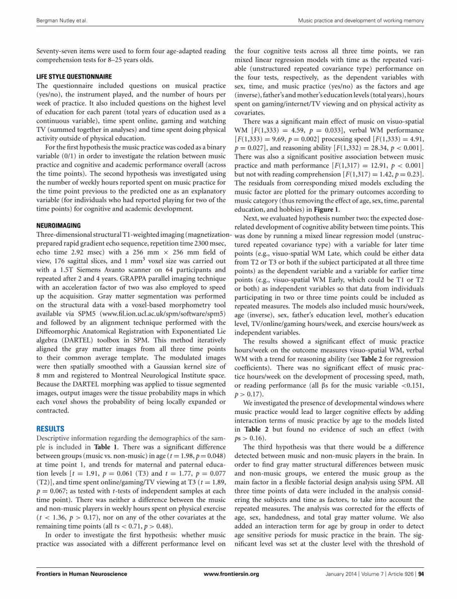
“fnhum-07-00926” — 2014/1/7 — 11:07 — page 4 — #4
Bergman Nutley et al. Music practice and development of working memory
Seventy-seven items were used to form four age-adapted readingcomprehension tests for 8–25 years olds.
LIFE STYLE QUESTIONNAIREThe questionnaire included questions on musical practice(yes/no), the instrument played, and the number of hours perweek of practice. It also included questions on the highest levelof education for each parent (total years of education used as acontinuous variable), time spent online, gaming and watchingTV (summed together in analyses) and time spent doing physicalactivity outside of physical education.
For the first hypothesis the music practice was coded as a binaryvariable (0/1) in order to investigate the relation between musicpractice and cognitive and academic performance overall (acrossthe time points). The second hypothesis was investigated usingthe number of weekly hours reported spent on music practice forthe time point previous to the predicted one as an explanatoryvariable (for individuals who had reported playing for two of thetime points) for cognitive and academic development.
NEUROIMAGINGThree-dimensional structural T1-weighted imaging (magnetization-prepared rapid gradient echo sequence, repetition time 2300 msec,echo time 2.92 msec) with a 256 mm × 256 mm field ofview, 176 sagittal slices, and 1 mm3 voxel size was carried outwith a 1.5T Siemens Avanto scanner on 64 participants andrepeated after 2 and 4 years. GRAPPA parallel imaging techniquewith an acceleration factor of two was also employed to speedup the acquisition. Gray matter segmentation was performedon the structural data with a voxel-based morphometry toolavailable via SPM5 (www.fil.ion.ucl.ac.uk/spm/software/spm5)and followed by an alignment technique performed with theDiffeomorphic Anatomical Registration with Exponentiated Liealgebra (DARTEL) toolbox in SPM. This method iterativelyaligned the gray matter images from all three time pointsto their common average template. The modulated imageswere then spatially smoothed with a Gaussian kernel size of8 mm and registered to Montreal Neurological Institute space.Because the DARTEL morphing was applied to tissue segmentedimages, output images were the tissue probability maps in whicheach voxel shows the probability of being locally expanded orcontracted.
RESULTSDescriptive information regarding the demographics of the sam-ple is included in Table 1. There was a significant differencebetween groups (music vs. non-music) in age (t = 1.98, p = 0.048)at time point 1, and trends for maternal and paternal educa-tion levels [t = 1.91, p = 0.061 (T3) and t = 1.77, p = 0.077(T2)], and time spent online/gaming/TV viewing at T3 (t = 1.89,p = 0.067; as tested with t-tests of independent samples at eachtime point). There was neither a difference between the musicand non-music players in weekly hours spent on physical exercise(t < 1.36, p > 0.17), nor on any of the other covariates at theremaining time points (all ts < 0.71, p > 0.48).
In order to investigate the first hypothesis: whether musicpractice was associated with a different performance level on
the four cognitive tests across all three time points, we ranmixed linear regression models with time as the repeated vari-able (unstructured repeated covariance type) performance onthe four tests, respectively, as the dependent variables withsex, time, and music practice (yes/no) as the factors and age(inverse), father’s and mother’s education levels (total years), hoursspent on gaming/internet/TV viewing and on physical activity ascovariates.
There was a significant main effect of music on visuo-spatialWM [F(1,333) = 4.59, p = 0.033], verbal WM performance[F(1,333) = 9.69, p = 0.002] processing speed [F(1,333) = 4.91,p = 0.027], and reasoning ability [F(1,332) = 28.34, p < 0.001].There was also a significant positive association between musicpractice and math performance [F(1,317) = 12.91, p < 0.001]but not with reading comprehension [F(1,317) = 1.42, p = 0.23].The residuals from corresponding mixed models excluding themusic factor are plotted for the primary outcomes according tomusic category (thus removing the effect of age, sex, time, parentaleducation, and hobbies) in Figure 1.
Next, we evaluated hypothesis number two: the expected dose-related development of cognitive ability between time points. Thiswas done by running a mixed linear regression model (unstruc-tured repeated covariance type) with a variable for later timepoints (e.g., visuo-spatial WM Late, which could be either datafrom T2 or T3 or both if the subject participated at all three timepoints) as the dependent variable and a variable for earlier timepoints (e.g., visuo-spatial WM Early, which could be T1 or T2or both) as independent variables so that data from individualsparticipating in two or three time points could be included asrepeated measures. The models also included music hours/week,age (inverse), sex, father’s education level, mother’s educationlevel, TV/online/gaming hours/week, and exercise hours/week asindependent variables.
The results showed a significant effect of music practicehours/week on the outcome measures visuo-spatial WM, verbalWM with a trend for reasoning ability (see Table 2 for regressioncoefficients). There was no significant effect of music prac-tice hours/week on the development of processing speed, math,or reading performance (all βs for the music variable <0.151,p > 0.17).
We investigated the presence of developmental windows wheremusic practice would lead to larger cognitive effects by addinginteraction terms of music practice by age to the models listedin Table 2 but found no evidence of such an effect (withps > 0.16).
The third hypothesis was that there would be a differencedetected between music and non-music players in the brain. Inorder to find gray matter structural differences between musicand non-music groups, we entered the music group as themain factor in a flexible factorial design analysis using SPM. Allthree time points of data were included in the analysis consid-ering the subjects and time as factors, to take into account therepeated measures. The analysis was corrected for the effects ofage, sex, handedness, and total gray matter volume. We alsoadded an interaction term for age by group in order to detectage sensitive periods for music practice in the brain. The sig-nificant level was set at the cluster level with the threshold of
Frontiers in Human Neuroscience www.frontiersin.org January 2014 | Volume 7 | Article 926 | 94

“fnhum-07-00926” — 2014/1/7 — 11:07 — page 5 — #5
Bergman Nutley et al. Music practice and development of working memory
FIGURE 1 | Cognitive performance across the three time points with theresiduals from mixed models (with age, sex, time, parental educationlevel, time spent on physical activity, gaming/internet/TV viewing as
independent variables) for the primary outcomes plotted according tomusic training category. Error bars represent standard error of the mean(SEM).
Table 2 | Regression coefficients for the models investigating the amount of change in cognitive performance on the three outcomes that couldbe explained by music hours/week and other possible factors.
Dependent variables and model summaries
Visuo-spatial WM (late) Verbal WM (late) Reasoning (late)
Independent variables β P β P β P
Constant – 0.002 – 0.032 – 0.171
Sex −0.568 0.242 0.300 0.422 −0.025 0.612
Age (Inverse) −150.70 0.241 57.692 0.517 −20.47 0.145
Mother Edu (years) 0.048 0.680 −0.107 0.242 0.018 0.139
Father Edu (years) 0.185 0.138 0.043 0.653 −0.010 0.423
Outcome (Early) 0.715 <0.001 0.773 <0.001 0.668 <0.001
Online/gaming/TV(h/w) −0.002 0.907 0.043 0.004 0.001 0.436
Exercise (h/w) −0.229 0.078 0.157 0.110 −0.005 0.712
Music (h/w) 0.351 0.003 0.261 0.006 0.021 0.090
p < 0.05 (corrected for the multiple comparisons of all voxelswithin gray matter volume). We found two clusters significantlyshowing differences in gray matter volume. One in the tem-poral lobe (p = 0.008, see Figure 2A) situated mainly in theinferior temporal and temporal-occipital fusiform gyri, basedon the MNI structural and Harvard-Oxford Cortical Structuralatlases (see Table 3 for coordinates and cluster sizes), and theother cluster was in the insula, caudate, and putamen (p = 0.002see Figure 2B). The interaction of age by group was notsignificant.
DISCUSSIONIn this study, the effect of musical practice on cognitive abil-ity was investigated in a developmental longitudinal study. Theresults showed that practicing a musical instrument was associ-ated with higher performance on tests of reasoning, processingspeed, and WM, as well as mathematics but not reading com-prehension. Importantly, the development between time pointswas related to the time spent on music practice per weekfor both WM tests. The effect was also seen after correctingfor baseline performance, parental education, and other after
Frontiers in Human Neuroscience www.frontiersin.org January 2014 | Volume 7 | Article 926 | 95

“fnhum-07-00926” — 2014/1/7 — 11:07 — page 6 — #6
Bergman Nutley et al. Music practice and development of working memory
FIGURE 2 |The main effect of playing a musical instrument on graymatter volume; (A) effects in temporal cortex, occipito-temporal area,and the insula; (B) effects in caudate nucleus shown in sagittal sectionfrom both left and right hemispheres.
school activities, including physical activity, TV-watching, andgaming. These results confirm previously reported associationsbetween musical practice and cognitive ability (Hurwitz et al.,1975; Schellenberg, 2006; Forgeard et al., 2008; Ruthsatz et al.,2008). The dose-response relation also supports the previouslyreported causal relation between the two (Schellenberg, 2004;Bugos et al., 2007).
We did not find a dose-response relation between music prac-tice and the development of processing speed and only found atrend for the development of reasoning ability. The positive effectson the secondary outcome of mathematics did not point to a causaleffect of music on academic development, and was only seen as anassociation with music practice across the time points. This is inline with previous findings of associations from correlational stud-ies between music practice and grades in mathematics (Schellen-berg, 2006; Cabanac et al., 2013) and mathematical performance(Schellenberg, 2006). As most of the music players in our sam-ple were already practicing an instrument at the beginning of thestudy, it is possible that potential pre-study music-related changesin the development of WM underlie the learning advantages seen
for mathematics, given its strong relation to visuo-spatial WMin particular (Bull et al., 2008). We did not see an associationbetween music practice and reading comprehension, a findingpreviously reported in a correlational study (Schellenberg, 2006).The reason for this is unclear and should be furtherstudied.
Differences were also seen in the gray matter volume of thebrain between music players and non-players in the temporo-occipital and insular cortex across the time points. One couldargue that since the music group outperformed the non-musicgroup on cognitive measures at the beginning of the study thatthese differences should be controlled for in the brain imaginganalysis. However, it is not certain and perhaps, given the liter-ature (e.g., Schlaug et al., 2005), even unlikely that the cognitivedifferences are independent of music, thus controlling for themwould also remove cognitive differences associated with music inthe brain. The differences observed in the brain are consistent withprevious findings showing higher gray matter volume for musi-cians in the fusiform gyrus (James et al., 2013), an area typicallyinvolved in visual pattern recognition. Specifically, this area hasbeen identified through fMRI investigations as important dur-ing musical notation decoding (Schon et al., 2002; Stewart et al.,2003). These regions do not include the fronto-parietal networksinvolved in top-down attention and WM (Olesen et al., 2004).However, the effect was seen in the caudate nucleus which also hasbeen implicated in WM training (Olesen et al., 2004; Dahlin et al.,2008; Backman et al., 2011). This demonstrates certain similari-ties between the neural correlates of cognitive training and musicpractice.
Over the past decade, research has shown that WM capacity issubject to training-induced improvements (Klingberg et al., 2002,2005; Holmes et al., 2009; Thorell et al., 2009; Brehmer et al., 2012;Green et al., 2012) and that the size of the transfer effects are lin-early related to practice time (Jaeggi et al., 2008; Bergman Nutleyet al., 2011). It is therefore likely that the time spent on otherWM taxing activities may affect WM capacity and other abili-ties partly depending on the same brain regions. Reasoning hasalso previously reported to improve by training on WM tasks(Klingberg et al., 2002, 2005; Jaeggi et al., 2008), non-verbal rea-soning tasks (Bergman Nutley et al., 2011; Mackey et al., 2011),as well as in strategy training studies (Klauer et al., 2002; Hamerset al., 1998). These types of interventions target different aspectsof reasoning and the performance improvements reported proba-bly have different origins. Interestingly, it has been shown thatit is possible to improve test performance in a single practicesession by learning to, e.g., attend to one stimulus dimension at a
Table 3 | Significant association between playing a musical instrument and gray matter volume.
Clusters overlap with* P corrected
cluster level
Number of
voxels
Z score x, y, z (MNI) for
the peak voxel
Temporo-occipital fusiform gyrus, Inferior temporal gyrus 0.008 4408 3.96 34, −51, −9
Insula, caudate, and putamen 0.002 5337 3.64 20, 18, 17
*The labeling is based on both MNI structural and Harvard-Oxford Cortical Structural atlases provided by FMRIB Software Library (FSL)(http://fsl.fmrib.ox.ac.uk/fsl/fslwiki/Atlases).
Frontiers in Human Neuroscience www.frontiersin.org January 2014 | Volume 7 | Article 926 | 96

“fnhum-07-00926” — 2014/1/7 — 11:07 — page 7 — #7
Bergman Nutley et al. Music practice and development of working memory
time (Denney and Heidrich, 1990). This indicates an immediateeffect, based on learned strategies, on how to best approach thevisuo-spatial structure of the task and is independent of practicetime.
The decoding of musical notation is one factor that may explainthe effect of music on cognitive functioning as it requires abil-ities such as spatial–temporal reasoning and visual perception(Gromko, 2004). Given that the results indicate linear effects ofmusic practice on WM and that previous studies have demon-strated effects of music practice on reasoning after as little as 1 yearof practice (Rauscher et al., 1997; Schellenberg, 2004), it may bethat the music-related processes taxing reasoning are trained whilelearning the musical notation code, whereas the processes taxingWM are trained while reading the code, keeping sections of codein WM and in control of attention. This would thus explain thelinear relation with time practiced between WM and music, as twomore years of music reading would further support developmentof WM. The effect of music training on reasoning development,on the other hand, may occur as quickly as the foundation ofthe musical systems are understood rather than linearly and grad-ual. That being said, regardless of beneficial strategies or basicunderstanding of the task, WM is required to solve reasoningproblems (Carpenter et al., 1990) and to read musical notation(which could explain the trend to linear effects on reasoningdevelopment).
Since the music players in this study showed superior per-formance on cognitive measures already at time point one it isnot possible to conclude that music practice caused the cogni-tive effects, although the dose-response relation indicates that thiscould be the case. Given that the groups differed in age and thatthe natural development of the cognitive functions assessed in thisstudy are not linear throughout the age span of the sample, itwas not possible to simply test an interaction between time andmusic in the mixed model. In order to address this, case-controlmatched analyses were attempted but resulted in too few controlcases to enable the analyses at sufficient statistical power. Hence,we instead performed linear regressions with the outcome at latertime points as the dependent variable while controlling for the out-come at earlier time points to inform about the change in cognitiveability related to music practice between time points.
Although prior research has suggested positive cognitive effectsof physical activity (Hillman et al., 2008; Hotting and Roder, 2013)as well as from playing computer games (Green and Bavelier,2012), we only found a positive effect for verbal WM developmentrelated to time spent on TV viewing/online and gaming activities.The reasons for a lack of a general effect on all cognitive tests areunclear, however, could be due to the crude measures of the covari-ates (combined score for the gaming/online/TV viewing variable,with no distinction made between verbal and more visuo-spatialactivities and no quantification of aerobic fitness or activity type).It may be that the combined score for gaming/online and TV view-ing represented activities relying on the use of verbal informationprocessing to a larger extent than taxing the visuo-spatial domain.
There may be alternative explanations to the different tra-jectories of WM development that in this study appear to bemusic-related, such as differences in personality. A recent studyshowed that personality type predicted the duration of music
training (along with parental education) to a larger degree thanIQ (Corrigall et al., 2013). This could be of importance as ourresults suggest that time spent on music practice is predictiveof WM development and could perhaps partly be explainedby the fact that certain personality types of high-functioningindividuals tend to show longer durations of practice and may,independently of music, have a different trajectory of their WMdevelopment. Another possibility is that there could be an inter-action between personality type and the cognitive training effectsassociated with music practice. This will be for future studies toinvestigate.
It is likely that part of the sample in our study is musicallytrained in the vocal domain, which is not accounted for. However,this should hardly affect the interpretation of the results as it islikely to add noise to the data, underestimating the strength ofthe effects, if anything. Future studies should continue to pursuethe question of causality, the mechanism through which musicimproves cognitive ability as well as potential effects on academicoutcome. Ideally, detailed information on musical notation read-ing skills should be recorded along with personality dimensionsand compared with performance on cognitive tests to furtherinvestigate the mechanisms involved in music practice and itseffects on cognitive ability.
CONCLUSIONThe results from this study show that music training is associ-ated with cognitive and mathematical benefits. This was madeapparent through the superior level of performance across timefor music players compared with non-players. There was also adifference in the gray matter density within brain in areas relatedto music notation decoding. Furthermore, the data suggest thatmusic players show a steeper development of both visuo-spatialand verbal WM over time, supporting previously reported causaleffects between music practice and cognitive performance. Timespent on music practice predicted both visuo-spatial WM and ver-bal WM development. More generally, these findings support theimportance of practice and learning for the development of WMduring childhood and adolescence.
REFERENCESAlloway, T. P. (2007). Automated Working Memory Assessment. London: Psychologi-
cal Corporation.Backman, L., Nyberg, L., Soveri, A., Johansson, J., Andersson, M., Dahlin, E., et al.
(2011). Effects of working-memory training on striatal dopamine release. Science333, 718. doi: 10.1126/science.1204978
Bengtsson, S. L., Nagy, Z., Skare, S., Forsman, L., Forssberg, H., and Ullen, F.(2005). Extensive piano practicing has regionally specific effects on white matterdevelopment. Nat. Neurosci. 8, 1148–1150. doi: 10.1038/nn1516
Bengtsson, S. L., and Ullen, F. (2006). Dissociation between melodic and rhythmicprocessing during piano performance from musical scores. Neuroimage 30, 272–284. doi: 10.1016/j.neuroimage.2005.09.019
Bergman Nutley, S., Söderqvist, S., Bryde, S., Thorell, L. B., Humphreys, K., andKlingberg, T. (2011). Gains in fluid intelligence after training non-verbal reason-ing in 4-year-old children – a controlled, randomized study. Dev. Sci. 14, 591–601.doi: 10.1111/j.1467-7687.2010.01022.x
Bilhartz, T. D., Bruhn, R. A., and Olson, J. E. (1999). The effect of early musictraining on child cognitive development. J. Appl. Dev. Psychol. 20, 615–636. doi:10.1016/S0193-3973(99)00033-7
Brehmer, Y., Westerberg, H., and Backman, L. (2012). Working-memory training inyounger and older adults: training gains, transfer, and maintenance. Front. Hum.Neurosci. 6:63. doi: 10.3389/fnhum.2012.00063
Frontiers in Human Neuroscience www.frontiersin.org January 2014 | Volume 7 | Article 926 | 97

“fnhum-07-00926” — 2014/1/7 — 11:07 — page 8 — #8
Bergman Nutley et al. Music practice and development of working memory
Bugos, J. A., Perlstein, W. M., McCrae, C. S., Brophy, T. S., and Bedenbaugh,P. H. (2007). Individualized Piano Instruction enhances executive functioningand working memory in older adults. Aging Ment. Health 11, 464–471. doi:10.1080/13607860601086504
Bull, R., Espy, K. A., and Wiebe, S. A. (2008). Short-term memory, workingmemory, and executive functioning in preschoolers: longitudinal predictors ofmathematical achievement at age 7 years. Dev. Neuropsychol. 33, 205–228 doi:10.1080/87565640801982312
Cabanac, A., Perlovsky, L., Bonniot-Cabanac, M.-C., and Cabanac, M. (2013)Music and academic performance. Behav. Brain Res. 256, 257–260. doi:10.1016/j.bbr.2013.08.023
Carpenter, P. A., Just, M. A., and Shell, P. (1990). What one intelligence test measures:a theoretical account of the processing in the Raven Progressive Matrices Test.Psychol. Rev. 97, 404–431. doi: 10.1037/0033-295X.97.3.404
Corrigall, K. A., Schellenberg, E. G., and Misura, N. M. (2013). Music training,cognition, and personality. Front. Psychol. 4:222. doi: 10.3389/fpsyg.2013.00222
Costa-Giomi, E. (1999). The effects of three years of piano instruction on children’scognitive development. J. Res. Music Educ. 47, 198–212. doi: 10.2307/3345779
Dahlin, E., Neely, A. S., Larsson, A., Backman, L., and Nyberg, L. (2008). Transferof learning after updating training mediated by the striatum. Science 320, 1510–1512. doi: 10.1126/science.1155466
Dahlin, K. I. E. (2010). Effects of working memory training on reading in childrenwith special needs. Read. Writ. 24, 479–491. doi: 10.1007/s11145-010-9238-y
Denney, N. W., and Heidrich, S. M. (1990). Training effects on Raven’s progressivematrices in young, middle-aged, and elderly adults. Psychol. Aging 5, 144–145.doi: 10.1037/0882-7974.5.1.144
Egeland, J., Aarlien, A. K., and Saunes, B.-K. (2013). Few effects of far transfer ofworking memory training in ADHD: a randomized controlled trial. PLoS ONE8:e75660. doi: 10.1371/journal.pone.0075660
Elbert, T., Pantev, C., Wienbruch, C., Rockstroh, B., and Taub, E. (1995). Increasedcortical representation of the fingers of the left hand in string players. Science 270,305–307. doi: 10.1126/science.270.5234.305
Ericsson, K. A., Charness, N., and Hoffman, R. R. (ed.). (2006). The CambridgeHandbook of Expertise and Expert Performance. Cambridge: Cambridge UniversityPress. doi: 10.1017/CBO9780511816796
Ericsson, K. A., and Lehmann, A. C. (1996). Expert and exceptional performance:evidence of maximal adaptation to task constraints. Annu. Rev. Psychol. 47, 273–305. doi: 10.1146/annurev.psych.47.1.273
Forgeard, M., Winner, E., Norton, A., and Schlaug, G. (2008). Practicing a musicalinstrument in childhood is associated with enhanced verbal ability and nonverbalreasoning. PLoS ONE 3:e3566. doi: 10.1371/journal.pone.0003566
Gaser, C., and Schlaug, G. (2003). Brain structures differ between musicians andnon-musicians. J. Neurosci. 23, 9240–9245.
Gillham, B., and Hesse, K. (2001). Basic Number Screening Test. London: Hodderand Stoughton International.
Green, C. S., and Bavelier, D. (2012). Learning, attentional control, and action videogames. Curr. Biol. 22, R197–R206. doi: 10.1016/j.cub.2012.02.012
Green, C. T., Long, D. L., Green, D., Iosif, A. M., Dixon, J. F., Miller, M. R., et al.(2012). Will working memory training generalize to improve off-task behavior inchildren with attention-deficit/hyperactivity disorder? Neurotherapeutics 9, 639–648. doi: 10.1007/s13311-012-0124-y
Gromko, J. E. (2004). Predictors of music sight-reading ability in high school windplayers. J. Res. Music Educ. 52, 6–15. doi: 10.2307/3345521
Gromko, J. E., and Poorman, A. S. (1998). The effect of music training on preschool-ers’ spatial-temporal task performance. J. Res. Music Educ. 46, 173–181. doi:10.2307/3345621
Gustafsson, J. E., and Rosén, M. (2004). Förändringar i läskompetens 1991–2001: Enjämförelse över tid och Länder [Trends in Reading Comprehension. 1991–2001: AComparison across Periods and Countries. The Swedish National Agency for SchoolImprovement]. Forskning i Fokus 22. Stockholm: Myndigheten för Skolutveckling.
Hamers, J. H. M., de Koning, E., and Sijtsma, K. (1998). Inductive reasoning inthird grade: intervention promises, and constraints. Contemp. Educ. Psychol. 23,132–148. doi: 10.1006/ceps.1998.0966
Hempel, A., Giesel, F. L., Garcia Caraballo, N. M., Amann, M., Meyer, H., Wusten-berg, T., et al. (2004). Plasticity of cortical activation related to working memoryduring training. Am. J. Psychiatry 161, 745–747. doi: 10.1176/appi.ajp.161.4.745
Hillman, C. H., Erickson, K. I., and Kramer, A. F. (2008). Be smart, exercise yourheart: exercise effects on brain and cognition. Nat. Rev. Neurosci. 9, 58–65. doi:10.1038/nrn2298
Holmes, J., Gathercole, S. E., and Dunning, D. L. (2009). Adaptive training leadsto sustained enhancement of poor working memory in children. Dev. Sci. 12,F9–F15. doi: 10.1111/j.1467-7687.2009.00848.x
Hotting, K., and Roder, B. (2013). Beneficial effects of physical exercise onneuroplasticity and cognition. Neurosci. Biobehav. Rev. 37, 2243–2257. doi:10.1016/j.neubiorev.2013.04.005
Hurwitz, I., Wolff, P. H., Bortnick, B. D., and Kokas, K. (1975). Nonmusical effects ofkodaly music curriculum in primary grade children. J. Learn. Disabil. 8, 167–174.doi: 10.1177/002221947500800310
Hyde, K. L., Lerch, J., Norton, A., Forgeard, M., Winner, E., Evans, A. C., et al. (2009).Musical training shapes structural brain development. J. Neurosci. 29, 3019–3025.doi: 10.1523/JNEUROSCI.5118-08.2009
Jaeggi, S. M., Buschkuehl, M., Jonides, J., and Perrig, W. J. (2008). Improving fluidintelligence with training on working memory. Proc. Natl. Acad. Sci. U.S.A. 105,6829–6833. doi: 10.1073/pnas.0801268105
James, C. E., Oechslin, M. S., Van De Ville, D., Hauert, C. A., Descloux, C., andLazeyras, F. (2013). Musical training intensity yields opposite effects on greymatter density in cognitive versus sensorimotor networks. Brain Struct. Funct.doi: 10.1007/s00429-013-0504-z
Jolles, D. D., Grol, M. J., Van Buchem, M. A., Rombouts, S. A., and Crone, E. A.(2010). Practice effects in the brain: changes in cerebral activation after work-ing memory practice depend on task demands. Neuroimage 52, 658–668. doi:10.1016/j.neuroimage.2010.04.028
Klauer, K. J., Willmes, K., and Phye, G. D. (2002). Inducing inductive reason-ing: does it transfer to fluid intelligence. Contemp. Educ. Psychol. 27, 1–25. doi:10.1006/ceps.2001.1079
Klingberg, T. (2010). Training and plasticity of working memory. Trends Cogn. Sci.14, 317–324. doi: 10.1016/j.tics.2010.05.002
Klingberg, T., Fernell, E., Olesen, P. J., Johnson, M., Gustafsson, P., Dahlstrom,K., et al. (2005). Computerized training of working memory in children withADHD–a randomized, controlled trial. J. Am. Acad. Child Adolesc. Psychiatry 44,177–186. doi: 10.1097/00004583-200502000-00010
Klingberg, T., Forssberg, H., and Westerberg, H. (2002). Training of workingmemory in children with ADHD. J. Clin. Exp. Neuropsychol. 24, 781–791. doi:10.1076/jcen.24.6.781.8395
Mackey, A. P., Hill, S. S., Stone, S. I., and Bunge, S. A. (2011). Differentialeffects of reasoning and speed training in children. Dev. Sci. 14, 582–590. doi:10.1111/j.1467-7687.2010.01005.x
Martin, M., Mullis, S., and Chrostowski, S. (eds). (2004). TIMSS 2003 TechnicalReport. Chestnut Hill: TIMSS& PIRLS International Study Centre Boston College.
Meinz, E. J., and Hambrick, D. Z. (2010). Deliberate practice is necessarybut not sufficient to explain individual differences in piano sight-readingskill: the role of working memory capacity. Psychol. Sci. 21, 914–919. doi:10.1177/0956797610373933
Nakada, T., Fujii, Y., Suzuki, K., and Kwee, I. L. (1998). ‘Musical brain’revealed by high-field (3 Tesla) functional MRI. Neuroreport 9, 3853–3856. doi:10.1097/00001756-199812010-00016
Norton, A., Winner, E., Cronin, K., Overy, K., Lee, D. J., and Schlaug, G. (2005). Arethere pre-existing neural, cognitive, or motoric markers for musical ability? BrainCogn. 59, 124–134. doi: 10.1016/j.bandc.2005.05.009
Olesen, P. J., Westerberg, H., and Klingberg, T. (2004). Increased prefrontal andparietal activity after training of working memory. Nat. Neurosci. 7, 75–79. doi:10.1038/nn1165
Pallesen, K. J., Brattico, E., Bailey, C. J., Korvenoja, A., Koivisto, J., Gjedde, A., et al.(2010). Cognitive control in auditory working memory is enhanced in musicians.PLoS ONE 5:e11120. doi: 10.1371/journal.pone.0011120
Pascual-Leone, A. (2001). The brain that plays music and is changed by it. Ann. N.Y. Acad. Sci. 930, 315–329. doi: 10.1111/j.1749-6632.2001.tb05741.x
Rauscher, F. H., Shaw, G. L., Levine, L. J., Wright, E. L., Dennis, W. R., and New-comb, R. L. (1997). Music training causes long-term enhancement of preschoolchildren’s spatial-temporal reasoning. Neurol. Res. 19, 2–8.
Raven, J. C. (2003). Raven’s Advanced Progressive Matrices: Pearson Assessment.London: Pearson, Inc.
Ruthsatz, J., Detterman, D., Griscom, W. S., and Cirullo, B. A. (2008). Becomingan expert in the musical domain: it takes more than just practice. Intelligence 36,330–338. doi: 10.1016/j.intell.2007.08.003
Salis, D. L. (1978). The Identification and Assessment of Cognitive Variables Associatedwith Reading of Advanced Music at the Piano. Ph.D. dissertation, University ofPittsburgh, Pittsburgh. Dissertation Abstracts International, A 38:12:7239:40.
Frontiers in Human Neuroscience www.frontiersin.org January 2014 | Volume 7 | Article 926 | 98

“fnhum-07-00926” — 2014/1/7 — 11:07 — page 9 — #9
Bergman Nutley et al. Music practice and development of working memory
Schellenberg, E. G. (2004). Music lessons enhance IQ. Psychol. Sci. 15, 511–514. doi:10.1111/j.0956-7976.2004.00711.x
Schellenberg, E. G. (2006). Long-term positive associations between music lessonsand IQ. J. Educ. Psychol. 98, 457–468. doi: 10.1037/0022-0663.98.2.457
Schellenberg, E. G. and Weiss, M. W. (2013). “Music and cognitive abilities,” inPsychology of Music, 3rd Edn, ed. D. Deutsch (Amsterdam: Elsevier Inc.), 499–550.doi: 10.1016/B978-0-12-381460-9.00012-2
Schlaug, G., Norton, A., Overy, K., and Winner, E. (2005). Effects of music trainingon the child’s brain and cognitive development. Ann. N. Y. Acad. Sci. 1060,219–230. doi: 10.1196/annals.1360.015
Schon, D., Anton, J. L., Roth, M., and Besson, M. (2002). AnfMRI study of music sight-reading. Neuroreport 13, 2285–2289. doi:10.1097/01.wnr.0000044224.79663.f5
Stewart, L., Henson, R., Kampe, K., Walsh, V., Turner, R., and Frith, U. (2003).Becoming a pianist. An fMRI study of musical literacy acquisition. Ann. N. Y.Acad. Sci. 999, 204–208. doi: 10.1196/annals.1284.030
Thorell, L. B., Lindqvist, S., Bergman Nutley, S., Bohlin, G., and Klingberg, T. (2009).Training and transfer effects of executive functions in preschool children. Dev.Sci. 12, 106–113. doi: 10.1111/j.1467-7687.2008.00745.x
Westerberg, H., Jacobaeus, H., Hirvikoski, T., Clevberger, P., Ostensson, M. L.,Bartfai, A., et al. (2007). Computerized working memory training after stroke–apilot study. Brain Inj. 21, 21–29. doi: 10.1080/02699050601148726
Wolinsky, F. D., Vander Weg, M. W., Howren, M. B., Jones, M. P., and Dotson, M. M.(2013). A randomized controlled trial of cognitive training using a visual speedof processing intervention in middle aged and older adults. PLoS ONE 8:e61624.doi:10.1371/journal.pone.0061624
Conflict of Interest Statement: Sissela Bergman Nutley is an employee ofPearson/Cogmed. Torkel Klingberg has had consultancy agreement with Pear-son/Cogmed.
Received: 28 August 2013; accepted: 18 December 2013; published online: 07 January2014.Citation: Bergman Nutley S, Darki F and Klingberg T (2014) Music practice is associ-ated with development of working memory during childhood and adolescence. Front.Hum. Neurosci. 7:926. doi: 10.3389/fnhum.2013.00926This article was submitted to the journal Frontiers in Human Neuroscience.Copyright © 2014 Bergman Nutley, Darki and Klingberg. This is an open-accessarticle distributed under the terms of the Creative Commons Attribution License(CC BY). The use, distribution or reproduction in other forums is permitted,provided the original author(s) or licensor are credited and that the original pub-lication in this journal is cited, in accordance with accepted academic practice. Nouse, distribution or reproduction is permitted which does not comply with theseterms.
Frontiers in Human Neuroscience www.frontiersin.org January 2014 | Volume 7 | Article 926 | 99

“fnhum-07-00693” — 2013/10/21 — 10:25 — page 1 — #1
REVIEW ARTICLEpublished: 22 October 2013
doi: 10.3389/fnhum.2013.00693
Neural implementation of musical expertise and cognitivtransfers: could they be promising in the framework ofnormal cognitive aging?
e
Baptiste Fauvel 1,2 ,3 ,4 , Mathilde Groussard 1,2 ,3 ,4 , Francis Eustache1,2 ,3 ,4 , Béatrice Desgranges1,2 ,3 ,4 andHervé Platel1,2 ,3 ,4*1 INSERM, U1077, Caen, France2 Université de Caen Basse-Normandie, UMR-S1077, Caen, France3 Ecole Pratique des Hautes Etudes, UMR-S1077, Caen, France4 CHU de Caen, U1077, Caen, France
Edited by:Merim Bilalic, University Tübingen,Germany
Reviewed by:Sebastian J. Lipina, Unidad deNeurobiología Aplicada, ArgentinaTilo Strobach, Humboldt UniversityBerlin, Germany
*Correspondence:Hervé Platel, INSERM, U1077,EPHE-Université de CaenBasse-Normandie, Caen, France;U.F.R. de Psychologie, Université deCaen Basse-Normandie, Esplanade dela Paix, 14032 Caen Cedex, Francee-mail: [email protected]
Brain plasticity allows the central nervous system of a given organism to cope withenvironmental demands. Therefore, the quality of mental processes relies partly on theinteraction between the brain’s physiological maturation and individual daily experiences.In this review, we focus on the neural implementation of musical expertise at both ananatomical and a functional level. We then discuss how this neural implementation canexplain transfers from musical learning to a broad range of non-musical cognitive functions,including language, especially during child development. Finally, given that brain plasticityis still present in aging, we gather arguments to propose that musical practice could bea good environmental enrichment to promote cerebral and cognitive reserves, therebyreducing the deleterious effect of aging on cognitive functions.
Keywords: musical training, expertise, brain plasticity, cognitive transfer, aging, brain reserve
INTRODUCTIONTo adapt to changing environmental requirements, the brainhas structural and functional dynamic properties that allow reg-ularities to be encoded and skills to be learned and refinedthrough regular practice (Draganski and May, 2008). In return,the efficiency of the cognitive system is partly influenced by envi-ronmental features and individual daily experiences (Plomin et al.,1999).
Mastering a musical instrument is a particularly complex mul-timodal and multiprocesses behavior because it requires a theoret-ical learning of the solfeggios rules, as well as a repeated trainingfor translating visual codes (the written scores) into precise motorsequences and auditory events (Wan and Schlaug, 2010). Musicaltraining and musicians have therefore been often used in studiesfocusing on practice-related brain plasticity. Interestingly, theseinvestigations have brought some empirical evidences arguing thatthe regular commitment in musical activities does not only leadto behavioral improvements in perceptivo-motor skills, but alsoin a wide range of non-specific cognitive functions (Schellenberg,2006).
In this article, we review (1) the structural plasticity effectsinduced by musical practice, to point out that they can belocated in general cognitive related-areas outside the auditory-motor domain; (2) the functional differences that exist betweenmusically naïve and musically trained subject when they performauditory-motor tasks, to show that trained subjects can rely ongeneral cognitive processes when the task become harder; (3) thetransfers effect that occur from musical practice toward other
cognitive functions, because it justify the use of music, notablyin rehabilitation program for developmental disorders or braininjuries. Finally, (4) we present works and arguments to suggestthat musical practice could also be encourage as a particularlygood environmental enrichment to promote a successful brainaging.
NEURAL IMPLEMENTATION OF MUSICAL EXPERTISESTRUCTURAL PLASTICITY INDUCED BY MUSICAL PRACTICEStructural brain plasticity phenomenon (i.e., changes in cellsanatomy and connections) can be longitudinally and cross-sectionally recorded in human using morphometric method ofMRI data analysis (Draganski and May, 2008). In a longitudi-nal MRI study conducted by Hyde et al. (2009), 31 children agedaround 6 years were scanned before being assigned to 15 monthsof either individual keyboard lessons or a non-instrumental musicclass. Whereas no structural differences were observed before theexperiment, those children who had learned to play the keyboardexhibited significantly greater increases in gray matter volume afterthe lessons within the primary motor cortex (right pre-centralgyrus) and motor-related areas (part of the corpus callosum), aswell as within the primary auditory cortex (right Heschl’s gyrus).The extent of the rearrangements in these motor and auditoryareas was correlated with improvements in finger tapping andauditory frequency discrimination tasks, respectively. Interest-ingly, the authors also found a greater increase in gray mattervolume in brain regions dedicated to the integration of multi-modal information (bilateral frontal areas and left pericingulate
Frontiers in Human Neuroscience www.frontiersin.org October 2013 | Volume 7 | Article 693 | 100

“fnhum-07-00693” — 2013/10/21 — 10:25 — page 2 — #2
Fauvel et al. Effects of musical training on the brain and cognition
gyrus). In the context of musical practice, these regions may sus-tain the translation of the visual score into motor sequences andauditory-induced emotional events (Hyde et al., 2009).
Cross-sectional studies conducted with adult expert musicianshave confirmed this anatomical brain shaping on several occa-sions. Adult violinists have a more highly elaborated topographicrepresentation of their left hand in the right somatosensory cortex(Elbert et al., 1995). Moreover, the gray matter volume of the pri-mary motor cortex (pre-central gyri) has been shown to increase inrelation with the degree of musical expertise (Gaser and Schlaug,2003), with a left-hemisphere advantage for pianists and a right-hemisphere one for string players (Bangert and Schlaug, 2006),illustrating the close link between anatomical changes and trainingdemands. Regarding the auditory cortex, Bermudez and Zatorre(2005) found greater gray matter volume in the right superior tem-poral gyrus of 43 adult musicians compared with non-musicians.Beyond the motor and auditory cortices, other local gray matterincreases, possibly induced by score reading, have been found inthe visuospatial and visuomotor areas (right fusiform gyrus, leftintraparietal sulcus; James et al., 2013) and left inferior temporalgyrus (Gaser and Schlaug, 2003), as well as in multimodal asso-ciation areas (right superior parietal gyrus; Gaser and Schlaug,2003), and general cognitive-related areas (left inferior frontalgyrus; James et al., 2013).
Structural brain plasticity phenomenon also appears to takeplace in white matter tracts. The corpus callosum exhibits enlarge-ment in musicians because they engage more frequently inbimanual behavior (Öztürk et al., 2002; Hyde et al., 2009), andpossibly because this structure is involved in visuoauditory pro-cesses too (Bengtsson et al., 2005). Bengtsson and colleagues alsofound an increase in the diffusivity value of the internal capsule ofthe corticospinal tract, and a positive correlation between hoursof musical practice and fiber tract diffusivity in frontal areas andthe right arcuate fasciculus, which are crucial for language pro-cessing, and have been shown to be larger and more structured inmusicians, especially singers (Halwani et al., 2011).
To resume, performing music involves elaborated, coordinated,and rules-based motor, auditory, and visual skills. Regular musi-cal training therefore leads to an anatomical shaping of auditory,motor and visual-related areas, but some results showed thatbrain regions involved in more general non-musical cognitive pro-cesses, such as language, attention, or executive functions, are alsoaffected by musical training-related structural plasticity (Hydeet al., 2009; James et al., 2013). This indirectly argues that theselater functions could also play a crucial role in musical expertiseachievement.
FUNCTIONAL PLASTICITY INDUCED BY MUSICAL PRACTICEThe impressive auditory skills and manual dexterity of musiciansare not solely explained by the anatomical remodeling of audi-tory and motor-related areas, as they also exhibit functional braindifferences with non-musicians when they perform tasks similarto music exercises (e.g., finger tapping tasks, tonal, or temporalauditory discrimination tasks).
fMRI studies have shown that when professional pianists andviolinists are asked to perform complex uni- or bi-manual tappingtasks, they exhibit reduced activation of the primary and secondary
motor areas (Jäncke et al., 2000; Lotze et al., 2003) as well asthe supplementary and pre-motor areas (Krings et al., 2000),compared with matched controls. The authors of these studiesconcluded that motor skills are more automatized, and thus lesscostly for musicians, who need therefore to recruit fewer neuronsfor performing as well as controls subjects do. This automatizationfrees up resources that allow for better spontaneity and flexibil-ity, as reflected in the greater activation of bilateral pre-frontaland parietal areas (Lotze et al., 2003; Landau and D’Esposito,2006).
Regarding auditory skills, once again, besides the structuralshaping of the auditory areas, particularities in neural responseshave been recorded when expert musicians process musicallyrelevant auditory stimuli. They have an enhanced cortical rep-resentation of the musical scale tones in the tonotopic map ofthe auditory cortex (Pantev et al., 1998) that leads them to exhibitgreater expectancy and more effective attentional processes formusical sounds. Indeed, when they have to identify a sound witha deviant frequency (pitch) or rhythm embedded in a sequenceof standard sounds, they display shorter latencies and/or higheramplitudes of the electrophysiological components known toreflect conscious sound discrimination and target detection [N2band P3; Tervaniemi et al., 2005; Late Positive Component (LPC);Besson et al., 1994; P3, Ungan et al., 2013].
Musicians’ enhanced cortical representations of musical rulesturn also in a pre-attentive memory-based processing for musi-cal sound encoding, maybe through the auditory corticofugalpathway. Indeed, modulations in both cortical and subcorticalelectrophysiological responses to the perception of musical irreg-ularities have even been recorded when musicians’ attention isdiverted away from the stimuli. It is reflected in the appearance orincrease of the MisMatch Negativity (MMN) component in theauditory cortex of musicians versus non-musicians when they areexposed to a melodic contour or interval change (Koelsch et al.,1999; Fujioka et al., 2004), and in the modulation of the EarlyRight Anterior Negative (ERAN) component when the changesconsist of more complex musical irregularities (e.g., unexpectedchords; Koelsch et al., 2002).
Concerning the subcortical level, musicians display faster neu-ral synchronization and stronger brainstem encoding of chordarpeggios in both in tune and out of tune conditions. Interestingly,the magnitude of the brainstem response is predictive of the par-ticipants’ performances in a pitch discrimination task (Bidelmanet al., 2011).
Neuroimaging studies using fMRI have also been designed toexplore whether musically trained individuals process the audi-tory aspects of music differently from non-musicians. When theyare passively exposed to music or have to judge whether chordsare consonant or dissonant, musically trained participants dis-play a right to left shift in the activation of auditory temporalareas (Ohnishi et al., 2001) and frontal and inferior parietal areas(Minati et al., 2009). According to the authors, this suggests theuse of more abstract and analytical strategies by the experts.Studies featuring pitch comparison tasks that involve greaterworking memory load (Gaab and Schlaug, 2003) or cognitivecontrol and updating in working memory (Pallesen et al., 2010)report that, compared with non-musicians, musically trained
Frontiers in Human Neuroscience www.frontiersin.org October 2013 | Volume 7 | Article 693 | 101

“fnhum-07-00693” — 2013/10/21 — 10:25 — page 3 — #3
Fauvel et al. Effects of musical training on the brain and cognition
participants recruit fewer early perceptual areas, and more audi-tory working memory-related areas (right posterior temporal andsupramarginal gyri and bilateral superior parietal areas) or asso-ciative areas (right pre-frontal, parietal lateral, anterior cingulated,and dorsolateral frontal cortices). The more difficult the task is,the more the experienced musicians resort to these areas and themore their performances are better compared with those of con-trols (Oechslin et al., 2012). Finally, regarding musical semanticmemory, a study conducted in our laboratory revealed that whenmusicians have to rate their familiarity with a melody, several auto-biographical episodic memory-related areas are activated morestrongly compared with non-musicians (bilateral hippocampus,visual primary cortex, cingulate cortex, and bilateral superior tem-poral areas; Groussard et al., 2010). This suggests that, owing totheir musical experience, musicians engage self-referential pro-cesses to perform the task and gain access to richer and more vividsensory details.
Moreover, musical expertise also leads to an auditory-motorcoupling, as reflected by the activity displayed within the primarymotor cortex when musicians hear a piece they have learned toplay (Haueisen and Knösche, 2001; Bangert et al., 2006). Con-versely, when they tinkle on a mute keyboard, the temporal areasdedicated to hearing are activated. In the same way, musical exper-tise is also built on auditory-visual and visuomotor associations.Indeed, it has been shown that when an atonal event (a wrong note)is written on a score, musicians are able to anticipate it, as reflectedin their behavioral performance and physiological response tothis event (Schön and Besson, 2005). Finally, when motionlesspianists imagine themselves playing a piece from a score, theydisplay the same pattern of neural activity within the secondaryand associative motor areas as they do during an actual instru-mental performance, although the degree of activation is reducedand does not concern the primary motor cortex (Meister et al.,2004).
To sum up so far, the expert auditory and motor performancesof musicians are sustained by the functional reorganization oftypical underlying neural processes, due to automation and bet-ter memory-based (top-down) processing. This makes the basicauditory and motor skills less costly for musicians, thereby allow-ing them to allocate resources to strategic processes when tasksbecome harder. Similar effects have also been observed for expertperformance in other activities such as object and pattern recog-nition by chess master players (Bilalic et al., 2012) or workingmemory-related tasks (Guida et al., 2012).
COGNITIVE TRANSFER OF MUSICAL TRAININGMusical hearing and practice seem not to be sustained by specificbrain areas, but involve rather general pre-existing skills and cog-nitive functions. As musical learning put a high demand on it, itcan result in transfers of improvements from one activity (musicmaking) to others (e.g., language skills, executive functions).
LANGUAGE SKILLSThere is a longstanding debate about the division or sharingof the brain substrates of language and music. Although somefamous neuropsychological cases of double dissociation have beenreported (Stewart et al., 2006), we know that music and language
perception and production share a lot of features, ranging from thebasic sensorimotor level to auditory-cognitive processes. Indeed,it appears that humans perceive these two auditory stimuli usingthe same acoustic cues (rhythm, pitch, and timbre) and relying onsimilar resources for syntactic integration processing or memory.Furthermore, both language and music have a visual form of nota-tion that allows them to be red or written, and producing either ofthese two behaviors involves auditory-motor coupling. Accordingto Patel’s OPERA hypothesis, transfers from musical training tolanguage skills occur because (i) there is an anatomical overlapin their brain networks, (ii) musical practice implies more preci-sion in processing features shared with language, (iii) this practicetakes place in a repeated manner, and is associated with (iv) morefocused attention, and (v) emotion (Patel, 2011).
Although they are less central to language than to music, pitchmodulations are still important for speech in order to convey emo-tion. Cross-sectional (Magne et al., 2006), and longitudinal studies(Moreno and Besson, 2005) found that musically trained childrenwere better at detecting pitch violations in a foreign or nativelanguage than their musically untrained counterparts. Moreover,the authors reported a cortical Late Positive Component (LPC) inresponse to pitch violations that was far less pronounced, or evenabsent, in the untrained children. Rather similar results have beenreported for adults, with musicians displaying a shorter latencyof this LPC when exposed to prosodic incongruities in foreignsentences (Schön et al., 2004; Marques et al., 2007).
Musicians also process the temporal features of speech soundsdifferently than non-musicians. Faster cortical neural responsesto voice onset time, as well as to vowel or syllable duration, havebeen attested both attentively and pre-attentively for children whotake part in music lessons (Chobert et al., 2012). As electrophysi-ological modulations have also been found at the brainstem level(Musacchia et al., 2007; Wong et al., 2007), Kraus and Chan-drasekaran (2010) suggest that transfer effects from processingacoustic cues in a musical context to processing them in a speechcontext are mostly due to the reinforcement of the auditorycorticofugal pathways (as in the reverse theory hypothesis thatstates that long-term cortically stored representations guide earlyperceptual encoding via descending pathways and top-down pro-cesses). The overtraining of musicians to detect, sequence, andencode relevant aspects of musical sound patterns shared withspeech endows them with heightened phonological awareness.
Apart from the benefit of perceiving prosody in native and for-eign languages, phonological awareness is also crucial for learningtone languages (Milovanov and Tervaniemi, 2011), hearing speechin noise, and reading skills (Strait and Kraus, 2011). This makemusic lessons potentially well suited to the rehabilitation of chil-dren with reading impairments such as dyslexia (Strait and Kraus,2011).
Another point that music and language have in common is thatthey consist of auditory elements that unfold over time accord-ing to complex rules referred to collectively as syntax. Althoughthe neural representations of these regularities are stored in differ-ent parts of the brain, their processing requires the same limitedneural resources, allowing for transfer effects (Patel, 2011). Whenexposed to syntactic incongruities in speech, musicians have dif-ferent Event-Related Potential (ERP) responses compared with
Frontiers in Human Neuroscience www.frontiersin.org October 2013 | Volume 7 | Article 693 | 102

“fnhum-07-00693” — 2013/10/21 — 10:25 — page 4 — #4
Fauvel et al. Effects of musical training on the brain and cognition
non-musicians. These responses are more bilateral in adult musi-cians (Fitzroy and Sanders, 2013), and appear earlier duringthe development in children who participate to music lessons(Jentschke and Koelsch, 2009).
We have seen that regular musical training results in the rein-forcement of auditory-motor coupling. This coupling is alsoessential for speech, and its stimulation through musical prac-tice seems to contribute to the rehabilitation of stroke patientswith language impairments (Rodriguez-Fornelis et al., 2012).
Finally, it seems that the short-term storage and manipulationof speech relies partly on the same neural networks and cog-nitive mechanisms in working memory as music (Besson et al.,2011; Schulze et al., 2011; Strait and Kraus, 2011). Regarding long-term episodic storage, a study conducted in our laboratory showedthat the brain areas engaged in musical episodic retrieval matchthose known to be activated for the retrieval of non-musical stim-uli (bilateral middle and superior frontal gyri and pre-cuneus;Platel, 2005). This could explain why musicians, who frequentlyuse short- and long-term memory resources for processing music,often perform better than non-musicians on verbal working mem-ory tasks (Bialystok and DePape, 2009), and sometimes on verbalepisodic memory tasks, too (Chan et al., 1998).
To conclude, it appears that the brain areas thought to bededicated to language and speech processing are rather involvedwhenever a behavior calls for fine-grained auditory analysis andimplies an auditory motor coupling. Because musical experts relyon these brain mechanisms to process and play music, it turns toimplications of language-related areas for processing music and totransfers from musical practice to language processes.
EXECUTIVE FUNCTIONS AND IQThe positive effects of musical practice on cognition are notrestricted to auditory and language skills. Considering literature,it seems that playing or learning music is linked to better perfor-mances on an astonishing range of cognitive measures. Even aftertaking the effects of potential confounding variables (e.g., fam-ily income, parents’ education, and involvement in non-musicalactivities) into account, Schellenberg (2006) observed a positiveassociation between the duration of music lessons in childhoodand full-scale IQ results, as well as academic achievement. No evi-dence was highlighted for a particular strong correlation betweenmusical training and one specific IQ subtests. Moreover, all the cor-relations disappeared when the IQ score was held constant, arguingfor a homogenous and non-specific effect of musical training onintelligence. To certify a little the causal link, the authors replicatedtheir findings in a follow-up study where children were assignedeither to music, drama, or no lessons groups (Schellenberg, 2004).After the lessons, the improvement in IQ performances was signifi-cantly greater in the groups that had taken music lessons than in theother groups. Given that Schellenberg (2006) subsequently failedto find any effect on IQ test results when full-time music studentswere compared with non-musicians studying psychology, law, orphysics, he concluded that musical learning is an extra-scholar, butscholar-like activity that requires more concentration, attention,and discipline than other everyday leisure activities. Therefore,according to this theory, music lessons could enhance the abil-ity to plan and make decisions, correct errors, ignore irrelevant or
distracting information, produce novel responses and avoid habit-ual ones, and cope well in difficult situations. These skills, referredto as executive functions, serve all the other cognitive domains andall kinds of learning. Accordingly, just like school learning does,taking music lessons as an out-of-school activity should enhanceIQ in a non-specific manner (Hannon and Trainor, 2007).
To conclude, according to some authors, taking music lessonsduring childhood seem to act as a particularly powerful envi-ronmental enrichment to potentiate all cognitive developmentthrough the potentiation of general cognitive resources such asexecutive functions.
MUSICAL PRACTICE AS A SHIELD AGAINST AGINGCEREBRAL PLASTICITY AND COGNITIVE AGINGWith the ongoing increase of life expectancy in industrializedcountries, cognitive and brain aging have become key issuesin neuropsychology. It is known that interindividual differ-ences regarding cognitive performances increase with aging andthat clinical consequences of aging-related cerebral atrophy dif-fer consistently from one individual to another (Villeneuve andBelleville, 2010). This has led researchers to come up withthe concepts of brain and cognitive reserve. Brain reserve isbased on the brain’s anatomical characteristics and the fact thata higher gray matter volume counteracts atrophy and delaysthe appearance of the first clinical symptoms. Cognitive reserverefers to neurocognitive mechanisms, such as enhancing theefficiency of the brain networks engaged to carry out a task,or using either supplementary or entirely alternative networks,reflecting recourse to compensatory strategies. These functionalchanges must allow maintaining efficient cognitive performancesin the face of age-related physiological disturbances (Stern, 2009).Researchers have shown that the quality of such reserves ispartly determined by early environmental features such as edu-cational level, but also actual environmental variables, such asoccupational level. In this framework, we think that musicalactivities could be a particularly suited occupation to promotebrain and cognitive reserves. Indeed, (1) as we have seen, resultsfrom children and adults subjects indicate that musical prac-tice is both a multimodal and a multiprocess activity, leading towide cerebral plasticity phenomenon and putting heavy demandson executive functions that might help in this way to pro-mote cognitive development (Schellenberg, 2006; Hannon andTrainor, 2007). It is not obvious that researcher will find simi-lar results in elderly subjects (Strobach et al., 2012a,b), but thereis good hopes that musical activities could be appropriate inthe field of normal cognitive aging, where deterioration mostlyaffects executive functions (Verhaeghen, 2011), partly becauseof the neural disconnection that disrupts the functional inte-gration of multiple systems (Madden et al., 2011). (2) Music(as other leisure activities) owns implicitly several characteris-tics of rehabilitative paradigms (e.g., gradual increment in taskdifficulty, motivation and arousal states elicited by the exer-cise, useful performance-related feedback and rewards; Greenand Bavelier, 2008) that greatly increase training efficacy, gen-eralization, and flexibility. Moreover, (3) music is not onlya cognitively costly activity, but also an artistic occupation,a new field of research try now to demonstrate that musical
Frontiers in Human Neuroscience www.frontiersin.org October 2013 | Volume 7 | Article 693 | 103

“fnhum-07-00693” — 2013/10/21 — 10:25 — page 5 — #5
Fauvel et al. Effects of musical training on the brain and cognition
enjoyment and chills are sustained by the release of chemicalneurotransmitters (Blood and Zatorre, 2001: Salimpoor et al.,2011) and regulate some hormones level (Boso et al., 2006; Fukuiand Toyoshima, 2008). Interestingly, some of these neurochem-icals mechanisms could influence brain healing in positive wayby partly controlling neurogenesis (Fukui and Toyoshima, 2008)and synaptogenesis (Kuo et al., 2008), as well as blood pres-sure level (Ramchandra et al., 2005). But the speculation thatmusic could favor brain healing through its esthetic experience-related neuroendocrine mediators remains to be well empiricallyestablished.
At this moment, to our knowledge, only two studies havefocused on the influence of musical practice on cognitive agingquality. One study, comparing professional musicians with ama-teur musicians and control participants aged from 60 to 83 yearsold, revealed that performances on the deferred recall of a geo-metrical shape, verbal denomination, and executive function tasksdiffered significantly in favor of professional musicians, with theamateur musicians’ performances lying midway between those ofthe professional musicians and the controls (Hanna-Pladdy andMacKay, 2011). As it has been proposed for child development,the authors argued that musical practice has a strong effect onexecutive functions, indirectly improving a wide range of mentalprocesses. In the other study, individualized piano lessons weregiven to non-musicians aged from 60 to 85 years (Bugos et al.,2007). After 6 months of lessons, those participants who had beenassigned to piano lessons exhibited significant improvements inexecutive tasks (Trail Making Test and WAIS codes).
To resume, among others determinants, socio-cognitivelifestyle behavior influences the quality of cognitive aging, throughbrain and cognitive reserve potentiation. We think that theoreticalarguments (e.g., ecological nature of musical training and “chem-ical substrate” of musical experiences) as well as empirical results
from studies conducted with children and adults subjects mustencourage works aiming at validate its use in the field of aging[as done by Hanna-Pladdy and MacKay (2011) and Bugos et al.(2007)].
CONCLUSIONTo conclude, music is built on and constrained by the human brainand its cognitive capacities. Becoming a musician places a heavydemand on these capacities, in engaging them simultaneously andin a coordinated manner. Through regular practice, behavioralimprovements are sustained by anatomical and functional brainchanges, as well as by the application of strategic processes. Trans-fers occur from regular musical practice to non-musical skills,such as language, that rely upon the same neural resources andcognitive mechanisms. Moreover, during childhood, some authorsargue that music lessons act as a particularly comprehensive sourceof environmental enrichment for promoting general cognitivedevelopment, perhaps through executive functions potentiation.
During aging, the main cognitive difficulties seems to deal withexecutive processes (Verhaeghen, 2011), therefore, there is a goodreason to speculate that musical practice could also have a positiveinfluence on cognition in this period of life. In fact, prelimi-nary studies focusing on musical practice and cognition in elderlysubjects tend to confirm this assumption (Bugos et al., 2007;Hanna-Pladdy and MacKay, 2011), but others well-controlled andcomparative studies are needed to (1) confirm that musical activ-ities engagement during life has a positive influence on cognitiveefficiency during aging, (2) specify when, how, and under whichconditions this influence occurs, (3) confront this influence withthose of other leisure activities or cognitive intervention programs.If these investigations are conclusive, it would validate a little morethe status of music as a “transformative technology of the mind”that benefits the very brain which created it (Patel, 2010).
REFERENCESBangert, M., Peschel, T., Schlaug, G.,
Rotte, M., Drescher, D., Hinrichs,H., et al. (2006). Shared networksfor auditory and motor processingin professional pianists: evidencefrom fMRI conjunction. Neuroim-age 30, 917–926. doi: 10.1016/j.neuroimage.2005.10.044
Bangert, M., and Schlaug, G. (2006).Specialization of the specialized infeatures of external human brainmorphology. Eur. J. Neurosci. 24,1832–1834. doi: 10.1111/j.1460-9568.2006.05031.x
Bengtsson, S. L., Nagy, Z., Skare,S., Forsman, L., Forssberg, H.,and Ullén, F. (2005). Extensivepiano practicing has regionally spe-cific effects on white matter develop-ment. Nat. Neurosci. 8, 1148–1150.doi: 10.1038/nn1516
Bermudez, P., and Zatorre, R. J. (2005).Differences in gray matter betweenmusicians and nonmusicians. Ann.N. Y. Acad. Sci. 1060, 395–399. doi:10.1196/annals.1360.057
Besson, M., Chobert, J., and Marie, C.(2011). Transfer of training betweenmusic and speech: common pro-cessing, attention, and memory.Front. Psychol. 2:94. doi: 10.3389/fpsyg.2011.00094
Besson, M., Faïta, F., and Requin,J. (1994). Brain waves associatedwith musical incongruities differfrom musicians and non-musicians.Neurosci. Lett. 168, 101–105. doi:10.1016/0304-3940(94)90426-X
Bialystok, E., and DePape, A. M. (2009).Musical expertise, bilingualism, andexecutive functioning. J. Exp. Psychol.Hum. Percept. Perform. 35, 565–574.doi: 10.1037/a0012735
Bidelman, G. M., Krishnan, A., andGandour, J. T. (2011). Enhancedbrainstem encoding predicts musi-cians’ perceptual advantages withpitch. Eur. J. Neurosci. 33, 530–538. doi: 10.1111/j.1460-9568.2010.07527.x
Bilalic, M., Turella, L., Campitelli, G.,Erb, M., and Grodd, W. (2012).Expertise modulates the neural basis
of context dependant of objects andtheir relation. Hum. Brain Mapp. 33,2728–2740. doi: 10.1002/hbm.21396
Blood, A. J., and Zatorre, R. J. (2001).Intensely pleasurable responses tomusic correlate with activity inbrain regions implicated in rewardand emotion. Proc. Natl. Acad.Sci. U.S.A. 98, 11818–11823. doi:10.1073/pnas.191355898
Boso, M., Politi, P., Barale, F., andEmanuele, E. (2006). Neurophysiol-ogy and neurobiology of the musi-cal experience. Funct. Neurol. 21,187–191.
Bugos, J. A., Perlstein, W. M., McCrae,C. S., Brophy, T. S., and Beden-baugh, P. H. (2007). Individu-alized piano instruction enhancesexecutive functioning and workingmemory in older adults. AgingMent. Health 11, 464–471. doi:10.1080/13607860601086504
Chan, A. S., Ho, Y.-C., and Cheung, M.-C. (1998). Music training improvesverbal memory. Nature 396, 128. doi:10.1038/24075
Chobert, J., Marie, C., François, C.,Schön, D., and Besson, M. (2012).Enhanced passive and active process-ing of syllables in musician children.J. Cogn. Neurosci. 23, 3874–3887. doi:10.1162/jocn_a_00088
Draganski, B., and May, A. (2008).Training-induced structural changesin the adult human brain. Behav.Brain Res. 192, 137–142. doi:10.1016/j.bbr.2008.02.015
Elbert, T., Pantev, C., Wienbruch, C.,Rockstroh, B., and Taub, E. (1995).Increased cortical representation ofthe fingers of the left hand in stringplayers. Science 270, 305–307. doi:10.1126/science.270.5234.305
Fitzroy, A. B., and Sanders, L. D. (2013).Musical expertise modulates earlyprocessing of syntactic violations inlanguage. Front. Psychol. 3:603. doi:10.3389/fpsyg.2012.00603
Fujioka, T., Trainor, L. J., Ross,B., Kakigi, R., and Pantev, C.(2004). Musical training enhancesautomatic encoding of melodic con-tour and interval structure. J.
Frontiers in Human Neuroscience www.frontiersin.org October 2013 | Volume 7 | Article 693 | 104

“fnhum-07-00693” — 2013/10/21 — 10:25 — page 6 — #6
Fauvel et al. Effects of musical training on the brain and cognition
Cogn. Neurosci. 16, 1010–1021. doi:10.1162/0898929041502706
Fukui, H., and Toyoshima, K. (2008).Music facilitate the neurogenesis,regeneration and repair of neurons.Med. Hypotheses 71, 765–769. doi:10.1016/j.mehy.2008.06.019
Gaab, N., and Schlaug, G. (2003).Musicians differ from nonmusiciansin brain activation despite perfor-mance matching. Ann. N. Y. Acad.Sci. 999, 385–388. doi: 10.1196/annals.1284.048
Gaser, C., and Schlaug, G. (2003). Brainstructures differ between musiciansand non-musicians. J. Neurosci. 23,9240–9245.
Green, C. S., and Bavelier, D. (2008).Exercising your brain: a review ofhuman brain plasticity and training-induced learning. Psychol. Aging 23,692–701. doi: 10.1037/a0014345
Groussard, M., La Joie, R., Rauchs, G.,Landeau, B., Chételat, G., Viader,F., et al. (2010). When music andlong-term memory interact: effectsof musical expertise on functionaland structural plasticity in the hip-pocampus. PLoS ONE 5:e13225. doi:10.1371/journal.pone.0013225
Guida, A., Gobert, F., Tardieu, H., andNicolas, S. (2012). How chuncks,long-term working memory, andtemplates offer a cognitive explana-tion for neuroimaging data on exper-tise acquisition : a two-stage frame-work. Brain Cogn. 79, 221–244. doi:10.1016/j.bandc.2012.01.010
Halwani, G. F., Loui, P., Rüber, T.,and Schlaug, G. (2011). Effectsof practice and experience onthe arcuate fasciculus: comparingsingers, instrumentalists, and non-musicians. Front. Psychol. 2:156. doi:10.3389/fpsyg.2011.00156
Hanna-Pladdy, B., and MacKay, A.(2011). The relation between instru-mental musical activity and cognitiveaging. Neuropsychology 25, 378–386.doi: 10.1037/a0021895
Hannon, E., and Trainor, L. (2007).Music acquisition: effects of encul-turation and formal training ondevelopment. Cogn. Sci. 11, 466–472.doi: 10.1016/j.tics.2007.08.008
Haueisen, J., and Knösche, R. T.(2001). Involuntary motor activity inpianists evoked by music perception.J. Cogn. Neurosci. 13, 786–792. doi:10.1162/0898920152541449
Hyde, K. L., Lerch, J., Norton, A.,Forgeard, M., Winner, E., Evans,A. C., et al. (2009). Musical train-ing shapes structural brain develop-ment. J. Neurosci. 29, 3019–3025. doi:10.1523/JNEUROSCI.5118-08.2009
James, C. E., Oechslin, M. S., Van DeVille, D., Hauert, C.-A., Descloux,
C., and Lazyras, F. (2013). Musi-cal training yields opposite effectson grey matter density in cognitiveversus sensorimotor networks. BrainStruct. Funct. doi: 10.1007/s00429-013-0504-z [Epub ahead of print].
Jäncke, L., Shah, N. J., and Peters,M. (2000). Cortical activations inprimary and secondary motor areasfor complex bimanual movements inprofessional pianists. Cogn. Brain Res.10, 177–183. doi: 10.1007/s00429-013-0504-z
Jentschke, S., and Koelsch, S.(2009). Musical training modu-lates the development of syntaxprocessing in children. Neuroim-age 47, 735–744. doi: 10.1016/j.neuroimage.2009.04.090
Koelsch, S., Schmidt, B.-H., and Kansok,J. (2002). Effect of musical expertiseon the early right anterior negativ-ity: an event-related brain potentialstudy. Psychophysiology 29, 657–663.doi: 10.1111/1469-8986.39506567 x
Koelsch, S., Schröger, E., and Ter-vaniemi, M. (1999). Superior pre-attentive auditory processing inmusicians. Neuroreport 10, 1309–1313.
Kraus, N., and Chandrasekaran, B.(2010). Music training for the devel-opment of auditory skills. Nat.Rev. Neurosci. 11, 599. doi:10.1038/nrn2882
Krings, T., Töpper, R., Foltys, H.,Erberich, S., Sparing, R., Willmes,K., et al. (2000). Cortical activa-tion patterns during complex motortasks in piano players and controlsubjects. A functional magnetic res-onance imaging study. Neurosci. Lett.278, 189–193. doi: 10.1038/nrn2882
Kuo, M.-F., Paulus, W., and Nitsche, M.A. (2008). Boosting focally-inducedbrain plasticity by dopamine. Cereb.Cortex 18, 648–651. doi: 10.1093/cer-cor/bhm098
Landau, S. M., and D’Esposito, M.(2006). Sequence learning in pianistsand nonpianists: an fMRI studyof motor expertise. Cogn. Affect.Behav. Neurosci. 6, 246–259. doi:10.3758/CABN.6.3.246
Lotze, M., Scheler, G., Tan, H.-R. M.,Braun, C., and Birbaumer, N. (2003).The musician’s brain: functionalimaging of amateurs and profession-als during performance and imagery.Neuroimage 20, 1817–1829. doi:10.1016/j.neuroimage.2003.07.018
Madden, D. J., Bennett, I. J., Burzynska,A., Potter, G. G., Chen, N.-K., andSong, A. W. (2011). Diffusion ten-sor imaging of cerebral white matterintegrity in cognitive aging. Biochim.Biophys. Acta 1822, 386–400. doi:10.1016/j.bbadis.2011.08.003
Magne, C., Schön, D., and Besson,M. (2006). Musician children detectpitch violations in both musicand language better than non-musician children: behavioral andelectrophysiological approaches. J.Cogn. Neurosci. 18, 199–211. doi:10.1162/jocn.2006.18.2.199
Marques, C., Moreno, S., Luís Cas-tro, S., and Besson, M. (2007).Musicians detect pitch violation in aforeign language better than nonmu-sicians: behavioral and electrophysi-ological evidence. J. Cogn. Neurosci.19, 1453–1463. doi: 10.1162/jocn.2007.19.9.1453
Meister, I. G., Krings, T., Foltys, H.,Boroojerdi, B., Müller, M., Töpper,R., et al. (2004). Playing piano inthe mind-an fMRI study on musicimagery and performance in pianists.Cogn. Brain Res. 19, 219–228.doi: 10.1016.j.cogbrainres.2003.12.005
Milovanov, R., and Tervaniemi, M.(2011). The interplay between musi-cal and linguistic aptitudes: areview. Front. Psychol. 2:321. doi:10.3389/fpsyg.2011.00321
Minati, L., Rosazza, C., D’Incerti,L., Pietrocini, E., Valentini, L.,Scaioli, V., et al. (2009). Func-tional MRI/event-related potentialstudy of sensory consonance and dis-sonance in musicians and nonmu-sicians. Neuroreport 20, 87–92. doi:10.1097/WNR.0b013e32830c694b
Moreno, S., and Besson, M. (2005).Influence of musical training onpitch processing: event-related brainpotential studies of adults and chil-dren. Ann. N. Y. Acad. Sci. 1060,93–97. doi: 10.1196/annals.1360.054
Musacchia, G., Strait, D., and Kraus,N. (2007). Relationships betweenbehaviour, brainstem and corticalencoding of seen and heard speech inmusicians and non-musicians. Hear.Res. 241, 34–42. doi: 10.1016/j.heares.2008.04.013
Oechslin, M. S., Van de Ville, D.,Lazeyras, F., Hauert, C.-A., andJames, C. E. (2012). Degree ofmusical expertise modulates higherorder brain functioning. Cereb. Cor-tex 23, 2213–2224. doi: 10.1093/cer-cor/bhs206
Ohnishi, T., Matsuda, H., Asada, T.,Aruga, M., Hirakata, M., Nishikawa,M., et al. (2001). Functional anatomyof musical perception in musicians.Cereb. Cortex 11, 754–760. doi:10.1093/cercor/11.8.754
Öztürk, H. A., Tascioglu, B., Aktekin,M., Kurtoglu, Z., and Erden, I.(2002). Morphometric comparisonof the human corpus callosum in
professional musicians and non-musicians by using in vivo mag-netic resonance imaging. J. Neuro-radial. 29, 29–34. doi: 10.1093/cer-cor/bhs206
Patel, A. D. (2010). “Music, biologicalevolution, and the brain,” in EmergingDisciplines, ed. M. Balar (Houston:Rice University Press), 91–144.
Patel, A. D. (2011). Why would musicaltraining benefit the neural encod-ing of speech? The OPERA hypoth-esis. Front. Psychol. 2:142 doi:10.3389/fpsyg.2011.00142
Pallesen, K. J., Brattico, E., Bailey,C. J., Korvenoja, A., Koivisto, J.,Gjedde, A., et al. (2010). Cognitivecontrol in auditory working mem-ory is enhanced in musicians. PLoSONE 5:e11120. doi: 10.1371/jour-nal.pone.0011120
Pantev, C., Oostenveld, R., Engelien,A., Ross, B., Roberts, L. E., andHoke, M. (1998). Increased auditorycortical representation in musicians.Nature 392, 811–814. doi: 10.1038/33918
Platel, H. (2005). Functional neu-roimaging of semantic and episodicmusical memory. Ann. N. Y.Acad. Sci. 1060, 136–147. doi:10.1196/annals.1360.010
Plomin, R., Defries, J. C., and McClearn,G. E. (1999). Des Gènes au Comporte-ment. Paris: De Boeck Université doi:10.1038/33918
Ramchandra, R., Barret, C. J., andMalas, S. C. (2005). Nitric oxide andsympathetic nerve activity in the con-trol of blood pressure. Clin. Exp.Pharmacol. Physiol. 32, 440–446. doi:10.1111/j.1440.1681.2005.04208.x
Rodriguez-Fornelis, A., Rojo, N., Amen-gual, J. L., Ripollés, P., Altenmüller, E.,and Münte, T. F. (2012). The involve-ment of audio-motor coupling in themusic-supported therapy applied tostroke patients. Ann. N. Y. Acad. Sci.1252, 282–293. doi: 10.1111/j.1749-6632.2011.06495.x
Salimpoor, V. N., Benovoy, M.,Larcher, K., Dagher, A., and Zatorre,R. J. (2011). Anatomically distinctdopamine release during anticipationand experience of peak emotion tomusic. Nat. Neurosci. 14, 257–262.doi: 10.1038/nn.2726
Schellenberg, E. G. (2004). Musiclessons enhance IQ. Psychol. Sci. 15,511–514.
Schellenberg, E. G. (2006). Long-termpositive associations between musiclessons and IQ. J. Educ. Psychol.98, 457–468. doi: 10.1037/0022-0663.98.2.457
Schön D., and Besson, M. (2005).Visually induced auditory expectancyin music reading: a behavioral
Frontiers in Human Neuroscience www.frontiersin.org October 2013 | Volume 7 | Article 693 | 105

“fnhum-07-00693” — 2013/10/21 — 10:25 — page 7 — #7
Fauvel et al. Effects of musical training on the brain and cognition
and electrophysiological study. J.Cogn. Neurosci. 17, 694–705. doi:10.1162/089829053467532
Schön, D., Magne, C., and Besson,M. (2004). The music of speech:music training facilitates pitch pro-cessing in both music and language.Psychophysiology 41, 341–349. doi:10.1111/1469-8986.00172.x
Schulze, K., Mueller, K., and Koelsch,S. (2011). Neural correlates of strat-egy use during auditory workingmemory in musicians and non-musicians. Eur. J. Neurosci. 33, 189–196. doi: 10.1111/j.1460-9568.2010.07470.x
Stern, Y. (2009). Cognitive reserve.Neuropsychologia 47, 2015–2028. doi:10.1016/j.neuropsychologia.2009.03.004
Stewart, L., von Kriegstein, K., War-ren, J. D., and Griffiths, T. D.(2006). Music and the brain: dis-orders of musical listening. Brain129, 2533–2553. doi: 10.1093/brain/awl171
Strait, D., and Kraus, N. (2011).Playing music for a smarter ear:cognitive, perceptual and neurobio-logical evidence. Music Percept. 29,
133–146. doi: 10.1525/MP.2011.29.2.133
Strobach, T., Frensch, P., Müller, H.,and Schubert, T. (2012a). Age- andpractice-related influences on dualtask costs mechanisms under optimalconditions of dual-task performance.Neuropsychol. Dev. Cogn. B AgingNeuropsychol. Cogn. 19, 222–247. doi:10.1080/13825585.2011.630973
Strobach, T., Frensch, P., Müller, H.J., and Schubert, T. (2012b). Test-ing the limits of optimizing dual-task performance in younger andolder adults. Front. Hum. Neu-rosci. 6:39. doi: 10.3389/fnhum.2012.00039
Tervaniemi, M., Just, V., Koelsch, S.,Widmann, A., and Schröger, E.(2005). Pitch discrimination accu-racy in musicians vs nonmusi-cians: an event-related potential andbehavioral study. Exp. Brain Res.161, 1–10. doi: 10.1007/s00221-004-2044-5
Ungan, P., Berki, T., Erbil, N., Yagcioglu,S.,Yüksel, M., and Utkucal, R. (2013).Event-related potentials to changes ofrhythmic unit: differences betweenmusicians and nonmusicians. Neurol.
Sci. 34, 25–39. doi: 10.1007/s10072-012-0961-9
Verhaeghen, P. (2011). Aging andexecutive control: reports ofa demise greatly exaggerated.Curr. Dir. Psychol. Sci. 20, 174–180. doi: 10.1177/0963721411408772
Villeneuve, S., and Belleville, S. (2010).Cognitive reserve and neuronalchanges associated with aging. Geri-atr. Psychol. Neuropsychiatr. Vieil.8, 133–140. doi: 10.1684/pnv.2010.0214
Wan, C. Y., and Schlaug, G. (2010).Music making as a tool for promot-ing brain plasticity across the lifespan. Neuroscientist 16, 566–577. doi:10.1177/1073858410377805
Wong, P. C. M., Skoe, E., Russo, N. M.,Dees, T., and Kraus, N. (2007). Musi-cal experience shapes human brain-stem encoding of linguistic pitchpatterns. Nat. Neurosci. 10, 4. doi:10.1038/nn1872
Conflict of Interest Statement: Theauthors declare that the research wasconducted in the absence of any com-mercial or financial relationships that
could be construed as a potential con-flict of interest.
Received: 31 July 2013; accepted: 01 Octo-ber 2013; published online: 22 October2013.Citation: Fauvel B, Groussard M,Eustache F, Desgranges B and Platel H(2013) Neural implementation of musi-cal expertise and cognitive transfers: couldthey be promising in the framework ofnormal cognitive aging? Front. Hum.Neurosci. 7:693. doi: 10.3389/fnhum.2013.00693This article was submitted to the journalFrontiers in Human Neuroscience.Copyright © 2013 Fauvel, Groussard,Eustache, Desgranges and Platel. This isan open-access article distributed underthe terms of the Creative CommonsAttribution License (CC BY). The use,distribution or reproduction in otherforums is permitted, provided the orig-inal author(s) or licensor are creditedand that the original publication inthis journal is cited, in accordance withaccepted academic practice. No use,distribution or reproduction is permit-ted which does not comply with theseterms.
Frontiers in Human Neuroscience www.frontiersin.org October 2013 | Volume 7 | Article 693 | 106

MINI REVIEW ARTICLEpublished: 15 July 2013
doi: 10.3389/fnhum.2013.00361
Neurophysiological constraints on the eye-mind linkErik D. Reichle1* and Eyal M. Reingold2
1 School of Psychology, University of Southampton, Southampton, UK2 Psychology, University of Toronto, Mississauga, ON, Canada
Edited by:Merim Bilalic, University Tübingen;University Clinic, Germany
Reviewed by:Raymond Bertram, University ofTurku, FinlandPetar Milin, Eberhardt KarlsUniversity Tuebingen, Germany
*Correspondence:Erik D. Reichle, School ofPsychology, University ofSouthampton, SouthamptonSO17 1BJ, UKe-mail: [email protected]
Several current computational models of eye-movement control in reading posit a tight linkbetween the eye and mind, with lexical processing directly triggering most “decisions”about when to start programming a saccade to move the eyes from one word to the next.One potential problem with this theoretical assumption, however, is that it may violateneurophysiological constraints imposed by the time required to encode visual information,complete some amount of lexical processing, and then program a saccade. In this article,we review what has been learned about these timing constraints from studies using ERPand MEG. On the basis of this review, it would appear that the temporal constraints aretoo severe to permit direct lexical control of eye movements without a significant amountof parafoveal processing (i.e., pre-processing of word n + 1 from word n). This conclusionunderscores the degree to which the perceptual, cognitive, and motor processes involvedin reading must be highly coordinated to support skilled reading, a par excellence exampleof a task requiring visual-cognitive expertise.
Keywords: ERP, MEG, computational models, reading, saccades
INTRODUCTIONReading is one of the most complex tasks that we routinely per-form. Part of this complexity reflects the fact that the visual acuityneeded to identify the features of printed text seems to be largelylimited to a 2◦ region of the central visual field, the fovea. Becauseof this limitation, readers must direct their eyes to the majorityof words in a text (Rayner, 1998). And although the eyes nor-mally move through the text so rapidly as to make the “task” ofmoving the eyes appear effortless, this impression is misleadingbecause individual eye movements, called saccades, require timeto program and execute (Becker and Jürgens, 1979) and are sub-ject to both random and systematic motor error (McConkie et al.,1988). For those reasons, the programming and execution of sac-cades during reading is itself a highly skilled activity. Adding tothis complexity is the fact that the “decisions” about when tomove the eyes must be coordinated with the other cognitive pro-cesses involved in reading, such as the identification of wordsand the allocation of covert attention. Attempts to better under-stand these interactions have produced computational modelsthat describe how lexical processing and attention are coordinatedwith the programming and execution of saccades to produce thepatterns of eye movements that are observed with skilled readers(see Reichle et al., 2003).
Although the assumptions of these models are complex andvaried, the two most successful of these models, E-Z Reader(Reichle et al., 1998, 2009) and SWIFT (Engbert et al., 2002,2005), posit that the eyes are tightly coupled to the mind, withmoment-to-moment “decisions” about when to move the eyesbeing controlled by lexical processing. For example, accordingto E-Z Reader, the completion of a preliminary stage of lexicalprocessing (called the familiarity check) on a word initiates theprogramming of a saccade to move the eyes to the next word.And according to the SWIFT model, a saccadic program to move
the eyes off of a word is initiated by an autonomous (random)timer that can be inhibited if the fixated word is difficult to pro-cess. In this way, both models can explain the ubiquitous findingthat difficult (e.g., low frequency) words tend to be the recip-ients of longer fixations than easy (e.g., high frequency) words(Just and Carpenter, 1980; Inhoff and Rayner, 1986; Rayner andDuffy, 1986; Schilling et al., 1998; Rayner et al., 2004; Kliegl et al.,2006).
Because models of eye-movement control have been used toboth simulate the “benchmark” findings related to eye move-ments in reading and examine various theoretical issues related toreading (e.g., how aging affects readers’ eye movements; Laubrocket al., 2006; Rayner et al., 2006), the models represent seriousattempts to explain the eye-mind link, or interface between lexi-cal processing, on one hand, and eye-movement control, on theother. To the extent that they are successful in this capacity, how-ever, the models raise an important question: How can somethingas slow as lexical processing mediate the decisions about whento move the eyes? To fully appreciate the paradoxical nature ofthis question, consider that, although fixation durations are quitevariable during reading, occasionally being as short as 50 ms or aslong as 800 ms, most are 200–250 ms in duration (Rayner, 1998).Because of this, and because of the fact that some non-trivialamount of time is required to program a saccade (Becker andJürgens, 1979), it is not immediately obvious how there can beenough time available during each fixation to allow lexical pro-cessing to intervene in the decisions about when to move theeyes.
In the remainder of this article, we will attempt to resolvethis paradox by reviewing what has been learned from neuro-physiology studies about the time course of those processes thatare known to play important functional roles in reading and—according to models like E-Z Reader and SWIFT—eye-movement
Frontiers in Human Neuroscience www.frontiersin.org July 2013 | Volume 7 | Article 361 |
HUMAN NEUROSCIENCE
107

Reichle and Reingold Neurophysiological constraints on the eye-mind link
control during reading. The studies that will be reviewed (seeTable 1) employ two basic methods, event-related potentials(ERPs) and megnetoencephalography (MEG), to examine thetimes required to propagate visual information from the eyesto the brain, and to then visually encode and engage in lexi-cal processing of printed words. It is important to note, how-ever, that although these studies provide estimates of the timesrequired to complete these processes, these estimates are inher-ently conservative because they correspond to the first statisticallyreliable effects of, for example, some variable (e.g., word fre-quency) on ERP markers of lexical processing. We will thereforeprovide the minimum, mean, and maximum values of eachestimate.
RESULTSRETINA-BRAIN LAGThe retina-brain lag is the time required for visual information topropagate from the eyes to the earliest cortical areas of the brain.The duration of this lag has been estimated using ERPs by havingsubjects attend to a visual stimulus (e.g., checkerboard pattern)that is suddenly displayed on a computer monitor and measuringwhen a visual-evoked potential (VEP) occurs relative to the onset
of the stimulus. Because early cortical areas maintain a retinotopicmapping between spatial locations in the visual field and cortex(Courtney and Ungerleider, 1997), it is possible to localize theneural generators of the VEP and thereby confirm that it reflectsearly visual processing.
Using this procedure, Clark et al. (1995) found that the VEPhad a mean latency of 40–45 ms post-stimulus onset. Using asimilar paradigm but having subjects make recognition deci-sions about images of faces, George et al. (1997) found thatVEPs differentiate previously seen versus novel faces as early as50 ms post-stimulus onset, with these repetition effects peakingat around 80 ms. This finding has been replicated (Mouchetant-Rostaing et al., 2000; Seeck et al., 1997), providing additionalevidence that 45–50 ms is sufficient for visual information toreach the brain. Finally, in an experiment designed to examinethe time course of both early and late visual processing, Foxeand Simpson (2002) observed a 50–63 ms VEP onset latencywhen subjects viewed pairs of bilaterally displayed disks with thetask of indicating whenever one was displaced from the other.Thus, as Table 1 indicates, estimates of the retina-brain lag rangefrom 47–73 ms across the studies that were reviewed, with amean of 60 ms.
Table 1 | Studies (listed chronologically) examining the time course the retina-brain lag, visual encoding, and lexical processing, including
their method, task and stimuli, and estimates (in ms) of when the processes occur.
Process Study Method Task and Stimuli Mean Estimates [Min, Max] (ms)
Retina-Brain Lag Clark et al. (1995) ERP viewing checkerboard patterns 42.5 [40–45]
George et al. (1997) ERP recognition of face images 65 [50–80]
Seeck et al. (1997) ERP recognition of face images 70 [50–90]
Mouchetant-Rostaing et al. (2000) ERP recognition of face images 65 [45–85]
Foxe and Simpson (2002) ERP detecting displaced visual disks 57.5 [50–63]
Mean Estimates 60 [47–73]
Visual Encoding Van Rullen and Thorpe (2001) ERP categorizing vehicles vs. animals 77.5 [75–80]
Foxe and Simpson (2002) ERP detecting displaced visual disks 77.5 [70–85]
Assadollahi and Pulvermüller (2001) MEG detecting novel words 105 [90–120]
Assadollahi and Pulvermüller (2003) MEG detecting novel words 90 [60–120]
Hauk and Pulvermüller (2004) ERP lexical decision of letter strings 102.5 [80–125]
Hauk et al. (2006) ERP lexical decision of letter strings 95 [90–100]
Mean Estimates 91.3 [77.5–105]
Lexical Processing Sereno et al. (1998) ERP lexical decision of letter strings 148 [132–164]
Assadollahi and Pulvermüller (2001) MEG detecting novel words 145 [120–170]
Assadollahi and Pulvermüller (2003) MEG detecting novel words 145 [120–170]
Sereno et al. (2003) ERP word-by-word reading 162 [132–192]
Hauk and Pulvermüller (2004) ERP lexical decision of letter strings 170 [150–190]
Proverbio et al. (2004) ERP phoneme detection in words 155 [135–175]
Baccino and Manunta (2005) ERP semantic relatedness judgment 167 [119–215]
Dambacher et al. (2006) ERP natural sentence reading 170 [140–200]
Hauk et al. (2006) ERP lexical decision of letter strings 110 [100–120]
Penolazzi et al. (2007) ERP word-by-word reading 120 [110–130]
Reichle et al. (2011) ERP lexical decision of letter strings 132 [102–162]
Mean Estimates 147.6 [126.6–171.8]
Frontiers in Human Neuroscience www.frontiersin.org July 2013 | Volume 7 | Article 361 | 108
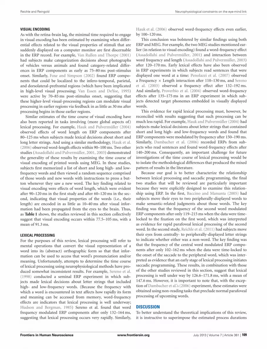
Reichle and Reingold Neurophysiological constraints on the eye-mind link
VISUAL ENCODINGAs with the retina-brain lag, the minimal time required to engagein visual encoding has been estimated by examining when differ-ential effects related to the visual properties of stimuli that aresuddenly displayed on a computer monitor are first discernablein the ERP record. For example, Van Rullen and Thorpe (2001)had subjects make categorization decisions about photographsof vehicles versus animals and found category-related differ-ences in ERP components as early as 75–80 ms post-stimulusonset. Similarly, Foxe and Simpson (2002) found ERP compo-nents that could be localized to the infero-temporal, parietal,and dorsolateral-prefrontal regions (which have been implicatedin high-level visual processing; Van Essen and DeYoe, 1995)were active by 70–85 ms post-stimulus onset, suggesting thatthese higher-level visual-processing regions can modulate visualprocessing in earlier regions via feedback in as little as 30 ms afterprocessing begins in those earlier regions.
Similar estimates of the time course of visual encoding havealso been reported in tasks involving (more global aspects of)lexical processing. For example, Hauk and Pulvermüller (2004)observed effects of word length on ERP components after80–125 ms when subjects made lexical decisions about short andlong letter strings. And using a similar methodology, Hauk et al.(2006) observed word-length effects within 90–100 ms. Two otherstudies (Assadollahi and Pulvermüller, 2001, 2003) demonstratedthe generality of these results by examining the time course ofvisual encoding of printed words using MEG. In these studies,subjects first memorized a list of short and long high- and low-frequency words and then viewed a random sequence comprisedof those words and new words with instructions to press a but-ton whenever they saw a new word. The key finding related tovisual encoding were effects of word length, which were evidentafter 90–120 ms in the first study and after 60–120 ms in the sec-ond, indicating that visual properties of the words (i.e., theirlength) are encoded in as little as 10–40 ms after visual infor-mation had been propagated from the eyes to the brain. Thus,as Table 1 shows, the studies reviewed in this section collectivelysuggest that visual encoding occurs within 77.5–105 ms, with amean of 91.3 ms.
LEXICAL PROCESSINGFor the purposes of this review, lexical processing will refer tomental operations that convert the visual representation of aword into its (abstract) orthographic form so that that infor-mation can be used to access that word’s pronunciation and/ormeaning. Unfortunately, attempts to determine the time courseof lexical processing using neurophysiological methods have pro-duced somewhat inconsistent results. For example, Sereno et al.(1998) conducted a seminal ERP experiment in which sub-jects made lexical decisions about letter strings that includedhigh- and low-frequency words. (Because the frequency withwhich a word is encountered in text affects how rapidly its formand meaning can be accessed from memory, word-frequencyeffects are indicators that lexical processing is well underway;Hudson and Bergman, 1985) Sereno et al. found that wordfrequency modulated ERP components after only 132–164 ms,suggesting that lexical processing occurs very rapidly. Similarly,
Hauk et al. (2006) observed word-frequency effects even earlier,by 100–120 ms.
This conclusion was bolstered by similar findings using bothERP and MEG. For example, the two MEG studies mentioned ear-lier (in relation to visual encoding) found a word-frequency effect(Assadollahi and Pulvermüller, 2001) and interaction betweenword frequency and length (Assadollahi and Pulvermüller, 2003)after 120–170 ms. Early lexical effects have also been observedin ERP experiments in which subjects read sentences that weredisplayed one word at a time: Penolazzi et al. (2007) observeda Frequency × Length interaction after 110–130 ms, and Serenoet al. (2003) observed a frequency effect after 132–192 ms.And similarly, Proverbio et al. (2004) observed word-frequencyeffects after 135–175 ms in an ERP experiment in which sub-jects detected target phonemes embedded in visually displayedwords.
This evidence for rapid lexical processing must, however, bereconciled with results suggesting that such processing can bemuch less rapid. For example, Hauk and Pulvermüller (2004) hadsubjects make lexical decisions about letter strings that containedshort and long high- and low-frequency words and found thatERP components were modulated by frequency after 150–190 ms.Similarly, Dambacher et al. (2006) recorded ERPs from sub-jects who read sentences and found word-frequency effects after140–200 ms. Consequently, an important challenge for futureinvestigations of the time course of lexical processing would beto isolate the methodological differences that produced the mixedpattern of results in the literature.
Because our goal is to better characterize the relationshipbetween lexical processing and saccadic programming, the finaltwo studies that will be reviewed are particularly importantbecause they were explicitly designed to examine this relation-ship using ERP. In the first, Baccino and Manunta (2005) hadsubjects move their eyes to two peripherally-displayed words tomake semantic-related judgments about those words. The keyfinding was that the frequency of the second word modulatedERP components after only 119–215 ms when the data were time-locked to the fixation on the first word, which was interpretedas evidence for rapid parafoveal lexical processing of the secondword. In the second study, Reichle et al. (2011) had subjects movetheir eyes from centrally- to peripherally-displayed letter stringsto indicate whether either was a non-word. The key finding wasthat the frequency of the central word modulated ERP compo-nents after only 102–162 ms when the data were time-locked tothe onset of the saccade to the peripheral word, which was inter-preted as evidence that an early stage of lexical processing initiatessaccadic programming. These results, in combination with thoseof the other studies reviewed in this section, suggest that lexicalprocessing is well under way by 126.6–171.8 ms, with a mean of147.6 ms. However, it is important to note that, with the excep-tion of Dambacher et al.’s (2006) experiment, these estimates wereobtained using non-reading tasks that preclude normal parafovealprocessing of upcoming words.
DISCUSSIONTo better understand the theoretical implications of this review,it is instructive to superimpose the estimated process durations
Frontiers in Human Neuroscience www.frontiersin.org July 2013 | Volume 7 | Article 361 | 109

Reichle and Reingold Neurophysiological constraints on the eye-mind link
(Table 1) on a time-line corresponding to the amount of timeavailable for cognitive processing during a single fixation of read-ing. Figure 1A thus shows the time course of the retina-brain lag,visual encoding, and lexical processing are aligned to the onsetof a single 240-ms fixation. (Although this particular duration isarbitrary and ignores the issue of variability, it corresponds to themean single-fixation duration observed on low-frequency wordsand is therefore a conservative estimate of the time available forlexical processing during most fixations; Reingold et al., 2012.)
As Figure 1A shows, the neurophysiological estimates suggestthat, on average, 148 ms is required to visually encode a printed
word and then complete some amount of lexical processing ofthat word. However, because the fixation is only 240 ms in dura-tion, there is seemingly little time to complete all of the operationsthat are necessary to move the eyes off of the word 92 ms later.These operations (at a minimum) include the transmission of asignal to the oculomotor system to start programming a saccade,the actual programming of that saccade, and whatever affer-ent delay occurs in the brainstem circuitry prior to moving theeyes. The conclusion that so little time is available to completethese operations is seemingly at odds with eye-movement exper-iments suggesting that saccades require 125–200 ms to program
FIGURE 1 | The time course of processing during a single, 240-msfixation on a word, including: (1) the propagation of information fromthe retina to brain (green); (2) visual encoding of the word features(blue); (3) lexical processing (purple); (4) saccadic programming (red);and (5) shifting attention from one word to the next (orange). (PanelA) Neurophysiological estimates for the times required for the retina-brainlag, visual encoding, and lexical processing are indicated by the coloredbars superimposed on the time line that is shown at the bottom of thepanel, with the three numbers above each colored bar indicating theestimated minimum, mean, and maximal times to complete eachrespective process (e.g., the estimated minimal time needed for theretina-brain lag is 43 ms). Based on these estimates, there should be littletime (92 ms) available for saccadic programming and whatevertransmission delays are necessary, e.g., to transmit a signal about thestate of lexical processing to the oculomotor system. (Panel B) If someamount of lexical processing of the fixated word is actually completedparafoveally, from the previously fixated word, then the amount of (foveal)
lexical processing of the word being fixated is reduced (e.g., to 25 ms) andcan thereby accommodate more realistic estimates of the time required toprogram saccades (e.g., approximately 124 ms). (Panel C) The time courseof processing if one assumes direct lexical control of saccadicprogramming and the strict serial allocation of attention (e.g., see Reichle,2011); as shown, the termination of whatever foveal lexical processing isnecessary to initiate saccadic programming causes attention to shift to thenext word, so that parafoveal lexical processing of that word can beginusing visual information acquired from the fixated word. (Note that(Panel C) is meant to be theoretically neutral with respect to specificserial-attention models of eye-movement control, and lexical processing isthus shown as a single stage rather than, e.g., being divided into the twostages posited by E-Z Reader. However, the depicted time course mapsonto the assumptions of E-Z Reader if: (a) the model’s first stage of lexicalprocessing corresponds to whatever lexical processing is completed priorto the initiation of a saccade, and (b) the model’s second stage of lexicalprocessing is subsumed in the time required to shift attention.)
Frontiers in Human Neuroscience www.frontiersin.org July 2013 | Volume 7 | Article 361 | 110

Reichle and Reingold Neurophysiological constraints on the eye-mind link
(Becker and Jürgens, 1979; Rayner et al., 1983; Reingold et al.,2012). It is also at odds with models of eye-movement control inreading, which posit that lexical processing is the “engine” thatcause the eyes to progress through the text (Reichle et al., 1998,2009; Engbert et al., 2002, 2005). Our analysis of the time courseof lexical processing thus poses a paradox if one is to maintain theposition that the completion of some amount of lexical processingis what determines when the eyes move during reading.
The solution to this paradox is that a significant portion of thelexical processing of a word that must be completed to “trigger”saccadic programming is actually completed from the preced-ing word, using visual information that was acquired from theparafovea. How this happens is illustrated in Figure 1B, whichis similar to Figure 1A except that lexical processing of the cur-rently fixated word begins from the previously fixated word,so that only 25 ms of lexical processing of the fixated word isactually completed from that word. (Again, this precise value isarbitrary, ignores variability, and is only meant to provide anexample.) Under this assumption, there is ample time (∼124 ms)for whatever neural transmission is required to signal the ocu-lomotor system to program and then initiate a saccade. Thishypothesis about the importance of parafoveal processing is con-sistent with survival analyses of fixation durations on high- andlow-frequency words with versus without parafoveal preview:Word-frequency effects were discernable more than 110 ms earlierwith than without preview (Reingold et al., 2012).
Finally, to make this hypothesis more concrete, Figure 1Cshows the typical sequence of events that are posited to occur byeye-movement models in which attention is allocated to supportthe lexical processing of exactly one word at any given time (e.g.,E-Z Reader or EMMA; see Reichle, 2011). As shown, the lexicalprocessing of any given word is completed from two locations—from the previously fixated word and from a fixation on the worditself. Then, upon completing whatever lexical processing is nec-essary to initiate saccadic programming, attention shifts to the
next word so that lexical processing of that word can begin usinginformation acquired from the current fixation location. (Notethat, according to the E-Z Reader model, the time required toshift attention also includes whatever additional time is neededto complete lexical access of the fixated word.)
Of course, the manner in which E-Z Reader instantiates eye-movement control places the most severe temporal constraintson lexical processing and its coordination with the oculomo-tor system because only one word is processed at a time. Theseconstraints are significantly relaxed to the extent that multi-ple words are processed in parallel, as posited by the SWIFTmodel (Engbert et al., 2002, 2005). According to this alternativetheoretical perspective, difficulty associated with processing thefixated word can inhibit the autonomous timer that otherwiseinitiates a saccadic program to move the eyes to a new view-ing location. Recent studies on saccadic inhibition (Reingold andStampe, 1999, 2000, 2002, 2003, 2004) and prior neurophysio-logical findings (Munoz et al., 1996) suggest that this type ofhypothesized mechanism might produce a very rapid inhibitoryeffect in as little as 20–30 ms. Consequently, there seems to beample time for an inhibitory mechanism to intervene in the deci-sions about when to move the eyes during reading (e.g., seeReingold et al., 2012 for a proposal of a hybrid eye-movementcontrol mechanism incorporating both facilitatory and inhibitorylexical influences).
This review thus indicates that lexical processing is sufficientlyrapid to permit direct control of the decisions about when tomove the eyes during reading, but that such control also requiresa substantial amount of lexical processing from the parafovea—perhaps more that has been acknowledged by reading researchers.This latter conclusion underscores the more basic claim abouteye-movement control during reading being a highly skilledactivity—one that requires a tremendous degree of coordinationbetween the systems that support attention, word identification,and the programming and execution of saccades.
REFERENCESAssadollahi, R., and Pulvermüller, F.
(2001). Neuromagnetic evidence forearly access to cognitive represen-tations. Neuroreport 12, 207–213.doi: 10.1097/00001756-200102120-00007
Assadollahi, R., and Pulvermüller, F.(2003). Early influences of wordlength and frequency: a groupstudy using MEG. Neuroreport 14,1183–1187. doi: 10.1097/00001756-200306110-00016
Baccino, T., and Manunta, Y. (2005).Eye-fixation-related potentials:insights into parafoveal processing.J. Psychophysiol. 19, 204–215. doi:10.1027/0269-8803.19.3.204
Becker, W., and Jürgens, R. (1979). Ananalysis of the saccadic system bymeans of double step stimuli. Vis.Res. 19, 967–983. doi: 10.1016/0042-6989(79)90222-0
Clark, V. P., Fan, S., and Hillyard, S.A. (1995). Identification of early
visual evoked potential generatorsby retinotopic and topographicanalyses. Hum. Brain Mapp.2, 170–187. doi: 10.1002/hbm.460020306
Courtney, S. M., and Ungerleider,L. G. (1997). What fMRIhas taught us about humanvision. Curr. Opin. Neurobiol. 7,554–561. doi: 10.1016/S0959-4388(97)80036-0
Dambacher, M., Kliegl, R., Hofmann,M., and Jacobs, A. M. (2006).Frequency and predictabilityeffects on event-related potentialsduring reading. Brain Res. 1084,89–103. doi: 10.1016/j.brainres.2006.02.010
Engbert, R., Longtin, A., and Kliegl,R. (2002). A dynamical modelof saccade generation in readingbased on spatially distributedlexical processing. Vis. Res. 42,621–636. doi: 10.1016/S0042-6989(01)00301-7
Engbert, R., Nuthmann, A., Richter,E., and Kliegl, R. (2005). SWIFT: adynamical model of saccade gener-ation during reading. Psychol. Rev.112, 777–813. doi: 10.1037/0033-295X.112.4.777
Foxe, J. J., and Simpson, G. V. (2002).Flow of activation from V1 tofrontal cortex in humans: a frame-work for defining “early” visualprocessing. Exp. Brain Res. 142,139–150. doi: 10.1007/s00221-001-0906-7
George, N., Jemel, B., Fiori, N.,and Renault, B. (1997). Faceand shape repetition effects inhumans: a spatio-temporal ERPstudy. Neuroreport 8, 1417–1423.doi: 10.1097/00001756-199704140-00019
Hauk, O., Davis, M. H., Ford, M.,Pulvermüller, F., and Marslen-Wilson, W. D. (2006). Thetime course of visual wordrecognition as revealed by
linear analysis of ERP data.Neuroimage 30, 1383–1400. doi:10.1016/j.neuroimage.2005.11.048
Hauk, O., and Pulvermüller, F.(2004). Effects of word lengthand frequency on the humanevent-related potential. Clin.Neurophysiol. 115, 1090–1103. doi:10.1016/j.clinph.2003.12.020
Hudson, P. T., and Bergman, M.W. (1985). Lexical knowledge inword recognition: word lengthand word frequency in nam-ing and lexical decision tasks.J. Mem. Lang. 24, 46–58. doi:10.1016/0749-596X(85)90015-4
Inhoff, A. W., and Rayner, K. (1986).Parafoveal word processing dur-ing eye fixations in reading:effects of word frequency. Percept.Psychophys. 40, 431–439. doi:10.3758/BF03208203
Just, M. A., and Carpenter, P. A.(1980). A theory of reading: omeye fixations to comprehension.
Frontiers in Human Neuroscience www.frontiersin.org July 2013 | Volume 7 | Article 361 | 111

Reichle and Reingold Neurophysiological constraints on the eye-mind link
Psychol. Rev. 87, 329–354. doi:10.1037/0033-295X.87.4.329
Kliegl, R., Nuthmann, A., and Engbert,R. (2006). Tracking the mind dur-ing reading: the influence of past,present, and future words on fix-ation durations. J. Exp. Psychol.Gen. 135, 12–35. doi: 10.1037/0096-3445.135.1.12
Laubrock, J., Kliegl, R., and Engbert,R. (2006). SWIFT explorationsof age differences in eye move-ments during reading. Neurosci.Behav. Rev. 30, 872–884. doi:10.1016/j.neubiorev.2006.06.013
McConkie, G. W., Kerr, P. W., Reddix,M. D., and Zola, D. (1988). Eyemovement control during reading:I. The location of initial fixations onwords. Vis. Res. 28, 1107–1118. doi:10.1016/0042-6989(88)90137-X
Mouchetant-Rostaing, Y., Giard,M.-H., Bentin, S., Aguera,P.-E., and Pernier, J. (2000).Neurophysiological correlates offace gender processing in humans.Eur. J. Neurosci. 12, 303–310. doi:10.1046/j.1460-9568.2000.00888.x
Munoz, D. P., Waitzman, D. M., andWurtz, R. H. (1996). Activity ofneurons in monkey superior col-liculus during interrupted saccades.J. Neurophysiol. 75, 2562–2580.
Penolazzi, B., Hauk, O., andPulvermüller, F. (2007). Earlysemantic context integrationand lexical access as revealed byevent-related brain potentials.Biol. Psychol. 74, 374–388. doi:10.1016/j.biopsycho.2006.09.008
Proverbio, A. M., Vecchi, L., andZani, A. (2004). From orthogra-phy to phonetics: ERP measuresof grapheme-to-phoneme con-version mechanisms in reading.J. Cogn. Neurosci. 16, 301–317. doi:10.1162/089892904322984580
Rayner, K. (1998). Eye movementsin reading and information pro-cessing: 20 years of research.Psychol. Bull. 124, 372–422. doi:10.1037/0033-2909.124.3.372
Rayner, K., Ashby, J., Pollatsek,A., and Reichle, E. D. (2004).The effects of frequency and
predictability on eye fixations inreading: implications for the E-ZReader model. J. Exp. Psychol. Hum.Percept. Perform. 30, 720–732. doi:10.1037/0096-1523.30.4.720
Rayner, K., and Duffy, S. A. (1986).Lexical complexity and fixationtimes in reading: effects of wordfrequency, verb complexity, andlexical ambiguity. Mem. Cogn. 14,191–201. doi: 10.3758/BF03197692
Rayner, K., Reichle, E. D., Stroud, M.J., Williams, C. C., and Pollatsek,A. (2006). The effects of wordfrequency, word predictability, andfont difficulty on the eye move-ments of young and older read-ers. Psychol. Aging 21, 448–465. doi:10.1037/0882-7974.21.3.448
Rayner, K., Slowiaczek, M. L.,Clifton, C., and Bertera, J. H.(1983). Latency of sequentialeye movements: implications forreading. J. Exp. Psychol. Hum.Percept. Perform. 9, 912–922. doi:10.1037/0096-1523.9.6.912
Reichle, E. D. (2011). “Serial atten-tion models of reading,” in OxfordHandbook on Eye Movements, eds S.P. Liversedge, I. D. Gilchrist, and S.Everling (Oxford: Oxford UniversityPress), 767–786.
Reichle, E. D., Pollatsek, A., Fisher, D.L., and Rayner, K. (1998). Towarda model of eye movement con-trol in reading. Psychol. Rev. 105,125–157. doi: 10.1037/0033-295X.105.1.125
Reichle, E. D., Rayner, K., and Pollatsek,A. (2003). The E-Z Reader modelof eye movement control in read-ing: comparisons to other models.Behav. Brain Sci. 26, 445–476. doi:10.1017/S0140525X03000104
Reichle, E. D., Tokowicz, N., Liu,Y., and Perfetti, C. A. (2011).Testing and assumption of the E-Z Reader model of eye-movementcontrol in reading: using event-related potentials to examine thefamiliarity check. Psychophysiology48, 993–1003. doi: 10.1111/j.1469-8986.2011.01169.x
Reichle, E. D., Warren, T., andMcConnell, K. (2009). Using
E-Z Reader to model the effects ofhigher-level language processingon eye movements during reading.Psychon. Bull. Rev. 16, 1–21. doi:10.3758/PBR.16.1.1
Reingold, E. M., Reichle, E. D., Glaholt,M. G., and Sheridan, H. (2012).Direct lexical control of eye move-ments in reading: evidence fromsurvival analysis of fixation dura-tions. Cogn. Psychology 65, 177–206.doi: 10.1016/j.cogpsych.2012.03.001
Reingold, E. M., and Stampe, D.M. (1999). “Saccadic inhibition incomplex visual tasks,” in CurrentOculomotor Research: Physiologicaland Psychological Aspects, eds W. B.Cer, H. Deubel, and T. Mergner(London: Plenum), 249–255.
Reingold, E. M., and Stampe, D. M.(2000). “Saccadic inhibition andgaze contingent research programs,”in Reading as a Perceptual Process,eds A. Kennedy, R. Radach, D.Heller, and J. Pynte (Amsterdam,Holland: Elsevier), 119–145.
Reingold, E. M., and Stampe, D.M. (2002). Saccadic inhibition involuntary and reflexive saccades.J. Cogn. Neurosci. 14, 371–388. doi:10.1162/089892902317361903
Reingold, E. M., and Stampe, D. M.(2003). “Using the saccadic inhi-bition paradigm to investigate sac-cadic control in reading,” in Themind’s Eye: Cognitive and AppliedAspects of Eye Movement Research,eds J. Hyönä, R. Radach, andH. Deubel (Amsterdam, Holland:Elsevier), 347–360.
Reingold, E. M., and Stampe, D.M. (2004). Saccadic inhibitionin reading. J. Exp. Psychol. Hum.Percept. Perform. 30, 194–211. doi:10.1037/0096-1523.30.1.194
Seeck, M., Michel, C. M., Mainwaring,N., Cosgrove, R., Blume, H.,Ives, J., et al. (1997). Evidencefor rapid face recognition fromhuman scalp and intracranial elec-trodes. Neuroreport 8, 2749–2754.doi: 10.1097/00001756-199708180-00021
Sereno, S. C., Brewer, C. C., andO’Donnell, P. J. (2003). Context
effects in word recognition: evi-dence for early interactive process-ing. Psychol. Sci. 14, 328–333.
Sereno, S. C., Rayner, K., and Posner,M. I. (1998). Establishing a time-line of word recognition: evidencefrom eye movements and event-related potentials. Neuroreport 9,2195–2200. doi: 10.1097/00001756-199807130-00009
Schilling, H. E. H., Rayner, K., andChumbley, J. I. (1998). Comparingnaming, lexical decision, and eyefixation times: word frequencyeffects and individual differences.Mem. Cogn. 26, 1270–1281. doi:10.3758/BF03201199
Van Essen, D. C., and DeYoe, E. A.(1995). “Concurrent processing inthe primate visual cortex,” in TheCognitive Neurosciences, ed M. S.Gazzaniga (Cambridge, MA: MITPress), 383–400.
Van Rullen, R., and Thorpe, S. J.(2001). The time course of visualprocessing: from early perception todecision-making. J. Cogn. Neurosci.13, 454–461. doi: 10.1162/08989290152001880
Conflict of Interest Statement: Theauthors declare that the researchwas conducted in the absence of anycommercial or financial relationshipsthat could be construed as a potentialconflict of interest.
Received: 01 May 2013; paper pend-ing published: 28 May 2013; accepted:23 June 2013; published online: 15 July2013.Citation: Reichle ED and Reingold EM(2013) Neurophysiological constraints onthe eye-mind link. Front. Hum. Neurosci.7:361. doi: 10.3389/fnhum.2013.00361Copyright © 2013 Reichle andReingold. This is an open-access articledistributed under the terms of theCreative Commons Attribution License,which permits use, distribution andreproduction in other forums, providedthe original authors and source arecredited and subject to any copyrightnotices concerning any third-partygraphics etc.
Frontiers in Human Neuroscience www.frontiersin.org July 2013 | Volume 7 | Article 361 | 112

ORIGINAL RESEARCH ARTICLEpublished: 14 May 2014
doi: 10.3389/fnhum.2014.00279
Assimilation of L2 vowels to L1 phonemes governs L2learning in adulthood: a behavioral and ERP studyMirko Grimaldi1*, Bianca Sisinni1, Barbara Gili Fivela1, Sara Invitto2, Donatella Resta1, Paavo Alku3
and Elvira Brattico4,5
1 Dipartimento di Studi Umanistici, Centro di Ricerca Interdisciplinare sul Linguaggio, Università del Salento, Lecce, Italy2 Laboratorio di Anatomia Umana e Neuroscience, Dipartimento di Scienze e Tecnologie Biologiche e Ambientali, Università del Salento, Lecce, Italy3 Department of Signal Processing and Acoustics, Aalto University, Espoo, Finland4 Brain & Mind Laboratory, Department of Biomedical Engineering and Computational Science, Aalto University, Espoo, Finland5 Cognitive Brain Research Unit, Institute of Behavioral Sciences, University of Helsinki, Helsinki, Finland
Edited by:Merim Bilalic, Alpen Adria UniversityKlagenfurt, Austria
Reviewed by:Antoine Tremblay, DalhousieUniversity, CanadaOlli Kalervo Aaltonen, University ofHelsinki, Finland
*Correspondence:Mirko Grimaldi, Dipartimento diStudi Umanistici, Centro di RicercaInterdisciplinare sul Linguaggio,Università del Salento, PiazzaAngelo Rizzo 1, Lecce 73100, Italye-mail: [email protected]
According to the Perceptual Assimilation Model (PAM), articulatory similarity/dissimilaritybetween sounds of the second language (L2) and the native language (L1) governsL2 learnability in adulthood and predicts L2 sound perception by naïve listeners. Weperformed behavioral and neurophysiological experiments on two groups of universitystudents at the first and fifth years of the English language curriculum and on a group ofnaïve listeners. Categorization and discrimination tests, as well as the mismatch negativity(MMN) brain response to L2 sound changes, showed that the discriminatory capabilitiesof the students did not significantly differ from those of the naïve subjects. In line withthe PAM model, we extend the findings of previous behavioral studies showing that, atthe neural level, classroom instruction in adulthood relies on assimilation of L2 vowels toL1 phoneme categories and does not trigger improvement in L2 phonetic discrimination.Implications for L2 classroom teaching practices are discussed.
Keywords: adult phoneme perception, mismatch negativity (MMN), foreign language acquisition, L2 classroomlearning, event-related potentials, vowel perception
INTRODUCTIONLearning a second language (L2) in adulthood challenges ourbrains. As mother tongue phoneme representations are formed inthe brains of 6–12 months old children (Werker and Tees, 1983;Kuhl et al., 1992; Cheour et al., 1998; Kuhl, 2008) non-nativespeech sounds become increasingly difficult to discriminate andL2 perception generally turns into a demanding task for learn-ers (Iverson et al., 2003). This loss of sensitivity does not preventL2 learning in adulthood (Flege, 1995). The extent of successmay depend nonetheless on numerous variables: i.e., age of L2learning, length of residence in an L2-speaking country, gender,formal instruction, motivation, language learning aptitude andamount of native language (L1) use (see Piske et al., 2001 for anoverview). When L2 learners are immersed in an L2 environment,the contribution of age toward learning to perceive and produceL2 sounds occurs primarily through interactions with the amountof L1 use and the amount of L2 native speaker input received(Flege et al., 1995, 1997, 1999; Flege and Liu, 2001; Flege andMacKay, 2004; Tsukada et al., 2005; see Piske, 2007 for a criticalreview). However, when learners are immersed in an L1 environ-ment and have a reduced L2 exposure, primarily in a restrictedsetting (namely, with little or unsystematic conversational experi-ence with native speakers) learning of L2 phonemes at the nativespeaker level becomes very difficult if not impossible. Accordingto Best and Tyler (2007: 16), the perception of L2 in these individ-uals receiving only formal instruction in adulthood may resemblethat of L2 naïve listeners. In other words, they are functionalmonolinguals, not actively learning or using L2 when compared
with L2-learning listeners, i.e., learners who are in the processof actively learning an L2 to achieve functional, communicativegoals within natural L2 context.
Cross-linguistic and L2 speech perception studies have shownthat adult learners of L2 have difficulty with both the perceptionand production of non-native phonological segments, i.e., con-sonants and vowels that either do not occur or are phoneticallydifferent in their L1 (see Flege, 2003 for a discussion). Indeed,it is commonly thought that a major determinant of L2 foreignaccent is the underlying problem associated with the perceptionof L2 phonological structures. In turn, acquisition of phoneticcontrasts involves not only the detection of differences in theacoustic signal but also the accessing of internalized categories,which in the brain are most likely associated with definite neu-ral representations. Within the behavioral literature, there are twomajor theoretical frameworks on L2 speech learning in adult-hood, the Speech Learning Model (SLM, Flege, 1995) and thePerceptual Assimilation Model (PAM, Best, 1995). The SLM hasbeen primarily concerned with the ultimate attainment of L2 pro-duction and perception and mainly deals with highly experiencedL2 learners immersed in an L2 environment, whereas the PAMis mainly interested in explaining the initial L2 perception of L2learners through the non-native perception of naïve listeners, whoare in fact functional monolinguals (but see Best and Tyler, 2007,for an extension to L2 learning). Both SLM and PAM posit thatthe degree of success listeners will have in perceiving non-nativeL2 sounds depends on the perceived relationship between pho-netic elements found in the L1 and the L2 systems. These models
Frontiers in Human Neuroscience www.frontiersin.org May 2014 | Volume 8 | Article 279 |
HUMAN NEUROSCIENCE
113

Grimaldi et al. Assimilation of L2 vowels to L1 phonemes
make predictions about performance in non-native segmentalperception based on the perceived distance between L1 and L2sounds (Guion et al., 2000).
This study investigated the thus far little studied L2 percep-tion in functional monolinguals, by behaviorally and neurallytesting the predictions posed by the PAM framework. The PAMpredicts that if two non-native sounds are perceived as accept-able exemplars of two distinct native phonemes (Two-Categoryassimilation), their discrimination will be easy, while if both non-native sounds are perceived to be equally poor/good exemplarsof the same native phoneme (Single-Category assimilation), theirdiscrimination will be difficult. An intermediate discrimination ispredicted when the two non-native sounds are both perceived asthe same native sound but differ in goodness rating (Category-Goodness assimilation). Finally, when an L2 category is perceivedas more than one L1 phoneme and the other L2 category isperceived as a single native phoneme, a good discriminationis predicted (Uncategorized-Categorized assimilation). For pre-dictions to be generated by PAM (or the SLM), cross-languagephonetic distance data need to be obtained by means of behav-ioral experiments. The degree of perceptual distance betweenphonemes is usually examined using an identification and rat-ing methodology. The foreign (or L2) sounds are first classified asinstances of a phonetic category(s) in the listener’s L1, then ratedfor goodness-of-fit to the L1 category.
Whereas the studies on L2 and non-native phoneme percep-tion discussed above have used only behavioral techniques toaddress this question, we chose to adopt both behavioral (cat-egorization and discrimination tests) and electrophysiological(event-related potential, ERP) techniques to examine the L2 per-ceptual abilities of our subjects. The ERP technique provides notonly a millisecond precise measurement of information process-ing in the brain but also, depending upon the task, can allow oneto disentangle automatic detection from attentional processes.ERP studies on L2 phoneme processing have used the odd-ball paradigm, alternating repetitive (standard) and infrequent(deviant) sounds (80–20% of occurrence respectively) while sub-jects are distracted from listening by a primary task (e.g., watchinga silent movie), to measure the so-called mismatch negativity(MMN) response to L2 contrasts. The MMN is an ERP com-ponent, elicited by stimulus change at ≈100–250 ms, mainlygenerated in the auditory cortex and with additional generatorsin the inferior frontal cortex, reflecting the neural detection ofa change in a constant property of the auditory environment(Picton et al., 2000; Näätänen et al., 2007). A large body of evi-dence supports the notion that the discriminative MMN processrelies both on auditory sensory and categorical phonetic represen-tations of speech stimuli and that these two codes are utilized inparallel by the pre-attentive change detection process reflected inthe MMN component (Näätänen et al., 2001, 2011; Pulvermüllerand Shtyrov, 2006). The MMN results from prediction violationson the basis of the repetitive standard presentation (Winkler andCzigler, 2012). It has been proposed that the standard presen-tation resembles perceptual learning during which hierarchicalsensory levels of processing receive bottom-up sensory input fromlower levels and receive top-down predictions from higher lev-els (Garrido et al., 2009). As a result of the repetition of the
standard presentation, prediction errors are reduced by repetitivesuppression or adaptation (Friston, 2005). A deviant presentationthen leads to a violation of bottom-up prediction that is reflectedin MMN generation (see also the discussion in Scharinger et al.,2012). Furthermore, the amplitude and peak latency of the MMNis directly correlated with the magnitude of the perceived changeand, hence, it is considered a measure of individual discrimina-tion accuracy (see Amenedo and Escera, 2000; Näätänen, 2001;Sussman et al., 2013 for a critical discussion).
The results of MMN studies, mainly focused on L2-learninglisteners, are mixed. For instance, Winkler et al. (1999a) foundthat Hungarian adult late L2 learners who had been immersedfor several years in the L2 context perceived non-native contrasts(in Finnish) as well as native speakers, as evidenced by compa-rable MMN amplitudes elicited by both native Finns and fluentHungarians in response to a Finnish across category-boundaryvowel contrast, when opposed to naïve Hungarians. The resultsby Winkler et al. (1999a) were not replicated in a population ofadvanced adult L2 learners (of English) who were not immersed,since advanced Finnish students of English did not show MMN toEnglish phonemes that would be comparable to the one elicitedby native Finnish phonemes, hence suggesting that learning inthe classroom environment may not lead to the formation of newlong-term native-like memory traces (Peltola et al., 2003). Thesebrain responses to new phonemes probably develop in childrenat a very fast pace: i.e., within three months of intensive expo-sure, as evidenced by MMN to L2 phoneme contrasts in Finnishchildren participating in French language immersion education(Cheour et al., 2002; Shestakova et al., 2003; Peltola et al., 2005).Again, however, subsequent works did not confirm these findingswhen the L2 was English both for Finnish listeners (Peltola et al.,2007) and Japanese listeners (Bomba et al., 2011). Finally, Rinkeret al. (2010) for bilingual Turkish–German kindergarten childrengrowing up in Germany have shown that the MMN response isless robust in Turkish–German children to the German vowel,when compared to a German control group. Thus, immersioneducation and natural acquisition contexts did not guaranteenative-like L2 vowel discrimination. Also, native-like L2 voweldiscrimination is not guaranteed after a short training (50 min on5 consecutive days) via associative/statistical learning: as showedby Dobel et al. (2009), who neurally investigates the perceptualacquisition of an L2 consonant (/φ/) in a group of adult Germanspeakers using the MEG methodology. Instead of establishing anovel category the subjects integrated /φ/ into the native category/f/, demonstrating that native categories are powerful attractorshampering the mastery of non-native contrasts. None of thesestudies, though, have tried to explain the L2 perceptual processesaccording to any of the well-established models for L2 learning.Hence they left open the question of which mechanisms governthe acquisition of L2 phonemes in adult learners from formalinstruction and with restricted L2 exposure.
The present study aims at studying the behavioral and neu-ral (MMN) correlates of L2 learning in adulthood while directlytesting the hypotheses that these correlates would index the per-ceptual mechanisms posed by the PAM model. Specifically, ourstudy addressed two questions: (i) Do the predictions generatedby the PAM through behavioral methods hold when they are
Frontiers in Human Neuroscience www.frontiersin.org May 2014 | Volume 8 | Article 279 | 114

Grimaldi et al. Assimilation of L2 vowels to L1 phonemes
neurophysiologically investigated, namely can the discriminationpatterns predicted by the PAM for L2 naïve listeners be also mir-rored in MMN amplitudes or latencies? (ii) Is L2 classroom learn-ing associated with the typology of L2 naïve listeners, as recentlysuggested by Best and Tyler (2007)? To answer these questions,we measured the behavioral and electrophysiological data of twogroups of Salento Italian (SI) undergraduate students of BritishEnglish (BE) attending the first and the fifth year of the ForeignLanguages and Literatures Faculty. Crucially, SI, the Italian varietyspoken in Southern Apulia, presents a five stressed vowel sys-tem (i.e., /i, ε, a, c, u/; Grimaldi, 2009; Grimaldi et al., 2010)contrary to the richer vowel system of BE that shows, excludingdiphthongs, eleven stressed vowels (see Stimuli). Therefore, forSI speakers, it could be relatively difficult to learn a complex L2vowel system, supporting the idea that the L1 plays an importantrole and enables one to predict the relative difficulty of acquisitionof a given L2 contrast (Iverson and Evans, 2007). Firstly, we behav-iorally tested the two groups of students by means of an identi-fication test. On the basis of the results of this test, the contrasts/i:/-/u:/ and /æ/-/ v/ (for which the PAM’s framework predicted anexcellent and a good discrimination, respectively) were selectedfor a behavioral discrimination test. In the ERPs experiment, thegroups of students were compared with a control group of listen-ers who were much more linguistically inexperienced of the L2, astheir knowledge of English derived only from compulsory schoolstudies. Moreover, as a control condition we introduced the L1within-category contrast /ε/-[e], for which poor discriminationis predicted (cf. Phillips et al., 1995; Dehaene-Lambertz, 1997;Winkler et al., 1999b; see also Miglietta et al., 2013). These twovowels are phonologically contrastive in standard Italian and theyare used to create lexical contrast (i.e., /"pεska/ “peach” vs. /"peska/“fishing”) whereas SI has the phoneme /ε/ only. Consequently, forSI speakers these stimuli belong to the same category, as /ε/ is theunderlying phoneme and [e] represents an allophone (generallytranscribed between brackets), namely a within-category variantof the same phoneme.
METHODSBEHAVIORAL EXPERIMENTSSubjectsTwo groups of 10 normal-hearing (tested prior to the experi-ment), right-handed, undergraduate male students of the ForeignLanguages and Literatures Faculty voluntarily participated in theexperiments. One group was enrolled in its first year (age 21.4 ±1.71; 9.4 ± 1.34 years of English studies in formal context),whereas the other was in its fifth year (age 25.6 ± 1.98; 14.3 ±2.11 years of English studies in formal context). As assessed bya questionnaire of language use, all the subjects neither partici-pated in Erasmus programs in England nor have had L2 nativeteachers prior to attending university. English instruction uni-versity classes are taught by Italian native-speakers prevalently,although for at least 6 months per year (3–5 h per week) thesestudents had been attending lessons also with native English lec-turers. However, in the last case, language classes are only a fewhours per week and are just based on lexical and morphosyn-tactic formal instructions; no systematic and explicit phoneticinstruction or training is administered.
StimuliThe stimuli consisted of the 11 BE monophthong vowels, i.e., /i:/,/I/, /ε/, /æ/, / v/, /A:/, / A/, / ε:/, / c:/, /U/ and /u:/ (Ladefoged, 2001).These sounds were produced by three male native BE speakers(age 47.3 ± 4.9; years in Italy: 22.3 ± 5.13), two of them comingfrom London, one coming from Birmingham. The speakers reada list of monosyllabic words with the phonemes /i:/, /I/, /ε/, /æ/,/ v/, /A:/, / A/ and / ε:/ placed in a /p_t/ context and the phonemes/i:/, / c:/, /U/ and /u:/ in an /s_t/ context, for a total of 36 stim-uli (3 speakers × 12 phonemes). Given that /i:/-/u:/ and /u:/-/U/were part of the discrimination task as control and target con-trasts, respectively, /i:, U and u:/ needed to be recorded in thesame consonant context. Thus, the extra context /s_t/ was usedfor these three vowels because there is no English word with /u:/in the /p_t/ context. These stimuli were recorded in the CRILsoundproof room by a CSL 4500 at a sampling rate of 22.05 kHzand were segmented and normalized in peak amplitude using thesoftware Praat 4.2. Each of the student groups performed twoperceptual tests: the identification and the oddity discriminationtest. All subjects were individually tested in the CRIL soundproofroom using a computer and with sounds (set at a comfortablesound level) delivered via headphones, for a total duration ofapproximately 40 min.
Identification testThe aim of the identification test was to examine the perceivedphonetic distance between the L1 and L2 sounds: i.e., to detectwhich L2 sounds are more similar/dissimilar to the L1 soundsand, consequently, are more difficult/easy to discriminate by per-ception (Flege and MacKay, 2004). The 36 stimuli were randomlypresented 3 times, and subjects identified each of them in terms ofone of the 5 SI vowels /i, ε, a, c/ or /u/ by clicking on the computerscreen. Students could not rehear a stimulus, but they were toldto guess if they were unsure. Before performing the test, studentsreceived instructions orally and a training test of 10 stimuli wasadministered in the presence of the experimenter to ensure thatthe students understood the task. No subject was rejected on thebasis of the training test because they all found the task easy toperform.
Oddity discrimination testThe purpose of the oddity discrimination test was to measure theability of listeners to discriminate L2 sounds. For each of the twocontrasts, 8 change trials and 8 catch trials (32 total trials per stu-dent) were executed. The change trials were made up of 3 items,each one produced by one of the three BE speakers, with an odditem belonging to a different phonological category that subjectshad to detect. The odd item was alternatively placed in the first,second or third position in a nearly balanced way (Tsukada et al.,2005) to avoid response bias (Bion et al., 2006). Additionally, thethree native English speakers produced the catch trials, where allof the items contained the same phonological category. Thesekinds of trials test subjects’ ability to ignore the acoustical dif-ferences among the stimuli belonging to the same phonologicalcategory. For instance, to test the contrast /i:/-/u:/ the changetrials were /i:/-/i:/-/u:/ − /i:/-/u:/-/i:/ − /u:/-/i:/-/i:/ − /u:/-/u:/-/i:/ − /u:/-/i:/-/u:/ − /i:/-/u:/-/u:/, and the catch trials were
Frontiers in Human Neuroscience www.frontiersin.org May 2014 | Volume 8 | Article 279 | 115

Grimaldi et al. Assimilation of L2 vowels to L1 phonemes
/i:/-/i:/-/i:/ − /u:/-/u:/-/u:/. Subjects clicked the computer screenon “1,” “2,” “3,” corresponding to the position of the item theyperceived as different or to “none” if they perceived all items asequal. The results of this test, i.e., A′ scores, were calculated foreach contrast by applying the formula of Snodgrass et al. (1985).These scores reduce the effects of response bias by calculating theproportion of hits (i.e., the number of correct selections of theodd item in the change trials) and the proportion of false alarms(i.e., the number of incorrect selections of an odd item in thecatch trials). An A′ score of 1.0 indicates perfect discriminationand an A′ score of 0.5 indicates a null discrimination. Subjectswere first given the instructions and then administered a train-ing test in the presence of the experimenter to verify that theyhad understood the task. No subject was rejected on the basis ofthe training test because they all found the task easy to perform.This test was also executed by a control group of 10 male BE lis-teners (mean age: 20.5 ± 1.95), native speakers of the Londonvariety.
Statistical analysis of oddity discrimination test results.Discrimination accuracy (A′ score) was analyzed in repeated-
measures ANOVA with “contrast” (/æ/-/ v/ and /i:/-/u:/) as thewithin-subject factor and “group” (first and fifth year) as thebetween-subject factor. In all of the statistical analyses, the alphalevel was set to p < 0.05, and type I errors were controlled for bydecreasing the degrees of freedom with the Greenhouse–Geisserepsilon. Post-hoc tests were conducted by Fisher’s least-significantdifference (LSD) comparisons.
ERP EXPERIMENTSubjectsThe two groups of students involved in the behavioral experi-ments participated in the ERP sessions. Additionally, a third con-trol group of normally hearing (tested prior to the experiment),right-handed subjects with only compulsory school education(10 subjects; age 25 ± 4.26; years of English studies in formalcontext 5 ± 2.9) performed the electrophysiological test. Thecontrol group was primarily composed of carpenters, plasterers,or unemployed, and each participant received a small monetarycompensation for participating in the experiment. If one con-siders that in Italy a foreign language is usually taught startingfrom the last two years of primary school (when children arenormally 8 years old), we can suppose that the student groupsand the control group have a similar starting age of L2 expo-sure. However, the student groups have more formal exposureto the L2, particularly the fifth year group. In contrast, the con-trol group’s L2 exposure was limited to compulsory school, wherethey passively received impoverished lexical or morphosyntacticinputs by non-native L2 teachers for approximately 3 h per week.Additionally, in Italy foreign programs are dubbed, so that theexposure to foreign languages in informal contexts is very low. Wealso excluded that the ordinary listening of English music couldrepresent an involuntary L2 training, as the acquisition of L2 inadulthood presupposes a strong motivation and a continuous useof L2 in different conversational contexts (cf. Gardner, 1991). Allof the subjects signed the informed consent form. The local EthicsCommittee approved the experimental procedure.
Stimuli and procedureWe used the same contrast pairs as in the oddity discriminationtest but the stimuli consisted of synthetic vowels whose durationwas 350 ms (edited with Praat 4.2). Thus the contrasts tested were/i/-/u/ and /æ/-/ v/. A third contrast was added as control, i.e.,/ε/-[e] where the former is a mid-opened vowel and the latter amid-closed one. This is a within-category contrast for SI speakersand poor discrimination is predicted. In Table 1, we provide theacoustic characteristics of stimuli. First formant frequency (F1)and second formant frequency (F2) are given in Hz.
To avoid confounding the effects of acoustic variations innatural utterances with the ERP responses, the stimuli for theERP experiment were created using the Semisynthetic SpeechGeneration method (SSG, Alku et al., 1999), which mathemat-ically models the functioning of the human voice productionmechanism. To obtain raw material for the SSG synthesis forthe ERP experiment, short words produced by a native male BEspeaker (44 years old coming from London) and by a native malespeaker of Standard Italian (45 years old, coming from Florence)were recorded in a soundproof room using a Sennheiser MKH20 P48 high-frequency condenser, omnidirectional microphone,and a response frequency of 20–20,000 Hz, and further processedwith a sampling frequency of 22050 Hz and a resolution of 16 bits.Signal sections corresponding to the desired vowels to be syn-thesized were cut from the recorded words. From these selectedsections, the corresponding vocal tract filters were computedwith SSG using digital all-pole filtering (Oppenheim and Schafer,1989) of 22.
The three contrasts /æ/-/ v/, /i/-/u/ and /ε/-[e] were presentedin separate blocks lasting 15 min each, and each with 86% fre-quency of occurrence (582 trials) for the standard stimulus (thefirst vowel of each above listed pair) and 14% frequency (114 tri-als) for the deviant stimulus (the second vowel of each pair). Theorder of presentation was pseudo-randomized, since a deviantstimulus was never presented before three standards. The inter-stimulus interval was 750 ms. During the EEG recording, partici-pants sat in a comfortable armchair and were instructed to watcha silent movie while paying no attention to the stimuli, which werebinaurally presented in a soundproof room through loudspeakersat 65/70 dB.
Electrophysiological recordingsThe EEG was recorded from the scalp using a 64 Ag/AgCl elec-trode cap (BrainCap, Brain Products) with a sampling frequencyof 500 Hz. Eye movements were monitored with electrodesattached at the top and the bottom of the left eye and at the top
Table 1 | Values of the first formant (F1) and the second formant (F2)
given in Hz and Euclidean distances of the stimulus contrasts utilized
in the ERP experiment.
Formants /i/ /u/ /æ/ / v/ /ε/ [e] Contrast Euclidean
distance
F1 Hz 322 347 823 678 563 435 /i/-/u/ 1209 Mel
F2 Hz 2363 1015 1535 1090 1712 1986 /æ/-/ v/ 581 Mel
/ε/-[e] 404 Mel
Frontiers in Human Neuroscience www.frontiersin.org May 2014 | Volume 8 | Article 279 | 116

Grimaldi et al. Assimilation of L2 vowels to L1 phonemes
of the right eye. The reference electrodes were attached on the earlobes. Impedance was kept under 15 k�. The signal was off-linefiltered (0.5–50 Hz, 24 dB), and the threshold for artifact rejec-tion was set at > ±125 μV. The numbers of trials accepted afterartifact rejection are reported in Table 2. Each standard follow-ing a deviant was removed from the averaging. The ERP epochsincluded a pre-stimulus interval of 100 ms, used for baselinecorrection, and lasted until 450 ms.
Statistical analysis of ERP dataTo quantify the MMN, we first identified the most negativepeaks at Fz around the time interval 120–300 ms for each con-trast and group from the grand-average difference waveforms.Subsequently, the individual MMN amplitudes were calculated bytaking the mean values from the same 40-ms interval around thegrand-average MMN peaks for each contrast and group obtainedas described above. The significance of the individual MMNamplitudes at Fz was verified by paired t-tests against the zerobaseline. To test our hypotheses on the effects of contrast typesand language exposure on the MMN amplitudes measured at F3,F4, C3, C4, P3, and P4, we used repeated-measures ANOVAsand linear mixed-effect models with the between-subject fac-tor Group (first year, fifth year students and control group)and the within-factors Language (the within-category contrast/ε/-[e] and the English pairs /i/-/u/ and /æ/-/ v/), Contrast (/i/-/u/, /æ/-/ v/, and /ε/-[e]), Frontality (frontal, central, and parietalelectrodes) and Laterality (right or left hemisphere). We alsoextracted the individual peak latencies of the MMN responserecorded at Fz by searching for the most negative peak withinthe time interval 120–300 ms per each subject and each condi-tion. For testing the hypotheses on the MMN peak latencies, asimilar ANOVA as above (with Group, Language and Contrastas factors) was conducted but without the two electrode factors.For all statistical tests, the alpha level was chosen to correspondto p < 0.05. Type I errors were controlled for by decreasing thedegrees of freedom with the Greenhouse–Geisser epsilon (orig-inal degrees of freedom are reported) or by adding subjects asrandom effect including it as intercept or random slopes, whenappropriate as assessed by the Bayesian information criteria in alinear mixed-effect model. The difference threshold for accept-ing or rejecting a more complex model was set to 4. Post-hoctests were conducted by Fisher’s least-significant difference (LSD)comparisons.
Table 2 | The average number of accepted standard (stand) and
deviant (dev) trials for each contrast and each group (control group,
first year students, fifth year students).
Contrasts Control First year Fifth year
stand dev stand dev stand dev
/æ/-/
v
/ 496 (85%) 97 (85%) 510 (88%) 99 (87%) 491 (85%) 98 (86%)
/i/-/u/ 472 (86%) 93 (86%) 500 (86%) 98 (86%) 512 (88%) 101 (89%)
/ε/-[e] 501 (86%) 98 (86%) 495 (85%) 99 (86%) 491 (84%) 98 (86%)
The percentages with respect to the total number of trials are also given in
parentheses.
RESULTSIDENTIFICATION TESTThe identification test results were considered in terms of the per-centage of identification of BE phonemes with respect to the SIones. The percentages indicate the frequency with which L1 SIvowels were used to classify the L2 BE vowels. The percentages ofidentification obtained by first (I) and fifth (V) year students aresummarized in Table 3.
The percentages of identification of the L2 phonemes to theL1 phonemes are very useful for understanding how the formerare perceived and categorized with respect to the latter. The L2phonemes associated with an L1 phoneme with an identifica-tion percentage ≥ 80% were considered consistently identifiedto the L1 and only that identification was taken into account.Conversely, those L2 phonemes associated with two or more L1phonemes (identification percentage < 80%) were considered asnot consistently assimilated, and the first two identifications weretaken into account.
The data summarized in Table 3 show that both the firstand the fifth year students adopted the same assimilation strate-gies, albeit with slightly different percentages. According to theidentification consistency threshold identified above, the resultsdepict the following scenario: /æ/ was consistently assimilatedwith the native phoneme /a/; / v/ was identified to /a/ or /o/, so wasnot assimilated to either of these two native phonemes. Finally,/i:/ and /u:/ were each consistently identified with the nativephonemes, /i/ and /u/, respectively. In fact, BE /i:/ and /u:/ (seeTable 1) share some formant features with SI /i/ (F1 326, F2 2244)and /u/ (F1 368, F2 867) (Grimaldi, 2009) and consequently areperceived by SI listeners as their native counterpart.
According to the PAM typologies of assimilation, the vow-els /æ/, / v/, /i:/ and /u:/, can be grouped into two contrasts ofL2 vowels (see Table 3): (i) the contrast /æ/-/ v/ falls into theUncategorized-Categorized assimilation, for which good discrim-ination is predicted, as the non-native vowel /æ/ is consistentlyassimilated to a native phoneme (/a/), whereas the other vowel/ v/ is not categorized with any native phoneme; (ii) the contrast/i:/-/u:/ falls into the two-category assimilation, for which excel-lent discrimination is predicted, as they have been consistently
Table 3 | Mean percentage of identification of L2 vs. L1 vowels by first
(I) and fifth (V) year students.
L2L1 /i/ /ε/ /a/ /
c
/ /u/
I (%) V (%) I (%) V (%) I (%) V (%) I (%) V (%) I (%) V (%)
/i:/ 99 100
/I/ 84 83 15 17
/ε/ 99 100
/æ/ 19 19 81 81
/ v/ 1 67 79 33 20
/A:/ 73 91 26 9
/ A/ 2 13 98 87
/ c
:/ 6 94 97 3
/ ε
:/ 33 52 23 18 32 27 11 3
/u:/ 2 98 100
/U/ 3 53 36 47 61
Frontiers in Human Neuroscience www.frontiersin.org May 2014 | Volume 8 | Article 279 | 117

Grimaldi et al. Assimilation of L2 vowels to L1 phonemes
identified with two different native phonemes: i.e., /i/ and /u/.The discrimination ability by the two groups of students for thesecontrasts was further tested with the oddity discrimination test.
ODDITY DISCRIMINATION TESTThe repeated-measures ANOVA on A′ scores (Table 4 andFigure 1) did not yield differences between the two groups,[F(1, 18) = 0.40, p > 0.05, η2
p = 0.02] but it yielded a significant
effect for the contrasts [F(1, 18) = 18.24, p = 0.000, η2p = 0.50].
The post-hoc analysis revealed that the contrast /i:/-/u:/ was dis-criminated with a higher A′ with regard to the contrast /æ/-/ v/.The interaction Group × Contrast was not significant [F(1, 18) =0.26, p > 0.05, η2
p = 0.01].
ERPsFigures 2–4 show the grand-average difference waveforms for allgroups and for each stimulus contrast (see also Figure S1 in theSupplementary Material). The mean MMN amplitudes and peaklatencies are displayed in Table 5 and Figure 5.
For all conditions and for all groups, we obtained a sig-nificant MMN response. In the ANOVA, the MMN amplitudewas slightly significantly modulated by Contrast [F(2, 52) = 3.02,p = 0.05, η2
p = 0.10; this result corresponded to an only marginalsignificance in the linear mixed-effects model with by-subjectsrandom intercepts where by-stimulus random intercepts andby-subject random slopes for Contrast were tested for inclu-sion: F(2, 54) = 2.9, p = 0.07]. The post-hoc tests showed thatthere was a significant difference between the L2 /æ/-/ v/ and thewithin-category contrast /ε/-[e] (p < 0.05) and a tendency towarda significant difference between /i/-/u/ and the within-category
Table 4 | The A′ scores obtained by the first year group (I) and the
fifth year group (V).
Contrasts I year group V year group
/æ/-/ v/ 0.69 (0.23) 0.67 (0.27)
/i:/-/u:/ 0.95 (0.04) 0.87 (0.15)
Standard deviations are in parentheses.
FIGURE 1 | The A′ score obtained by the first year group (dotted bar)and the fifth year group (striped bar).
contrast /ε/-[e] (p = 0.06). Namely, the within-category contrast/ε/-[e] had the lowest amplitude, while the L2 contrasts /i/-/u/ and/æ/-/ v/ showed similar amplitudes. The MMN amplitude was alsomodulated by Frontality [F(2, 52) = 112.16, p < 0.001, η2
p = 0.81;also replicated in the linear mixed-effects model: F(2, 400) = 2.4,p < 0.0001] and the post-hoc showed that the amplitudes werehighest in the frontal area, then in the central and finally in theparietal area. Additionally, we found a modulation of the frontalMMN amplitudes by group expertise with the significant interac-tion Group × Frontality [F(4, 52) = 4.56, p < 0.001, η2
p = 0.26;confirmed also in the linear mixed-effects model: F(4, 400) = 10.7,p < 0.001]. This interaction derived from the larger MMN ampli-tudes at frontal electrodes to any stimulus found in the controlstudents as compared with the fifth year students (p = 0.06).
Moreover, the significant interaction Contrast × Frontality[F(4, 104) = 3.38, p < 0.05, η2
p = 0.15; this result was replicatedin the linear mixed-effects model: F(4, 400) = 4, p = 0.004] con-firmed that in the frontal area the within-category contrast /ε/-[e]had lower amplitudes than /i/-/u/ and /æ/-/ v/ (/i/-/u/ vs. /ε/-[e]: p < 0.05; /æ/-/ v/ vs. /ε/-[e]: p = 0.01; /i/-/u/ vs. /æ/-/ v/: p >
0.05). The typical fronto-central MMN scalp distribution was alsoconfirmed by the significant interaction Frontality × Laterality[F(2, 52) = 4.48, p = 0.01, η2
p = 0.14; this result was not repli-cated though in the linear mixed-effect model: F(2, 400) = 1.6,p = 0.2] and the post-hoc showed that this pattern was present inboth the right and left hemispheres. The amplitude of the MMNpresented a difference in the frontal area only, where it was largerover the right than the left hemisphere (cf. Table 6 for the repeatedmeasures ANOVA results).
The MMN peak latency differed according to the vowel con-trasts, as testified by the significant main effect of Contrast[F(2, 52) = 10.35, p < 0.001, η2
p = 0.28] (cf. Table 7 for all statis-tical results).
This effect obtained with a general linear model with fixedeffects was confirmed also in a linear mixed-effects model ofMMN peak latency as a function of Contrast with by-subjectsrandom intercepts where by-stimulus random intercepts forContrast were tested for inclusion (by-subject random slopeswere not included instead, since they did not improve themodel fit according to the Bayesian information criteria). Alsoin this more generalizable mixed-effects model the main effectof Contrast reached significance [F(2, 52) = 11.2, p < 0.001]. Inpost-hoc tests, the contrasts /i/-/u/ evoked a faster MMN than thecontrast /æ/-/ v/ (p = 0.01) and the within-category contrast /ε/-[e] (p = 0.000), and in turn the contrast /æ/-/ v/ evoked a fasterMMN than the contrast /ε/-[e] (p < 0.05).
DISCUSSIONThis study tested whether the L2 discrimination patterns pre-dicted by the PAM for L2 contrasts are mirrored in the MMNamplitudes and peak latencies to the same contrasts. The behav-ioral findings suggest that the first and the fifth year studentsdid not differ in their discrimination processes, notwithstand-ing the different classroom and educational backgrounds. Inparticular, these two groups of subjects exhibited excellent dis-crimination of /i:/-/u:/ (belonging to Two-Category assimilation)and moderate to good discrimination of /æ/-/ v/ (belonging to
Frontiers in Human Neuroscience www.frontiersin.org May 2014 | Volume 8 | Article 279 | 118

Grimaldi et al. Assimilation of L2 vowels to L1 phonemes
FIGURE 2 | (A) Grand-average difference waveforms for the first (blue pointedline) and fifth (red dashed line) year students and the control group (black solidline) in response to the contrast /i/-/u/; (B) The grand-average difference
waveforms for the three groups at the frontal electrode (Fz) are enlarged; (C)Voltage maps for the groups are plotted at the MMN peaks of the grand-average waveforms, referenced to the algebraic mean of the electrodes.
Frontiers in Human Neuroscience www.frontiersin.org May 2014 | Volume 8 | Article 279 | 119

Grimaldi et al. Assimilation of L2 vowels to L1 phonemes
FIGURE 3 | (A) Grand-average difference waveforms for the first (blue pointedline) and fifth (red dashed line) year students and the control group (black solidline) in response to the contrast /æ/-/ v/; (B) The grand-average difference
waveforms for the three groups at the frontal electrode (Fz) are enlarged; (C)Voltage maps for the groups are plotted at the MMN peaks of the grand-average waveforms, referenced to the algebraic mean of the electrodes.
Frontiers in Human Neuroscience www.frontiersin.org May 2014 | Volume 8 | Article 279 | 120

Grimaldi et al. Assimilation of L2 vowels to L1 phonemes
FIGURE 4 | (A) Grand-average difference waveforms for the first (blue pointedline) and fifth (red dashed line) year students and the control group (black solidline) in response to the contrast /ε/-[e]; (B) The grand-average difference
waveforms for the three groups at the frontal electrode (Fz) are enlarged; (C)Voltage maps for the groups are plotted at the MMN peaks of the grand-average waveforms, referenced to the algebraic mean of the electrodes.
Frontiers in Human Neuroscience www.frontiersin.org May 2014 | Volume 8 | Article 279 | 121

Grimaldi et al. Assimilation of L2 vowels to L1 phonemes
Table 5 | The mean MMN amplitudes and peak latencies at Fz.
Vowel contrasts I year group V year group Control group
Amplitude Latency Amplitude Latency Amplitude Latency
/i/-/u/ 3.37 (1.58) 187 (34) −2.62 (1.45) 176 (13) −4.26 (1.78) 182 (18)
/æ/-/ v/ −2.88 (1.00) 185 (21) −2.73 (1.87) 207 (38) −3.97 (1.28) 202 (18)
/ε/-[e] −2.29 (1.36) 230 (35) −2.39 (1.45) 212 (49) −3.09 (2.45) 209 (51)
Standard deviations are given in parentheses.
FIGURE 5 | (A) The average amplitude (μV) for each contrast. The resultsare merged since there were no significant differences among the groups.(B) The average latency (ms) for each contrast. The results are mergedsince there were no significant differences among the groups.
Uncategorized-Categorized assimilation). The findings obtainedin the behavioral experiments are in accordance with the PAMpredictions, as the PAM framework foresees excellent discrimina-tion of /i:/-/u:/ and moderate-to-good discrimination of /æ/-/ v/.
Notably, PAM assimilation types describe the possible per-ceptive outcomes of first contact with an unfamiliar phonolog-ical system and its phonetic patterns. Hence, PAM assimilationtypes predict how naïve listeners will identify and discriminatenon-native phonological contrasts. When a good or an excellentdiscrimination is predicted, this does not mean that L2 listen-ers are able to differentiate phonetic and phonological patternsin non-native stimuli, but that they can only easily recognizethe acoustic deviations of the unfamiliar phones from their L1phonemes (Best and Tyler, 2007). According to (Best and Tyler,2007), this is a starting condition that may or not evolve inthe formation of L2 phonetic and phonological categories dur-ing the acquisition process, depending on numerous variables:i.e., age of L2 learning, length of residence in an L2-speakingcountry, gender, formal instruction, motivation, language learn-ing aptitude and amount of native language (L1) use (Piske et al.,2001). The current behavioral findings from both the identifi-cation and discrimination tests confirmed in perception thoseobtained in production by Suter’s (1976) seminal work, accord-ing to which formal instruction was a factor which did not greatlycontribute to the improvement of pronunciation. Suter’s studyshowed that the pronunciation of students does not necessarilyimprove during their university education. Within the PAM andthe SLM framework, supportive evidence, concerning both per-ception and production, was also behaviorally provided by Simonand D’Hulster (2012). Indeed, L2 university experience in Dutch-speaking learners of English did not have an important effect on
Table 6 | Degrees of freedom (df ), F and p values of the repeated
measures ANOVA performed for the MMN amplitudes.
Factor df F p-Value
Contrast 2, 52 3.02 0.05
Contrast × group 4, 52 0.21 0.93
Frontality 2, 52 112.16 <0.001
Frontality × group 4, 52 4.56 <0.001
Laterality 1, 26 0.06 0.80
Laterality × group 2, 26 0.97 0.39
Contrast × frontality 4, 104 3.38 <0.001
Contrast × frontality × group 8, 104 0.83 0.57
Contrast × laterality 2, 52 0.58 0.56
Contrast × laterality × group 4, 52 0.39 0.81
Frontality × laterality 2, 52 4.48 0.01
Frontality × laterality × group 4, 52 1.36 0.26
Contrast × frontality × laterality 4, 104 0.47 0.75
Contrast × frontality × laterality × group 8, 104 1.68 0.11
Table 7 | Degrees of freedom (df ), F and p values of the repeated
measures ANOVA performed for the MMN latencies.
Term df F p-Value
Contrast 2, 52 10.35 <0.001
Contrast × group 4, 52 1.57 0.19
their production performance. That is, learners who were almostat the end of their university studies did not produce the Englishvowel contrast /ε/-/æ/ significantly more native-likely than learn-ers who had only just begun their university studies in English. Inparallel, according to PAM, Simon and D’Hulster (2012) foundthat in perception both inexperienced and experienced learnerswere able to discriminate the vowel contrast /ε/-/æ/ similarly,since they displayed a Category-Goodness assimilation for whichintermediate discrimination is predicted (Best and Tyler, 2007).
In the ERP experiment we introduced a control group of lis-teners with English knowledge derived only from compulsoryschool, thus much more inexperienced than the students groups.Furthermore, we introduced a third contrast as control, i.e., theL1 within-category contrast /ε/-[e]. Based on the vowel spaceof SI, spoken by our subjects (cf. Grimaldi, 2009 and Table 1),we predicted that those two vowels should be perceived as goodexemplars of the same native phoneme /ε/. Hence, we expecteddifficult discrimination for that contrast (Phillips et al., 1995;
Frontiers in Human Neuroscience www.frontiersin.org May 2014 | Volume 8 | Article 279 | 122

Grimaldi et al. Assimilation of L2 vowels to L1 phonemes
Dehaene-Lambertz, 1997; Winkler et al., 1999b). Indeed, ourelectrophysiological results confirmed that in all subjects the twoL2 contrasts, /i/-/u/ and /æ/-/ v/, elicited larger MMN ampli-tudes than the L1 within-category contrast /ε/-[e] (cf. Table 6).According to PAM predictions, this finding indicates that oursubjects discriminated well the two non-native contrasts.
MMN peak latencies, on the other hands, were modulatedby the contrast type: the contrast /i/-/u/ elicited a faster MMNthan the contrast /æ/-/ v/ and the within-category contrast /ε/-[e]; in turn, the contrast /æ/-/ v/ evoked a faster MMN thanthe contrast /ε/-[e]. This result reflected the acoustic distancesbetween the stimuli (see Table 1), i.e., the smallest between thewithin-category contrast /ε/-[e] and the largest between the L2contrast /i/-/u/. As a consequence, the MMN peak latency steadilydecreased with increasing acoustic deviation (cf. Näätänen et al.,1997). Actually, the behavioral findings showed that the /i/-/u/contrast is better discriminated than the /æ/-/ v/ contrast. So, suchfine mirroring of the MMN peak latencies to the behavioral dis-crimination performances suggests that the perceptual processesmanifested by our subjects are influenced by stimulus representa-tions containing mainly auditory (sensory) information.
Furthermore, the MMN peaked at frontal electrodes, was min-imal over supra-temporal regions, and was right lateralized. Thiscan shed further light on the nature of the perceptual pro-cesses of our subjects (cf. Näätänen et al., 1993; Rinne et al.,2000; Deouell, 2007). Indeed, the MMN generators are usuallyleft lateralized over supra-temporal regions for speech stimuli,whereas the acoustical MMN is bilaterally generated, suggestingthat the neural phoneme traces are located in the left auditorycortex (Näätänen et al., 1997; Rinne et al., 1997; Shestakovaet al., 2002; Pulvermüller et al., 2003; Shtyrov et al., 2005; seeNäätänen et al., 2007 for a discussion). Consequently, the similar-ity in MMN amplitudes between the groups and the predominantfrontal right hemispheric activation suggest a discrimination ofauditory sensory information rather than permanent phonemetraces.
Overall, these results confirmed our view based on PAM pre-dictions, namely that both our student groups responded to L2contrasts as they assimilate them to L1 phonemes, similarly toL2 naïve listeners. If native L2 perceptual abilities had emerged,we would have found significant differences in the MMN ampli-tude and peak latency responses between the three groups, whichwas not the case. However, we did find a slight difference inthe MMN topography between the groups, although irrespec-tive of the stimulus category: in the frontal electrodes the controlgroup showed more negative MMN amplitudes than the fifthyear group of students (Figures 2–4). This effect is most likelyderiving from the overlap of the attention-related N2b compo-nent on the MMN response (Näätänen, 1992; Escera et al., 1998,2000), so that the alternating effect of the L2 standard and deviantstimuli produced an attention-modulated neural processing inthe less experienced subjects than in the ones more experiencedwith those speech sounds in general (Näätänen, 1990; Sussmanet al., 1998). However, this effect was observed for all stimuli andnot modulated by the sound category; hence, is not alone suf-ficient to claim for neuroplasticity to L2 sounds in the studentgroups.
Our findings suggest that the amount and the quality of class-room inputs received by our students might be insufficient toform long-term traces of the L2 sounds in their auditory cortex, asindexed by the MMN. This picture is consistent with earlier stud-ies on Finnish children participating in English immersion educa-tion and on advanced adult classroom Finnish learners of English(Peltola et al., 2003, 2007) where no MMN traces were foundfor the development of a new L2 vowel category. Also, the samescenario emerged in studies on limited passive training (Dobelet al., 2009) where MEG data showed that L1 phonemic cate-gories are powerful attractors in that they absorb the non-nativestimulus, which is a considerable stumbling block on the path tothe mastery of non-native contrasts. Based on these findings, theauthors proposed that the maturation of new native-like memorytraces is associated with the authenticity of the learning context.However, none of these studies have tested these processes withina theoretical framework on L2 speech learning in adulthood.
CONCLUSIONS AND IMPLICATIONS FOR FUTURE WORKSOur study for the first time provides an electrophysiologicalconfirmation of the PAM predictions. Specifically, our resultsconfirm that the PAM framework is able to make predictionson non-native speech perception by L2 listeners who have notactively learned an L2 to achieve functional, communicative goalsand that within this typology of learners one has to include L2classroom learners (Best and Tyler, 2007: 16). Actually, foreignlanguage acquisition usually happens in a pervasive L1 setting(where L2 pronunciation receives little attention) and does notextend much outside the classroom: it often employs formalinstruction on lexical and grammatical information and lacksintensive perceptual and pronunciation training (Best and Tyler,2007). When spoken in the classroom, the L2 is often utteredby L1-accented teachers or, at best, by speakers from diverseL2 varieties, which interferes with perception even for nativelisteners of the L2 (Bundgaard-Nielsen and Bohn, 2004). Thus,foreign language acquisition is a fairly impoverished contextfor L2 learning. Indeed, starting from the Suter’s (1976) work,behavioral studies examining the influence of formal instructionon the acquisition of L2 foreign perception and production skillshave not produced favorable results for language teachers (Flegeet al., 1995). The amount of formal inputs received by L2 studentshas been shown to have a rather limited or null influence, exceptfor the case in which specific training in the perception andproduction of L2 sounds or a substantial amount of high-qualityinput over a period of many years is administered (see Piskeet al., 2001; Simon and D’Hulster, 2012, and the literature withincited). Thereby, we confirmed and extended the findings ofprevious behavioral studies (Flege and Fletcher, 1992; Flege,1995; Flege et al., 1999) in neurally showing that long-term L2language classroom has no influence on degree of L2 perceptionand foreign accent. Further studies might, however, utilize novelmethods of signal processing to investigate whether differencesin neural processing depending on classroom learning mightbe hidden in narrow EEG frequency bands or in trial-to-trialvariations or in corticocortical transfer of information (e.g., Choiet al., 2013; Lieder et al., 2013), which could not be detected withthe conventional approach adopted here.
Frontiers in Human Neuroscience www.frontiersin.org May 2014 | Volume 8 | Article 279 | 123

Grimaldi et al. Assimilation of L2 vowels to L1 phonemes
Overall, this and earlier studies support the hypothesis thatstudents in a foreign language classroom should particularlybenefit from learning environments only where: (i) receive afocused amount of high-quality input from L2 native teachers;(ii) use pervasively the L2 to achieve functional and communica-tive goals; and (iii) receive intensive training in the perceptionand production of L2 sounds in order to reactivate neuroplas-ticity of auditory cortex (see the issues and studies discussed inPiske, 2007). In fact, recent behavioral and neurophysiologicalstudies (Kraus et al., 1995; Pisoni and Lively, 1995; Tremblayet al., 1997, 1998; Tremblay and Kraus, 2002; Iverson et al., 2005;Ylinen et al., 2009; Zhang et al., 2009) suggest that the sensoryresolution of phonetic features can be improved by targetedtraining, even in adults, and new phonetic representations maybe stably developed.
ACKNOWLEDGMENTSThe authors thank Maija Peltola, Anna Shestakova, Sari Ylinenand Friedemann Pulvermüller for their helpful suggestions. Wealso wish to thank Francesco Sigona and Chao Liu for theirhelp with signal processing in various stages of the study andJari Lipsanen and Enrico Ciavolino for their help in statisti-cal analyses. Finally we thank David Ellison for his support inimproving the English text. This research was co-financed by anE.C. grant within the National Operational Program “Scientificresearch, technological development, higher education” (D.D.MIUR 1312), the 3-year grant of the University of Helsinki(project number 490083), and the post-doctoral project of theAcademy of Finland (project number 133673).
SUPPLEMENTARY MATERIALThe Supplementary Material for this article can be foundonline at: http://www.frontiersin.org/journal/10.3389/fnhum.
2014.00279/abstract
Figure S1 | Power spectral density curves representing the EEG
spectrogram recorded at the channel Oz for each subject and the three
experimental contrasts. To plot the curves, the Fourier Transform has
been computed over the whole recording of the EEG time series for
each subject and condition by using the function Matplotlib in Matlab
environment.
REFERENCESAlku, P., Tiitinen, H., and Näätänen, R. (1999). A method for generating natural
sounding stimuli for brain research. Clin. Neurophysiol. 110, 1329–1333. doi:10.1016/S1388-2457(99)00088-7
Amenedo, E., and Escera, C. (2000). The accuracy of sound duration representationin the human brain determines the accuracy of behavioral perception. Eur. J.Neurosci. 12, 2570–2574. doi: 10.1046/j.1460-9568.2000.00114.x
Best, C.T. (1995). “A direct realist view of cross-language speech perception,” inSpeech Perception and Linguistic Experience: Issues in Cross-language Research,ed W. Strange (Timonium, MD: York Press), 171–204.
Best, C. T., and Tyler, M. D. (2007). “Nonnative and second-language speechperception: Commonalities and complementarities,” in Second LanguageSpeech Learning: the Role of Language Experience in Speech Perception andProduction, eds M. J. Munro and O.-S. Bohn (Amsterdam: John Benjamins),13–34.
Bion, R. A. H., Escudero, P., Rauber, A. S., and Baptista, B. O. (2006). “Categoryformation and the role of spectral quality in the perception and productionof English front vowels,” in 9th International Conference on Spoken LanguageProcessing, Interspeech 2006 (Pittsburgh, PA), 1363–1366.
Bomba, M. D., Choly, D., and Pang, E. W. (2011). Phoneme discrimination andmismatch negativity in English and Japanese speakers. Neuroreport 13, 22,479–483. doi: 10.1097/WNR.0b013e328347dada
Bundgaard-Nielsen, R. L., and Bohn, O.-S. (2004). Exploring the intelligibility offoreign-accented English vowels when “English” is ill-defined. J. Acoust. Soc. Am.115, 2605. doi: 10.1121/1.4784667
Cheour, M., Ceponiene, R., Lehtokoski, A., Luuk, A., Allik, J., Alho, K., et al. (1998).Development of language-specific phoneme representations in the infant brain.Nat. Neurosci. 1, 351–353. doi: 10.1038/1561
Cheour, M., Shestakova, A., Alku, P., Ceponiene, R., and Näätänen, R. (2002).Mismatch negativity (MMN) shows that 3-6-years-old children can learn todiscriminate nonnative speech sounds within two months. Neurosci. Lett. 325,187–190. doi: 10.1016/S0304-3940(02)00269-0
Choi, J. W., Lee, J. K., Ko, D., Lee, G. T., Jung, K. Y., and Kim, K. H. (2013). Fronto-temporal interactions in the theta-band during auditory deviant processing.Neurosci. Lett. 548, 120–125. doi: 10.1016/j.neulet.2013.05.079
Dehaene-Lambertz, G. (1997). Electrophysiological correlates of categoricalphoneme perception in adults. Neuroreport 8, 919–924. doi: 10.1097/00001756-199703030-00021
Deouell, L. Y. (2007). The frontal generator of the mismatch negativity revisited.J. Psychophysiol. 21, 188–203. doi: 10.1027/0269-8803.21.34.188
Dobel, C., Lagemann, L., and Zwitserlood, P. (2009). Non-native phonemes in adultword learning: evidence from the N400m. Philos. Trans. R. Soc. Lond. B Biol. Sci.364, 3697–3709. doi: 10.1098/rstb.2009.0158
Escera, C., Alho, K., Schröger, E., and Winkler, I. (2000). Involuntary atten-tion and distractibility as evaluated with event-related brain potentials. Audiol.Neurootol. 5, 151–166. doi: 10.1159/000013877
Escera, C., Alho, K., Winkler, I., and Näätänen, R. (1998). Neural mechanismsof involuntary attention switching to novelty and change in the acousticenvironment. J. Cogn. Neurosci. 10, 590–604. doi: 10.1162/089892998562997
Flege, J. E. (1995). “Second-language speech learning: theory, findings and prob-lems,” in Speech Perception and Linguistic Experience: Issues in Cross-languageResearch, ed W. Strange (Timonium, MD: York Press), 233–273.
Flege, J. E. (2003). “Assessing constraints on second-language segmental produc-tion and perception,” in Phonetics and Phonology in Language Comprehensionand Production, eds A. Meyer and N. Schiller (Berlin: Mouton de Gruyter),319–357.
Flege, J. E., and Fletcher, K. L. (1992). Talker and listener effects on the perceptionof degree of foreign accent. J. Acoust. Soc. Am. 91, 370–389.
Flege, J. E., Frieda, E. M., and Nozawa, T. (1997). Amount of native-language(L1) use affects the pronunciation of an L2. J. Phonetics 25, 169–186. doi:10.1006/jpho.1996.0040
Flege, J. E., and Liu, S. (2001). The effect of experience on adults’ acquisition of asecond language. Stud. Sec. Lang. Acquis. 23, 527–552.
Flege, J. E., and MacKay, I. R. A. (2004). Perceiving vowels in a second language.Stud. Sec. Lang. Acquis. 24, 1–34. doi: 10.1017/S0272263104026117
Flege, J. E., Munro, M. J., and MacKay, I. R. A. (1995). Factors affecting strength ofperceived foreign accent in a second language. J. Acoust. Soc. Am. 97, 3125–3134.doi: 10.1121/1.413041
Flege, J. E., Yeni-Komshian, G., and Liu, H. (1999). Age constraints on secondlanguage acquisition, J. Mem. Lang. 41, 78–104. doi: 10.1006/jmla.1999.2638
Friston, K. (2005). A theory of cortical responses. Philos. Trans. R. Soc. Lond. B Biol.Sci. 360, 815–836. doi: 10.1098/rstb.2005.1622
Gardner, R. C. (1991). Integrative motivation, inducted anxiety and languagelearning in a controlled environment. Stud. Sec. Lang. Acquis. 13, 57–72. doi:10.1017/S0272263100010822
Garrido, M. I., Kilner, J. M., Stephan, K. E., and Friston, K. J. (2009). The mismatchnegativity: a review of under- lying mechanisms. Clin. Neurophys. 120, 453–463.doi: 10.1016/j.clinph.2008.11.029
Grimaldi, M. (2009). “Acoustic correlates of phonological microvariations: thecase of unsuspected micro-parametric metaphonetic processes in a small areaof Southern Salento (Apulia),” in Romance Languages and Linguistic Theory2006, eds D. Tock and W. Leo Wetzels (Amsterdam; Philadelphia, PA: JohnBenjamins), 89–109. doi: 10.1353/lan.2006.0021
Grimaldi, M., Calabrese, A., Sigona, F., Garrapa, L., and Sisinni, B. (2010).“Articulatory grounding of southern salentino harmony processes,”in 11th Annual Conference of the International Speech CommunicationAssociation (ISCA), Interspeech, Spoken Language Processing for All (Makurai),1561–1564.
Frontiers in Human Neuroscience www.frontiersin.org May 2014 | Volume 8 | Article 279 | 124

Grimaldi et al. Assimilation of L2 vowels to L1 phonemes
Guion, S. G., Flege J. E., Ahahane-Yamada, R., and Pruitt, J. C. (2000). An inves-tigation of current models of second language speech perception: the case ofJapanese adults’ perception of English consonants. J. Acoust. Soc. Am. 107,2711–2725. doi: 10.1121/1.428657
Iverson, P., and Evans, B. G. (2007). Learning English vowels with different first-language vowel systems: perception of formant targets, formant movement, andduration. J. Acoust. Soc. Am. 122, 2842–2854. doi: 10.1121/1.2783198
Iverson, P., Hazan, V., and Bannister, K. (2005). Phonetic training with acoustic cuemanipulations: a comparison of methods for teaching English /r/-/l/ to Japaneseadults. J. Acoust. Soc. Am. 118, 3267–3278. doi: 10.1121/1.2062307
Iverson, P., Kuhl, P. K., Akahaene-Yamada, R., Diesch, E., Tohkura, Y., Kettermann,A., et al. (2003). A perceptual interference account of acquisition difficultiesfor nonnative phonemes. Cognition 87, 47–57. doi: 10.1016/S0010-0277(02)00198-1
Kraus, N., McGee, T., Carrell, T. D., King, C., Tremblay, K., and Nicol, N. (1995).Central auditory plasticity associated with speech discrimination training.J. Cogn. Neurosci. 7, 25–32. doi: 10.1162/jocn.1995.7.1.25
Kuhl, P. K. (2008). “Linking infant speech perception to language acquisition:phonetic learning predicts language growth,” in Infant Pathways to Language:Methods, Models, and Research Directions, eds P. McCardle, J. Colombo,and L. Freund (New York, NY: Erlbaum), 213–243. Available online at:http://ilabs.washington.edu/kuhl/pdf/2008_Kuhl.pdf
Kuhl, P. K., Williams, K. A., Lacerda, W. F., Stevens, K. N., and Lindblom, B. (1992).Linguistic experiences alter phonetic perception in infants by 6 months of age.Science 255, 606–608. doi: 10.1126/science.1736364
Ladefoged, P. (2001). Vowels and Consonants. An Introduction to the Sounds ofLanguages. Oxford: Blackwell Publishing.
Lieder, F., Daunizeau, J., Garrido, M. I., Friston, K. J., and Stephan, K. E. (2013).Modelling trial-by-trial changes in the mismatch negativity. PLoS Comput. Biol.9:e1002911. doi: 10.1371/journal.pcbi.1002911
Miglietta, S., Grimaldi, M., and Calabrese, A. (2013). Conditioned allophonyin speech perception: an ERPs study. Brain Lang. 126, 285–290. doi:10.1016/j.bandl.2013.06.001
Näätänen, R. (1990). The role of attention in auditory information processingas revealed by event-related potentials and other brain measures of cognitivefunction. Behav. Brain Sci. 13, 201–288. doi: 10.1017/S0140525X00078407
Näätänen, R. (1992). Attention and Brain Function. Hillsdale, NJ: LawrenceErlbaum Associates.
Näätänen, R. (2001). The perception of speech sounds by the human brain asreflected by the Mismatch Negativity (MMN) and its magnetic equivalent(MMNm). Psychophysiology 38, 1–21. doi: 10.1111/1469-8986.3810001
Näätänen, R., Kujala, T., and Winkler, I. N. (2011). Auditory processing that leads toconscious perception: a unique window to central auditory processing openedby the mismatch negativity and related responses. Psychophysiology 48, 4–22.doi: 10.1111/j.1469-8986.2010.01114.x
Näätänen, R., Lehtokoski, A., Lennes, M., Cheour, M., Huotilainen, M., Iivonen,A., et al. (1997). Language specific phoneme representations revealed by elec-tric and magnetic brain responses. Nature 358, 392–397. doi: 10.1038/385432a0
Näätänen, R., Paavilainen, P., Rinne, T., and Alho, K. (2007). The mismatch neg-ativity (MMN) in basic research of central auditory processing: a review. Clin.Neurophysiol. 118, 2544–2590. doi: 10.1016/j.clinph.2007.04.0260.
Näätänen, R., Paavilainen, P., Tiitinen, H., Jiang, D., and Alho, K. (1993). Attentionand mismatch negativity. Psychophysiology 30, 436–450. doi: 10.1111/j.1469-8986.1993.tb02067.x
Näätänen, R., Tervaniemi, M., Sussman, E., Paavilainen, P., and Winkler, I. (2001).“Primitive intelligence” in the auditory cortex. Trends Neurosci. 24, 283–288.doi: 10.1016/S0166-2236(00)01790-2
Oppenheim, A. V., and Schafer, R. W. (1989). Discrete-time Signal Processing.Engelwood, NJ: Prentice-Hall.
Peltola, M. S., Kujala, T., Tuomainen, J., Ek, M., Aaltonen, O., and Näätänen,R. (2003). Native and foreign vowel discrimination as indexed by themismatch negativity (MMN) response. Neurosci. Lett. 352, 25–28. doi:10.1016/j.neulet.2003.08.013
Peltola, M. S., Kuntola, M., Tamminen, H., Hämäläinen, H., and Aaltonen, O.(2005). Early exposure to a nonnative language alters preattentive vowel dis-crimination. Neurosci. Lett. 388, 121–125. doi: 10.1016/j.neulet.2005.06.037
Peltola, M. S., Tuomainen, O., and Aaltonen, O. (2007). The effect of languageimmersion education on the preattentive perception of native and nonnative
vowel contrasts. J. Psycholinguist Res. 36, 15–23. doi: 10.1007/s10936-006-9030-y
Phillips, C., Marantz, A., McGinnis, M., Pesetsky, D., Wexler, K., and Yellin, E.(1995). “Brain mechanisms of speech perception: a preliminary report,” inPapers on Language Processing and Acquisition, eds C. T. Schutze, J. B. Ganger,and K. Broihier (Cambridge, MA: MIT Working Papers in Linguistics, MITPress), 153–191.
Picton, T. W., Alain, C., Otten, L., Ritter, W., and Achim, A. (2000). Mismatchnegativity: different water in the same river. Audiol. Neurootol. 5, 111–139. doi:10.1159/000013875
Piske, T. (2007). “Implications of James E. Flege’s research for the foreign languageclassroom,” in Language Experience in Second Language Speech Learning, edsO.-S. Bohn and M. J. Munro (Amsterdam; Philadelphia, PA: John Benjamins),301–314. doi: 10.1075/lllt.17.26pis
Piske, T., MacKay, I. R. A., and Flege, J. E. (2001). Factors affecting degree of foreignaccent in an L2: a review. J. Phonetics 29, 191–215. doi: 10.1006/jpho.2001.0134
Pisoni, D. B., and Lively, S. E. (1995). “Variability and invariance in speech per-ception: a new look at some old problems in perceptual learning,” in SpeechPerception and Linguistic Experience: Issues in Cross-language Research, ed W.Strange (Timonium, MD: York Press), 429–455.
Pulvermüller, F., and Shtyrov, Y. (2006). Language outside the focus of attention:the mismatch negativity as a tool for studying higher cognitive processes. Prog.Neurobiol. 79, 49–71. doi: 10.1016/j.pneurobio.2006.04.004
Pulvermüller, F., Shtyrov, Y., and Ilmoniemi, R. (2003). Spatiotemporal dynamicsof neural language processing: an MEG study using minimum-norm currentestimates. Neuroimage 20, 1020–1025. doi: 10.1016/S1053-8119(03)00356-2
Rinker, T., Alku, P., Brosch, S., and Kiefer, M. (2010). Discrimination of nativeand nonnative vowel contrasts in bilingual Turkish–German and monolingualGerman children: insight from the Mismatch Negativity ERP component. BrainLang. 113, 90–95. doi: 10.1016/j.bandl.2010.01.007
Rinne, T., Alho, K., Alku, P., Holi, M., Sinkkonen, J., Virtanen, J., et al. (1997).“Hemispheric asymmetry of cortical activation as reflected by the mismatchnegativity reveals when a sound is processed as speech,” in Abstracts, 27th AnnualMeeting of the Society of Neuroscience (New Orleans, LA), 1058.
Rinne, T., Alho, K., Ilmoniemi, R. J., Virtanen, J., and Näätänen, R. (2000). Separatetime behaviors of the temporal and frontal Mismatch Negativity sources.Neuroimage, 12, 14–19. doi: 10.1006/nimg.2000.0591
Scharinger, M., Mohanan, P. J., and Idsardi, W. J. (2012). Asymmetries in theprocessing of vowel height. J. Speech Lang. Hear. Res. 55, 903–918. doi:10.1044/1092-4388(2011/11-0065)
Shestakova, A., Brattico, E., Huotilainen, M., Galunov, V., Soloviev, A., andSams, M. (2002). Abstract phoneme representations in the left temporal cor-tex: magnetic mismatch negativity study. Neuroreport 13, 1813–1816. doi:10.1097/00001756-200210070-00025
Shestakova, A., Huotilainen, M., Ceponiené, R., and Cheour, M. (2003). Event-related potentials associated with second language learning in children. Clin.Neurophysiol. 114, 1507–1512. doi: 10.1016/S1388-2457(03)00134-2
Shtyrov, Y., Pihko, E., and Pulvermüller, F. (2005). Determinants of dominance:is language laterality explained by physical or linguistic features of speech?Neuroimage 27, 37–47. doi: 10.1016/j.neuroimage.2005.02.003
Simon, E., and D’Hulster, T. (2012). The effect of experience on the acquisition ofa non-native vowel contrast. Lang. Sci. 34, 269–283. doi: 10.1016/j.langsci.2011.10.002
Snodgrass, J., Levy Berger, G., and Haydon, M. (1985). Human ExperimentalPsychology. New York, NY: Oxford University Press.
Sussman, E., Ritter, W., and Vaughan, H. G. Jr. (1998). Attention affects the organi-zation of auditory input associated with the mismatch negativity system. BrainRes. 789, 130–138. doi: 10.1016/S0006-8993(97)01443-1
Sussman, E. S., Chen, S.Sussman-Fort, J., and Dinces, E. (2013). The five myths ofMMN: redefining how to use MMN in basic and clinical research. Brain Topogr.doi: 10.1007/s10548-013-0326-6. [Epub ahead of print].
Suter, R. W. (1976). Predictors of pronunciation accuracy in second languagelearning. Lang. Learn. 26, 233–253. doi: 10.1111/j.1467-1770.1976.tb00275.x
Tremblay, K., and Kraus, N. (2002). Auditory training induces asymmetricalchanges in cortical neural activity. J. Speech. Lang. Hear. Res. 45, 564–572. doi:10.1044/1092-4388(2002/045)
Tremblay, K., Kraus, N., Carrell, T.D., and McGee, T. (1997). Central auditorysystem plasticity: generalization to novel stimuli following listening training.J. Acoust. Soc. Am. 102, 3762–3773. doi: 10.1121/1.420139
Frontiers in Human Neuroscience www.frontiersin.org May 2014 | Volume 8 | Article 279 | 125

Grimaldi et al. Assimilation of L2 vowels to L1 phonemes
Tremblay, K., Kraus, N., and McGee, T. (1998). The time course of auditory per-ceptual learning: neurophysiological changes during speech-sound training.Neuroreport 9, 3557–3560. doi: 10.1097/00001756-199811160-00003
Tsukada, K., Birdsong, D., Bialystok, E., Mack, M., Sung, H., and Flege, J. E.(2005). A developmental study of English vowel production and percep-tion by native Korean adults and children. J. Phonetics 33, 263–290. doi:10.1016/j.wocn.2004.10.002
Werker, J. F., and Tees, R. C. (1983). Developmental changes across childhood inthe perception of non-native speech sounds. Can. J. Psychol. 37, 278–286. doi:10.1037/h0080725
Winkler, I. F., and Czigler, I. (2012). Evidence from auditory and visual event-related potential (ERP) studies of deviance detection (MMN and vMMN)linking predictive coding theories and perceptual object representations. Int. J.Psychophysiol. 83, 132–143. doi: 10.1016/j.ijpsycho.2011.10.001
Winkler, I., Kujala, T., Tiitinen, H., Sivonen, P., Alku, P., Lehtokoski, A., et al.(1999a). Brain responses reveal the learning of foreign language phonemes.Psychophysiology 36, 638–642. doi: 10.1111/1469-8986.3650638
Winkler, I., Lehtokoski, A., Alku, P., Vainio, M., Czigler, I., Csépe, V., et al. (1999b).Pre-attentive detection of vowel contrasts utilizes both phonetic and audi-tory memory representations. Brain Res. Cogn. Brain Res. 7, 357–369. doi:10.1016/S0926-6410(98)00039-1
Ylinen, S., Uther, M., Latvala, A., Vepsäläinen, S., Iverson, P., Akahane-Yamada, R.,et al. (2009). Training the brain to weight speech cues differently: a study of
finnish second-language users of english. J. Cogn. Neurosci. 22, 1319–1332. doi:10.1162/jocn.2009.21272
Zhang, Y., Kuhl, P. K., Imada, T., Iverson, P., Pruitt, P., Stevens, E. B., et al.(2009). Neural signatures of phonetic learning in adulthood: a magnetoen-cephalography study. Neuroimage 46, 226–240. doi: 10.1016/j.neuroimage.2009.01.028
Conflict of Interest Statement: The authors declare that the research was con-ducted in the absence of any commercial or financial relationships that could beconstrued as a potential conflict of interest.
Received: 25 July 2013; accepted: 15 April 2014; published online: 14 May 2014.Citation: Grimaldi M, Sisinni B, Gili Fivela B, Invitto S, Resta D, Alku P and BratticoE (2014) Assimilation of L2 vowels to L1 phonemes governs L2 learning in adult-hood: a behavioral and ERP study. Front. Hum. Neurosci. 8:279. doi: 10.3389/fnhum.2014.00279This article was submitted to the journal Frontiers in Human Neuroscience.Copyright © 2014 Grimaldi, Sisinni, Gili Fivela, Invitto, Resta, Alku and Brattico.This is an open-access article distributed under the terms of the Creative CommonsAttribution License (CC BY). The use, distribution or reproduction in other forums ispermitted, provided the original author(s) or licensor are credited and that the originalpublication in this journal is cited, in accordance with accepted academic practice. Nouse, distribution or reproduction is permitted which does not comply with these terms.
Frontiers in Human Neuroscience www.frontiersin.org May 2014 | Volume 8 | Article 279 | 126

ORIGINAL RESEARCH ARTICLEpublished: 23 October 2013
doi: 10.3389/fnhum.2013.00701
Training of ultra-fast speech comprehension inducesfunctional reorganization of the central-visual system inlate-blind humansSusanne Dietrich*, Ingo Hertrich and Hermann Ackermann
Department of General Neurology, Center for Neurology, Hertie Institute for Clinical Brain Research, University of Tübingen, Tübingen, Germany
Edited by:Merim Bilalic, University Tübingen,Germany
Reviewed by:Shanqing Cai, Boston University,USAAlessandro Guida, University ofRennes 2, France
*Correspondence:Susanne Dietrich, Department ofGeneral Neurology, Center forNeurology, Hertie Institute forClinical Brain Research, University ofTübingen, Hoppe-Seyler-Str. 3,D-72076 Tübingen, Germanye-mail: [email protected]
Individuals suffering from vision loss of a peripheral origin may learn to understandspoken language at a rate of up to about 22 syllables (syl) per seconds (s)—exceedingby far the maximum performance level of untrained listeners (ca. 8 syl/s). Previousfindings indicate the central-visual system to contribute to the processing of acceleratedspeech in blind subjects. As an extension, the present training study addresses theissue whether acquisition of ultra-fast (18 syl/s) speech perception skills induces de novocentral-visual hemodynamic activation in late-blind participants. Furthermore, we askedto what extent subjects with normal or residual vision can improve understanding ofaccelerated verbal utterances by means of specific training measures. To these ends,functional magnetic resonance imaging (fMRI) was performed while subjects werelistening to forward and reversed sentence utterances of moderately fast and ultra-fastsyllable rates (8 or 18 syl/s) prior to and after a training period of ca. 6 months. Fourof six participants showed—independently from residual visual functions—considerableenhancement of ultra-fast speech perception (about 70% points correctly repeated words)whereas behavioral performance did not change in the two remaining participants. Onlysubjects with very low visual acuity displayed training-induced hemodynamic activationof the central-visual system. By contrast, participants with moderately impaired or evennormal visual acuity showed, instead, increased right-hemispheric frontal or bilateralanterior temporal lobe responses after training. All subjects with significant training effectsdisplayed a concomitant increase of hemodynamic activation of left-hemispheric SMA. Inspite of similar behavioral performance, trained “experts” appear to use distinct strategiesof ultra-fast speech processing depending on whether the occipital cortex is still deployedfor visual processing.
Keywords: cross-modal plasticity, speech perception, residual vision, strategies, blindness
INTRODUCTIONThe acquisition of sensorimotor or perceptual skills is associatedwith functional brain reorganization, and these processes mayemerge within a single or extend across several modality-specificcortical regions. For example, training-induced neuroplasticity ofauditory association areas has been observed in normal-sightedsubjects who learned to understand time-compressed speech(Adank and Devlin, 2010). By contrast, cross-modal mechanismsappear to contribute to the processing of non-visual (tactile orauditory) stimuli or language-related tasks in blind individuals.Striate cortex, e.g., has been found to show significant hemody-namic activation during Braille reading (e.g., Büchel et al., 1998;Sadato et al., 1998; Gizewski et al., 2003; Sadato, 2005; Burtonet al., 2006), auditory motion detection (Poirier et al., 2006),syntactic and semantic speech processing (Röder et al., 2002),verb generation, production of mental images based upon ani-mal names, and verbal episodic memory retrieval (Amedi et al.,2003; Lambert et al., 2004; Raz et al., 2005). As a further example,vision-impaired individuals may learn to understand acceleratedspoken language at a rate of up to about 22 syllables (syl) per
second (s)—exceeding by far the capacities of normal sighted lis-teners whose upper limit is at ca. 8 syl/s (Moos and Trouvain,2007). This exceptional skill allows for the processing of largeamounts of written materials using screen-reading text-to-speechdevices and may help, e.g., to better cope with the demands ofcollege or university education. As a first approach to the eluci-dation of the cerebral mechanisms underlying these intriguingperceptual/cognitive capacities, a previous fMRI study of ourgroup (Dietrich et al., 2013) delineated the hemodynamic activa-tion pattern of late-blind and sighted individuals while listeningto sentence utterances of a moderately fast (8 syl/s) or ultra-fast (16 syl/s) syllable rate. The proficiency of the blind subjectsextended from low to high comprehension capabilities (up to>90%—in terms of the percentage of syllables in correctly repro-duced words in a sentence repetition task) at 16 syl/s, whereasthe performance level of sighted subjects fell consistently below20%. Besides the classical perisylvian “language zones” of theleft hemisphere [inferior frontal gyrus (IFG)/superior temporalcortex] and the supplementary motor area (SMA), blind peoplehighly skilled in ultra-fast speech perception showed significant
Frontiers in Human Neuroscience www.frontiersin.org October 2013 | Volume 7 | Article 701 |
HUMAN NEUROSCIENCE
127

Dietrich et al. Training of ultra-fast speech comprehension
hemodynamic activation of right-hemispheric primary visualcortex (V1), contralateral fusiform gyrus (FG), and bilateral pul-vinar (Pv) (Dietrich et al., 2013). In a recent Hypothesis andTheory paper (Hertrich et al., 2013b), an expanded model ofspeech perception was introduced to describe how blind subjectsmight use their visual system for ultra-fast speech perception.Thereby, right visual cortex enhances time-critical speech pro-cessing due to its cross-links to (i) the afferent auditory pathway(e.g., via Pv) and (ii) to frontal action-related representations(e.g., SMA, IFG). Experimental data have shown that the visualsystem can impact auditory perception at basic computationalstages such as temporal signal resolution. For instance, magne-toencephalographic measurements revealed an early field compo-nent in right occipital cortex phase-locked to the syllable onsets ofaccelerated speech (Hertrich et al., 2013a). In normal sighted peo-ple, the “bottleneck” for understanding time-compressed speechseems related to higher demands on the buffering of phonologi-cal materials and is presumably linked to frontal brain structures.Thus, occipito-frontal interaction via SMA might be an impor-tant factor for overcoming this bottleneck.
Moos and Trouvain (2007) as well as our previous groupstudy (Dietrich et al., 2013) used as participants a group ofblind performers and normal sighted non-performers and, thus,the factors blindness and performance were confounded. Thus,the question whether vision might be a limitation for ultra-fast speech comprehension is not yet answered. Training studiescan be expected to further elucidate the relationship betweenneuroimaging data and behavioral performance during ultra-fast speech perception. More specifically, we hypothesized—basedupon our preceding work—that, first, the acquisition of ultra-fast speech comprehension skills translates into hemodynamicactivation of the visual system, indicating that enhanced spo-ken language processing might be associated with the recruit-ment of occipital cortex. Second, the strength of training-inducedresponses of the visual system was expected to parallel the extentof vision impairment. Third, activation of left IFG and SMA—as components of inner speech representations—might reflectspeech understanding. In particular, left SMA is hypothesized toplay a role in coordinating prosodic features such as the timing ofsyllable onsets with phonetic representations in IFG which is alsoassumed to strongly interact with the mental lexicon. The data,furthermore, will be interpreted in terms of different neuroplas-ticity patterns as suggested by Kelly and Garavan (2005), i.e., thedistinction between reorganization and redistribution during theacquisition of a new skill. Thereby, functional reorganization—in terms of recruiting an additional area with training associatedwith the shift in the cognitive processes underlying performance(see also Guida et al., 2012)—is expected in blind subjects withstrongly reduced vision. By contrast, subjects with residual visionafter training may just increase activation within the alreadyexisting classical language network.
As a first and still preliminary test of these suggestions, fivelate-blind subjects varying in residual vision capacities and onenormal sighted individual—all of them never exposed to acceler-ated spoken language before—were instructed to train ultra-fastspeech comprehension capacities over a period of ca. 6 months.The participants underwent behavioral performance tests as well
as fMRI measurements prior to and after the training sessionswhile listening to sentence utterances of a moderately fast (8 syl/s)and ultra-fast (18 syl/s) speaking rate. As a control condition, thesame test materials were applied as time-reversed events to theparticipants, representing unintelligible signals of a matched dis-tribution of spectral energy. Given the relatively small sample sizeof the training study, each of the six individuals (differing in theirresidual vision) was evaluated based on whole-brain analyses asa single case. Furthermore, the “activation spots” of our previousgroup study (Dietrich et al., 2013) which displayed a significantcovariance between BOLD responses and ultra-fast speech per-ception capabilities (right V1, ipsilateral Pv, left SMA, and leftIFG) were considered regions-of-interest (ROI) and analyzed inmore detail at the group level.
MATERIALS AND METHODSPARTICIPANTSFive blind subjects and a single normal-sighted individual (3males; mean age = 34.3 years, SD = 11.99) participated in thisfunctional imaging experiment (Table 1). All of them were right-handed (Edinburgh handedness inventory) native German speak-ers without a history of neurological problems or hearing deficitsas determined by means of an audiogram. The study designhad been approved by the ethics committee of the University ofTübingen. All blind participants received a set of written infor-mation (MRI guidelines, data protection, and consent form) aspdf-files by email. Prior to fMRI measurements, the experimenterread, in addition, the materials aloud to each blind individual,and the consent form was signed in the presence of a sighted wit-ness. Since the blind participants were recruited from communityorganizations, a detailed clinical data bank was not available to theauthors and, thus, information on etiology and follow-up of theophthalmological disorders had to be drawn from personal inter-views and previous medical records. In all instances, a peripheralorigin of blindness could be established, but the participants rep-resented a rather heterogeneous group with respect to residualvision capabilities, i.e., peripheral visual field and visual acu-ity (see Table 1). The evaluation of the hemodynamic responsepatterns of each participant, therefore, had to take into accounthis/her individual profile of visual functions. Three of the six par-ticipants (see Table 1) showed no or low visual capabilities (nos.147, 151, 150), residual or normal functions (nos. 144, 146, 142).
TRAINING PROCEDUREAfter a baseline session (behavioral testing and fMRI mea-surements), the participants were instructed to use the screen-reader JAWS (male voice, synthesizer “Eloquence,” http://www.
freedomsci.de) for at least 1 h per day. They received digital news-papers on a regular basis, but might also “read” other texts, e.g.,e-books. Furthermore, the subjects were encouraged to speed upmore and more the syllable rate according to their actual traininglevel. When they themselves had the impression to understandmore than 80% of the texts at a level of 13 syl/s, they were invitedto a second series of fMRI measurements (i.e., first training tar-get). A third functional imaging session was conducted as soon asa syllable rate of 18 syl/s (final training target) could be mastered.The time span between the recording sessions amounted to ca. 3
Frontiers in Human Neuroscience www.frontiersin.org October 2013 | Volume 7 | Article 701 | 128

Dietrich et al. Training of ultra-fast speech comprehension
Table 1 | Clinical and behavioral data of the vision-impaired and healthy subjects.
Subject Behavioral performance (%) Etiology
Pre-training Post-training Visual acuity (%) Peripheralvisual field
Cause of blindness
8 syl/s 13 syl/s 18 syl/s 8 syl/s 13 syl/s 18 syl/s Left Right
147 100 89 0 83 97 68 <2 <2 None Retinitis pigmentosa sinceage of 7, severely deterioratedafter the age of 36 years
151 100 56 0 100 82 36 <2 <2 Intact Macular degeneration(dystrophy of rods and cones)since age of 29
150 100 62 0 100 97 70 2 10 Reduced Myopia (short-sightedness)severely deteriorated afterthe age of 45 years
144 100 69 8 100 93 79 20 5 Reduced Retinitis pigmentosa sinceage of 13
146 100 80 14 100 92 14 50 50 Reduced Retinitis pigmentosa sinceage of 3
142 100 56 0 100 96 67 Normal-sighted
months, but could ultimately be determined by the subjects them-selves. The present study primarily compares the pre- (baseline)and post-training (trained at 18 syl/s) measurements, whereas theresults of the intermediate session (trained at 13 syl/s), includ-ing complex de-/activation patterns, are not in the focus of thediscussion.
STIMULI OF THE fMRI EXPERIMENTThree sets (to be used at different sessions) of 90 differenttext passages comprising one or two sentences each were col-lected from newspapers. All these test items had a duration ofca. 4 s after text-to-speech conversion (formant synthesizer “elo-quence” implemented in the synthesizer JAWS), at the threespeaking rates considered (30 stimuli each at 8, 13, or 18 syl/s;see Supplementary files 1, 2 for examples). Thus, the faster testsentences encompassed more text than the slower ones. In acalibration experiment prior to the training study, the relation-ship between the internal speed parameters of the JAWS systemand the respective mean syllable rate was determined. In addi-tion, all stimuli were converted into time-reversed speech signals,serving as spectrally matched, but unintelligible control items(Supplementary files 3, 4). Altogether, thus, each stimulus setcontained 30 (items) × 3 (rates) × 2 (forward/backward) = 180stimuli that were presented in pseudo-randomized order withinone session.
BEHAVIORAL DATA ACQUISITION AND ANALYSISTo obtain a quantitative behavioral measure of an individual’scapability to understand time-compressed speech, each sub-ject performed—outside the scanner and prior to the fMRImeasurements—a sentence repetition task, encompassing a set of33 sentences of a length of 18 syllables each. These verbal utter-ances were played to the participants at different speaking ratesamounting from 6 syl/s up to 22 syl/s, using a manual staircaseprocedure in order to determine the actual syllable rate at whichrepetition performance amounted to about 80% (total percent-age of syllables across all correctly repeated words). The stimulus
materials were presented to the participants via a loudspeaker(Fostex, 6301B) within a sound-attenuated room, subjects beingasked to repeat them “as good as possible,” even when theyfailed to “grasp” all words (see Supplementary file 5 for an exam-ple). The subjects’ repetitions were digitally recorded (M-audioMicrotrack 2496) and underwent subsequent quantitative eval-uation of speech comprehension (percentage of correctly repro-duced syllables (see above), ignoring minor errors such as pluralendings). Admittedly, this repetition task has a memory com-ponent that might interfere with spoken language intelligibility.However, in a previous study (Dietrich et al., 2013) we observeda strong ceiling effect for moderately fast speech, indicating thatmemory is not the limiting factor in the subjects’ performance.
fMRI—DATA ACQUISITIONBased upon an event-related design, the set of 180 stimuli of eachfunctional imaging session (30 items at three speaking rates plusthe respective backward signals) as well as 40 silent baseline events(scanner noise only) were applied in a randomized order, sub-divided into five runs of 44 stimuli each as binaural-symmetricsignals to the participants via headphones (Sennheiser HD 570;modified by removal of the permanent magnet, see Baumgartet al., 1998). Since these headphones show sufficient dampen-ing of environmental noise, it was not necessary to provideadditional earplugs during the experiments. The inter-stimulusinterval amounted to 9.6 s (jitter ± 1.4 s, steps of 0.2 s). Sincethe design did not allow for an explicit control of speech com-prehension during the fMRI experiment, behavioral performancewas evaluated offline, and during scanning subjects were justinstructed to listen carefully to the applied auditory stimuli andto try to understand them. Thus, the design did not allow foran explicit control of speech comprehension during the fMRIexperiment. However, the brain structures sensitive to speechintelligibility have been found to “light up” even under listening-only conditions (Scott et al., 2000). And activation of languageprocessing areas such as IFG has been considered an indicatorof actual speech comprehension (Poldrack et al., 2001). Subjects
Frontiers in Human Neuroscience www.frontiersin.org October 2013 | Volume 7 | Article 701 | 129

Dietrich et al. Training of ultra-fast speech comprehension
were asked to close their eyes during scanning and to report tothe experimenters whether they could adequately hear the testmaterials in the presence of scanner noise, otherwise the soundamplitude of the stimuli was further adjusted.
The experiment was run on a 3 Tesla MRI system (MagnetomTRIO; Siemens, Erlangen, Germany), using an echo-planar imag-ing sequence (echo-time = 30 ms, 64 × 64 matrix with a res-olution of 3 × 3 mm2, 27 axial slices across the whole brainvolume, TR = 1.6 s, slice thickness = 4 mm, flip angle = 90◦,270 scans per run). The scanner generated a constant back-ground noise throughout fMRI measurements, serving as thebaseline condition of the experimental design during the nullevents. Anatomical images required for the localization of thehemodynamic responses were obtained by means of a MDEFTsequence (T1-weighted images, TR = 2.3 s, TE = 2.92 ms, flipangle = 8◦, slice thickness = 1 mm, resolution = 1 × 1 mm2) ofa bi-commissural (AC-PC) orientation.
fMRI—DATA ANALYSESPreprocessing of the data encompassed slice time and motioncorrection, normalization to the Montreal Neurological Institute(MNI) template space, and smoothing by means of an 8 mmfull-width half-maximum Gaussian kernel (SPM5 software pack-age; http://www.fil.ion.ucl.ac.uk/spm). For the sake of statisticalanalysis, the blood oxygen level-dependent (BOLD) responseswere modeled by means of a prototypical hemodynamic func-tion within the context of a general linear model, event durationsbeing specified as 4 s epochs. Any low-frequency temporal driftswere removed using a 128 s high-pass filter.
The evaluation of the functional imaging data encompassedthe following steps of signal analysis:
(1) In order to delineate the brain regions engaged in the process-ing of moderately fast (=8 syl/s) and ultra-fast (=18 syl/s)verbal utterances before and after the learning interval,pre- and post-training runs were analyzed in a concate-nated sequence (1st level SPM analysis). First, the SPMT-contrast of hemodynamic activation vs. baseline (andvice versa) was computed separately for each subject (theSPM coordinates resulting from the contrasts are listedin Supplementary files 6, 7). Second, direct comparisonsbetween post- and pre-training fMRI data during the ultra-fast speech condition (not vs. baseline) as well as during thebaseline (=null-event) were additionally analyzed separatelyfor each subject (the SPM coordinates resulting from the con-trasts are listed in Supplementary files 8, 9). Whole-brainSPM T-contrasts were threshold at p < 0.005 uncorrected atvoxel level with an extent threshold k = 10 voxels.
(2) For the evaluation of the interaction of the experimentalconditions considered (vs. baseline), a whole-brain ANOVAincluding the factors Mode (forward/backward speech),Speech rate (8/18 syl/s), and Training (pre-/post-training) wasconducted within two single subjects—no. 147 with no resid-ual vision and no. 142 normal sighted both with significanttraining effects (whole-brain full factorial ANOVA, thresh-old p < 0.005 uncorrected at voxel level, extent threshold
k = 10 voxels; the SPM coordinates resulting from the three-way interaction Mode × Speech rate × Training are listed inSupplementary file 10).
(3) Additionally, it was hypothesized that primary visual cortexactivation depends on low residual vision. Thus, a whole-brain covariance analysis was performed with residual visionas a rank covariate (SPM T-contrast “post- minus pre-training” regarding the forward ultra-fast speech condition,calculated across the six subjects; whole-brain one-sample T-test, threshold p < 0.005 uncorrected at voxel level, extentthreshold k = 10 voxels).
(4) In order to obtain quantitative data for a comparison ofthe six participants with each other, four activation clusters(see Figure 4) hypothesized to be strongly related to ultra-fast speech comprehension capabilities were selected fromour previous group study of late-blind subjects (Dietrichet al., 2013): Right V1 activation was considered an index ofvisual system recruitment (peak coordinates = 15, −102, 6;cluster size = 368 voxels), ipsilateral Pv (18, −30, 6; 23 vox-els) had emerged as an input region projecting toward rightV1 via the secondary visual pathway, left-hemispheric SMA(−6, 9, 60; 112 voxels) may serve as a potential interfacebetween syllabic event timing and inner speech representa-tions, and left-hemispheric IFG (−48, 18, 18; 162 voxels)seems to be involved in the phonological encoding of innerspeech representations. Against the background of our modelof ultra-fast speech understanding derived from previouswork (Hertrich et al., 2013b), only blind, but not sightedsubjects were expected to recruit the central-visual systemwhereas, by contrast, participants with residual or normalvision should deactivate the visual-system during listeningto speech. As concerns SMA, already identified as a “bot-tleneck” of time-compressed spoken language perception(Vagharchakian et al., 2012), hemodynamic activation wasexpected (i) to reflect gains in behavioral performance and(ii) to increase with speech rate in high-performing subjects.As a control, hemodynamic IFG activation during ultra-fast speech perception is also hypothesized to increase withtraining state, but will not show comparable effects that aredirectly related to speech rate. Spearman’s rank correlationanalysis (coefficient ρ) was applied to test for the impactof residual vision upon BOLD responses (rank 1–6, 1 =no residual vision, 6 = normal sighted) as well as Pearson’scorrelation (coefficient r) to evaluate the relation betweenindividual behavioral performance and BOLD responses pre-and post-training. Spearman’s rank correlation was chosenbecause the relationship between residual vision and BOLDresponses of the visual system is not expected to be linear(compared to a more linear relationship between behav-ioral performance and IFG/SMA activation). Furthermore, Tstatistic was used testing individual de- or activation patterns,differences between forward and reversed speech conditions,and differences between the slopes of regression lines corre-sponding to pre- vs. post-training data. Since our hypothesesmake predictions regarding the direction of effects, one-sidedstatistical tests were applied.
Frontiers in Human Neuroscience www.frontiersin.org October 2013 | Volume 7 | Article 701 | 130

Dietrich et al. Training of ultra-fast speech comprehension
RESULTSBEHAVIORAL PERFORMANCE AFTER THE TRAININGPrior to the training procedure, none of the subjects was ableto understand ultra-fast speech at a level of 18 syl/s. Morespecifically, the percentage of correctly reproduced syllables fellbelow 14% in all instances. At (the second) follow-up exami-nation, four subjects (nos. 147, 150, 144, and 142) showed animproved behavioral performance of up to ca. 70% points (mean= 71%, SE = 2.74%). This subgroup included participants withlow or high residual or even normal vision. A single partici-pant (no. 146) failed to show any learning effects, and anotherone (no. 151) achieved only modest accomplishments amount-ing to about 36% points (Table 1). Regarding moderately fast(8 syl/s) and accelerated speech at 13 syl/s, after training all sub-jects performed with >80% (8 syl/s: mean = 97%, SE = 6.94%;13 syl/s: mean = 93%, SE = 5.71%) correctly reproduced sylla-bles (Table 1).
WHOLE-BRAIN ANALYSESUltra-fast speech vs. baseline pre-trainingPrior to the training period, hemodynamic responses to ultra-fastverbal utterances emerged within primary auditory areas of eitherhemisphere as well as adjacent structures of the superior/middletemporal cortex in all subjects (SPM T-contrast “forwardultra-fast speech vs. baseline,” p < 0.005 uncorrected with anextent threshold k = 10 voxels; Figure 1; Supplementary file 6).Additionally, some participants showed activation within left-and right-hemispheric frontal, and parietal regions, the left cere-bellum, and subcortical supratentorial areas such as the hip-pocampus, thalamus, or pallidum (Supplementary file 6).
Ultra-fast speech vs. baseline post-trainingAfter completion of the training, all subjects displayed significantresponses within primary auditory areas of either hemisphere aswell as adjacent structures of the superior/middle temporal cortex
FIGURE 1 | Whole-brain analyses from six single subjects differing inresidual vision, based upon the SPM T -contrasts for forward ultra-fastspeech vs. baseline pre- and post-training (red = condition vs. baseline,green = baseline vs. condition), the direct contrast post- vs. pre-training
with respect to the ultra-fast condition as well as the training-inducedbaseline shift in the null-event (red = post- vs. pre-training, green = pre-vs. post-training). Threshold at p < 0.005 uncorrected with an extentthreshold k = 10 voxels (for peak coordinates see Supplementary files 6–9).
Frontiers in Human Neuroscience www.frontiersin.org October 2013 | Volume 7 | Article 701 | 131

Dietrich et al. Training of ultra-fast speech comprehension
(Figure 1; Supplementary file 6). Regarding well-trained partic-ipants (nos. 147, 150, 144, and 142), visual inspection of theactivation clusters within the temporal lobe showed less exten-sion toward the anterior part of the superior temporal gyrus(STG) and sulcus (STS) in subjects with no or low residualvision (nos. 147, 150) as compared to the others (nos. 144,142). Particularly, the normal-sighted subject (no. 142) displayedstrong anterior temporal lobe activation (Figure 1). Furthermore,participants with low residual vision (nos. 147, 151, 150) acti-vated the occipital lobe, e.g., primary and secondary visual areas(BA 17/18) (Figures 1, 2; Supplementary file 6). The FG wasfound to “light up” in the subject with no residual vision (no.147), whereas in other participants (nos. 151, 144, 142) BOLDsignal changes were restricted to the inferior temporal gyrus(Supplementary file 6). All subjects showed activation within theleft-hemispheric inferior frontal gyrus (IFG). Under the appliedthreshold, left IFG activation occurred sometimes as a secondarypeak linked to the temporal cluster (Supplementary file 6). Allwell-trained subjects displayed hemodynamic responses of leftSMA (Figure 1; Supplementary file 6), however, in subject no.147 SMA activation fell below the applied threshold (x, y, z = −3,6, 69, T = 2.24). One subject with high residual vision (no.144) showed, obviously, BOLD responses within the left- andright-hemispheric frontal cortex, i.e., precentral gyrus (PrCG),middle frontal gyrus (MFG), orbital gyrus (OrbG), and IFG(Figure 1; Supplementary file 6). Some participants presentedwith additional activation clusters within the parietal lobe andsubcortical areas such as thalamus and caudate nucleus. In all sub-jects, left and/or right cerebellar activation was noted (Figure 1;Supplementary file 6).
Regarding the SPM T-contrast vice versa (baseline vs. ultra-fast speech condition), a widespread pattern concerning theparietal, occipital, and frontal lobe occurred, primarily in sub-jects with normal or residual vision (nos. 144, 146, 142)and predominantly after the training period (Figures 1, 2;Supplementary file 7; for occipital clusters).
Post- minus pre-training regarding the ultra-fast speech conditionRegarding the training effect on hemodynamic activation dur-ing ultra-fast speech perception, the six subjects showed differentindividual response patterns:
Direct comparison of hemodynamic activation after andbefore the learning procedure (SPM T-contrast post- vs. pre-training; Figure 1; Supplementary file 8) revealed only in thesole subject (no. 147) with no residual vision and a high train-ing effect significant BOLD signal changes at the level of theoccipital lobe, i.e., right primary and bilateral secondary visualcortex (BA 17/18), and the cerebellum. Additionally, this partic-ipant displayed significant hemodynamic responses within thebilateral temporal lobe (STG, middle temporal gyrus = MTG,inferior temporal gyrus = ITG, temporal pole = Tp), and toa lesser extent within the frontal (bilateral PrCG, left IFG) andleft parietal lobe (postcentral gyrus = PoCG, supramarginalgyrus = SmG). By contrast, subject no. 151, also afflicted withsignificantly reduced visual acuity, who, nevertheless, had onlymoderate training success (36% points), did not show enhancedoccipital and cerebellar activation after training. An increase ofBOLD responses in this subject occurred predominantly withinparietal (left PoCG, bilateral angular gyrus = AG, bilateralinferior parietal lobule = IPL), frontal (left PrCG, left IFG,
FIGURE 2 | Special location of the right-hemispheric visual cluster(fixation cross) overlaid on lateral and medial brain structuresin each subject (T1 MNI template) based upon the SPMT -contrast for forward ultra-fast speech vs. baseline
post-training (red = condition vs. baseline, green = baselinevs. condition). Threshold at p < 0.005 uncorrected at voxel levelwith an extent threshold k = 10 (for peak coordinates seeSupplementary files 6, 7).
Frontiers in Human Neuroscience www.frontiersin.org October 2013 | Volume 7 | Article 701 | 132
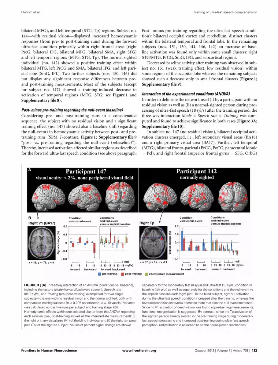
Dietrich et al. Training of ultra-fast speech comprehension
bilateral MFG), and left temporal (STG, Tp) regions. Subject no.144—with residual vision—displayed increased hemodynamicresponses (from pre- to post-training runs) during the forwardultra-fast condition primarily within right frontal areas (rightPrcG, bilateral IFG, bilateral MFG, bilateral SMA, right SFG)and left temporal regions (MTG, STG, Tp). The normal sightedindividual (no. 142) showed a positive training effect withinbilateral MTG, left PrCG and SMA, bilateral IFG, and left pari-etal lobe (SmG, IPL). Two further subjects (nos. 150, 146) didnot display any significant response differences between pre-and post-training measurements. Most of the subjects (exceptfor subject no. 147) showed a training-induced decrease inactivation of temporal regions (MTG, STG; see Figure 1 andSupplementary file 8).
Post- minus pre-training regarding the null-event (baseline)Considering pre- and post-training runs in a concatenatedsequence, the subject with no residual vision and a significanttraining effect (no. 147) showed also a baseline shift (regardingthe null-event) in hemodynamic activity between post- and pre-training runs (SPM T-contrast, Figure 1; Supplementary file 9“post- vs. pre-training regarding the null-event (=baseline)”).Thereby, increased activation affected similar regions as describedfor the forward ultra-fast speech condition (see above paragraph:
Post- minus pre-training regarding the ultra-fast speech condi-tion): bilateral occipital cortex and cerebellum, distinct clusterswithin the bilateral temporal and frontal lobe. In the remainingsubjects (nos. 151, 150, 144, 146, 142) an increase of base-line activation was found only within some small clusters: rightSTG/MTG, PrCG, SmG, IFG, and subcortical regions.
Decreased baseline activity after training was observed in sub-ject no. 151 (weak training effect, low residual vision) withinsome regions of the occipital lobe whereas the remaining subjectsshowed such a decrease only in small frontal clusters (Figure 1;Supplementary file 9).
Interaction of the experimental conditions (ANOVA)In order to delineate the network used (i) by a participant with noresidual vision as well as (ii) a normal-sighted person during pro-cessing of ultra-fast speech (18 syl/s) after the training period, thethree-way interaction Mode × Speech rate × Training was com-puted and found to achieve significance in both cases (Figure 3A;Supplementary file 10).
In subject no. 147 (no residual vision), bilateral occipital acti-vation clusters emerged, i.e., left secondary visual areas (BA18)and a right primary visual area (BA17). Further, left temporal(MTG), bilateral fronto-parietal (PrCG, PoCG, paracentral lobule= Pcl), and right frontal (superior frontal gyrus = SFG, OrbG)
FIGURE 3 | (A) Three-Way interaction of an ANOVA (conditions vs. baseline),including the factors Mode (forward/backward speech), Speech rate(8/18 syl/s), and Training (pre-/post-training) exemplified for two singlesubjects—the one with no residual vision and the normal sighted, both withcomparable training success [p < 0.005 uncorrected, k = 10 voxels]. Variancewas calculated across five runs per subject and training stage. (B)Hemodynamic effects within one selected cluster from the ANOVA regardingeach session (pre-, post-training as well as the intermediate measurement): (i)the right primary visual area (V1) of the blind individual and (ii) the right temporalpole (Tp) of the sighted subject. Values of percent signal change are shown
separately for the moderately fast (8 syl/s) and ultra-fast (18 syl/s) condition vs.baseline (left plot) as well as separately for the conditions and the null-event vs.the implicit baseline each (right plot). In the blind subject, right V1 activationduring the ultra-fast speech condition increased after the training, whereas thereversed condition showed a decrease (note that also the null-event increased).Since no V1 activation or deactivation was found at pre-training measurements,functional reorganization is suggested. By contrast, since the Tp activation ofthe sighted person already existed in the pre-training stage during moderatelyfast speech processing and increased post-training during ultra-fast speechperception, redistribution is assumed to be the neuro-plastic mechanism.
Frontiers in Human Neuroscience www.frontiersin.org October 2013 | Volume 7 | Article 701 | 133

Dietrich et al. Training of ultra-fast speech comprehension
areas were found to be activated. Considering post-hoc hemody-namic effects (values of percentage signal change) of the forwardand reversed moderately fast (8 syl/s) and ultra-fast (18 syl/s) con-ditions (vs. baseline) within the right-hemispheric primary visualarea, BOLD response increased after the training (comparedwith pre-training) during the forward ultra-fast—but not mod-erately fast—condition, whereas both reversed speech conditionsremained at the level of zero or showed deactivation (Figure 3B,left plot).
In the sighted participant (no. 142), bilateral temporal (MTG,Tp), bilateral cerebellar (Cb), left frontal (SMA, IFG, PrCG),and parietal regions (left SmG, right Pcl) showed a signifi-cant three-way interaction. Post-hoc analysis found increasedright temporal pole activation after training during the for-ward ultra-fast speech condition, but revealed this area alsoto be activated before training during the moderately fastspeech condition. Additionally, this region showed selectivityto forward (compared with reversed) speech (Figure 3B,left plot).
Regarding the training-induced baseline shift (null-event;Figure 3B, right plot), hemodynamic activity during the null-event was stronger after the training period in the blind individualwhereas the normal-sighted participant showed an opposite ten-dency. The baseline shift in the blind subject was even strongerduring the intermediate measurement at a training stage of13 syl/s (not included in the ANOVA).
REGIONS-OF-INTEREST (ROI) ANALYSESRight-hemispheric visual cortexConcerning right V1 (Figure 4A), the tests of percent sig-nal changes addressed the difference of the values from zeroand the differences between conditions. Only subject no. 147(no residual vision) showed significantly positive values underthe forward ultra-fast speech condition following the trainingsessions (T = 2.870, p < 0.05) and, additionally, higher val-ues under the forward as compared to the reversed ultra-fastspeech condition post-training (T = 2.479, p < 0.05). Subjectno. 151 (low residual vision) achieved positive values under
FIGURE 4 | Regions-of-interest (ROI) analyses, (A) right primary visualarea (V1) and (B) right pulvinar (Pv), (C) left supplementary motor area(SMA) as well as the (D) left inferior frontal gyrus (IFG)—all clusters derivedfrom Dietrich et al. (2013), overlaid on an anatomical T1 template. Upperplot (red): Behavioral performance of understanding ultra-fast speech (18 syl/s)
pre- and post-training. Participants are arranged according to their visual acuity.Lower plots (blue): Percent signal change during forward (left) and reversed(middle) ultra-fast speech (18 syl/s) as well as forward moderately fast (8 syl/s)speech (right) pre- and post-training (vs. baseline). Significant differences fromzero (one sample T -tests): ∗p < 0.05, ∗∗p < 0.01, ∗∗∗p < 0.001, ∼ p < 0.1.
Frontiers in Human Neuroscience www.frontiersin.org October 2013 | Volume 7 | Article 701 | 134

Dietrich et al. Training of ultra-fast speech comprehension
the reversed ultra-fast condition post-training (T = 2.248, p <
0.05), whereby forward ultra-fast speech not did yield any sig-nificant differences as compared to reversed utterances. Subjectno. 150 (low residual vision) displayed neither significant posi-tive/negative values nor significant differences between forwardand reversed speech under the ultra-fast speech condition afterthe training sessions. Subjects nos. 144 (T = −3.123, p < 0.05),146 (T = −1.637, p = 0.09), and 142 (T = −0.598, p = 0.291)showed negative values or did not differ from zero during theforward ultra-fast speech condition post-training. Furthermore,these latter subjects did not show any differences between for-ward and reversed ultra-fast speech post-training. In addition,significantly positive values under the moderately fast speech con-dition pre-training (T = 3.735, p <.05) and selectivity to forwardcompared with reversed speech (T = 1.944, p < 0.05) could beobserved exclusively in subject no. 144.
Furthermore, a significant negative correlation between resid-ual vision and the percent signal change during the forwardultra-fast speech condition (vs. baseline) could be detectedbefore (ρ = −0.77, p < 0.05) and after the training sessions(ρ = −0.83, p < 0.05), with a tendency toward a steeper regres-sion line at the post-training examination (T = −1.569, p < 0.1)(Figure 5A). By contrast, no significant correlations emerged dur-ing application of moderately fast speech or during the reversedultra-fast speech condition—neither at pre- nor post-trainingmeasurements.
All three subjects with no/low residual vision (rank 1, 2, 3)showed positive values of percent signal change compared tosubjects with residual vision (rank 4, 5, 6) with respect to thepost- and pre-training stages (Figure 5A). Therefore, a whole-brain covariance analysis was performed with the residual visionas non-linear covariate (rank 1, 2, 3, 4, 4, 4), based on thehypothesis that training-induced enhancement of primary visualactivation occurs only in subjects with no or low residual vision.Indeed, the training effect for forward ultra-fast speech (SPM T-contrast post- vs. pre-training) was associated with hemodynamicresponses of right V1 (Figure 5B) at a similar location as the acti-vation cluster observed in our previous group study (Dietrichet al., 2013; Figure 5C).
Right-hemispheric pulvinarAs concerns right Pv, neither pre- nor post-training fMRI eval-uation yielded any percent signal change values which signifi-cantly differed from zero (Figure 4B). Furthermore, no signifi-cant differences emerged between forward and reversed ultra-fastspeech conditions post-training (Figure 4B). However, across theentire group of participants, a significant negative correlationbetween residual vision and percent signal change during for-ward ultra-fast speech was found at post-training measurements(ρ = −0.77, p < 0.05) in right Pv. Prior to the learning proce-dure, the correlation also showed a negative trend (ρ = −0.71,p = 0.055).
Left-hemispheric SMA and IFGAs concerns left-hemispheric SMA, all subjects with trainingeffects (= all except no. 146) showed a positive change in per-cent signal change across follow-up under the forward ultra-fast
FIGURE 5 | (A) Correlations between BOLD responses during the forwardultra-fast (18 syl/s) speech condition (vs. baseline) and the participants,arranged according to their residual vision (given as ranks 1–6: 1 = noresidual vision and 6 = normally sighted within the right-hemisphericprimary visual activation cluster, drawn from Dietrich et al. (2013) (see panelC). Values of percent signal changes refer to the pre- (blue) and post-training(red) measurements of each subject. (B) Whole-brain covariance analysis(SPM T -contrast “post- minus pre-training,” forward ultra-fast speechcondition) with residual vision as a non-linear covariate (rank 1, 2, 3, 4, 4,4)—hypothesizing that primary visual activation will only change in subjectswith no or very low residual vision. The location of this effect in visual cortexwas similar to the activation cluster observed in Dietrich et al. (2013) (C).
speech condition (Figure 4C, left plot). By contrast, negativechanges from the pre- to the post-training values of this measurecould be observed at the level of SMA under the forward moder-ately fast condition across most subjects (descriptive; Figure 4C,right plot). Considering left IFG, significant positive values of per-cent signal change were found during the forward moderatelyfast speech condition across most subjects pre- as well as post-training (Figure 4D). By contrast, only two subjects (nos. 151,142) showed significant positive values of percent signal changeduring the forward ultra-fast speech condition post-training,whereas during the backward ultra-fast speech condition neg-ative values could be observed within one subject (no. 144)(Figure 4D).
Before training, a significant positive correlation betweenBOLD responses and behavioral performance (Figure 6A) wasfound across all three speech rates (8, 13, 18 syl/s) of the for-ward speech conditions and across all subjects within left SMA(r = 0.58, p < 0.05) as well as IFG (r = 0.72, p < 0.001). After
Frontiers in Human Neuroscience www.frontiersin.org October 2013 | Volume 7 | Article 701 | 135

Dietrich et al. Training of ultra-fast speech comprehension
FIGURE 6 | (A) Correlation between BOLD responses during forward speechpre-training and individual behavioral performance within left SMA and inferiorfrontal gyrus (IFG) across all three speech rates (8, 13, 18 syl/s) and across allsubjects. (B) BOLD responses during forward speech post-training for each
speech rate (8, 13, 18 syl/s) within left SMA and IFG. Only subjects with aperformance level of more than ca. 70% at speech rate (8 syl/s: n = 6; 13 syl/s:n = 6; 18 syl/s: n = 4) are included. Significant differences (paired T -test)emerged between moderately fast and ultra-fast speech within left SMA.
training, most subjects (except of nos. 151 and 146 with respectto the ultra-fast speech condition) performed all speech rates (8,13, 18 syl/s) up to a level of at least ca. 70% correctly repro-duced syllables. However, post-training BOLD responses of thesesubjects with at least ca. 70% performance during the ultra-fast speech condition (vs. baseline, n = 4) were significantlyhigher than during moderately fast speech (n = 6) within SMA(T = −4.009, p < 0.05), but not within IFG (T = 1.639, p =0.162) (Figure 6B). Expectedly, against the background that fourof the six subjects reached comparable training effects, their resid-ual vision did not significantly correlate with BOLD responses inleft SMA or IFG neither pre- nor post-training (p > 0.1 for alltests).
DISCUSSIONBEHAVIORAL TRAINING EFFECTSFour of the six participants showed—independently of theirresidual visual functions—a significant improvement of ultra-fast (18 syl/s) speech comprehension, in terms of a performancelevel of ca. 70% points after daily training sessions. And the threevision-impaired subjects of this subgroup reported to benefitduring daily life from their ability to use screen-reading text-to-speech devices. By contrast, poor (36% points) or no trainingeffects could be observed in the two remaining individuals. Sofar, none of the participants achieved the pre-specified target ofmore than 80 % correctly repeated words at a speech rate of18 syl/s. Since the participants underwent fMRI measurements
as soon as they themselves had the impression to meet this cri-terion, they lacked an external control over actual performance.The sighted control subject of the present study reported that shehad “reached her limits” and that she feels additional trainingwould not yield further improvements of ultra-fast speech per-ception. By contrast, a further extension of the training period(so far ca. 6 months) might have been beneficial in case of visionloss, since higher performance levels have been documented inother blind individuals (Moos and Trouvain, 2007). Most note-worthy, residual vision in terms of visual acuity in the four sub-jects with a significant—and similar—increase of post-trainingbehavioral performance ranged from none to 100%. Thus, thisclinical parameter, obviously, did not constrain the acquisition ofultra-fast speech perception capabilities. It cannot be excluded,however, whether and in how far subjects with low residual visionmight be able to further enhance ultra-fast speech comprehensionin case of more expanded training procedures.
POST-TRAINING BRAIN NETWORKS OF ULTRA-FAST SPEECHCOMPREHENSION (WHOLE-BRAIN ANALYSES)Common activation “nodes” across subjectsClinical and functional imaging data indicate a contribution ofleft IFG to speech perception—at least in case more demand-ing segmentation processes and/or working memory operationsare involved (Burton et al., 2000). Furthermore, hemodynamicactivation of language processing areas such as IFG has beenassumed to represent an index of actual speech comprehension
Frontiers in Human Neuroscience www.frontiersin.org October 2013 | Volume 7 | Article 701 | 136

Dietrich et al. Training of ultra-fast speech comprehension
(Poldrack et al., 2001). The present study revealed post-trainingleft-hemispheric IFG responses across all subjects, indicatingintelligibility of ultra-fast spoken language even if the train-ing effect was low (subject no. 146 showed no training effects,nevertheless, performance level at baseline had amounted to14%). Furthermore, left-hemispheric PrCG and the cerebellumdisplayed significant BOLD signal changes during forward ultra-fast speech post-training. These observations are in line withclinical and functional imaging data pointing at a contributionof those structures—under specific circumstances—to auditoryspeech perception. For example, the cerebellum has been foundengaged in the encoding of specific temporal-linguistic informa-tion during word identification tasks (Mathiak et al., 2002).
Descriptive analyses of individual response patternsFour participants showed a significant—and similar—increaseof post-training behavioral performance (ca. 70% points), inde-pendent of residual vision capabilities (<2, 10, 20, 100% visualacuity). Most noteworthy, different individual hemodynamicresponse patterns could be observed in these cases, indicatingprocessing strategies of ultra-fast speech understanding to varyacross these subjects.
Training of ultra-fast speech comprehension was found toinduce hemodynamic activation at the level of the occipital lobein three subjects with no or low residual vision. The strongesteffects (also significant in the post- minus pre-training com-parison) emerged in subject no. 147, a participant lacking anyresidual visual functions, and such responses to acceleratedspoken language could not be observed at the baseline exami-nation. These findings provide tentative support for a relation-ship between ultra-fast speech perception and right-hemisphericV1 activation—in case the central-visual system is deprived ofmodality-specific afferent input. Based on our previous studies,it has been suggested that collaboration of right auditory cortexand ipsilateral primary visual area allows for the implementationof a signal-driven timing mechanism related to syllabic modu-lation of the speech signal (Dietrich et al., 2013; Hertrich et al.,2013a,b). More specifically, subjects able to recruit the visual cor-tex during spoken language comprehension appear to install anadditional information channel conveying temporal cues on syl-lable structure to the frontal lobe which then help to triggerphonological encoding processes and, as a consequence, to sup-port verbal working memory operations (Hertrich et al., 2013b).Since the visual system seems to compensate for the temporallimitations of the auditory system under these conditions, thisstrategy might allow for a further increase in ultra-fast speechcomprehension capabilities beyond the level that was achieved inthe present training study.
Besides increased V1 activation during the ultra-fast speechcondition, subject 147 also showed significant changes in the null-event if the runs across the pre- and post-training sessions wereconcatenated for analysis. Interestingly, this increase was strongestin the session at an intermediate training level when the ultra-fastcondition did not yet activate right V1 (vs. baseline). This “base-line shift” might result from actual activity during the null event.However, it could also reflect a discrepancy between the explicit(null-event) and the implicit baseline, which might indicate that
the BOLD response in V1 is not strictly aligned with the stim-uli across training stages. This inconsistency might be boundto the emergence of reorganization processes—rather than justredistribution of brain activity as reported by Kelly and Garavan(2005)–since occipital cortex was not active prior to the pre-training session and since a new area has been recruited duringthe learning procedure.
Interestingly, subject no. 151 (with low visual acuity similarto subject no. 147) showed a strong reduction of the null-eventactivity within the occipital lobe at the post- as compared tothe pre-training session, but no increase of activation during theultra-fast speech condition. The low training effect of this subjectseems to be in line with the absence of significant V1 activa-tion during the ultra-fast condition. However, the strong changesof the null-event activity might indicate that also in this subjecttraining-induced reorganization is possible.
By contrast, subject no. 150 (10% visual acuity) did not showany training-induced changes in occipital cortex activity and,thus, visual system recruitment for the sake of ultra-fast speechperception seems to be impossible.
It should be noted that the post-training hemodynamicresponse pattern of subject no. 147, displaying the strongestresponse of right visual cortex, encroaches, in addition, uponleft-hemispheric primary (BA17) and bilateral secondary (SOG,MOG) visual areas. As an explanation, the recruitment of occipi-tal areas at either side of the brain might reflect central-visual sup-port of several linguistic functions during ultra-fast speech com-prehension, e.g., both segmental (left hemisphere) and supraseg-mental/prosodic (right hemisphere) processing. Against the back-ground of previous findings (Dietrich et al., 2013; Hertrichet al., 2013b), strong right-hemispheric lateralization of theprimary visual area seems to be associated with a prosodicfunction, i.e., triggering syllable onsets within a metric pat-tern. Thus, a somehow unfocused reorganization pattern mightbe further stabilized after getting more experienced with ultra-fast speech materials. Continuation of the training procedure athigh rates could result, conceivably, in a less distributed occip-ital activation pattern centered around the right primary visualarea.
In line with our preceding single-case and group studies onultra-fast speech perception (Hertrich et al., 2009; Dietrich et al.,2013), post-training fMRI revealed hemodynamic activation ofleft FG in response to ultra-fast speech materials in subjectno. 147, characterized by the strongest engagement of right-hemisphere visual cortex. FG is embedded into the so-called ven-tral route of the central-visual system which, especially, supportsobject recognition (e.g., Haxby et al., 1991), but may contribute tophonological operations as well (e.g., Cone et al., 2008). Thus, leftFG as a secondary phonological area might expand the phonolog-ical network and help to cope with the higher processing demandsduring ultra-fast speech perception.
Presumably, right-hemispheric frontal lobe activation, includ-ing PrCG, MFG, OrbG, and IFG as observed in subject no.144, might reflect a network associated with phonological wordstress processing during ultra-fast speech comprehension. Inprevious studies, neural correlates of word stress as comparedto vowel quality encoding (Klein et al., 2011) or auditory
Frontiers in Human Neuroscience www.frontiersin.org October 2013 | Volume 7 | Article 701 | 137

Dietrich et al. Training of ultra-fast speech comprehension
processing of bi-syllabic pseudo-words (Tervaniemi et al., 2009)were found within a widespread fronto-temporal activation pat-tern including, among others, right-hemispheric activation clus-ters resembling the response pattern of subject no. 144. Againstthis background, it may be assumed that a prosodic/metricpattern was generated, predicting continuous word stress ormetrical stress sequences during listening to ultra-fast speech.Such a focus on stressed syllables—ignoring more or less theunstressed ones—might help to overcome the backward maskingeffects of high speaking rates. However, this strategy of ultra-fast speech comprehension, conceivably, poses higher demandson the auditory system with regard to the processing of wordstress information, interpolation of unstressed syllables, andthe mapping of these patterns onto articulatory motor plans.This assumption is in line with reports of increased activationof the auditory association cortices and left ventral premotorcortex after training to understand time-compressed speech insighted individuals (Adank and Devlin, 2010). Although sucha compensation strategy based upon prosodic/metric redun-dancies of verbal utterances might facilitate ultra-fast speechcomprehension to a certain extent, it may not be able to reallyenhance the temporal resolution at the level of the syllable and,therefore, might require increased top-down effort of languageprocessing.
Distinct hemodynamic activation of the temporal pole ateither side was found after training of ultra-fast speech compre-hension in the sighted subject no. 142. Combinatorial processes atsentence-level (Hickok and Poeppel, 2007) and the evaluation ofaffective semantics (Ethofer et al., 2006) have been associated withhemodynamic responses of the anterior temporal lobe, indicat-ing holistic rather than compositional (i.e., hierarchical analysis ofsegments, syllables, and morphemes, and phrases) semantic pro-cessing under these conditions. Such top-down mechanisms—based upon contextual/semantic pre-information—may facilitatesound-to-semantic mapping processes even in case of incompletephonological decoding. Although the use of this strategy shouldovercome to a certain extent the backward masking effects ofultra-fast speech signals, we may speculate that subjects applyingthis strategy will have difficulty understanding the logical struc-ture of more complex sentences. Moreover, the present (sighted)subject who might have used this strategy did not recruit any newarea as in the case of the blind participant. Instead, activity exist-ing pre-training during the moderately fast speech condition wasfound increased post-training during the ultra-fast speech con-dition. Thus, redistribution of activity as defined in Kelly andGaravan (2005) is assumed to be the underlying mechanism forplasticity.
Summarizing these descriptive analyses of distinct individ-ual activation patterns, subjects appear to use different strate-gies during ultra-fast speech perception depending, among otherthings, on residual visual functions. In case of very low resid-ual vision, the visual system seems to be recruited as a meansto enhance actual temporal resolution of the acoustic signal viaimplementation of an extra-channel conveying information onsyllabic structure to verbal working memory buffers. This newstrategy concomitant with the recruitment of an additional areacan be considered a variant of functional reorganization. If the
visual system is not at a subject’s disposal for spoken languagecomprehension, listeners may rely on already available semantictop-down strategies facilitating lexical access, based upon, e.g.,redundant aspects of verbal materials. Under these conditions, astronger engagement of (some components of) the classical lan-guage network might help to speed up the encoding processes,providing an example of neuroplasticity due to redistribution ofbrain activity.
THE IMPACT OF RESIDUAL VISION AND BEHAVIORAL PERFORMANCEON THE RESPONSES OF THE VISUAL SYSTEM (ROI ANALYSES)During forward ultra-fast speech perception, residual vision wasfound to have a significant impact on the activation of the right-hemispheric primary visual cortex (V1) and ipsilateral pulvinar(Pv), a subcortical structure projecting to the occipital lobes.More specifically, a negative correlation emerged in terms ofdecreased hemodynamic activation concomitant with increasedresidual vision. The respective regression line showed a trendtoward a steeper decline at the post-training measurements, indi-cating a differential impact of residual vision on the trainingeffects. Thus, residual visual capacities appear to constrain theresources of audiovisual reorganization during ultra-fast speechperception. The critical threshold seems to be centered aroundacuity values of 10 to 20%, whereas a more efficient use of thevisual cortex such as in subject no. 147 even may require residualvision to fall below a level of only 2%.
A previous study investigating cross-modal reorganization in avision-impaired subject who had suffered from severe visual acu-ity reduction with no evidence of visual field loss, reported popu-lations of visual neurons not critical for a subject’s remaining lowvision to be recruited for tactile information processing (Cheunget al., 2009). The observation of such a retinotopy-related func-tional segregation of neurons bound to residual vision, on the onehand, and cross-modal processing, on the other, might explainwhy audiovisual plasticity did not occur in the subjects of ourgroup with normal or high residual vision. Furthermore, visualacuity rather than the peripheral visual field seems to be the cru-cial prerequisite of a recruitment of the visual system (Cheunget al., 2009). The present data confirm this suggestion since, e.g.,subject no. 151 with no visual acuity, but intact peripheral visualfields showed increased hemodynamic activation of the visual sys-tem at post-training measurements although the subject showed amoderate training effect only. By contrast, participant no. 144 suf-fering from a strongly reduced visual field did not activate rightvisual cortex after the training sessions during the forward ultra-fast speech condition, presumably, because visual acuity was notmarkedly reduced. Consequently, the foveal representation withinthe visual system seems to be the target of cross-modal reor-ganization processes during the acquisition of ultra-fast speechcomprehension. In line with this hypothesis, training of ultra-fastspeech comprehension induced strong visual cortex activation ina subject with no residual vision (no. 147). In spite of good train-ing effects, only low hemodynamic responses within the visualsystem could be observed in the subject suffering from residual10% visual acuity (no. 150).
Interestingly, forward moderately fast speech did not induceany distinct hemodynamic activation of visual cortex after
Frontiers in Human Neuroscience www.frontiersin.org October 2013 | Volume 7 | Article 701 | 138

Dietrich et al. Training of ultra-fast speech comprehension
training. It can be assumed, thus, that the speech task has tobe sufficiently challenging in order to induce significant acti-vation in the visual system. In fact, passive listening to soundsdoes not give rise to occipital activation in blind subjects (Arnoet al., 2001). Accordingly, visual areas in the blind were notactivated by the mere presence of sound, but were involvedin attentive perception of changes in the auditory environment(Kujala et al., 2005). Attention is suggested to play an impor-tant role during ultra-fast speech comprehension as well—interms of the detection of almost entirely masked syllable onsets(Dietrich et al., 2013; Hertrich et al., 2013b). However, sig-nificant visual cortex activation was also found at pre-trainingexamination during the moderately fast speech condition selec-tively to forward (compared to reversed) speech in a subjectwith high residual vision (no. 144). As an explanation, hemody-namic responses of visual cortex during speech perception mightreflect actual visual imagery. The subjects’ high familiarity withspeech signals in association with speaking faces during languageacquisition might facilitate such effects also in late-blind sub-jects. In a previous study, addressing the reverse relationshipof the two sensory modalities, perception of visual lip move-ments during silent syllable production was found to evokeprimary auditory BOLD responses, indicating strong audio-visual interactions during speech perception (Hertrich et al.,2010).
Taken together, the highest activation levels of the visual sys-tem were found in case of (i) absent residual vision and (ii)good training effects. Only subject no. 147 met these two crite-ria while the remaining subjects fell short to either one or bothcharacteristics. As concerns the latter participant, visual systemrecruitment was selectively limited to forward—and, thus, prin-cipally intelligible—utterances. As a rule, right Pv displayed aprofile of BOLD responses similar to right visual cortex. Again,these findings point at a contribution of this subcortical struc-ture to ultra-fast speech perception. Conceivably, the observedinteractions of Pv and visual cortex reflect the operation of anaudiovisual interface in that the secondary visual pathway pro-vides the central-visual system with auditory input (see Hertrichet al., 2013b, for more details).
VISION-INDEPENDENT AND PERFORMANCE-DEPENDENTCHANGES OF HEMODYNAMIC ACTIVATION OF LEFT SMA AND IFG(ROI ANALYSIS)Pre-training BOLD responses within left IFG and SMAwere found to be significantly correlated—independently fromresidual vision—with the percentage of correctly understoodspeech materials, pooled across speech rates (8, 13, 18 syl/s).Consequently, both left IFG and SMA activation are associatedwith speech comprehension. More specifically, it is suggested thatleft IFG is involved in the speech production-related phonolog-ical reconstruction of perceived speech material (Burton et al.,2000). However, not all participants with significant trainingeffect showed significant left IFG activation with respect to thecluster resulting from Dietrich et al. (2013) after the train-ing period. Therefore, some subjects seem not to reach thethreshold with respect to the relatively large cluster resultedfrom (Dietrich et al., 2013). However, the whole-brain analyses
revealed post-training left-hemispheric IFG activation in subjectswith significant training effect.
Prior to training, hemodynamic activation within left SMAduring the “moderately fast condition” showed considerable vari-ation across individuals. Recruitment of left SMA, thus, doesnot seem to be a prerequisite to speech comprehension per se assuch. However, all subjects with training effects showed a positivechange in percent signal change across follow-up under the for-ward ultra-fast speech condition. Moreover, left SMA, but not IFGactivation post-training was significantly higher during the ultra-fast than the moderately fast speech condition in well-performingsubjects. Neither the pre- nor post-training responses of SMA andIFG depended on residual vision, an observation supporting theview of a supra-modal function of these frontal structures, irre-spective of input modality. Several studies indicate SMA to engagein the syllabic organization of verbal utterances during speechproduction (Ziegler et al., 1997; Riecker et al., 2005; Brendelet al., 2010). On a broader scale, this region appears to sup-port timing processes across various sensorimotor and cognitivedomains (Rubia and Smith, 2004; Paz et al., 2005). Furthermore,clinical as well as experimental data point at a contribution ofSMA to spoken language perception and verbal working mem-ory operations as well (Schirmer et al., 2001; Smith et al., 2003;Schirmer, 2004; Chung et al., 2005; Geiser et al., 2008). Giventhat SMA might act as a platform for timing operations relatedto verbal working memory functions, sensory systems might con-vey temporal information on syllable structure via left SMA intothis short-term storage system. More specifically, the joint acti-vation of auditory and visual areas with left SMA could reflect asignal-driven timing mechanism facilitating the transformationof the perceived acoustic signal into an active pre-articulatoryphonetic/phonological output structure in our speech generationsystem under time-critical conditions (see Hertrich et al., 2013b,for more details).
CONCLUSIONSSeveral participants were found able to increase ultra-fast speech(18 syl/s) comprehension up to a level of ca. 70% points, irre-spective of residual vision, across a training session of about6 months. However, subjects seem to deploy distinct patternsof neuroplasticity in order to overcome the bottleneck of tem-poral resolution associated with high speaking rates: In caseof very low residual vision, the right-hemispheric visual sys-tem (V1, Pv) was, apparently, recruited indicating functionalreorganization at the level of the occipital lobes. By contrast,participants with normal or high residual vision did not showany training-induced responses of the central-visual system.However, they displayed increased activation (redistribution) ofseveral parts of the classical speech processing network. Althoughboth patterns of neuroplasticity resulted in comparable train-ing effects, the strategy of processing ultra-fast speech mightsignificantly differ: In case of a deployment of the visual sys-tem, actual temporal resolution of the acoustic signal mightincrease via the representation of a syllabic trigger cues. If, how-ever, the visual system is not at a subject’s disposal for spokenlanguage processing, they may use, as an alternative, a strat-egy based on informational redundancies of the speech signal.
Frontiers in Human Neuroscience www.frontiersin.org October 2013 | Volume 7 | Article 701 | 139
Dietrich et al. Training of ultra-fast speech comprehension
Independent of residual vision, left-hemispheric SMA and IFGactivation emerged as a common indicator of speech comprehen-sion. In case of proficient ultra-fast speech comprehension, how-ever, only SMA showed stronger activation during application ofultra-fast as compared to moderately fast test materials, pointingat a critical contribution to spoken language understanding undertime-critical conditions.
AUTHORS CONTRIBUTIONHermann Ackermann, Ingo Hertrich, and Susanne Dietrichdelineated the rationale and developed the design of the study.IH and SD were engaged in data collection and development ofanalyses methods. SD performed the behavioral and fMRI dataanalyses, and drafted the first version of the paper. All authorscontributed to the final version of the manuscript and approvedits content.
ACKNOWLEDGMENTSThis study was supported by the German Research Foundation(DFG; AC 55 09/01) and the Hertie Institute for Clinical BrainResearch, Tübingen. The authors would like to thank MaikeBorutta for excellent technical assistance, recruiting and takingcare of the participants.
SUPPLEMENTARY MATERIALThe Supplementary Material for this article can be found onlineat: http://www.frontiersin.org/journal/10.3389/fnhum.2013.00701/abstract
Supplementary file 1 | An example for forward moderately fast speech
(8 syl/s). “Wegen den anstehenden wichtigen Prüfungen muss er viel
lernen.”
Supplementary file 2 | An example for forward ultra-fast speech (18 syl/s).
“Die Mitfahrerin stellte sich als gute Gesprächspartnerin heraus.”
Supplementary file 3 | An example for reversed moderately fast speech(8 syl/s). See Supplementary file 1.
Supplementary file 4 | An example for reversed ultra-fast speech (18 syl/s).See Supplementary file 2.
Supplementary file 5 | An example of the repetition task for ultra-fast
speech of blind listeners with ca. 70% comprehension capability. “Die
Karten für das Konzert nächstes Jahr sind bereits jetzt ausverkauft.”
Supplementary file 6 | Coordinates describing the SPM T-contrasts“forward ultra-fast speech condition versus baseline” pre- andpost-training separately.
Supplementary file 7 | Coordinates describing the SPM T-contrasts“baseline versus forward ultra-fast speech condition” pre- andpost-training separately.
Supplementary file 8 | Coordinates describing the SPM T -contrasts “post-versus pre-training” during the forward ultra-fast speech condition (andvice versa).
Supplementary file 9 | Coordinates describing the SPM T -contrasts “post-versus pre-training” (and vice versa) during the null-event (=baseline).
Supplementary file 10 | Coordinates describing the Three-Way interaction“Mode × Speech rate × Training” resulting from the ANOVA exemplifiedto subject nos. 147 and 142.
REFERENCESAdank, P., and Devlin, J. T. (2010).
On-line plasticity in spokensentence comprehension: adapt-ing to time-compressed speech.Neuroimage 49, 1124–1132. doi:10.1016/j.neuroimage.2009.07.032
Amedi, A., Raz, N., Pianka, P.,Malach, R., and Zohary, E. (2003).Early “visual” cortex activationcorrelates with superior verbalmemory performance in the blind.Nat. Neurosci. 6, 758–766. doi:10.1038/nn1072
Arno, P., De Volder, A. G., Vanlierde,A., Wanet-Defalque, M. C., Streel,E., Robert, A., et al. (2001).Occipital activation by patternrecognition in the early blind usingauditory substitution for vision.Neuroimage 13, 632–645. doi:10.1006/nimg.2000.0731
Baumgart, F., Kaulisch, T.,Tempelmann, C., Gaschler-Markefski, B., Tegeler, C., Schindler,F., et al. (1998). Electrodynamicheadphones and woofers for appli-cation in magnetic resonanceimaging scanners. Med. Phys. 25,2068–2070. doi: 10.1118/1.598368
Brendel, B., Hertrich, I., Erb, M.,Lindner, A., Riecker, A., Grodd, W.,et al. (2010). The contribution of
mesiofrontal cortex to the prepa-ration and execution of repetitivesyllable productions: an fMRIstudy. Neuroimage 50, 1219–1230.doi: 10.1016/j.neuroimage.2010.01.039
Büchel, C., Price, C., Frackowiak, R. S.J., and Friston, K. (1998). Differentactivation patterns in the visual cor-tex of late and congenitally blindsubjects. Brain 121, 409–419. doi:10.1093/brain/121.3.409
Burton, H., McLaren, D. G., andSinclair, R. J. (2006). Readingembossed capital letters: an fMRIstudy in blind and sighted individu-als. Hum. Brain Mapp. 27, 325–339.doi: 10.1002/hbm.20188
Burton, M. W., Small, S. L., andBlumstein, S. E. (2000). The roleof segmentation in phonologicalprocessing: an fMRI investigation.J. Cogn. Neurosci. 12, 679–690. doi:10.1162/089892900562309
Cheung, S. H., Fang, F., He, S., andLegge, S. E. (2009). Retinotopicallyspecific reorganization of visualcortex for tactile pattern recogni-tion. Curr. Biol. 19, 596–601. doi:10.1016/j.cub.2009.02.063
Chung, G. H., Han, Y. M., Jeong, S. H.,and Jack, C. R. (2005). Functionalheterogeneity of the supplementary
motor area. Am. J. Neuroradiol. 26,1819–1823.
Cone, N. E., Burman, D. D., Bitan,T., Bolger, D. J., and Booth, J. R.(2008). Developmental changes inbrain regions involved in phono-logical and orthographic process-ing during spoken language process-ing. Neuroimage 41, 623–635. doi:10.1016/j.neuroimage.2008.02.055
Dietrich, S., Hertrich, I., andAckermann, H. (2013).Ultra-fastspeech comprehension in blind sub-jects engages primary visual cortex,fusiform gyrus, and pulvinar—a functional magnetic imaging(fMRI) study. BMC Neurosci. 14:74.doi: 10.1186/1471-2202-14-74
Ethofer, T., Anders, S., Erb, M.,Herbert, C., Wiethoff, S., Kissler,J., et al. (2006). Cerebral pathwaysin processing of affective prosody:a dynamic causal modeling study.Neuroimage 30, 580–587. doi:10.1016/j.neuroimage.2005.09.059
Geiser, E., Zaehle, T., Jäncke, L., andMeyer, M. (2008). The neural cor-relate of speech rhythm as evi-denced by metrical speech process-ing. J. Cogn. Neurosci. 20, 541–552.doi: 10.1162/jocn.2008.20029
Gizewski, E. R., Gasser, T., de Greiff,A., Boehm, A., and Forsting, M.
(2003). Cross-modal plasticity forsensory and motor activation pat-terns in blind subjects. Neuroimage19, 968–975. doi: 10.1016/S1053-8119(03)00114-9
Guida, A., Gobet, F., Tardieu, H., andNicolas, S. (2012). How chunks,long-term working memory andtemplates offer a cognitive expla-nation for neuroimaging dataon expertise acquisition: a two-stage framework. Brain Cogn. 79,221–244. doi: 10.1016/j.bandc.2012.01.010
Haxby, J. V., Grady, C. L., Horwitz,B., Ungerleider, L. G., Mishkin,M., Carson, R. E., et al. (1991).Dissociation of object and par-tial visual processing pathwaysin human extrastriate cortex.Proc. Natl. Acad. Sci. U.S.A. 88,1621–1625. doi: 10.1073/pnas.88.5.1621
Hertrich, I., Dietrich, S., andAckermann, H. (2010). Cross-modal interactions duringperception of audiovisual speechand nonspeech signals: an fMRIstudy. J. Cogn. Neurosci. 23,221–237. doi: 10.1162/jocn.2010.21421
Hertrich, I., Dietrich, S., andAckermann, H. (2013a). Tracking
Frontiers in Human Neuroscience www.frontiersin.org October 2013 | Volume 7 | Article 701 | 140

Dietrich et al. Training of ultra-fast speech comprehension
the speech signal—time-lockedMEG signals during perceptionof ultra-fast and moderately fastspeech in blind and in sightedlisteners. Brain Lang. 124, 9–12. doi:10.1016/j.bandl.2012.10.006
Hertrich, I., Dietrich, S., andAckermann, H. (2013b). Howcan audiovisual pathways enhancethe temporal resolution of time-compressed speech in blindsubjects? Front. Psychol. 4:350. doi:10.3389/fpsyg.2013.00530
Hertrich, I., Dietrich, S., Moos, A.,Trouvain, J., and Ackermann, H.(2009). Enhanced speech percep-tion capabilities in a blind listenerare associated with activation offusiform gyrus and primary visualcortex. Neurocase 15, 163–170. doi:10.1080/13554790802709054
Hickok, G., and Poeppel, D. (2007).The cortical organization of speechprocessing. Nat. Neurosci. Rev. 8,393–402. doi: 10.1038/nrn2113
Kelly, A. M. C., and Garavan, H. (2005).Human functional neuroimaging ofbrain changes associated with prac-tice. Cereb. Cortex 15, 1089–1102.doi: 10.1093/cercor/bhi005
Klein, E., Domah, U., Grande, M.,and Domah, F. (2011). Neuro-cognitive foundations of wordstress processing—evidence fromfMRI. Behav. Brain Funct. 7:15. doi:10.1186/1744-9081-7-15
Kujala, T., Palva, M. J., Salonene,O., Alkuf, P., Huotilainena, M.,Järvinenf, A., et al. (2005). Therole of blind humans’ visual cor-tex in auditory change detection.Neurosci. Lett. 379, 127–131. doi:10.1016/j.neulet.2004.12.070
Lambert, S., Sampaio, E., Mauss, Y.,and Schreiber, C. (2004). Blindnessand brain plasticity: contributionof mental imagery? An fMRI study.Cogn. Brain Res. 20, 1–11. doi:10.1016/j.cogbrainres.2003.12.012
Mathiak, K., Hertrich, I., Grodd,W., and Ackermann, H. (2002).
Cerebellum and speech perception:a functional magnetic reso-nance imaging study. J. Cogn.Neurosci. 14, 902–912. doi:10.1162/089892902760191126
Moos, A., and Trouvain, J. (2007).“Comprehension of ultra-fastspeech—blind vs. “normallyhearing” persons,” in Proceedingsof the International Congress ofPhonetic Sciences, eds J. Trouvainand W. J. Barry (Saarbrücken:University of Saarbrücken),677–680.
Paz, R., Natan, C., Boraud, T.,Bergman, H., and Vaadia, E.(2005). Emerging patterns ofneuronal responses in supple-mentary and primary motor areasduring sensorimotor adaptation.J. Neurosci. 25, 10941–10951. doi:10.1523/JNEUROSCI.0164-05.2005
Poirier, C., Collignon, O., Scheiber, C.,Renier, L., Vanlierde, A., Tranduy,D., et al. (2006). Auditory motionperception activates visual motionareas in early blind subjects.Neuroimage 31, 279–285. doi:10.1016/j.neuroimage.2005.11.036
Poldrack, R. A., Temple, E., Protopapas,A., Nagarajan, S., Tallal, P.,Merzenich, M., et al. (2001).Relations between the neural basesof dynamic auditory processing andphonological processing: evidencefrom fMRI. J. Cogn. Neurosci. 13,687–697. doi: 10.1162/089892901750363235
Raz, N., Amedi, A., and Zohary,E. (2005). V1 activation incongenitally blind humans isassociated with episodic retrieval.Cereb. Cortex 15, 1459–1468. doi:10.1093/cercor/bhi026
Riecker, A., Mathiak, K., Wildgruber,D., Erb, M., Hertrich, I., Grodd,W., et al. (2005). fMRI reveals twodistinct cerebral networks subserv-ing speech motor control. Neurology64, 700–706. doi: 10.1212/01.WNL.0000152156.90779.89
Röder, B., Stock, O., Bien, S., Neville,H., and Rösler, F. (2002). Speechprocessing activates visual cortexin congenitally blind humans.Eur. J. Neurosci. 16, 930–936. doi:10.1046/j.1460-9568.2002.02147.x
Rubia, K., and Smith, A. (2004).The neural correlates of cog-nitive time management: areview. Acta Neurobiol. Exp. 64,329–340.
Sadato, N. (2005). How the blind“see” Braille: lessons from func-tional magnetic resonance imag-ing. Neuroscientist 11, 577–582. doi:10.1177/1073858405277314
Sadato, N., Pascual-Leone, A.,Grafman, J., Deiber, M. P., Ibanez,V., and Hallett, M. (1998). Neuralnetworks for Braille reading by theblind. Brain 121, 1213–1229. doi:10.1093/brain/121.7.1213
Schirmer, A. (2004). Timing speech:a review of lesion and neu-roimaging findings. Cogn.Brain Res. 21, 269–287. doi:10.1016/j.cogbrainres.2004.04.003
Schirmer, A., Alter, K., Kotz, S. A.,and Friederici, A. D. (2001).Lateralization of prosody duringlanguage production: a lesionstudy. Brain Lang. 76, 1–17. doi:10.1006/brln.2000.2381
Scott, S. K., Blank, C. C., Rosen,S., and Wise, R. J. S. (2000).Identification of a pathway for intel-ligible speech in the left tempo-ral lobe. Brain 123, 2400–2406. doi:10.1093/brain/123.12.2400
Smith, A., Taylor, E., Lidzba, K., andRubia, K. (2003). A right hemi-spheric frontocerebellar network fortime discrimination of several hun-dreds of milliseconds. Neuroimage20, 344–350. doi: 10.1016/S1053-8119(03)00337-9
Tervaniemi, M., Kruck, S., DeBaene,W., Schröger, E., Alter, K., andFriederici, A. D. (2009).Top-downmodulation of auditory process-ing: effects of sound context,
musical expertise and atten-tional focus. Eur. J. Neurosci. 30,1636–1642. doi: 10.1111/j.1460-9568.2009.06955.x
Vagharchakian, L., Dehaene-Lambertz,G., Pallier, C., and Dehaene, S.(2012). A temporal bottleneck in thelanguage comprehension network.J. Neurosci. 32, 9089–9102. doi:10.1523/JNEUROSCI.5685-11.2012
Ziegler, W., Kilian, B., and Deger,K. (1997). The role of the leftmesial frontal cortex in fluentspeech: Evidence from a case ofleft supplementary motor areahemorrhage. Neuropsychologia 35,1197–1208. doi: 10.1016/S0028-3932(97)00040-7
Conflict of Interest Statement: Theauthors declare that the researchwas conducted in the absence of anycommercial or financial relationshipsthat could be construed as a potentialconflict of interest.
Received: 30 July 2013; accepted: 03October 2013; published online: 23October 2013.Citation: Dietrich S, Hertrich I andAckermann H (2013) Training ofultra-fast speech comprehension inducesfunctional reorganization of the central-visual system in late-blind humans.Front. Hum. Neurosci. 7:701. doi:10.3389/fnhum.2013.00701This article was submitted to the journalFrontiers in Human Neuroscience.Copyright © 2013 Dietrich, Hertrichand Ackermann. This is an open-accessarticle distributed under the terms of theCreative Commons Attribution License(CC BY). The use, distribution or repro-duction in other forums is permitted,provided the original author(s) or licen-sor are credited and that the originalpublication in this journal is cited, inaccordance with accepted academic prac-tice. No use, distribution or reproductionis permitted which does not comply withthese terms.
Frontiers in Human Neuroscience www.frontiersin.org October 2013 | Volume 7 | Article 701 | 141

HUMAN NEUROSCIENCEORIGINAL RESEARCH ARTICLE
published: 15 July 2014doi: 10.3389/fnhum.2014.00516
Neural correlates of verbal creativity: differences inresting-state functional connectivity associated withexpertise in creative writingMartin Lotze1*†, Katharina Erhard1†, Nicola Neumann1, Simon B. Eickhoff2,3 and Robert Langner2,3
1 Functional Imaging Unit, Center for Diagnostic Radiology and Neuroradiology, University of Greifswald, Greifswald, Germany2 Institute of Clinical Neuroscience and Medical Psychology, Heinrich Heine University Düsseldorf, Düsseldorf, Germany3 Institute of Neuroscience and Medicine, Research Centre Jülich, Jülich, Germany
Edited by:Merim Bilalic, Alpen AdriaUniversity Klagenfurt, Austria
Reviewed by:Moritz F. Wurm, University ofTrento, ItalyUlrike Halsband, University ofFreiburg, Germany
*Correspondence:Martin Lotze, Functional ImagingUnit, Center for DiagnosticRadiology and Neuroradiology,University of Greifswald,Walther-Rathenau-Straße 46,D-17475 Greifswald, Germanye-mail: [email protected]†These authors have contributedequally to this work.
Neural characteristics of verbal creativity as assessed by word generation tasks havebeen recently identified, but differences in resting-state functional connectivity (rFC)between experts and non-experts in creative writing have not been reported yet. Previouselectroencephalography (EEG) coherence measures during rest demonstrated a decreasedcooperation between brain areas in association with creative thinking ability. Here, weused resting-state functional magnetic resonance imaging to compare 20 experts increative writing and 23 age-matched non-experts with respect to rFC strengths withina brain network previously found to be associated with creative writing. Decreased rFC forexperts was found between areas 44 of both hemispheres. Increased rFC for expertswas observed between right hemispheric caudate and intraparietal sulcus. Correlationanalysis of verbal creativity indices (VCIs) with rFC values in the expert group revealedpredominantly negative associations, particularly of rFC between left area 44 and lefttemporal pole. Overall, our data support previous findings of reduced connectivity betweeninterhemispheric areas and increased right-hemispheric connectivity during rest in highlyverbally creative individuals.
Keywords: creativity, expertise, resting-state-fMRI, functional connectivity, temporal pole, interhemispheric con-nectivity, basal ganglia, brain
INTRODUCTIONCreativity is considered as the ability to produce original andunexpected work, which is appropriate for a given goal (Stein,1953). Recent reviews (Dietrich, 2007; Abraham, 2013) empha-sized the need for cognitive models that define different aspectsof creativity and specify the underlying cognitive processes. Ina preliminary framework to distinguish between branches ofcreativity, Abraham (2013) grouped a problem-solving domainand an expression domain. In that framework, creative expressiondenotes the ability to express oneself in a unique manner. Withinthe expression domain, there are subgroups depending on thenature of the task (verbal, art, music, etc.). Referring to this frame-work, the present study represents a between-group approach, inwhich groups differ with regard to their ability to write creativetexts (verbal expression domain).
Two previous studies have investigated the neural correlates ofcreative story writing. In a positron-emission-tomography study,Bechtereva et al. (2004) found left parieto-temporal regions (BA39, 40) active during a difficult story generation condition incomparison to an easier one as well as conditions that controlledfor syntactic and memory related aspects of the task. The authorsconcluded that these areas are required to provide the necessaryflexibility for creative thinking. In contrast, Howard-Jones et al.
(2005) using fMRI found right prefrontal areas as well as the ante-rior cingulate cortex associated with creative versus uncreativestory generation. These activations were connected to episodicretrieval, monitoring, and higher cognitive control. Neither ofthe two studies investigating story generation, however, involvedactual writing in the scanner.
When using a text continuation task, we recently demonstratedthat creative writing involved bilateral hippocampi, temporalpoles (BA 38) and the cingulate cortex (CC; Shah et al., 2013).These areas have been associated with episodic memory retrieval,free-associative and spontaneous cognition and semantic integra-tion. In addition, there were correlations of the verbal creativityindex (VCI; Schoppe, 1975) with activations in the left inferiorfrontal gyrus (BA 44), the left middle frontal gyrus (BA 9/46)and the left temporal pole (BA 38). In a recent study (Erhardet al., 2014), we compared functional activation during creativewriting in groups of expert and non-expert writers using the sameparadigm. Experts showed increased left-hemispheric activationin the caudate nucleus and superior medial prefrontal cortex.
Apart from task-related activation sites, studies on the inter-action between brain areas in specific networks are informative,especially in creativity research (Jung et al., 2013). Functionalconnectivity is defined as the statistical association among two
Frontiers in Human Neuroscience www.frontiersin.org July 2014 | Volume 8 | Article 516 | 142

Lotze et al. Resting-state-fMRI in creative writers
or more anatomically distinct time-series (Friston et al., 1993)and can inter alia be assessed with electroencephalography (EEG)coherence measures or fMRI resting state functional connectivity.Generally, creative achievement has been connected to decreasedcortical arousal, as demonstrated by an increased EEG alphapower (Martindale and Hines, 1975; Fink and Benedek, 2012) andreduced scores on “latent inhibition”, the capacity to screen fromconscious awareness stimuli previously experienced as irrelevant(Carson et al., 2003). EEG coherence measures determined duringrest have indicated less cooperation between brain areas in morecreative individuals. This decoupling of brain areas has beenfound equally distributed over the right and left hemispheres,showing also significant interhemispheric decoupling (Jausovecand Jausovec, 2000). fMRI resting-state functional connectivity(rFC) studies revealed that higher creativity scores were associatedwith an increased rFC between the medial prefrontal cortex(mPFC) and the posterior cingulate cortex (Takeuchi et al., 2012),which was linked to a stronger interaction within the defaultmode network (Raichle et al., 2001). Wei et al. (2014) found a pos-itive correlation of rFC between the left mPFC and the left middletemporal gyrus with creativity scores that was interpreted as rep-resenting another hub associated with the default mode network.Increased rFC between the mPFC and the posterior cingulatecortex, however, was not confirmed. Taken together, the two exist-ing studies on rFC and creativity were not consistent, but bothidentified connections associated with the default mode network.
In contrast to the above-mentioned studies, in the presentstudy we investigated rFC in a group of experts in creative writingand compared it to non-experts. rFC was not only correlated tocreativity scores assessed by normative creativity tests, but also tothe rating of the actual texts written inside the scanner. Seeds forFC analysis were selected from activation maxima calculated dur-ing a text continuation task performed by participants included intwo previous studies (Shah et al., 2013; Erhard et al., 2014). Theseareas were the pars opercularis of the inferior frontal gyrus (area44), the CC, the superior temporal gyrus (STG), the hippocam-pus, the caudate nucleus (caudate), and the intraparietal sulcus(IPS). According to the findings of EEG-resting state studies, wehypothesized a decreased interhemispheric and left hemisphericFC and a more right hemispheric FC in the expert writers.
METHODSPARTICIPANTSWe investigated 43 native German participants. Twenty expertstudents of Creative Writing and Culture Journalism from theonly two universities in Germany that offer academic coursesin creative writing: the Universities of Hildesheim and Leipzig(8 females and 12 males; mean age: 25.2, standard deviation (±)2.7; mean semester: 7.1 ± 3.9). These students can be consideredas well-selected and domain-specific talented people, becausethe selection criteria for the programs are extremely competitiveand only 6% percent of applications are accepted. Twenty-threestudents from the University of Greifswald (non-experts in cre-ative writing; 11 female and 12 male; mean age: 24.0 ± 1.9)formed the control group. For the non-expert group, we inves-tigated 22 students of medicine, four students of the humanities(psychology, history, english, philosophy), one student from the
faculty of law, and one student from the faculty of business. Allparticipants were right-handed (as assessed by the EdinburghHandedness Inventory; Oldfield, 1971) and reported no history ofneurological or psychiatric disorders. Written informed consentwas obtained from all participants before entering the study,which was approved by the Ethics Committee of the MedicalFaculty of the University of Greifswald.
EXPERTISE MEASURESAll participants were asked about their experience and prac-tice of creative writing. Experts reported writing experience of11.7 ± 4.8 years on average, including their studies of creativewriting, whereas the non-experts claimed an average of 3.1 ± 5.2years. Weekly writing practice during the last three months beforescanning amounted to 21.0 ± 10.2 h for the expert and 0.5 ± 0.8 hfor the non-expert group (t(19) = 9.0; p < 0.001). Likewise, expertshad more years of experience (t(46) = 5.80; p < 0.001). Adaptinga method commonly used in music research, we calculated anindividual “practice index” (PI) by multiplication of creativewriting experience with weekly writing practice [(semester + yearsof writing practice) × practice of writing per week].
TASKAll participants continued a text of two different literary texts (textA written by Ror Wolf; text B written by Durs Grünbein) over atime of 2 min and 20 s, respectively. In accordance with the CAT(Amabile, 1996), all produced texts were typewritten and sent in arandomized order to four independent judges, who were generallyfamiliar with the domain (two professors and two lecturers fromthe department of Creative Writing and Culture Journalism at theUniversity of Hildesheim). All judges rated the creativity of eachtext on a 10-cm-long visual analog scale (VAS; from 0: not creativeat all, to 10: extremely creative). The creative writing performancein the scanner (creative writing ranking; CR) was calculated forevery participant using the mean value of both texts A and B ofthe “creativity” rating from all judges.
The verbal creativity test (Schoppe, 1975) yielding a summaryVCI, consisted of nine subtests analyzing the participants’ verbalfluency and verbal production skills, whereas some subtests werealso including aspects of flexibility and originality. We evalu-ated these verbal creativity tests according to its standardizedinstructions.
DEFINITION OF THE SEED REGIONS RELATED TO CREATIVE WRITINGThe regions of interest (“seeds”) for the present investigationhad previously been identified by fMRI observed during a textcontinuation task (Shah et al., 2013; Erhard et al., 2014). Themain effects for both groups were calculated and thresholdedat p < 0.05 (false discovery rate (FDR)-corrected for multiplecomparisons across the whole brain). The following seeds showedsignificance and were therefore tested in the present study: leftposterior area 44 (MNI coordinates: −57, 6, 27), right posteriorarea 44 (54, 6, 33), medial cingulate cortex (−9, 12, 42), left IPS(−36, −39, 48), right IPS (42, −33, 45), left hippocampus (−27,−9, −24), left temporal pole (−48, 9, −18), left posterior superiortemporal sulcus (STS) (−54, −42, 6), left (−9, 3, 15), and right(15, 21, 15) caudate nucleus. The peak activation foci of each
Frontiers in Human Neuroscience www.frontiersin.org July 2014 | Volume 8 | Article 516 | 143

Lotze et al. Resting-state-fMRI in creative writers
cluster were taken as centers of spheres with 5 mm radius to definethe volumes of interest for the present analysis.
MAGNETIC RESONANCE IMAGINGData were acquired at a 3T Siemens Magnetom Verio (Siemens,Erlangen, Germany) with a 32-channel headcoil. Two-dimensional echo-planar images (EPI) were acquired withrepetition time TR = 2000 ms, echo time TE = 30 ms, flip angle =90◦, field of view = 192 × 192 mm2. Each volume consisted of34 slices with a voxel size of 3 × 3 × 3 mm3 with a 1-mm gapbetween them. The first two volumes of each run were discardedto allow for T1 equilibration effects.
We used baseline scans interspersed in an experimental blockdesign alternating task-related activation and rest. Overall, eachof six different activation conditions was presented twice toeach participant. Experimental blocks between baseline periodsused for resting-state analysis were five blocks of 60 s duration(experimental conditions: reading, copying, silent speech, brain-storming, correcting) and one block of 140 s duration (creativewriting). Participants were presented instructions on a scanner-adapted desk. A double mirror affixed on the headcoil enabledthe view on the in-scanner desk with the instruction sheets, thetext material, the writing sheet, and the fixation cross. Duringrest, participants were instructed to stop thinking about the taskand fixate a fixation cross (eyes-open baseline). Rest periods hada total duration of 20 s (10 volumes). The first three scans of eachbaseline period were not used in order to reduce BOLD effectsfrom the activation period. The procedure for using baseline scansfrom block design fMRI experiments was adapted for our purposefrom Fair et al. (2007). For rFC analysis, 90 volumes for eachparticipant were used (5 × 8 volumes before and between blocks,plus five volumes at the end of each scanning session). Details onfluctuations during baseline have been described more recentlyby Garrett et al. (2010). In total, 90 baseline scans of resting statedata from two consecutive measurement runs were available foranalysis.
RESTING-STATE CONNECTIVITY ANALYSISData were jointly preprocessed using SPM8 (Wellcome Depart-ment of Cognitive Neuroscience, London, UK). Images were firstcorrected for head movement by affine registration using a two-pass procedure by which images were initially realigned to the firstimage and subsequently to the mean of the realigned images. Eachparticipant’s mean image was then spatially normalized to theMontreal Neurological Institute (MNI) single-subject templatebrain using the “unified segmentation” approach (Ashburner andFriston, 2005), and the ensuing deformation was applied to theindividual EPI volumes. Hereby, volumes were resampled at 1.5 ×
1.5 × 1.5 × mm3 voxel size. Images were then smoothed by a 5-mm full-width at half-maximum Gaussian kernel to increase thesignal-to-noise ratio and compensate for remaining differences inindividual anatomy.
rFC measures can be influenced by several confounds such ashead movements and physiological processes (e.g., fluctuationsdue to cardiac and respiratory cycles; cf. Fox et al. (2009)).In order to reduce spurious correlations, variance explainedby the following nuisance variables was removed from each
voxel’s BOLD signal time series (for a detailed evaluation of thisprocedure see Satterthwaite et al. (2013)): (i) the six motionparameters derived from the image realignment; (ii) the firstderivatives of the six motion parameters; (iii) mean tissue-classspecific signal intensity per time point (Cieslik et al., 2013). Allnuisance variables entered the regression model as first- andsecond-order terms, resulting in a total of 30 nuisance regressors.After confound removal, data were band-pass filtered preservingfrequencies between 0.01 and 0.08 Hz, as meaningful resting-statecorrelations will predominantly be found in these frequenciesgiven that the BOLD response acts as a low-pass filter (Greiciuset al., 2003).
The time course of each seed region’s BOLD signal was thenextracted for each participant as the first eigenvariate of activityin all gray-matter voxels located within the respective seed. Foreach participant, the time-series data of each seed region werecorrelated with each other, and the resulting Pearson correlationcoefficients were transformed into Fisher’s Z scores. Subsequently,the influence of age and sex was partialled out of both the resting-state correlations and the covariates of interest. Main effects ofrFC (across the entire sample) were tested by one-sample t-tests, applying a significance threshold of p < 0.05 (adjusted formultiple comparisons by FDR correction). Median rFC in expertsand non-experts was compared via a non-parametric approachusing 10,000 realizations of the null hypothesis (group-labelexchangeability) in a Monte-Carlo simulation to create an empir-ical null distribution of group differences (posterior-probabilitysignificance threshold: p > 0.95, uncorrected). Additionally, weapplied effect-size criteria: first, differences were only consideredpotentially relevant if the rFC score in either group (or both)corresponded at least to a small effect (i.e., r ≥ 0.10). Second, thebetween-group difference in rFC itself needed to correspond to alarge effect (i.e., Cohen’s d ≥ 0.80) to be considered relevant here.Finally, creativity-related changes in interregional coupling wereexamined by rank-correlating participants’ Fisher-Z-transformedrFC values with creativity scores across both the entire sampleand the expert subgroup alone. The results of these Spearmancorrelation analyses were regarded significant if they passed athreshold of p < 0.05 (uncorrected). Again, we applied an effect-size criterion: accordingly, correlations were regarded as relevantif they were of at least medium size according to Cohen’s effect-size categorization (i.e., r ≥ 0.24).
RESULTSCREATIVITY SCORESMean VCI (Schoppe, 1975) was 116.5 ± 9.9 for expert writers and107.1 ± 8.8 for non-experts (t(46) = 3.42; p < 0.01). Creative per-formance of experts (creativity rating; CR) was commonly judgedhigher than those of non-experts (t(46) = 3.36, p < 0.01). Weobserved a positive correlation between creative performance inthe scanner and individual verbal creativity scores (CR and VCI:r = 0.38, p < 0.01). Expert writers had much more experiencein writing creative texts (PI) than non-experts (t(19) = 6.24; p <
0.001), and this correlated positively with performance (PI andCR: r = 0.46, p < 0.01) and individual verbal creativity (PI andVCI: r = 0.43, p < 0.01).
Frontiers in Human Neuroscience www.frontiersin.org July 2014 | Volume 8 | Article 516 | 144

Lotze et al. Resting-state-fMRI in creative writers
BASIC RESTING-STATE FUNCTIONAL CONNECTIVITY (rFC)Across both groups, there was significant positive rFC betweenthe following regions: bilateral IPS, area 44, and caudate nucleus,respectively, were all highly interconnected between hemispheres(IPS: z = 5.37; areas 44: z = 3.09; caudate nuclei: z = 3.19). IPS wasadditionally coupled to ipsilateral areas 44 on both hemispheres(right: z = 4.87, left: z = 5.14), and left IPS was coupled with rightarea 44 (z = 4.91). The left hippocampus and the left temporalpole also showed high rFC (z = 3.77; see Figure 1). In addition,left area 44 was significantly interconnected with MCC (z = 2.82).
RESTING-STATE FC DIFFERENCES BETWEEN EXPERTS ANDNON-EXPERTSExperts showed significantly increased rFC between right IPSand right caudate nucleus (p > 0.99, d = 1.00; see Figure 2A).Furthermore, experts showed reduced rFC between hemispheresin bilateral area 44 (p > 0.99, d = 1.08; see Figure 2B), betweenright area 44 and left IPS (p = 0.99, d = 1.12), as well as betweenright area 44 and left caudate nucleus (p = 0.97, d = 0.83).
CORRELATIONS BETWEEN CREATIVITY SCORES AND rFC ACROSS ALLPARTICIPANTSWe observed positive associations of the creativity ratings of thetexts (CR) with rFC between left IPS and right caudate (r = 0.31,p = 0.041). Negative correlations of CR with rFC were foundbetween left area 44 and right IPS (r = −0.36; p = 0.018) as well
as rFC between left area 44 and left aSTG (r = −0.35; p = 0.022).Furthermore, we observed several negative correlations betweenthe VCI and FC, specifically interhemispheric rFC between bilat-eral area 44 (r = −0.43; p = 0.004) as well as rFC between lefttemporal pole and left caudate (r = −0.37; p = 0.015), left IPS andleft hippocampus (r = −0.34; p = 0.03), and right IPS and MCC(r = −0.31; p = 0.041).
CORRELATIONS BETWEEN CREATIVITY SCORES AND rFC IN EXPERTSIn experts, rFC between left area 44 and left temporal polecorrelated negatively with CR (r = −0.62; p = 0.004; Figure 3A),while a positive correlation of CR was observed with rFC betweenright area 44 and left posterior STS (r = 0.55; p = 0.013) aswell as rFC between left IPS and right caudate (r = 0.45; p =0.0498). Furthermore, rFC between left temporal pole and leftcaudate was negatively associated with VCI (r = −0.54; p =0.014; Figure 3B). Conversely, rFC between left IPS and rightcaudate were positively correlated with VCI (r = 0.44; p = 0.0496)in experts. Comparisons of the correlations between creativityscores and rFC in experts and non-experts yielded no significantresults.
DISCUSSIONWe here compared rFC between experts and non-experts in thefield of creative writing. Experts showed considerable higher cre-ativity scores in the verbal creativity tests and the creativity ratings
FIGURE 1 | Connectivity between seeds found to be relevant during restfor all participants. Top: In the left (L) hemisphere area 44 (red) and theintraparietal sulcus (green; IPS; z = 5.14) and hippocampus (yellow) andtemporal pole (yellow, TP; z = 3.77) were highly interconnected. On the right
(R) hemisphere the IPS and area 44 (z = 4.87). Interhemispheric connectivity(bottom) was relevant for bilateral IPS (z = 5.37), left IPS and right area 44 (z =4.91), between both caudate (orange; z = 3.19) and between both areas 44(z = 3.09).
Frontiers in Human Neuroscience www.frontiersin.org July 2014 | Volume 8 | Article 516 | 145

Lotze et al. Resting-state-fMRI in creative writers
FIGURE 2 | Functional connectivity differences between the subject groups. (A) Experts showed increased FC between right intraparietal sulcus (IPS) andcaudate. (B) Naive showed increased connectivity between area 44 of both hemispheres.
FIGURE 3 | Correlation of behavioral data and FC in the expert group. (A) Negative correlation between verbal creativity rating (Amabile, 1996) andconnectivity between left area 44 and left temporal pole (TP). (B) Negative correlation between creativity index (CI; Schoppe, 1975) and connectivity betweenleft TP and caudate.
Frontiers in Human Neuroscience www.frontiersin.org July 2014 | Volume 8 | Article 516 | 146

Lotze et al. Resting-state-fMRI in creative writers
of their written texts. These differences in performance mighttherefore well be associated with differential connectivity duringrest. For investigating rFC, we did not investigate the default modenetwork as recently done by others (Wei et al., 2014), but usedseed regions based on our previous studies with the same storygeneration task (Shah et al., 2013; Erhard et al., 2014). Acrossboth groups, interhemispheric rFC during rest was high betweenthe inferior parietal sulci, the caudate nuclei, and the areas 44.Additionally, the IPS was significantly connected to the ipsilateralarea 44 on both hemispheres, as well as the left hippocampus tothe left temporal pole. In addition, left area 44 was significantlyinterconnected with the MCC. Experts in creative writing differedfrom non-experts by an increased rFC between right caudate andright IPS and a reduced interhemispheric rFC between BA 44 andIPS and caudate.
Across all participants, behavioral creativity scores were pre-dominantly inversely correlated to interhemispheric and left-intrahemispheric rFC. Only rFC between the left IPS and theright caudate correlated positively with the creativity rating ofthe text. The same pattern was observed in experts, since left-hemispheric and interhemispheric rFC was negatively associatedwith creativity, apart from positive correlations with the rFC ofthe right caudate and left IPS and of the right area 44 and theright posterior STS.
DECREASED LEFT- AND INTERHEMISPHERIC rFC IN EXPERTS OFCREATIVE WRITINGrFC in experts was characterized by a reduced left- and inter-hemipheric integration. These findings corroborate previousresults of EEG coherence measures that demonstrated less coop-eration of brain areas related to creative thinking (Jausovec andJausovec, 2000). Here verbal creativity scores were negativelyassociated with right hemispheric and interhemispheric con-nectivity between cortical areas (Jausovec and Jausovec, 2000)supporting previous suggestions (Petsche, 1996) that it is thefunctional relations between brain regions, rather than thelocalized power measures that prove to be better indicators ofindividual differences. In a recent review, Jung et al. (2013)described creative cognition as characterized by “blind vari-ation” (idea generation) and “selective retention” (convergentthinking) processes (Simonton, 2013). Within this framework,the default mode network (Raichle et al., 2001) could serve asa system operating disinhibitory mental simulation processes,whereas specific associated cognitive control networks basedon excitatory processes would initiate selection processes andrefine ideas (Jung et al., 2013). Our data fit to this modelinsofar as it provided evidence for a reduced left- and inter-hemispheric integration of language areas (especially left area44) that may lead to a more autonomous and less constrainingfunctioning of separate elements of the network. Previous rFCdata (Takeuchi et al., 2012; Wei et al., 2014) have stressed theinvolvement of the default mode network in creative cogni-tion.
THE ROLE OF THE LEFT CAUDATE IN VERBAL CREATIVITY TASKSConcerning the role of the basal ganglia in creative cognition,Abraham et al. (2012a) found better scores in a creativity task
that demands to overcome the constraining influences of salientexamples in a group of patients with basal ganglia lesions. Thefact that patients with lesions of the basal ganglia may performbetter in problem solving tasks requiring to ignore pieces ofinformation fits well with the literature on inhibitory controloperations that are considered a central function of the basalganglia. Basal ganglia lesions thus result in poor inhibitory con-trol, inattention and increased distractibility (Aron et al., 2003),which is advantageous in overcoming knowledge constraints. Inthe present study, verbal creativity of experts was correlated toreduced FC of the left caudate with left temporal pole, whatis in agreement with an enhanced verbal creativity going alongwith a decreased inhibition. The left temporal pole is considereda semantic “hub” in the brain (Patterson et al., 2007) and hasbeen found involved in verbal (Abraham et al., 2012b) and non-verbal (Ellamil et al., 2012) creativity tasks, as well as in figurativelanguage comprehension, such as metaphors (Schmidt and Seger,2009; Mihov et al., 2010). Disinhibition of the left temporal polemay thus contribute to excellent verbal skills in experts of creativewriting.
INCREASED rFC BETWEEN INTRAPARIETAL SULCUS AND RIGHTCAUDATE IN EXPERTSRemarkably, in our study experts showed decreased cortico-cortical left and interhemispheric connectivity but increasedright-hemispheric interactions of the caudate with the intrapari-etal sulcus. Further, rFC between right caudate and left IPS werepositively correlated with creativity measures. The IPS is involvedin verbal short-term memory and functions as an attentionalmodulator of distant neural networks which themselves are spe-cialized in processing language representations (Majerus et al.,2006). Bilateral caudate activation in turn has been observedduring an untrained working memory task (Moore et al., 2013)as well as during long-term working memory training (Kühnet al., 2013) and seems to mediate changes in underlying work-ing memory ability. Increased rFC between the right caudateand bilateral IPS in experts of creative writing may thus beconnected to their special expertise and practice with handlingverbal information and not be an expression for verbal creativityper se.
Although rFC between two regions need not be based ondirect anatomical connectivity, the following white-matter fibersinterconnecting our seeds might be relevant here: all the inter-hemispheric connections (commissural fibers) are passing thecorpus callosum. These connections have been identified betweenthe inferior parietal sulci, the caudate nuclei, and the areas 44.As for intrahemispheric association fibers, the superior longi-tudinal fascicle III (Schmahmann et al., 2007) might be mostrelevant. This structure connects the IPS with ipsilateral area44 on both hemispheres. Furthermore, the inferior longitudinalfascicle might be the relevant association fiber connecting theleft hippocampus to the left temporal pole. Parts of the cingularbundle (Schmahmann et al., 2007) might interconnect left area44 with the MCC. In addition, the right caudate and right IPS,whose functional interconnection was changed in the expertgroup, might be connected by the fronto-occipital fasciculus(Schmahmann et al., 2007).
Frontiers in Human Neuroscience www.frontiersin.org July 2014 | Volume 8 | Article 516 | 147

Lotze et al. Resting-state-fMRI in creative writers
LIMITATIONSThere are several limitations for the approach used in this study.One is that we selected baseline periods of a rest/activationblocked design study instead of measuring a single continuousresting-state period (cf. Fair et al., 2007). Since the number ofscans used for our analysis is lower than in comparable resting-state analyses, our approach may have reduced statistical power,potentially leading us to miss relevant but smaller expertiseeffects. Therefore, further investigations of the effects of verbalcreativity on brain networks using longer, continuous resting-state time series would be desirable.
In addition, it can not be excluded that there may be a carry-over of the BOLD response from the previous block. Since wedid not investigate ROIs of the default mode networks, as didothers (Takeuchi et al., 2012; Wei et al., 2014), we are not ableto comment on their findings of increased medial prefrontal loberFC for creativity. Therefore, future studies might take a moreexploratory approach to be able to encompass a wider set ofregions associated with verbal creativity.
Finally, it has to be kept in mind that verbal creativity scoresand practice in professional writing were associated in our par-ticipants. This association is inherent in the expertise approachchosen here to study neural correlates of verbal creativity. There-fore, our approach does not allow for disentangling influences ofpractice and innate predisposition (i.e., talent).
CONCLUSIONWe here reported on the first comparison of rFC in an expertgroup in creative writing relative to a closely matched controlgroup. Experts exhibited a reduced interhemispheric rFC andnegative correlations of creativity scores with rFC between leftcaudate and left temporal pole which may indicate less inhibitionand more autonomous functioning of language areas. On theother hand, rFC between the right caudate and IPS may reflectlong-term experience with verbal information processing. Futurestudies might use modulation procedures to investigate changesin cortical interaction for different phases of creative verbalprocesses. In addition, a closer focus on basal ganglia corticalinteraction, for instance in patients with basal ganglia lesions,could be an interesting direction for new research.
ACKNOWLEDGMENTSWe thank the University of Hildesheim and especially FlorianKessler and Hans-Josef Ortheil for the support and valuable helpon this study.
REFERENCESAbraham, A. (2013). The promises and perils of the neuroscience of creativity.
Front. Hum. Neurosci. 7:246. doi: 10.3389/fnhum.2013.00246Abraham, A., Beudt, S., Ott, D. V. M., and Von Cramon, D. Y. (2012a). Creative
cognition and the brain: dissociations between frontal, parietal-temporal andbasal ganglia groups. Brain Res. 1482, 55–70. doi: 10.1016/j.brainres.2012.09.007
Abraham, A., Pieritz, K., Thybusch, K., Rutter, B., Kröger, S., Schweckendiek,J., et al. (2012b). Creativity and the brain: uncovering the neural signa-ture of conceptual expansion. Neuropsychologia 50, 1906–1917. doi: 10.1016/j.neuropsychologia.2012.04.015
Amabile, T. M. (1996). Creativity in Context. Boulder, CO: Westview Press, 19–127.
Aron, A. R., Watkins, L., Sahakian, B. J., Monsell, S., Barker, R. A., and Robbins,T. W. (2003). Task-set switching deficits in early-stage Huntington’s disease:implications for basal ganglia function. J. Cogn. Neurosci. 15, 629–642. doi: 10.1162/089892903322307357
Ashburner, J., and Friston, K. J. (2005). Unified segmentation. Neuroimage 26, 839–851. doi: 10.1016/j.ijpsycho.2004.01.001
Bechtereva, N. P., Korotkov, A. D., Pakhomov, S. V., Roudas, M. S., Starchenko,M. G., and Medvedev, S. V. (2004). PET study of brain maintenance of verbalcreative activity. Int. J. Psychophysiol. 53, 11–20. doi: 10.1016/j.ijpsycho.2004.01.001
Carson, S. H., Peterson, J. B., and Higgins, D. M. (2003). Decreased latent inhibitionis associated with increased creative achievement in high-functioning individu-als. J. Pers. Soc. Psychol. 85, 499–506. doi: 10.1037/0022-3514.85.3.499
Cieslik, E. C., Zilles, K., Caspers, S., Roski, C., Kellermann, T. S., Jakobs, O.,et al. (2013). Is there “one” DLPFC in cognitive action control? Evidence forheterogeneity from co-activation-based parcellation. Cereb. Cortex 23, 2677–2689. doi: 10.1093/cercor/bhs256
Dietrich, A. (2007). Who’s afraid of a cognitive neuroscience of creativity? Methods42, 22–27. doi: 10.1016/j.ymeth.2006.12.009
Ellamil, M., Dobson, C., Beeman, M., and Christoff, K. (2012). Evaluative andgenerative modes of thought during the creative process. Neuroimage 59, 1783–1794. doi: 10.1016/j.neuroimage.2011.08.008
Erhard, K., Kessler, F., Neumann, N., Ortheil, H.-J., and Lotze, M. (2014).Professional training in creative writing is associated with enhanced fronto-striatal activity in a literary text continuation task. Neuroimage. doi: 10.1016/j.neuroimage.2014.05.076. [Epub ahead of print].
Fair, D. A., Schlaggar, B. L., Cohen, A. L., Miezin, F. M., Dosenbach, N. U., Wenger,K. K., et al. (2007). A method for using blocked and event-related fMRI datato study"resting state" functional connectivity. Neuroimage 35, 396–405. doi: 10.1016/j.neuroimage.2006.11.051
Fink, A., and Benedek, M. (2012). EEG alpha power and creative ideation. Neurosci.Biobehav. Rev. doi: 10.1016/j.neubiorev.2012.12.002. [Epub ahead of print].
Fox, M. D., Zhang, D., Snyder, A. Z., and Raichle, M. E. (2009). The global signaland observed anticorrelated resting state brain networks. J. Neurophysiol. 101,3270–3283. doi: 10.1152/jn.90777.2008
Friston, K. J., Frith, C. D., Liddle, P. F., and Frackowiak, R. S. (1993). Functionalconnectivity: the principal-component analysis of large (PET) data sets. J. Cereb.Blood Flow Metab. 13, 5–14. doi: 10.1038/jcbfm.1993.4
Garrett, D. D., Kovacevic, N., Mcintosh, A. R., and Grady, C. L. (2010). Bloodoxygen level-dependent signal variability is more than just noise. J. Neurosci.30, 4914–4921. doi: 10.1523/jneurosci.5166-09.2010
Greicius, M. D., Krasnow, B., Reiss, A. L., and Menon, V. (2003). Functional con-nectivity in the resting brain: a network analysis of the default mode hypothesis.Proc. Natl. Acad. Sci. U S A 100, 253–258. doi: 10.1073/pnas.0135058100
Howard-Jones, P. A., Blakemore, S. J., Samuel, E. A., Summers, I. R., and Claxton,G. (2005). Semantic divergence and creative story generation: an fMRI investi-gation. Brain Res. Cogn. Brain Res. 25, 240–250. doi: 10.1016/j.cogbrainres.2005.05.013
Jausovec, N., and Jausovec, K. (2000). Differences in resting EEG related to ability.Brain Topogr. 12, 229–240. doi: 10.1023/A:1023446024923
Jung, R. E., Mead, B. S., Carrasco, J., and Flores, R. A. (2013). The structure ofcreative cognition in the human brain. Front. Hum. Neurosci. 7:330. doi: 10.3389/fnhum.2013.00330
Kühn, S., Schmiedek, F., Noack, H., Wenger, E., Bodammer, N. C., Lindenberger,U., et al. (2013). The dynamics of change in striatal activity following updatingtraining. Hum. Brain Mapp. 34, 1530–1541. doi: 10.1002/hbm.22007
Majerus, S., Poncelet, M., Van Der Linden, M., Albouy, G., Salmon, E., Sterpenich,V., et al. (2006). The left intraparietal sulcus and verbal short-term memory:focus of attention or serial order? Neuroimage 32, 880–891. doi: 10.1016/j.neuroimage.2006.03.048
Martindale, C., and Hines, D. (1975). Creativity and cortical activation duringcreative, intellectual and EEG feedback tasks. Biol. Psychol. 3, 91–100. doi: 10.1016/0301-0511(75)90011-3
Mihov, K. M., Denzler, M., and Förster, J. (2010). Hemispheric specialization andcreative thinking: a meta-analytic review of lateralization of creativity. BrainCogn. 72, 442–448. doi: 10.1016/j.bandc.2009.12.007
Moore, A. B., Li, Z., Tyner, C. E., Hu, X., and Crosson, B. (2013). Bilateral basalganglia activity in verbal working memory. Brain Lang. 125, 316–323. doi: 10.1016/j.bandl.2012.05.003
Frontiers in Human Neuroscience www.frontiersin.org July 2014 | Volume 8 | Article 516 | 148
Lotze et al. Resting-state-fMRI in creative writers
Oldfield, R. C. (1971). The assessment and analysis of handedness: the Edinburghinventory. Neuropsychologia 9, 97–113. doi: 10.1016/0028-3932(71)90067-4
Patterson, K., Nestor, P. J., and Rogers, T. T. (2007). Where do you know what youknow? The representation of semantic knowledge in the human brain. Nat. Rev.Neurosci. 8, 976–987. doi: 10.1038/nrn2277
Petsche, H. (1996). Approaches to verbal, visual and musical creativity by EEGcoherence analysis. Int. J. Psychophysiol. 24, 145–159. doi: 10.1016/S0167-8760(96)00050-5
Raichle, M. E., Macleod, A. M., Snyder, A. Z., Powers, W. J., Gusnard, D. A., andShulman, G. L. (2001). A default mode of brain function. Proc. Natl. Acad. Sci.U S A 98, 676–682. doi: 10.1073/pnas.98.2.676
Satterthwaite, T. D., Elliott, M. A., Gerraty, R. T., Ruparel, K., Loughead, J., Calkins,M. E., et al. (2013). An improved framework for confound regression and filter-ing for control of motion artifact in the preprocessing of resting-state functionalconnectivity data. Neuroimage 64, 240–256. doi: 10.1016/j.neuroimage.2012.08.052
Schmahmann, J. D., Pandya, D. N., Wang, R., Dai, G., D’Arceuil, H. E., deCrespigny, A. J., et al. (2007). Association fibre pathways of the brain: parallelobservations from diffusion spectrum imaging and autoradiography. Brain 130,630–653. doi: 10.1093/brain/awl359
Schmidt, G. L., and Seger, C. A. (2009). Neural correlates of metaphor processing:the roles of figurativeness, familiarity and difficulty. Brain Cogn. 71, 375–386.doi: 10.1016/j.bandc.2009.06.001
Schoppe, K.-J. (Ed.). (1975). Verbaler Kreativitätstest (V-K-T) [Verbal creativitytest]. Göttingen: Hogrefe.
Shah, C., Erhard, K., Ortheil, H. J., Kaza, E., Kessler, C., and Lotze, M. (2013).Neural correlates of creative writing: an fMRI study. Hum. Brain Mapp. 34,1088–1101. doi: 10.1002/hbm.21493
Simonton, D. K. (2013). After Einstein: scientific genius is extinct. Nature 493:602.doi: 10.1038/493602a
Stein, M. I. (1953). Creativity and culture. J. Psychol. 36, 31–322.Takeuchi, H., Taki, Y., Hashizume, H., Sassa, Y., Nagase, T., Nouchi, R., et al. (2012).
The association between resting functional connectivity and creativity. Cereb.Cortex 22, 2921–2929. doi: 10.1093/cercor/bhr371
Wei, D., Yang, J., Li, W., Wang, K., Zhang, Q., and Qiu, J. (2014). Increasedresting functional connectivity of the medial prefrontal cortex in creativity bymeans of cognitive stimulation. Cortex 51, 92–102. doi: 10.1016/j.cortex.2013.09.004
Conflict of Interest Statement: The authors declare that the research was conductedin the absence of any commercial or financial relationships that could be construedas a potential conflict of interest.
Received: 31 March 2014; paper pending published: 26 May 2014; accepted: 26 June2014; published online: 15 July 2014.Citation: Lotze M, Erhard K, Neumann N, Eickhoff SB and Langner R (2014)Neural correlates of verbal creativity: differences in resting-state functional connec-tivity associated with expertise in creative writing. Front. Hum. Neurosci. 8:516.doi: 10.3389/fnhum.2014.00516This article was submitted to the journal Frontiers in Human Neuroscience.Copyright © 2014 Lotze, Erhard, Neumann, Eickhoff and Langner. This is an open-access article distributed under the terms of the Creative Commons Attribution License(CC BY). The use, distribution or reproduction in other forums is permitted, providedthe original author(s) or licensor are credited and that the original publication in thisjournal is cited, in accordance with accepted academic practice. No use, distribution orreproduction is permitted which does not comply with these terms.
Frontiers in Human Neuroscience www.frontiersin.org July 2014 | Volume 8 | Article 516 | 149

ORIGINAL RESEARCH ARTICLEpublished: 24 December 2013
doi: 10.3389/fnhum.2013.00898
Do neural correlates of face expertise vary with taskdemands? Event-related potential correlates of own- andother-race face inversionHolger Wiese*
DFG Research Unit Person Perception, Institute of Psychology, Friedrich Schiller University of Jena, Jena, Germany
Edited by:Merim Bilalic, University Tübingen,Germany
Reviewed by:Benjamin J. Balas, MassachusettsInstitute of Technology, USAXin Zheng, York University, Canada
*Correspondence:Holger Wiese, DFG Research UnitPerson Perception, Institute ofPsychology, Friedrich SchillerUniversity of Jena, Am Steiger 3/1,07743 Jena, Germanye-mail: [email protected]
We are typically more accurate at remembering own- than other-race faces. This“own-race bias” has been suggested to result from enhanced expertise with and moreefficient perceptual processing of own-race than other-race faces. In line with this idea, theN170, an event-related potential correlate of face perception, has been repeatedly foundto be larger for other-race faces. Other studies, however, found no difference in N170amplitude for faces from diverse ethnic groups. The present study tested whether theseseemingly incongruent findings can be explained by varying task demands. Europeanparticipants were presented with upright and inverted European and Asian faces (aswell as European and Asian houses), and asked to either indicate the ethnicity or theorientation of the stimuli. Larger N170s for other-race faces were observed in the ethnicitybut not in the orientation task, suggesting that the necessity to process facial categoryinformation is a minimum prerequisite for the occurrence of the effect. In addition, N170inversion effects, with larger amplitudes for inverted relative to upright stimuli, were morepronounced for own- relative to other-race faces in both tasks. Overall, the present findingssuggest that the occurrence of ethnicity effects in N170 for upright faces depends onthe amount of facial detail required for the task at hand. At the same time, the largerinversion effects for own- than other-race faces occur independent of task and may reflectthe fine-tuning of perceptual processing to faces of maximum expertise.
Keywords: faces, event-related potentials, N170, own-race bias, inversion
INTRODUCTIONHumans can typically recognize an immense number of previ-ously seen faces and are therefore often considered to be expertsin face recognition. Importantly, however, such expertise con-siderably varies depending on an individual’s experience with aspecific category of faces. The maybe best known example for thisclaim is the so-called own-race1 bias (Malpass and Kravitz, 1969;Meissner and Brigham, 2001), i.e., the finding that participantsare typically more accurate at recognizing faces from their owncompared to another ethnic group. This phenomenon has beenexplained by the substantially larger experience that most peo-ple have with faces from their own relative to other ethnic groups,resulting in a fine-tuning of face perception mechanisms (Rossionand Michel, 2011). For instance, it has been found that so-calledholistic face processing, i.e., the merging of facial features into asingle Gestalt-like representation, but also the processing of thefeatures themselves, is more efficient for own- relative to other-race faces (Tanaka et al., 2004; Rhodes et al., 2006; Hayward et al.,2008).
Neural correlates of face perception have been extensivelystudied using event-related potentials (ERPs). Most researchersagree that the first component with a high degree of selectively
1The term “race” is exclusively used to refer to visually distinct ethnic groups.
for faces is the N170 (Bentin et al., 1996; Rossion and Jacques,2008), a negative component peaking at approximately 170 msat occipito-temporal scalp sites. The N170 is larger for facesthan for most objects (e.g., Rossion et al., 2000; Itier and Taylor,2004), and it has been suggested to reflect the structural encoding(Eimer, 2011) or detection of a face-like pattern (Schweinbergerand Burton, 2003; Amihai et al., 2011).
Following-up on the suggestion that the own-race bias resultsfrom differences in perceptual face processing, a number of stud-ies examined whether the ethnicity of a face affects the amplitudeof the N170. At first sight, the results of these studies are ratherdiscrepant, with roughly half of them reporting no significant dif-ference in N170 for own- vs. other-race faces (James et al., 2001;Caldara et al., 2003, 2004; Wiese et al., 2009; Vizioli et al., 2010a,b;Herzmann et al., 2011; Ofan et al., 2011, 2013; Chen et al., 2013),and the other half finding larger amplitudes for other- relativeto own-race faces (Herrmann et al., 2007; Gajewski et al., 2008;Stahl et al., 2008, 2010; Walker et al., 2008; He et al., 2009; Balasand Nelson, 2010; Brebner et al., 2011; Caharel et al., 2011; Wiese,2012; Montalan et al., 2013; Wiese et al., 2013). Importantly, ithas been suggested that varying task demands may contribute tothese discrepant findings (e.g., Ito and Bartholow, 2009; Caharelet al., 2011). A larger N170 for other-race faces might occur whenthe identity of the face is relevant for the task, whereas N170might be similar for own- and other-race faces when such detailed
Frontiers in Human Neuroscience www.frontiersin.org December 2013 | Volume 7 | Article 898 |
HUMAN NEUROSCIENCE
150

Wiese Task modulates N170 ethnicity effects
facial information is not task-relevant (as suggested by Ito andBartholow, 2009).
Broadly in line with this idea, the publications which I amaware of to date can be roughly assigned to one of three categories(see Table 1): (i) studies with tasks that are based on superficialinformation in which specific facial detail was not directly task-relevant (e.g., orientation tasks, detection of target objects etc.),(ii) studies in which categorical information was task-relevant(e.g., race categorization, gender categorization etc.), and (iii)studies in which the encoding of identity information of individualfaces was necessary to perform the task (e.g., recognition mem-ory, identity-repetition tasks etc.) 2 . As can be seen in Table 1,when studies are split into these categories the results are relativelyconsistent, with few exceptions from a clear-cut overall pattern:Whereas seven out of nine studies examining identity informa-tion reported an N170 ethnicity effect, with larger amplitudes forother- relative to own-race faces, only two out of ten studies usinga task based on superficial information reported such an effect.When the task was based on categorical information, four out ofsix studies observed larger N170 amplitudes for other-race faces,with the remaining two either showing no effect or larger ampli-tudes for own-race faces. A crosstab χ2 test (with studies classifiedby task category as dependent variables and coding a larger N170for other-race faces as 1 and all other results as 0) resulted in asignificant effect (χ2 = 7.016, p = 0.030), suggesting a relevantinfluence of task demands on the occurrence of the N170 ethnic-ity effect. As is also evident from Table 1, other factors that mightpotentially contribute to the presence vs. absence of an N170 eth-nicity effect (e.g., the use of color vs. grayscale images, or thespecific other-race tested) do not offer a similarly straightforwardexplanation.
In a recent study, Senholzi and Ito (2013) directly tested theinfluence of task on the N170 ethnicity effect. In line with theabove-described pattern, they found that ethnicity effects wereabsent in a butterfly detection task, whereas N170 was larger forother-race faces in an identity task. In a categorization task, how-ever, larger amplitudes for own-race faces were observed. In sum,the systematic review of the literature provided here points toan important contribution of task on the presence vs. absenceof the N170 ethnicity effect, but differences between studies mayhave also resulted from additional factors varying between partic-ipant groups (such as varying long-term expertise with other-racepeople in different countries, see Rossion and Michel, 2011).Moreover, in the study by Senholzi and Ito (2013) task wasmanipulated as a between-subjects factor, which introduced thepossibility that group differences other than task that were notcontrolled in this study (such as differences in quality or quantityof contact to other-race people, or the distribution of participantgender in the three tasks), might have affected the results.
Furthermore, a number of the above-cited studies examinedthe so-called face inversion effect (FIE) for own- and other-racefaces. It is well established that the picture-plane rotation of aface by 180◦ substantially impairs its recognition, and this effect is
2Please note that I do not consider the experiments by Ito and Urland (2005)in this review, as N170 is unusually small in this study, presumably due to theuntypical use of an average mastoid reference.
disproportionally stronger for faces relative to other objects (Yin,1969). The FIE has been suggested to result from a substantial dif-ficulty to process configural or holistic information from invertedfaces (Maurer et al., 2002; Rossion, 2008). Given that other-racefaces are processed less holistically, one might assume that theFIE should be smaller for these faces, a finding which has indeedbeen observed repeatedly (e.g., Rhodes et al., 1989; Hancock andRhodes, 2008). Moreover, it is known that face inversion affectsthe N170, which is increased and delayed for inverted relative toupright faces (e.g., Eimer, 2000; Rossion et al., 2000; Itier andTaylor, 2002). Accordingly, one might expect a larger N170 FIEfor own- relative to other-race faces. However, results on this issueare also mixed, with some studies showing larger N170 inversioneffects for own-race faces (Vizioli et al., 2010a; Caharel et al., 2011;Montalan et al., 2013), whereas others do not (Wiese et al., 2009;Chen et al., 2013). As can be seen in Table 1, the number of rele-vant studies is relatively small to date, but an enhanced N170 FIEfor own-race faces has been shown repeatedly in categorizationtasks, while the situation with more superficial tasks is less clear.
Finally, a number of ERP studies tested effects of face ethnicityon the amplitude of the occipito-temporal P2, a positive-goingcomponent subsequent to N170, which has been suggested toreflect the processing of second-order configurations (Mercureet al., 2008), i.e., the metric distances between facial features, orthe typicality of a face relative to a prototype (Schulz et al., 2012).Previous studies have reported substantially larger P2 amplitudesat both left- and right-hemispheric electrode sites for own- rela-tive to other-race faces in participants without particular expertisefor other-race faces (Stahl et al., 2008; Lucas et al., 2011), whereasparticipants with substantial contact to people from the otherethnic background showed only small ethnicity effects in the P2(Stahl et al., 2008; Wiese et al., 2013). Importantly for the presentpurpose, the P2 effect has also been found to be modulated bytask demands (Stahl et al., 2010), as it was substantially smallerand restricted to right-hemispheric electrodes when participantswere asked to rate own- and other-race faces for attractiveness ascompared to categorizing them according to ethnicity. However,it is unclear whether the higher amount of facial detail neces-sary for the successful completion of the attractiveness task or thereduced salience of ethnicity information in this condition led tothe reduced P2 effect.
The present study aimed at testing the following predic-tions. (i) If the presence vs. absence of the N170 ethnicity effectfor upright faces depended on task demands, this should bedetectable using a within-subjects manipulation, which excludesthe possibility of confounds by uncontrolled group variables.N170 amplitude should be similar for own- and other-race facesin a more superficial task, in which detailed facial informa-tion is not task-relevant (orientation task). By contrast, N170amplitudes should be larger for other-race faces when categoryinformation is task-relevant (ethnicity task). Only few studies oncategorization tasks are available, and the present study aimed atadding further evidence to this least often tested task category3.(ii) A larger N170 FIE for own-race faces has been observed in
3I chose to not test all three tasks, as this would have made the experimentinappropriately long.
Frontiers in Human Neuroscience www.frontiersin.org December 2013 | Volume 7 | Article 898 | 151

Wiese Task modulates N170 ethnicity effects
Table 1 | Previous studies on N170 ethnicity effects, sorted by task categories.
Study Participants’ethnic group
Other-race faces Images Task N170 Amplitude
Upright faces Inversioneffect
SUPERFICIAL TASKS; CATEGORY OR IDENTITY INFORMATION NOT TASK-RELEVANT
Caldara et al., 2003 Caucasian Asian Grayscale “Count butterfly stimuli” Own = other Not tested
Gajewski et al., 2008 Exp. 1:Caucasian
Asian, African Grayscale “Detect door stimuli” Own < other No basiceffect
Exp. 2: Asian Caucasian, African Grayscale “Detect door stimuli” Own = other No basiceffect
Wiese et al., 2009 Caucasian Asian Grayscale “Stimulusupright/inverted?”
Own = other Own = other
Vizioli et al., 2010a Asian/Cauc. African, Asian/Cauc. Grayscale “Detect red/greenfaces”
Own = other Own > other
Vizioli et al., 2010b Asian/Cauc. Asian/Cauc. Grayscale “Press if stimulus isinverted”
Own = other Not tested
Balas and Nelson, 2010 Caucasian African-American Color “Stimulusupright/inverted?”
Own < other No basiceffect
Ofan et al., 2011 82% Caucasian,18% Asian
African-American Two-tone “Target wordpleasant/unpleasant?”
Own = other Not tested
Ofan et al., 2013 Caucasian African-American Two-tone “Target wordpleasant/unpleasant?”
Own = other Not tested
Senholzi and Ito, 2013 Caucasian African-American Color “Detect butterflystimuli”
Own = other Not tested
Chen et al., 2013 Asian Caucasian Grayscale “Count flowers” Own = other Own = other
FACE CATEGORY TASK-RELEVANT
Caldara et al., 2004 Caucasian Asian Grayscale “Face Asian orEuropean?”
Own = other Not tested
He et al., 2009 Caucasian Asian, African-American Color “Face female or male?” Own < other Not tested
Brebner et al., 2011 Caucasian African-American Color “Face older or youngerthan 21?”
Own < other Not tested
Caharel et al., 2011 Caucasian Asian, African Color “Face own- orother-race?”
Own < other Own > other
Montalan et al., 2013 Caucasian African Grayscale “Face African orCaucasian?”
Own < other Own > other
Senholzi and Ito, 2013 Caucasian African-American Color “Race same as in lastimage?”
Own > other Not tested
FACIAL IDENTITY TASK-RELEVANT
James et al., 2001 Caucasian Asian Grayscale “Face learned or new?” Own = other Notdifferentiallytested
Herrmann et al., 2007 Caucasian Asian ? “Face same as in lastimage?”
Own < other Not tested
Stahl et al., 2008 Caucasian Asian Grayscale “Face learned or new?” Own < other Not tested
Walker et al., 2008 Caucasian African Grayscale “Face same as in lastimage?”
Own < other Notdifferentiallytested
Stahl et al., 2010 Caucasian Asian Grayscale “Face learned or new?” Own < other Not tested
Herzmann et al., 2011 Asian/Cauc. Asian/Cauc. Grayscale “Face learned or new?” Own = other Not tested
Wiese, 2012 Caucasian Asian Grayscale “Face learned or new?” own < Other Not tested
Senholzi and Ito, 2013 Caucasian African-American Color “Face same as in lastimage?”
Own < other Not tested
Wiese et al., 2013 Asian/Cauc. Asian/Cauc. Grayscale “Face learned or new?” Own < other Not tested
Frontiers in Human Neuroscience www.frontiersin.org December 2013 | Volume 7 | Article 898 | 152

Wiese Task modulates N170 ethnicity effects
tasks which either emphasized the processing of detailed facialinformation (Caharel et al., 2011; Montalan et al., 2013) or not(Vizioli et al., 2010a). Moreover, two further studies with superfi-cial tasks did not find a larger FIE for own-race faces (Wiese et al.,2009; Chen et al., 2013). I thus expected to find an increased N170FIE for own-race faces in the ethnicity task, whereas it was lessclear whether a corresponding effect would emerge in the moresuperficial orientation task or not. (iii) If the previously observedreduction of the P2 ethnicity effect in an attractiveness judgmentrelative to an ethnicity categorization task was related to the largeramount of detailed face processing required for the attractivenessdecision, a more superficial orientation task should result in asimilar (or even larger) P2 effect as a categorization task. If, how-ever, salience of ethnicity information contributes to the P2 effect,it should be larger in the categorization relative to the orientationtask. Finally, to test whether any potential task effects were selec-tive for faces, I added non-facial control stimuli to the experiment(i.e., Asian and European houses).
METHODSPARTICIPANTSTwenty right-handed Caucasian students from the University ofJena (13 female, mean age = 22.0 years ± 2.1 SD) contributeddata. All participants reported normal or corrected-to-normalvision and no history of neurological or psychiatric disorders.Participants received course credits or a monetary reward of 5C/hfor partaking. All participants gave written informed consent andthe study was approved by the ethics committee of the Faculty ofSocial and Behavioral Sciences at Jena University.
STIMULIColor images depicting 50 Asian and 50 Caucasian full-frontalfaces with neutral or moderately happy expressions (50% femalerespectively), as well as 50 Asian and 50 European houses weretaken from various internet resources. The house stimuli con-sisted of traditional buildings only to ensure easy differentiationof their cultural origin. Faces and houses were cut out and pastedin front of a uniform black background, such that no clothingor background information was visible. All stimuli were croppedto a frame of 300 × 380 pixels, resulting in a visual angle of6.7◦ × 8.5◦ at a viewing distance of 90 cm. Stimuli were matchedfor luminance and contrast using Adobe Photoshop. Inverted ver-sions of all stimuli were created by picture-plane rotations of theimages by 180◦.
Five participants (all female, mean age = 22.2 years ± 2.8 SD),who did not take part in the main experiment, rated all face stim-uli in upright orientation for emotional expressions on a 7-pointscale (ranging from 1 = very angry to 7 = very happy; 4 = neu-tral). An item analysis showed that both Caucasian and Asianfaces were rated as showing neutral expressions (Caucasian faces:M = 3.93 ± 0.71SD; Asian faces: M = 4.29 ± 0.60 SD). At thesame time, Asian faces were rated more happy than Caucasianfaces [t(98) = 2.74, p = 0.007, Cohen’s d = 0.55].
PROCEDUREParticipants were seated in a dimly lit, electrically shielded andnoise-attenuated cabin (400-A-CT-Special, Industrial Acoustics,
Niederkrüchten, Germany) with their heads in a chin rest. Theexperiment consisted of two practice blocks (one for each of thetwo tasks) using additional stimuli and eight experimental blocks.Each trial started with the presentation of a fixation cross that ran-domly varied in duration between 1000 and 1500 ms, followed bythe presentation of a face or house stimulus for 1000 ms. In dif-ferent experimental blocks, participants were asked to indicate theorientation (upright, inverted) or the ethnic/cultural backgroundof each stimulus (Asian or European). Participants were asked torespond via key presses using their left and right index fingers asquickly as possible without compromising accuracy. The assign-ment of keys to response categories was counterbalanced acrossparticipants.
The experimental design varied the factors stimulus type (facevs. house), ethnicity (Asian vs. European), orientation (uprightvs. inverted), and task (orientation task, ethnicity task) within-subjects. The task changed after each block, and task order wasbalanced across participants (ABABABAB vs. BABABABA). Eachblock contained 100 trials with either 12 or 13 trials per condi-tion, which were presented randomly intermixed. Each individualimage was presented twice in the course of the experiment, oncein the ethnicity and once in the orientation task.
After the main experiment, all participants completed a ques-tionnaire (see Wiese, 2012) asking them to indicate the amount ofcontact they have with Asian and European people (in h/week),the number of contact persons (per week) from both ethnicgroups, and the intensity of contact (0 = no contact, 1 = verysuperficial to 4 = very intense) with Asian and European people indaily-life situations (such as job/university, meeting friends/sparetime activities, family, domestic circumstances). Total scores werecalculated for each participant by summing up (h/week, num-ber of persons/week) or averaging (contact quality) self-reportmeasures from the different situations separately for Asian andEuropean contacts.
EEG RECORDING AND ANALYSISEEG was recorded using a 64-channel BioSemi Active II system(BioSemi, Amsterdam, Netherlands). Active sintered Ag/AgCl-electrodes were mounted in an elastic cap, and EEG wasrecorded continuously with a 512 Hz sampling rate from DCto 155 Hz. Note that BioSemi systems work with a “zero-ref”setup with ground and reference electrodes replaced by a so-calledCMS/DRL circuit (cf. to http://www.biosemi.com/faq/cms&drl.htm for further information).
Blink artifacts were corrected using the algorithm imple-mented in BESA 5.3 (MEGIS Software GmbH, Graefelfing,Germany). EEG was segmented relative to stimulus onsetfrom −200 to 1000 ms, with a 200 ms baseline. Trials contam-inated by non-ocular artifacts and saccades were rejected usingthe BESA 5.3 tool, with an amplitude threshold of 100 μV and agradient criterion of 75 μV. Remaining trials were re-calculatedto average reference, averaged according to experimental condi-tion and digitally low-pass filtered at 40 Hz (12 db/oct, zero phaseshift).
Latency of early ERP components (P1, N170) was analyzedat the electrodes of their respective maximum and the respec-tive contralateral homologue (O1/O2 for P1; P9/P10 for N170).
Frontiers in Human Neuroscience www.frontiersin.org December 2013 | Volume 7 | Article 898 | 153

Wiese Task modulates N170 ethnicity effects
P1 peak amplitude was measured at electrodes O1 and O2 ina time window from 90 to 140 ms, N170 peak amplitude wasmeasured at P7/P8, PO7/PO8, P9/P10, and PO9/PO10 in a timewindow from 140 to 210 ms. For P2, mean amplitudes wereanalyzed at electrodes P7/P8, PO7/PO8, P9/P10, and PO9/PO10from 210 to 300 ms. Separate repeated-measures analyses of vari-ance (ANOVAs) were calculated for each component. Whenappropriate, degrees of freedom were corrected according to theHuynh-Feldt procedure.
RESULTSCONTACT QUESTIONNAIREParticipants reported substantially more contact to Europeanrelative to Asian people [contact time: MEuropean =52.4 h/week ± 27.1 SD, MAsian = 3.0 h/week ± 7.5 SD,t(19) = 7.76, p < 0.001, Cohen’s d = 2.638; number of contactpersons: MEuropean = 31.5 ± 22.9 SD, MAsian = 1.0 ± 1.6 SD,t(19) = 6.20, p < 0.001, Cohen’s d = 1.879]. Moreover, partic-ipants indicated more intense contact to European relative toAsian people [MEuropean = 2.6 ± 1.3 SD, MAsian = 0.7 ± 1.3 SD,t(19) = 4.42, p < 0.001, Cohen’s d = 1.385].
PERFORMANCEA repeated-measures ANOVA on mean reaction times for cor-rect responses (see Figure 1A) with the within-subject factorstask (orientation vs. ethnicity task), stimulus type (face vs.house), ethnicity (Asian vs. European), and orientation (uprightvs. inverted) revealed significant main effects of task [F(1, 19) =114.98, p < 0.001, η2
p = 0.858], stimulus type [F(1, 19) = 15.10,
p = 0.001, η2p = 0.443], ethnicity [F(1, 19) = 28.73, p < 0.001,
η2p = 0.602], and orientation [F(1, 19) = 5.15, p = 0.035, η2
p =0.213]. These main effects were qualified by significant inter-actions of task × ethnicity [F(1, 19) = 44.58, p < 0.001, η2
p =0.701], reflecting faster responses for Asian stimuli in the ethnic-ity but not in the orientation task, task × orientation [F(1, 19) =5.58, p = 0.029, η2
p = 0.227], with faster responses for uprightstimuli in the ethnicity but not in the orientation task, and stimu-lus type × orientation [F(1, 19) = 16.57, p = 0.001, η2
p = 0.466],with faster responses to upright relative to inverted faces but nothouses.
A corresponding analysis on accuracies (see Figure 1B)revealed significant main effects of task [F(1, 19) = 11.53, p =0.003, η2
p = 0.378], ethnicity [F(1, 19) = 11.36, p = 0.003, η2p =
0.374], and orientation [F(1, 19) = 5.94, p = 0.025, η2p = 0.238],
as well as significant two-way interactions of task × ethnic-ity [F(1, 19) = 15.10, p = 0.001, η2
p = 0.443], with more accurateresponses for Asian than European stimuli in the ethnicity but notin the orientation task, and task × orientation [F(1, 19) = 13.84,p = 0.001, η2
p = 0.422], with more accurate responses for uprightrelative to inverted stimuli in the ethnicity but not in the orien-tation task. Finally, an interaction of stimulus type × orientation[F(1, 19) = 15.42, p = 0.001, η2
p = 0.448] was further qualified bya three-way interaction of ethnicity × stimulus type × orienta-tion [F(1, 19) = 10.30, p = 0.005, η2
p = 0.352]. Follow-up anal-yses revealed significant inversion effects, with more accurateresponses for upright than inverted stimuli for European faces[F(1, 19) = 6.79, p = 0.017, η2
p = 0.263], Asian faces [F(1, 19) =
6.99, p = 0.016, η2p = 0.269], and Asian houses [F(1, 19) = 5.26,
p = 0.033, η2p = 0.217], but more accurate responses for inverted
relative to upright European houses [F(1, 19) = 6.14, p = 0.023,η2
p = 0.244].
EVENT-RELATED POTENTIALSERPs are depicted in Figures 2 and 3. The number of trials percondition in an individual participant included in the statisticalanalyses ranged from 27 to 50 (M = 44 ± 5 SD). In the followingparagraphs, main effects of hemisphere or site, as well interactionscontaining only those factors are not reported. In addition, maineffects and interactions qualified by higher-order interactions arenot described in the text. A complete list of all significant effectsand all statistical indices for the omnibus tests can be found inTable 2.
P1. A repeated-measures ANOVA on P1 peak amplitude withthe factors hemisphere (left vs. right), task, stimulus type, ethnic-ity and orientation revealed a significant interaction of stimulustype × orientation, reflecting similar amplitudes for invertedrelative to upright houses, but larger amplitudes for invertedrelative to upright faces. Additionally, an interaction of hemi-sphere × ethnicity × stimulus type reflected larger amplitudes forEuropean relative to Asian houses but not faces, an effect whichwas slightly larger at electrode O2.
Analysis of P1 latencies at O1 and O2 revealed significant maineffects of stimulus type, reflecting earlier P1 peaks for housesthan faces, ethnicity, with slightly earlier peaks for European stim-uli, and orientation, with slightly earlier peaks for upright thaninverted stimuli.
N170. Analysis of N170 peak amplitude yielded a significantinteraction of task × stimulus type, reflecting larger amplitudesfor houses in the orientation than ethnicity task. Moreover, aninteraction of site × ethnicity × stimulus type × orientationwas observed. Follow-up tests for face stimuli revealed significantinteractions of ethnicity × orientation, reflecting larger inver-sion effects for European relative to Asian faces, at electrodesPO9/PO10 [F(1, 19) = 18.87, p = 0.003, η2
p = 0.385], P9/P10
[F(1, 19) = 10.50, p = 0.004, η2p = 0.356], P7/P8 [F(1, 19) = 5.89,
p = 0.025, η2p = 0.237], and PO7/PO8 [F(1, 19) = 6.41, p =
0.020, η2p = 0.252; see Figure 4]. For houses a significant inter-
action of ethnicity × orientation was detected at PO7/PO8[F(1, 19) = 10.76, p = 0.004, η2
p = 0.362], with larger inversioneffects for European relative to Asian houses, but not at any ofthe other electrode sites (0.16 < Fs < 4.06, 0.058 < ps < 0.898).The interactions of task × stimulus type × ethnicity [F(1, 19) =0.56, p = 0.464, η2
p = 0.029] and task × stimulus type × ethnic-
ity × orientation [F(1, 19) = 0.34, p = 0.569, η2p = 0.017] were
not significant in the omnibus ANOVA.As the substantial between-category effects in N170 (i.e.,
houses vs. faces) may have obscured more subtle within-categoryeffects (i.e., Asian faces vs. Caucasian faces) in the analysesdescribed above, an additional ANOVA was carried out, inwhich only faces were used (see Wiese et al., 2009 for a sim-ilar approach). This analysis revealed significant main effectsof site [F(3, 57) = 15.89, p < 0.001, η2
p = 0.455] and orientation
[F(1, 19) = 30.19, p < 0.001, η2p = 0.614], as well as significant
Frontiers in Human Neuroscience www.frontiersin.org December 2013 | Volume 7 | Article 898 | 154

Wiese Task modulates N170 ethnicity effects
FIGURE 1 | Mean reaction times (A) and accuracies (B), error bars denote standard errors of the mean.
two-way interactions of site × orientation [F(3, 57) = 2.60, p =0.043, η2
p = 0.151], and ethnicity × orientation [F(1, 19) = 10.35,
p = 0.005, η2p = 0.353], reflecting larger inversion effects for
Caucasian relative to Asian faces. Importantly, the five-way inter-action of hemisphere × site × task × race × orientation wassignificant [F(3, 57) = 2.91, p = 0.042, η2
p = 0.133]. Post-hoc t-tests were calculated to see whether N170 amplitude differedbetween Asian and Caucasian faces, but I restricted these anal-yses to those electrode sites that had shown ethnicity effectsin previous studies (P9/P10, PO9/PO10; see e.g., Wiese et al.,2013). For upright faces, N170 in the ethnicity task was largerfor Asian relative to Caucasian faces at both P9 [t(19) = 2.27, p =0.035, Cohen’s d = 0.274] 4 and PO10 [t(19) = 2.28, p = 0.034,Cohen’s d = 0.180], but neither at PO9 [t(19) = 0.98, p = 0.339,Cohen’s d = 0.108] or P10 [t(19) = 1.34, p = 0.197, Cohen’sd = 0.119]. In the orientation task, no significant differencesbetween upright Asian and Caucasian faces were detected [0.69 <
4Please note that the t tests in this section are not corrected for multiple com-parisons, as they were calculated to clarify the significant interaction in theANOVA.
ts < 1.94, 0.067 < ps < 0.499]. For inverted faces, no significantdifferences were observed in the ethnicity categorization task(0.17 < ts < 1.32, 0.203 < ps < 0.864), whereas in the orienta-tion task N170 was larger for Caucasian faces at PO10 [t(19) =3.07, p = 0.006, Cohen’s d = 0.226], but not at P9 [t(19) = 1.35,p = 0.193, Cohen’s d = 0.134], PO9 [t(19) = 1.99, p = 0.061,Cohen’s d = 0.208], or P10 [t(19) = 1.39, p = 0.179, Cohen’s d =0.155]. Thus, whereas ethnicity effects in N170 for upright faceswere only evident in the ethnicity task (with larger amplitudesfor other-race faces), ethnicity effects for inverted faces were onlyobserved in the orientation task (with larger amplitudes for own-race faces). Finally, two ANOVAs were conducted to confirm thatlarger inversion effects for own-race relative to other-race facesoccurred in both tasks. The critical interaction of ethnicity × ori-entation was significant both in the orientation [F(1, 19) = 7.73,p = 0.012, η2
p = 0.289] and in the ethnicity categorization task
[F(1, 19) = 5.30, p = 0.033, η2p = 0.218].
Analysis of N170 latency at P9/P10 revealed significant maineffects of stimulus type, with delayed peaks for houses relative tofaces, and orientation, reflecting delayed peaks for inverted rel-ative to upright stimuli (see Table 2). Moreover, an interaction
Frontiers in Human Neuroscience www.frontiersin.org December 2013 | Volume 7 | Article 898 | 155

Wiese Task modulates N170 ethnicity effects
FIGURE 2 | Grand mean ERPs from the ethnicity task. Vertical lines indicate the N170 time window.
Frontiers in Human Neuroscience www.frontiersin.org December 2013 | Volume 7 | Article 898 | 156

Wiese Task modulates N170 ethnicity effects
FIGURE 3 | Grand mean ERPs from the orientation task. Vertical lines indicate the N170 time window.
Frontiers in Human Neuroscience www.frontiersin.org December 2013 | Volume 7 | Article 898 | 157

Wiese Task modulates N170 ethnicity effects
Table 2 | Significant effects of omnibus ANOVAs on ERP data.
F df p η2p
P1 PEAK AMPLITUDEHemisphere 7.96 1, 19 0.011 0.295Ethnicity 25.69 1, 19 <0.001 0.575Stimulus type 30.59 1, 19 <0.001 0.617Orientation 15.70 1, 19 <0.001 0.452Ethnicity × stimulus type 19.28 1, 19 <0.001 0.504Hemisphere × ethnicity ×stimulus type
4.79 1, 19 0.041 0.201
Stimulus type × orientation 23.96 1, 19 <0.001 0.558P1 PEAK LATENCYEthnicity 4.94 1, 19 0.039 0.206Stimulus type 21.63 1, 19 <0.001 0.532Orientation 12.96 1, 19 0.002 0.406N170 PEAK AMPLITUDESite 25.15 3, 57 <0.001 0.570Task 9.30 1, 19 0.007 0.329Ethnicity 8.05 1, 19 0.011 0.297Stimulus type 122.56 1, 19 <0.001 0.866Orientation 33.04 1, 19 <0.001 0.635Site × task 4.11 3, 37 0.010 0.178Task × stimulus type 10.01 1, 19 0.005 0.345Site × ethnicity × stimulustype
3.13 3, 57 0.033 0.141
Site × orientation 13.42 3, 57 <0.001 0.414Ethnicity × orientation 11.84 1, 19 0.003 0.384Site × ethnicity × orientation 5.96 3, 57 0.001 0.239Stimulus type × orientation 11.15 1, 19 0.003 0.370Site × stimulus type ×orientation
9.01 3, 57 <0.001 0.322
Site × ethnicity × stimulustype × orientation
3.12 3, 57 0.033 0.141
N170 PEAK LATENCYHemisphere 5.19 1, 19 0.034 0.215Stimulus type 41.20 1, 19 <0.001 0.684Orientation 17.67 1, 19 <0.001 0.482Ethnicity × stimulus type 18.04 1, 19 <0.001 0.487P2 MEAN AMPLITUDESite 100.36 3, 57 <0.001 0.841Ethnicity 29.09 1, 19 <0.001 0.605Stimulus type 43.68 1, 19 <0.001 0.697Orientation 54.28 1, 19 <0.001 0.741Task × ethnicity 4.76 1, 19 0.042 0.200Site × task × ethnicity 5.82 3, 57 0.002 0.235Task × stimulus type 5.82 1, 19 0.026 0.234Ethnicity × stimulus type 60.86 1, 19 <0.001 0.762Hemisphere × task ×ethnicity × stimulus type
4.93 1, 19 0.039 0.206
Stimulus type × orientation 5.64 1, 19 0.028 0.229Task × stimulus type ×orientation
5.35 1, 19 0.032 0.220
of ethnicity × stimulus type was observed, with delayed N170responses for Asian relative to European faces [F(1, 19) = 34.28,p < 0.001, η2
p = 0.643], but not houses [F(1, 19) = 2.60, p =0.123, η2
p = 0.120].
P2. A repeated-measures ANOVA on P2 amplitude yieldedsignificant interactions of ethnicity × stimulus type, with largeramplitudes for European relative to Asian faces but not houses,and site × task × ethnicity, reflecting particularly pronouncedethnicity effects in the categorization task at P9/P10. A furtherinteraction of task × stimulus type × orientation was indicativeof a larger inversion effect for houses in the orientation com-pared to the ethnicity task, whereas faces elicited similar inversioneffects in the two tasks. Finally, a significant interaction of hemi-sphere × task × ethnicity × stimulus type was detected. Post-hocanalyses for face stimuli revealed significantly larger ethnicityeffects over the left hemisphere in the ethnicity relative to theorientation task [interaction of ethnicity × task: F(1, 19) = 5.59,p = 0.029, η2
p = 0.227], whereas ethnicity effects over the righthemisphere did not interact with task (F < 1). At the same time,European faces elicited significantly more positive amplitudesthan Asian faces in both tasks and over both the left and righthemisphere, which was reflected in significant main effects of eth-nicity in all possible combinations of these factors [all F(1, 19) >
27, all p < 0.001, all η2p > 0.592]. Post-hoc tests for house stimuli
revealed no significant ethnicity effects, neither in the ethnicitynor in the orientation task (all p > 0.1).
DISCUSSIONThe present study tested the effect of task demands on the neuralprocessing of own- (i.e., European) and other-race (i.e., Asian)faces and non-facial control stimuli (i.e., houses). Concerningthe predictions outlined in the introduction, the following mainresults can be summarized: First, task demands affected the N170ethnicity effect for upright faces. More specifically, the ERP anal-ysis revealed a larger N170 for upright other- relative to own-racefaces in the categorization task, but no such effect in the orien-tation task. Second, a larger N170 FIE was observed for own-relative to other-race faces. This interaction of face ethnicity andorientation generalized across tasks. Finally, the P2 ethnicity effectobserved over the left hemisphere was reduced in the orientationrelative to the ethnicity task, suggesting that a high saliency ofethnicity information amplifies this effect, but is not an essentialprerequisite for its emergence. These ERP results and additionalbehavioral findings are discussed in the following paragraphs.
Most importantly, the analysis of ERP data revealed largerN170 amplitudes for upright other- relative to own-race facesin the ethnicity categorization task but not in the orientationtask. This finding is in line with the literature reviewed in theintroduction (see Table 1), in which the majority of studies usingsuperficial tasks found no N170 ethnicity effect, whereas themajority of studies using categorization or identity tasks foundlarger N170 amplitudes for other-race faces. Similarly, a recentstudy by Senholzi and Ito (2013) observed no N170 ethnicityeffect in a superficial task and larger amplitudes for other-racefaces in an identity task. The present experiment adds to thisprevious finding by showing that even tasks that do not explic-itly require the processing of identity information result in N170ethnicity effects. It may therefore be seen as a stronger test tothe idea that the N170 ethnicity effect is not elicited by par-ticularly superficial tasks. At variance with the present findings,however, Senholzi and Ito (2013) reported larger amplitudes for
Frontiers in Human Neuroscience www.frontiersin.org December 2013 | Volume 7 | Article 898 | 158

Wiese Task modulates N170 ethnicity effects
FIGURE 4 | Scalp-topographical voltage maps (spherical spline interpolation, 90◦ equidistant projection) depicting the inversion effect (upright—inverted conditions) in the N170 time window.
own-race than other-race faces in a categorization task. This pat-tern has not been reported previously, with the exception of onestudy (Ito and Urland, 2005), which used an average mastoidreference potentially obscuring any experimental effects at thenearby T5/T6 electrodes where N170 was measured. While theexact reason for this discrepancy remains unclear, the presentresults are in line with the majority of studies using categorizationtasks.
It should be noted that the larger N170 for upright other- rel-ative to own-group faces seems quite specific to face ethnicity,and does not reflect a more general mechanism differentiatingbetween any social in-group vs. out-group faces. For instance,while the N170 ethnicity effect occurs in both Asian and Europeanparticipants (Wiese et al., 2013), a similar interaction of stimuluscategory by participant group with larger amplitudes for other-group faces is not observed for own- vs. other-age or own- vs.other-gender faces (see e.g., Wiese et al., 2008, 2012b; Melinderet al., 2010; Wolff et al., 2013). In sum, the present findings sup-port the idea that ethnicity effects in N170 amplitude criticallydepend on task demands.
Interestingly, although N170 was larger for inverted own-racefaces in the orientation task only, the N170 FIE was larger forown-race faces in both tasks. The N170 inversion effect has beensuggested to reflect perceptual expertise for a given class of stim-uli (Rossion et al., 2002), and larger effects for own-race faces aretherefore well in line with expertise accounts of the own-race bias(e.g., Tanaka et al., 2004; Rossion and Michel, 2011). The presentfinding is also in line with those previous studies that observed alarger N170 FIE for own-race faces (Vizioli et al., 2010a; Caharelet al., 2011), but not with others that did not (Wiese et al., 2009;Chen et al., 2013). While the reason for this discrepancy remainssomewhat unclear, it may be related to the fact that Chen et al.(2013) tested exclusively Chinese participants. Recent evidencesuggests that Asian participants show a similar degree of holisticprocessing for own- and other-race faces (Crookes et al., 2013),possibly reflecting a larger degree of variability tolerated by theface processing system of this participant group. Moreover, it hasbeen suggested that the presence of non-face stimuli in our pre-vious study (Wiese et al., 2009) may have affected the results, asthose previous studies showing a larger own-race FIE did not use
Frontiers in Human Neuroscience www.frontiersin.org December 2013 | Volume 7 | Article 898 | 159

Wiese Task modulates N170 ethnicity effects
additional non-face stimuli (Caharel et al., 2011). This suggestionis not supported by the present experiment, which demonstrateda corresponding result even though house stimuli were randomlyintermixed in all conditions.
The typical finding that N170 amplitudes are larger forinverted relative to upright faces has been explained by Itier et al.(2007) and Itier and Batty (2009) by suggesting that N170 toupright faces reflected the activation of face-sensitive neurons,whereas N170 to inverted faces was elicited by the combinedactivity of both face- and eye-sensitive neurons (see also Klothet al., 2013). When upright faces are presented, activity of eyecells is inhibited. This framework offers a possible explanation ofthe present results (see Table 3), as the larger N170 for uprightother-race relative to upright own-race faces may reflect less effi-cient inhibition of eye cells for other-race faces. At the same time,larger N170 amplitudes for inverted own-race faces may resultfrom the fact that the eye region for faces from different ethnicgroups substantially differs, and that eyes of other-race faces maynot be able to elicit eye cell activity to the same extent as own-race faces. Importantly, the efficiency of inhibition may to somedegree depend on top-down modulations such as task demands.A task that requires more in-depth face processing (such as a cat-egorization task) may lead to a sharpening of neural processingto face cells, and thus to strong inhibition of eye cells. This sharp-ening, however, may be possible only for own-race faces as theseare more prototypical. In the orientation task, neural processingmay not be tuned as specifically toward face cells, resulting in anincomplete (and similar) inhibition of eye cells for upright own-and other-race faces. Moreover, for inverted faces eye cell activ-ity may reach the respective maximum possible level for bothown- and other-race faces, resulting in larger N170 amplitudesfor inverted own-race faces. Importantly, this interpretation sug-gests that eye rather than face cells are responsible for ethnicityeffects in N170. Please note, however, that this interpretationof the present results is speculative and needs further testing infuture studies.
Of note, inversion effects in the N170 time range were alsoobserved for house stimuli. As can be seen in Figure 4, theseeffects did not occur at sites where the N170 FIE was at maximum(P10/PO10) but had a clearly more dorsal scalp distribution,with largest effects occurring at PO7/PO8. This finding is in linewith previous reports of inversion effects for houses at similar
Table 3 | A potential mechanism for N170 ethnicity effects.
Ethnicity task Orientation task
Face cells Eye cells Face cells Eye cells
Upright Own-race + + + + + + + + + +++Other-race + + + ++ + + + ++
Inverted Own-race + + + + + + + + + + + +Other-race + + + ++ + + + ++
Black crosses indicate actual activity, gray crosses indicate maximum possible
activity.
scalp positions (Eimer, 2000; Itier et al., 2006) and at lateraloccipito-temporal intracranial electrodes (Rosburg et al., 2010).These findings may suggest that different neural populations elicitthe house and face inversion effects. If so, these populationsseem to respond differently under varying processing demands, ashouse inversion effects were larger in the orientation task, whileFIEs were larger in the ethnicity task.
House inversion effects in the N170 time range were larger forEuropean (“own-race”) relative to Asian (“other-race”) houses.Importantly, the larger FIE for own-race faces was detected at alltested electrode sites, whereas the enhanced effect for Europeanhouses was restricted to the more dorsal electrodes PO7/PO8.Generally in line with this finding, previous studies detectedlarger N170 amplitudes for objects of particular expertise rela-tive to control stimuli at similar scalp sites (Tanaka and Curran,2001). The present results suggest that larger inversion effects toown-culture stimuli are not completely face-selective. Instead, themore dorsal portion may reflect overall enhanced familiarity withown-culture stimuli, whereas the more ventral part may moreselectively represent the fine-tuning of facial expertise. In sum,N170 inversion effects were observed to be larger for own- relativeto other-race faces, which was independent of task and cannot befully explained by generally enhanced familiarity with Europeanstimuli. Instead, the larger N170 FIE for own-race faces appearsto be at least partly related to the fine-tuning of processes selectivefor face stimuli.
Subsequent to N170, larger P2 amplitudes were observed forown- relative to other-race faces. This P2 ethnicity effect wasmodulated by task, with larger effects in the categorization rel-ative to the orientation task, which is reminiscent of a previousstudy from our group (Stahl et al., 2010). In this previous exper-iment, we observed clearly bilateral P2 effects in an ethnicitycategorization task, but only a small and right-lateralized effectin an individualization task, in which participants had to rateeach of the faces for attractiveness. The finding in the presentstudy, in which a reduced P2 effect in a superficial orientationtask relative to a categorization task was observed, suggests thatit is not the amount of facial detail necessary for a given task thataffects the magnitude of the P2 ethnicity effect. Instead, it appearsthat the explicit processing of ethnicity information boosts theP2 effect over the left hemisphere. The right-hemispheric effectseems less affected by task demands, but is reduced by long-termexpertise with other-race faces (Stahl et al., 2008). In contrast toN170, however, P2 effects were not correlated with the own-racebias in memory in a recent study (Wiese et al., 2013), and the roleof P2 during the processing of own- and other-race faces thereforeremains somewhat unclear.
In a recent study, Balas and Nelson (2010) presented own-and other-race faces with either consistent shape and pigmen-tation information (own-race shape + pigmentation, other-raceshape + pigmentation) or inconsistent information (own-raceshape/other-race pigmentation, other-race shape/own-race pig-mentation). In a time window similar to the P2 in the presentstudy (230–300 ms) the authors observed larger amplitudes forown- relative to other-race shape information. Interestingly, pig-mentation information was observed to have the opposite effect,with more positive amplitudes for other-race pigmentation.
Frontiers in Human Neuroscience www.frontiersin.org December 2013 | Volume 7 | Article 898 | 160

Wiese Task modulates N170 ethnicity effects
These findings suggest that the P2 effects observed in the presentand previous studies from our group (Stahl et al., 2008, 2010;Wiese et al., 2013) largely reflect differences in shape. It is note-worthy in this context that we used Asian and Caucasian own-and other-race faces, whereas Balas and Nelson (2010) usedCaucasian and African-American faces. It is thus likely that pig-mentation differences between own- and other-race faces weremore pronounced in the latter study and relatively less percep-tually salient in the present and our previous experiments.
It should be noted that each individual stimulus was pre-sented twice in the present experiment, and it is thus possible thatparticipants recognized some of the previously presented faces,even though the processing of individual identity was never task-relevant. One might suggest that the recognition of repeated facesmay have been stronger in the categorization task, in which moredetailed facial information was processed. Consequently, largerP2 ethnicity effects in the categorization relative to the orienta-tion task may have not been related to ethnicity categorizationper se but to more pronounced recognition of repeated faces.Although I cannot definitely exclude this possibility on the basisof the present data, it does not appear parsimonious when resultsof previous experiments are taken into account. If larger ethnicityeffects in P2 were related to face recognition, this would suggestthat in our previous study (Stahl et al., 2010) identity process-ing was stronger in an ethnicity categorization compared to anattractiveness rating task. This latter task, however, presumablyrequired stronger processing of individual face information thanthe ethnicity task. In sum, while the suggestion that recognition ofrepeated faces enhanced the P2 effect in the present study appearsless parsimonious than the alternative interpretation of strongereffects in case of ethnicity categorizations, further studies thatavoid face repetition would be needed to definitely decide thisquestion.
In addition to these ERP findings, two aspects of the behavioralresults appear noteworthy. First, participants were faster to makeethnicity decisions for other-race than own-race stimuli. Similarfindings have been reported by a number of previous studies, andhave been interpreted to reflect the fast detection of an out-groupdefining feature in other-race faces (Levin, 1996, 2000). Accordingto socio-cognitive theories of the own-race bias, this categoriza-tion advantage resulted in increased attention to general categorycompared to individuating information in other-race faces, whichin turn led to less accurate memory (Hugenberg et al., 2010). Thepresent results demonstrate that such a categorization advantageis not restricted to faces, but can also be observed for housesfrom a different culture. This general categorization advantagefor “other-race” stimuli suggests that it is not face-selective, butmay reflect an effect of overall familiarity extending to variousstimulus classes.
Second, while no inversion effect was observed in the RTdata of the orientation task, inversion slowed down participants’responses in the ethnicity task. Considering that the FIE is typi-cally interpreted to reflect the disturbance of configural or holisticface processing (Rossion, 2008), this finding indicates that thecategorization of facial ethnicity is not solely based on feature pro-cessing (for a recent demonstration of inversion effects in othercategorization tasks, see Wiese et al., 2012a). This is at some
variance with socio-cognitive accounts suggesting that the detec-tion of race-specifying features drives ethnicity categorizations(Levin, 2000).
Finally, a potential limitation of the present study may be seenin the finding that Asian faces showed slightly but significantlyhappier expressions than Caucasian faces. Accordingly, this dif-ference in expression may in principle have affected the presentresults, and may have led for instance to an increased N170 forhappy rather than other-race faces. From my perspective, thisassumption is not particularly likely for the following reasons:First, as noted above, a recent study from our group that used asimilar face set found that effects of face ethnicity in the N170 andP2 interacted with participant ethnicity, and that both Caucasianand Asian participants demonstrated larger N170 amplitudes forthe respective other-race category (Wiese et al., 2013). It is hardto see how this finding could be explained in terms of happierexpressions in Asian faces. Second, a previous study did not detectdifferences in N170 amplitude for upright happy vs. neutral faces(Ashley et al., 2004). It should be further noted that this previousexperiment used clearly emotional faces whereas in the presentstudy, although a significant difference was detected, both Asianand Caucasian faces were rated as neutral on average.
In conclusion, the present results support the idea that differ-ential processing of own- vs. other-race faces at early perceptualprocessing stages is modulated by task demands. More specifi-cally, the necessity to process faces at a categorical or individuallevel appears to result in a larger N170 for own-race than other-race faces, while the processing of more superficial stimulusproperties does not. At the same time, the N170 FIE is substan-tially larger for own-race than other-race faces in both tasks. Thislatter finding can only partly be explained by larger overall famil-iarity with more commonly seen stimuli and may thus reflectthe fine-tuning of early perceptual processing stages to faces ofmaximum expertise.
ACKNOWLEDGMENTSThis work was supported by a grant of the DeutscheForschungsgemeinschaft (DFG; Wi 3219/5-2). I am grateful toCarolin S. Altmann for help during stimulus preparation andfor programming the emotion ratings, and to Kathrin Rauscherfor support during data collection. I am furthermore indebtedto Nadine Kloth for commenting on an earlier version of themanuscript.
REFERENCESAmihai, I., Deouell, L. Y., and Bentin, S. (2011). Neural adaptation is
related to face repetition irrespective of identity: a reappraisal of theN170 effect. Exp. Brain Res. 209, 193–204. doi: 10.1007/s00221-011-2546-x
Ashley, V., Vuilleumier, P., and Swick, D. (2004). Time course and specificity ofevent-related potentials to emotional expressions. Neuroreport 15, 211–216. doi:10.1097/00001756-200401190-00041
Balas, B., and Nelson, C. A. (2010). The role of face shape and pigmen-tation in other-race face perception: an electrophysiological study.Neuropsychologia 48, 498–506. doi: 10.1016/j.neuropsychologia.2009.10.007
Bentin, S., Allison, T., Puce, A., Perez, E., and McCarthy, G. (1996).Electrophysiological studies of face perception in humans. J. Cogn. Neurosci. 8,551–565. doi: 10.1162/jocn.1996.8.6.551
Frontiers in Human Neuroscience www.frontiersin.org December 2013 | Volume 7 | Article 898 | 161

Wiese Task modulates N170 ethnicity effects
Brebner, J. L., Krigolson, O., Handy, T. C., Quadflieg, S., and Turk, D. J. (2011).The importance of skin color and facial structure in perceiving and remem-bering others: an electrophysiological study. Brain Res. 1388, 123–133. doi:10.1016/j.brainres.2011.02.090
Caharel, S., Montalan, B., Fromager, E., Bernard, C., Lalonde, R., and Mohamed,R. (2011). Other-race and inversion effects during the structural encodingstage of face processing in a race categorization task: an event-related brainpotential study. Int. J. Psychophysiol. 79, 266–271. doi: 10.1016/j.ijpsycho.2010.10.018
Caldara, R., Rossion, B., Bovet, P., and Hauert, C. A. (2004). Event-relatedpotentials and time course of the ‘other-race’ face classification advantage.Neuroreport 15, 905–910. doi: 10.1097/00001756-200404090-00034
Caldara, R., Thut, G., Servoir, P., Michel, C. M., Bovet, P., and Renault, B. (2003).Face versus non-face object perception and the ‘other-race’ effect: a spatio-temporal event-related potential study. Clin. Neurophysiol. 114, 515–528. doi:10.1016/S1388-2457(02)00407-8
Chen, Y., Pan, F., Wang, H., Xiao, S., and Zhao, L. (2013). Electrophysiological cor-relates of processing own- and other-race faces. Brain Topogr. 26, 605–616. doi:10.1007/s10548-013-0286-x
Crookes, K., Favelle, S., and Hayward, W. G. (2013). Holistic processing for other-race faces in chinese participants occurs for upright but not inverted faces. Front.Psychol. 4:29. doi: 10.3389/fpsyg.2013.00029
Eimer, M. (2000). Effects of face inversion on the structural encoding and recogni-tion of faces - evidence from event-related brain potentials. Cogn. Brain Res. 10,145–158. doi: 10.1016/S0926-6410(00)00038-0
Eimer, M. (2011). “The face-sensitive N170 component of the event-related brainpotential,” in The Oxford Handbook of Face Perception, eds A. Calder, G.Rhodes, M. H. Johnson, and J. V. Haxby (Oxford: Oxford University Press),329–344.
Gajewski, P. D., Schlegel, K., and Stoerig, P. (2008). Effects of human race and faceinversion on the N170 a cross-race study. J. Psychophysiol. 22, 157–165. doi:10.1027/0269-8803.22.4.157
Hancock, K. J., and Rhodes, G. (2008). Contact, configural coding andthe other-race effect in face recognition. Br. J. Psychol. 99, 45–56. doi:10.1348/000712607X199981
Hayward, W. G., Rhodes, G., and Schwaninger, A. (2008). An own-race advantagefor components as well as configurations in face recognition. Cognition 106,1017–1027. doi: 10.1016/j.cognition.2007.04.002
He, Y., Johnson, M. K., Dovidio, J. F., and McCarthy, G. (2009). The relationbetween race-related implicit associations and scalp-recorded neural activ-ity evoked by faces from different races. Soc. Neurosci. 4, 426–442. doi:10.1080/17470910902949184
Herrmann, M. J., Schreppel, T., Jager, D., Koehler, S., Ehlis, A. C., and Fallgatter,A. J. (2007). The other-race effect for face perception: an event-related potentialstudy. J. Neural Transm. 114, 951–957. doi: 10.1007/s00702-007-0624-9
Herzmann, G., Willenbockel, V., Tanaka, J. W., and Curran, T. (2011).The neural correlates of memory encoding and recognition forown-race and other-race faces. Neuropsychologia 49, 3103–3115. doi:10.1016/j.neuropsychologia.2011.07.019
Hugenberg, K., Young, S. G., Bernstein, M. J., and Sacco, D. F. (2010). Thecategorization-individuation model: an integrative account of the other-racerecognition deficit. Psychol. Rev. 117, 1168–1187. doi: 10.1037/a0020463
Itier, R. J., Alain, C., Sedore, K., and McIntosh, A. R. (2007). Early face pro-cessing specificity: it’s in the eyes! J. Cogn. Neurosci. 19, 1815–1826. doi:10.1162/jocn.2007.19.11.1815
Itier, R. J., and Batty, M. (2009). Neural bases of eye and gaze process-ing: the core of social cognition. Neurosci. Biobehav. Rev. 33, 843–863. doi:10.1016/j.neubiorev.2009.02.004
Itier, R. J., Latinus, M., and Taylor, M. J. (2006). Face, eye and objectearly processing: What is the face specificity? Neuroimage 29, 667–676. doi:10.1016/j.neuroimage.2005.07.041
Itier, R. J., and Taylor, M. J. (2002). Inversion and contrast polarity reversal affectboth encoding and recognition processes of unfamiliar faces: a repetition studyusing ERPs. Neuroimage 15, 353–372. doi: 10.1006/nimg.2001.0982
Itier, R. J., and Taylor, M. J. (2004). N170 or N1? Spatiotemporal differencesbetween object and face processing using ERPs. Cereb. Cortex 14, 132–142. doi:10.1093/cercor/bhg111
Ito, T. A., and Bartholow, B. D. (2009). The neural correlates of race. Trends Cogn.Sci. 13, 524–531. doi: 10.1016/j.tics.2009.10.002
Ito, T. A., and Urland, G. R. (2005). The influence of processing objectives on theperception of faces: An ERP study of race and gender perception. Cogn. Affect.Behav. Neurosci. 5, 21–36. doi: 10.3758/CABN.5.1.21
James, M. S., Johnstone, S. J., and Hayward, W. G. (2001). Event-related poten-tials, configural encoding, and feature-based encoding in face recognition.J. Psychophysiol. 15, 275–285. doi: 10.1027//0269-8803.15.4.275
Kloth, N., Itier, R. J., and Schweinberger, S. R. (2013). Combined effects of inversionand feature removal on N170 responses elicited by faces and car fronts. BrainCogn. 81, 321–328. doi: 10.1016/j.bandc.2013.01.002
Levin, D. T. (1996). Classifying faces by race: The structure of face categories. J. Exp.Psychol. Learn. Mem. Cogn. 22, 1364–1382. doi: 10.1037/0278-7393.22.6.1364
Levin, D. T. (2000). Race as a visual feature: using visual search and perceptual dis-crimination tasks to understand face categories and the cross-race recognition.J. Exp. Psychol. Gen. 129, 559–574. doi: 10.1037/0096-3445.129.4.559
Lucas, H. D., Chiao, J. Y., and Paller, K. A. (2011). Why some faces won’t beremembered: brain potentials illuminate successful versus unsuccessful encod-ing for same-race and other-race faces. Front. Hum. Neurosci. 5:20. doi:10.3389/fnhum.2011.00020
Malpass, R. S., and Kravitz, J. (1969). Recognition for faces of own and other race.J. Pers. Soc. Psychol. 13, 330–334. doi: 10.1037/h0028434
Maurer, D., Le Grand, R., and Mondloch, C. J. (2002). The many faces of con-figural processing. Trends Cogn. Sci. 6, 255–260. doi: 10.1016/S1364-6613(02)01903-4
Meissner, C. A., and Brigham, J. C. (2001). Thirty years of investigating the own-race bias in memory for faces - a meta-analytic review. Psychol. Public Policy Law7, 3–35. doi: 10.1037/1076-8971.7.1.3
Melinder, A., Gredeback, G., Westerlund, A., and Nelson, C. A. (2010). Brainactivation during upright and inverted encoding of own- and other-age faces:ERP evidence for an own-age bias. Dev. Sci. 13, 588–598. doi: 10.1111/j.1467-7687.2009.00910.x
Mercure, E., Dick, F., and Johnson, M. H. (2008). Featural and configural face pro-cessing differentially modulate ERP components. Brain Res. 1239, 162–170. doi:10.1016/j.brainres.2008.07.098
Montalan, B., Veujoz, M., Boitout, A., Leleu, A., Camus, O., Lalonde, R., et al.(2013). Investigation of effects of face rotation on race processing: an ERPsstudy. Brain Cogn. 81, 360–369. doi: 10.1016/j.bandc.2012.12.004
Ofan, R. H., Rubin, N., and Amodio, D. M. (2011). Seeing race: N170 responses torace and their relation to automatic racial attitudes and controlled processing.J. Cogn. Neurosci. 23, 3153–3161. doi: 10.1162/jocn_a_00014
Ofan, R. H., Rubin, N., and Amodio, D. M. (2013). Situation-based social anxietyenhances the neutral processing of faces: evidence from an intergroup context.Soc. Cogn. Affect. Neurosci. doi: 10.1093/scan/nst087. [Epub ahead of print].
Rhodes, G., Brake, S., Taylor, K., and Tan, S. (1989). Expertise and configuralcoding in face recognition. Br. J. Psychol. 80, 313–331. doi: 10.1111/j.2044-8295.1989.tb02323.x
Rhodes, G., Hayward, W. G., and Winkler, C. (2006). Expert face coding: configuraland component coding of own-race and other-race faces. Psychon. Bull. Rev. 13,499–505. doi: 10.3758/BF03193876
Rosburg, T., Ludowig, E., Dumpelmann, M., Alba-Ferrara, L., Urbach, H., andElger, C. E. (2010). The effect of face inversion on intracranial and scalprecordings of event-related potentials. Psychophysiology 47, 147–157. doi:10.1111/j.1469-8986.2009.00881.x
Rossion, B. (2008). Picture-plane inversion leads to qualitative changes of face per-ception. Acta Psychol. (Amst.) 128, 274–289. doi: 10.1016/j.actpsy.2008.02.003
Rossion, B., Gauthier, I., Goffaux, V., Tarr, M. J., and Crommelinck, M.(2002). Expertise training with novel objects leads to left-lateralized facelikeelectrophysiological responses. Psychol. Sci. 13, 250–257. doi: 10.1111/1467-9280.00446
Rossion, B., Gauthier, I., Tarr, M. J., Despland, P., Bruyer, R., Linotte, S., et al.(2000). The N170 occipito-temporal component is delayed and enhanced toinverted faces but not to inverted objects: an electrophysiological accountof face-specific processes in the human brain. Neuroreport 11, 69–74. doi:10.1097/00001756-200001170-00014
Rossion, B., and Jacques, C. (2008). Does physical interstimulus vari-ance account for early electrophysiological face sensitive responses in thehuman brain? Ten lessons on the N170. Neuroimage 39, 1959–1979. doi:10.1016/j.neuroimage.2007.10.011
Rossion, B., and Michel, C. (2011). “An experience-based holistic account of theother-race face effect,” in The Oxford Handbook of Face Perception, eds. A. J.
Frontiers in Human Neuroscience www.frontiersin.org December 2013 | Volume 7 | Article 898 | 162

Wiese Task modulates N170 ethnicity effects
Calder, G. Rhodes, M. H. Johnson, and J. V. Haxby (Oxford: Oxford UniversityPress), 215–243.
Schulz, C., Kaufmann, J. M., Walther, L., and Schweinberger, S. R. (2012).Effects of anticaricaturing vs. caricaturing and their neural correlates eluci-date a role of shape for face learning. Neuropsychologia 50, 2426–2434. doi:10.1016/j.neuropsychologia.2012.06.013
Schweinberger, S. R., and Burton, A. M. (2003). Covert recognition and the neu-ral system for face processing. Cortex 39, 9–30. doi: 10.1016/S0010-9452(08)70071-6
Senholzi, K. B., and Ito, T. A. (2013). Structural face encoding: how task affectsthe N170’s sensitivity to race. Soc. Cogn. Affect. Neurosci. 8, 937–942. doi:10.1093/scan/nss091
Stahl, J., Wiese, H., and Schweinberger, S. R. (2008). Expertise and own-race bias inface processing: an event-related potential study. Neuroreport 19, 583–587. doi:10.1097/WNR.0b013e3282f97b4d
Stahl, J., Wiese, H., and Schweinberger, S. R. (2010). Learning task affects ERP-correlates of the own-race bias, but not recognition memory performance.Neuropsychologia 48, 2027–2040. doi: 10.1016/j.neuropsychologia.2010.03.024
Tanaka, J. W., and Curran, T. (2001). A neural basis for expert object recognition.Psychol. Sci. 12, 43–47. doi: 10.1111/1467-9280.00308
Tanaka, J. W., Kiefer, M., and Bukach, C. M. (2004). A holistic account of the own-race effect in face recognition: evidence from a cross-cultural study. Cognition93, B1–B9. doi: 10.1016/j.cognition.2003.09.011
Vizioli, L., Foreman, K., Rousselet, G. A., and Caldara, R. (2010a). Inverting faceselicits sensitivity to race on the N170 component: a cross-cultural study. J. Vis.10, 1–23. doi: 10.1167/10.1.15
Vizioli, L., Rousselet, G. A., and Caldara, R. (2010b). Neural repetition suppressionto identity is abolished by other-race faces. Proc. Natl. Acad. Sci. U.S.A. 107,20081–20086. doi: 10.1073/pnas.1005751107
Walker, P. M., Silvert, L., Hewstone, M., and Nobre, A. C. (2008). Social contactand other-race face processing in the human brain. Soc. Cogn. Affect. Neurosci.3, 16–25. doi: 10.1093/scan/nsm035
Wiese, H. (2012). The role of age and ethnic group in face recognition memory:ERP evidence from a combined own-age and own-race bias study. Biol. Psychol.89, 137–147. doi: 10.1016/j.biopsycho.2011.10.002
Wiese, H., Kaufmann, J. M., and Schweinberger, S. R. (2013). The neural signatureof the own-race bias: Evidence from event-related potentials. Cereb. Cortex. doi:10.1093/cercor/bhs369. [Epub ahead of print].
Wiese, H., Kloth, N., Güllmar, D., Reichenbach, J. R., and Schweinberger, S.R. (2012a). Perceiving age and gender in unfamiliar faces: an fMRI studyon face categorization. Brain Cogn. 78, 163–168. doi: 10.1016/j.bandc.2011.10.012
Wiese, H., Komes, J., and Schweinberger, S. R. (2012b). Daily-life contact affectsthe own-age bias and neural correlates of face memory in elderly partici-pants. Neuropsychologia 50, 3496–3508. doi: 10.1016/j.neuropsychologia.2012.09.022
Wiese, H., Schweinberger, S. R., and Hansen, K. (2008). The age of the beholder:ERP evidence of an own-age bias in face memory. Neuropsychologia 46,2973–2985. doi: 10.1016/j.neuropsychologia.2008.06.007
Wiese, H., Stahl, J., and Schweinberger, S. R. (2009). Configural processing ofother-race faces is delayed but not decreased. Biol. Psychol. 81, 103–109. doi:10.1016/j.biopsycho.2009.03.002
Wolff, N., Schweinberger, S. R., and Wiese, H. (2013). What drives social in-groupbiases in face recognition memory? ERP evidence from the own-gender bias.Soc. Cogn. Affect. Neurosci. doi: 10.1093/scan/nst024. [Epub ahead of print].
Yin, R. K. (1969). Looking at upside-down faces. J. Exp. Psychol. 81, 141–145. doi:10.1037/h0027474
Conflict of Interest Statement: The author declares that the research was con-ducted in the absence of any commercial or financial relationships that could beconstrued as a potential conflict of interest.
Received: 29 July 2013; accepted: 09 December 2013; published online: 24 December2013.Citation: Wiese H (2013) Do neural correlates of face expertise vary with taskdemands? Event-related potential correlates of own- and other-race face inversion.Front. Hum. Neurosci. 7:898. doi: 10.3389/fnhum.2013.00898This article was submitted to the journal Frontiers in Human Neuroscience.Copyright © 2013 Wiese. This is an open-access article distributed under the terms ofthe Creative Commons Attribution License (CC BY). The use, distribution or repro-duction in other forums is permitted, provided the original author(s) or licensor arecredited and that the original publication in this journal is cited, in accordance withaccepted academic practice. No use, distribution or reproduction is permitted whichdoes not comply with these terms.
Frontiers in Human Neuroscience www.frontiersin.org December 2013 | Volume 7 | Article 898 | 163

ORIGINAL RESEARCH ARTICLEpublished: 04 March 2014
doi: 10.3389/fnhum.2014.00117
Stimulus familiarity modulates functional connectivity ofthe perirhinal cortex and anterior hippocampus duringvisual discrimination of faces and objectsVictoria C. McLelland1*, David Chan1, Susanne Ferber1,2 and Morgan D. Barense1,2
1 Department of Psychology, University of Toronto, Toronto, ON, Canada2 Rotman Research Institute, Baycrest, Toronto, ON, Canada
Edited by:Wolfgang Grodd, University HospitalAachen, Germany
Reviewed by:Ute Habel, RWTH AachenUniversity, GermanyBirgit Derntl, RWTH AachenUniversity, Germany
*Correspondence:Victoria C. McLelland, Departmentof Psychology, University of Toronto,100 St. George Street, Toronto, ONM5S 3G3, Canadae-mail: [email protected]
Recent research suggests that the medial temporal lobe (MTL) is involved in perceptionas well as in declarative memory. Amnesic patients with focal MTL lesions andsemantic dementia patients showed perceptual deficits when discriminating facesand objects. Interestingly, these two patient groups showed different profiles ofimpairment for familiar and unfamiliar stimuli. For MTL amnesics, the use of familiarrelative to unfamiliar stimuli improved discrimination performance. By contrast, patientswith semantic dementia—a neurodegenerative condition associated with anterolateraltemporal lobe damage—showed no such facilitation from familiar stimuli. Given thatthe two patient groups had highly overlapping patterns of damage to the perirhinalcortex, hippocampus, and temporal pole, the neuroanatomical substrates underlyingtheir performance discrepancy were unclear. Here, we addressed this question with amultivariate reanalysis of the data presented by Barense et al. (2011), using functionalconnectivity to examine how stimulus familiarity affected the broader networks withwhich the perirhinal cortex, hippocampus, and temporal poles interact. In this study,healthy participants were scanned while they performed an odd-one-out perceptual taskinvolving familiar and novel faces or objects. Seed-based analyses revealed that functionalconnectivity of the right perirhinal cortex and right anterior hippocampus was modulatedby the degree of stimulus familiarity. For familiar relative to unfamiliar faces and objects,both right perirhinal cortex and right anterior hippocampus showed enhanced functionalcorrelations with anterior/lateral temporal cortex, temporal pole, and medial/lateral parietalcortex. These findings suggest that in order to benefit from stimulus familiarity, it isnecessary to engage not only the perirhinal cortex and hippocampus, but also a networkof regions known to represent semantic information.
Keywords: functional connectivity, perirhinal, hippocampus, perception, semantic memory, familiarity
INTRODUCTIONThe medial temporal lobe (MTL) is comprised of several highly-interconnected structures including the hippocampus, entorhi-nal, perirhinal, and parahippocampal cortices. These regions havegenerally been thought to exclusively support functions relatedto long-term declarative memory (Squire et al., 2004; Squire andWixted, 2011). Recently, however, it has become apparent thatsome of these structures play an important role in other cognitivefunctions, such as certain perceptual processes. For example, theperirhinal cortex is important for making perceptual discrimi-nations among items that share a large number of overlappingfeatures, particularly when it is necessary to process conjunctionsof these features (Bussey and Saksida, 2002; Barense et al., 2005,2007; Bartko et al., 2007; Devlin and Price, 2007; O’Neil et al.,2009). The involvement of the perirhinal cortex in perception hasbeen demonstrated for a variety of stimulus classes with complexfeatures, including inanimate objects, but also faces (Lee et al.,2005a,b, 2008; Barense et al., 2010a). Based on this evidence for aperceptual function of the perirhinal cortex, as well as on findings
that the hippocampus is involved in the discrimination of three-dimensional scenes (Lee et al., 2012), it has been argued that therecruitment of structures within the MTL depends more on thenature of the items being represented, as opposed to whether thetask explicitly targets long-term memory (Bussey and Saksida,2007; Graham et al., 2010).
The perirhinal cortex, located caudally in relation to the tem-poral pole, but also exhibiting strong connectivity with moreposterior inferior temporal visual regions (Suzuki and Amaral,1994), is well-placed to serve as an interface between perceptionand semantic memory. Within the domain of semantic memory,the perirhinal cortex seems to be particularly important for stor-ing and binding conceptual information about objects (Murrayand Bussey, 1999; Taylor et al., 2006; Chan et al., 2011), and alsofor differentiating between similar members of a single categorythat share many conceptual features, particularly among livingthings (Moss et al., 2005; Tyler et al., 2013). It is one of severalregions affected by semantic dementia—a neurodegenerative dis-ease characterized by progressive loss of semantic knowledge
Frontiers in Human Neuroscience www.frontiersin.org March 2014 | Volume 8 | Article 117 |
HUMAN NEUROSCIENCE
164

McLelland et al. Functional connectivity during complex discrimination
and degeneration of the anterior temporal lobes (Hodges et al.,1992)—and perirhinal cortex atrophy in the disorder has beenassociated with deficits on a large battery of tasks assessing con-ceptual knowledge (Davies et al., 2004).
There is evidence that semantic memory and perceptual func-tions interact, such that the process of perceiving everyday itemslike faces and objects is influenced by stimulus familiarity (inthis case, the term “familiarity” refers to the degree to whichparticipants know and have previous experience with the stim-uli, and should not be confused with the concept denotinga feeling of knowing without vivid recollection of contextualdetails; e.g., Yonelinas, 2002). For example, the perirhinal cor-tex mediates aspects of the interaction between perception andconceptual knowledge, as this structure is necessary for the per-ception and detection of familiar object feature configurations infigure-ground tasks (Barense et al., 2012; Peterson et al., 2012). Ingeneral, familiarity significantly affects the efficiency with whichitems such as faces and objects are recognized (Bülthoff andNewell, 2006), and familiarity can alter the default level at whichclasses of objects are categorized, such that participants are ableto more quickly categorize familiar items at an individual level(e.g., Bill Clinton, the Eiffel Tower), but are slower to categorizethese same items at the basic level (e.g., a human face, a building),which is the reverse of the pattern typically seen with unfamiliarstimuli (Anaki and Bentin, 2009). Perceptual expertise, which is aform of familiarity that results from the acquisition of experiencewith particular natural classes of objects outside of a laboratorysetting, can alter even the neural representation of these objectcategories, such that they come to be processed more in lateraloccipital cortex and fusiform gyrus instead of in earlier visualcortical regions (McGugin et al., 2012; Wong et al., 2012).
Items with which participants have had some previous experi-ence automatically prompt the retrieval of more related semanticor general conceptual information than do novel items. For exam-ple, viewing unfamiliar faces will result in retrieval of some lim-ited conceptual information about the individuals’ age, sex, andemotional expression, whereas viewing familiar faces is accompa-nied by retrieval of more detailed identity-specific information(Bruce and Young, 1986). Moreover, faces appear to be a typeof stimulus that is particularly associated with semantic infor-mation, relative to other personal characteristics such as voices(Barsics and Brédart, 2012). The retrieval of semantic mate-rial associated with a stimulus appears to be spontaneous, withsemantic information retrieved automatically even when partic-ipants are engaged in another irrelevant task (Jung et al., 2013).Semantic processes are therefore likely to be involved in any taskthat contains elements with which participants are familiar.
The automatic retrieval of semantic knowledge associated withfamiliar stimuli affects performance on perceptual discriminationtasks, even when the completion of these tasks does not overtlyrequire the use of semantic information. In Barense et al. (2010b),two patient groups with differing profiles of temporal lobedamage completed perceptual discrimination tasks that requiredchoosing the odd-one-out among concurrently-presented com-plex stimuli. The stimuli were either everyday familiar objects(e.g., cars) or unfamiliar, novel objects (e.g., “greebles,” Gauthierand Tarr, 1997). Results revealed differential effects of stimulus
familiarity in amnesic patients with non-progressive MTL dam-age (“MTL amnesics”) vs. patients with neurodegeneration ofthe temporal lobes caused by semantic dementia (“SD patients”).Whereas both healthy controls and MTL amnesics benefittedfrom stimulus familiarity, SD patients did not show this facili-tation. The MTL amnesics did in fact perform significantly worsethan controls when discriminating among both novel and famil-iar stimuli, but their deficit for familiar stimuli was attenuated bytheir relatively unimpaired access to semantic memory. However,as the lesions in both the MTL and SD patients were widespreadand variable, and both had significant damage to the perirhinalcortex and temporal pole, it was difficult to draw conclusionsabout the specific brain regions responsible for these differentialeffects.
To further investigate the neural correlates of familiarity effectsin perceptual discrimination tasks, Barense et al. (2011) scannedhealthy control participants while they identified the odd-one-out among sets of objects and faces that varied in familiarity(see Figure 1) and found that a number of regions throughoutthe MTL were sensitive to stimulus familiarity. Specifically, theperirhinal cortex, temporal pole, and anterior hippocampus wereall more active bilaterally during discriminations of familiar facesrelative to unfamiliar faces. Likewise, the perirhinal cortex andanterior/posterior hippocampus were more active bilaterally forfamiliar objects relative to unfamiliar objects. This observed activ-ity was not simply a reflection of successful encoding, as theseeffects were still evident when the analysis was restricted to tri-als in which participants did not later remember the stimuli ina surprise recognition memory test. However, the perirhinal cor-tex, hippocampus, and temporal poles cannot be the sole regionsunderlying the familiarity effects observed in patients with tem-poral lobe damage (i.e., facilitation from familiar stimuli in focalMTL amnesics, but no such facilitation in SD patients), becausethese MTL amnesics had suffered damage to all of these structuresand yet still showed facilitation from the use of familiar stimuli(Barense et al., 2010b).
Given that brain damage in humans is rarely restricted todiscrete anatomical areas of theoretical interest, insight into thedifferences between these patient groups can be gained by exam-ining how stimulus familiarity affects the broader networks ofregions with which the perirhinal cortex, hippocampus, and tem-poral poles interact. The measurement of functional connectivityis one method of acquiring such information. This techniqueinvolves the identification of regions throughout the brain inwhich changes in activation occur at the same time and at a sim-ilar magnitude to the changes in activation in specific regionsof interest, or seeds. Any areas exhibiting such a correlation arebroadly thought to be functionally interacting with the seedregions in some way (Rogers et al., 2007; Friston, 2011). Usingthese techniques, it has already been established that the func-tional connectivity of the MTL can be affected by changing taskdemands (Martin et al., 2011; O’Neil et al., 2012).
In the present study, we examined how the functional con-nectivity of regions in the perirhinal cortex, temporal pole, andanterior and posterior hippocampus identified by Barense et al.(2011) varied during perceptual discrimination, depending onthe degree to which participants were familiar with the items to be
Frontiers in Human Neuroscience www.frontiersin.org March 2014 | Volume 8 | Article 117 | 165

McLelland et al. Functional connectivity during complex discrimination
FIGURE 1 | Example stimuli from five conditions in the odditydiscrimination task. On each trial, participants were presented with a set ofthree trial-unique items. Two of these items were pictures of the same stimulusshown from different viewpoints, while the remaining item was a picture of aslightly different item shown from another viewpoint. Participants were askedto select the different item (the “odd-one-out”). Stimuli on each trial were either
familiar (famous faces or everyday objects) or novel (unfamiliar faces orgreebles). On the size baseline task, two of the squares were the same size,and the third square was slightly smaller or larger; participants were instructedto choose the different-sized square. In the current study, our primary contrastof interest was between the familiar and unfamiliar conditions; consequentlythe size contrast condition was not analyzed.
discriminated. We hypothesized that for discriminations involv-ing familiar stimuli (relative to unfamiliar stimuli), at least someof these four subregions would exhibit significantly greater inter-action with structures thought to represent semantic information,including anterior temporal (e.g., Patterson et al., 2007; Binneyet al., 2010; Visser et al., 2010), lateral temporal (e.g., Schmolcket al., 2002; Levy et al., 2004), and inferior parietal cortex (Binderand Desai, 2011; Fairhall and Caramazza, 2013). If confirmed, thefindings would offer insight into the performance discrepanciesreported between SD patients and MTL amnesics during percep-tual discrimination of familiar stimuli (Barense et al., 2010b). Tothis end, we conducted a partial least squares (PLS) analysis offunctional connectivity on the data described in Barense et al.(2011).
MATERIALS AND METHODSThis study involved a re-analysis of the data presented inBarense et al. (2011) using multivariate statistical imaging tech-niques, and thus, a full description of the methods can befound there. Accordingly, only the aspects of the experimentaldesign that are relevant to the current analyses are presentedhere.
PARTICIPANTSEighteen young adult participants (12 female, M = 27.3 yearsold, SD = 5.5 years) were recruited. All were right-handed anddid not suffer from any neurological abnormalities. All partici-pants gave informed written consent, and this research received
ethical approval from the Cambridgeshire Local Research EthicsCommittee (LREC reference 05/Q0108/127).
PROCEDUREAn oddity discrimination paradigm was employed, with each par-ticipant completing 405 trials (81 trials per condition, 105 trialsin each of the first three runs, and 90 trials in the fourth run).Participants were simultaneously presented with pictures of threeitems: two of these pictures were of the same stimulus and theother was a picture of a different stimulus. Participants were askedto identify the odd one out (see Figure 1), and indicated theirresponses by pressing one of three specified buttons on a four-button response box held in the right hand. The sets of stimulialways appeared in the same layout, with two items next to eachother and a third item above the other two, though the locationof the odd-one-out was counterbalanced across trials. All stim-uli were trial-unique and therefore not repeated across trials. Thestimuli in each trial belonged to one of four possible experimen-tal conditions: familiar (famous) faces, unfamiliar faces, familiarobjects, and unfamiliar objects (greebles). In addition, there was asize control condition, in which the stimuli were black squares.The sets of stimuli were each displayed for 5.5 s, during whichparticipants indicated the odd stimulus with a corresponding but-ton press as quickly and as accurately as possible. The inter-trialinterval was 0.25 s, except that on every 11th trial there was anadditional 0.60 s delay, during which the experimental programchecked to ensure that it was synced to the appropriate scan-ner pulse. Each condition was presented across “mini-blocks” of
Frontiers in Human Neuroscience www.frontiersin.org March 2014 | Volume 8 | Article 117 | 166
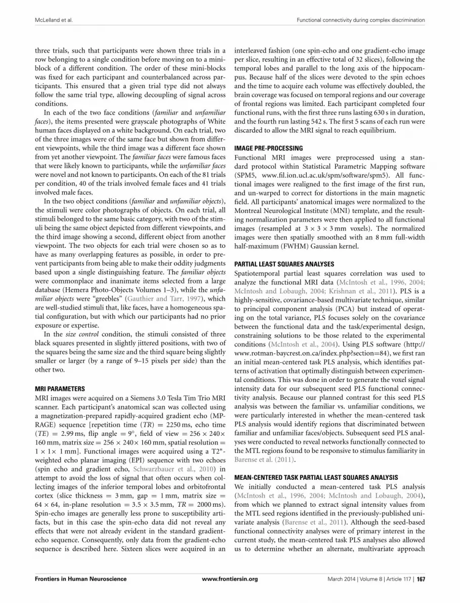
McLelland et al. Functional connectivity during complex discrimination
three trials, such that participants were shown three trials in arow belonging to a single condition before moving on to a mini-block of a different condition. The order of these mini-blockswas fixed for each participant and counterbalanced across par-ticipants. This ensured that a given trial type did not alwaysfollow the same trial type, allowing decoupling of signal acrossconditions.
In each of the two face conditions (familiar and unfamiliarfaces), the items presented were grayscale photographs of Whitehuman faces displayed on a white background. On each trial, twoof the three images were of the same face but shown from differ-ent viewpoints, while the third image was a different face shownfrom yet another viewpoint. The familiar faces were famous facesthat were likely known to participants, while the unfamiliar faceswere novel and not known to participants. On each of the 81 trialsper condition, 40 of the trials involved female faces and 41 trialsinvolved male faces.
In the two object conditions (familiar and unfamiliar objects),the stimuli were color photographs of objects. On each trial, allstimuli belonged to the same basic category, with two of the stim-uli being the same object depicted from different viewpoints, andthe third image showing a second, different object from anotherviewpoint. The two objects for each trial were chosen so as tohave as many overlapping features as possible, in order to pre-vent participants from being able to make their oddity judgmentsbased upon a single distinguishing feature. The familiar objectswere commonplace and inanimate items selected from a largedatabase (Hemera Photo-Objects Volumes 1–3), while the unfa-miliar objects were “greebles” (Gauthier and Tarr, 1997), whichare well-studied stimuli that, like faces, have a homogeneous spa-tial configuration, but with which our participants had no priorexposure or expertise.
In the size control condition, the stimuli consisted of threeblack squares presented in slightly jittered positions, with two ofthe squares being the same size and the third square being slightlysmaller or larger (by a range of 9–15 pixels per side) than theother two.
MRI PARAMETERSMRI images were acquired on a Siemens 3.0 Tesla Tim Trio MRIscanner. Each participant’s anatomical scan was collected usinga magnetization-prepared rapidly-acquired gradient echo (MP-RAGE) sequence [repetition time (TR) = 2250 ms, echo time(TE) = 2.99 ms, flip angle = 9◦, field of view = 256 × 240×160 mm, matrix size = 256 × 240× 160 mm, spatial resolution =1 × 1× 1 mm]. Functional images were acquired using a T2∗-weighted echo planar imaging (EPI) sequence with two echoes(spin echo and gradient echo, Schwarzbauer et al., 2010) inattempt to avoid the loss of signal that often occurs when col-lecting images of the inferior temporal lobes and orbitofrontalcortex (slice thickness = 3 mm, gap = 1 mm, matrix size =64 × 64, in-plane resolution = 3.5 × 3.5 mm, TR = 2000 ms).Spin-echo images are generally less prone to susceptibility arti-facts, but in this case the spin-echo data did not reveal anyeffects that were not already evident in the standard gradient-echo sequence. Consequently, only data from the gradient-echosequence is described here. Sixteen slices were acquired in an
interleaved fashion (one spin-echo and one gradient-echo imageper slice, resulting in an effective total of 32 slices), following thetemporal lobes and parallel to the long axis of the hippocam-pus. Because half of the slices were devoted to the spin echoesand the time to acquire each volume was effectively doubled, thebrain coverage was focused on temporal regions and our coverageof frontal regions was limited. Each participant completed fourfunctional runs, with the first three runs lasting 630 s in duration,and the fourth run lasting 542 s. The first 5 scans of each run werediscarded to allow the MRI signal to reach equilibrium.
IMAGE PRE-PROCESSINGFunctional MRI images were preprocessed using a stan-dard protocol within Statistical Parametric Mapping software(SPM5, www.fil.ion.ucl.ac.uk/spm/software/spm5). All func-tional images were realigned to the first image of the first run,and un-warped to correct for distortions in the main magneticfield. All participants’ anatomical images were normalized to theMontreal Neurological Institute (MNI) template, and the result-ing normalization parameters were then applied to all functionalimages (resampled at 3 × 3 × 3 mm voxels). The normalizedimages were then spatially smoothed with an 8 mm full-widthhalf-maximum (FWHM) Gaussian kernel.
PARTIAL LEAST SQUARES ANALYSESSpatiotemporal partial least squares correlation was used toanalyze the functional MRI data (McIntosh et al., 1996, 2004;McIntosh and Lobaugh, 2004; Krishnan et al., 2011). PLS is ahighly-sensitive, covariance-based multivariate technique, similarto principal component analysis (PCA) but instead of operat-ing on the total variance, PLS focuses solely on the covariancebetween the functional data and the task/experimental design,constraining solutions to be those related to the experimentalconditions (McIntosh et al., 2004). Using PLS software (http://www.rotman-baycrest.on.ca/index.php?section=84), we first ranan initial mean-centered task PLS analysis, which identifies pat-terns of activation that optimally distinguish between experimen-tal conditions. This was done in order to generate the voxel signalintensity data for our subsequent seed PLS functional connec-tivity analysis. Because our planned contrast for this seed PLSanalysis was between the familiar vs. unfamiliar conditions, wewere particularly interested in whether the mean-centered taskPLS analysis would identify regions that discriminated betweenfamiliar and unfamiliar faces/objects. Subsequent seed PLS anal-yses were conducted to reveal networks functionally connected tothe MTL regions found to be responsive to stimulus familiarity inBarense et al. (2011).
MEAN-CENTERED TASK PARTIAL LEAST SQUARES ANALYSISWe initially conducted a mean-centered task PLS analysis(McIntosh et al., 1996, 2004; McIntosh and Lobaugh, 2004),from which we planned to extract signal intensity values fromthe MTL seed regions identified in the previously-published uni-variate analysis (Barense et al., 2011). Although the seed-basedfunctional connectivity analyses were of primary interest in thecurrent study, the mean-centered task PLS analyses also allowedus to determine whether an alternate, multivariate approach
Frontiers in Human Neuroscience www.frontiersin.org March 2014 | Volume 8 | Article 117 | 167

McLelland et al. Functional connectivity during complex discrimination
would also implicate the MTLs in the perceptual discriminationof familiar stimuli, as described by Barense et al. (2011). Mean-centered task PLS is an exploratory, data-driven form of PLSin which no a priori contrasts or hypotheses are specified, anda series of latent variables (LVs) that optimally account for thecovariance between functional data and experimental conditionsare derived using singular value decomposition (McIntosh et al.,2004). These LVs, which are similar to eigenvectors in PCA, havethree components: a singular value, which describes the propor-tion of the covariance between the task and the functional dataaccounted for by that particular LV, voxel saliences, which iden-tify the distributed spatial pattern of brain voxels that is mostrelated to the effect characterized by that LV, and task saliences,which illustrate the extent to which each experimental conditionis associated with that pattern of voxels. Effects were assessedover a 16 s temporal window from the onset of each trial. Aswe were primarily interested in the differences between the fourexperimental conditions (familiar faces, unfamiliar faces, familiarobjects, and unfamiliar objects), as opposed to differences betweenthe experimental and control conditions, the size control condi-tion was excluded from these analyses. The functional MRI datawere transformed into a 72 × 282,440 matrix, in which the rowsrepresented each of the four conditions for each of the 18 par-ticipants, while the columns contained the signal values for everyvoxel at each of the eight 2 s time lags contained within the 16 stemporal window. Singular value decomposition was performedon a mean-centered, columnwise averaged form of this matrix,yielding the three components mentioned above.
The statistical significance of each LV was evaluated with500 permutation tests, calculated with a threshold of p < 0.05(McIntosh et al., 1996; McIntosh and Lobaugh, 2004). Each per-mutation test involves random reordering and reassignment ofevery participant’s data to the specified experimental conditions,determining the likelihood of each LV’s singular value occurringby chance. Additionally, the reliability of the voxel saliences (i.e.,the clusters of brain regions identified by each LV) was mea-sured by bootstrap estimation of their standard errors, entailingrandom sampling of participants with replacement 300 times,determining which voxel responses appear reliably (and thereforeare not heavily influenced by which participants are included inthe sample). Here, clusters with bootstrap ratios (BSR) of greaterthan ±3.5 were classified as reliable (A BSR is approximatelyequivalent to a z-score, and in this case corresponds to a p-valueof roughly 0.001; McIntosh and Lobaugh, 2004). As PLS analy-ses are conducted in a single analytic step, correction for multiplecomparisons is not required.
SEED PARTIAL LEAST SQUARES ANALYSESFollowing this preliminary mean-centered task PLS analysis, wethen used a “seed” PLS analysis (McIntosh et al., 2004) to examinethe functional connectivity of the four regions identified as beingsensitive to stimulus familiarity in Barense et al. (2011). This formof PLS can be used to assess functional connectivity by pinpoint-ing regions across the whole brain in which signal is correlatedwith that of user-specified seed regions of interest, and determin-ing how this connectivity changes across experimental conditions.In this case, the seed regions were the peak MNI coordinates
that had been identified in the univariate contrasts of the famil-iar vs. the unfamiliar conditions in Barense et al. (2011). Theseregions included the perirhinal cortex, the anterior hippocam-pus, the posterior hippocampus, and the temporal pole (one seedin each hemisphere). The voxel signal from each of these seedswas extracted from the mean-centered task PLS result using PLSsoftware’s multiple voxel extraction tool, centered on the follow-ing peak MNI coordinates identified by Barense et al. (2011):left perirhinal cortex (x, y, z = −33,−12,−27), left anterior hip-pocampus (x, y, z = −21,−9,−18), left posterior hippocam-pus (x, y, z = −33,−27,−15), left temporal pole (x, y, z = −36, 18,−27), right perirhinal cortex (x, y, z = 36,−9, −30),right anterior hippocampus (x, y, z = 27,−15,−18), right pos-terior hippocampus (x, y, z = 33,−33,−12), and right temporalpole (x, y, z = 63, 3,−18), averaging signal intensity across thetwo nearest neighboring voxels. Peak signal intensity values wereextracted for each voxel from lag 3 (6 s from trial onset) of themean-centered task analysis, as this timepoint corresponds to thetypical peak of the hemodynamic response function (cf. O’Neilet al., 2012).
These signal intensity values for each participant were enteredin matrix form as the behavioral values in a single non-rotatedseed PLS analysis. This 72 × 8 matrix contained four rows foreach of the 18 participants, and one column for each of the seedregions. Correlations were computed between this matrix and thematrix of functional MRI data containing all voxel signal valuesas described earlier for the mean-centered task PLS analysis. Theresulting correlation maps were stacked and again analyzed withsingular value decomposition. The non-rotated version of PLS,instead of being data-driven, constrains the possible solutions toallow for the explicit testing of hypotheses via specification of apriori contrasts. Non-rotated PLS analyses yield one LV corre-sponding to each specified contrast. Considering that we wereprimarily interested in the effects of stimulus familiarity, withinthis model we specified a contrast for each seed investigatingwhether the functional connectivity of that seed differed betweenthe familiar and unfamiliar conditions (irrespective of whetherthe stimuli were faces or objects). The significance of the LVsand reliability of the voxel saliences were evaluated in the samemanner as the mean-centered task analysis, with 500 permutationtests (p < 0.05) and 300 bootstrap estimations (BSR = ±3.5).Peak MNI coordinates from reliable clusters identified in both themean-centered and seed PLS analyses are reported in Tables 2–4,with anatomical labels assigned using the Automated AnatomicalLabeling atlas (Tzourio-Mazoyer et al., 2002). In all figures forboth the mean-centered and seed PLS analyses, conditions andbrain regions with positive saliences are always displayed in redand those with negative saliences are always displayed in blue.
RESULTSBEHAVIORAL RESULTSAccuracy and reaction time (RT) for each of the five condi-tions are shown in Table 1 (reproduced from Barense et al.,2011). A repeated-measures ANOVA indicated that there wasno main effect of stimulus familiarity on participants’ accuracyin the discrimination task, F(1, 17) = 0.90, p = 0.357, indicatingthat differences in functional connectivity between the familiar
Frontiers in Human Neuroscience www.frontiersin.org March 2014 | Volume 8 | Article 117 | 168

McLelland et al. Functional connectivity during complex discrimination
Table 1 | Mean accuracy and reaction time (correct trials only) for each condition (reproduced from Barense et al., 2011).
Familiar faces Unfamiliar faces Familiar objects Unfamiliar objects Size
Proportion correct 0.87 (0.07) 0.84 (0.07) 0.77 (0.07) 0.77 (0.09) 0.79 (0.10)
RT (ms) 2616 (415) 2796 (349) 3009 (448) 3300 (386) 2110 (366)
RT, reaction time; standard deviation values are shown in parentheses.
and unfamiliar conditions did not stem from factors related toaccuracy. However, there was a significant main effect of famil-iarity on RT, F(1, 17) = 41.47, p < 0.001, due to the fact that themean RT for the familiar conditions (M = 2813.13, SE = 96.56)was significantly faster than for the unfamiliar conditions (M =3048.00, SE = 80.50). Despite this effect, it is unlikely that thedifference in RT underlies any observed differences in functionalconnectivity between the familiar and unfamiliar conditions. Themean RT discrepancy of 234 ms is far shorter than a single TR;consequently, peak correlations between functionally interactingregions during both the familiar and unfamiliar conditions wouldstill fall within the same time lag in our PLS analyses.
MEAN-CENTERED TASK PLS ANALYSISThe mean-centered task PLS analysis yielded three significant LVs(see Table 2). The first LV explained 42.53% of the crossblockcovariance (p < 0.001), and differentiated between the face andnon-face conditions. Two negatively-correlated patterns of acti-vation were identified, one associated with both the familiar andunfamiliar objects conditions (shown in red in Figure 2A), andanother associated with the familiar faces condition (shown inblue in Figure 2A). The regions associated more highly with thefamiliar faces condition relative to the familiar and unfamiliarobjects conditions included bilateral clusters in the anterior hip-pocampus and anterior lateral temporal cortex, which are areasthat correspond well to the regions that responded to face famil-iarity from the univariate general linear model analysis in Barenseet al. (2011). The regions associated more with the two objectconditions relative to the familiar faces condition included largesections of occipital cortex and bilateral fusiform gyrus.
The second LV explained 33.08% of the crossblock covari-ance (p < 0.001) and appeared to primarily distinguish theunfamiliar objects condition from the familiar objects and unfa-miliar faces conditions. The regions associated more with theunfamiliar objects condition relative to the familiar objects andunfamiliar faces conditions included right inferior temporal andsuperior/middle occipital gyrus (shown in red in Figure 2B).The regions associated with the familiar objects and unfamil-iar faces conditions relative to the unfamiliar objects conditionincluded the right anterior hippocampus and bilateral insular cor-tex (shown in blue in Figure 2B). The familiar faces condition wasnot significantly associated with either of these two patterns ofactivation in LV 2.
Finally, the third LV was of most interest to the current study,as it differentiated between the conditions involving familiar andunfamiliar stimuli (explaining 24.39% of the crossblock covari-ance, p < 0.048). Specifically, this LV highlighted one patternof activation that was correlated more with the familiar facesand familiar objects conditions relative to the unfamiliar faces
condition (shown in blue in Figure 2C), and another correlatedwith the unfamiliar faces condition relative to the two familiarconditions (shown in red in Figure 2C). The unfamiliar objectscondition was not significantly associated with either of thesetwo negatively-correlated activation patterns. The regions asso-ciated with the familiar conditions consisted of strong bilateralactivation along the entire extent of the parahippocampal gyrus,extending into the anterior hippocampus, and also included thebilateral temporal poles. This activation corresponded well withthe MTL regions identified as being sensitive to stimulus famil-iarity in Barense et al. (2011). In contrast, the unfamiliar facescondition was associated primarily with bilateral medial parietalactivation.
SEED PLS ANALYSISFollowing this mean-centered task analysis, we conducted ournon-rotated seed PLS analysis with signal intensity valuesextracted from the mean-centered task PLS analysis for each seed.This analysis revealed that the functional connectivity of two ofthe eight seeds (the right perirhinal cortex and the right ante-rior hippocampus) was modulated by stimulus familiarity, suchthat these regions were functionally interacting with different net-works depending on whether the stimuli to be discriminated werefamiliar or unfamiliar. Specifically, the right perirhinal cortex seed(x, y, z = 36,−9,−30) displayed this pattern (16.43% of cross-block covariance, p < 0.016). Figure 3 illustrates that during thetwo familiar conditions (shown in red), signal in the right perirhi-nal cortex was highly correlated with a bilateral network includinglateral temporal, anterior temporal, and medial and lateral pari-etal cortex (see Table 3). In contrast, during the two unfamiliarconditions, the functional connectivity of this right perirhinalseed region was significantly different, and instead correlated withlarge sections of occipital cortex.
This same contrast of the familiar vs. unfamiliar conditionswas also significant in the right anterior hippocampal seed(x, y, z = 27,−15,−18, 15.38% of crossblock covariance, p <
0.036). Like the right perirhinal cortex, the right anterior hip-pocampus showed differential functional connectivity for thefamiliar vs. unfamiliar conditions, and many of the regions identi-fied in the two functionally-connected networks overlapped withthose found in the two networks showing connectivity with theright perirhinal cortex (see Table 4 and Figure 4). During thefamiliar conditions, the network associated with the right anteriorhippocampus included right anterior lateral temporal and lateralparietal cortex, cuneus and precuneus. For the unfamiliar con-ditions, the functionally-connected network consisted of regionssuch as bilateral occipital cortex and fusiform gyrus. Figure 5shows the time course of the correlation between signal in theright perirhinal cortex and right anterior hippocampal seeds
Frontiers in Human Neuroscience www.frontiersin.org March 2014 | Volume 8 | Article 117 | 169

McLelland et al. Functional connectivity during complex discrimination
Table 2 | Regions associated with the latent variables from the mean-centered task PLS analysis.
Latent variable Cluster size (k)* Brain region x y z BSR
1 FAMILIAR OBJECTS AND UNFAMILIAR OBJECTS > FAMILIAR FACES
1170 R Middle occipital gyrus 39 −75 21 10.101001 L Superior occipital gyrus −21 −81 36 9.37463 R Fusiform gyrus 33 −36 −15 8.27167 L Fusiform gyrus −27 −51 −12 4.9673 R Inferior frontal gyrus 33 24 −12 4.2344 L Thalamus −18 −27 6 4.2220 L Lingual gyrus −6 −69 −6 3.9416 R Insula 42 −12 6 3.9117 R Thalamus 12 −21 12 3.8611 R Insula 42 3 3 3.65
1 FAMILIAR FACES > FAMILIAR OBJECTS AND UNFAMILIAR OBJECTS
338 Precuneus/Posterior cingulate gyrus 0 −60 21 −7.04177 R Hippocampus 21 −9 −15 −6.70197 L Hippocampus/Parahippocampal gyrus −21 −6 −9 −6.52311 R Middle temporal gyrus 66 −48 12 −6.39101 R Middle temporal gyrus 60 −3 −21 −6.05109 L Middle temporal gyrus −60 −9 −18 −6.0252 L Middle occipital gyrus −45 −75 33 −5.6411 R Cerebellum 42 −45 −27 −4.1513 L Temporal pole −36 15 −24 −4.04
2 UNFAMILIAR OBJECTS > FAMILIAR OBJECTS AND UNFAMILIAR FACES
1268 R Inferior temporal gyrus 57 −63 −12 7.04673 L Middle temporal gyrus −45 −63 0 5.98277 R Superior occipital gyrus 27 −81 36 5.60140 L Superior occipital gyrus −21 −75 42 5.47
2 FAMILIAR OBJECTS AND UNFAMILIAR FACES > UNFAMILIAR OBJECTS
456 L Postcentral gyrus −51 −21 18 −6.40103 L Superior temporal gyrus −51 3 −6 −5.34456 R Insula 36 12 −9 −4.8577 L Hippocampus −33 −9 −24 −4.7772 R Middle temporal gyrus 54 −39 0 −4.4955 R Parahippocampal gyrus 30 −6 −27 −3.8630 R Putamen 24 12 −9 −3.6533 R Cuneus 9 −90 6 −3.5539 L Cuneus −3 −93 15 −3.55
3 UNFAMILIAR FACES > FAMILIAR FACES AND FAMILIAR OBJECTS
624 R Precuneus 6 −72 42 5.45
3 FAMILIAR FACES AND FAMILIAR OBJECTS > UNFAMILIAR FACES
636 R Parahippocampal gyrus 30 −33 −18 −6.44440 L Parahippocampal gyrus −27 −18 −21 −5.71170 L Cerebellum −6 −54 0 −5.30141 L Temporal pole −42 9 −30 −4.2857 R Inferior frontal gyrus 39 24 −15 −3.9193 R Middle occipital gyrus 27 −93 18 −3.68
Note: Only clusters evident during the peak timepoint (TR 3) with a bootstrap ratio of greater than +/−3.5 are reported. *Cluster size (k) indicates the number of
voxels comprising the cluster; only clusters with a minimum extent of 10 voxels are reported. BSR, Bootstrap ratio; LV, Latent Variable; L, left; R, right.
Frontiers in Human Neuroscience www.frontiersin.org March 2014 | Volume 8 | Article 117 | 170

McLelland et al. Functional connectivity during complex discrimination
FIGURE 2 | Latent variables identified in the mean-centered task PLSanalysis. On the left, voxel salience maps are shown. On the right, bargraphs for each of the three LVs illustrate the extent to which eachcondition corresponds to each of the positively-weighted (shown in red onbrain images) and negatively-weighted (shown in blue on brain images)networks. These networks, in the form of voxel salience maps for each LV,are all shown for TR 3, superimposed on the ch2bet template in MRIcron
(Rorden et al., 2007). (A) LV 1 highlighted the differences between theface and object conditions, (B) LV 2 highlighted the difference between theunfamiliar objects condition and the unfamiliar faces and familiar objectsconditions, and (C) LV 3 identified differences between the familiar andunfamiliar conditions. Maps are thresholded at the equivalent of p < 0.05for visualization purposes. BSR, bootstrap ratio. Error bars reflect 95%confidence intervals.
and a sample of the regions identified as functionally connectedduring the familiar conditions.
In these same two perirhinal and anterior hippocampalregions in the left hemisphere, this contrast did not reach
significance. Functional connectivity was not significantly mod-ulated by familiarity in either the left perirhinal cortex (x, y, z =−33,−12,−27, 11.14% of crossblock covariance, p < 0.317) orthe left anterior hippocampus (x, y, z = −21,−9,−18, 14.05%
Frontiers in Human Neuroscience www.frontiersin.org March 2014 | Volume 8 | Article 117 | 171

McLelland et al. Functional connectivity during complex discrimination
FIGURE 3 | Regions functionally connected with the right perirhinalcortex seed. A seed PLS analysis demonstrated that the functionalconnectivity of the right perirhinal cortex differed depending on stimulusfamiliarity. (A) Regions shown in red are those with which the right perirhinalcortex seed was functionally connected during the two familiar conditions inTR 3, whereas (B) displays regions shown in blue with which the perirhinal
seed was correlated during the two unfamiliar conditions in TR 3. The bargraph in (C) depicts the correlation of the signal in the perirhinal seed withthe two networks during the four experimental conditions. Maps arethresholded at the equivalent of p < 0.05 for visualization purposes, andnetworks are overlaid on the ch2bet template in MRIcron (Rorden et al.,2007). BSR, bootstrap ratio. Error bars reflect 95% confidence intervals.
Table 3 | Regions showing significant functional connectivity with the right perirhinal cortex seed.
Cluster size (k)* Brain region x y z BSR
REGIONS FUNCTIONALLY CONNECTED DURING THE FAMILIAR RELATIVE TO UNFAMILIAR CONDITIONS
442 R Superior temporal gyrus/Angular gyrus 60 −57 18 5.93
44 R Superior temporal gyrus 57 −30 12 5.24
56 R Inferior/Middle temporal gyrus 63 −18 −24 4.79
151 L Middle temporal gyrus −42 −51 12 4.66
54 R Superior temporal gyrus 51 −6 6 4.35
70 L Inferior temporal gyrus −60 −21 −21 4.34
241 L Cuneus 0 −69 30 4.05
86 R Temporal pole 42 3 −30 3.62
59 L Angular gyrus −42 −69 36 3.53
37 R Precuneus 18 −48 33 3.53
REGIONS FUNCTIONALLY CONNECTED DURING THE UNFAMILIAR RELATIVE TO FAMILIAR CONDITIONS
329 L Superior occipital gyrus −18 −81 42 −5.73
740 R Middle occipital gyrus 54 −69 −9 −4.51
31 L Putamen −27 3 −6 −4.47
Only clusters evident during the peak timepoint (TR 3) with a bootstrap ratio of greater than +/−3.5 are reported. *Cluster size (k) indicates the number of voxels
comprising the cluster; only clusters with a minimum extent of 10 voxels are reported. BSR, Bootstrap ratio; L, left; R, right.
of crossblock covariance, p < 0.078) seeds. Similarly, the contrastof connectivity for the familiar conditions vs. the unfamiliar con-ditions was not significant in either hemisphere for the other tworemaining seeds in the posterior hippocampus and temporal pole.
Permutation testing for the LVs in the right posterior hippocam-pus (x, y, z = 33,−33,−12, 8.41% of crossblock covariance, p <
0.884), left posterior hippocampus (x, y, z = −33,−27,−15,
12.73% of crossblock covariance, p < 0.158), right temporal
Frontiers in Human Neuroscience www.frontiersin.org March 2014 | Volume 8 | Article 117 | 172

McLelland et al. Functional connectivity during complex discrimination
Table 4 | Regions showing significant functional connectivity with the right anterior hippocampal seed.
Cluster size (k)* Brain region x y z BSR
REGIONS FUNCTIONALLY CONNECTED DURING THE FAMILIAR RELATIVE TO UNFAMILIAR CONDITIONS
382 L Cuneus 0 −81 15 4.94
177 R Superior temporal gyrus 60 −57 18 4.67
70 R Middle temporal gyrus 60 −9 −21 4.56
41 R Middle temporal gyrus 60 −39 −6 4.12
21 L Middle temporal gyrus −42 −51 15 3.82
15 L Inferior temporal gyrus −57 −24 −21 3.75
48 L Middle temporal gyrus −66 −51 0 3.63
REGIONS FUNCTIONALLY CONNECTED DURING THE UNFAMILIAR RELATIVE TO FAMILIAR CONDITIONS
1023 R Middle occipital gyrus 33 −84 6 −6.93
150 L Middle occipital gyrus −30 −81 9 −6.14
63 L Fusiform gyrus −48 −42 −15 −5.43
55 L Putamen −30 6 −6 −4.66
31 L Fusiform gyrus −36 −21 −24 −4.13
10 Parahippocampal gyrus −24 0 −33 −4.12
Only clusters evident during the peak timepoint (TR 3) with a bootstrap ratio of greater than +/−3.5 are reported. *Cluster size (k) indicates the number of voxels
comprising the cluster; only clusters with a minimum extent of 10 voxels are reported. BSR, Bootstrap ratio; L, left; R, right.
FIGURE 4 | Regions functionally connected with the right anteriorhippocampal seed. The functional connectivity of the right anteriorhippocampus also differed depending on stimulus familiarity. (A)Regions shown in red are those with which the right anteriorhippocampal seed was functionally connected during the two familiarconditions in TR 3. (B) Regions shown in blue are those with
which the seed was correlated during the two unfamiliar conditionsin TR 3. The bar graph in (C) depicts the correlation of the signalin the anterior hippocampal seed with the two networks during thefour experimental conditions. Maps are thresholded at the equivalentof p < 0.05 for visualization purposes. BSR, bootstrap ratio. Errorbars reflect 95% confidence intervals.
pole (x, y, z = 63, 3,−18, 8.94% of crossblock covariance, p <
0.828), and left temporal pole (x, y, z = −36, 18,−27, 12.92%of crossblock covariance, p < 0.152) indicated that the functionalconnectivity of these four seed regions did not differ dependingon stimulus familiarity.
DISCUSSIONThe aim of this study was to examine the functional connec-tivity of several MTL structures during a complex perceptualdiscrimination task. Specifically, we were interested in whetherMTL functional connectivity during the task would be affected
Frontiers in Human Neuroscience www.frontiersin.org March 2014 | Volume 8 | Article 117 | 173

McLelland et al. Functional connectivity during complex discrimination
FIGURE 5 | Time course of correlations between the right perirhinal cortexand anterior hippocampus and selected functionally-connected regions.Line graphs illustrate the correlations over time following stimulus onsetbetween signal in the seed regions and selected regions within theirfunctionally-connected networks. (A) Correlations are shown between theright perirhinal cortex seed and sample peak regions within its
functionally-connected network for the two familiar conditions. (B) Correlationsare shown between the anterior hippocampus seed and sample peak regionswithin its functionally-connected network for the two familiar conditions. Eachlag is equivalent to a TR, which in this case is 2 s in duration. Voxel saliencemaps are thresholded at the equivalent of p < 0.05 for visualization purposes,and are overlaid on the ch2bet template in MRIcron (Rorden et al., 2007).
by participants’ prior familiarity with the stimuli to be discrim-inated. The perirhinal cortex, anterior and posterior hippocam-pus, and temporal pole were previously shown to be more activeduring perceptual discrimination of familiar, relative to unfa-miliar, faces and objects (Barense et al., 2011). We anticipatedthat when participants discriminated between familiar stimuli,some or all of these areas would show increased connectivity withanterior temporal, lateral temporal, and inferior parietal regionsknown to be involved in semantic memory (Binder and Desai,2011), relative to when participants discriminated between novelor unfamiliar stimuli. Our findings indicate that the functionalconnectivity of the right perirhinal cortex and right anteriorhippocampus did in fact differ across familiar and unfamiliar con-ditions, while the connectivity of the left perirhinal cortex, leftanterior hippocampus, and bilateral posterior hippocampus andtemporal pole was unaffected by stimulus familiarity.
During the two familiar conditions, signal in the right perirhi-nal cortex covaried with signal in bilateral anterior portions ofthe middle and inferior temporal gyri, the right temporal pole,bilateral posterior aspects of the middle and superior tempo-ral gyri, and bilateral angular gyrus, cuneus and precuneus. Incontrast, during the unfamiliar conditions, the right perirhinalcortex instead showed connectivity with bilateral occipital cortex
(Figure 3). The pattern of differential functional connectivityexhibited by the right anterior hippocampus was nearly identi-cal to that of the right perirhinal cortex for both the familiar andunfamiliar conditions, though during the familiar conditions,the network associated with the right anterior hippocampus wasslightly more right-lateralized (Figure 4).
RELATIONSHIP OF OBSERVED FUNCTIONAL CONNECTIVITYTO ANATOMICAL CONNECTIVITYThe regions identified as being functionally connected to theperirhinal cortex and anterior hippocampus during perceptualdiscrimination correspond well with what is already known aboutthe anatomical connections of the MTL. Functional correla-tions between regions during resting states are thought to reflectintrinsic anatomical connections, and studies using such meth-ods have demonstrated that the perirhinal cortex and anteriorhippocampus have very similar intrinsic functional connectiv-ity, while the parahippocampal gyrus and posterior hippocampusshow functional correlations with a separate, more posterior net-work (Kahn et al., 2008; Ranganath and Ritchey, 2012). Giventhe similar resting-state connectivity of the perirhinal cortex andanterior hippocampus, it therefore is not surprising that the task-related functional connectivity of these two regions was similarly
Frontiers in Human Neuroscience www.frontiersin.org March 2014 | Volume 8 | Article 117 | 174
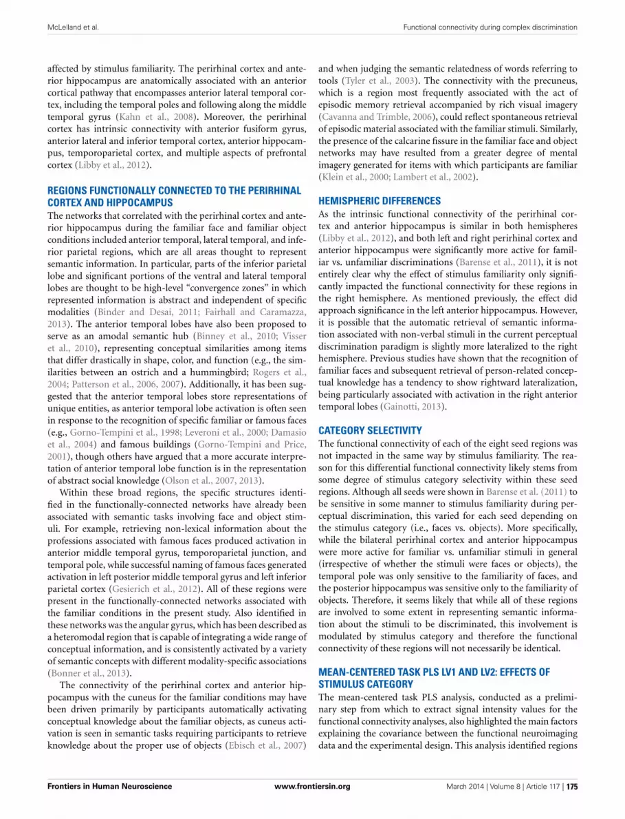
McLelland et al. Functional connectivity during complex discrimination
affected by stimulus familiarity. The perirhinal cortex and ante-rior hippocampus are anatomically associated with an anteriorcortical pathway that encompasses anterior lateral temporal cor-tex, including the temporal poles and following along the middletemporal gyrus (Kahn et al., 2008). Moreover, the perirhinalcortex has intrinsic connectivity with anterior fusiform gyrus,anterior lateral and inferior temporal cortex, anterior hippocam-pus, temporoparietal cortex, and multiple aspects of prefrontalcortex (Libby et al., 2012).
REGIONS FUNCTIONALLY CONNECTED TO THE PERIRHINALCORTEX AND HIPPOCAMPUSThe networks that correlated with the perirhinal cortex and ante-rior hippocampus during the familiar face and familiar objectconditions included anterior temporal, lateral temporal, and infe-rior parietal regions, which are all areas thought to representsemantic information. In particular, parts of the inferior parietallobe and significant portions of the ventral and lateral temporallobes are thought to be high-level “convergence zones” in whichrepresented information is abstract and independent of specificmodalities (Binder and Desai, 2011; Fairhall and Caramazza,2013). The anterior temporal lobes have also been proposed toserve as an amodal semantic hub (Binney et al., 2010; Visseret al., 2010), representing conceptual similarities among itemsthat differ drastically in shape, color, and function (e.g., the sim-ilarities between an ostrich and a hummingbird; Rogers et al.,2004; Patterson et al., 2006, 2007). Additionally, it has been sug-gested that the anterior temporal lobes store representations ofunique entities, as anterior temporal lobe activation is often seenin response to the recognition of specific familiar or famous faces(e.g., Gorno-Tempini et al., 1998; Leveroni et al., 2000; Damasioet al., 2004) and famous buildings (Gorno-Tempini and Price,2001), though others have argued that a more accurate interpre-tation of anterior temporal lobe function is in the representationof abstract social knowledge (Olson et al., 2007, 2013).
Within these broad regions, the specific structures identi-fied in the functionally-connected networks have already beenassociated with semantic tasks involving face and object stim-uli. For example, retrieving non-lexical information about theprofessions associated with famous faces produced activation inanterior middle temporal gyrus, temporoparietal junction, andtemporal pole, while successful naming of famous faces generatedactivation in left posterior middle temporal gyrus and left inferiorparietal cortex (Gesierich et al., 2012). All of these regions werepresent in the functionally-connected networks associated withthe familiar conditions in the present study. Also identified inthese networks was the angular gyrus, which has been described asa heteromodal region that is capable of integrating a wide range ofconceptual information, and is consistently activated by a varietyof semantic concepts with different modality-specific associations(Bonner et al., 2013).
The connectivity of the perirhinal cortex and anterior hip-pocampus with the cuneus for the familiar conditions may havebeen driven primarily by participants automatically activatingconceptual knowledge about the familiar objects, as cuneus acti-vation is seen in semantic tasks requiring participants to retrieveknowledge about the proper use of objects (Ebisch et al., 2007)
and when judging the semantic relatedness of words referring totools (Tyler et al., 2003). The connectivity with the precuneus,which is a region most frequently associated with the act ofepisodic memory retrieval accompanied by rich visual imagery(Cavanna and Trimble, 2006), could reflect spontaneous retrievalof episodic material associated with the familiar stimuli. Similarly,the presence of the calcarine fissure in the familiar face and objectnetworks may have resulted from a greater degree of mentalimagery generated for items with which participants are familiar(Klein et al., 2000; Lambert et al., 2002).
HEMISPHERIC DIFFERENCESAs the intrinsic functional connectivity of the perirhinal cor-tex and anterior hippocampus is similar in both hemispheres(Libby et al., 2012), and both left and right perirhinal cortex andanterior hippocampus were significantly more active for famil-iar vs. unfamiliar discriminations (Barense et al., 2011), it is notentirely clear why the effect of stimulus familiarity only signifi-cantly impacted the functional connectivity for these regions inthe right hemisphere. As mentioned previously, the effect didapproach significance in the left anterior hippocampus. However,it is possible that the automatic retrieval of semantic informa-tion associated with non-verbal stimuli in the current perceptualdiscrimination paradigm is slightly more lateralized to the righthemisphere. Previous studies have shown that the recognition offamiliar faces and subsequent retrieval of person-related concep-tual knowledge has a tendency to show rightward lateralization,being particularly associated with activation in the right anteriortemporal lobes (Gainotti, 2013).
CATEGORY SELECTIVITYThe functional connectivity of each of the eight seed regions wasnot impacted in the same way by stimulus familiarity. The rea-son for this differential functional connectivity likely stems fromsome degree of stimulus category selectivity within these seedregions. Although all seeds were shown in Barense et al. (2011) tobe sensitive in some manner to stimulus familiarity during per-ceptual discrimination, this varied for each seed depending onthe stimulus category (i.e., faces vs. objects). More specifically,while the bilateral perirhinal cortex and anterior hippocampuswere more active for familiar vs. unfamiliar stimuli in general(irrespective of whether the stimuli were faces or objects), thetemporal pole was only sensitive to the familiarity of faces, andthe posterior hippocampus was sensitive only to the familiarity ofobjects. Therefore, it seems likely that while all of these regionsare involved to some extent in representing semantic informa-tion about the stimuli to be discriminated, this involvement ismodulated by stimulus category and therefore the functionalconnectivity of these regions will not necessarily be identical.
MEAN-CENTERED TASK PLS LV1 AND LV2: EFFECTS OFSTIMULUS CATEGORYThe mean-centered task PLS analysis, conducted as a prelimi-nary step from which to extract signal intensity values for thefunctional connectivity analyses, also highlighted the main factorsexplaining the covariance between the functional neuroimagingdata and the experimental design. This analysis identified regions
Frontiers in Human Neuroscience www.frontiersin.org March 2014 | Volume 8 | Article 117 | 175

McLelland et al. Functional connectivity during complex discrimination
associated with optimal combinations of the familiar faces, unfa-miliar faces, familiar objects, and unfamiliar objects conditions.The first two latent variables primarily distinguished betweenspecific stimulus categories irrespective of their familiarity. LV1highlighted differences between the face and object conditions,and the existence of such differences is unsurprising given thenumerous dissimilarities between faces and objects. LV2 dis-tinguished the unfamiliar objects (greebles) condition from theremaining three conditions involving more everyday items, sug-gesting that there is a fundamental difference between discrimina-tions involving a completely novel type of stimulus that has neverbeen encountered before compared to discriminations involvingstimuli to which participants have had some exposure—whetherit be to the stimulus category in general (e.g., faces in gen-eral) or to the actual exemplars themselves (e.g., specific famousfaces).
In contrast to these first two LVs, LV3 revealed differencesin neural representation that resulted from the varying levels ofstimulus familiarity, irrespective of stimulus type, which was ourmain a priori factor of interest. The relative importance of thesethree LVs indicate that while differences among stimulus cate-gories account for a greater degree of covariance than differencesin stimulus familiarity, both of these constructs make significantand simultaneous contributions.
MEAN-CENTERED TASK PLS LV3: EFFECTS OF STIMULUSFAMILIARITYThe focus of the current study was on the familiarity-relatedeffects identified in the third LV. Nevertheless, some of the regionsidentified in the first latent variable were also relevant to this issue.The first LV identified the anterior hippocampus and anteriorlateral temporal cortex as being particularly involved in makingperceptual discriminations among familiar faces, relative to dis-criminations among familiar objects. The fact that this activationwas located more anteriorly in the temporal lobes supports thefindings of previous studies demonstrating some degree of stim-ulus selectivity along the longitudinal axis of the MTL, such thatanterior regions are more content-general, responding to face,object, and some scene stimuli, while posterior regions respondselectively to scenes (Litman et al., 2009; Liang et al., 2012).Moreover, the anterior hippocampal and anterior lateral tempo-ral activation seen in response to familiar faces corroborates thefindings of Trinkler et al. (2009), who found that activation inthe anterior hippocampus and anterior middle temporal gyruswas associated with greater pre-experimental knowledge aboutface stimuli, and that anterior hippocampal activation increasedwith the degree to which the faces were personally-known toparticipants. The anterior hippocampal activation seen for thefamiliar faces condition in the present study may therefore reflectthe retrieval of episodic autobiographical information associatedwith the famous individuals whose faces were presented. Whilethe current implication of the anterior hippocampus in percep-tual discrimination of familiar faces is consistent with previousfindings (e.g., Lee et al., 2008), it is at odds with a recent sug-gestion that a bias toward pattern completion processes in theanterior hippocampus renders it of no use for fine perceptual dis-crimination tasks (Poppenk et al., 2013). The perirhinal cortex
alone does in fact appear to be capable of making such discrim-inations among object and face stimuli (Barense et al., 2010a),and the involvement of the anterior hippocampus may thereforesimply stem from its strong connectivity with the perirhinal cor-tex (Kahn et al., 2008; Libby et al., 2012). However, the repeatedimplication of the anterior hippocampus in the perceptual dis-crimination of faces suggests the possibility of an additional,as yet unspecified, role for the anterior hippocampus in suchtasks.
The third latent variable implicated large sections of the MTLin perceptual discriminations of familiar stimuli in general. Forthe combination of the familiar faces and familiar objects condi-tions, activation was localized bilaterally along the entire extentof the parahippocampal gyrus, extending into the anterior hip-pocampus, perirhinal cortex, and slightly into the posterior hip-pocampus, as well as in the temporal poles. These regions aregenerally consistent with those identified as showing greater activ-ity during the familiar vs. unfamiliar conditions in the univariategeneral linear model (GLM) used by Barense et al. (2011), sug-gesting that the multivariate PLS and univariate GLM methodsdetected similar effects. Widespread involvement of the MTL inperceptual discrimination offers support for the proposition thataspects of this region serve as an extension of a representationalhierarchy in the ventral visual stream (Cowell et al., 2010; Leeet al., 2012). Moreover, the observed sensitivity of the MTL tostimulus familiarity in perceptual discrimination suggests thatthese regions also represent semantic information (Murray andBussey, 1999; Taylor et al., 2006; Barense et al., 2011).
IMPLICATIONS FOR SEMANTIC DEMENTIA AND MTLAMNESIAThe current findings offer insight into an intriguing and pre-viously unexplained pattern of results from Barense et al.(2010b). As mentioned previously, this study found that twopatient groups—amnesics with non-progressive MTL damage(“MTL amnesics”) and patients with semantic dementia (“SDpatients”)—were both impaired relative to controls at perceptualdiscriminations of complex and visually similar stimuli. However,only the controls and MTL amnesics showed a benefit fromstimulus familiarity when making such discriminations. By con-trast, the SD patients failed to show this facilitation from theuse of familiar stimuli, likely because they were unable to engagesupport from their impaired semantic system. Nonetheless,the neuroanatomical correlates of these behavioral differenceswere unclear. The two patient groups had largely overlappingMTL damage, particularly in the perirhinal cortex. Additionally,although Barense et al. (2011) illustrated that multiple MTLsubregions were sensitive to stimulus familiarity during percep-tual discrimination, none of these regions were clearly moredamaged in the SD patients than the MTL amnesics. The cur-rent results suggest that even though the ability to discriminatebetween items with overlapping features depends heavily uponthe perirhinal cortex, this structure (and those structures withwhich it is closely connected, such as the anterior hippocam-pus) receives relevant input from other functionally-connectedregions depending on the familiarity of the stimuli to be dis-criminated. Both the SD patients and the MTL amnesics had
Frontiers in Human Neuroscience www.frontiersin.org March 2014 | Volume 8 | Article 117 | 176

McLelland et al. Functional connectivity during complex discrimination
damage to the perirhinal cortex, which impaired their discrim-ination performance, but the SD patients’ additional damageto anterior and lateral temporal cortex, identified here as beingfunctionally connected with the perirhinal cortex and anteriorhippocampus for familiar stimuli, may have prevented them frombenefitting from the support provided by intact access to semanticmemory.
CONCLUSIONSIn summary, during a perceptual discrimination task in whichparticipants selected the odd-one-out from a set of three complexfaces or objects with many shared visual features, the functionalconnectivity of the right perirhinal cortex and right anterior hip-pocampus was significantly modulated by the degree to whichparticipants were familiar with the stimuli being discriminated.This was the case despite the fact that the task did not explic-itly require the retrieval of any conceptual information about theitems to be discriminated and could be completed without draw-ing upon semantic memory. For familiar relative to unfamiliarfaces and objects, both the right perirhinal cortex and right ante-rior hippocampus showed enhanced functional correlations witha network of regions associated with semantic knowledge. Thesefindings illustrate that experience and expertise with particularclasses of objects influences not only the location of their neu-ral representation (McKeeff et al., 2010; McGugin et al., 2012),but also the functional interactions of these representations withbroader whole-brain networks. The results have potential impli-cations for semantic dementia patients, as the results suggest thatit was the patients’ inability to engage a network of regions includ-ing the lateral temporal cortex and the temporal pole, as opposedto localized damage in the perirhinal cortex or hippocampus, thatimpaired their ability to benefit from the use of familiar stim-uli in non-semantic perceptual tasks (Binder and Desai, 2011).Future research in semantic dementia showing diminished inter-actions between perirhinal cortex, anterior hippocampus, andanterior/lateral temporal regions relative to normal controls dur-ing perceptual discrimination tasks will confirm this hypothesis.
AUTHOR CONTRIBUTIONSMorgan D. Barense was responsible for the original experimentaldesign, programming, and fMRI data collection. The data analy-sis was completed by Victoria C. McLelland with assistance fromDavid Chan and Morgan D. Barense. Victoria C. McLelland inter-preted the results and wrote the paper with input from Morgan D.Barense, Susanne Ferber, and David Chan.
ACKNOWLEDGMENTSMorgan D. Barense and Susanne Ferber are supported byDiscovery Grants from the Natural Sciences and EngineeringResearch Council of Canada and operating grants from theCanadian Institutes of Health Research. We thank RichardHenson and Kim Graham for support of the original univari-ate fMRI study, which was funded by the UK Medical ResearchCouncil (WBSE U.1055.05.012.00001.01). We also thank MichaelJ. Tarr (Carnegie Mellon University) for providing the greeblestimuli.
REFERENCESAnaki, D., and Bentin, S. (2009). Familiarity effects on categorization levels of faces
and objects. Cognition 111, 144–149. doi: 10.1016/j.cognition.2009.01.002Barense, M. D., Bussey, T. J., Lee, A. C. H., Rogers, T. T., Rhys Davies, R., Saksida, L.
M., et al. (2005). Functional specialization in the human medial temporal lobe.J. Neurosci. 25, 10239–10246. doi: 10.1523/JNEUROSCI.2704-05.2005
Barense, M. D., Gaffan, D., and Graham, K. S. (2007). The human medial temporallobe processes online representations of complex objects. Neuropsychologia 45,2963–2974. doi: 10.1016/j.neuropsychologia.2007.05.023
Barense, M. D., Henson, R. N., and Graham, K. S. (2011). Perception andconception: temporal lobe activity during complex discriminations of famil-iar and novel faces and objects. J. Cogn. Neurosci. 23, 3052–3067. doi:10.1162/jocn_a_00010
Barense, M. D., Henson, R. N., Lee, A. C. H., and Graham, K. S. (2010a). Medialtemporal lobe activity during complex discrimination of faces, objects, andscenes: effects of viewpoint. Hippocampus 20, 389–401. doi: 10.1002/hipo.20641
Barense, M. D., Rogers, T. T., Bussey, T. J., Saksida, L. M., and Graham, K.S. (2010b). Influence of conceptual knowledge on visual object discrimina-tion: insights from semantic dementia and MTL amnesia. Cereb. Cortex 20,2568–2582. doi: 10.1093/cercor/bhq004
Barense, M. D., Ngo, J. K. W., Hung, L. H. T., and Peterson, M. A. (2012).Interactions of memory and perception in amnesia: the figure-ground perspec-tive. Cereb. Cortex 22, 2680–2691. doi: 10.1093/cercor/bhr347
Barsics, C., and Brédart, S. (2012). Recalling semantic informationabout newly learned faces and voices. Memory 20, 527–534. doi:10.1080/09658211.2012.683012
Bartko, S. J., Winters, B. D., Cowell, R. A., Saksida, L. M., and Bussey, T. J. (2007).Perceptual functions of perirhinal cortex in rats: zero-delay object recogni-tion and simultaneous oddity discriminations. J. Neurosci. 27, 2548–2559. doi:10.1523/JNEUROSCI.5171-06.2007
Binder, J. R., and Desai, R. H. (2011). The neurobiology of semantic memory.Trends Cogn. Sci. 15, 527–536. doi: 10.1016/j.tics.2011.10.001
Binney, R. J., Embleton, K. V., Jefferies, E., Parker, G. J., and Lambon Ralph, M.A. (2010). The ventral and inferolateral aspects of the anterior temporal lobeare crucial in semantic memory: evidence from a novel direct comparison ofdistortion-corrected fMRI, rTMS, and semantic dementia. Cereb. Cortex 20,2728–2738. doi: 10.1093/cercor/bhq019
Bonner, M. F., Peelle, J. E., Cook, P. A., and Grossman, M. (2013). Heteromodalconceptual processing in the angular gyrus. Neuroimage 71, 175–186. doi:10.1016/j.neuroimage.2013.01.006
Bruce, V., and Young, A. (1986). Understanding face recognition. Br. J. Psychol. 77,305–327. doi: 10.1111/j.2044-8295.1986.tb02199.x
Bülthoff, I., and Newell, F. N. (2006). The role of familiarity in the recognition ofstatic and dynamic objects. Prog. Brain Res. 154A, 315–325. doi: 10.1016/S0079-6123(06)54017-8
Bussey, T. J., and Saksida, L. M. (2002). The organization of visual object represen-tations: a connectionist model of effects of lesions in perirhinal cortex. Eur. J.Neurosci. 15, 355–364. doi: 10.1046/j.0953-816x.2001.01850.x
Bussey, T. J., and Saksida, L. M. (2007). Memory, perception, and the ventral visual-perirhinal-hippocampal stream: thinking outside of the boxes. Hippocampus 17,898–908. doi: 10.1002/hipo.20320
Cavanna, A. E., and Trimble, M. R. (2006). The precuneus: a review of itsfunctional anatomy and behavioural correlates. Brain 129, 564–583. doi:10.1093/brain/awl004
Chan, A. M., Baker, J. M., Eskandar, E., Schomer, D., Ulbert, I., Marinkovic, K., et al.(2011). First-pass selectivity for semantic categories in human anteroventraltemporal lobe. J. Neurosci. 31, 18119–18129. doi: 10.1523/JNEUROSCI.3122-11.2011
Cowell, R. A., Bussey, T. J., and Saksida, L. M. (2010). Functional dissociationswithin the ventral object processing pathway: cognitive modules or a hierarchi-cal continuum? J. Cogn. Neurosci. 22, 2460–2479. doi: 10.1162/jocn.2009.21373
Damasio, H., Tranel, D., Grabowski, T., Adolphs, R., and Damasio, A. (2004).Neural systems behind word and concept retrieval. Cognition 92, 179–229. doi:10.1016/j.cognition.2002.07.001
Davies, R. R., Graham, K. S., Xuereb, J. H., Williams, G. B., and Hodges, J. R.(2004). The human perirhinal cortex and semantic memory. Eur. J. Neurosci.20, 2441–2446. doi: 10.1111/j.1460-9568.2004.03710.x
Devlin, J. T., and Price, C. J. (2007). Perirhinal contributions to human visualperception. Curr. Biol. 17, 1484–1488. doi: 10.1016/j.cub.2007.07.066
Frontiers in Human Neuroscience www.frontiersin.org March 2014 | Volume 8 | Article 117 | 177

McLelland et al. Functional connectivity during complex discrimination
Ebisch, S. J. H., Babiloni, C., Del Gratta, C., Ferretti, A., Perrucci, M. G., Caulo,M., et al. (2007). Human neural systems for conceptual knowledge of properobject use: a functional magnetic resonance imaging study. Cereb. Cortex 17,2744–2751. doi: 10.1093/cercor/bhm001
Fairhall, S. L., and Caramazza, A. (2013). Brain regions that repre-sent amodal conceptual knowledge. J. Neurosci. 33, 10552–10558. doi:10.1523/JNEUROSCI.0051-13.2013
Friston, K. J. (2011). Functional and effective connectivity: a review. Brain Connect.1, 13–36. doi: 10.1089/brain.2011.0008
Gainotti, G. (2013). Laterality effects in normal subjects’ recognition offamiliar faces, voices, and names. Perceptual and representational compo-nents. Neuropsychologia 51, 1151–1160. doi: 10.1016/j.neuropsychologia.2013.03.009
Gauthier, I., and Tarr, M. J. (1997). Becoming a greeble expert: exploring mech-anisms for face recognition. Vision Res. 37, 1673–1682. doi: 10.1016/S0042-6989(96)00286-6
Gesierich, B., Jovicich, J., Riello, M., Adriani, M., Monti, A., Brentari, V., et al.(2012). Distinct neural substrates for semantic knowledge and naming inthe temporoparietal network. Cereb. Cortex 22, 2217–2226. doi: 10.1093/cer-cor/bhr286
Gorno-Tempini, M. L., and Price, C. J. (2001). Identification of famous faces andbuildings: a functional neuroimaging study of semantically unique items. Brain124, 2087–2097. doi: 10.1093/brain/124.10.2087
Gorno-Tempini, M. L., Price, C. J., Josephs, O., Vandenberghe, R., Cappa, S. F.,Kapur, N., et al. (1998). The neural systems sustaining face and proper-nameprocessing. Brain 121(pt 11), 2103–2118. doi: 10.1093/brain/121.11.2103
Graham, K. S., Barense, M. D., and Lee, A. C. H. (2010). Going beyond LTM in theMTL: a synthesis of neuropsychological and neuroimaging findings on the roleof the medial temporal lobe in memory and perception. Neuropsychologia 48,831–853. doi: 10.1016/j.neuropsychologia.2010.01.001
Hodges, J. R., Patterson, K., Oxbury, S., and Funnell, E. (1992). Semantic dementia:progressive fluent aphasia with temporal-lobe atrophy. Brain 115, 1783–1806.doi: 10.1093/brain/115.6.1783
Jung, K., Ruthruff, E., and Gaspelin, N. (2013). Automatic identification of famil-iar faces. Atten. Percept. Psychophys. 75, 1438–1450. doi: 10.3758/s13414-013-0468-3
Kahn, I., Andrews-Hanna, J. R., Vincent, J. L., Snyder, A. Z., and Buckner, R. L.(2008). Distinct cortical anatomy linked to subregions of the medial temporallobe revealed by intrinsic functional connectivity. J. Neurophysiol. 100, 129–139.doi: 10.1152/jn.00077.2008
Klein, I., Paradis, A.-L., Poline, J.-B., Kosslyn, S. M., and Le Bihan, D.(2000). Transient activity in the human calcarine cortex during visual-mentalimagery: an event-related fMRI study. J. Cogn. Neurosci. 12, 15–23. doi:10.1162/089892900564037
Krishnan, A., Williams, L. J., McIntosh, A. R., and Abdi, H. (2011). Partial LeastSquares (PLS) methods for neuroimaging: a tutorial and review. Neuroimage56, 455–475. doi: 10.1016/j.neuroimage.2010.07.034
Lambert, S., Sampaio, E., Schieber, C., and Mauss, Y. (2002). Neural substratesof animal mental imagery: calcarine sulcus and dorsal pathway involvement- an fMRI study. Brain Res. 2002, 176–183. doi: 10.1016/S0006-8993(01)03232-2
Lee, A. C. H., Buckley, M. J., Pegman, S. J., Spiers, H., Scahill, V. L., Gaffan, D., et al.(2005a). Specialization in the medial temporal lobe for processing of objects andscenes. Hippocampus 15, 782–797. doi: 10.1002/hipo.20101
Lee, A. C. H., Bussey, T. J., Murray, E. A., Saksida, L. M., Epstein, R.,Kapur, N., et al. (2005b). Perceptual deficits in amnesia: challenging themedial temporal lobe mnemonic view. Neuropsychologia 43, 1–11. doi:10.1016/j.neuropsychologia.2004.07.017
Lee, A. C. H., Scahill, V. L., and Graham, K. S. (2008). Activating the medial tempo-ral lobe during oddity judgment for faces and scenes. Cereb. Cortex 18, 683–696.doi: 10.1093/cercor/bhm104
Lee, A. C. H., Yeung, L. K., and Barense, M. D. (2012). The hippocampus and visualperception. Front. Hum. Neurosci. 17:91. doi: 10.3389/fnhum.2012.00091
Leveroni, C. L., Seidenberg, M., Mayer, A. R., Mead, L. A., Binder, J. R., and Rao,S. M. (2000). Neural systems underlying the recognition of familiar and newlylearned faces. J. Neurosci. 20, 878–886.
Levy, D. A., Bayley, P. J., and Squire, L. R. (2004). The anatomy of semanticknowledge: medial vs. lateral temporal lobe. Proc. Natl. Acad. Sci. U.S.A. 101,6710–6715. doi: 10.1073/pnas.0401679101
Liang, J. C., Wagner, A. D., and Preston, A. R. (2012). Content representation inthe human medial temporal lobe. Cereb. Cortex 23, 80–96. doi: 10.1093/cer-cor/bhr379
Libby, L. A., Ekstrom, A. D., Ragland, J. D., and Ranganath, C. (2012).Differential connectivity of perirhinal and parahippocampal cortices withinhuman hippocampal subregions revealed by high-resolution functionalimaging. J. Neurosci. 32, 6550–6560. doi: 10.1523/JNEUROSCI.3711-11.2012
Litman, L., Awipi, T., and Davachi, L. (2009). Category-specificity in thehuman medial temporal lobe cortex. Hippocampus 19, 308–319. doi:10.1002/hipo.20515
Martin, V. C., Schacter, D. L., Corballis, M. C., and Addis, D. R. (2011). A role forthe hippocampus in encoding simulations of future events. Proc. Natl. Acad. Sci.U.S.A. 108, 13858–13863. doi: 10.1073/pnas.1105816108
McGugin, R. W., Gatenby, J. C., Gore, J. C., and Gauthier, I. (2012). High-resolution imaging of expertise reveals reliable object selectivity in the fusiformface area related to perceptual performance. Proc. Natl. Acad. Sci. U.S.A. 109,17063–17068. doi: 10.1073/pnas.1116333109
McIntosh, A. R., Bookstein, F. L., Haxby, J. V., and Grady, C. L. (1996). Spatial pat-tern analysis of functional brain images using partial least squares. Neuroimage3, 143–157. doi: 10.1006/nimg.1996.0016
McIntosh, A. R., Chau, W. K., and Protzner, A. B. (2004). Spatiotemporal analysisof event-related fMRI data using partial least squares. Neuroimage 23, 764–775.doi: 10.1016/j.neuroimage.2004.05.018
McIntosh, A. R., and Lobaugh, N. J. (2004). Partial least squares analysis of neu-roimaging data: applications and advances. Neuroimage 23, S250–S263. doi:10.1016/j.neuroimage.2004.07.020
McKeeff, T. J., McGugin, R. W., Tong, F., and Gauthier, I. (2010). Expertise increasesthe functional overlap between face and object perception. Cognition 117,355–360. doi: 10.1016/j.cognition.2010.09.002
Moss, H. E., Rodd, J. M., Stamatakis, E. A., Bright, P., and Tyler, L. K. (2005).Anteromedial temporal cortex supports fine-grained differentiation amongobjects. Cereb. Cortex 15, 616–627. doi: 10.1093/cercor/bhh163
Murray, E. A., and Bussey, T. J. (1999). Perceptual-mnemonic functions ofthe perirhinal cortex. Trends Cogn. Sci. 3, 142–151. doi: 10.1016/S1364-6613(99)01303-0
Olson, I. R., McCoy, D., Klobusicky, E., and Ross, L. A. (2013). Social cognitionand the anterior temporal lobes: a review and theoretical framework. Soc. Cogn.Affect. Neurosci. 8, 123–133. doi: 10.1093/scan/nss119
Olson, I. R., Plotzker, A., and Ezzyat, Y. (2007). The enigmatic temporal pole: areview of findings on social and emotional processing. Brain 130, 1718–1731.doi: 10.1093/brain/awm052
O’Neil, E. B., Cate, A. D., and Kohler, S. (2009). Perirhinal cortex contributes toaccuracy in recognition memory and perceptual discriminations. J. Neurosci.29, 8329–8334. doi: 10.1523/JNEUROSCI.0374-09.2009
O’Neil, E. B., Protzner, A. B., McCormick, C., McLean, D. A., Poppenk, J., Cate,A. D., et al. (2012). Distinct patterns of functional and effective connectivitybetween perirhinal cortex and other cortical regions in recognition memoryand perceptual discrimination. Cereb. Cortex 22, 74–85. doi: 10.1093/cercor/bhr075
Patterson, K., Lambon Ralph, M. A., Jefferies, E., Woollams, A., Jones, R.,Hodges, J. R., et al. (2006). Presemantic cognition in semantic dementia:six deficits in search of an explanation. J. Cogn. Neurosci. 18, 169–183. doi:10.1162/jocn.2006.18.2.169
Patterson, K., Nestor, P. J., and Rogers, T. T. (2007). Where do you know what youknow? The representation of semantic knowledge in the human brain. Nat. Rev.Neurosci. 8, 976–987. doi: 10.1038/nrn2277
Peterson, M. A., Cacciamani, L., Barense, M. D., and Scalf, P. E. (2012). Theperirhinal cortex modulates V2 activity in response to the agreement betweenpart familiarity and configuration familiarity. Hippocampus 22, 1965–1977. doi:10.1002/hipo.22065
Poppenk, J., Evensmoen, H. R., Moscovitch, M., and Nadel, L. (2013). Long-axisspecialization of the human hippocampus. Trends Cogn. Sci. 17, 230–240. doi:10.1016/j.tics.2013.03.005
Ranganath, C., and Ritchey, M. (2012). Two cortical systems for memory-guidedbehaviour. Nat. Rev. Neurosci. 13, 713–726. doi: 10.1038/nrn3338
Rogers, B. P., Morgan, V. L., Newton, A. T., and Gore, J. C. (2007). Assessing func-tional connectivity in the human brain by fMRI. Magn. Reson. Imaging 25,1347–1357. doi: 10.1016/j.mri.2007.03.007
Frontiers in Human Neuroscience www.frontiersin.org March 2014 | Volume 8 | Article 117 | 178
McLelland et al. Functional connectivity during complex discrimination
Rogers, T. T., Lambon Ralph, M. A., Garrard, P., Bozeat, S., McClelland, J. L.,Hodges, J. R., et al. (2004). Structure and deterioration of semantic mem-ory: a neuropsychological and computational investigation. Psychol. Rev. 111,205–235. doi: 10.1037/0033-295X.111.1.205
Rorden, C., Karnath, H. O., and Bonilha, L. (2007). Improving lesion-symptommapping. J. Cogn. Neurosci. 19, 1081–1088. doi: 10.1162/jocn.2007.19.7.1081
Schmolck, H., Kensinger, E. A., Corkin, S., and Squire, L. R. (2002). Semanticknowledge in patient H.M. and other patients with bilateral medial and lateraltemporal lobe lesions. Hippocampus 12, 520–533. doi: 10.1002/hipo.10039
Schwarzbauer, C., Mildner, T., Heinke, W., Brett, M., and Deichmann, R.(2010). Dual echo EPI–the method of choice for fMRI in the pres-ence of magnetic field inhomogeneities? Neuroimage 49, 316–326. doi:10.1016/j.neuroimage.2009.08.032
Squire, L. R., Stark, C. E. L., and Clark, R. E. (2004). The medial temporal lobe.Ann. Rev. Neurosci. 27, 279–306. doi: 10.1146/annurev.neuro.27.070203.144130
Squire, L. R., and Wixted, J. T. (2011). The cognitive neuroscience of humanmemory since H.M. Ann. Rev. Neurosci. 34, 259–288. doi: 10.1146/annurev-neuro-061010-113720
Suzuki, W. A., and Amaral, D. G. (1994). Perirhinal and parahippocampal corticesof the macaque monkey: cortical afferents. J. Comp. Neurol. 350, 497–533. doi:10.1002/cne.903500402
Taylor, K. I., Moss, H. E., Stamatakis, E. A., and Tyler, L. K. (2006). Binding cross-modal object features in perirhinal cortex. Proc. Natl. Acad. Sci. U.S.A. 103,8239–8244. doi: 10.1073/pnas.0509704103
Trinkler, I., King, J. A., Doeller, C. F., Rugg, M. D., and Burgess, N. (2009).Neural bases of autobiographical support for episodic recollection of faces.Hippocampus 19, 718–730. doi: 10.1002/hipo.20556
Tyler, L. K., Chiu, S., Zhuang, J., Randall, B., Devereux, B. J., Wright, P., et al. (2013).Objects and categories: feature statistics and object processing in the ventralstream. J. Cogn. Neurosci. 25, 1723–1735. doi: 10.1162/jocn_a_00419
Tyler, L. K., Stamatakis, E. A., Dick, E., Bright, P., Fletcher, P., and Moss, H. (2003).Objects and their actions: evidence for a neurally distributed semantic system.Neuroimage 18, 542–557. doi: 10.1016/S1053-8119(02)00047-2
Tzourio-Mazoyer, N., Landeau, B., Papathanassiou, D., Crivello, F., Etard,O., Delcroix, N., et al. (2002). Automated anatomical labeling of activa-tions in SPM using a macroscopic anatomical parcellation of the MNIMRI single-subject brain. Neuroimage 15, 273–289. doi: 10.1006/nimg.2001.0978
Visser, M., Jefferies, E., and Lambon Ralph, M. A. (2010). Semantic process-ing in the anterior temporal lobes: a meta-analysis of the functional neu-roimaging literature. J. Cogn. Neurosci. 22, 1083–1094. doi: 10.1162/jocn.2009.21309
Wong, Y. K., Folstein, J. R., and Gauthier, I. (2012). The nature of experience deter-mines object representations in the visual system. J. Exp. Psychol. Gen. 141,682–698. doi: 10.1037/a0027822
Yonelinas, A. R. (2002). The nature of recollection and familiarity: a review of 30years of research. J. Mem. Lang. 46, 441–517. doi: 10.1006/jmla.2002.2864
Conflict of Interest Statement: The authors declare that the research was con-ducted in the absence of any commercial or financial relationships that could beconstrued as a potential conflict of interest.
Received: 01 October 2013; accepted: 17 February 2014; published online: 04 March2014.Citation: McLelland VC, Chan D, Ferber S and Barense MD (2014) Stimulusfamiliarity modulates functional connectivity of the perirhinal cortex and anteriorhippocampus during visual discrimination of faces and objects. Front. Hum. Neurosci.8:117. doi: 10.3389/fnhum.2014.00117This article was submitted to the journal Frontiers in Human Neuroscience.Copyright © 2014 McLelland, Chan, Ferber and Barense. This is an open-accessarticle distributed under the terms of the Creative Commons Attribution License(CC BY). The use, distribution or reproduction in other forums is permitted, pro-vided the original author(s) or licensor are credited and that the original publi-cation in this journal is cited, in accordance with accepted academic practice. Nouse, distribution or reproduction is permitted which does not comply with theseterms.
Frontiers in Human Neuroscience www.frontiersin.org March 2014 | Volume 8 | Article 117 | 179

HUMAN NEUROSCIENCEREVIEW ARTICLE
published: 27 December 2013doi: 10.3389/fnhum.2013.00885
Beyond perceptual expertise: revisiting the neuralsubstrates of expert object recognitionAssaf Harel*, Dwight Kravitz and Chris I. Baker
Laboratory of Brain and Cognition, National Institute of Mental Health, National Institutes of Health, Bethesda, MD, USA
Edited by:Merim Bilalic, University Tübingen,Germany
Reviewed by:Guillermo Campitelli, Edith CowanUniversity, AustraliaElinor McKone, Australian NationalUniversity, Australia
*Correspondence:Assaf Harel, Laboratory of Brain andCognition, National Institute of MentalHealth, National Institutes of Health,10 Center Drive, Bethesda, MD20892, USAe-mail: [email protected]
Real-world expertise provides a valuable opportunity to understand how experienceshapes human behavior and neural function. In the visual domain, the study of expertobject recognition, such as in car enthusiasts or bird watchers, has produced a large,growing, and often-controversial literature. Here, we synthesize this literature, focusingprimarily on results from functional brain imaging, and propose an interactive frameworkthat incorporates the impact of high-level factors, such as attention and conceptualknowledge, in supporting expertise. This framework contrasts with the perceptual viewof object expertise that has concentrated largely on stimulus-driven processing in visualcortex. One prominent version of this perceptual account has almost exclusively focusedon the relation of expertise to face processing and, in terms of the neural substrates,has centered on face-selective cortical regions such as the Fusiform Face Area (FFA). Wediscuss the limitations of this face-centric approach as well as the more general perceptualview, and highlight that expert related activity is: (i) found throughout visual cortex, not justFFA, with a strong relationship between neural response and behavioral expertise even inthe earliest stages of visual processing, (ii) found outside visual cortex in areas such asparietal and prefrontal cortices, and (iii) modulated by the attentional engagement of theobserver suggesting that it is neither automatic nor driven solely by stimulus properties.These findings strongly support a framework in which object expertise emerges fromextensive interactions within and between the visual system and other cognitive systems,resulting in widespread, distributed patterns of expertise-related activity across the entirecortex.
Keywords: expertise, object recognition, visual perception, fMRI, review, visual cortex
WHAT IS EXPERTISE AND WHY IS IT IMPORTANT TOSTUDY IT?Understanding the impact of experience on human behavior andbrain function is a central and longstanding issue in psychologyand neuroscience. One approach to this question has been toinvestigate people with exceptional skill, or expertise, in variousdomains (e.g., chess, wine-tasting, bird watching) and determinehow expert processing and the neural substrates supporting itdiffer from those in novices. Most broadly, expertise is definedas consistently superior performance within a specific domainrelative to novices and relative to other domains (Ericsson andLehmann, 1996). For example, top soccer players such as Cris-tiano Ronaldo, may excel at kicking soccer balls but not at pitchingbaseballs.1 While there are many possible domains of expertiseengaging diverse facets of human cognition, including perception,attention, memory, problem solving, motor coordination andaction (Ericsson et al., 2006), they all provide an opportunityto study the effect of some of the most extreme and prolongednaturally occurring forms of experience on neural function.
1http://www.washingtonpost.com/blogs/soccer-insider/wp/2013/08/01/soccer-and-society-cristiano-ronaldo-juggles-baseball-at-dodger-stadium/
In this article, we will focus on expert visual object recognition,which is an acquired skill certain people show in discriminatingbetween similar members of a homogenous object category, aparticularly demanding perceptual task (Jolicoeur et al., 1984;Hamm and Mcmullen, 1998). Face recognition is, arguably, thequintessential example of object expertise, as almost all humanshave extensive experience with faces and show remarkable facerecognition abilities (Carey, 1992; Tanaka, 2001; although seeevidence of significant individual differences: Bowles et al., 2009;Zhu et al., 2010; Wilmer et al., 2012). However, some individualsdevelop expertise for other very specific object categories. Forexample, ornithologists are very adept at identifying differenttypes of birds, which all share common features (e.g., feathers,beak) but are distinct from other animals (Rosch et al., 1976;Johnson and Mervis, 1997; see Figure 1A for examples of differentdomains of object expertise). Such expertise may extend intoeven more homogenous groups such as different kinds of wadingbirds (Johnson and Mervis, 1997; Tanaka et al., 2005). At an evenmore specific level, dog show judges have enhanced recognitionof individual dogs only within the particular breeds they arefamiliar with (Diamond and Carey, 1986; Robbins and Mckone,2007). Similarly, car experts can distinguish between different car
Frontiers in Human Neuroscience www.frontiersin.org December 2013 | Volume 7 | Article 885 | 180

Harel et al. Interactive view of object expertise
FIGURE 1 | Expert visual object recognition. (A) Expertise in visual objectrecognition has been demonstrated in several domains, including cars (e.g.,Kanwisher, 2000; Rossion and Curran, 2010; Harel and Bentin, 2013), dogs(Diamond and Carey, 1986; Robbins and Mckone, 2007), birds (e.g.,Johnson and Mervis, 1997; Kanwisher, 2000), x-rays (Harley et al., 2009),fingerprints (Busey and Vanderkolk, 2005), and chess (e.g., Krawczyk et al.,2011; Bilalic et al., 2012). (B) Discrimination performance of car experts andcar novices with cars and airplanes. Relative to naïve observers (novices),car experts are very good at telling whether two car images varying in color,view and orientation are of the same model or not. However, when thesecar experts have to perform a similar task with airplane images, theirperformance drops dramatically and is as equally poor as of novices. Thisexemplifies the definition of expertise as consistently superior performancewithin a domain relative to other people and other domains. Figure adaptedfrom Harel et al. (2010). (C) A schematic representation of the differentlevels of visual representation that may be modified by expertise (simplefeatures, intermediate complexity features, holistic and conceptualrepresentations). Here we highlight the interaction between these differentrepresentational levels in the visual system. There will be furtherinteractions between visual representations and the higher-level conceptualsystem representing domain-specific knowledge.
models (Bukach et al., 2010; Harel et al., 2010), or make ad-hoc distinctions, such as between Japanese and European cars(Harel and Bentin, 2013), even across variations in color, viewand orientation. However, this car expertise does not extend toother similar domains, such as other modes of transportation(e.g., airplanes) (Figure 1B).
In this article, we primarily focus on the mechanisms thatsupport expert visual object recognition through an examina-tion of their neural correlates. We argue that the neural sub-strates of expert object recognition are not discretely localized in
visual areas but distributed (e.g., Haxby et al., 2001) and highlyinteractive (e.g., Mahon et al., 2007), with the specific regionsengaged defined by the domain of object expertise and the partic-ular information utilized by the expert (Op de Beeck and Baker,2010a,b; Van Der Linden et al., 2014). Through experience, thisinformation comes to be extracted and processed through specificobserver-based mechanisms both within the visual system (e.g.,tuning changes) and between visual regions and extrinsic systems,key amongst which are those supporting long-term conceptualknowledge and top-down attention (Figure 1C). More broadly,we suggest that such interplay between different neural systems isa common feature of all forms of expertise. This interactive frame-work contrasts with the view of expertise as a predominantlysensory or perceptual skill supported by automatic stimulus-driven processes localized within category-selective visual regionsin occipitotemporal cortex (e.g., Bukach et al., 2006).
We will first describe the perceptual view of visual objectexpertise, contrasting it with an interactive view, before focusingon the face-centric account of expert object recognition. Thisaccount has had a large influence the field of object expertise butwe will highlight its major theoretical and empirical limitations.Finally, we will discuss evidence in favor of an interactive accountand conclude by suggesting how this account can be generalizedto explain other forms of expertise.
THE NEURAL SUBSTRATES OF EXPERT VISUAL OBJECTRECOGNITIONPERCEPTUAL VIEW OF EXPERTISEWhat underlies expertise in object recognition? Since the hall-mark of expert object recognition is making very fine discrimi-nations between similar stimuli, one intuitive possibility is thatexpert object recognition primarily entails changes to sensoryor perceptual processing (Palmeri et al., 2004). Thus, attainingany form of visual expertise should be supported primarily byqualitative changes in processing within specific regions of visualcortex (Palmeri and Gauthier, 2004). We refer to this notion as theperceptual view of expertise. To the extent that any changes affectthe bottom-up, sensory processing of visual information, expertprocessing under this perceptual view is automatic and stimulus-driven, with little impact of attentional, task demands or otherhigher-level cognitive factors (Tarr and Gauthier, 2000; Palmeriet al., 2004).2
This perceptual view of expertise is supported by theexperience-dependent changes in neural tuning in areas of visualcortex reported in studies of perceptual learning (e.g., Karni andSagi, 1991), that is, “practice-induced improvement in the abilityto perform specific perceptual tasks” (Ahissar and Hochstein,
2Note that the perceptual view of expertise does not claim that the task usedfor the training is irrelevant (in fact it is critical for inducing expertise, seeTanaka et al., 2005), but rather, that real-world experts (who are superior inwithin category discrimination) automatically try to individuate objects fromtheir domain of expertise irrespective of the task at hand. In other words,once experts master the ability to individuate exemplars, they cannot viewtheir objects of expertise without attempting to individuate them. Thus, oneshould distinguish between task-specific learning effects (e.g., Tanaka et al.,2005; Wong et al., 2009b) and task-dependence following expertise training(e.g., Rhodes et al., 2004).
Frontiers in Human Neuroscience www.frontiersin.org December 2013 | Volume 7 | Article 885 | 181

Harel et al. Interactive view of object expertise
2004). For example, neurons in early visual areas (V1–V4) havebeen reported to show stronger responses and narrower orien-tation tuning curves following extensive training on orientationdiscrimination tasks (e.g., Monkey: Schoups et al., 2001; Yangand Maunsell, 2004. Human: Schiltz et al., 1999; Schwartz et al.,2002; Furmanski et al., 2004; Yotsumoto et al., 2008; for a recentreview see Lu et al., 2011). Further, long-term training withartificial objects in both human (e.g., Op de Beeck et al., 2006;Yue et al., 2006; Wong et al., 2009b; Zhang et al., 2010) andnon-human primates (e.g., Kobatake et al., 1998; Op de Beecket al., 2001; Baker et al., 2002; Woloszyn and Sheinberg, 2012)have revealed specific changes in the response of high-level visualcortex such as increases or decreases in response magnitude andincreased selectivity for trained objects and task-relevant stimulusdimensions (for review, see Op de Beeck and Baker, 2010b). Forexample, Op de Beeck et al. (2006) trained human subjects forapproximately 10 h to discriminate between exemplars in one ofthree novel object classes (“smoothies”, “spikies”, and “cubies”).Comparison of fMRI data before and after training revealedtraining-dependent increases and decreases in response acrossdistributed areas of occipitotemporal.
INTERACTIVE VIEW OF EXPERTISEWhile these perceptual learning and training studies demonstratechanges in visual cortex with experience, such visual perceptualexperience is only one aspect of real world object expertise.Objects, particularly real world natural objects embody richinformation not only in terms of their appearance, but also intheir function, motor affordances, and other semantic proper-ties.3 Given these extended properties, the cortical representationsof objects can be considered conceptual and distributed ratherthan sensory and localized (Mahon et al., 2007; Martin, 2009;Carlson et al., 2014). Experts and novices are distinguished bydifferences in these conceptual associations, since long-term realworld expert object recognition is accompanied by the ability toaccess relevant and meaningful conceptual information that isnot available to non-experts (Johnson and Mervis, 1997; Bartonet al., 2009; Harel and Bentin, 2009; Gilaie-Dotan et al., 2012).However, conceptual properties of objects have not typically beenmanipulated in training studies such as those described above(but see Gauthier et al., 2003; Weisberg et al., 2007). Thus,in the acquisition of expertise, conceptual knowledge develops,along with other observer-based high-level factors (e.g., autobio-graphical memories, emotional associations) in conjunction withexperience-dependent changes in perceptual processing (Johnsonand Mervis, 1997; Johnson, 2001; Medin and Atran, 2004), lead-ing to a correlation between discrimination ability and conceptualknowledge within the domain of expertise (Barton et al., 2009;Dennett et al., 2012; McGugin et al., 2012a).
A complete account of real world expert object recognitioncannot ignore these factors, and must specify how stimulus-based sensory-driven processing interacts with observer-based
3One of the most striking examples of the importance of semantic informationto object recognition comes from visual associative agnosia, in which patientsshow intact shape processing but are unable to connect it to visual knowledgeof the object (McCarthy and Warrington, 1986; Farah, 2004).
high-level factors. For example, the expert’s increased knowledgeand engagement may guide the extraction of diagnostic visualinformation, which in turn, may be used to expand existingconceptual knowledge. We refer to this experience-based interplaybetween conceptual and perceptual processing as the interactiveview of expertise. This interactive view of expertise contrastswith the perceptual view of expertise (i.e., as automatic, domain-specific, and attention-invariant) and echoes a more generalview of visual recognition as an interaction between stimulusinformation (“bottom-up”) and observer-based cognitive (“top-down”) factors such as goals, expectations, and prior knowledge(Schyns, 1998; Schyns et al., 1998; Lupyan et al., 2010). It isimportant to note while the interactive view does not support astrict stimulus-driven view of expert processing, it also does notsuggest that the effects of experience are driven solely by top-downfactors that operate independently of the perceptual processing insensory cortex (for such a view, see Pylyshyn, 1999). Rather, weargue expertise arises from the interaction of sensory-driven andobserver-based processing.
In terms of natural experience, faces perhaps best exemplifythe combination of visual and conceptual properties that underlieobject expertise. Faces are not only a distinct category of stimuluswithin which we make fine-grained discriminations, but are alsotypically associated with rich social, biographic and semanticinformation. Thus, faces seem the ideal domain to study real-world expert object recognition. And indeed, such considera-tions have led to an approach of studying expertise through theprism of face recognition. However, somewhat unfortunately,this approach has been dominated by the perceptual approachto expertise, focusing almost entirely on the visual aspects ofprocessing while ignoring the influences of higher-level cognitivefactors on the visual processing. We discuss this perceptually dom-inated face account of expertise in the following section, beforepresenting our interactive view of expertise in greater detail.
THE FACE ACCOUNT OF EXPERT OBJECT RECOGNITION AND FUSIFORMFACE AREA (FFA)Face perception shows a number of specific behavioral mark-ers (e.g., stronger effects of inversion (Yin, 1969)) not typicallyobserved for other categories of visual stimuli that are thought toreflect specialized processing mechanisms. However, it has beenclaimed that some of these same markers can be observed forexpert object recognition, leading to the suggestion that the faceprocessing and expertise shared a common mechanism. In theirseminal paper, Diamond and Carey (1986) reported that dogexperts display a similar decrement in recognition of invertedcompared to upright dogs (but see Robbins and Mckone, 2007for a failure to replicate). They reasoned that the inversion effectemerges if three conditions are met: (1) members of an objectcategory must share a prototypical configuration of parts; (2)it must be possible to individuate the members of the categoryon the basis of second-order relational features (spatial relationof the parts relative to their prototypical arrangement); and (3)the observers must have the expertise to exploit such features.According to this perceptual theory of expertise, acquiring exper-tise in object recognition leads to a unique mode of perceptualprocessing, namely, transitioning from a feature-based mode of
Frontiers in Human Neuroscience www.frontiersin.org December 2013 | Volume 7 | Article 885 | 182

Harel et al. Interactive view of object expertise
processing into what is often referred to as a “holistic” mode ofprocessing.4 Consequently, this processing strategy was suggestedto underlie expertise with objects in general (Gauthier et al.,2003).
In this context, many studies have compared expert and faceprocessing to provide insight into the mechanisms of objectexpertise. When experts view objects from their domain of exper-tise, some studies have reported effects analogous to those foundwith faces. These include behavioral (Gauthier and Tarr, 1997,2002), electrophysiological (Tanaka and Curran, 2001; Rossionet al., 2002; Gauthier et al., 2003; Scott et al., 2008) and neu-roimaging (Gauthier et al., 1999, 2000) measures. However, otherstudies find conflicting results (Carmel and Bentin, 2002; Xu et al.,2005; Robbins and Mckone, 2007; Harel and Bentin, 2013) andmuch of evidence supporting the face account of expertise is con-troversial. In particular, it has been argued that the data presentedin these studies is not sufficient to conclude that object expertiseengages the same mechanisms as face perception (for detaileddiscussion see McKone and Kanwisher, 2005; McKone et al.,2007; McKone and Robbins, 2011). Here, we will focus on theneuroimaging evidence on expertise, which has predominantlyinvestigated the role of the Fusiform Face Area (FFA; Kanwisheret al., 1997), a region in ventral temporal cortex that respondsmore when people view faces compared to other objects.
Broadly, there are two possible accounts of the face selectivityin FFA: (i) Stimulus driven—this region is specialized for pro-cessing faces only (Kanwisher, 2010)5 or (ii) Process-driven—this region is specialized for a specific computation (i.e., holisticprocessing) that is recruited when processing faces but can alsobe recruited for any object of expertise (Tarr and Gauthier,2000). Under this process-driven account, any category of objectsthat share a prototypical configuration of features and requireexperience to discriminate between its members will engage theFFA (Gauthier and Tarr, 2002; McGugin et al., 2012b).
Supporting the process-driven account, Gauthier and col-leagues reported that FFA showed a higher response to objects ofexpertise than to other everyday objects both in real-world experts(bird and car experts) (Gauthier et al., 2000; see also Xu, 2005)and in laboratory-trained experts with novel objects—“Greebles”(Gauthier et al., 1999). They suggested that FFA is recruited when-ever expert fine discriminations among homogeneous stimuliare required. Thus, the expertise-enhanced response of FFA wassuggested to be: (i) specific to categories with exemplars sharing aprototypical configuration of parts and (ii) independent of visualshape, as the increase in response was found for diverse objectsof expertise (Greebles, cars, and birds). Later studies reported
4Broadly defined, holistic processing refers to the calculation of the relationsbetween the parts of the object rather than the piecemeal processing ofindividual object parts (for a review see Maurer et al., 2002). The term holisticis notorious in the face perception literature for its many definitions andassociations (Gauthier and Tarr, 2002). In the present article, we use the termholistic in its most general, inclusive sense subsuming first- and second-orderconfigural representations as well as holistic (integral) processing.5Although the stimulus-driven account has often been linked to the notion ofinnate face processing, this is a separate issue. This account does not reject arole of experience, but suggests that experience contributes to the formationof stimulus-driven representations.
similar response enhancement in FFA (or in its vicinity) usingchess configurations in chess experts (Bilalic et al., 2011; Righiet al., 2010). Response enhancement in FFA was also observedin children who were experts with Pokémon cartoon charactersbut not for Digimon characters with which they had no expertise(James and James, 2013).
However, the claim that the FFA supports expert object recog-nition is highly debated and is subject to much controversy. Inparticular, many studies have failed to find an increased responseto objects of expertise in FFA: with real world expertise (Grill-Spector et al., 2004; Rhodes et al., 2004; Krawczyk et al., 2011),with short-term laboratory training (Op de Beeck et al., 2006;Yue et al., 2006) and even with the Greeble stimuli used inthe original studies (Brants et al., 2011). Further, the presenceof any expertise effect in FFA may reflect the perceived natureof the stimuli, particularly their resemblance to faces (Op deBeeck et al., 2006; for a discussion, see Sheinberg and Tarr,2010).
Beyond these empirical concerns, it is important to note,that while this perceptual face-centric approach has generated aconsiderable body of research, it has major theoretical drawbacksfor understanding the general nature of expert object recognition.These limitations are particularly evident in neuroimaging, wherethe theoretical discussion of the neural substrates of expert objectrecognition has seemingly reduced to the question of whetherFFA is critically engaged in expertise (Xu, 2005; Bilalic et al.,2011; McGugin et al., 2012b) or not (Grill-Spector et al., 2004;Rhodes et al., 2004; Krawczyk et al., 2011), largely ignoring anyneural signatures of expert object recognition beyond FFA thatare nonetheless unique to expertise. In fact, even faces themselveselicit selective activation in many more regions than just theFFA, recruiting a whole network of cortical regions includingthe Occipital Face Area (OFA), Superior Temporal Sulcus (STS),Anterior Temporal Lobe (ATL), Ventrolateral Prefrontal Cortex(VLPFC), and the amygdala (for a review see Haxby and Gobbini,2011). Further, information about faces is not restricted to theseface-selective regions but is distributed across the ventral occip-itotemporal cortex (Haxby et al., 2001; Susilo et al., 2010). Allthese regions may be highly relevant to different aspects of faceexpertise, for example distinguishing facial expressions supportedby STS (Said et al., 2010; Pitcher et al., 2011), accessing infor-mation about unique identity invariant to visual transformationssupported by ATL (Quiroga et al., 2005; Simmons et al., 2010),and processing of specialized facial features, such as the eyes,supported by VLPFC (Chan and Downing, 2011; for a review seeChan, 2013).
Thus, there is little theoretical justification for focusing solelyon the FFA when many other regions, including those outsidevisual cortex, show the ability to support expertise with faces.Indeed, while faces are certainly a central domain of human visualexpertise there are actually no a-priori reasons why the uniquecharacteristics associated with their perceptual processing (suchas holistic processing or activation of the FFA) should serve as abenchmark for all domains of object expertise. More generally, aswe discuss in the next section, there is ample evidence that theneural manifestations of object expertise can be found not only invisual cortex, but also in many other cortical areas.
Frontiers in Human Neuroscience www.frontiersin.org December 2013 | Volume 7 | Article 885 | 183

Harel et al. Interactive view of object expertise
BEYOND FUSIFORM FACE AREA (FFA): EVIDENCE FOR THE BROADLYDISTRIBUTED NATURE OF EXPERTISEDespite the strong focus on FFA in the perceptual account ofexpertise, it’s clear that expertise-related activations for non-face objects are found outside FFA and even outside otherface-selective regions. In fact, even the early fMRI studies ofGauthier and colleagues revealed expertise-related activations inthe face-selective OFA and in other regions of occipitotemporalcortex including object-selective Lateral Occipital Complex (LOC;Malach et al., 1995), and scene-selective Parahippocampal PlaceArea (PPA; Epstein and Kanwisher, 1998). Subsequent fMRI stud-ies of expert object recognition also reported expertise-specificactivity outside of FFA (Harley et al., 2009; Krawczyk et al., 2011),and long-term training with artificial objects has been reportedto elicit changes in many parts of occipitotemporal cortex (Opde Beeck et al., 2006; Yue et al., 2006; Wong et al., 2009b; Brantset al., 2011; Wong et al., 2012) as well as in areas outside visualcortex such as STS (Van Der Linden et al., 2010), posterior parietalcortex (Moore et al., 2006) and prefrontal cortex (Moore et al.,2006; Jiang et al., 2007; Van Der Linden et al., 2014).
To test the full extent of the neural substrates of expert objectrecognition across the entire brain, Harel and colleagues pre-sented car expert and novice participants with images of cars,faces, and airplanes while performing a standard one-back task,requiring detection of image repeats (Harel et al., 2010, Exper-iment 1). Directly contrasting the car-selective activation (carsvs. airplanes) of the car experts with that of the novices revealedwidespread effects of expertise, which encompassed not onlyoccipitotemporal cortex, but also retinotopic early visual cortexas well as areas outside of visual cortex including the precuneus,intraparietal sulcus, and lateral prefrontal cortex (Figure 2A).These distributed effects of expertise suggest the involvementof non-visual factors, such as attention, memory and decision-making in expert object recognition (Harel et al., 2010; Krawczyket al., 2011; Bilalic et al., 2012). Note that these patterns ofactivation represent the interaction between object category andgroup (experts/naïve observers), that is, reflecting car-selectiveactivity that is greater in experts relative to novices. Thus theexpert modulation of early visual cortex cannot be explainedaway by suggesting that low-level differences in the categoriescompared are driving the effect (McGugin et al., 2012b). Further,the lack of a difference in activation for faces between the expertsand novices argues against a general motivational explanation.
The work discussed so far has focused on the activation dif-ferences between experts and novices at a group level. However,recently it has also been suggested that the critical test of theinvolvement of a region in object expertise is whether its responseto objects of expertise correlates with the degree of expertise(Gauthier et al., 2005; Harley et al., 2009). Using this criterion,McGugin et al. (2012b), in a high-resolution fMRI study at 7T,reported that car selectivity in FFA correlates with car expertise(but see Grill-Spector et al., 2004 for a conflicting result). Whilethese data, if taken alone, would appear to support the process-driven account of FFA, the focus on FFA may again be mis-leading. Importantly, significant correlations were found in manyareas outside occipitotemporal cortex including lingual gyrus, andprecuneus, strongly resembling the spatial distribution of expert
activations of Harel et al. (2010; Figures 2A, B). Furthermore,within visual areas, significant correlations between car selectivityand expertise were found not only in face-selective voxels but alsoin non-selective voxels. Overall, if correlation between degree ofexpertise and response to objects of expertise is the critical markerfor the neural substrates of expertise, these results suggest theinvolvement of a number of distributed regions and suggest noprivileged status of face selectivity.
While the correlation findings of McGugin and colleagues sug-gest widespread effects of expertise, due to the nature of the high-resolution scanning the imaged volume was restricted to partsof occipitotemporal cortex. Importantly, data was not acquiredfrom early visual cortex, a region implicated in expertise effectsby Harel and colleagues. To replicate the findings of McGuginand colleagues and see if the correlation effects extend even toearly visual cortex (suggesting task-based attentional modulationof visual activity: Watanabe et al., 1998), data from Harel et al.(2010) was re-analyzed computing the correlation between abehavioral measure of expertise (pooled across car experts andnovices) and the response to cars in a number of functionally-defined regions in visual cortex (Harel et al., 2012). Not only wasa positive correlation found in FFA, but also in scene-selective PPAand object-selective LOC. Critically, a positive correlation wasalso found in early visual cortex, highlighting a general tendencyacross cortex for car selectivity to correlate with behavioral exper-tise (Figure 2C). Together, these results suggest that even whenconsidering the specific correlation between activity and level ofexpertise, the neural basis of visual expertise is not relegated tospecific “hot spots” in high-level visual cortex such as FFA (orany other single localized region, for that matter), but is rathermanifest in a widespread pattern of activity specific to the domainof expertise, which may reflect the engagement of large-scale top-down attentional networks (Downar et al., 2001; Corbetta andShulman, 2002).
These findings of widespread expertise effects across the cortexargue strongly against the perceptual view of expertise and insteadsupport a framework in which a wide variety of different regionsand processes generate expert performance. This characterizationis in keeping with the critical role that non-perceptual factorsplay in distinguishing experts from novices. Having discussed theevidence for the engagement of both stimulus-driven and high-level cortical regions, we now turn to studies demonstrating howtheir interaction supports expertise.
BEYOND PERCEPTION: EVIDENCE FOR THE INTERACTIVENATURE OF EXPERTISEThe interactive view of object expertise proposes that expertobject recognition depends on both sensory stimulus-drivenprocessing as well as more high-level cognitive factors with acritical interaction between these processes, whereby the expert’sincreased knowledge and attention guides the extraction of diag-nostic visual information. Indeed, we suggest that a theoryof expert object recognition cannot be complete without tak-ing both perceptual and top-down contributions into account.Evidence for this interaction comes from behavioral and neu-roimaging studies from various domains of visual expertise that
Frontiers in Human Neuroscience www.frontiersin.org December 2013 | Volume 7 | Article 885 | 184

Harel et al. Interactive view of object expertise
FIGURE 2 | Widespread distributed effects of expertise across thecortex. Comparison of car expertise-related effects reported by (A)McGugin et al. (2012b) and (B) Harel et al. (2010). Common regions outsideFFA visible in both maps include lingual gyrus/collateral sulcus (CoS),precuneus, and STS. Importantly, the field of view used by McGugin et al.(outlined in black) did not include early visual cortex, but expertise effectswere observed by Harel et al. (2010) in these areas. (C) Re-analyzed datafrom Harel et al. (2010), Experiment 1 showing correlations betweenbehavioral car expertise (car discrimination relative to airplane
discrimination) and car-selective activity (expressed as the differencebetween percent signal change for cars and the mean of percent signalchange of the other two categories tested) in the four independentlydefined regions used in that study (for details see Harel et al., 2010): FFA,early visual cortex, object-selective cortex and scene-selective cortex.Together, the distributed expertise effects (a, b) and the widespreadcorrelations between expertise and car selectivity (c) strongly suggest thatthe expertise effect reported by McGugin et al. reflects attentionalengagement.
involve interactions among diverse high-level cognitive processes,particularly task-based attentional engagement and domain-specific conceptual knowledge. We first focus on two of thedomains of expertise that have been most intensively investigated(cars, chess), followed by a brief review of other domains ofexpertise, focusing in particular on spatial navigation and reading.
EXAMPLE OF INTERACTIONS WITH TASK-BASED ATTENTION INCAR EXPERTISEAs noted above, the expertise effects found in Harel et al. (2010),Experiment 1 are so widespread, it seems most plausible thatthey reflect some non-specific effect, such as the increased levelof top-down engagement that the experts have with their objectsof expertise. For example, experts may direct more attention totheir objects of expertise (Hershler and Hochstein, 2009; Golanet al., 2014), leading both to the increased activation observedinside (Kanwisher, 2000; McKone et al., 2007) and outside (Harelet al., 2010) FFA. Thus, an alternative account is that the enhancedactivation observed for objects of expertise reflects a top-downattentional effect rather than the operation of an automaticstimulus-driven perceptual mechanism (Harel et al., 2010).
To directly test the role of attention in expertise, Harelet al. (2010), Experiment 2 explicitly manipulated the attentionalengagement of both car experts and novices. Participants were
presented with interleaved images of cars and airplanes but wereinstructed to attend only to cars in one half of the trials, and toattend only to airplanes the other half of the trials, respondingwhenever they saw an immediate image repeat in the attended cat-egory only. A purely perceptual view of expertise as an automaticprocess would predict that the spatial extent of expert car-selectiveactivation would be similar in both conditions, that is, irrespectiveof the engagement of the experts (Gauthier et al., 2000; Tarrand Gauthier, 2000). Contrary to this prediction, experts showedwidespread selectivity for cars only when they were task-relevant(Figure 3, top row). When the same car images were presented,but were task-irrelevant, the car selectivity in experts diminishedconsiderably, to the extent that there were almost no differencesbetween the experts and novices (Figure 3, bottom row). Thesefindings strikingly demonstrate that the neural activity character-istic of visual object expertise reflects the enhanced engagementof the experts rather than the mandatory operation of perceptual,stimulus-driven expert recognition mechanisms.
Further support for the role of attention comes from a behav-ioral study showing expert categorization of even car fragmentsinvolves top-down mechanisms (Harel et al., 2011). Specifically,when car experts categorized car fragments of intermediate com-plexity varying in their diagnostic value (Ullman et al., 2002;Harel et al., 2007), they did not utilize the information differently
Frontiers in Human Neuroscience www.frontiersin.org December 2013 | Volume 7 | Article 885 | 185

Harel et al. Interactive view of object expertise
FIGURE 3 | The effect of attentional engagement on the neuralcorrelates of expertise. Data from Harel et al. (2010), Experiment 2demonstrating that when experts are attending to their category ofexpertise (high engagement, top row), there are widespread effects ofexpertise compared with novices. However, these effects diminishdrastically when car experts (compared with novices) engagement is drawntoward another object category (low engagement, bottom row). For furtherdetails see Harel et al. (2010).
from novices, as might have been expected had their perceptualrepresentations changed, but rather showed a general enhance-ment of response speed, indicative of a general bias or atten-tional effect. Further, when car experts search for cars amongother common objects, they show a more efficient deploymentof attention to cars relative to other object targets. The efficiencyof visual search can be assessed by calculating search slopes, thatis, estimating the linear increase in search speed as a functionof the number of distractors displayed, with less efficient searchresulting in greater increase in reaction times with increasingdisplay size (Wolfe, 1994). Accordingly, car experts showed ashallower search slopes for objects from their domain of expertiserelative to objects they are not experts with, suggesting a moreefficient search (Hershler and Hochstein, 2009; Golan et al., 2014).Interestingly, the search for objects of expertise was still muchless efficient than that for faces, which often result in nearlyflat search slopes (Hershler and Hochstein, 2005), indicative ofautomatic and preattentive processing. This difference betweennon-face objects of expertise and faces is another demonstrationthat expertise in object recognition does not involve automaticperceptual processing.
While these neuroimaging and behavioral findings highlightthe importance of top-down attention in expertise, experts notonly direct more attention to objects of expertise, they engagein a multitude of other unique cognitive and affective processes,including accessing domain-specific knowledge. Ironically, thecentral role of top-down cognitive factors in object expertise canbe illustrated in a domain of expertise that has been extensively
studied from a perceptual perspective (e.g., Gauthier et al., 2000,2003, 2005; Rossion et al., 2007; Bukach et al., 2010; but see Hareland Bentin, 2013). However, car experts are also more knowl-edgeable about cars, both about their shape and function, oftenpossessing highly-specialized domain-specific knowledge (e.g.,acceleration, horsepower). We suggest that this domain-specificconceptual knowledge interacts with and guides the extractionof visual information (Figure 1C). Several behavioral studiesshow that car discrimination ability is highly correlated withconceptual knowledge of cars (Barton et al., 2009; Dennett et al.,2012; McGugin et al., 2012a). These behavioral studies convergeon the conclusion that car expertise integrates both visual andconceptual knowledge (for a similar conclusion, Van Gulick andGauthier, 2013).
Finally, in addition to the fMRI studies discussed above whichhighlight the role of attentional engagement in car expertise, evi-dence for the involvement of non-visual factors can also be foundin a structural MRI study. Gilaie-Dotan et al. (2012) showed thatcar discrimination ability is positively correlated with increasinggray matter density in prefrontal cortex. This finding is in contrastto the prediction of the perceptual view of expertise of specificchanges to category-selective regions in visual cortex.
Taken together, the behavioral, structural and functional imag-ing studies suggest that when experts view objects from theirdomain of expertise, they differ from novices not only in theirstimulus-driven perceptual processing of the objects, but they alsodirect more attention to them and access domain-specific knowl-edge. It is important to note that the interactive view of expertobject recognition does not exclude the involvement of perceptualmechanisms in expertise that may or may not engage the FFA.Rather, changes in brain activity induced by expertise with objectsreflect a multitude of interacting factors, both stimulus-drivenand observer-based.
EXAMPLES OF INTERACTIONS WITH TASK-BASED ATTENTION ANDPRIOR KNOWLEDGE IN CHESS EXPERTISESo far we have discussed evidence for the involvement of atten-tion and conceptual knowledge in expertise, however, studies ofchess expertise suggest that these two factors may operate intandem. Chess employs multiple cognitive functions, includingobject recognition, conceptual knowledge, memory, and the pro-cessing of spatial configurations (Gobet and Charness, 2006).And while chess expertise has been associated with selectiveactivations in visual cortex, and in particular FFA (Bilalic et al.,2011, but see Krawczyk et al., 2011; Righi et al., 2010), a mul-titude of cortical regions are reported to be active in chessexperts when viewing chessboards (Bilalic et al., 2010, 2012;Krawczyk et al., 2011). Expert-related activity was found to bewidespread, extending beyond visual cortex to include activa-tions in collateral sulcus (CoS), posterior middle temporal gyrus(pMTG), occipitotemporal junction (OTJ), supplementary motorarea (SMA), primary motor cortex (M1), and left anterior insula.These regions have been suggested to support pattern recogni-tion, perception of complex relations, and action-related func-tional knowledge of chess objects (Bilalic et al., 2010). The exactnature of the interactions between the different areas supportingchess expertise is yet to be determined, especially how visual
Frontiers in Human Neuroscience www.frontiersin.org December 2013 | Volume 7 | Article 885 | 186

Harel et al. Interactive view of object expertise
information is utilized and accessed by higher-level cognitive pro-cesses ubiquitous to chess, such as problem solving and decision-making.
Critically, Bilalic and colleagues demonstrated that task con-text and prior knowledge play an essential role in driving corticalactivations in chess experts (Bilalic et al., 2010, 2011, 2012). Theexpert-specific pattern of activation manifested only when thetask was specific to the domain of expertise (e.g., searching forparticular chess pieces), and not when a comparable control taskwas used (i.e., a task that did not require the recognition of partic-ular chess pieces) with identical visual input. In other words, therewas little activity that distinguished experts and novices when theywere not engaged, directly echoing the findings of Harel et al.(2010). Further, activity in some of the visual areas that displayedtask-specific expertise effects (e.g., CoS) were also modulated byprior knowledge, demonstrated in a lower magnitude of responsewhen the chess displays represented random, impossible chesspositions relative to possible ones.
INTERACTIONS IN OTHER DOMAINS OF EXPERTISEThe interactive view of expert object recognition can be expandedto account for the neural manifestations of other types of exper-tise involving visual information based on the totality of thecognitive processes they recruit. In essence, the interactive viewsuggests that expertise is supported by a multitude of brain areas,the identity of which determined by the informational demandsimposed by the particular domain of expertise. Critically, thesedifferent brain areas do not operate independently, as activityin one area is mutually constrained by activity in the others,reflecting the interactive nature of visual processing in general,and expertise in particular.
The interactive view is supported by the extensive and var-ied activations observed for many domains of expertise (e.g.,architecture: Kirk et al., 2009; reading musical notation: Wongand Gauthier, 2010; archery: Kim et al., 2011; basketball: Abreuet al., 2012). Critically, the specific networks involved are definedby the diagnostic information for those domains. For example,professional basketball players also excel at anticipating the con-sequences of the actions of other players (i.e., success of free shotsat a basket: Aglioti et al., 2008), reflected in activations in frontaland parietal areas traditionally involved in action observation, aswell as in the extrastriate body area (EBA, a body-selective regionin a occipitotemporal cortex: Downing et al., 2001), probably dueto their expert reading of the observed action kinematics (Abreuet al., 2012).
Whereas many examples of visual expertise involve recognitionof objects or discrete stimuli, expertise can also be found for large-scale spatial environments, for example taxi drivers navigatingLondon (Woollett et al., 2009). Structural MRI studies havereported an increased hippocampal volume in taxi drivers relativeto controls (Maguire et al., 2000; Woollett and Maguire, 2011).Importantly, these changes in hippocampus were not observed inLondon bus drivers with equivalent driving experience, indicatingthat specific navigation strategies interact with experience inproducing changes to neural substrates (Maguire et al., 2006).However, in accord with the interactive view, the hippocampus isnot the only region involved in navigation expertise. For example,
visual inspection of landmark objects in city scenes by Londontaxi drivers (Spiers and Maguire, 2006) results in widespreadpatterns of activation along the dorsal (Kravitz et al., 2011) andventral (Kravitz et al., 2013) visual pathways, as well as parahip-pocampal cortex, retrosplenial cortex, and various prefrontalstructures all strongly associated with scene processing (Epstein,2008), navigation, and spatial processing generally (Kravitz et al.,2011). Of course, all of these areas are strongly interconnectedwith the hippocampus, and thus constitute a network whereinmultiple types of information are integrated to support complexspatial behavior.
Reading is an example of a domain in which the neuralsubstrates supporting the interaction between conceptual andperceptual processing may be more predictable. Reading is ameans of accessing the language system through vision, hence,involving the activation of multiple brain regions and intercon-nections supporting the processing and representation of differenttypes of linguistic information (phonological, lexical, semantic;for a review, see Price, 2012). The visual component of read-ing, word processing, has been primarily linked to experience-dependent activations in ventral occipitotemporal cortex (Bakeret al., 2007; Wong et al., 2009a; Dehaene et al., 2010) in a regionoften referred to as the Visual Word Form Area (VWFA; for areview see Dehaene and Cohen, 2011). Exemplifying the inter-action between orthography and other language systems, VFWAactivity following training with novel orthography was found torepresent not only visual form, but also phonological and seman-tic information (Xue et al., 2006). In contrast to face-selectiveactivation, which is typically stronger in the right relative to theleft hemisphere, VWFA shows the opposite lateralization, withstronger responses in the left hemisphere. To explain the relativelocations of face- and word-selective regions, Plaut, Behrmannand colleagues proposed a competitive interaction between faceand word representation for foveally-biased cortex, constrainedby the need to integrate reading with the language system thatis primarily left-lateralized (Behrmann and Plaut, 2013; Dundaset al., 2013). This computational approach, which attempts tounderstand how higher-level, non-visual information constrainscategory specialization in visual areas, is likely to be a fruitfulavenue for future research.
Together, these studies demonstrate that the neural substratesof visual expertise extend well beyond visual cortex, and are man-ifest in regions supporting attention, memory, spatial cognition,language, and action observation. Importantly, the involvementof these systems is predictable from their general functions, sug-gesting that expertise evolves largely within the same systems thatinitially process the stimuli. Overall, it is clear that more complexforms of visual expertise recruit broad and diverse arrays of corti-cal and subcortical regions. Visual expertise, in its broadest sense,engages multiple cognitive processes in addition to perception,and the interplay between these different cognitive systems is whatunites these seemingly different domains of expertise. Notably,studying the different networks that form the neural correlatesof expertise may inform us of the diverse cognitive processesinvolved in particular domains of expertise, as these processesare often not consciously accessible for the experts themselves(Palmeri et al., 2004).
Frontiers in Human Neuroscience www.frontiersin.org December 2013 | Volume 7 | Article 885 | 187

Harel et al. Interactive view of object expertise
SUMMARY AND FUTURE DIRECTIONSReal-world expertise provides a unique opportunity to studyhow neural representations change with experience in humans.In this article, we focused on expertise in visual object recog-nition, reassessing its common view as a predominantly auto-matic stimulus-driven perceptual skill that is supported bycategory-selective areas in high-level visual cortex. We pro-pose an interactive framework for expert object recognition,which posits that expertise emerges from multiple interactionswithin and between the visual system and other cognitive sys-tems, such as top-down attention and conceptual memory.These interactions are manifest in widespread distributed pat-terns of activity across the entire cortex, and are highly sus-ceptible to high-level factors, such as task relevance and priorknowledge.
While the interactive framework provides a more completeaccount of the neural correlates of visual expertise across itsdiverse domains, many questions are still open. Having estab-lished the involvement of multiple cortical networks in objectexpertise, the next natural question is what are the relativecontributions of each of these processes to the unique behaviordisplayed by experts. For example, examining the role of top-down attention in expertise, what is the precise effect of the highengagement of experts with their objects of expertise (inherent toreal world expertise) on the perceptual processing of these objects?Using experimental paradigms that are known to affect top-downattention, such as divided attention, will allow researchers to testthe extent of the involvement of top-down attention in expertise.Further, given the modulation of activation by task relevance, howdo different tasks affect the neural manifestations of expertise?Similar questions can be asked about the role of conceptualknowledge in guiding perceptual processing. Of particular interesthere is how accumulating knowledge over time interacts with andaffects the way experts extract information from their objects ofexpertise.
Finally, it should be noted that the great advantage providedby studying real-world expertise—its high ecological validity—also poses a real challenge. How can the perceptual elements beteased apart from the other high-level top-down factors in real-world experts, which possess both qualities? One potential way toaddress this challenge is by studying long-term expertise in morecontrolled settings, which allow the researcher to tease apart thedifferent factors involved in a particular domain of expertise. Forexample, one can study the time course of intensive, relativelyshort-term training with real world objects while controlling thevisual input, the conceptual knowledge, and the level of engage-ment to manipulate the relationship between conceptual andsensory information. For example, Weisberg et al. (2007) showedthat training participants to treat novel objects as tools engagesaction-related “tool” areas (left intraparietal sulcus and premotorcortex) that were not active before training or for objects nottreated as tools. These findings demonstrate how a particulartype of experience with objects is incorporated with perceptualvisual information to form new object concepts. This approachcan be extended to further our understanding of complex anddiverse cortical networks and interactions underlying real-worldexpertise.
ACKNOWLEDGMENTSThe authors thank Hans Op de Beeck, Marlene Behrmann, andAlex Martin for helpful discussions. This research was supportedby the Intramural Research Program of the US National Institutesof Health (NIH), National Institute of Mental Health (NIMH).
REFERENCESAbreu, A. M., Macaluso, E., Azevedo, R. T., Cesari, P., Urgesi, C., and Aglioti, S. M.
(2012). Action anticipation beyond the action observation network: a functionalmagnetic resonance imaging study in expert basketball players. Eur. J. Neurosci.35, 1646–1654. doi: 10.1111/j.1460-9568.2012.08104.x
Aglioti, S. M., Cesari, P., Romani, M., and Urgesi, C. (2008). Action anticipationand motor resonance in elite basketball players. Nat. Neurosci. 11, 1109–1116.doi: 10.1038/nn.2182
Ahissar, M., and Hochstein, S. (2004). The reverse hierarchy theory of visualperceptual learning. Trends Cogn. Sci. 8, 457–464. doi: 10.1016/j.tics.2004.08.011
Baker, C. I., Behrmann, M., and Olson, C. R. (2002). Impact of learning onrepresentation of parts and wholes in monkey inferotemporal cortex. Nat.Neurosci. 5, 1210–1216. doi: 10.1038/nn960
Baker, C. I., Liu, J., Wald, L. L., Kwong, K. K., Benner, T., and Kanwisher, N.(2007). Visual word processing and experiential origins of functional selectivityin human extrastriate cortex. Proc. Natl. Acad. Sci. U S A 104, 9087–9092. doi: 10.1073/pnas.0703300104
Barton, J. J. S., Hanif, H., and Ashraf, S. (2009). Relating visual to verbal semanticknowledge: the evaluation of object recognition in prosopagnosia. Brain 132,3456–3466. doi: 10.1093/brain/awp252
Behrmann, M., and Plaut, D. C. (2013). Distributed circuits, not circum-scribed centers, mediate visual recognition. Trends Cogn. Sci. 17, 210–219.doi: 10.1016/j.tics.2013.03.007
Bilalic, M., Langner, R., Erb, M., and Grodd, W. (2010). Mechanisms and neuralbasis of object and pattern recognition: a study with chess experts. J. Exp.Psychol. Gen. 139, 728–742. doi: 10.1037/a0020756
Bilalic, M., Langner, R., Ulrich, R., and Grodd, W. (2011). Many faces of expertise:fusiform face area in chess experts and novices. J. Neurosci. 31, 10206–10214.doi: 10.1523/JNEUROSCI.5727-10.2011
Bilalic, M., Turella, L., Campitelli, G., Erb, M., and Grodd, W. (2012). Expertisemodulates the neural basis of context dependent recognition of objects and theirrelations. Hum. Brain Mapp. 33, 2728–2740. doi: 10.1002/hbm.21396
Bowles, D. C., McKone, E., Dawel, A., Duchaine, B., Palermo, R., Schmalzl,L., et al. (2009). Diagnosing prosopagnosia: effects of ageing, sex andparticipant–stimulus ethnic match on the Cambridge face memory test andCambridge face perception test. Cogn. Neuropsychol. 26, 423–455. doi: 10.1080/02643290903343149
Brants, M., Wagemans, J., and Op De Beeck, H. P. (2011). Activation of fusiformface area by Greebles is related to face similarity but not expertise. J. Cogn.Neurosci. 23, 3949–3958. doi: 10.1162/jocn_a_00072
Bukach, C. M., Gauthier, I., and Tarr, M. J. (2006). Beyond faces and modularity:the power of an expertise framework. Trends Cogn. Sci. 10, 159–166. doi: 10.1016/j.tics.2006.02.004
Bukach, C. M., Phillips, W. S., and Gauthier, I. (2010). Limits of generalizationbetween categories and implications for theories of category specificity. Atten.Percept. Psychophys. 72, 1865–1874. doi: 10.3758/app.72.7.1865
Busey, T. A., and Vanderkolk, J. R. (2005). Behavioral and electrophysiologicalevidence for configural processing in fingerprint experts. Vision Res. 45, 431–448. doi: 10.1016/j.visres.2004.08.021
Carey, S. (1992). Becoming a face expert. Philos. Trans. R. Soc. Lond. B Biol. Sci. 335,95–102; discussion 102–103. doi: 10.1098/rstb.1992.0012
Carlson, T. A., Simmons, R. A., Kriegeskorte, N., and Slevc, L. R. (2014). Theemergence of semantic meaning in the ventral temporal pathway. J. Cogn.Neurosci. 26, 132–142. doi: 10.1162/jocn_a_00458
Carmel, D., and Bentin, S. (2002). Domain specificity versus expertise: factorsinfluencing distinct processing of faces. Cognition 83, 1–29. doi: 10.1016/s0010-0277(01)00162-7
Chan, A. W. (2013). Functional organization and visual representations in humanventral lateral prefrontal cortex. Front. Psychol. 4:371. doi: 10.3389/fpsyg.2013.00371
Frontiers in Human Neuroscience www.frontiersin.org December 2013 | Volume 7 | Article 885 | 188

Harel et al. Interactive view of object expertise
Chan, A. W., and Downing, P. E. (2011). Faces and eyes in human lateral prefrontalcortex. Front. Hum. Neurosci. 5:51. doi: 10.3389/fnhum.2011.00051
Corbetta, M., and Shulman, G. L. (2002). Control of goal-directed and stimulus-driven attention in the brain. Nat. Rev. Neurosci. 3, 201–215. doi: 10.1038/nrn755
Dehaene, S., and Cohen, L. (2011). The unique role of the visual word form area inreading. Trends Cogn. Sci. 15, 254–262. doi: 10.1016/j.tics.2011.04.003
Dehaene, S., Pegado, F., Braga, L. W., Ventura, P., Nunes Filho, G., Jobert, A.,et al. (2010). How learning to read changes the cortical networks for vision andlanguage. Science 330, 1359–1364. doi: 10.1126/science.1194140
Dennett, H. W., McKone, E., Tavashmi, R., Hall, A., Pidcock, M., Edwards, M.,et al. (2012). The cambridge car memory test: a task matched in formatto the Cambridge face memory test, with norms, reliability, sex differences,dissociations from face memory and expertise effects. Behavior Res. Methods44, 587–605. doi: 10.3758/s13428-011-0160-2
Diamond, R., and Carey, S. (1986). Why faces are and are not special. An effect ofexpertise. J. Exp. Psychol. Gen. 115, 107–117. doi: 10.1037/0096-3445.115.2.107
Downar, J., Crawley, A. P., Mikulis, D. J., and Davis, K. D. (2001). The effect of taskrelevance on the cortical response to changes in visual and auditory stimuli: anevent-related fMRI study. Neuroimage 14, 1256–1267. doi: 10.1006/nimg.2001.0946
Downing, P. E., Jiang, Y., Shuman, M., and Kanwisher, N. (2001). A cortical areaselective for visual processing of the human body. Science 293, 2470–2473.doi: 10.1126/science.1063414
Dundas, E. M., Plaut, D. C., and Behrmann, M. (2013). The joint development ofhemispheric lateralization for words and faces. J. Exp. Psychol. Gen. 142, 348–358. doi: 10.1037/a0029503
Epstein, R. A. (2008). Parahippocampal and retrosplenial contributions to humanspatial navigation. Trends Cogn. Sci. 12, 388–396. doi: 10.1016/j.tics.2008.07.004
Epstein, R., and Kanwisher, N. (1998). A cortical representation of the local visualenvironment. Nature 392, 598–601. doi: 10.1038/33402
Ericsson, K. A., Charness, N., Feltovich, P. J., and Hoffman, R. R. (2006). CambridgeHandbook of Expertise and Expert Performance. New York: Cambridge UniversityPress.
Ericsson, K. A., and Lehmann, A. C. (1996). Expert and exceptional performance:evidence of maximal adaptation to task constraints. Annu. Rev. Psychol. 47, 273–305. doi: 10.1146/annurev.psych.47.1.273
Farah, M. J. (2004). Visual Agnosia. Cambridge, MA: The MIT Press.Furmanski, C. S., Schluppeck, D., and Engel, S. A. (2004). Learning strengthens the
response of primary visual cortex to simple patterns. Curr. Biol. 14, 573–578.doi: 10.1016/j.cub.2004.03.032
Gauthier, I., and Tarr, M. J. (1997). Becoming a “Greeble” expert: exploringmechanisms for face recognition. Vision Res. 37, 1673–1682. doi: 10.1016/S0042-6989(96)00286-6
Gauthier, I., and Tarr, M. J. (2002). Unraveling mechanisms for expert objectrecognition: bridging brain activity and behavior. J. Exp. Psychol. Hum. Percept.Perform. 28, 431–446. doi: 10.1037/0096-1523.28.2.431
Gauthier, I., Curby, K. M., Skudlarski, P., and Epstein, R. A. (2005). Individualdifferences in FFA activity suggest independent processing at different spatialscales. Cogn. Affect. Behav. Neurosci. 5, 222–234. doi: 10.3758/cabn.5.2.222
Gauthier, I., Curran, T., Curby, K. M., and Collins, D. (2003). Perceptual inter-ference supports a non-modular account of face processing. Nat. Neurosci. 6,428–432. doi: 10.1038/nn1029
Gauthier, I., Skudlarski, P., Gore, J. C., and Anderson, A. W. (2000). Expertise forcars and birds recruits brain areas involved in face recognition. Nat. Neurosci.3, 191–197. doi: 10.1038/72140
Gauthier, I., Tarr, M. J., Anderson, A. W., Skudlarski, P., and Gore, J. C. (1999).Activation of the middle fusiform ‘face area’ increases with expertise in recog-nizing novel objects. Nat. Neurosci. 2, 568–573. doi: 10.1038/9224
Gilaie-Dotan, S., Harel, A., Bentin, S., Kanai, R., and Rees, G. (2012). Neu-roanatomical correlates of visual car expertise. Neuroimage 62, 147–153. doi: 10.1016/j.neuroimage.2012.05.017
Gobet, F., and Charness, N. (2006). “Expertise in chess,” in Cambridge Handbookon Expertise and Expert Performance, eds K. Ericsson, N. Charness, P. J. Feltovichand R. R. Hoffman (Cambridge, MA: Cambridge University Press), 523–538.
Golan, T., Bentin, S., DeGutis, J. M., Robertson, L. C., and Harel, A.(2014). Association and dissociation between detection and discrimination ofobjects of expertise: evidence from visual search. Atten. Percept. Psychophys.doi: 10.3758/s13414-013-0562-6. [Epub ahead of print].
Grill-Spector, K., Knouf, N., and Kanwisher, N. (2004). The fusiform face area sub-serves face perception, not generic within-category identification. Nat. Neurosci.7, 555–562. doi: 10.1038/nn1224
Hamm, J. P., and Mcmullen, P. A. (1998). Effects of orientation on the identifi-cation of rotated objects depend on the level of identity. J. Exp. Psychol. Hum.Percept. Perform. 24, 413–426. doi: 10.1037/0096-1523.24.2.413
Harel, A., and Bentin, S. (2009). Stimulus type, level of categorization and spatial-frequencies utilization: implications for perceptual categorization hierarchies. J.Exp. Psychol. Hum. Percept. Perform. 35, 1264–1273. doi: 10.1037/a0013621
Harel, A., and Bentin, S. (2013). Are all types of expertise created equal? Car expertsuse different spatial frequency scales for subordinate categorization of cars andfaces. PLoS One 8:e67024. doi: 10.1371/journal.pone.0067024
Harel, A., Gilaie-Dotan, S., and Bentin, S. (2012). How perceptual is perceptualexpertise? Neural and behavioral evidence for the involvement of top-downfactors in visual expertise. Program No. 317.08. Neuroscience Meeting Planner.New Orleans, LA: Society for Neuroscience. Online.
Harel, A., Gilaie-Dotan, S., Malach, R., and Bentin, S. (2010). Top-down engage-ment modulates the neural expressions of visual expertise. Cereb. Cortex 20,2304–2318. doi: 10.1093/cercor/bhp316
Harel, A., Ullman, S., Epshtein, B., and Bentin, S. (2007). Mutual information ofimage fragments predicts categorization in humans: electrophysiological andbehavioral evidence. Vision Res. 47, 2010–2020. doi: 10.1016/j.visres.2007.04.004
Harel, A., Ullman, S., Harari, D., and Bentin, S. (2011). Basic-level categorizationof intermediate complexity fragments reveals top-down effects of expertise invisual perception. J. Vis. 11:18. doi: 10.1167/11.8.18
Harley, E. M., Pope, W. B., Villablanca, J. P., Mumford, J., Suh, R., Mazziotta,J. C., et al. (2009). Engagement of fusiform cortex and disengagement of lateraloccipital cortex in the acquisition of radiological expertise. Cereb. Cortex 19,2746–2754. doi: 10.1093/cercor/bhp051
Haxby, J. V., and Gobbini, M. I. (2011). “Distributed neural systems for faceperception,” in The Oxford Handbook of Face Perception, eds A. Calder, G.Rhodes, J. Haxby and M. Johnson (New York, NY: Oxford University Press),93–110.
Haxby, J. V., Gobbini, M. I., Furey, M. L., Ishai, A., Schouten, J. L., and Pietrini,P. (2001). Distributed and overlapping representations of faces and objects inventral temporal cortex. Science 293, 2425–2430. doi: 10.1126/science.1063736
Hershler, O., and Hochstein, S. (2005). At first sight: a high-level pop out effect forfaces. Vision Res. 45, 1707–1724. doi: 10.1016/j.visres.2004.12.021
Hershler, O., and Hochstein, S. (2009). The importance of being expert: top-downattentional control in visual search with photographs. Atten. Percept. Psychophys.71, 1478–1486. doi: 10.3758/APP.71.7.1478
James, T. W., and James, K. H. (2013). Expert individuation of objects increasesactivation in the fusiform face area of children. Neuroimage 67, 182–192.doi: 10.1016/j.neuroimage.2012.11.007
Jiang, X., Bradley, E., Rini, R. A., Zeffiro, T., Vanmeter, J., and Riesenhuber, M.(2007). Categorization training results in shape-and category-selective humanneural plasticity. Neuron 53, 891–903. doi: 10.1016/j.neuron.2007.02.015
Johnson, K. E. (2001). Impact of varying levels of expertise on decisions of categorytypicality. Mem. Cognit. 29, 1036–1050. doi: 10.3758/bf03195765
Johnson, K. E., and Mervis, C. B. (1997). Effects of varying levels of expertise onthe basic level of categorization. J. Exp. Psychol. Gen. 126, 248–277. doi: 10.1037/0096-3445.126.3.248
Jolicoeur, P., Gluck, M. A., and Kosslyn, S. M. (1984). Pictures and names: makingthe connection. Cogn. Psychol. 16, 243–275. doi: 10.1016/0010-0285(84)90009-4
Kanwisher, N. (2000). Domain specificity in face perception. Nat. Neurosci. 3, 759–763. doi: 10.1038/77664
Kanwisher, N. (2010). Functional specificity in the human brain: a window intothe functional architecture of the mind. Proc. Natl. Acad. Sci. U S A 107, 11163–11170. doi: 10.1073/pnas.1005062107
Kanwisher, N., Mcdermott, J., and Chun, M. M. (1997). The fusiform face area: amodule in human extrastriate cortex specialized for face perception. J. Neurosci.17, 4302–4311.
Karni, A., and Sagi, D. (1991). Where practice makes perfect in texture discrimina-tion: evidence for primary visual cortex plasticity. Proc. Natl. Acad. Sci. U S A 88,4966–4970. doi: 10.1073/pnas.88.11.4966
Kim, Y.-T., Seo, J.-H., Song, H.-J., Yoo, D.-S., Lee, H. J., Lee, J., et al. (2011). Neuralcorrelates related to action observation in expert archers. Behav. Brain Res. 223,342–347. doi: 10.1016/j.bbr.2011.04.053
Frontiers in Human Neuroscience www.frontiersin.org December 2013 | Volume 7 | Article 885 | 189

Harel et al. Interactive view of object expertise
Kirk, U., Skov, M., Christensen, M. S., and Nygaard, N. (2009). Brain correlates ofaesthetic expertise: a parametric fMRI study. Brain Cogn. 69, 306–315. doi: 10.1016/j.bandc.2008.08.004
Kobatake, E., Wang, G., and Tanaka, K. (1998). Effects of shape-discriminationtraining on the selectivity of inferotemporal cells in adult monkeys. J. Neuro-physiol. 80, 324–330.
Kravitz, D. J., Saleem, K. S., Baker, C. I., and Mishkin, M. (2011). A new neuralframework for visuospatial processing. Nat. Rev. Neurosci. 12, 217–230. doi: 10.1038/nrn3008
Kravitz, D. J., Saleem, K. S., Baker, C. I., Ungerleider, L. G., and Mishkin, M. (2013).The ventral visual pathway: an expanded neural framework for the processingof object quality. Trends Cogn. Sci. 17, 26–49. doi: 10.1016/j.tics.2012.10.011
Krawczyk, D. C., Boggan, A. L., Mcclelland, M. M., and Bartlett, J. C. (2011). Theneural organization of perception in chess experts. Neurosci. Lett. 499, 64–69.doi: 10.1016/j.neulet.2011.05.033
Lu, Z. L., Hua, T., Huang, C. B., Zhou, Y., and Dosher, B. A. (2011). Visualperceptual learning. Neurobiol. Learn. Mem. 95, 145–151. doi: 10.1016/j.nlm.2010.09.010
Lupyan, G., Thompson-Schill, S. L., and Swingley, D. (2010). Concep-tual penetration of visual processing. Psychol. Sci. 21, 682–691. doi: 10.1177/0956797610366099
Maguire, E. A., Gadian, D. G., Johnsrude, I. S., Good, C. D., Ashburner, J.,Frackowiak, R. S. J., et al. (2000). Navigation-related structural change in thehippocampi of taxi drivers. Proc. Natl. Acad. Sci. U S A 97, 4398–4403. doi: 10.1073/pnas.070039597
Maguire, E. A., Woollett, K., and Spiers, H. J. (2006). London taxi drivers andbus drivers: a structural MRI and neuropsychological analysis. Hippocampus 16,1091–1101. doi: 10.1002/hipo.20233
Mahon, B. Z., Milleville, S. C., Negri, G. A., Rumiati, R. I., Caramazza, A.,and Martin, A. (2007). Action-related properties shape object representationsin the ventral stream. Neuron 55, 507–520. doi: 10.1016/j.neuron.2007.07.011
Malach, R., Reppas, J. B., Benson, R. R., Kwong, K. K., Jiang, H., Kennedy, W. A.,et al. (1995). Object-related activity revealed by functional magnetic resonanceimaging in human occipital cortex. Proc. Natl. Acad. Sci. U S A 92, 8135–8139.doi: 10.1073/pnas.92.18.8135
Martin, A. (2009). “Circuits in mind: the neural foundations for object concepts,”in The Cognitive Neurosciences, 2nd Edn., ed M. S. Gazzaniga (Cambridge, MA:MIT Press), 1031–1046.
Maurer, D., Grand, R. L., and Mondloch, C. J. (2002). The many faces of config-ural processing. Trends Cogn. Sci. 6, 255–260. doi: 10.1016/s1364-6613(02)01903-4
McCarthy, R. A., and Warrington, E. K. (1986). Visual associative agnosia: a clinico-anatomical study of a single case. J. Neurol. Neurosurg. Psychiatry 49, 1233–1240.doi: 10.1136/jnnp.49.11.1233
McGugin, R. W., Richler, J. J., Herzmann, G., Speegle, M., and Gauthier, I. (2012a).The vanderbilt expertise test reveals domain-general and domain-specific sexeffects in object recognition. Vision Res. 69, 10–22. doi: 10.1016/j.visres.2012.07.014
McGugin, R. W., Gatenby, J. C., Gore, J. C., and Gauthier, I. (2012b). High-resolution imaging of expertise reveals reliable object selectivity in the fusiformface area related to perceptual performance. Proc. Natl. Acad. Sci. U S A 109,17063–17068. doi: 10.1073/pnas.1116333109
McKone, E., and Kanwisher, N. (2005). “Does the human brain process objects ofexpertise like faces? a review of the evidence,” in From Monkey Brain to HumanBrain [Electronic Resource]: A Fyssen Foundation Symposium, eds S. Dehaene, J.Duhamel, M. D. Hauser and G. Rizzolatti (Cambridge, MA: The MIT Press).
McKone, E., Kanwisher, N., and Duchaine, B. C. (2007). Can generic expertiseexplain special processing for faces? Trends Cogn. Sci. 11, 8–15. doi: 10.1016/j.tics.2006.11.002
McKone, E., and Robbins, R. (2011). “Are faces special?,” in Oxford Handbook ofFace Perception, eds A. Calder, G. Rhodes, J. Haxby and M. Johnson (New York,NY: Oxford University Press), 149–176.
Medin, D. L., and Atran, S. (2004). The native mind: biological categorizationand reasoning in development and across cultures. Psychol. Rev. 111, 960–983.doi: 10.1037/0033-295x.111.4.960
Moore, C. D., Cohen, M. X., and Ranganath, C. (2006). Neural mechanisms ofexpert skills in visual working memory. J. Neurosci. 26, 11187–11196. doi: 10.1523/jneurosci.1873-06.2006
Op de Beeck, H. P., and Baker, C. I. (2010a). Informativeness and learning: responseto Gauthier and colleagues. Trends Cogn. Sci. 14, 236–237. doi: 10.1016/j.tics.2010.03.010
Op de Beeck, H. P., and Baker, C. I. (2010b). The neural basis of visual objectlearning. Trends Cogn. Sci. 14, 22–30. doi: 10.1016/j.tics.2009.11.002
Op de Beeck, H. P., Baker, C. I., Dicarlo, J. J., and Kanwisher, N. G. (2006).Discrimination training alters object representations in human extrastriatecortex. J. Neurosci. 26, 13025–13036. doi: 10.1523/jneurosci.2481-06.2006
Op de Beeck, H. P., Wagemans, J., and Vogels, R. (2001). Inferotemporal neuronsrepresent low-dimensional configurations of parameterized shapes. Nat. Neu-rosci. 4, 1244–1252. doi: 10.1038/nn767
Palmeri, T. J., and Gauthier, I. (2004). Visual object understanding. Nat. Rev.Neurosci. 5, 291–303. doi: 10.1038/nrn1364
Palmeri, T. J., Wong, A. C., and Gauthier, I. (2004). Computational approaches tothe development of perceptual expertise. Trends Cogn. Sci. 8, 378–386. doi: 10.1016/j.tics.2004.06.001
Pitcher, D., Dilks, D. D., Saxe, R. R., Triantafyllou, C., and Kanwisher, N. (2011).Differential selectivity for dynamic versus static information in face-selectivecortical regions. Neuroimage 56, 2356–2363. doi: 10.1016/j.neuroimage.2011.03.067
Price, C. J. (2012). A review and synthesis of the first 20 years of PET and fMRIstudies of heard speech, spoken language and reading. Neuroimage 62, 816–847.doi: 10.1016/j.neuroimage.2012.04.062
Pylyshyn, Z. (1999). Is vision continuous with cognition? The case for cognitiveimpenetrability of visual perception. Behav. Brain Sci. 22, 341–365. doi: 10.1017/s0140525x99002022
Quiroga, R. Q., Reddy, L., Kreiman, G., Koch, C., and Fried, I. (2005). Invariantvisual representation by single neurons in the human brain. Nature 435, 1102–1107. doi: 10.1038/nature03687
Rhodes, G., Byatt, G., Michie, P. T., and Puce, A. (2004). Is the fusiform face areaspecialized for faces, individuation, or expert individuation? J. Cogn. Neurosci.16, 189–203. doi: 10.1162/089892904322984508
Righi, G., Tarr, M., and Kingon, A. (2010). “Category-selective recruitment ofthe fusiform gyrus with chess expertise,” in Expertise and Skill Acquisition: TheImpact of William G. Chase, ed J. J. Staszewski (New York, NY: Taylor & Francis),261–280.
Robbins, R., and Mckone, E. (2007). No face-like processing for objects-of-expertisein three behavioural tasks. Cognition 103, 34–79. doi: 10.1016/j.cognition.2006.02.008
Rosch, E., Mervis, C. B., Gray, W. D., Johnson, D. M., and Boyesbraem, P. (1976).Basic objects in natural categories. Cogn. Psychol. 8, 382–439. doi: 10.1016/0010-0285(76)90013-x
Rossion, B., Collins, D., Goffaux, V., and Curran, T. (2007). Long-term expertisewith artificial objects increases visual competition with early face categoriza-tion processes. J. Cogn. Neurosci. 19, 543–555. doi: 10.1162/jocn.2007.19.3.543
Rossion, B., and Curran, T. (2010). Visual expertise with pictures of cars correlateswith RT magnitude of the car inversion effect. Perception 39, 173–183. doi: 10.1068/p6270
Rossion, B., Gauthier, I., Goffaux, V., Tarr, M. J., and Crommelinck, M. (2002).Expertise training with novel objects leads to left-lateralized facelike elec-trophysiological responses. Psychol. Sci. 13, 250–257. doi: 10.1111/1467-9280.00446
Said, C. P., Moore, C. D., Engell, A. D., Todorov, A., and Haxby, J. V. (2010). Dis-tributed representations of dynamic facial expressions in the superior temporalsulcus. J. Vis. 10, 1–12. doi: 10.1167/10.5.11
Schiltz, C., Bodart, J. M., Dubois, S., Dejardin, S., Michel, C., Roucoux, A.,et al. (1999). Neuronal mechanisms of perceptual learning: changes in humanbrain activity with training in orientation discrimination. Neuroimage 9, 46–62.doi: 10.1006/nimg.1998.0394
Schoups, A., Vogels, R., Qian, N., and Orban, G. (2001). Practising orientationidentification improves orientation coding in V1 neurons. Nature 412, 549–553.doi: 10.1038/35087601
Schwartz, S., Maquet, P., and Frith, C. (2002). Neural correlates of perceptuallearning: a functional MRI study of visual texture discrimination. Proc. Natl.Acad. Sci. U S A 99, 17137–17142. doi: 10.1073/pnas.242414599
Schyns, P. G. (1998). Diagnostic recognition: task constraints, object infor-mation and their interactions. Cognition 67, 147–179. doi: 10.1016/s0010-0277(98)00016-x
Frontiers in Human Neuroscience www.frontiersin.org December 2013 | Volume 7 | Article 885 | 190
Harel et al. Interactive view of object expertise
Schyns, P. G., Goldstone, R. L., and Thibaut, J. P. (1998). The develop-ment of features in object concepts. Behav. Brain Sci. 21, 1–17. doi: 10.1017/s0140525x98000107
Scott, L. S., Tanaka, J. W., Sheinberg, D. L., and Curran, T. (2008). The role ofcategory learning in the acquisition and retention of perceptual expertise: abehavioral and neurophysiological study. Brain Res. 1210, 204–215. doi: 10.1016/j.brainres.2008.02.054
Sheinberg, D. L., and Tarr, M. J. (2010). “Objects of expertise,” in PerceptualExpertise: Bridging Brain and Behavior, eds I. Gauthier and D. N. Bub (New York:Oxford University Press), 41–65.
Simmons, W. K., Reddish, M., Bellgowan, P. S. F., and Martin, A. (2010). Theselectivity and functional connectivity of the anterior temporal lobes. Cereb.Cortex 20, 813–825. doi: 10.1093/cercor/bhp149
Spiers, H. J., and Maguire, E. A. (2006). Thoughts, behaviour and brain dynamicsduring navigation in the real world. Neuroimage 31, 1826–1840. doi: 10.1016/j.neuroimage.2006.01.037
Susilo, T., McKone, E., and Edwards, M. (2010). Solving the upside-down puzzle:why do upright and inverted face aftereffects look alike? J. Vis. 10, 1–16. doi: 10.1167/10.13.1
Tanaka, J. W. (2001). The entry point of face recognition: evidence for faceexpertise. J. Exp. Psychol. Gen. 130, 534–543. doi: 10.1037/0096-3445.130.3.534
Tanaka, J. W., and Curran, T. (2001). A neural basis for expert object recognition.Psychol. Sci. 12, 43–47. doi: 10.1111/1467-9280.00308
Tanaka, J. W., Curran, T., and Sheinberg, D. L. (2005). The training and transfer ofreal-world perceptual expertise. Psychol. Sci. 16, 145–151. doi: 10.1111/j.0956-7976.2005.00795.x
Tarr, M. J., and Gauthier, I. (2000). FFA: a flexible fusiform area for subordinate-level visual processing automatized by expertise. Nat. Neurosci. 3, 764–769.doi: 10.1038/77666
Ullman, S., Vidal-Naquet, M., and Sali, E. (2002). Visual features of intermediatecomplexity and their use in classification. Nat. Neurosci. 5, 682–687. doi: 10.1038/nn870
Van Der Linden, M., Van Turennout, M., and Indefrey, P. (2010). Formation ofcategory representations in superior temporal sulcus. J. Cogn. Neurosci. 22,1270–1282. doi: 10.1162/jocn.2009.21270
Van Der Linden, M., Wegman, J., and Fernández, G. (2014). Task- and experience-dependent cortical selectivity to features informative for categorization. J. Cogn.Neurosci. doi: 10.1162/jocn_a_00484. [Epub ahead of print].
Van Gulick, A., and Gauthier, I. (2013). Measurement of visual and semanticknowledge for cars and estimation of experience. J. Vis. 13, 984. doi: 10.1167/13.9.984
Watanabe, T., Harner, A. M., Miyauchi, S., Sasaki, Y., Nielsen, M., Palomo, D.,et al. (1998). Task-dependent influences of attention on the activation of humanprimary visual cortex. Proc. Natl. Acad. Sci. U S A 95, 11489–11492. doi: 10.1073/pnas.95.19.11489
Weisberg, J., Van Turennout, M., and Martin, A. (2007). A neural system for learn-ing about object function. Cereb. Cortex 17, 513–521. doi: 10.1093/cercor/bhj176
Wilmer, J. B., Germine, L., Chabris, C. F., Chatterjee, G., Gerbasi, M., andNakayama, K. (2012). Capturing specific abilities as a window into humanindividuality: the example of face recognition. Cogn. Neuropsychol. 29, 360–392.doi: 10.1080/02643294.2012.753433
Wolfe, J. M. (1994). Guided search 2.0 a revised model of visual search. Psychon.Bull. Rev. 1, 202–238. doi: 10.3758/bf03200774
Woloszyn, L., and Sheinberg, D. L. (2012). Effects of long-term visual experience onresponses of distinct classes of single units in inferior temporal cortex. Neuron74, 193–205. doi: 10.1016/j.neuron.2012.01.032
Wong, Y. K., Folstein, J. R., and Gauthier, I. (2012). The nature of experiencedetermines object representations in the visual system. J. Exp. Psychol. Gen. 141,682–698. doi: 10.1037/a0027822
Wong, Y. K., and Gauthier, I. (2010). A multimodal neural network recruitedby expertise with musical notation. J. Cogn. Neurosci. 22, 695–713. doi: 10.1162/jocn.2009.21229
Wong, A. C. N., Jobard, G., James, K. H., James, T. W., and Gauthier, I. (2009a).Expertise with characters in alphabetic and nonalphabetic writing systemsengage overlapping occipito-temporal areas. Cogn. Neuropsychol. 26, 111–127.doi: 10.1080/02643290802340972
Wong, A. C. N., Palmeri, T. J., Rogers, B. P., Gore, J. C., and Gauthier, I. (2009b).Beyond shape: how you learn about objects affects how they are represented invisual cortex. PLoS One 4:e8405. doi: 10.1371/journal.pone.0008405
Woollett, K., and Maguire, E. A. (2011). Acquiring “the knowledge” of London’slayout drives structural brain changes. Curr. Biol. 21, 2109–2114. doi: 10.1016/j.cub.2011.11.018
Woollett, K., Spiers, H. J., and Maguire, E. A. (2009). Talent in the taxi: a modelsystem for exploring expertise. Philos. Trans. R. Soc. Lond. B Biol. Sci. 364, 1407–1416. doi: 10.1098/rstb.2008.0288
Xu, Y. (2005). Revisiting the role of the fusiform face area in visual expertise. Cereb.Cortex 15, 1234–1242. doi: 10.1093/cercor/bhi006
Xu, Y., Liu, J., and Kanwisher, N. (2005). The M170 is selective for faces, not forexpertise. Neuropsychologia 43, 588–597. doi: 10.1016/j.neuropsychologia.2004.07.016
Xue, G., Chen, C., Jin, Z., and Dong, Q. (2006). Language experience shapesfusiform activation when processing a logographic artificial language: an fMRItraining study. Neuroimage 31, 1315–1326. doi: 10.1016/j.neuroimage.2005.11.055
Yang, T., and Maunsell, J. H. (2004). The effect of perceptual learning on neuronalresponses in monkey visual area V4. J. Neurosci. 24, 1617–1626. doi: 10.1523/jneurosci.4442-03.2004
Yin, R. K. (1969). Looking at upside-down faces. J. Exp. Psychol. 81, 141–145.doi: 10.1037/h0027474
Yotsumoto, Y., Watanabe, T., and Sasaki, Y. (2008). Different dynamics of perfor-mance and brain activation in the time course of perceptual learning. Neuron57, 827–833. doi: 10.1016/j.neuron.2008.02.034
Yue, X., Tjan, B. S., and Biederman, I. (2006). What makes faces special? Vision Res.46, 3802–3811. doi: 10.1016/j.visres.2006.06.017
Zhang, J., Meeson, A., Welchman, A. E., and Kourtzi, Z. (2010). Learningalters the tuning of functional magnetic resonance imaging patterns forvisual forms. J. Neurosci. 30, 14127–14133. doi: 10.1523/jneurosci.2204-10.2010
Zhu, Q., Song, Y., Hu, S., Li, X., Tian, M., Zhen, Z., et al. (2010). Heritability ofthe specific cognitive ability of face perception. Curr. Biol. 20, 137–142. doi: 10.1016/j.cub.2009.11.067
Conflict of Interest Statement: The authors declare that the research was con-ducted in the absence of any commercial or financial relationships that could beconstrued as a potential conflict of interest.
Received: 29 August 2013; paper pending published: 01 October 2013; accepted: 05December 2013; published online: 27 December 2013.Citation: Harel A, Kravitz D and Baker CI (2013) Beyond perceptual expertise:revisiting the neural substrates of expert object recognition. Front. Hum. Neurosci.7:885. doi: 10.3389/fnhum.2013.00885This article was submitted to the journal Frontiers in Human Neuroscience.Copyright © 2013 Harel, Kravitz and Baker. This is an open-access article distributedunder the terms of the Creative Commons Attribution License (CC BY). The use, dis-tribution or reproduction in other forums is permitted, provided the original author(s)or licensor are credited and that the original publication in this journal is cited, inaccordance with accepted academic practice. No use, distribution or reproduction ispermitted which does not comply with these terms.
Frontiers in Human Neuroscience www.frontiersin.org December 2013 | Volume 7 | Article 885 | 191
OPINION ARTICLEpublished: 20 June 2014
doi: 10.3389/fnhum.2014.00442
Holding a stick at both ends: on faces and expertiseAssaf Harel1*, Dwight J. Kravitz2 and Chris I. Baker1
1 Laboratory of Brain and Cognition, National Institute of Mental Health, National Institutes of Health, Bethesda, MD, USA2 Department of Psychology, The George Washington University, Washington, DC, USA*Correspondence: [email protected]
Edited by:Merim Bilalic, Alpen Adria University Klagenfurt, Austria
Reviewed by:Guillermo Campitelli, Edith Cowan University, Australia
Keywords: expertise, perceptual expertise, object recognition, visual perception, fMRI, review, visual cortex
Ever since Diamond and Carey’s (1986)seminal work, object expertise has oftenbeen viewed through the prism of faceperception (for a thorough discussion, seeTanaka and Gauthier, 1997; Sheinberg andTarr, 2010). According to Wong and Wong(2014, W&W), however, this emphasis hassimply been a response to the question ofmodularity of face perception, and has notbeen about expertise in and of itself. Itis precisely this conflation of questions ofexpertise and modularity, the consequentfocus on FFA, and the detrimental effectthis had on the field of object expertiseresearch that we discussed as part of ouroriginal review (Harel et al., 2013).
We fully acknowledge that somerecent works on visual expertise—particularly outside the domain of realworld object recognition (the focus of ourarticle)—have started to discuss objectexpertise beyond sensory cortex (e.g.,Wong and Gauthier, 2010; Wong et al.,2012). However, at the same time, otherhigh-profile works continue to focus onexpertise solely in the context of FFAand face-selectivity (McGugin et al., 2012,2014), arguing that their results are incon-sistent with the notion that “learningeffects are distributed throughout cor-tex with no relation to face selectivity”(McGugin et al., 2012, p. 17067).
Focusing on discrete regions when thequestion is modularity, but focusing ondistributed effects when the question isexpertise itself, comes across as hold-ing the stick at both ends and leads tothe widespread misconception that FFAplays a privileged role in expertise. Ofcourse, one can show that expertise effectsoccur within FFA while simultaneouslyacknowledging the widespread effects
of expertise across the cortex. However,the significance of the former result tothe understanding of object expertise isgreatly reduced by the latter. Put simply,the more distributed expertise effects are,the less significant is the role of one partic-ular region for our understanding of thegeneral mechanisms of object expertise.Take for example, the widespread effectsof car expertise, which includes even earlyvisual cortex (Harel et al., 2013, Figure 2).Thus, a continued focus on the relation-ship between expertise and face processingdetracts from the study of the generalprinciples underlying real-world objectexpertise.
Beyond the issue of modularity, W&Wsuggest we mischaracterized prior researchby stating it often emphasized expertise asan automatic, stimulus-driven skill, withlittle impact of attention, task and high-level cognitive factors. However, we foundthis criticism rather surprising given (i)the extensive discussion of automaticityand interference in the expertise literaturewith many studies suggesting that process-ing becomes more automatic with exper-tise (Tarr and Gauthier, 2000; Gauthierand Tarr, 2002; McCandliss et al., 2003;McGugin et al., 2011; Richler et al.,2011), and (ii) recent work explicitlytesting the hypothesis that car exper-tise effects are invariant to modulationsof attention or clutter (McGugin et al.,2014). In their response W&W suggestthat experts “tend to automatically pro-cess their objects of expertise in a certainway” but those processes can be “overrid-den by higher-level cognitive processing.”It is unclear how an automatic process canbe sometimes engaged and occasionallyoverridden. This comes across as another
instance of holding the stick at bothends.
Despite the points of contention wehave highlighted here, we are encour-aged that W&W fully agree with the dis-tributed interactive view of visual expertisewe discussed (Harel et al., 2010, 2013).We are certain that future research fullyfocused on addressing the distributed andhighly interactive nature of visual expertisewill provide new insights into the corticalmechanisms underlying real world objectexpertise.
REFERENCESDiamond, R., and Carey, S. (1986). Why faces are
and are not special. an effect of expertise. J. Exp.Psychol. Gen. 115, 107–117. doi: 10.1037/0096-3445.115.2.107
Gauthier, I., and Tarr, M. J. (2002). Unraveling mech-anisms for expert object recognition: bridgingbrain activity and behavior. J. Exp. Psychol. Hum.Percept. Perform. 28:431. doi: 10.1037/0096-1523.28.2.431
Harel, A., Gilaie-Dotan, S., Malach, R., and Bentin, S.(2010). Top-down engagement modulates the neu-ral expressions of visual expertise. Cereb. Cortex 20,2304–2318. doi: 10.1093/cercor/bhp316
Harel, A., Kravitz, D., and Baker, C. I. (2013). Beyondperceptual expertise: revisiting the neural sub-strates of expert object recognition. Front. Hum.Neurosci. 7:885. doi: 10.3389/fnhum.2013.00885
McCandliss, B. D., Cohen, L., and Dehaene, S. (2003).The visual word form area: expertise for reading inthe fusiform gyrus. Trends Cogn. Sci. 7, 293–299.doi: 10.1016/S1364-6613(03)00134-7
McGugin, R. W., Gatenby, J. C., Gore, J. C.,and Gauthier, I. (2012). High-resolution imag-ing of expertise reveals reliable object selectivityin the fusiform face area related to perceptualperformance. Proc. Natl. Acad. Sci. U.S.A. 109,17063–17068. doi: 10.1073/pnas.1116333109
McGugin, R. W., McKeeff, T. J., Tong, F., and Gauthier,I. (2011). Irrelevant objects of expertise competewith faces during visual search. Atten. Percept.Psychophys. 73, 309–317. doi: 10.3758/s13414-010-0006-5
Frontiers in Human Neuroscience www.frontiersin.org June 2014 | Volume 8 | Article 442 |
HUMAN NEUROSCIENCE
192

Harel et al. Response to Wong and Wong
McGugin, R. W., Van Gulick, A. E., Tamber-Rosenau,B. J., Ross, D. A., and Gauthier, I. (2014). Expertiseeffects in face-selective areas are robust to clut-ter and diverted attention, but not to competition.Cereb. Cortex. doi: 10.1093/cercor/bhu060. [Epubahead of print].
Richler, J. J., Wong, Y. K., and Gauthier, I. (2011).Perceptual expertise as a shift from strate-gic interference to automatic holistic process-ing. Curr. Dir. Psychol. Sci. 20, 129–134. doi:10.1177/0963721411402472
Sheinberg, D. L., and Tarr, M. J. (2010). “Objects ofexpertise,” in Perceptual Expertise: Bridging Brainand Behavior, eds I. Gauthier, M. J. Tarr, and D.Bub (New York, NY: Oxford University Press),41–65.
Tanaka, J. W., and Gauthier, I. (1997). “Expertisein object and face recognition,” in Psychologyof Learning and Motivation, Vol. 36, eds R. L.Goldstone, D. L. Medin, and P. G. Schyns (SanDiego, CA: Elsevier Academic Press), 83–125.
Tarr, M. J., and Gauthier, I. (2000). FFA: a flexiblefusiform area for subordinate-level visual process-ing automatized by expertise. Nat. Neurosci. 3,764–770. doi: 10.1038/77666
Wong, A. C.-N., and Wong, Y. K. (2014). Interactionbetween perceptual and cognitive processingwell acknowledged in perceptual expertiseresearch. Front. Hum. Neurosci. 8:308. doi:10.3389/fnhum.2014.00308
Wong, Y. K., Folstein, J. R., and Gauthier, I.(2012). The nature of experience determinesobject representations in the visual system. J. Exp.Psychol. Gen. 141, 682–698. doi: 10.1037/a0027822
Wong, Y. K., and Gauthier, I. (2010). A multimodalneural network recruited by expertise with musi-cal notation. J. Cogn. Neurosci. 22, 695–713. doi:10.1162/jocn.2009.21229
Conflict of Interest Statement: The authors declarethat the research was conducted in the absence of any
commercial or financial relationships that could beconstrued as a potential conflict of interest.
Received: 07 May 2014; paper pending published: 17May 2014; accepted: 02 June 2014; published online: 20June 2014.Citation: Harel A, Kravitz DJ and Baker CI (2014)Holding a stick at both ends: on faces and exper-tise. Front. Hum. Neurosci. 8:442. doi: 10.3389/fnhum.2014.00442This article was submitted to the journal Frontiers inHuman Neuroscience.Copyright © 2014 Harel, Kravitz and Baker. This isan open-access article distributed under the terms of theCreative Commons Attribution License (CC BY). Theuse, distribution or reproduction in other forums is per-mitted, provided the original author(s) or licensor arecredited and that the original publication in this journalis cited, in accordance with accepted academic practice.No use, distribution or reproduction is permitted whichdoes not comply with these terms.
Frontiers in Human Neuroscience www.frontiersin.org June 2014 | Volume 8 | Article 442 | 193

OPINION ARTICLEpublished: 20 May 2014
doi: 10.3389/fnhum.2014.00308
Interaction between perceptual and cognitive processingwell acknowledged in perceptual expertise researchAlan C.-N. Wong1* and Yetta K. Wong2*1 Perception and Experience Laboratory, Department of Psychology, The Chinese University of Hong Kong, Hong Kong, China2 Department of Applied Social Studies, City University of Hong Kong, Hong Kong, China*Correspondence: [email protected]; [email protected]
Edited by:Merim Bilalic, Alpen Adria University Klagenfurt, Austria
Reviewed by:Guillermo Campitelli, Edith Cowan University, AustraliaAssaf Harel, National Institutes of Health, USA
Keywords: perceptual expertise, object recognition, modularity, visual training, review
To understand the neural correlates ofexpert object recognition, Harel et al.(2013) proposed the use of an existing the-oretical framework (Mahon et al., 2007;Martin, 2007) that emphasizes the interac-tion between different parts of the visualpathway as well as between the visualand other cognitive systems. While weagree that focusing more on the role ofthese interactions in expertise acquisitionis a fruitful research direction, we wouldlike to clarify the position of percep-tual expertise researchers 1. In fact, per-ceptual expertise researchers never regardface-selective areas as the only neuralsubstrates important for expert objectrecognition. Nor do they deny the roleof interaction between the visual sys-tem and other cognitive systems. Instead,perceptual expertise researchers have beenconsidering the interaction between per-ceptual and cognitive processing as animportant component in understandingperceptual expertise for different objects.It is therefore unnecessary to create thedebate between the so-called “perceptualview” and “interactive view” of expertobject recognition, as the interactionbetween perceptual and cognitive process-ing has been well accommodated in per-ceptual expertise research. We elaborate onthis idea through the following two points:
1 Here “perceptual expertise researchers” refer to thosewho investigate perceptual expertise in object recog-nition. To evaluate our interpretation of the positiontaken by perceptual expertise researchers in general,we encourage readers to refer to the papers we cited,including mainly but not limited to the works ofresearchers from the Perceptual Expertise Network(PEN).
PERCEPTUAL EXPERTISERESEARCHERS DO EMPHASIZE THEROLE OF ATTENTIONAL ANDHIGHER-LEVEL COGNITIVE FACTORS INEXPERTISE ACQUISITION ANDEXPRESSIONHarel et al. (2013) states that accordingto perceptual expertise researchers, “expertprocessing. . . is automatic and stimulus-driven, with little impact of attentional,task demands or other higher-level cog-nitive factors” (p.2). Unfortunately, thischaracterization of the views of perceptualexpertise researchers is inaccurate. Palmeriand Gauthier (2004), for example, pro-poses the abandoning of the strict dis-tinction between perceptual and cognitiveprocesses in understanding expert objectrecognition. Bukach et al. (2006), anotherlandmark paper detailing the perceptualexpertise framework, affirms that thereare different kinds of perceptual expertisefor different objects, and to distinguishbetween them both physical and concep-tual (e.g., functional knowledge) proper-ties should be considered.
Research has shown the importance oftask demands in the development as wellas expression of expertise in object recog-nition. Task demand during training isalways a major factor determining whetheror what kind of expertise would be formed(e.g., Tanaka et al., 2005; Scott et al.,2006; Krigolson et al., 2009; Wong et al.,2009a,b, 2012). Task demand during test-ing also affects whether and how muchexpertise effects can be observed (Wonget al., 2009b, 2012, 2014). For example,in Wong et al. (2009b), two groups ofobservers learned to categorize the same
set of artificial objects (ziggerins) in dif-ferent ways, leading to different changes ofneural selectivity patterns for the trainedobjects. Importantly, the neural changeswere better observed when the testingtask matched the training task. Even theFFA shows higher activity to differentobjects in tasks that requires more atten-tion such as old/new recognition thanpassive viewing (Rhodes et al., 2004).Therefore, using Harel et al.’s (2013) terms(their footnote 2), both “task-specificlearning effects” and “task dependencefollowing expertise training” have beenwell identified among perceptual expertiseresearchers (see also Box 2 of Bukach et al.,2006).
Perceptual expertise researchers put alot of emphasis on the engagement ofnon-visual factors and the involvement ofvisual and non-visual areas outside theFFA (James and Gauthier, 2003, 2004,2006; James and Atwood, 2009; James andCree, 2010; Wong and Gauthier, 2010a;Bilalic et al., 2011a, 2010, 2012; Behrmannand Plaut, 2013; Kersey and James, 2013).For example, Wong and Gauthier (2010a)found that expert perception of musi-cal notes engages not only higher visualregions that are distinct from the face- orletter-selective regions, but also bilateralearly retinotopic cortex, and a wide rangeof multimodal regions including audi-tory, audiovisual, somatosensory, motor,parietal, frontal, and various subcorticalareas. Similarly, a distributed network ofareas including the motor and inferiorfrontal cortices is also engaged selectivelyfor visual judgments of letters (James andGauthier, 2006). A wide range of brain
Frontiers in Human Neuroscience www.frontiersin.org May 2014 | Volume 8 | Article 308 |
HUMAN NEUROSCIENCE
194

Wong and Wong Perceptual expertise research acknowledges interactions
regions in the occipital, temporal, andfrontal regions has also been found to bemore active when chess experts performedvisual judgment of chess pieces on chess-boards (Bilalic et al., 2010; JEPG).
Training studies also show clearlythe engagement of a widespread neu-ral network of areas for expert objectprocessing. When comparing the neuraltraining effects of two traditions of visualperceptual training protocols (namelyperceptual learning and perceptual exper-tise training), a wide range of brainregions has been investigated, includ-ing the recruitment and disengagementof early retinotopic cortex, higher visualcortex, parietal cortex, and the superiortemporal sulcus (Wong et al., 2012). Jamesand Gauthier (2003, 2004) also foundthat participants who verbally learned toassociate artificial objects with conceptualfeatures showed activations in non-visualareas during subsequent, perceptual judg-ment on these objects, including superiortemporal gyrus (hearing), inferior frontalgyrus (semantics), etc.
Perceptual expertise researchers oftenemphasize that experts tend to automati-cally process their objects of expertise in acertain way (e.g., holistically, at a subordi-nate level of abstraction) or by recruitingcertain brain areas even without explicittask instructions or requirements (e.g.,Gauthier et al., 2000; Wong et al., 2009a,b).Importantly, however, this does not meanthat such processes cannot be influencedor even overridden by higher-level cog-nitive processing 2. On the contrary, asdescribed above, both training studies andstudies with real-world expertise demon-strate that cognitive processing (e.g., atten-tion shaped by the current task demand,multimodal integration, and semantics) isoften engaged even in tasks requiring onlyperceptual judgments.
Furthermore, it has been postulatedthat non-visual processing not only is
2 Take as an example the composite task frequentlyused to measure holistic processing in expert objectrecognition (e.g., Richler et al., 2008; Wong andGauthier, 2010b; Wong et al., 2011). A typicalobserver would spontaneously process all parts of anobject of expertise even though the task instructionrequires the observer to focus on one part, leadingto imperfect performance. However, the performanceis mostly well above chance, indicating that one canoverride their natural tendency of holistic processingat least to a certain extent to fulfill task requirement.
engaged but also plays a crucial role inshaping neural selectivity for expert objectcategories. For example, writing trainingis found to be more effective than visualpractice in contributing to the formationof letter selectivity in the fusiform gyrus,indicating a close interaction betweenmotor and perceptual areas (James andAtwood, 2009; Kersey and James, 2013).Recently, Behrmann and Plaut (2013) pro-pose that, the selective engagement of theleft and right fusiform gyri for word andface processing respectively may originatefrom the constraint to keep the connec-tions between visual word processing areasand language processing areas (both left-lateralized) as short as possible. Therefore,even when accounting for selectivity invisual areas, a distributed network of brainareas should be and have been considered.
EARLY PERCEPTUAL EXPERTISERESEARCH FOCUSES MORE ON THEFACE-SELECTIVE AREAS IN ORDER TOADDRESS THE “FACE MODULARITY”DEBATE, BUT THAT DOES NOTNECESSITATE THAT RESEARCHERSREGARD FACE-SELECTIVE AREAS ASTHE ONLY BRAIN REGIONSIMPORTANT FOR EXPERT OBJECTRECOGNITIONDespite the abundant research on theinteraction between visual and cognitiveprocessing in expert object recognition,why may perceptual expertise researchersbe regarded as face-centric, as in Harelet al. (2013; p.4)? It has to do with the “facemodularity debate” that heat up from thelate 90’s in the field of face perception.
The face modularity debate concernsthe nature of the fusiform face area (FFA)in face processing: Is the FFA a mod-ule specialized for face recognition, or isit responsible for expert subordinate-levelrecognition of any objects? As stated inBukach et al. (2006) and McGugin et al.(2012), the degree to which FFA activ-ity is exclusive for faces lies in the centerof the debate. In support of the latterview, perceptual expertise researchers haveshown that acquisition of expertise withvarious object categories (e.g., cars, birds,“Digimon” cartoon characters, chess, andartificial objects like greebles) either leadsto or is associated with increased selec-tivity in the FFA (Bilalic et al., 2011b;e.g., Gauthier et al., 1999, 2000; Grelotti
et al., 2005; Xu, 2005). The modularity vs.expertise debate, however, is still ongoing(e.g., McGugin et al., 2012; Rezlescu et al.,2014). Therefore, a more accurate depic-tion of the field of expert object recogni-tion is that, researchers (including thoseholding the modular and expertise views)have been focusing a lot on the FFA dueto their research question concerning facemodularity.
It is important to note that, althoughearly perceptual expertise research focusesmore on the face-selective areas in orderto address the “face modularity” debate,that does not necessitate that researchersregard face-selective areas as the only brainregions important for expert object recog-nition. As an analogy, that one focuseson studying expert object recognition doesnot mean that one regards expert objectrecognition as the only important func-tion of vision. As detailed in our first point,perceptual expertise researchers have beentackling issues other than the face mod-ularity debate in recent years, and haveexpanded their investigations to differ-ent domains of perceptual expertise in awidespread network of brain areas.
CONCLUSIONPerceptual expertise researchers have beenactively investigating the neural changesassociated with expertise both inside andoutside of the visual cortex. Tackling theface modularity debate, the majority ofearly effort has been put into clarifyingthe nature of the FFA. However, muchwork has since been devoted to studyingthe role of other high-level cognitive fac-tors, including the effects of task demandon both the development and expressionof expertise, the involvement of visual andnon-visual areas in expert object recogni-tion, the effects of conceptual associations,the way non-visual processes helps deter-mine the pattern of visual object selectiveactivity, etc. In sum, there is no such thingas a rivalry between the so-called “per-ceptual view” and “interactive view” ofexpert object recognition, and the inter-action between perceptual and cognitiveprocessing has been well accommodated inperceptual expertise research.
REFERENCESBehrmann, M., and Plaut, D. C. (2013). Distributed
circuits, not circumscribed centers, mediate visual
Frontiers in Human Neuroscience www.frontiersin.org May 2014 | Volume 8 | Article 308 | 195

Wong and Wong Perceptual expertise research acknowledges interactions
cognition. Trends Cogn. Sci. 17, 210–219. doi:10.1016/j.tics.2013.03.007
Bilalic, M., Kiesel, A., Pohl, C., Erb, M., and Grodd,W. (2011a). It takes two–skilled recognition ofobjects engages lateral areas in both hemispheres.PLoS ONE 6:e16202. doi: 10.1371/journal.pone.0016202
Bilalic, M., Langner, R., Erb, M., and Grodd, W.(2010). Mechanisms and neural basis of objectand pattern recognition – a study with chessexperts. J. Exp. Psychol. Gen. 134, 728–742. doi:10.1037/a0020756
Bilalic, M., Langner, R., Ulrich, R., and Grodd, W.(2011b). Many faces of expertise: fusiform facearea in chess experts and novices. J. Neurosci.31, 10206–10214. doi: 10.1523/JNEUROSCI.5727-10.2011
Bilalic, M., Turella, L., Campitelli, G., Erb, M., andGrodd, W. (2012). Expertise modulates the neu-ral basis of context dependent recognition ofobjects and their relations. Hum. Brain Mapp. 33,2728–2740. doi: 10.1002/hbm.21396
Bukach, C. M., Gauthier, I., and Tarr, M. J. (2006).Beyond faces and modularity: the power ofan expertise framework. Trends Cogn. Sci. 10,159–166. doi: 10.1016/j.tics.2006.02.004
Gauthier, I., Skudlarski, P., Gore, J. C., and Anderson,A. W. (2000). Expertise for cars and birds recruitsbrain areas involved in face recognition. Nat.Neurosci. 3, 191–197. doi: 10.1038/72140
Gauthier, I., Tarr, M. J., Anderson, A. W., Skudlarski,P., and Gore, J. C. (1999). Activation of the middlefusiform ‘face area’ increases with expertise in rec-ognizing novel objects. Nat. Neurosci. 2, 568–573.doi: 10.1038/9224
Grelotti, D. J., Klin, A. J., Gauthier, I., Skudlarski,P., Cohen, D. J., Gore, J. C., et al. (2005). fMRIactivation of the fusiform gyrus and amygdalato cartoon characters but not to faces in a boywith autism. Neuropsychologia 43, 373–385. doi:10.1016/j.neuropsychologia.2004.06.015
Harel, A., Kravitz, D., and Baker, C. I. (2013). Beyondperceptual expertise: revisiting the neural sub-strates of expert object recognition. Front. Hum.Neurosci. 7:885. doi: 10.3389/fnhum.2013.00885
James, K. H., and Atwood, T. O. (2009). The role ofsensorimotor learning in the perception of letter-like forms: tracking the causes of neural special-ization for letters. Cogn. Neuropsychol. 26, 91–110.doi: 10.1080/02643290802425914
James, K. H., and Gauthier, I. (2006). Letter process-ing automatically recruits a sensory-motor brainnetwork. Neuropsychologia 44, 2937–2949. doi:10.1016/j.neuropsychologia.2006.06.026
James, T. W., and Cree, G. (2010). “Perceptual andconceptual interactions in object recognition andexpertise,” in Perceptual Expertise: Bridging Brainand Behavior, eds I. Gauthier, M. J. Tarr, and D.Bub (New York, NY: Oxford University Press),333–352.
James, T. W., and Gauthier, I. (2003). Auditoryand action semantic feature types activatesensory-specific perceptual brain regions. Curr.Biol. 13, 1792–1796. doi: 10.1016/j.cub.2003.09.039
James, T. W., and Gauthier, I. (2004). Brain areasengaged during visual judgments by involun-tary access to semantic information.. VisionRes. 44, 429–439. doi: 10.1016/j.visres.2003.10.004
Kersey, A. J., and James, K. H. (2013). Brain activa-tion patterns resulting from learning letter formsthrough active self-production and passive obser-vation in young children. Front. Cogn. Sci. 4:567.doi: 10.3389/fpsyg.2013.00567
Krigolson, O. E., Pierce, L. J., Holroyd, C. B., andTanaka, J. (2009). Learning to become an expert:reinforcement learning and the acquisition of per-ceptual expertise. J. Cogn. Neurosci. 21, 1833–1840.doi: 10.1162/jocn.2009.21128
Mahon, B. Z., Milleville, S. C., Negri, G. A. L.,Rumiati, R. I., Caramazza, A., and Martin, A.(2007). Action-related properties shape object rep-resentations in the ventral stream. Neuron 55, 507.doi: 10.1016/j.neuron.2007.07.011
Martin, A. (2007). The representation of object con-cepts in the brain. Ann. Rev. Psychol. 58, 25–45. doi:10.1146/annurev.psych.57.102904.190143
McGugin, R. W., Gatenby, J. C., Gore, J. C.,and Gauthier, I. (2012). High-resolution imag-ing of expertise reveals reliable object selectivityin the fusiform face area related to perceptualperformance. Proc. Natl. Acad. Sci. U.S.A. 109,17063–17068. doi: 10.1073/pnas.1116333109
Palmeri, T. J., and Gauthier, I. (2004). Visual objectunderstanding. Nat. Rev. Neurosci. 5, 291–303. doi:10.1038/nrn1364
Rezlescu, C., Barton, J. S., Pitcher, D., and Duchaine,B. (2014). Normal acquisition of expertise withgreebles in two cases of acquired prosopagnosia.Proc. Natl. Acad. Sci. U.S.A. 111, 5123–5128. doi:10.1073/pnas.1317125111
Rhodes, G., Byatt, G., Michie, P. T., and Puce,A. (2004). Is the fusiform face area special-ized for faces, individuation, or expert indi-viduation? J. Cogn. Neurosci. 16, 189–203. doi:10.1162/089892904322984508
Richler, J. J., Tanaka, J. W., Brown, D. D., and Gauthier,I. (2008). Why does selective attention fail in faceprocessing? J. Exp. Psychol. Learn. Mem. Cogn. 34,1356–1368. doi: 10.1037/a0013080
Scott, L. S., Tanaka, J. W., Sheinberg, D. L., andCurran, T. (2006). A reevaluation of the elec-trophysiological correlates of expert object pro-cessing. J. Cogn. Neurosci. 18, 1453–1465. doi:10.1162/jocn.2006.18.9.1453
Tanaka, J. W., Curran, T., and Sheinberg, D. L.(2005). The training and transfer of real-worldperceptual expertise. Psychol. Sci. 16, 145–151. doi:10.1111/j.0956-7976.2005.00795.x
Wong, A. C.-N., Bukach, C. M., Yuen, C., Yang,L., Leung, S., and Greenspon, E. (2011). Holisticprocessing of words modulated by reading expe-rience. PLoS ONE 6:e20753. doi: 10.1371/jour-nal.pone.0020753
Wong, A. C.-N., Palmeri, T. J., and Gauthier,I. (2009a). Conditions for face-like expertisewith objects: becoming a Ziggerin expert - butwhich type? Psychol. Sci. 20, 1108–1117. doi:10.1111/j.1467-9280.2009.02430.x
Wong, A. C.-N., Palmeri, T. J., Rogers, B. P., Gore, J.C., and Gauthier, I. (2009b). Beyond shape: howyou learn about objects affects how they are rep-resented in visual cortex. PLoS ONE 4:e8405. doi:10.1371/journal.pone.0008405
Wong, Y. K., Folstein, J. R., and Gauthier, I. (2012).The nature of experience determines object repre-sentations in the visual system. J. Exp. Psychol. Gen.141, 682–698. doi: 10.1037/a0027822
Wong, Y. K., and Gauthier, I. (2010a). A multimodalneural network recruited by expertise with musi-cal notation. J. Cogn. Neurosci. 22, 695–713. doi:10.1162/jocn.2009.21229
Wong, Y. K., and Gauthier, I. (2010b). Holisticprocessing of musical notation: dissociating fail-ures of selective attention in experts and novices.Cogn. Affect. Behav. Neurosci. 10, 541–551. doi:10.3758/CABN.10.4.541
Wong, Y. K., Peng, C., Fratus, K. N., Woodman, G. F.,and Gauthier, I. (2014). Perceptual expertise andtop-down expectation of musical notation engagesthe primary visual cortex. J. Cogn. Neurosci. doi:10.1162/jocn_a_00616. [Epub ahead of print].
Xu, Y. (2005). Revisiting the role of the fusiformface area in visual expertise. Cereb. Cortex 15,1234–1242. doi: 10.1093/cercor/bhi006
Conflict of Interest Statement: The authors declarethat the research was conducted in the absence of anycommercial or financial relationships that could beconstrued as a potential conflict of interest.
Received: 22 January 2014; paper pending published:21 February 2014; accepted: 25 April 2014; publishedonline: 20 May 2014.Citation: Wong AC-N and Wong YK (2014) Interactionbetween perceptual and cognitive processing wellacknowledged in perceptual expertise research. Front.Hum. Neurosci. 8:308. doi: 10.3389/fnhum.2014.00308This article was submitted to the journal Frontiers inHuman Neuroscience.Copyright © 2014 Wong and Wong. This is an open-access article distributed under the terms of the CreativeCommons Attribution License (CC BY). The use, dis-tribution or reproduction in other forums is permitted,provided the original author(s) or licensor are creditedand that the original publication in this journal is cited,in accordance with accepted academic practice. No use,distribution or reproduction is permitted which does notcomply with these terms.
Frontiers in Human Neuroscience www.frontiersin.org May 2014 | Volume 8 | Article 308 | 196

OPINION ARTICLEpublished: 01 November 2013
doi: 10.3389/fnhum.2013.00740
Expertise paradigms for investigating the neural substratesof stable memoriesGuillermo Campitelli* and Craig Speelman
School of Psychology and Social Science, Edith Cowan University, Perth, WA, Australia*Correspondence: [email protected]
Edited by:Robert Langner, Heinrich Heine University Düsseldorf, Germany
Reviewed by:Vaibhav A. Diwadkar, Wayne State University School of Medicine, USAKarl Friston, UCL, UK
Keywords: stable memories, long-term memory, expertise, experimental paradigm, brain imaging
One of the hallmarks of acquiring exper-tise in any area of life is the ability tomaintain relevant information over a longperiod of time (i.e., years or decades).Understanding the neural implementa-tions of this ability requires the elucidationof two issues. First, the processes wherebyrecently acquired pieces of informationbecome stable over time (i.e., memoryconsolidation); and second, the localiza-tion of these stable memories in the brain.
Unlike neurobiological and neuropsy-chological memory research, brain imag-ing research has paid little attentionto these issues. Instead, most memoryresearch in brain imaging has focused onthe processes of memory encoding andretrieval. In this article we first succinctlypresent the current debate on the local-ization of stable memories in neurobiol-ogy and neuropsychology. We then discussthe difficulties in studying the localizationof stable memories in human neuroimag-ing. After presenting the most traditionalparadigm in studying long-term mem-ory and the autobiographical memoryparadigm, we present three expertise brainimaging paradigms. We also discuss howthe latter help overcome the technical diffi-culties to investigate the neural localizationof stable memories.
NEUROBIOLOGY ANDNEUROPSYCHOLOGY OF STABLEMEMORIESMemory consolidation has been inten-sively studied in mainstream neurobiol-ogy of memory with animal models (e.g.,McGaugh, 2000; Dudai, 2004; Izquierdoet al., 2006). Moreover, neuropsychologi-cal studies of patients with brain lesionshave been relevant to investigate the
brain localization of stable memories (e.g.,Milner et al., 1968; Squire and Alvarez,1995; Nadel and Moscovitch, 1997).
A synthesis between these lines ofresearch led to the development of twomain models of the neural implementa-tions of stable memories: the standardconsolidation theory (SCT, Burnham,1904; Squire and Alvarez, 1995; McGaugh,2000; Dudai, 2004) and the multiple tracetheory (MTT, Nadel and Moscovitch,1997). A comparison between these theo-ries is beyond the scope of this article (seeWinocur et al., 2010, for further details).Suffice it to say that they agree on that,after acquiring new information, thereis a period of synaptic consolidation thatlasts from seconds to days, and that thehippocampus is essential in this process.They also agree on that there is a secondtype of consolidation—system consolida-tion—that lasts from months to decades,and that the result of this process is theformation of stable memories. However,there is no agreement whether these stablememories are localized in the hippocam-pus, in associative areas of the cortex, or inboth.
DIFFICULTIES IN STUDYING THELOCALIZATION OF STABLE MEMORIESIN BRAIN IMAGINGBrain imaging studies are paramount tothe testing of those theories in healthyhumans. In their influential review onbrain imaging, Cabeza and Nyberg (2000,p. 22) clearly indicated why this is a chal-lenging endeavor:
“Encoding refers to processes thatlead to the formation of new mem-ory traces. Storage designates the main-tenance of memory traces over time,
including consolidation operations thatmake memory traces more permanent.Retrieval refers to the process of access-ing stored memory traces. Encoding andretrieval processes are amenable to func-tional neuroimaging research, becausethey occur at specific points in time,whereas storage/consolidation processesare not, because they are temporally dis-tributed (Buckner and Koutstaal, 1998).”
TRADITIONAL PARADIGMLong-term memory tasks typically involvea learning phase and a test phase. In theformer, participants are presented with aset of items, and they are requested tomemorize them. In the test phase partic-ipants are presented with items and theyare requested to indicate whether or noteach item was present in the previouslypresented set. The top row of Table 1illustrates this approach. After subtractingthe brain activation of a perceptual-motorcontrol task, the activation due to the long-term memory task is believed to representthe neural correlates of long-term memory(e.g., Duncan et al., 2012).
Brain imaging studies that use thistraditional paradigm of long-term mem-ory provide information on the neuralimplementations of how people learn newinformation, and how people retrieveinformation learned a few minutes orhours ago. However, the traditionalparadigm fails to provide information onwhether stable memories have a specificlocalization, and if so where such stablememories are localized in the brain.
AUTOBIOGRAPHICAL MEMORYThe field of research that aims at fill-ing this gap is the field of autobiograph-ical memory. Instead of using a learning
Frontiers in Human Neuroscience www.frontiersin.org November 2013 | Volume 7 | Article 740 |
HUMAN NEUROSCIENCE
197

Campitelli and Speelman Expertise paradigms
Table 1 | Experimental paradigms to investigate neural correlates of long-term memory (LTM).
Main condition Control condition
Task Stimuli Task Stimuli
Traditional paradigm Learning-test General Perceptual-motor General
Pre-scan interview Test Autobiographic Test General
Expert archival paradigm Test Domain specific (own) Test Domain specific (others)
Expert memory paradigm Delayed response Domain specific Delayed response General
Expert vs. novice paradigm Simple domain-specific (experts) Domain specific (experts) Simple domain-specific (novices) Domain specific (novices)
phase and a test phase, autobiographicalmemory studies use personal informa-tion provided by participants to gener-ate experimental situations in which pastinformation or experiences are retrievedduring the experiment. For example,the pre-scan interview paradigm (e.g.,Maguire and Mummery, 1999; see secondrow of Table 1) uses information gatheredin a previous interview with participantsto generate cues that would trigger pastpersonal experiences. As a control condi-tion participants answer general knowl-edge questions. One of the problems ofthis paradigm is that the brain activity dur-ing the experiment may reflect aspects ofthe interview, rather than the targeted pastpersonal memories. A number of tech-niques have been proposed to overcomethis problem, but they also have draw-backs. For example, Cabeza et al. (2004)proposed the “photo paradigm,” in whichparticipants are given a camera to keeptrack of events of their lives. After a fewdays or weeks, participants are shown pho-tos taken by them or other photos in abrain imaging session. The assumption isthat the subtraction of the pattern of brainactivation between these conditions wouldreveal the neural implementation of pastpersonal memories. This paradigm solvesthe problem of contamination of memo-ries from the interview, but it does notenable the study of remote memories.
Below we present three paradigms—theexpert archival paradigm, the expert mem-ory paradigm and the expert vs. noviceparadigm—used in expertise studies thatshed light on the neural localizations ofstable memories. Memory theories basedon expertise research (e.g., chunking the-ory, Chase and Simon, 1973; templatetheory, Gobet and Simon, 1996) empha-size the role of stable memories acquiredthrough a period of practice of years or
decades. Therefore, it is not surprisingthat brain imaging studies with expertshave focused on the neural localization ofthese stable memories. We illustrate theseparadigms with studies using chess playersas participants.
EXPERT ARCHIVAL PARADIGMGiven that experts learn domain-specificpatterns, and that these patterns are sta-ble memories, expertise studies aimingto uncover the brain localization of sta-ble memories do not require a learn-ing phase as in the traditional paradigm.Indeed, the learning phase occurred yearsago. Moreover, if archival data is available,stimuli can be constructed, thus avoid-ing the interview of participants typical ofautobiographical memory studies.
These features afford the possibilityto design experiments with stimuli thatwould trigger the activation of well-consolidated memories. The third rowof Table 1 illustrates this paradigm. Forexample, Campitelli et al. (2008) used theexpert-archival paradigm, in which chessinternational masters were presented withpositions of games they played in the pastand positions belonging to other play-ers. The task was to identify whether thepositions belonged to their own gamesor not. In other words, this is a long-term memory task in which the learningphase occurred years before the experi-ment was conducted, and it is an autobio-graphical memory task in which a pre-scaninterview was not necessary. The authorsfound a left-lateralized pattern of brainactivation in the chess players. The pat-tern included activity at or near the lefttemporo-parietal junction, and a numberof areas in the left frontal lobe, which isconsistent with previous autobiographicalmemory studies (Maguire and Mummery,1999; Maguire et al., 2000; Gilboa et al.,
2004; Levine et al., 2004). The fact that thestudy with the expert archival paradigmshowed similar results to the typical auto-biographical memory paradigms providesevidence that the results found with thepre-scan interview are not an artifact of theparadigm.
EXPERT MEMORY PARADIGMThe previous paradigm sheds light onthe neural localization of autobiograph-ical stable memories. The expert mem-ory paradigm helps understanding of theneural substrate of stable episodic andsemantic memories. The expert mem-ory paradigm also takes advantage of thefact that experts possess well consolidatedmemories of domain-specific patterns. Itinvolves the comparison of experts’ brainactivity performing a task (e.g., a delayed-response task) using domain-specific stim-uli and the same task with anothertype of stimuli. For example, Campitelliet al. (2007) compared the brain activ-ity of chess experts performing a delayed-response task in two conditions: 1. stimuliwere chess positions; and 2. stimuli werescenes with gray and white backgroundsand black and white shapes. This con-trast is intriguing because it identifies theneural implementations of stable memo-ries of domain-specific material by usinga “working memory” task. This is becausethe working memory component of thedelayed-response task is canceled out inthe contrast. Incidentally, using the sametask in this subtraction avoids the problemof “pure insertion” (i.e., the assumptionthat adding a process component to a taskdoes not produce an interaction betweenthe new component and other compo-nents of the task; Friston et al., 1996).
With this paradigm Campitelli et al.(2007) found activation in medialtemporal areas. In a more localized study
Frontiers in Human Neuroscience www.frontiersin.org November 2013 | Volume 7 | Article 740 | 198

Campitelli and Speelman Expertise paradigms
Bilalic et al. (2011b) found activity in amedial temporal area—the fusiform facearea in the fusiform gyrus. This study pro-vides evidence in favor of the view thatthis area is involved in expertise acquiredto differentiate between members of thesame class (e.g., Curby and Gauthier,2010), as opposed to the view that thisis an area specialized in processing faces(e.g., Kanwisher and Yovel, 2006).
EXPERT vs. NOVICE PARADIGMThe expert vs. novice paradigm, popular-ized by Chase and Simon (1973), can alsoshed light on the neural localization ofstable memories. It involves recruiting agroup of non-experts, who are requestedto perform the same tasks as experts (i.e.,a simple task of the domain of expertise).For example, Bilalic et al. (2011a) askedexperts and novices to determine whetherthe king was in check in a chess posi-tion (see also Bilalic et al., 2010, 2011b,2012, for similar approaches). A compar-ison of the brain activity in experts tothat of non-experts affords the possibil-ity of identifying whether stable mem-ories are located in the same areas asnot-so-well consolidated long-term mem-ories. For example, Guida et al. (2012,2013) conducted a review of expertise andtraining studies, and they identified that,in comparison to non-experts, when per-forming “working memory” tasks expertsuse less brain activity in working memoryareas. Furthermore, experts show moreactivity than non-experts in long-termmemory areas. These results support atwo-stage model of neural implementa-tions of expertise; the first stage involvesefficiency in working-memory processing,and the second comprises a restructuringof brain areas involved in the consolidationof domain-specific long-term memories.
CONCLUDING REMARKSWe have described three expertiseparadigms that have contributed to inves-tigating the neural localization of stablememories. The expert archival paradigmaims at investigating the localization ofstable autobiographical memories, theexpert memory paradigm investigatesthe localization of episodic and seman-tic memories, and the expert vs. noviceparadigm is important when investigatingwhether stable memories are localized in
the same areas as the not-so-well consol-idated long-term memories, or whetherthey become stable in other areas of thebrain. An additional advantage of exper-tise paradigms is that they typically showlarge effect sizes, which increase the prob-ability of finding statistically significantresults.
Given that the ability to maintain rel-evant information over years or decadesis apparent in domain-specific experts andeveryday life experts these paradigms havethe potential to shed light not only onthe neural implementations of expertise,but also on the neural implementations oflong-term memory in general.
REFERENCESBilalic, M., Kiesel, A., Pohl, C., Erb, M., and Grodd, W.
(2011a). It takes two–Skilled recognition of objectsengages lateral areas in both hemispheres. PLoSONE 6:e16202. doi: 10.1371/journal.pone.0016202
Bilalic, M., Langner, R., Ulrich, R., and Grodd, W.(2011b). Many faces of expertise: fusiform facearea in chess experts and novices. J. Neurosci.31, 10206–10214. doi: 10.1523/JNEUROSCI.5727-10.2011
Bilalic, M., Langner, R., Erb, M., and Grodd, W.(2010). Mechanisms and neural basis of objectand pattern recognition: a study with chessexperts. J. Exp. Psychol. Gen. 139, 728–742. doi:10.1037/a0020756
Bilalic, M., Turella, L., Campitelli, G., Erb, M., andGrodd, W. (2012). Expertise modulates the neu-ral basis of context-dependent recognition ofobjects and their relations. Hum. Brain Mapp. 33,2728–2740. doi: 10.1002/hbm.21396
Buckner, R. L., and Koutstaal, W. (1998). Functionalneuroimaging studies of encoding, priming, andexplicit-memory retrieval. Proc. Natl. Acad. Sci.U.S.A. 95, 891–898. doi: 10.1073/pnas.95.3.891
Burnham, W. H. (1904). Retroactive amnesia: illus-trative cases and a tentative explanation. Am. J.Psychol. 14, 382–396.
Cabeza, R., and Nyberg, L. (2000). Imaging cog-nition II: an empirical review of 275 PET andfMRI studies. J. Cogn. Neurosci. 12, 1–47. doi:10.1162/08989290051137585
Cabeza, R., Prince, S. E., Daselaar, S. M., Greenberg, D.L., Budde, M., Dolcos, F., et al. (2004). Brain activ-ity during episodic retrieval of autobiographicaland laboratory events: an fMRI study using a novelphoto paradigm. J. Cogn. Neurosci. 16, 1583–1594.doi: 10.1162/0898929042568578
Campitelli, G., Gobet, F., Head, K., Buckley, M.,and Parker, A. (2007). Brain localisation of mem-ory chunks in chessplayers. Int. J. Neurosci. 117,1641–1659. doi: 10.1080/00207450601041955
Campitelli, G., Parker, A., Head, K., and Gobet,F. (2008). Left lateralization in autobiographi-cal memory: an fMRI study using the expertarchival paradigm. Int. J. Neurosci. 118, 1–19. doi:10.1080/00207450701668053
Chase, W. G., and Simon, H. A. (1973). Perception inchess. Cogn. Psychol. 4, 55–81. doi: 10.1016/0010-0285(73)90004-2
Curby, K. M., and Gauthier, I. (2010). To the trainedeye: perceptual expertise alters visual processing.Top. Cogn. Sci. 2, 189–201. doi: 10.1111/j.1756-8765.2009.01058.x
Dudai, Y. (2004). The neurobiology of con-solidations, or, how stable is the engram?Annu. Rev. Psychol. 55, 51–86. doi:10.1146/annurev.psych.55.090902.142050
Duncan, K., Ketz, N., Inati, S., and Davachi, L. (2012).Evidence for area CA1 as a match/mismatch detec-tor: a high-resolution fMRI study of the humanhippocampus. Hippocampus 22, 389–398. doi:10.1002/hipo.20933
Friston, K. J., Price, C. J., Fletcher, P., Moore, C.,Frackowiak, R. S., and Dolan, R. J. (1996). Thetrouble with cognitive subtraction. Neuroimage 4,97–104. doi: 10.1006/nimg.1996.0033
Gilboa, A., Winocur, G., Grady, C. L., Hevenor,S. J., and Moscovitch, M. (2004). Rememberingour past: functional neuroanatomy of recollec-tion of recent and very remote personal events.Cereb. Cortex 14, 1214–1225. doi: 10.1093/cercor/bhh082
Gobet, F., and Simon, H. A. (1996). Templatesin chess memory: a mechanism for recallingseveral boards. Cogn. Psychol. 31, 1–40. doi:10.1006/cogp.1996.0011
Guida, A., Gobet, F., and Nicolas, S. (2013).Functional cerebral reorganization: a sig-nature of expertise? Reexamining Guida,Gobet, Tardieu, and Nicolas’(2012) two-stageframework. Front. Hum. Neurosci. 7:590. doi:10.3389/fnhum.2013.00590
Guida, A., Gobet, F., Tardieu, H., and Nicolas, S.(2012). How chunks, long-term working mem-ory and templates offer a cognitive explanation forneuroimaging data on expertise acquisition: a two-stage framework. Brain Cogn. 79, 221–244. doi:10.1016/j.bandc.2012.01.010
Izquierdo, I., Bevilaqua, L. R., Rossato, J. I.,Bonini, J. S., Medina, J. H., and Cammarota,M. (2006). Different molecular cascades indifferent sites of the brain control memory con-solidation. Trends Neurosci. 29, 496–505. doi:10.1016/j.tins.2006.07.005
Kanwisher, N., and Yovel, G. (2006). The fusiform facearea: a cortical region specialized for the percep-tion of faces. Philos. Trans. R. Soc. B Biol. Sci. 361,2109–2128. doi: 10.1098/rstb.2006.1934
Levine, B., Turner, G. R., Tisserand, D., Hevenor, S.J., Graham, S. J., and McIntosh, A. R. (2004). Thefunctional neuroanatomy of episodic and seman-tic autobiographical remembering: a prospec-tive functional MRI study. J. Cogn. Neurosci. 16,1633–1646. doi: 10.1162/0898929042568587
Maguire, E. A., and Mummery, C. J. (1999).Differential modulation of a common memoryretrieval network revealed by positron emissiontomography. Hippocampus 9, 54–61. doi: 10.1002/(SICI)1098-1063(1999)9:1<54::AID-HIPO6>3.0.CO;2-O
Maguire, E. A., Mummery, C. J., and Buchel, C.(2000). Patterns of hippocampal-cortical interac-tion dissociate temporal lobe memory subsystems.Hippocampus 10, 475–482. doi: 10.1002/1098-1063(2000)10:4<475::AID-HIPO14>3.3.CO;2-O
McGaugh, J. L. (2000). Memory-A century of con-solidation. Science 287, 248–251. doi: 10.1126/sci-ence.287.5451.248
Frontiers in Human Neuroscience www.frontiersin.org November 2013 | Volume 7 | Article 740 | 199

Campitelli and Speelman Expertise paradigms
Milner, B., Corkin, S., and Teuber, H. L.(1968). Further analysis of the hippocam-pal amnesic syndrome: 14-year follow-upof H.M. Neuropsychologia 6, 215–234. doi:10.1016/0028-3932(68)90021-3
Nadel, L., and Moscovitch, M. (1997). Memory con-solidation, retrograde amnesia and the hippocam-pal complex. Curr. Opin. Neurobiol. 7, 217–227.doi: 10.1016/S0959-4388(97)80010-4
Squire, L. R., and Alvarez, P. (1995). Retrograde amne-sia and memory consolidation: a neurobiologicalperspective. Curr. Opin. Neurobiol. 5, 169–177. doi:10.1016/0959-4388(95)80023-9
Winocur, G., Moscovitch, M., and Bontempi, B.(2010). Memory formation and long-term reten-tion in humans and animals: convergence towardsa transformation account of hippocampal-neocortical interactions. Neuropsychologia 48,2339–2356. doi: 10.1016/j.neuropsychologia.2010.04.016
Received: 15 July 2013; accepted: 15 October 2013;published online: 01 November 2013.Citation: Campitelli G and Speelman C (2013)Expertise paradigms for investigating the neural
substrates of stable memories. Front. Hum. Neurosci.7:740. doi: 10.3389/fnhum.2013.00740This article was submitted to the journal Frontiers inHuman Neuroscience.Copyright © 2013 Campitelli and Speelman. This isan open-access article distributed under the terms of theCreative Commons Attribution License (CC BY). Theuse, distribution or reproduction in other forums is per-mitted, provided the original author(s) or licensor arecredited and that the original publication in this journalis cited, in accordance with accepted academic practice.No use, distribution or reproduction is permitted whichdoes not comply with these terms.
Frontiers in Human Neuroscience www.frontiersin.org November 2013 | Volume 7 | Article 740 | 200

ORIGINAL RESEARCH ARTICLEpublished: 28 April 2014
doi: 10.3389/fnhum.2014.00222
Sparse distributed memory: understanding the speed androbustness of expert memoryMarcelo S. Brogliato1, Daniel M. Chada1,2 and Alexandre Linhares1*1 Behavioral and Decision Sciences, EBAPE/Fundação Getulio Vargas, Rio de Janeiro, Brazil2 Computational Cognitive Science Lab., Department of Psychology, University of California, Berkeley, CA, USA
Edited by:Merim Bilalic, Alpen Adria UniversityKlagenfurt, Austria
Reviewed by:Michael Harre, The University ofSydney, AustraliaEric Nichols, Google, USA
*Correspondence:Alexandre Linhares, Behavioral andDecision Sciences,EBAPE/Fundação Getulio Vargas, P.Botafogo 190, Rio de Janeiro,22250-900, Brazile-mail: [email protected]
How can experts, sometimes in exacting detail, almost immediately and very preciselyrecall memory items from a vast repertoire? The problem in which we will be interestedconcerns models of theoretical neuroscience that could explain the speed and robustnessof an expert’s recollection. The approach is based on Sparse Distributed Memory, whichhas been shown to be plausible, both in a neuroscientific and in a psychological manner, ina number of ways. A crucial characteristic concerns the limits of human recollection, the“tip-of-tongue” memory event—which is found at a non-linearity in the model. We expandthe theoretical framework, deriving an optimization formula to solve this non-linearity.Numerical results demonstrate how the higher frequency of rehearsal, through work orstudy, immediately increases the robustness and speed associated with expert memory.
Keywords: sparse distributed memory, non-linearity, critical distance, theoretical neuroscience, expert memory
1. INTRODUCTION
Szilard told Einstein about the Columbia secondary-neutron exper-iments and his calculations toward a chain reaction in uraniumand graphite. Long afterward [Szilard] would recall his surprisethat Einstein had not yet heard of the possibility of a chain reac-tion. When he mentioned it Einstein interjected, “Daranhabe ich garnicht gedacht!”-“I never thought of that!” He was nevertheless, saysSzilard, “very quick to see the implications and perfectly willing to doanything that needed to be done.”
—July 16, 1941, meeting between Leo Szilard and AlbertEinstein concerning atomic weapons (Rhodes, 2012, p. 305).
How can experts—like Albert Einstein—immediately findmeaning given very few cues? How can experts—like LeoSzilard—recollect, sometimes in exacting detail, memories thatnon-experts would find baffling? These abilities span wide acrossthe spectrum of human activity: From full chess games playeddecades ago, to verses written by Dante, to exotic wines, or tothe script and actors involved in movie scenes, experts can almostimmediately and very precisely recall from a vast repertoire. Howcan neuroscience explain the speed and robustness of experts’recollection?
The work done herein can be related to the work doneby Shepard (1957) and further developed by Nosofsky (1986);Shepard (1987) in the sense that the models investigated hereuse conceptual approximation and distancing in what could beconsidered a psychological space. However, this work does notaim to continue these authors’ approaches to identification, cat-egorization, similarity and psychological distance. Here we aimat discovering the bounds and limits of conceptual retrievalin human memory via the Sparse Distributed Memory (SDM)proxy.
Recently, Abbott et al. (2013) explored a computational level(as defined by Marr, 1892) account of SDM as a model of
inference. We provide here an initial exploration that may furtherthe work done by these authors, providing a theoretical founda-tion for a computational account of the edges of recollection viaSparse Distributed Memory (and possibly other architectures, bymeans of the connectionist common-ground).
Other approaches that are neurally plausible could include thetemplate and chunk theory by Gobet et al. (Gobet and Simon,2000; Gobet et al., 2001; Harré et al., 2012; Harré, 2013). Chunksare stored memory items, and templates include slots in whichitems can vary.
Recent findings by Huth et al. (2012) suggest that humansemantic representation resides in a continuous psychologicalspace. The authors provide evidence in the form of fMRI resultssupporting that human semantic representation resides in acontinuous multidimensional space. The SDM model exploredherein is consistent with these findings in that SDM permits hier-archical relationships between concepts, and instantiates a multi-dimensional conceptual space which holds attractors to memoryitems that are, in fact, continuous (as a function of their distancefrom the reading point).
Two of the concepts with which we will deal here are reflectedin this 1941 meeting: information content, shown by Einstein’ssurprise involved in unexpected information; and the ability torapidly access memory, in detail, shown by Szilard’s “long after-ward” recollection of the meeting. A third concept we will use isthat evidence points toward memory being organized around cellassemblies, and Sparse Distributed Memory takes advantage ofthis concept.
2. CELL ASSEMBLIES AND THE SPARSE DISTRIBUTEDMEMORY MODEL
2.1. CELL ASSEMBLIESHow is information encoded in the brain? We postulate thatinformation is encoded by cell assemblies, not by individual
Frontiers in Human Neuroscience www.frontiersin.org April 2014 | Volume 8 | Article 222 |
HUMAN NEUROSCIENCE
201

Brogliato et al. Speed and robustness of expert memory
neurons (Sakurai, 1996, 1998, 1999). There are at least five rea-sons leading to this position. (1) Neurons constantly die—yetthe brain is robust to their loss. (2) There is large variabilityin the activity of individual neurons—as would be expected onanatomical and physiological grounds alone. (3) A single neu-ron does not participate in a single function; as Sakurai (1998)puts it:
Even the famous “face neurons” in the temporal cortex do notrespond to single unique faces but to several faces or to severalfeatures comprising the several faces. (p. 2)
(4) Studies of activity correlation between neighboring neuronsshow very low, if not zero, correlation. (5) Finally, while the num-ber of neurons is quite large, it is minute in comparison withthe different combinations of incoming stimuli one experiencesduring one’s lifetime.
Furthermore, recent literature suggests the connectionbetween the increased activation of the fusiform face area (FFA)and the acquisition of expertise (Gauthier et al., 1999; Xu, 2005;McGugin et al., 2012). Current results hold strong evidencethat FFA activation is correlated with domain-specific expertisein naturalistic settings (Bilalic et al., 2011b). Additionally, itis shown that expertise in object-recognition tasks modulatesactivation in different areas of the brain (Bilalic et al., 2011a),including homologous right-left hemispheric activation in bothobject and pattern recognition expertise (Bilalic et al., 2010,2012). This evidence and the preceding points serve to furtheremphasize the distributed role of activation in recognition andexpertise.
Hence we subscribe to the hypothesis that the unit of infor-mation encoding is not the individual neuron, but groups ofneurons, or cell assemblies (Sakurai, 1996, 1998, 1999). In thismodel, shown in Figure 1, a single neuron may participate in alarge number of assemblies, and the possible number of assem-blies is enormous. Cell assemblies, rather than being encumberedby such combinatorial explosions, actually take advantage ofthem, as we will see in Sparse Distributed Memory.
2.2. SPARSE DISTRIBUTED MEMORYA promising research programme in theoretical neuroscience iscentered around Sparse Distributed Memory, originally proposed
by Kanerva (1988). SDM is a neuroscientific and psychologicallyplausible model of human memory.
2.2.1. A large space for memory itemsSDM introduces many interesting mathematical properties ofn-dimensional binary space that, in a memory model, are psy-chologically plausible. Most notable among these are robustnessagainst noisy information, the tip-of-the-tongue phenomenon,conformity to the limits of short-term memory (Linhares et al.,2011), and robustness against loss of neurons. The model hasbeen explored in the study of vision and other senses (Olshausenet al., 1993; Laurent, 2002; Rao et al., 2002; Mazor and Laurent,2005). In spite of the increasing number of neuroscientists dis-playing interest in Sparse Distributed Memory (Ballard et al.,1997; French, 1999; Ludermir et al., 1999; Silva et al., 2004;Laurent, 2006; Bancroft et al., 2012), we still have limitedunderstanding of its properties.
As in some other neuroscientific models, inhibitory and exci-tatory signals are represented in binary form. In SDM, both thedata and the storage space belong to {0, 1}n, hence a particularmemory item is represented by a binary vector of length n, hence-forth called a bitstring. These binary bitstrings are stored (as withmost computational memory models) in addresses. In SDM, thesealso take the form of n-dimensional binary vectors.
The distance between two bitstrings is calculated using theHamming distance. Hamming distance is defined for two bit-strings of equal length as the number of positions in which the bitsdiffer. For example, 00110b and 01100b are bitstrings of length 5and their Hamming distance is 2.
The size of the {0, 1}n address space grows exponentiallywith the number of dimensions n; i.e., N = 2n. While Kanerva(1988) suggests n between 100 and 10, 000, recently he has pos-tulated 10, 000 as a desirable minimum n (Kanerva, 2009). Thisis, of course, an enormous space, unfeasible to be physicallyimplemented.
To solve the feasibility problem of implementing this mem-ory, SDM takes a uniformly distributed random sample of {0, 1}n,having N ′ elements, and instantiates only these points of thespace. These instantiated addresses in the sample are called hardlocations and each hard location implements a set of n counters,which we will see in more detail. The hard locations allow SDMto use the entire (virtual) {0, 1}n space through distributed read
FIGURE 1 | Cell assemblies: the information encoded in a single neuron is negligible and fragile. Multiple neurons may provide the best description ofhuman information processing.
Frontiers in Human Neuroscience www.frontiersin.org April 2014 | Volume 8 | Article 222 | 202

Brogliato et al. Speed and robustness of expert memory
and write operations (described in more detail below). A ran-dom bitstring is generated with equal probability of 0’s and 1’sin each dimension. Thus, the average distance between two ran-dom bitstrings has a binomial distribution with mean μ = n/2and standard deviation σ = √
n/4. For large n, the vast majorityof the space lies “close” to the mean (i.e., between μ − 3σ andμ + 3σ ) and has few shared hard locations: as n grows, two bit-strings with distance far from n/2 are very improbable. We definetwo bitstrings to be orthogonal when their distance is close to n/2.
Figure 2 provides a simplified view of the model, with a smallspace for hard locations and a large space for possible locations.The model instantiates a random sample of about one millionhard locations—which is in fact, a minute fraction of the space:for n = 100, only 100 · 106/2100 = 7 · 10−23 percent of the wholespace “exists” (i.e., is instantiated), and for n = 1000 only 100 ·106/21000 = 7 · 10−294 percent.
2.2.2. Creating a cell assembly by sampling the space at μ−3σThe activation of addresses takes place according to theirHamming distance from the datum. Suppose one is writingdatum η at address ξ , then all addresses inside an n-dimensionalcircle with center ξ and radius r are activated. So, η will be storedin all of these activated addresses, which are around address ξ , asshown in Figure 3. An address ξ ′ is inside the circle if its hammingdistance to the center ξ is less than or equal to the radius r, i.e.,distance(ξ, ξ ′) ≤ r. Generally, r = μ − 3σ . The radius is selectedto activate, on average, 1/1000th of the sample, that is, approx-imately 1000 hard locations for a model with one million hardlocations. To achieve this, a 1000-dimension memory uses anaccess radius r = 451, and a 256-dimensional memory, r = 103.This will generate a cell assembly to either store or retrieve amemory item. With this activation mechanism, SDM provides amethod to write and read any bitstring in the {0, 1}n space.
2.2.3. Writing an item to the memoryTable 1 shows an example of a write operation being performedin a 7-dimensional memory.
FIGURE 2 | Hard-locations randomly sampled from binary space.
One way to view the write and read operations is to visualizeneurons (hard locations) as vectors, that is vectors pointing to cer-tain areas of the space. In the SDM model, the cell assembly (i.e.,the set of active hard locations) work in unison, rather like a sumof vectors: as one writes bitstrings in memory, the counters of thehard locations are updated.
When a bitstring activates a set of hard locations, the activehard locations do not individually point to the bitstring that acti-vated them, but, taken together, they point to a coordinate inspace (that is, the bitstring that activated them). In this fashion,any one hard location can be said to simultaneously point to manydifferent areas of the space, and any point in space is representedby the set of hard locations it activates.
In other words, both reading and writing depend on manyhard locations to be successful. This effect is represented inFigure 4: where all hard locations inside the circle are activatedand they, individually, do not point to η. But, as vectors, their sumpoints to the general direction of η. If another datum ν is writteninto the memory near η, the shared hard locations will have infor-mation from both of them and would not point (directly) toeither. All hard locations, inside and outside of the circle, may alsopoint elsewhere to other additional data points: as we have seen,even “face” neurons have multiple functions.
The write operation works as follows: Suppose one is writ-ing datum η at address ξ : then all hard locations inside ann-dimensional circle with center ξ and radius r are activated. So,η will be stored in all these activated addresses, which are closeto address ξ . An address ξ ′ is inside the circle if its hammingdistance to the center ξ is less than or equal to the radius r, i.e.,
FIGURE 3 | Activated addresses inside access radius r around centeraddress.
Table 1 | Write operation example in a 7-dimensional memory of data
η being written to ξ , one of the activated addresses.
η 0 1 1 0 1 0 0
ξbefore 6 −3 12 −1 0 2 4
⇓ −1 ⇓ +1 ⇓ +1 ⇓ −1 ⇓ +1 ⇓ −1 ⇓ −1
ξafter 5 −2 13 −2 1 1 3
Frontiers in Human Neuroscience www.frontiersin.org April 2014 | Volume 8 | Article 222 | 203

Brogliato et al. Speed and robustness of expert memory
distance(ξ, ξ ′) ≤ r. The information will be written to the entirecell assembly: thus, all hard locations within the circle will beupdated.
Each hard location has both an address (given by its bitstring)and a value. The value is stored in counters. Each hard locationhas one counter for each dimension in the space. Each counterstores, for its dimension, the bit value that has been writtenmore frequently (0’s or 1’s) to its hard location. So each counter,corresponding to each dimension, is incremented for each bit1 and decremented for each bit 0 written to that hard location.Thus, if the counter is positive, the hard location has had more1’s than 0’s written to it, if the counter is negative, more 0’s than1’s, and if the counter is zero, there have been an equal number of1’s and 0’s written to that particular dimension in that particularhard location.
Each datum η is written into the counters of every activatedhard location inside the access radius, centered on the address ξ
that equals the datum: ξ = η. If some neurons are lost, only afraction of the datum is lost, and the memory remains capableof retrieving the right datum due to the high redundancy of themodel.
2.2.4. Reading an item from memoryTable 2 illustrates a read operation over a 7-dimensional memory.
The read operation is performed by polling each activated hardlocation and choosing the most-written bit for each dimension.A hard location is considered activated if it is within a hamming
FIGURE 4 | Hard-locations pointing, approximately, to the targetbitstring.
Table 2 | Read operation example.
ξ1 −2 12 4 0 −3
ξ2 −5 −4 2 8 −2
ξ3 −1 0 −1 −2 −1
ξ4 3 2 −1 3 1∑ −5 10 4 3 −5
⇓ ⇓ ⇓ ⇓ ⇓0 1 1 1 0
distance (radius) of the activating bitstring cue. Activated hardlocations are taken into account in calculating the result of a readoperation, while others are ignored. Reading consists of addingall n counters from the activated hard locations and, for each bit,setting it to 1 if the counter is positive, setting it to 0 if the counterif negative, and randomly setting it to 0 or 1 if the counter is zero.Thus, each bit of the returned bitstring is chosen according to allwritten bitstrings in the entire cell assembly (i.e., all active hardlocations) and is equal to the bit value most written in that dimen-sion. In short, the read operation depends on many hard locationsto be successful. If another datum ν is written into the memorynear η, the shared hard locations will have information from bothof them without directly pointing to ν either. In this way, anyone hard location may, in a fashion, simultaneously “point” tomultiple addresses.
An imprecise cue ηx shares hard locations with the target bit-string η—yet it should be possible to retrieve η correctly, evenif additional reading operations become necessary to retrieve η
exactly. When reading a cue ηx that is x bits away from η, the cueshares many hard locations with η (see Figure 5). The numberof shared hard locations decreases as the distance of the cue toη increases, in other words, as x = d(ηx, η) increases. The tar-get datum η is read in all addresses shared between η and ηx,thus they will bias the read output toward the direction of η.If the cue is sufficiently close to the target datum η, the outputof the read operation will be closer to η than ηx originally was.Iterating the read operation will obtain results increasingly closerto η, until it is exactly the same. So ηx0 will yield an ηx1 that iscloser, reading at ηx1 yields an ηx2 that is closer still and so on untilηxi = η, if the iteration converges. Hence, performing a sequenceof successive read operations will allow convergence onto thetarget data η.
Since a cue ηx near the target bitstring η shares many hardlocations with η, SDM can retrieve data from imprecise cues (i.e.,as an autoassociative memory). In spite of this characteristic, itis crucial to know how imprecise this cue could be while stillconverging. What is the maximum distance from our cue to the
FIGURE 5 | Shared addresses between the target datum η and thecue ηx.
Frontiers in Human Neuroscience www.frontiersin.org April 2014 | Volume 8 | Article 222 | 204

Brogliato et al. Speed and robustness of expert memory
original data that still retrieves the right answer? There is a pre-cise point in which a non-linearity occurs, and the qualitativebehavior of the model changes.
A striking feature of this model is its reflection of the psycho-logical “tip-of-tongue” phenomenon, which seems to reflect thelimits of human recollection. It is the psychological state in whichone knows that one knows some pre-registered memory item, yetone is unable to recollect it at a given time.
The tip-of-the-tongue phenomenon occurs when a personknows that he/she has been previously exposed to a certain stimu-lus, but is unable to recall some specifics. In SDM, a tip-of-tonguememory event occurs when the expected time to convergence(or divergence) approaches infinity. In other words, when succes-sive read iterations fail to converge or to diverge. Kanerva (1988)called this particular instance of x, where the output of the readoperation averages x, the critical distance. Intuitively, it is the dis-tance from which smaller distances converge and greater distancesdiverge. In Figure 6, the circle has radius equal to the critical dis-tance and every ηx inside the circle should converge. The figurealso shows an example of convergence in four readings. We putthat this is a proxy for the edge of human recall: a threshold untilwhich recollection occurs, and beyond which it no longer occurs.
Kanerva describes this critical distance as the threshold of con-vergence of a sequence of read words. It is “the distance beyondwhich divergence is more likely than convergence” (Kanerva,1988). Furthermore, “a very good estimate of the critical distancecan be obtained by finding the distance at which the arithmeticmean of the new distance to the target equals the old distance tothe target” (Kanerva, 1988).
Kanerva has analytically derived this non-linearity for a veryparticular set of circumstances. His original book analyzed a spe-cific situation with n = 1000 (N = 21000), 1,000,000 hard loca-tions, an access-radius of 451 (with 1000 hard locations in eachcircle) and 10,000 writes of random bitstrings in the memory.
FIGURE 6 | In this example, four iterative readings are required toconverge from ηx to η.
This is a very particular set of parameters, and doesn’t shed lighton questions of speed and robustness of expert recollection. Inthe next section we deal with this non-linearity and the issue ofanalyzing critical distance as an optimization problem.
In subsequent sections, we will derive an equation for the crit-ical distance, in terms of SDM’s parameters. We will then presentempirical results of the evolution of the critical distance undervarying conditions,which shed light on the model’s behavior. Itis worth noting that, since SDM is itself a computer simulation,what we call empirical results refer to conclusions obtained overdata from thousands of runs of the simulation. All data and con-clusions (aside from theory) herein refer to trials over computersimulations.
3. MATERIALS AND METHODS3.1. DERIVING THE CRITICAL DISTANCE AS A MINIMIZATION
PROBLEMKanerva has shown that, when 10, 000 items are stored in thememory, and the number of dimensions N = 1000, then the crit-ical distance is at a Hamming distance of 209 bits: if one readsthe item at a distance smaller than 209 bits, one is able to itera-tively converge toward the item. If, on the other hand, one readsthe item at a distance higher than 209 bits, the memory can-not retrieve the item. Furthermore at the juncture of about 209bits, expected time to convergence grows to infinite. This reflectsthe aforementioned tip-of-the-tongue phenomenon: when oneknows that one knows a particular bit of knowledge, yet is unableto retrieve it at that point. Psychologically, this would entail sometop–down mechanism which would force the iterated search tohalt. We establish a maximum number of iterated reads, based onrepeated simulations (see section 4.2).
Kanerva thus fixed a number of parameters in order to derivethis mathematical result:
1. the number of dimensions, N = 1000;2. the number of other items stored into the memory, at 10, 000;3. the reading method (by pooling all hard locations);4. a single write of the target bitstring in the memory;5. the access radius of 451, activating approximately 1000 hard
locations per read or write operation.
As Kanerva defined it, approximately half of read operations 209bits away from the target data will bring us closer to the target andapproximately half will move us away from the target. His mathcould be simplified to this: each item will activate approximately1000 hard locations, so writing 10, 000 items randomly will acti-vate a total of 10, 000, 000 hard locations, giving an average of 10different bitstrings written in each hard location. When one readsfrom a bitstring η200, 200 bits away of the target η, η200 will sharea mean of 97 hard locations with the target (Kanerva, 1988, Table7.1, p. 63). This way, it is possible to split the set of active hardlocations into two groups: one group having 903 hard locationswith 10 random bitstrings written into each; and other grouphaving 97 hard locations each with 9 random bitstrings plus ourtarget bitstring η.
Let us analyze what happens to each bit of the read bitstrings.To each bit we have 903 · 10 + 97 · 9 = 9903 random bits out of a
Frontiers in Human Neuroscience www.frontiersin.org April 2014 | Volume 8 | Article 222 | 205

Brogliato et al. Speed and robustness of expert memory
total of 10, 000 bits. The total number of 1-bits is a random vari-able that follows the Binomial distribution with 9903 samples andp = 0.5. It has a mean of 9903/2 = 4951.5 and standard deviation√
9903/4 = 49.75. If our target bit is 0 we will choose correctlywhen our sum is less than half total, or 10, 000/2 = 5000. If ourtarget bit is 1, our sum is the random variable of total 1-bits addedby 97 1-bits from our sample. Adding a constant number changesonly the mean and does not affect the standard deviation. Sowe will choose correctly when our sum of means 4951.5 + 97 =5048.5 and standard deviation 49.75 is greater than 5000. Bothprobabilities here equal 83% of choosing the same bit as the tar-get. As we have 1000 bits, in average, we can predict that the resultof the read operation will be 170 bits away from the target.
The critical distance is the point where the aforementionedprobability equals the distance from the bitstring ηx to the tar-get η, or x = n(1 − p), where x is the distance from the bitstringto the target, p is the probability of choosing the wrong valueof a bit (given by the above technique), and n is the number ofdimensions.
Given that we intend to study the critical distance as a theo-retical proxy for the limits of human recollection, we would liketo explore a larger number of possibilities and parameter settingsof the model. Hence we compute the non-linearity of the criticaldistance as minimization problem. Let:
• d: be the distance to the target;• h: be the number of hard locations activated during read and
write operations (note that this value depends on that accessradius);
• s: be the number of total stored bitstrings in the SDM;• H: be the number of instantiated hard locations;• w: be the number of times the target bitstring was written in
memory;• θ : be the total of random bitstrings in all h hard locations
activated by a read operation; i.e., the size of a cell assembly;and
• φ(d): be the mean number of shared hard locations activated bytwo bitstrings d bits away from each other. One can find valuesfor a 1000-dimensional SDM in Kanerva’s book, Table 7.1, p.63, or the equations to calculate to any SDM in (de Pádua Bragaand Aleksander, 1995; Kanerva, 1988, Appendix B, p. 125).
Consider a memory in which a total of s bitstrings have alreadybeen stored via write operations. Each of these write operationswould have activated approximately h hard locations. This way,on average, all write operations together activate a total of sh hardlocations. This gives an average of sh/H random bitstrings storedin each hard location.
Knowing the average number of bitstrings stored in each hardlocation, it is simple to find an equation for θ . Each read operationperformed for a cue ηd has φ(d) hard locations shared with thetarget bitstring η, and h − φ(d) non-shared hard locations. Thenon-shared hard locations have only random bitstrings stored inthemselves. However, the shared hard locations have the targetbitstring written w times, resulting in fewer random bitstrings. Asthe average number of bitstrings written in each hard locations is
sh/H, we have:
θ = s · h
H· [h − φ(d)] +
(s · h
H− w
)· φ(d)
θ = s · h2
H− w · φ(d)
Suppose the k-th bit of our target bitstring is zero. The read oper-ation will correctly choose bit 0 if, and only if, more than half ofthe bitstrings from the activated hard locations has the k-th bit0 (setting aside the case of an equal number of zeros and ones1).As each hard location has sh/H bitstrings and the read operationactivates h, half of the bitstrings equals h · sh/(2H) = sh2/(2H).Then, to choose correctly, we should have
∑θi = 1 Xi < sh2/(2H),
where Xi is the k-th bit of the i-th bitstring stored in each activatedhard location.
Suppose the k-th bit our target bitstring is 1. The read opera-tion will choose bit 1 when more than half of the bitstrings fromthe activated hard locations has the k-th bit 1. We have alreadyseen that half of the bitstrings is sh2/(2H). But here, as the bitequals 1 and there are w target bitstrings in each φ(d), we have toadd w · φ(d) to the sum. In other words, we must account for thenumber of times the target was written into the hard locationswhich are activated by both the target and the cue which is at adistance d. This gives us w · φ(d) +∑θ
i = 1 Xi > sh2/(2H).Summarizing, we have:
P(wrong|bit = 0) = 1 − P
(θ∑
i = 1
Xi <sh2
2H
)
P(wrong|bit = 1) = P
(θ∑
i = 1
Xi <sh2
2H− w · φ(d)
)
We already know that P(Xi = 1) = P(Xi = 0) = 1/2. Since eachXi corresponds to a Bernoulli trial,
∑θi = 1 Xi ∼ Binomial(θ; 0.5),
which has mean θ/2 and standard deviation√
θ/4.The critical distance is the distance where the chance of con-
vergence to the target equals the distance of divergence from thetarget. That is, in the critical distance, the probability of a wrongchoice of the bit, times the number of bits, is equal to the origi-nal distance to the target. Then, the critical distance is the d thatsatisfies equation P(wrong) · n = d or P(wrong) = d/n.
Using the theorem of total probability, we have:
P(wrong) = P(wrong|bit = 0) · P(bit = 0)
+ P(wrong|bit = 1) · P(bit = 1)
if we let
α = P
⎛⎝
θ(d)∑i = 1
Xi <sh2
2H
⎞⎠
1the case for a random coin toss is negligible, since, as θ becomes large, itsprobability tends toward 0 quickly.
Frontiers in Human Neuroscience www.frontiersin.org April 2014 | Volume 8 | Article 222 | 206

Brogliato et al. Speed and robustness of expert memory
and
β = P
⎛⎝
θ(d)∑i = 1
Xi <sh2
2H− w · φ(d)
⎞⎠
thus,
P(wrong) = 1
2· [(1 − α) + β]
This way, the equation to be solved is:
1
2· [(1 − α) + β] = d
n
Since d is an integer value and θ is a function of d, this equalitymay not be achievable (this describes a range, where for a cer-tain d: leftside > rightside and for d + 1: leftside < rightside). Inthese cases, the critical distance can be obtained minimizing thefollowing equation with the restriction of d ∈ N and d ≤ n:
f (d) ={
1
2· [(1 − α) + β] − d
n
}2
If the size of the cell assembly, θ , is large enough, a good approx-imation to the Binomial(θ; 0.5) is the normal distribution. LetN be the normalized normal distribution with mean zero andvariance one. We have:
α � N
(z <
sh2/(2H) − θ/2√θ/4
)= α
β � N
(z <
sh2/(2H) − w · φ(d) − θ/2√θ/4
)= β
Simplifying, we have:
N
(z <
sh2/(2H) − θ/2√θ/4
)= N
(z <
w · φ(d)√θ
)
N
(z <
sh2/(2H) − w · φ(d) − θ/2√θ/4
)= N
(z <
−w · φ(d)√θ
)
And we have to minimize the following function with restrictionsof d ∈ N and d ≤ n:
f (d) ={
1
2·[
1 − α + β]
− d
n
}2
In the case studied in Kanerva (1988), n = 1000, h = 1000,H = 1, 000, 000, s = 10, 000, w = 1, and θ = 10, 000 − φ(d).Replacing these values in the equation, we have to minimize:
f (d) ={
1
2·[
1 − α + β]
− d
1000
}2
When d = 209, we have φ(d) = 87 and f (209) ∼= 0.00032, whichis the global minimum.
We note once again that equations to calculate φ(d) have beenderived in Kanerva (1988, Appendix B) and need not be repeatedhere—see also the derivations for higher d by de Pádua Braga andAleksander (1995). The example calculated above used Table 7.1of Kanerva (1988), which has the values of φ(d) for d in a 1000-dimensional SDM with one million hard locations.
In the following section we briefly reiterate and discuss thecontribution of the theoretical model, and turn to empiricalresults pertaining to the exploration of the critical distance.We vary parameters of the memory model in order to explorethe changes to the critical distance. These empirical trials yieldenlightening results pertaining to the critical distance as a paral-lel for the edge of human recollection and for human expertisein SDM.
4. RESULTSIn this text, we show that, given α, β, and d, minimizing thefunction (repeated here from the previous derivation):
f (d) ={
1
2·[
1 − α + β]
− d
n
}2
solves the issue of non-linearity involved in the critical distance ofthe model, that is, the psychological limits of human recollectionat a given point in time. Such result should be valuable to assesswhether the memory is prone to convergence or divergence.
This result may help provide avenues of exploration in theoret-ical neuroscience and can be readily available to cognitive model-ers. Yet, it still falls short of giving us an intuitive understanding ofthe speed and robustness of the memory of experts. Therefore, wewill explore the critical distance behavior at different configura-tions. We have implemented the model and conducted a large setof computational experiments, whose visualizations illuminatethe issue of expert memory.
4.1. NUMERICAL SIMULATIONS: VISUALIZING THE MEMORYDYNAMICS
So far we have seen a single particular case with set parameters,and our goal is to understand the speed and robustness of expertmemory. Let us consider variations of these parameters, and com-pute, through simulations, the behavior of the critical distance.We vary the number of dimensions N ∈ {256, 1000}, we vary thenumber of stored items from the set {1000, 2000, . . . , 50000}, andwe vary the rehearsal number: the number of times an item hasbeen stored in the memory.
The following figures depict heat maps describing the behav-ior of the critical distance. In these simulations, all items arestored at their respective locations, that is, a bitstring x isstored at the location x. Generating each heat map provedcomputationally demanding: when N = 1000, approximately305, 000, 000, 000, 000 bit-compares are required (storage ofitems in memory: 5 · 1013, and to read items from memory:3 · 1015 bit-compares). Each individual pixel demands an averageof 7, 000, 000, 000 bit-compares.
All figures presented below have three colored lines. The greenline marks the first occurrence of non-convergence to the exacttarget bitstring. The red line marks the last occurrence of the
Frontiers in Human Neuroscience www.frontiersin.org April 2014 | Volume 8 | Article 222 | 207
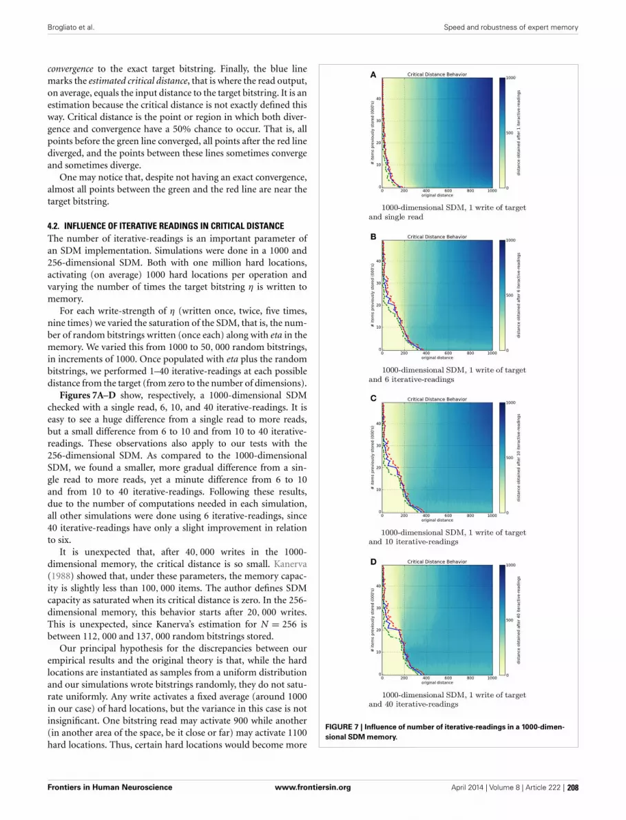
Brogliato et al. Speed and robustness of expert memory
convergence to the exact target bitstring. Finally, the blue linemarks the estimated critical distance, that is where the read output,on average, equals the input distance to the target bitstring. It is anestimation because the critical distance is not exactly defined thisway. Critical distance is the point or region in which both diver-gence and convergence have a 50% chance to occur. That is, allpoints before the green line converged, all points after the red linediverged, and the points between these lines sometimes convergeand sometimes diverge.
One may notice that, despite not having an exact convergence,almost all points between the green and the red line are near thetarget bitstring.
4.2. INFLUENCE OF ITERATIVE READINGS IN CRITICAL DISTANCEThe number of iterative-readings is an important parameter ofan SDM implementation. Simulations were done in a 1000 and256-dimensional SDM. Both with one million hard locations,activating (on average) 1000 hard locations per operation andvarying the number of times the target bitstring η is written tomemory.
For each write-strength of η (written once, twice, five times,nine times) we varied the saturation of the SDM, that is, the num-ber of random bitstrings written (once each) along with eta in thememory. We varied this from 1000 to 50, 000 random bitstrings,in increments of 1000. Once populated with eta plus the randombitstrings, we performed 1–40 iterative-readings at each possibledistance from the target (from zero to the number of dimensions).
Figures 7A–D show, respectively, a 1000-dimensional SDMchecked with a single read, 6, 10, and 40 iterative-readings. It iseasy to see a huge difference from a single read to more reads,but a small difference from 6 to 10 and from 10 to 40 iterative-readings. These observations also apply to our tests with the256-dimensional SDM. As compared to the 1000-dimensionalSDM, we found a smaller, more gradual difference from a sin-gle read to more reads, yet a minute difference from 6 to 10and from 10 to 40 iterative-readings. Following these results,due to the number of computations needed in each simulation,all other simulations were done using 6 iterative-readings, since40 iterative-readings have only a slight improvement in relationto six.
It is unexpected that, after 40, 000 writes in the 1000-dimensional memory, the critical distance is so small. Kanerva(1988) showed that, under these parameters, the memory capac-ity is slightly less than 100, 000 items. The author defines SDMcapacity as saturated when its critical distance is zero. In the 256-dimensional memory, this behavior starts after 20, 000 writes.This is unexpected, since Kanerva’s estimation for N = 256 isbetween 112, 000 and 137, 000 random bitstrings stored.
Our principal hypothesis for the discrepancies between ourempirical results and the original theory is that, while the hardlocations are instantiated as samples from a uniform distributionand our simulations wrote bitstrings randomly, they do not satu-rate uniformly. Any write activates a fixed average (around 1000in our case) of hard locations, but the variance in this case is notinsignificant. One bitstring read may activate 900 while another(in another area of the space, be it close or far) may activate 1100hard locations. Thus, certain hard locations would become more
FIGURE 7 | Influence of number of iterative-readings in a 1000-dimen-sional SDM memory.
Frontiers in Human Neuroscience www.frontiersin.org April 2014 | Volume 8 | Article 222 | 208

Brogliato et al. Speed and robustness of expert memory
noise than signal during activation sooner, rather than a uni-form degradation occurring. This discrepancy would cause, in theaggregate, a saturation of the SDM with fewer bitstrings storedthan expected in theory. This remains one possibility, though wehope the issue will be explored in future work.
4.3. INFLUENCE OF THE NUMBER OF WRITES ON THE CRITICALDISTANCE
The influence of the number of writes on the critical distance wasnot analyzed by Kanerva. It is important because, when a randombitstring is seen only once, it is psychologically plausible that itwill be gradually forgotten with new incoming information. Whatmatters is not exactly the number of writes, but the proportion ofthe number of times a bitstring was stored in relation to others.
A remark on cognitive psychology is in order here. Consider,as an example, the aforementioned exchange between Szilardand Einstein. As an expert confronts unexpected information,it is reasonable to expect that additional memory writes willoccur. If we presume that evolution brought the human memoryclose to optimality, as explored by the rational analysis approach(Anderson and Milson, 1989; Anderson, 1990; Anderson andSchooler, 1991), one would expect some mechanism akin toShannon’s idea of information content to be in play.
That is, as an expert is surprised by new, unforeseen, informa-tion, say, an outcome , with information I() = −log(P()),where I stands for the information content in outcome . Onewould therefore expect the expert’s memory to either place addi-tional attention to the outcome, leading to: (1) additional writesto memory, or (2) amplification of the write operation’s signals,or possibly (3) both effects.
Figures 8A–D show a 1000-dimensional SDM with 1, 2, 5, and9 writes of the target bitstring η. It is easy to see a huge differencefrom 1 to 2 writes. Although the green line has a strange behaviornear 50, 000 items stored, the critical distance was much greaterthan with 1 write. From 2 to 5 to 9 rehearsals, the critical distancestarts growing rapidly and slows down near six writes. This makessense, since it should have a threshold smaller than 500 bits.
The 256-dimensional memory has a similar behavior, but lessabrupt. It keeps growing, but slower than a 100-dimensionalmemory. It never crosses the 50 bits on x-axis in 256 bits, whilethe 1000-dimensional reaches the 200 bits on x-axis and almosthits 400 bits on the x-axis.
These figures display the immense power of reinforcement orrehearsal: additional writes of a memory item significantly raisethe attractor basin (critical distance) for that memory item.
This behavior is plausible, as the human brain rapidly recog-nizes a pattern when it is used to it. Many times, the patternsappear in different contexts, giving cues far from the target con-cept, much like a chess player, who looks at a position and rapidlyrecognizes what is happening (Bilalic et al., 2009; Rennig et al.,2013).
5. DISCUSSIONThis is the first work focused on better understanding the criti-cal distance behavior of a Sparse Distributed Memory (Kanerva,personal communication). Our future research intends to explorethe rehearsal mechanisms in cognitive architectures for one ofthe most studied domains of expertise: (Linhares, 2005; Linhares
FIGURE 8 | Influence of number of target writes in a 1000-dimensionalSDM memory.
Frontiers in Human Neuroscience www.frontiersin.org April 2014 | Volume 8 | Article 222 | 209

Brogliato et al. Speed and robustness of expert memory
and Brum, 2007; Linhares and Freitas, 2010; Linhares et al.,2012; Linhares and Chada, 2013; Linhares, 2014), and attemptto bridge the low-level world of neurons and their assemblieswith the high-level world of abstract thought and understand-ing of strategic scenarios. We have argued here that, as SDMremains both a psychologically plausible and a neuroscientificallyplausible model of human memory, the study of its critical dis-tance may provide insights into the edges of our own recollection.Without a precise understanding of the critical distance behavior,one cannot advance the theoretical model. Moreover, one can-not develop robust applications without knowing the limits ofconvergence.
The empirical tests shown here confirmed that the critical dis-tance in SDM constitutes a “band” wherein both convergenceand divergence become less and less likely. This is a palatableresult because, intuitively, the Tip-of-the-Tongue phenomenon inhumans seems like an attractor, something we sometimes “fallinto.” We argue this is a parallel between SDM and human rec-ollection, and posit that our theoretical and empirical resultsprovide evidence that the critical distance is a correlate to the edgeof human recollection.
While humans sometimes fall into the TotT, there are alsotimes when we almost fall into it and, after a bit of effort, are ableto recall the desired information. In the model, this would meanwe enter the critical band, but leave it after one or two iterationsand converge. Likewise, it seems one can be very certain of whatone is saying and, in mid-sentence, completely diverge from thenext piece of information we wished to recall. In SDM this wouldamount to entering the critical band, but then diverging.
As Figure 6 shows, the speed of convergence is a function ofthe number of read operations: additional read operations bringone closer to the memory item (assuming that the original cuewas not past the critical point). We also see that this effect isgreatly reduced after 6 to 10 read operations. As Figure 7 shows,expertise can be correlated with providing additional writes tothe memory, and we show that increasing the rehearsal num-ber greatly increases the margin for error or ambiguity, and greatlydecreases the relevant information needed for convergence, as thecritical threshold is increased. In human terms, experts “knowwhat you are talking about” with fewer cues. Their memory hasmuch greater robustness.
Yet, it is the combination of these two dynamics that sheds lighton experts’ speed. Taking the SDM model as a plausible accountof human memory, we can compare by saying that, for experts,having a much higher threshold may signify being able to convergewithin fewer, or even a single, read operation. As the hard locationshave been reinforced with the original information, read oper-ations converge faster. With very few cues and noisy, ambiguous,information, experts may still manage to recollect and understandalmost immediately the object, situation, or event in question. It isno wonder Albert Einstein could immediately grasp Leo Szilard’sconcerns.
5.1. DATA SHARINGAll the computational methods developed in this study are avail-able as an open-source project, and can be found at https://github.com/msbrogli/sdm.
FUNDINGThis work has been generously supported by grants fromthe Fulbright Foundation, and FAPERJ Foundation (grants E-26/110.540/2012 and E-26/111.846/2011), grants from the CNPqFoundation (grants 401883/2011-6 and 470341/2009-2), the Pro-Pesquisa program of FGV Foundation.
ACKNOWLEDGMENTSThe authors are enormously grateful for a series of discussionswith Dr. Pentti Kanerva, Dr. Eric Nichols, Dr. Robert M. French,Dr. Rafael Godszmidt, Dr. Alexandre Mendes, Dr. Christian N.Aranha, Felipe Buchbinder, Ariston D. de Oliveira, and ManuelDoria. We would also like to thank the referees for their lucid andhighly constructive reviews of our work.
SUPPLEMENTAL MATERIALThe supplemental data file consists of five parts: (1) An intro-duction to the computational methods available in https://github.
com/msbrogli/sdm; (2) A large set of additional heatmaps, doc-umenting the behavior of the model in a 1000-dimensionalmemory; (3) The same tests on 256-dimensional memory, (4) allrehearsal results for 256-dimensional memory, and (5) the sametests for 1000-dimensional memory.
The Supplementary Material for this article can be foundonline at: http://www.frontiersin.org/journal/10.3389/fnhum.
2014.00222/abstract
REFERENCESAbbott, J. T., Hamrick, J. B., and Griffiths, T. L. (2013). “Approximating bayesian
inference with a sparse distributed memory system,” in Proceedings of the 34thAnnual Conference of the Cognitive Science Society (Berlin).
Anderson, J. R. (1990). The Adaptive Character of Thought. New Jersey: LawrenceErlbaum Associates.
Anderson, J. R., and Milson, R. (1989). Human memory: an adaptive perspective.Psychol. Rev. 96, 703–719. doi: 10.1037/0033-295X.96.4.703
Anderson, J. R., and Schooler, L. J. (1991). Reflections of the environment inmemory. Psychol. Sci. 2, 396–408. doi: 10.1111/j.1467-9280.1991.tb00174.x
Ballard, D. H., Hayhoe, M. M., Pook, P. K., and Rao, R. P. (1997). Deicticcodes for the embodiment of cognition. Behav. Brain Sci. 20, 723–742. doi:10.1017/S0140525X97001611
Bancroft, T. D., Hockley, W. E., and Servos, P. (2012). Can vibrotactileworking memory store multiple items? Neurosci. Lett. 514, 31–34. doi:10.1016/j.neulet.2012.02.044
Bilalic, M., Kiesel, A., Pohl, C., Erb, M., and Grodd, W. (2011a). It takes twoskilledrecognition of objects engages lateral areas in both hemispheres. PLoS ONE6:e16202. doi: 10.1371/journal.pone.0016202
Bilalic, M., Langner, R., Ulrich, R., and Grodd, W. (2011b). Many faces of expertise:fusiform face area in chess experts and novices. J. Neurosci. 31, 10206–10214.doi: 10.1523/JNEUROSCI.5727-10.2011
Bilalic, M., Langner, R., Erb, M., and Grodd, W. (2010). Mechanisms and neu-ral basis of object and pattern recognition: a study with chess experts. J. Exp.Psychol. Gen. 139, 728. doi: 10.1037/a0020756
Bilalic, M., McLeod, P., and Gobet, F. (2009). Specialization effect and its influ-ence on memory and problem solving in expert chess players. Cogn. Sci. 33,1117–1143. doi: 10.1111/j.1551-6709.2009.01030.x
Bilalic, M., Turella, L., Campitelli, G., Erb, M., and Grodd, W. (2012). Expertisemodulates the neural basis of context dependent recognition of objects and theirrelations. Hum. Brain Mapp 33, 2728–2740. doi: 10.1002/hbm.21396
de Pádua Braga, A., and Aleksander, I. (1995). Geometrical treatment and statisticalmodelling of the distribution of patterns in the n-dimensional boolean space.Pattern Recogn. Lett. 16, 507–515. doi: 10.1016/0167-8655(95)00120-6
French, R. M. (1999). Catastrophic forgetting in connectionist networks. TrendsCogn. Sci. 3, 128–135. doi: 10.1016/S1364-6613(99)01294-2
Frontiers in Human Neuroscience www.frontiersin.org April 2014 | Volume 8 | Article 222 | 210

Brogliato et al. Speed and robustness of expert memory
Gauthier, I., Tarr, M. J., Anderson, A. W., Skudlarski, P., and Gore, J. C. (1999).Activation of the middle fusiform face area. Nat. Neurosci. 2, 568–573. doi:10.1038/9224
Gobet, F., Lane, P., Croker, S., Cheng, P., Jones, G., Oliver, I., et al. (2001).Chunking mechanisms in human learning. Trends Cogn. Sci. 5, 236–243. doi:10.1016/S1364-6613(00)01662-4
Gobet, F., and Simon, H. (2000). Five seconds or sixty? presentation time in expertmemory. Cogn. Sci. 24, 651–682. doi: 10.1207/s15516709cog2404_4
Harré, M. (2013). The neural circuitry of expertise: perceptual learning and socialcognition. Front. Hum. Neurosci. 7:852. doi: 10.3389/fnhum.2013.00852
Harré, M., Bossomaier, T., and Snyder, A. (2012). The perceptual cues that reshapeexpert reasoning. Sci. Rep. 2, 502. doi: 10.1038/srep00502
Huth, A. G., Nishimoto, S., Vu, A. T., and Gallant, J. L. (2012). A continu-ous semantic space describes the representation of thousands of object andaction categories across the human brain. Neuron 76, 1210–1224. doi: 10.1016/j.neuron.2012.10.014
Kanerva, P. (1988). Sparse Distributed Memory. Cambridge, MA: MIT Press.Kanerva, P. (2009). Hyperdimensional computing: an introduction to comput-
ing in distributed representation with high-dimensional random vectors. Cogn.Comput. 1, 139–159. doi: 10.1007/s12559-009-9009-8
Laurent, G. (2002). Olfactory network dynamics and the coding of multidimen-sional signals. Nat. Rev. Neurosci. 3, 884–895. doi: 10.1038/nrn964
Laurent, G. (2006). “Shall we even understand the fly’s brain, chapter 1,” in 23Problems in Systems Neuroscience, eds J. L. Van Hemmen, and T. J. Sejnowski(Oxford, NY: Oxford University Press), 3–21.
Linhares, A. (2005). An active symbols theory of chess intuition. Minds Mach. 15,131–181. doi: 10.1007/s11023-005-5045-7
Linhares, A. (2014). The emergence of choice: decision-making and strategicthinking through analogies. Inform. Sci. 259, 36–56. doi: 10.1016/j.ins.2013.08.006
Linhares, A., and Brum, P. (2007). Understanding our understanding of strategicscenarios: what role do chunks play? Cogn. Sci. 31, 989–1007. doi: 10.1080/03640210701703725
Linhares, A., and Chada, D. M. (2013). What is the nature of the mind’spattern-recognition process? New Ideas Psychol. 31, 108–121. doi: 10.1016/j.newideapsych.2012.08.001
Linhares, A., Chada, D. M., and Aranha, C. N. (2011). The emergence of miller’smagic number on a sparse distributed memory. PLOS ONE 6:e15592. doi:10.1371/journal.pone.0015592
Linhares, A., and Freitas, A. (2010). Questioning chase and simon’s (1973) “per-ception in chess”: the “experience recognition” hypothesis. New Ideas Psychol.28, 64–78. doi: 10.1016/j.newideapsych.2009.07.008
Linhares, A., Freitas, A. E. T. A., Mendes, A. S., and Silva, J. S. (2012). Entanglementof perception and reasoning in the combinatorial game of chess: differentialerrors of strategic reconstruction. Cogn. Syst. Res. 13, 72–86. doi: 10.1016/j.cogsys.2010.12.006
Ludermir, T. B., Carvalho, A., Braga, A., and Souto, M. (1999). Weightless neuralmodels: a review of current and past works. Neural Comput. Surv. 2, 41–61.
Marr, D. (1892). Vision. New York, NY: W.H. Freeman.Mazor, O., and Laurent, G. (2005). Transient dynamics versus fixed points in
odor representations by locust antennal lobe projection neurons. Neuron 48,661–673. doi: 10.1016/j.neuron.2005.09.032
McGugin, R. W., Gatenby, J. C., Gore, J. C., and Gauthier, I. (2012). High res-olution imaging of expertise reveals reliable object selectivity in the fusiformface area related to perceptual performance. Proc. Natl. Acad. Sci. U.S.A. 109,17063–17068. doi: 10.1073/pnas.1116333109
Nosofsky, R. M. (1986). Attention, similarity, and the identification-categorization relationship. J. Exp. Psychol. 115, 39–57. doi: 10.1037/0096-3445.115.1.39
Olshausen, B. A., Anderson, C. H., and Van Essen, D. C. (1993). A neurobiologicalmodel of visual attention and invariant pattern recognition based on dynamicrouting of information. J. Neurosci. 13, 4700–4719.
Rao, R. P., Zelinsky, G. J., Hayhoe, M. M., and Ballard, D. H. (2002). Eyemovements in iconic visual search. Vis. Res. 42, 1447–1463. doi: 10.1016/S0042-6989(02)00040-8
Rennig, J., Bilalic, M., Huberle, E., Karnath, H.-O., and Himmelbach, M.(2013). The temporo-parietal junction contributes to global gestalt percep-tion: evidence from studies in chess experts. Front. Hum. Neurosci. 7:513. doi:10.3389/fnhum.2013.00513
Rhodes, R. (2012). Making of the Atomic Bomb. New York, NY :Simon and Schuster.Sakurai, Y. (1996). Population coding by cell assemblies–what it really
is in the brain. Neurosci. Res. 26, 1–16. doi: 10.1016/0168-0102(96)01075-9
Sakurai, Y. (1998). The search for cell assemblies in the working brain. Behav. BrainRes. 91, 1–13. doi: 10.1016/S0166-4328(97)00106-X
Sakurai, Y. (1999). How do cell assemblies encode information in the brain?Neurosci. Biobehav. Rev. 23, 785–796. doi: 10.1016/S0149-7634(99)00017-2
Shepard, R. N. (1957). Stimulus and response generalization: a stochastic modelrelating generalization to distance in psychological space. Psychometrika 22,325–345. doi: 10.1007/BF02288967
Shepard, R. N. (1987). Toward a universal law of generalization for psychologicalscience. Science 237, 1317–1323. doi: 10.1126/science.3629243
Silva, M. T. P., Braga, A. P., and Lacerda, W. S. (2004). Reconfigurable co-processorfor kanerva’s sparse distributed memory. Microprocess. Microsyst. 28, 127–134.doi: 10.1016/j.micpro.2004.01.003
Xu, Y. (2005). Revisiting the role of the fusiform ‘face area’ in visual expertise.Cereb. Cortex 15, 1234–1242. doi: 10.1093/cercor/bhi006
Conflict of Interest Statement: The authors declare that the research was con-ducted in the absence of any commercial or financial relationships that could beconstrued as a potential conflict of interest.
Received: 25 September 2013; accepted: 28 March 2014; published online: 28 April2014.Citation: Brogliato MS, Chada DM and Linhares A (2014) Sparse distributed mem-ory: understanding the speed and robustness of expert memory. Front. Hum. Neurosci.8:222. doi: 10.3389/fnhum.2014.00222This article was submitted to the journal Frontiers in Human Neuroscience.Copyright © 2014 Brogliato, Chada and Linhares. This is an open-access article dis-tributed under the terms of the Creative Commons Attribution License (CC BY). Theuse, distribution or reproduction in other forums is permitted, provided the originalauthor(s) or licensor are credited and that the original publication in this jour-nal is cited, in accordance with accepted academic practice. No use, distribution orreproduction is permitted which does not comply with these terms.
Frontiers in Human Neuroscience www.frontiersin.org April 2014 | Volume 8 | Article 222 | 211

OPINION ARTICLEpublished: 20 September 2013
doi: 10.3389/fnhum.2013.00590
Functional cerebral reorganization: a signature ofexpertise? Reexamining Guida, Gobet, Tardieu, andNicolas’ (2012) two-stage frameworkAlessandro Guida1*, Fernand Gobet2 and Serge Nicolas3
1 Département de Psychologie, Centre de Recherche en Psychologie, Cognition et Communication, Université Rennes 2, Rennes, France2 Department of Psychological Sciences, University of Liverpool, Liverpool, UK3 Institut de Psychologie, Université Paris Descartes, Boulogne Billancourt, France*Correspondence: [email protected]; [email protected]
Edited by:Merim Bilalic, University Tübingen, University Clinic, Germany
Reviewed by:Guillermo Campitelli, Edith Cowan University, AustraliaRobert Langner, Heinrich Heine University Düsseldorf, Germany
Keywords: expertise, working memory, functional cerebral reorganization, chunks, templates, retrieval structures
In 2012, Guida, Gobet, Tardieu andNicolas proposed a two-stage frameworkto explain how cognitive changes due topractice could shape experts’ brain physi-ologically and thus explain neuroimagingdata of expertise acquisition. In this paper,after presenting the motivations for sucha framework and the framework itself, weexamine the idea that functional cerebralreorganization (FCR) could be used as asignature for expertise.
CHUNKS, TEMPLATES AND RETRIEVALSTRUCTURESIn the mid-nineties, Ericsson and Kintsch(1995) and Gobet and Simon (1996) pro-posed Long-Term Working Memory the-ory (LTWMT) and Template Theory (TT),respectively, in order to account for behav-ioral data in the domain of expertise.These data were difficult to explain withthe sole concept of chunk (Chase andSimon, 1973), given the severe limita-tions of working memory (WM) (7 ± 2for an optimistic estimation, Miller, 1956;but for recent reevaluations, see Cowan,2001; Gobet and Clarkson, 2004; Mathyand Feldman, 2012). For example, sev-eral experiments (e.g., Charness, 1976;Frey and Adesman, 1976; Glanzer et al.,1984) showed that interfering tasks hadalmost no effect on WM performanceor text comprehension with experts. Yet,according to chunking theory (Chase andSimon, 1973), interfering tasks shouldwipe out the content of WM where infor-mation is stored. This led Ericsson andKintsch (1995) and Gobet and Simon(1996) to suggest that information was
not stored in WM as initially proposed,but was rapidly and efficiently trans-ferred in LTM, where the interferingtasks has no effect. Both theories pro-posed that this was possible only ifknowledge structures were built. Thesestructures were called templates with TTand retrieval structures with LTWMT.Even if differences exist between thetwo theories (e.g., Ericsson and Kintsch,2000; Gobet, 2000a,b), LTWMT and TTrevolve around the same fundamentalcore idea: Fast and reliable transfer inLTM becomes possible with expertise viaknowledge structures, which enables LTMto be used during WM tasks, thus giv-ing the appearance of expanding individ-uals’ WM capacity. These two cognitivetheories have been used to explain notonly behavioral but also neuroscientificdata (e.g., Pesenti et al., 2001; Ericsson,2003; Campitelli et al., 2007; Bilalic et al.,2010).
EXPLAINING NEUROIMAGING DATA INEXPERTISE ACQUISITION: ATWO-STAGE FRAMEWORKRecently, the core idea of the two theo-ries has been used by Guida et al. (2012)to bridge together, for the first time, (a)neuroimaging data acquired from noviceundergoing practice in WM-related tasksand (b) neuroimaging data acquired fromexperts in WM-related tasks. The results ofthe two groups of studies, which belong totwo separate domains of research, diverge.Neuroimaging of novices practicing from2 h up to 5 weeks show mainly a decreaseof activation in prefrontal and parietal
areas (for similar conclusions, see Kellyand Garavan, 2005; Hill and Schneider,2006; Buschkuehl et al., 2012). Conversely,neuroimaging studies of experts who arecompared to novices are more compati-ble with FCR, viz., experts and novices usedifferent brain areas and different men-tal operations to perform similar tasks(for similar conclusions, see Ericsson,2003). Notwithstanding these divergentfindings (brain activation decrease vs.FCR), the core idea behind LTMWT andTT allows bridging these two neuroimag-ing patterns into a coherent two-stageframework.
FIRST STAGE: DECREASE OFACTIVATION DUE TO CHUNKCREATION AND RETRIEVALWhen novices start practicing, and if theactivity is new, the first important processis chunk creation. While executing theirnew activity several times, novices willstart gradually chunking separate elementstogether through binding, viz., encodingthe relations among stimuli that co-occur(Cohen and Eichenbaum, 1993). Oncechunks have been created and thus storedin LTM, chunk can be retrieved and there-fore used, allowing encoding multiple ele-ments in WM with one chunk (e.g., “f,”“b,” “i,” can be encoded as one elementin WM instead of three), using fewerresources.
At a physiological level, chunk creation(through binding) and chunk retrievalare two reasons to expect brain activa-tion decrease. First, if the binding processoccurs in prefrontal regions (Prabhakaran
Frontiers in Human Neuroscience www.frontiersin.org September 2013 | Volume 7 | Article 590 |
HUMAN NEUROSCIENCE
212

Guida et al. Expertise and functional cerebral reorganization
et al., 2000; Raffone and Wolters, 2001)and in parietal regions (Shafritz et al.,2002; Oakes et al., 2006), less activationshould be observed in these regions aftera period of training, because as trainingprogresses, fewer chunks will be created.Second, the use of chunks through chunkretrieval makes it possible to encode infor-mation in WM with less resources, aselements are grouped. Several researchershave shown that, physiologically, thereis a correlation between the number ofelements in WM and brain activity inprefrontal and parietal areas1 (Todd andMarois, 2004; Vogel and Machizawa, 2004;Cowan, 2011). Therefore, if less WM spaceis used through chunk retrieval, decreaseof brain activity should be expectedin prefrontal and parietal WM areas(Figure 1).
1 It is particularly the case in the intra-parietal sulcus(e.g., Majerus et al., 2010; Cowan et al., 2011).
SECOND STAGE: FUNCTIONALCEREBRAL REORGANIZATION DUE TOKNOWLEDGE STRUCTURES CREATIONAND RETRIEVALWith practice (e.g., Cowan et al., 2004;Chen and Cowan, 2005) and expertise(e.g., Chase and Simon, 1973; Gobet andSimon, 1996), chunks get larger and morecomplex, and with years of training theybecome knowledge structures. For experts,the peculiarity of these structures, whenused in their domain of expertise, isto allow rapid and reliable encoding inepisodic LTM, even in WM-like conditions(fast presentation times of multiple ele-ments) when usually elements can only beencoded reliably in WM (Figure 1).
In terms of brain activation, at thisstage, not only a cerebral activation pat-tern compatible with WM activities isexpected but conjunctly a pattern com-patible with episodic LTM, that is, medialtemporal lobe (MTL) activations (Gabrieli
et al., 1997; Young et al., 1997; Lepageet al., 1998; for reviews, see Squire et al.,2004; Eichenbaum et al., 2007) due to theutilization of knowledge structures. Froma longitudinal standpoint, this implies aFCR, which can be defined by two changesoccurring with practice: (a) the decreaseof brain activity undergirding cognitiveprocesses that are used less with prac-tice (here WM in stage 1), and (b) theemergence of brain activity in new areassupporting new cognitive processes (hereepisodic LTM in stage 2). Therefore, aFCR involving episodic LTM2 is expected(Figure 1). Unfortunately, to our knowl-edge, nobody has followed the develop-ment of expertise in a WM-related taskwith neuroimaging long enough to test
2 FCR, in this article, means, a shift of a way of per-forming a task to another way, without specifying thenew way. FCR involving episodic LTM, in this arti-cle, means that the new way of performing the taskis through episodic LTM
FIGURE 1 | Schematic representation of the Two-Stage Frameworklinking the cognitive and cerebral levels in expertise acquisition,through two examples. The “Examples” section shows the evolution ofthe effect of knowledge on how items to-be-remembered are processed:at first, items are processed almost separately, later, items are regroupedin chunks, and finally in knowledge structures, which can be viewed assuper-chunks that regroup multiple chunks into a high-level pattern. In the“Cerebral Level” section, the representation of brain activity is at anordinal scale. SST stands for statistical significance threshold; if brainactivity is beneath this threshold, it goes undetected. PFC stands for
prefrontal cortex, PL for parietal lobe, and MTL for medial temporal lobe.The first MTL activity on the left is almost at the same level than thestatistical significance threshold in order to indicate that for novices, brainactivity is sometimes detected (see section “Concluding Remarks”). Fornovices, detection seems to vary according to the kind of experimentalparadigm, the parameters and maybe the participants of the experiments.If one considers that the MTL activity is above the statistical significancethreshold for novices then functional cerebral reorganization is bettersuited to describe expertise acquisition; if it is beneath, then functionalcerebral redistribution is better suited.
Frontiers in Human Neuroscience www.frontiersin.org September 2013 | Volume 7 | Article 590 | 213

Guida et al. Expertise and functional cerebral reorganization
this hypothesis. Instead, what is possible isto compare novices against experts. Thiswas the aim of Guida et al.’s (2012) review,which showed that most of the studieswere compatible with FCR involving LTM.
FUNCTIONAL CEREBRALREORGANIZATION: A SIGNATURE OFEXPERTISE?Given that a link has been establishedbetween expertise and FCR, an importantquestion is to know whether FCR could beused as a signature for expertise. A simpleway to answer this question is to exam-ine empirically whether the implication“expertise thus FCR” observed by Guidaet al. (2012) could be reversed. In otherwords, when one looks for patterns com-patible with FCR—viz. a decrease of brainactivity concerning one cognitive processand the emergence of brain activity con-cerning new cognitive processes—is exper-tise found? If it is not the case then FCRdoes not imply expertise.
At first glance, this does not seem to betrue. There are multiple examples show-ing that the simple utilization of differentstrategies can involve patterns of activa-tion similar to FCR. For instance, the lit-erature of WM-related tasks shows thatwhen different groups of individuals usespontaneously different strategies—verbalstrategy vs. visual strategy (Burbaud et al.,2000), or verbal strategy vs. spatial strategy(Glabus et al., 2003)—then completely dif-ferent patterns of activation are detected.This seems to be true even when thestrategies are dictated by the experimenter,as observed by Bernstein et al. (2002)when imposing different encoding strate-gies in a task of face recognition. Thesethree between-subject studies bring onlyindirect evidence, but they are confirmedby a within-subject study. When Reichleet al. (2000) asked the same individualsto process a sentence-picture verificationtask with different strategies (linguistic vs.visual), completely different patterns ofactivity appeared: there was a decrease ofbrain activity concerning cognitive pro-cesses (e.g., linguistic) and the emergenceof brain activity concerning new cognitiveprocesses (e.g., visual). In all these cases,a pattern consistent with FCR is presentbut no expertise is found. Therefore, theimplication “expertise thus FCR” does notseem reversible.
However, when considering preciselyFCR involving episodic LTM areas, thepicture is different. First, we found onlyone study (Kondo et al., 2005); secondly, itis the only study where participants weretaught how to use knowledge structures.Kondo et al. (2005) asked their noviceparticipants to encode ten object picturesusing the method of loci, basing them-selves on the visuospatial knowledge oftheir house. When comparing neuroimag-ing before and after using the methodof loci, they observed a pattern consis-tent with FCR at retrieval. Hence, if oneargues that the method of loci is based onthe utilization of expertise (Guida et al.,2009, 2013), the conclusion from Kondoet al. (2005) could be that for FCR involv-ing episodic LTM areas, the implication“expertise thus FCR” can be reversed,making the proposal that FCR is a signa-ture for expertise verisimilar (when involv-ing episodic LTM).
However, when trying to relateexpertise and FCR and before one canbe conclusive on the link between thesetwo concepts, two elements need to betaken into consideration, functional cere-bral redistribution and brain connectivity.These will constitute our concludingremarks.
CONCLUDING REMARKSA very recent growing body of data sug-gests that in some cases, functional cere-bral redistribution could also occur withpractice. Both FCR and functional cerebralredistribution involve a combination ofincreases and decreases in activation (Kellyand Garavan, 2005); however, only FCRnecessitates the emergence of new areaswith practice. Recent evidence suggeststhat MTL could also be involved in WMtasks with no practice (e.g., Ranganathand Blumenfeld, 2005; Olson et al., 2006;Lee and Rudebeck, 2010; Campo et al.,2013). The debate is still ongoing and theseresults are considered artifactual by some,mainly because the tasks used seem moreLTM-like than WM-like (Jonides et al.,2008). Squire and Wixted (2011) observedthat if WM capacity were not exceeded,MTL was not involved (e.g., Shrager et al.,2008; Jeneson et al., 2012). Nonetheless,these data suggest that in some cases, MTLactivation could be expected at the earlystages of training. It is plausible that this
activation could increase with expertisewhen knowledge structures are available,which means that this pattern would bebetter described by functional redistribu-tion than FCR, because this last patternimplies no MTL activation at the initialstage of practice (Figure 1). However, theadditional areas of experts are sometimesthe same structures than that of novicesbut on the opposite hemisphere (e.g.,Bilalic et al., 2011, 2012), a.k.a. “doubletake” phenomenon (e.g., Scalf et al., 2007),which complicates sometime the distinc-tion between functional redistribution andFCR. To conclude concerning MTL, morework needs to be done to ascertain itsinvolvement, especially in practice-relatedstudies where this kind of evidence isscarce (but, see Dahlin et al., 2008), there-fore, presently, these are only assumptions.
Finally, when considering expertise-related FCR, which constitutes a com-bined increase and decrease in activationacross the brain, it is also crucial to under-stand how the different brain areas worktogether in terms of network connectiv-ity. Fundamental in this respect is the ideaof “neural context” proposed by McIntosh(1998; also, see Bressler and McIntosh,2007), according to which the frame ofactivation (or the neural context of acti-vation) around a determined brain areais at least as important as the activationof that brain area. If one relates this ideato practice, then the consequence is thateven if the activation of a region does notchange with practice, it can still be cru-cial, by influencing the increase or decreaseof activation in other brain areas (Kellyand Garavan, 2005). The neural contextcould thus be important for the study offunctional reorganization, and its applica-tion should be disseminated (Bressler andMenon, 2010).
ACKNOWLEDGMENTSIn memory of Hubert Tardieu, our dearfriend, collaborator, and colleague. We feelprivileged to have shared moments of hislife. We will never forget his sense ofhumor, wit and genuine kindness.
REFERENCESBernstein, L. J., Beig, S., Siegenthaler, A. L., and
Grady, C. L. (2002). The effect of encoding strat-egy on the neural correlates of memory for faces.Neuropsychologia 40, 86–98. doi: 10.1016/S0028-3932(01)00070-7
Frontiers in Human Neuroscience www.frontiersin.org September 2013 | Volume 7 | Article 590 | 214

Guida et al. Expertise and functional cerebral reorganization
Bilalic, M., Kiesel, A., Pohl, C., Erb, M., and Grodd, W.(2011). It takes two-skilled recognition of objectsengages lateral areas in both hemispheres. PLoSONE 6:e16202. doi: 10.1371/journal.pone.0016202
Bilalic, M., Langner, R., Erb, M., and Grodd, W.(2010). Mechanisms and neural basis of objectand pattern recognition – a study with chessexperts. J. Exp. Psychol. Gen. 139, 728–742. doi:10.1037/a0020756
Bilalic, M., Turella, L., Campitelli, G., Erb, M., andGrodd, W. (2012). Expertise modulates the neu-ral basis of context dependent recognition ofobjects and their relations. Hum. Brain Mapp. 33,2728–2740. doi: 10.1002/hbm.21396
Bressler, S. L., and McIntosh, A. R. (2007). “Therole of neural context in large-scale neurocogni-tive network operations,” in Handbook of BrainConnectivity, eds V. K. Jirsa and A. R. McIntosh(New York, NY: Springer), 403–419. doi: 10.1007/978-3-540-71512-2_14
Bressler, S. L., and Menon, V. (2010). Large-scalebrain networks in cognition: emerging methodsand principles. Trends Cogn. Sci. 14, 277–290. doi:10.1016/j.tics.2010.04.004
Burbaud, P., Camus, O., Guehl, D., Bioulac, B.,Caille, J. M., and Allard, M. (2000). Influenceof cognitive strategies on the pattern of corticalactivation during mental subtraction. A func-tional imaging study in human subjects. Neurosci.Lett. 287, 76–80. doi: 10.1016/S0304-3940(00)01099-5
Buschkuehl, M., Jaeggi, S. M., and Jonides, J. (2012).Neuronal effects following working memory train-ing. Dev. Cogn. Neurosci. 2(Suppl. 1), S167–S179.doi: 10.1016/j.dcn.2011.10.001
Campitelli, G., Gobet, F., Head, K., Buckley, M.,and Parker, A. (2007). Brain localisation of mem-ory chunks in chessplayers. Int. J. Neurosci. 117,1641–1659. doi: 10.1080/00207450601041955
Campo, P., Garrido, M. I., Moran, R. J., García-Morales, I., and Poch, C. (2013). Network recon-figuration and working memory impairment inmesial temporal lobe epilepsy. Neuroimage 72,48–54. doi: 10.1016/j.neuroimage.2013.01.036
Charness, N. (1976). Memory for chess positions:resistance to interference. J. Exp. Psychol. LearnMem. Cogn. 2, 641–653. doi: 10.1037/0278-7393.2.6.641
Chase, W. G., and Simon, H. A. (1973). Perception inchess. Cogn. Psychol. 4, 55–81. doi: 10.1016/0010-0285(73)90004-2
Chen, Z., and Cowan, N. (2005). Chunk limits andlength limits in immediate recall: a reconciliation.J. Exp. Psychol. Learn. Mem. Cogn. 31, 1235–1249.doi: 10.1037/0278-7393.31.6.1235
Cohen, N. J., and Eichenbaum, H. (1993). Memory,Amnesia, and the Hippocampal System. Cambridge,MA: MIT Press.
Cowan, N. (2001). The magical number 4 in short-term memory: a reconsideration of mental stor-age capacity. Behav. Brain Sci. 24, 87–185. doi:10.1017/S0140525X01003922
Cowan, N. (2011). The focus of attentionas observed in visual working memorytasks: making sense of competing claims.Neuropsychologia 49, 1401–1406. doi:10.1016/j.neuropsychologia.2011.01.035
Cowan, N., Chen, Z., and Rouder, J. N. (2004).Constant capacity in an immediate serial-recall
task: a logical sequel to Miller (1956). Psychol.Sci. 15, 634–640. doi: 10.1111/j.0956-7976.2004.00732.x
Cowan, N., Li, D., Moffitt, A., Becker, T. M., Martin,E. A., Saults, J. S., et al. (2011). A neural regionof abstract working memory. J. Cogn. Neurosci. 23,2852–2863. doi: 10.1162/jocn.2011.21625
Dahlin, E., Stigsdotter-Neely, A., Larsson, A.,Bäckman, L., and Nyberg, L. (2008). Transferof learning after updating training mediatedby the striatum. Science 320, 1510–1512. doi:10.1126/science.1155466
Eichenbaum, H., Yonelinas, A. R., and Ranganath, C.(2007). The medial temporal lobe and recognitionmemory. Annu. Rev. Neurosci. 30, 123–152. doi:10.1146/annurev.neuro.30.051606.094328
Ericsson, K. (2003). Exceptional memorizers: made,not born. Trends Cogn. Sci. 7, 233–235. doi:10.1016/S1364-6613(03)00103-7
Ericsson, K. A., and Kintsch, W. (1995). Long-termworking memory. Psychol. Rev. 102, 211–245. doi:10.1037/0033-295X.102.2.211
Ericsson, K. A., and Kintsch, W. (2000). Shortcomingsof generic retrieval structures with slots ofthe type of Gobet (1993) proposed andmodeled. Br. J. Psychol. 91, 571–590. doi:10.1348/000712600161998
Frey, P. W., and Adesman, P. (1976). Recall mem-ory for visually presented chess positions. Mem.Cognit. 4, 541–547. doi: 10.3758/BF03213216
Gabrieli, J. D., Brewer, J. B., Desmond, J. E., andGlover, G. H. (1997). Separate neural bases oftwo fundamental memory processes in the humanmedial temporal lobe. Science 276, 264–266. doi:10.1126/science.276.5310.264
Glabus, M. F., Horwitz, B., Holt, J. L., Kohn, P.D., Gerton, B. K., Callicott, J. H., et al. (2003).Interindividual differences in functional interac-tions among prefrontal, parietal and parahip-pocampal regions during working memory. Cereb.Cortex 13, 1352–1361. doi: 10.1093/cercor/bhg082
Glanzer, M., Fisher, B., and Dorfman, D. (1984).Short-term storage in reading. J. Verbal Learn.Verbal Behav. 23, 467–486. doi: 10.1016/S0022-5371(84)90300-1
Gobet, F. (2000a). Some shortcomings of long-termworking memory. Br. J. Psychol. 91, 551–570. doi:10.1348/000712600161989
Gobet, F. (2000b). Retrieval structures and schemata:a brief reply to Ericsson and Kintsch. Br. J. Psychol.91, 591–594. doi: 10.1348/000712600162005
Gobet, F., and Clarkson, G. (2004). Chunks inexpert memory: evidence for the magical num-ber four . . . or is it two. Memory 12, 732–747. doi:10.1080/09658210344000530
Gobet, F., and Simon, H. A. (1996). Templatesin chess memory: a mechanism for recallingseveral boards. Cogn. Psychol. 31, 1–40. doi:10.1006/cogp.1996.0011
Guida, A., Gobet, F., Tardieu, H., and Nicolas, S.(2012). How chunks, long-term working mem-ory and templates offer a cognitive explanation forneuroimaging data on expertise acquisition: a two-stage framework. Brain Cogn. 79, 221–244. doi:10.1016/j.bandc.2012.01.010
Guida, A., Gras, D., Noel, Y., Le Bohec, O., Quaireau,C., and Nicolas. S. (2013). The effect of long-term working memory through personalizationapplied to free recall: uncurbing the primacy
effect enthusiasm. Mem. Cognit. 41, 571–587. doi:10.3758/s13421-012-0284-3
Guida, A., Tardieu, H., and Nicolas. S. (2009).The personalisation method applied to a work-ing memory task: evidence of long-term workingmemory effects. Eur. J. Cogn. Psychol. 21, 862–896.doi: 10.1080/09541440802236369
Hill, N. M., and Schneider, W. (2006). “Brain changesin the development of expertise: Neurological evi-dence on skill-based adaptations,” in CambridgeHandbook of Expertise and Expert Performance, edsK. A. Ericsson, N. Charness, P. Feltovich, and R.Hoffman (New York, NY: Cambridge University),653–682. doi: 10.1017/CBO9780511816796.037
Jeneson, A., Wixted, J. T., Hopkins, R. O., and Squire,L. R. (2012). Visual working memory capac-ity and the medial temporal lobe. J. Neurosci.32, 3584–3589. doi: 10.1523/JNEUROSCI.6444-11.2012
Jonides, J., Lewis, R. L., Nee, D. E., Lustig, C. A.,Berman, M. G., and Moore, K. S. (2008). Themind and brain of short-term memory. Annu. Rev.Psychol. 59, 193–224. doi: 10.1146/annurev.psych.59.103006.093615
Kelly, A. M. C., and Garavan, H. (2005). Human func-tional neuroimaging of brain changes associatedwith practice. Cereb. Cortex 15, 1089–1102. doi:10.1093/cercor/bhi005
Kondo, Y., Suzuki, M., Mugikura, S., Abe, N.,Takahashi, S., Iijima, T., et al. (2005). Changes inbrain activation associated with use of a mem-ory strategy: a functional MRI study. Neuroimage24, 1154–1163. doi: 10.1016/j.neuroimage.2004.10.033
Lee, A. C. H., and Rudebeck, S. R. (2010).Investigating the interaction between spatial per-ception and working memory in the humanmedial temporal lobe. J. Cogn. Neurosci. 22,2823–2835. doi: 10.1162/jocn.2009.21396
Lepage, M., Habib, R., and Tulving, E. (1998).Hippocampal PET activations of memory encod-ing and retrieval: the HIPER model. Hippocampus8, 313–322. doi: 10.1002/(SICI)1098-1063(1998)8:4<313::AID-HIPO1> 3.0.CO;2-I
McIntosh, A. R. (1998). Understanding neural inter-actions in learning and memory using functionalneuroimaging. Ann. N.Y. Acad. Sci. 85, 556–571.doi: 10.1111/j.1749-6632.1998.tb10625.x
Majerus, S., D’Argembeau, A., Perez, T. M., Belayachi,S., Van der Linden, M., Collette, F., et al. (2010).The commonality of neural networks for ver-bal and visual short-term memory. J. Cogn.Neurosci. 22, 2570–2593. doi: 10.1162/jocn.2009.21378
Mathy, F., and Feldman, J. (2012). What’s magic aboutmagic numbers. Chunking and data compressionin short-term memory. Cognition 122, 346–362.doi: 10.1016/j.cognition.2011.11.003
Miller, G. A. (1956). The magical number seven, plusor minus two: some limits of our capacity for pro-cessing information. Psychol. Rev. 63, 81–97. doi:10.1037/h0043158
Oakes, L. M., Ross-Sheehy, S., and Luck, S. J. (2006).Rapid development of feature binding in visualshort-term memory. Psychol. Sci. 17, 781–787. doi:10.1111/j.1467-9280.2006.01782.x
Olson, I. R., Moore, K., Stark, M., and Chatterjee, A.(2006). Visual working memory is impaired whenthe medial temporal lobe is damaged. J. Cogn.
Frontiers in Human Neuroscience www.frontiersin.org September 2013 | Volume 7 | Article 590 | 215

Guida et al. Expertise and functional cerebral reorganization
Neurosci. 18, 1087–1097. doi: 10.1162/jocn.2006.18.7.1087
Pesenti, M., Zago, L., Crivello, F., Mellet, E., Samson,D., Duroux, B., et al. (2001). Mental calculationin a prodigy is sustained by right prefrontal andmedial temporal areas. Nat. Neurosci. 4, 103–107.doi: 10.1038/82831
Prabhakaran, V., Narayanan, K., Zhao, Z., andGabrieli. J. D. E. (2000). Integration of diverseinformation in working memory within the frontallobe. Nat. Neurosci. 3, 85–90. doi: 10.1038/71156
Raffone, A., and Wolters, G. (2001). A corti-cal mechanism for binding in visual workingmemory. J. Cogn. Neurosci. 13, 766–785. doi:10.1162/08989290152541430
Ranganath, C., and Blumenfeld, R. S. (2005). Doubtsabout double dissociations between short- andlong-term memory. Trends Cogn. Sci. 9, 374–380.doi: 10.1016/j.tics.2005.06.009
Reichle, E. D., Carpenter, P. A., and Just, M. A. (2000).The neural bases of strategy and skill in sentence-picture verification. Cogn. Psychol. 40, 261–295.doi: 10.1006/cogp.2000.0733
Scalf, P. E., Banich, M. T., Kramer, A. F., Narechania,K., and Simon, C. D. (2007). Double take:parallel processing by the cerebral hemispheres
reduces attentional blink. J. Exp. Psychol. Hum.Percept. Perform. 33, 298–329. doi: 10.1037/0096-1523.33.2.298
Shafritz, K. M., Gore, J. C., and Marois, R. (2002). Therole of the parietal cortex in visual feature binding.Proc. Natl. Acad. Sci. U.S.A. 99, 10917–10922. doi:10.1073/pnas.152694799
Shrager, Y., Levy, D. A., Hopkins, R. O., andSquire, L. R. (2008). Working memory andthe organization of brain systems. J. Neurosci.28, 4818–4822. doi: 10.1523/JNEUROSCI.0710-08.2008
Squire, L. R., Stark, C. E. L., and Clark, R. E. (2004).The medial temporal lobe. Annu. Rev. Neurosci.27, 279–306. doi: 10.1146/annurev.neuro.27.070203.144130
Squire, L. R., and Wixted, J. (2011). The cognitiveneuroscience of human memory since H.M. Annu.Rev. Neurosci. 34, 259–288. doi: 10.1146/annurev-neuro-061010-113720
Todd, J. J., and Marois, R. (2004). Capacity limitof visual short-term memory in human poste-rior parietal cortex. Nature 428, 751–754. doi:10.1038/nature02466
Vogel, E. K., and Machizawa, M. G. (2004). Neuralactivity predicts individual differences in visual
working memory capacity. Nature 428, 748–751.doi: 10.1038/nature02447
Young, B. J., Otto, T., Fox, G. D., and Eichenbaum,H. (1997). Memory representation withinthe parahippocampal region. J. Neurosci. 17,5183–5195.
Received: 23 July 2013; accepted: 02 September 2013;published online: 20 September 2013.Citation: Guida A, Gobet F and Nicolas S (2013)Functional cerebral reorganization: a signature of exper-tise? Reexamining Guida, Gobet, Tardieu, and Nicolas’(2012) two-stage framework. Front. Hum. Neurosci.7:590. doi: 10.3389/fnhum.2013.00590This article was submitted to the journal Frontiers inHuman Neuroscience.Copyright © 2013 Guida, Gobet and Nicolas. This isan open-access article distributed under the terms ofthe Creative Commons Attribution License (CC BY).The use, distribution or reproduction in other forumsis permitted, provided the original author(s) or licen-sor are credited and that the original publicationin this journal is cited, in accordance with acceptedacademic practice. No use, distribution or repro-duction is permitted which does not comply withthese terms.
Frontiers in Human Neuroscience www.frontiersin.org September 2013 | Volume 7 | Article 590 | 216

“fnhum-07-00640” — 2013/10/5 — 17:43 — page 1 — #1
PERSPECTIVE ARTICLEpublished: 08 October 2013
doi: 10.3389/fnhum.2013.00640
Task decomposition: a framework for comparing diversetraining models in human brain plasticity studiesEmily B. J. Coffey 1,2 and Sibylle C. Herholz 1,2,3*1 Montreal Neurological Institute, McGill University, Montreal, QC, Canada2 International Laboratory for Brain, Music and Sound Research, Université de Montreal, Montreal, QC, Canada3 German Center for Neurodegenerative Diseases (DZNE), Bonn, Germany
Edited by:Merim Bilalic, University Clinic,University of Tübingen, Germany
Reviewed by:Luca Turella, University of Trento, ItalyAlessandro Guida, University ofRennes 2, France
*Correspondence:Sibylle C. Herholz, DeutschesZentrum für NeurodegenerativeErkrankungen, Holbeinstr. 13-15,53175 Bonn, Germanye-mail: [email protected];[email protected]
Training studies, in which the structural or functional neurophysiology is compared beforeand after expertise is acquired, are increasingly being used as models for understandingthe human brain’s potential for reorganization. It is proving difficult to use these resultsto answer basic and important questions like how task training leads to both specific andgeneral changes in behavior and how these changes correspond with modifications inthe brain. The main culprit is the diversity of paradigms used as complex task models.An assortment of activities ranging from juggling to deciphering Morse code has beenreported. Even when working in the same general domain, few researchers use similartraining models. New ways to meaningfully compare complex tasks are needed. Wepropose a method for characterizing and deconstructing the task requirements of complextraining paradigms, which is suitable for application to both structural and functionalneuroimaging studies. We believe this approach will aid brain plasticity research by makingit easier to compare training paradigms, identify “missing puzzle pieces,” and encourageresearchers to design training protocols to bridge these gaps.
Keywords: expertise, plasticity, training, MRI, multisensory learning
INTRODUCTIONThe idea that the structure and function of the human brainremains somewhat open to alteration by experience over the lifes-pan is now well established (Wan and Schlaug, 2010; Zatorre et al.,2012), although researchers have not yet formed a comprehensiveview of how – and under which conditions – this occurs.
In this paper, we focus on the research looking at training-related plasticity in human subjects that uses complex skills asmodels, such as juggling (e.g., Draganski et al., 2004; Boyke et al.,2008; Scholz et al., 2009), golfing (e.g., Bezzola et al., 2011), or var-ious aspects of making music (e.g., Lappe et al., 2008; Hyde et al.,2009). Work using such skills complements earlier and ongoingresearch on more basic aspects of brain–behavior relationships,such as learning a simple finger-tapping task (e.g., Ungerleideret al., 2002).
Complex tasks offer several advantages over simpler tasks, asmodels: they involve more ecologically valid learning experiences;they offer an opportunity to study higher-order and domain-general aspects of learning; they are often inherently interestingto subjects, which offers benefits particularly for longitudinalstudies in motivation and compliance; and most significantly,recent evidence suggests that the multisensory and sensorimotornature of such tasks is particularly effective in inducing plasticityboth in sensory and association cortical areas (Lappe et al., 2008;Paraskevopoulos et al., 2012).
The complex nature of the tasks also introduces major chal-lenges for the comparison and integration of results across trainingstudies. These studies usually produce complex results, includingchanges in activity or structure in many different brain regions.Strictly speaking, only a direct comparison can demonstrate
specificity of plastic changes. Rarely are these available, though;the majority of training studies have used either control groupswithout training, or comparison with a within-subject baselineto assess the possibility of developmental or other non-specificchanges. In a recent review of 20 studies on the structural effectsof a range of cognitive and multisensory training paradigms,for example, only three compared the task of interest with asecond task (Thomas and Baker, 2013). Since few direct com-parisons are available, inferences regarding the specificity of taskeffects rest on arguments about the relevance of the brain struc-ture to apparently related tasks, or on correlational evidence inthe form of relationships with behavioral change. Typically, thisworks well when outcomes can be predicted beforehand, andindeed training studies report changes in brain areas (or otherphysiological measurements) that are known from previous workto be involved in related activities (e.g., in auditory and motorareas for musical training; Hyde et al., 2009). Findings whichare not predicted, for example because relationships of higher-order cognitive systems to training are yet unknown, can posegreater interpretation problems. This is due to the dissimilarityof the studies available for comparison – studies using unalikeparadigms offer only very weak and caveated support for oneanother.
As well as for explaining unexpected results, the diversity ofcomplex task paradigms makes it hard for researchers to drawgeneral bigger-picture conclusions about brain plasticity fromthe aggregate results. As Erickson (2012) notes, “it is difficultto retrieve much homogeneity in the outcomes from such aheterogeneous set of studies and primary aims.” There are basicquestions about brain plasticity which remain unresolved. For
Frontiers in Human Neuroscience www.frontiersin.org October 2013 | Volume 7 | Article 640 | 217

“fnhum-07-00640” — 2013/10/5 — 17:43 — page 2 — #2
Coffey and Herholz Task comparison for neuroplasticity studies
example, why does acquiring some skills lead to increases inmeasures of brain structure or activity (generally interpreted asstrengthening existing capacity or recruiting additional machin-ery), whereas others lead to decreases (generally interpreted asimproved efficiency and requiring less processing effort)? Whatdetermines whether a skill is transferable to other behaviors orresults in a highly specific behavioral gain? It will be difficultto piece together answers from such a heterogeneous group ofstudies.
In sum, complex tasks are problematic because their studydesign space is vast. Because neuroimaging studies such as these areresource-intensive, particularly longitudinal studies which allowus to test causal hypotheses directly, systematically varying eachaspect of the training paradigms is an impractical solution. Thewide and sparse coverage of potential complex tasks that is alreadyrepresented in literature implicates nearly every cognitive system,and in various combinations. Even when studies ostensibly usesimilar tasks, they may differ on tens of potentially importanttraining design parameters, among them the control conditionused (i.e., none, between subjects, or within subjects), the samplesize, population characteristics, the duration and intensity of train-ing, and the subjects’ attained proficiency. Concurrently, rapidevolution of neuroimaging and analysis methods further reducethe comparability of studies.
One might argue for a return to simpler training paradigmsuntil basic mechanisms of plasticity are more fully understood,were it not for the fact that a better understanding of complextask training-related plasticity and its underlying mechanisms isneeded now. Important motivations fuelling the observed increasein research comes from promising yet early attempts to improveneurological rehabilitation after injury or stroke (Altenmülleret al., 2009), to prevent of cognitive decline in old age (Wanand Schlaug, 2010), to develop auditory training tools that tar-get the brain to treat auditory processing disorder (Musiek et al.,2002; Loo et al., 2010), and possibly to transform the way theeffectiveness of therapeutics and training techniques is evaluated(Erickson, 2012).
We must find new ways in which to integrate knowledge gen-erated using many models. In the remainder of this paper, wepropose one such approach.
A FRAMEWORK TO CHARACTERIZE TRAINING TASKSIn professional environments in which training of personnelmust be both effective and efficient, instructional designers haverefined the art of training; i.e., producing trainees with spe-cific skills and knowledge. Briefly, one such instructional designprocess known as the Dick and Carey Systems Approach Model(Dick et al., 2004) begins with the definition of a set of con-crete goals called “performance objectives” (POs). Flow chartsare then used to illustrate the analysis of complex activities intosmaller activities or functions. The POs and task breakdownserve as a reference when designers create evaluation measures,define an appropriate instructional strategy, develop and selectinstructional materials, and finally, evaluate the effectiveness ofthe training.
Whereas the goal of instructional designers is the successfuland measurable transmission of skills and knowledge, the goals
of researchers are usually either to design a training paradigmwhich provokes change in a certain brain structure or function,or to better understand what changes might have been causedby an existing training paradigm or naturalistic learning experi-ence. In either case, two ideas can be borrowed from instructionaldesign; the use of POs, and the task analysis. These are useful bothwhen designing studies and when evaluating existing designs forcomparability.
PERFORMANCE OBJECTIVESA PO consists of a description of the desired outcome behav-ior; the circumstances under which the outcome should be metincluding any equipment, instructions, environmental variableslike condition of the subjects and availability of feedback or coach-ing; and the criteria used to judge the learner’s performance. It isworthwhile to create POs for a training study because they helpresearchers to maintain coherence between the performed task,the subjects’ instructions, and the behavioral-dependent variables,and to consider addressing possible alternative explanations withadditional controls or measures. In Figure 1A, we include somesuggestions for writing and using POs.
The POs for some training studies are relatively straightforwardand can be easily deduced from the methods description. Forexample, a recent study (Landi et al., 2011) investigated structuralchanges associated with motor adaptation. The PO for this taskcould have been written as follows:
• Objective: Minimize the average target–cursor distance over thesession.
• Circumstances: Controlling a joystick using the thumb andindex fingers of the right hand to follow a moving target, whilea complex perturbation is applied.
• Criteria of performance: The distance between target and cur-sor averaged for each block and expressed as a percentage of thebaseline.
Extracting POs from other training studies, particularly onesinvolving naturalistic designs on leisure activities, is sometimes lessstraightforward because the tasks are not always comprehensivelydescribed. This might be because a detailed account of a verypopular activity seems unnecessary, because the training is notstrictly under the experimenter’s control, or because the aim of astudy might be only to show that any change was caused by doingsome activity.
In the study reports, conclusions are nevertheless almost alwaysdrawn about the relationships between many specific physiolog-ical findings and possible task-relevant cognitive activity. Thedetails of how real-life complex tasks are taught and learnedmay be relevant for this interpretation. For example, a noviceviolinist who is encouraged to play entirely by ear will exer-cise a different set of cognitive skills than one who is learningby reading musical notation, which could explain differencesin activity in visual and auditory areas. Making a clear state-ment about the intended focus of the training early in the studydesign phase makes it easier to identify supplementary measuresand controls that might have explanatory value (e.g., a post-practice questionnaire to provide insight into the instructor’sstrategy).
Frontiers in Human Neuroscience www.frontiersin.org October 2013 | Volume 7 | Article 640 | 218

“fnhum-07-00640” — 2013/10/5 — 17:43 — page 3 — #3
Coffey and Herholz Task comparison for neuroplasticity studies
FIGURE 1 | Suggested steps and considerations for writing performance objectives (A) and for task decomposition (B).
TASK DECOMPOSITIONTask analysis has a long tradition in cognitive psychology andbehaviorism where it has been applied to develop models forbehavioral contingencies (e.g., Skinner, 1954), to create compu-tational models (Newell and Simon, 1972), to analyze individualdifferences in reasoning (Sternberg, 1977), and to build computermodels of cognitive architecture (Anderson et al., 2003, 2009; Qinet al., 2003).
Unlike previous work in which creating models of behav-ior and cognition was the goal, our motivation is to be able tocompare the neuroplasticity results from multifaceted tasks andfrom researchers of different theoretical persuasions. We musttherefore target a level of generality that can be linked to the func-tional networks and modules accessible to neuroimaging methods,rather than on finer-grained analyses, such as specific thoughtprocesses. We must also prioritize training-related changes, andwe must try to remain as theoretically neutral as possible, suchthat two researchers studying complex tasks need not first agreeon cognitive and mechanistic models for each of many taskcomponents.
We propose a “task decomposition” in which a complex taskis broken down into elements that are necessary to achieve thePO (see Figure 1B for suggested steps). To facilitate agreementand limit inherent theoretical assumptions, we suggest that the
elements be formulated as potentially measurable behaviors (e.g.,hold a sequence of notes in mind) rather than as cognitive con-structs (e.g., auditory working memory). Choices must be madeas to the generality of the elements, for example, whether ahand movement is broken down into finger movements. Thiswill depend on the ability of the experimental design to resolvesmaller elements, but if modeled hierarchically, elements could beexpanded or collapsed to different levels of detail to accommodatedifferent comparative goals. A common taxonomy of behavioralelements would aid task comparison. To the best of our knowl-edge a suitable taxonomy does not exist, but one readily observesmultiple reoccurring elements when working through severaldecompositions. Enumerating and standardizing the wording ofthese would be a necessary step for any meta-analysis.
We relate the elements temporally, which is straightforward anddoes not introduce many cognitive assumptions. Elements mayoccur in sequence or concurrently, and series of events may occuras a discrete unit or as a loop. We have included behaviors normallyconsidered both lower and higher-order cognition as elements(e.g., visual observation vs. evaluating the success of an action).Metacognitive elements like the selection of different strategiescould also be included, but this would add a level of complexity, forexample, if one evaluation element switched between two possiblestructures. It should first be considered if the component is a focus
Frontiers in Human Neuroscience www.frontiersin.org October 2013 | Volume 7 | Article 640 | 219

“fnhum-07-00640” — 2013/10/5 — 17:43 — page 4 — #4
Coffey and Herholz Task comparison for neuroplasticity studies
of neuroplasticity; i.e., likely to have changed with the training ina way that was measurable.
For some tasks, the selection of elements and their arrangementmay lead to several competing structures which represent neuro-physiologically relevant differences. In the case of a longitudinalstudy in which transient effects are observed over several measure-ment points (e.g., Taubert et al., 2010), different structures couldusefully be related to expertise acquisition. Different structurescould be caused by incorrect assumptions or inter-individual dif-ferences; we believe that even in these cases it will be valuable todocument the task as a basis for discussion – problems can thenbe resolved empirically.
In the following section, we illustrate how task decompositionmight be used to compare two tasks. We focus on multisensorytraining tasks in this paper, but this approach might also be appliedto more purely cognitive training (see for example reviews byBuschkuehl et al., 2012; Guida et al., 2012).
AN EXAMPLE OF USING TASK DECOMPOSITIONUsing this approach, we start to explore how changes in gray mat-ter concentration as measured by voxel-based morphometry arerelated to two training tasks; visuomotor tracking described previ-ously (Landi et al., 2011) and 40 h of amateur-level golf practice asan uncontrolled leisure activity (Bezzola et al., 2011). The conclu-sions we can draw from this two-task analysis might seem trivial asit is not difficult to compare two studies without decomposition.However, our goal here is to offer examples of task decomposi-tion diagrams (TDDs) and a simple illustration of the principle ofusing them to compare tasks.
We have prepared a possible task decomposition for each study(see Figure 2) and highlighted the elements that differ betweenthe tasks (bold font). We expect to find similar neurophysiologicalchanges in tasks that share a component, and no change in thisarea with other tasks that do not have this component or do notstress adaptation and learning of this component.
FIGURE 2 |Task decomposition diagrams for two training paradigms,(A) the visuomotor tracking task of Landi et al. (2011), and (B) a golfswing which we presume was a major part of golf training (Bezzolaet al., 2011). Sub-tasks of the main activity are shown with boxes. We havegrouped similar elements into classes for the purposes of visualization
(perception – green, motor – blue, evaluation/error calculation – red,memory – yellow), though the elements themselves are likely to be moreuseful for task comparison. Arrows show dependencies between sub-tasksand thick bars indicate concurrent activities. Components that differbetween the visuomotor tracking task and the golf swing are in bold font.
Frontiers in Human Neuroscience www.frontiersin.org October 2013 | Volume 7 | Article 640 | 220
“fnhum-07-00640” — 2013/10/5 — 17:43 — page 5 — #5
Coffey and Herholz Task comparison for neuroplasticity studies
The PO of the visuomotor tracking task was to minimize theaverage target–cursor distance over the session, whereas the POof the golf practice was presumably to execute a golf swing so asto move ball to target location. The TDDs show overlap of taskrequirements relating to motor planning and execution. Thesecan account for the convergent findings of changes in motor areasin the dorsal stream encompassing primary motor cortex (M1)contra-lateral to the (most) trained hand in both studies. In con-trast, the divergence of the tasks in some aspects, in particularvisuomotor control in two vs. three dimensions, hand vs. fullbody action including balance, and a tight coupling of actionand outcome vs. integration of several separate movements intoa larger sequence that involves more planning, could account fora discrepancy in findings in the frontal and parietal associationareas, as these areas have been shown to be related to plan-ning action sequences and visuomotor integration (Andersen andBuneo, 2002; Molnar-Szakacs et al., 2006) and the representationof one’s own body in spatial reference frames (Vallar et al., 1999) –elements that are important parts of golf, but not visuomotortracking.
This sort of analysis could then be used to investigate explana-tory hypotheses. For example, based on a previous functionalmagnetic resonance imaging (fMRI) study using the same task(Della-Maggiore and McIntosh, 2005), Landi et al. (2011) hadexpected changes in a network including M1, posterior parietalcortex (PPC), and cerebellum, but found only the M1 result.Cerebellum and PPC are relevant functionally for online errorcorrection, but the lack of structural changes might be due to thesimilarity of the manual tracking task to everyday tasks in theserespects. The more novel kinds of whole-body and multisensoryerror corrections that are necessary for learning golf swings mightstimulate greater neurophysiological adaptation. Next steps mightbe to compare these results with those from other manual taskswith these error correction requirements, or to design one.
OTHER APPLICATIONS AND CAUTIONSBeyond uncovering patterns of task demands and neurophysio-logical effect across training studies, task decomposition couldbe used in other ways in plasticity research. Characterizing tasksused in human and animal research could facilitate cross-fieldcomparisons from systems to circuit level (e.g., Sagi et al., 2012).Hypotheses as to the cause for divergent empirical findings canbe tested by designing tasks in which only those sub-components
suspected of causing the change are manipulated. It would alsobe possible to start with a brain region of interest, identifycommon characteristics or components of trainings that lead toenhancements, and design rehabilitative tasks emphasizing thoseelements.
There are several possible pitfalls to this approach: post hocmodels could be biased toward task components that have knownneural correlates that are in line with the results of the study;omitting crucial task components due to oversight or bias mightresult in incorrect assignment of neuroimaging results to task com-ponents that are included in the model; and since most brainregions are involved in multiple, different cognitive processes,changes in the same brain region may be due to different taskcomponents depending on context. These challenges parallel chal-lenges interpreting neuroimaging data in general (e.g., Vul et al.,2009; Simmons et al., 2011), and can be partially addressed bya priori model setup and awareness of these limitations duringinterpretation.
CONCLUSIONIn the rapidly evolving field of training-related plasticity, integra-tion of results across studies will be crucial. For this, an approachlike task decomposition could be useful to disentangle the respec-tive influences of task demands on neuroplasticity, and increasethe informational value and impact of each resource-intensivetraining study. By integrating across studies, we will be able toreveal specific and general mechanisms of plasticity within andacross modalities such as the motor, visual, and auditory sys-tems, and enhance our understanding of the role of higher-orderfunctions and association areas in cortical plasticity. We arguethat if researchers systematically consider what sub-tasks par-ticipants must perform in order to achieve training goals, andcommunicate them in the literature along with other aspects oftheir study design, it may turn the diversity in training studiesinto an advantage rather than an impediment by allowing us toextract meaning from aggregate results and to target future studiesefficiently.
ACKNOWLEDGMENTSWe would like to acknowledge the support of the Canadian Insti-tutes of Health Research (Vanier Canada Graduate Scholarship;Emily B. J. Coffey) and the Deutsche Forschungsgemeinschaft(HE6067/1-1 and 3-1; Sibylle C. Herholz).
REFERENCESAltenmüller, E., Marco-Pallares, J.,
Münte, T. F., and Schneider, S.(2009). Neural reorganizationunderlies improvement in stroke-induced motor dysfunction bymusic-supported therapy. Ann. N.Y. Acad. Sci. 1169, 395–405. doi:10.1111/j.1749-6632.2009.04580.x
Andersen, R. A., and Buneo, C.A. (2002). Intentional maps inposterior parietal cortex. Annu.Rev. Neurosci. 25, 189–220. doi:10.1146/annurev.neuro.25.112701.142922
Anderson, J. R., Anderson, J. F., Fer-ris, J. L., Fincham, J. M., andJung, K. J. (2009). Lateral infe-rior prefrontal cortex and anteriorcingulate cortex are engaged at dif-ferent stages in the solution ofinsight problems. Proc. Natl. Acad.Sci. U.S.A. 106, 10799–10804. doi:10.1073/pnas.0903953106
Anderson, J. R., Qin, Y., Sohn, M.H., Stenger, V. A., and Carter,C. S. (2003). An information-processing model of the BOLDresponse in symbol manipula-tion tasks. Psychon. Bull. Rev.
10, 241–261. doi: 10.3758/BF03196490
Bezzola, L., Merillat, S., Gaser, C., andJancke, L. (2011). Training-inducedneural plasticity in golf novices. J.Neurosci. 31, 12444–12448. doi:10.1523/JNEUROSCI.1996-11.2011
Boyke, J., Driemeyer, J., Gaser, C.,Buchel, C., and May, A. (2008).Training-induced brain structurechanges in the elderly. J. Neu-rosci. 28, 7031–7035. doi: 10.1523/JNEUROSCI.0742-08.2008
Buschkuehl, M., Jaeggi, S. M.,and Jonides, J. (2012). Neuronal
effects following working mem-ory training. Dev. Cogn.Neurosci. 2(Suppl. 1), S167–S179. doi: 10.1016/j.dcn.2011.10.001
Della-Maggiore, V., and McIntosh,A. R. (2005). Time course ofchanges in brain activity andfunctional connectivity associatedwith long-term adaptation to arotational transformation. J. Neu-rophysiol. 93, 2254–2262. doi:10.1152/jn.00984.2004
Dick, W., Carey, L., and Carey, J.O. (2004). The Systematic Design of
Frontiers in Human Neuroscience www.frontiersin.org October 2013 | Volume 7 | Article 640 | 221

“fnhum-07-00640” — 2013/10/5 — 17:43 — page 6 — #6
Coffey and Herholz Task comparison for neuroplasticity studies
Instruction. Boston, MA: Allyn &Bacon.
Draganski, B., Gaser, C., Busch, V.,Schuierer, G., Bogdahn, U., and May,A. (2004). Neuroplasticity: changesin grey matter induced by train-ing. Nature 427, 311–312. doi:10.1038/427311a
Erickson, K. I. (2012). Evidence forstructural plasticity in humans:comment on Thomas andBaker (2012). Neuroimage 73,237–238; discussion 265–267.doi: 10.1016/j.neuroimage.2012.07.003
Guida, A., Gobet, F., Tardieu,H., and Nicolas, S. (2012). Howchunks, long-term working mem-ory and templates offer a cogni-tive explanation for neuroimagingdata on expertise acquisition: a two-stage framework. Brain Cogn. 79,221–244. doi: 10.1016/j.bandc.2012.01.010
Hyde, K. L., Lerch, J., Norton, A.,Forgeard, M., Winner, E., Evans,A. C., et al. (2009). Musical train-ing shapes structural brain develop-ment. J. Neurosci. 29, 3019–3025.doi: 10.1523/JNEUROSCI.5118-08.2009
Landi, S. M., Baguear, F., and Della-Maggiore, V. (2011). One week ofmotor adaption induces structuralchanges in primary motor cortex thatpredict long-term memory one yearlater. J. Neurosci. 31, 11808–11813.doi: 10.1523/JNEUROSCI.2253-11.2011
Lappe, C., Herholz, S. C., Trainor, L.J., and Pantev, C. (2008). Corti-cal plasticity induced by short-termunimodal and multimodal musicaltraining. J. Neurosci. 28, 9632–9639.doi: 10.1523/JNEUROSCI.2254-08.2008
Loo, J. H., Bamiou, D. E., Campbell, N.,and Luxon, L. M. (2010). Computer-based auditory training (CBAT):
benefits for children with language-and reading-related learning diffi-culties. Dev. Med. Child Neurol.52, 708–717. doi: 10.1111/j.1469-8749.2010.03654.x
Molnar-Szakacs, I., Kaplan, J., Green-field, P. M., and Iacoboni, M. (2006).Observing complex action sequences:the role of the fronto-parietal mirrorneuron system. Neuroimage 33, 923–935. doi: 10.1016/j.neuroimage.2006.07.035
Musiek, F. E., Shinn, J., and Hare,C. (2002). Plasticity, auditory train-ing, and auditory processing disor-ders. Semin. Hear. 23, 263–276. doi:10.1055/s-2002-35862
Newell, A., and Simon, H. A. (1972).Human Problem Solving. EnglewoodCliffs, NJ: Prentice-Hall.
Paraskevopoulos, E., Kuchenbuch,A., Herholz, S. C., and Pantev,C. (2012). Evidence for training-induced plasticity in multisensorybrain structures: an MEG study. PLoSONE 7:e36534. doi: 10.1371/jour-nal.pone.0036534
Qin, Y., Sohn, M. H., Anderson, J. R.,Stenger, V. A., Fissell, K., Goode, A.,et al. (2003). Predicting the practiceeffects on the blood oxygenationlevel-dependent (BOLD) function offMRI in a symbolic manipulationtask. Proc. Natl. Acad. Sci. U.S.A. 100,4951–4956. doi: 10.1073/pnas.0431053100
Sagi, Y., Tavor, I., Hofstetter, S., Tzur-Moryosef, S., Blumenfeld-Katzir, T.,and Assaf, Y. (2012). Learning inthe fast lane: new insights intoneuroplasticity. Neuron 73, 1195–1203. doi: 10.1016/j.neuron.2012.01.025
Scholz, J., Klein, M. C., Behrens, T.E., and Johansen-Berg, H. (2009).Training induces changes in white-matter architecture. Nat. Neurosci.12, 1370–1371. doi: 10.1038/nn.2412
Simmons, J. P., Nelson, L. D.,and Simonsohn, U. (2011). False-positive psychology: undisclosedflexibility in data collection andanalysis allows presenting anythingas significant. Psychol. Sci. 22,1359–1366. doi: 10.1177/0956797611417632
Skinner, B. F. (1954). The science oflearning and the art of teaching. Harv.Educ. Rev. 24, 86–97.
Sternberg, R. J. (1977). Com-ponent processes in analogicalreasoning. Psychol. Rev. 84, 353–378. doi: 10.1037/0033-295X.84.4.353
Taubert, M., Draganski, B., Anwan-der, A., Muller, K., Horstmann,A., Villringer, A., et al. (2010).Dynamic properties of humanbrain structure: learning-relatedchanges in cortical areas andassociated fiber connections. J.Neurosci. 30, 11670–11677. doi:10.1523/JNEUROSCI.2567-10.2010
Thomas, C., and Baker, C. I. (2013).Teaching an adult brain new tricks:a critical review of evidence fortraining-dependent structural plas-ticity in humans. Neuroimage 73,225–236. doi: 10.1016/j.neuroimage.2012.03.069
Ungerleider, L. G., Doyon, J., andKarni, A. (2002). Imaging brainplasticity during motor skill learn-ing. Neurobiol. Learn. Mem. 78,553–564. doi: 10.1006/nlme.2002.4091
Vallar, G., Lobel, E., Galati, G.,Berthoz, A., Pizzamiglio, L., andLe Bihan, D. (1999). A fronto-parietal system for computing theegocentric spatial frame of refer-ence in humans. Exp. Brain Res.124, 281–286. doi: 10.1007/s002210050624
Vul, E., Harris, C., Winkielman, P.,and Pashler, H. (2009). Puzzlinglyhigh correlations in fMRI studies of
emotion, personality, and social cog-nition. Perspect. Psychol. Sci. 4, 274–290. doi: 10.1111/j.1745-6924.2009.01125.x
Wan, C. Y., and Schlaug, G.(2010). Music making as a toolfor promoting brain plasticity acrossthe life span. Neuroscientist 16,566–577. doi: 10.1177/1073858410377805
Zatorre, R. J., Fields, R. D., andJohansen-Berg, H. (2012). Plasticityin gray and white: neuroimag-ing changes in brain structureduring learning. Nat. Neu-rosci. 15, 528–536. doi: 10.1038/nn.3045
Conflict of Interest Statement: Theauthors declare that the research wasconducted in the absence of any com-mercial or financial relationships thatcould be construed as a potential con-flict of interest.
Received: 25 July 2013; accepted: 16September 2013; published online: 08October 2013.Citation: Coffey EBJ and Herholz SC(2013) Task decomposition: a frame-work for comparing diverse trainingmodels in human brain plasticity stud-ies. Front. Hum. Neurosci. 7:640. doi:10.3389/fnhum.2013.00640This article was submitted to the journalFrontiers in Human NeuroscienceCopyright © 2013 Coffey and Her-holz. This is an open-access article dis-tributed under the terms of the CreativeCommons Attribution License (CC BY).The use, distribution or reproduction inother forums is permitted, provided theoriginal author(s) or licensor are cred-ited and that the original publicationin this journal is cited, in accordancewith accepted academic practice. No use,distribution or reproduction is permit-ted which does not comply with theseterms.
Frontiers in Human Neuroscience www.frontiersin.org October 2013 | Volume 7 | Article 640 | 222

HYPOTHESIS AND THEORY ARTICLEpublished: 17 December 2013
doi: 10.3389/fnhum.2013.00852
The neural circuitry of expertise: perceptual learning andsocial cognitionMichael Harré*
Complex Systems Research Group, School of Civil Engineering, The University of Sydney, Sydney, NSW, Australia
Edited by:Merim Bilalic, University Tübingen,Germany
Reviewed by:Guillermo Campitelli, Edith CowanUniversity, AustraliaFernand Gobet, University ofLiverpool, UK
*Correspondence:Michael Harré, Complex SystemsResearch Group, School of CivilEngineering, The University ofSydney, Building J05, Sydney, NSW2006, Australiae-mail: [email protected]
Amongst the most significant questions we are confronted with today include theintegration of the brain’s micro-circuitry, our ability to build the complex social networksthat underpin society and how our society impacts on our ecological environment. In tryingto unravel these issues one place to begin is at the level of the individual: to considerhow we accumulate information about our environment, how this information leads todecisions and how our individual decisions in turn create our social environment. Whilethis is an enormous task, we may already have at hand many of the tools we need. Thisarticle is intended to review some of the recent results in neuro-cognitive research andshow how they can be extended to two very specific and interrelated types of expertise:perceptual expertise and social cognition. These two cognitive skills span a vast range ofour genetic heritage. Perceptual expertise developed very early in our evolutionary historyand is a highly developed part of all mammals’ cognitive ability. On the other hand socialcognition is most highly developed in humans in that we are able to maintain larger andmore stable long term social connections with more behaviorally diverse individuals thanany other species. To illustrate these ideas I will discuss board games as a toy modelof social interactions as they include many of the relevant concepts: perceptual learning,decision-making, long term planning and understanding the mental states of other people.Using techniques that have been developed in mathematical psychology, I show that wecan represent some of the key features of expertise using stochastic differential equations(SDEs). Such models demonstrate how an expert’s long exposure to a particular contextinfluences the information they accumulate in order to make a decision.These processesare not confined to board games, we are all experts in our daily lives through long exposureto the many regularities of daily tasks and social contexts.
Keywords: expertise, perceptual template, theory of mind, social cognition, neural networks, stochastic differentialequations
1. INTRODUCTIONThose that have spent decades mastering a complex task havebeen the subject of considerable research and popular interestthroughout the twentieth century and it is now a mature and wellestablished area of research for the twenty-first century. Much ofthe interest in popular culture comes from the remarkable featsof accuracy, memory and speed these experts demonstrate withsuch relative ease when compared with the rest of us. It is hardnot to be impressed by a chess Grand Master who can play againsta half dozen other Grand Masters with little effect on the qualityof their play (Gobet and Simon, 1996). Even more impressive isthe ability of some chess players to play dozens of simultaneousgames while blindfolded (Saariluoma, 1991). These are remark-able cognitive feats, but the basis of much current research is theidea that experts are not naturally or genetically privileged. Thereare aspects that are important; at what age training begins andthe many hours of deliberate practice (Ericsson et al., 1993) playa vital role, but these are external factors. In principle at least, weall have the basic mechanisms that enable similar feats of excel-lence to be developed, up to some (possibly significant) degree
of personal variation (Hambrick et al., 2013). On the other handmuch of the scientific interest in this area stems from new waysin which neuro-imaging can be combined with expertise-specifictasks in order to analyze the basic neural processes that underpinthese cognitive abilities and their development.
This article begins with the principle that there are universalmechanisms that exist in all of us but that experts have exploitedthese mechanisms to the very limits of their abilities. The sig-nificance of the universality of these mechanisms lies in the role“expertise” plays in all of our lives. For example we are all expertsin facial recognition, we have a specific part of the brain dedicatedto this task called the fusiform face area (Sergent et al., 1992). Weare also experts in social reasoning, we have dedicated regions ofthe brain [such as the precuneus (Huth et al., 2012)] that enableus to perceive some situations as specifically social in nature. Fromthis point of view, board games are a highly focused, complex taskthat involves multiply interacting processes, the constituent partsof which may be compared to what is already understood regard-ing simpler and comparatively well understood systems that maybe differentially integrated and activated in experts.
Frontiers in Human Neuroscience www.frontiersin.org December 2013 | Volume 7 | Article 852 |
HUMAN NEUROSCIENCE
223

Harré Perceptual and social expertise
This article addresses two specific aspects of expertise in boardgames using stochastic differential equations (SDEs), this is anapproach to expertise that has not yet been explored in the lit-erature but has been used extensively as a realistic model ofneural dynamics in decision making and has lead to significantinsights into the theoretical and computational modeling of neu-ral dynamics (Bogacz et al., 2006; McMillen and Holmes, 2006).The first is the categorization of board positions by a fast feedfor-ward mechanism and its integration with other neural processesas a source of contextual information (Harré et al., 2012). Thisframework captures the rapidity with which an expert can uncon-sciously appreciate a game’s gestalt (Simon, 1986) and generategood options for the next move without conscious deliberation.The second is the ability of experts to understand the perspec-tive of their opponent in deciding their next move. While strategicperspective taking is a little studied aspect of board game expertisethere is considerable neuro-imaging evidence suggesting that per-spective taking in economic games has mechanisms in commonwith board game expertise. With this in mind, the key contri-butions of this article are threefold: the introduction of an SDEformulation of expertise, its application to perceptual categoriza-tion in board games and its application to perspective taking inboard games.
2. PERCEPTUAL CATEGORIZATION FOR EXPERTSIt has been hypothesized that expert perception in complex tasksis based on implicit learning of the statistical regularities of theenvironment to which the expertise pertains (Kahneman andKlein, 2009; Kellman and Garrigan, 2009; Harré, 2013). Forexample these statistical regularities allow a Grand Master chessplayer to rapidly categorize the current state of a game and gen-erate good intuitive guesses as to what the next move might be.This requires a neural process of rapid consolidation of experi-ence weighted percepts into a single categorical “whole” (Serreet al., 2007; Kriegeskorte et al., 2008; Wan et al., 2011; Harré andSnyder, 2012; Huth et al., 2012). An important implication is thatfor unconscious perceptual categorization of this sort there is noneed for reward feedback, it is an unsupervised learning pro-cess as suggested by the early visual processing model in Serreet al. (2007). Such processes have been the subject of psycholog-ical studies at least since the early work of Ratcliff (1978) on thebrain’s statistical accumulation of percepts to a decision boundaryand Nosofsky’s (1984) work on categorical similarity for decisionmaking.
2.1. STOCHASTIC PROCESSES AS MODELS OF DECISION-MAKINGThis section introduces the mathematical framework in whichperceptual decisions are modeled by SDEs. While the focus ofthis work is on binary decisions due to their simplicity of expo-sition, real decisions are the selection of one option from manyalternatives. So while the work on stochastic decision boundaries(the basis of what follows) has recently been extended to multi-ple alternatives (McMillen and Holmes, 2006), the focus here ison the binary case. The simplest form of these equations is thatof a time series of the incremental changes in a noisy variable xthat has a constant (fixed) “drift” component μ and a statistical“diffusion” term σdW (dW is a standard Wiener process with unit
variance). During a time interval dt the change in x is given by:
dx = μdt + σdW . (1)
With σ → 0 the noise term reduces to zero and x = μt + c withintegration constant c, i.e., a straight line with gradient μ andso the drift is thought of as the linear change in the accumula-tion of a signal x. With σ �= 0 the path followed by x fluctuatesaround the expected increase μt with the fluctuations propor-tional to the variance σ2. In the perceptual decision literature thisdrift diffusion model is often interpreted as a threshold decisionprocess: if x > Z1 or x < Z2 then x has crossed a decision bound-ary, Z1 or Z2, and a decision has been reached favoring one oftwo hypotheses, H1 or H2, represented by these two boundaryvalues. This accumulation to a decision threshold is shown inFigure 1. A common interpretation, and the one adopted here,is that a neuron (or an assembly of neurons) can be thought ofas a noisy accumulator of signals from other neurons such thatwhen the neuron is excited to a level Z it fires thereby signalinga decision to those neurons to which it is (forward) connected.This is loosely described as a neuron’s decision-making process ora neuron having “made a decision.”
There are three variations to this model that are interestingin the current context. The first is that the μ term can be splitinto as many components as needed to represent the dynamicmicrostructure within the interval dt providing these values areconstants. For instance the composite term μ∗ = μ1 − μ2 + μ3
is the “net drift” in x as the term μ∗ is still constant, but the
FIGURE 1 | A schematic of the net accumulation of evidence in favor ofone choice over another and the mean trajectory followed by the“average” time course of evidence accumulation. μ is the averageamount of information received during dt over the time course of evidenceaccumulation. Note that because there is some statistical variation in thetime course of the accumulation of information, for a non-zero σ (cf.Equations 1–4) there is a finite chance that the signal will cross the wrongboundary (and therefore the neuron signals the wrong decision) in thesense that the underlying signal is distorted to the extent that the boundarythat is crossed is different from the boundary that would have beencrossed in the absence of noise. So as σ increases so too does the chancethat the wrong boundary is crossed.
Frontiers in Human Neuroscience www.frontiersin.org December 2013 | Volume 7 | Article 852 | 224

Harré Perceptual and social expertise
component parts μi may have neurological, psychological orperceptual interpretations that are useful to distinguish.
The second generalization is to split the process into twoseparate and independent stochastic variables: If μ1 and μ2
are independent stochastic processes with fluctuations σi thenEquation (1) can be generalized:
dx1 = μ1dt + σ1dW1 (2)
dx2 = μ2dt + σ2dW2 (3)
This is often referred to as the race model: x1 races against x2 toreach their respective decision boundaries. In neurological mod-eling studies the xi are seen as independent accumulators, theycollect noisy signals from other neurons until they cross theirrespective decision boundaries. The average signals they receiveare the μi plus fluctuations σidWi, so these are thought of as feed-forward neural models, neurons that fire at a pervious time feedan average signal of μi that is aggregated in the decision variablexi encoded by a neuron.
A third generalization is to introduce a dependency of the rateof change in x on the current state of x. In this case dx is driven bya constant drift term μ as well as the present state of x:
dx = (μ + λx)dt + σdW (4)
again noting that μ can be split into its (constant) constituentparts. This is called an Ornstein-Uhlenbeck (O-U) SDE, a com-mon model of the neural processes involved in stochastic deci-sions (Bogacz et al., 2006). The interpretations of these modelswill be introduced as needed.
2.2. STOCHASTIC DECISIONS REGARDING CATEGORY MEMBERSHIPThese SDEs can be used as a model of neural interactions andcategory formation in the early stage, feedforward perceptual sys-tems of the brain (DiCarlo et al., 2012). From this point of viewa neuron receives signals from other neurons at an earlier stageof perceptual processing that encode a simpler set of perceptsand this later neuron then aggregates these signals into a morecomplex representation. This is a process with some statisticalvariation due to the inherently noisy nature of the external envi-ronment as well as neural activity, but over a large populationof neurons the information a higher level “scene categorization”neuron receives will represent the correct category. Figure 3Ashows an abstract representation of the set of neurons that eitherexcite or inhibit one of two higher level category neurons as mod-eled by Equations (2, 3). Figure 3B shows how two neurons (E1
and I2) can encode a signal that excites one categorical neuronwhile inhibiting the other.
The decision neurons C1 and C2 represent two differenthypotheses regarding the state of the world. H1 : the current scenecan be represented as a category encoded by C1, H2 : the currentscene can be represented as a category encoded by C2. A deci-sion regarding the state of the world is reached when evidencefor one category accumulates to one boundary before an alter-native boundary is crossed and so either C1 or C2 fires signalingthe category for which there is more evidence. This process canbe thought of as a stream of signals arriving at the retina and
being processed by neurons that encode increasingly more andmore complex representations that span an ever larger range ofthe visual field as the signals pass through a feedforward neuralnetwork (Serre et al., 2007).
An SDE representing changes in the level of excitation of neu-rons C1 and C2 is given by (see the caption of Figure 3 for the cellgroups notation):
dx1 = [C1 excitatory signals︷ ︸︸ ︷
E1 + E1E2 + E1I2 − (
C1 inhibitory signals︷ ︸︸ ︷I1 + I1E2 + I1I2)]dt + σ1dW1
= μ∗1dt + σ1dW1 (5)
dx2 = [E2 + E2E1 + E2I1︸ ︷︷ ︸C2 excitatory signals
− (I2 + I2E1 + I2I1︸ ︷︷ ︸C2 inhibitory signal
)]dt + σ2dW2
= μ∗2dt + σ2dW2 (6)
Note the similarities of Equations (5) and (6) to Equations (2)and (3) and that the μ∗
i are simply constants independent ofthe xi (also note the discussion regarding μ∗ just before theseequations). In Equations (5) and (6) the μ∗
i terms represent thedeterministic component of the net drift of the stochastic vari-able xi toward a threshold value Z, this threshold represents theevidence necessary to recognize a category. This drift in dxi is anaggregation of visual signals and such aggregation of signals is notperfect, so some signal (such as the detection of a “spikey hat” onthe top of a chess piece) might suggest a Queen piece or a Kingpiece when in fact it is a bishop, however, when aggregated overmany different visual cues we often accurately differentiate Kingsfrom Queens from Bishops. These stochastic infelicities occur atall levels of the processing of visual signals, so we model thesenoisy signals using SDEs just as is done in studies of simple per-ceptual decision making, the key difference here lying principallyin how far along the perceptual hierarchy the neurons in ques-tion happen to lie, this perceptual hierarchy runs along the ventralstream from right to left in Figure 2 and from top to bottom inFigure 3. So in order to categorize a whole board, a bishop in acertain position might inhibit the recognition of a certain gameopening (because it never occurs in that position for that par-ticular opening) while it excites the recognition of an alternativeopening, but such a recognition is again imperfect, the square onwhich the bishop is placed may be misidentified by the player ora critical nearby pawn might be overlooked. Some of these infe-licities might be addressed through slower and more deliberateanalysis of the board position, but this may not always be effec-tive as the perception of the game category can set the context onwhich further deliberate analysis of the board is based.
The visual processing of scene information goes from the per-ception of simple lines and angles in region V1 through to actualobjects such as single chess pieces and ultimately to a representa-tion of the category to which the board configuration belongs,if the player is experienced enough to have learned such cat-egorical representations, see Figure 2 that summarizes some ofthe key ideas in the literature. This is different from recogniz-ing every individual game element on the board, a category inthe current sense means the broad strategic layout of the game,usually indicated by a number of key game pieces in particular
Frontiers in Human Neuroscience www.frontiersin.org December 2013 | Volume 7 | Article 852 | 225

Harré Perceptual and social expertise
FIGURE 2 | A representation of the category formation mechanism.Information arrives at the eye and passes to the anterior regions of thebrain where these signals arrive at V1 (an early visual processing region ofthe cortex). These signals then pass along the ventral stream of theinferotemporal cortex (IT). Most people from a western country are likely tobe able to recognize a single chess piece, more experienced playersdevelop “Chunks” that are aggregates of games pieces as well asschematics for the whole board that include key pieces and their spatialrelationships, these “Templates” enable the rapid comprehension of thecurrent state of the game and provide “slots” into which the smaller chunkscan be fitted [see for example Gobet et al. (2001) and Gobet and Lane(2010) who have modeled unsupervised template learning in their CHRESTmodel and suggested a neural model for it (Chassy and Gobet, 2011), asdoes the perceptual templates in Harré (2013) and Harré and Snyder (2012)].Some of the information (individual pieces) are shown obscured in theTemplate (far left), only information which is necessary to distinguish onecategory from the many other possibilities is needed, such game piecesneed to have frequently co-occurred in the same places over and over againthrough a player’s training and experience of the game. In this diagram onlyone template is shown, however, an expert will have encoded thousands ofsuch templates in their IT and each one “competes” to be recognized:signals that originate from earlier neurons encode simpler representations(a line detected by V1 is simpler than a game piece, a game piece issimpler than a chunk and a chunk is simpler than a template) that combineto make larger and more complex representations of the game, eventuallysignaling the most complex learned representation such as a template. Theneuron that accumulates enough (typically noisy) information to cross thatneuron’s threshold is the winner [see the race model, Equations (2) and (3)]i.e., for two categories the competition would be between neurons C1 andC2 in Figure 3 and the thresholds would be Z1 and Z2 in Figure 1.
key positions (Harré and Snyder, 2012; Harré, 2013). So minorvariations on a particular chess opening can belong to a single cat-egory, indicated by key pieces in key positions that have frequentlyoccurred in these same positions. This is what is meant by the sta-tistical regularity of the environment, it enables a chess player toimplicitly learn the strategic context of a game and then, as theygrow in experience, to use this context in their search for goodmoves based on the implicitly recognized cues involving spatialrelationships, color and some pieces. But an expert player willhave acquired many thousands of different such categories overtheir career so the i in dxi of Equations (5) and (6) runs into the1000’s. Recent work has extracted these templates and enumer-ated them using an artificial neural network and real games ofamateurs and professionals (Harré, 2013). And just as identifyinga single game piece can be a statistically uncertain process, so too
is identifying the current game’s categorical membership out ofthe many thousands of possibilities.
These SDEs are similar in nature to those described in Bogaczet al. (2006) as well as the hierarchical structure recently proposedby Serre et al. (2007) and DiCarlo et al. (2012) where feedfor-ward inhibitory and excitatory signals compete to accumulateevidence for one category over another. The key notion of thisnew approach is the use of SDEs to describe the neural mecha-nisms and to apply these ideas to expertise. So in this model avisual signal will elicit a combination of signals from neurons inprecursor neural assemblies (of intermediate complexity) E1, E2,I1, and I2 (Equations 5, 6). The solutions to these equations canbe usefully expressed in terms of the probability that one of thecategory boundaries is crossed (Bogacz et al., 2006):
p(Zj is crossed) = eβμ∗
j
∑k eβμ∗
kj, k ∈ {1, . . . , 1000’s} (7)
where β = 2z/σ2 (for simplicity symmetrical decision boundariesare used: j ∈ {1, 2}, Z1 = Z2 = z), see Figure 1 for a schematic ofthe binary categorization dynamics and the probability of cross-ing one threshold versus another. Equation (7) has a very simpleinterpretation: The probability of recognizing “board category” jis a function of the sum of the evidence μ∗
j in favor of that cate-
gory (relative to the evidence for other categories μ∗k ) subject to
some statistical variability parameterized by β.
3. PERSPECTIVE TAKING AND OUR “STRATEGIC THEORY OFMIND”
The previous section extended the well studied modelingparadigm of SDEs to the issue of expert board perception andrapid categorization. While this is a novel extension of recentwork the goal is relatively modest in that it aims to connect thetheoretical principles of two approaches to the modeling of bothsimple and complex perceptual decisions.
This next section has a more ambitious goal: to use an exten-sion of these SDEs (a hybrid combining Equations 2, 3 withEquation 4) as a model of decision-making processes whereby thedecision-maker has an internal representation of the perspectiveof another person such as a chess opponent. One of the assump-tions made in what follows is that in order to understand anotherperson’s perspective an individual needs a representation of theother’s internal mental states e.g., their constraints and goals, andthat these might be different from those of the first person. Atthis point two concerns arise: Is this a reasonable assumption? andWhat is the evidence for such an assumption? The latter will be cov-ered in sections 3.1–3.3 but a few words are needed first to justifythe reasonableness of this approach.
When a skilled player looks at a game in progress there maybe sufficient information available in the first few moments ofviewing the game for the player to make a decision as to whereto move their eyes in order to refine their search such that onlythe most promising areas of the board are explored (De Grootet al., 1996). This fast perceptual comprehension is an uncon-scious aspect of expertise and it is an important part of an expert’sremarkable speed in selecting a good move from a very short
Frontiers in Human Neuroscience www.frontiersin.org December 2013 | Volume 7 | Article 852 | 226

Harré Perceptual and social expertise
FIGURE 3 | A feedforward schematic of how percepts of intermediatecomplexity either excite or inhibit more complex percepts. C1 and C2 areneurons that encode high order scene categories that are composed ofpercepts of an intermediate complexity. (A) A conceptual framework of cellassemblies that act to excite and/or inhibit more complex categoricalrepresentations. The groups of neurons E1, E2, I1, and I2 are each a set ofneurons some of which have overlapping functionality between excitation
and inhibition. For example the cells in the group E1I2 = E1 ∩ I2 excite C1 andinhibit C2 whereas the cells in group I1I2 = I1 ∩ I2 inhibit both C1 and C2. (B)A more detailed model showing how a neuron in assembly E1 receives noisyinhibitory and excitatory signals from other percepts. It can then directlyexcite percept C1 while indirectly inhibiting C2 via the excitation ofintermediary inhibitory neurons that terminate on C2. The “Ventral Stream” isthe feedforward direction of the neural signaling as shown in Figure 4.
exposure to a game position (de Groot and de Groot, 1978). Theidea is that for exceptionally familiar positions the next move isso well understood that very little (if any) further analysis is nec-essary in order to know what the best move is. In such situationsno comprehension of the other player’s mental state is necessary,purely perceptual processes based on their extensive experienceare sufficient to explain an expert’s behavior and performance.If this is the case then very little planning and control beyondthe early stages of perception is needed and a player’s decision-making can circumvent the slow and computationally expensivesequential process of searching multiple alternative branches ofplay in order to find the best strategy and instead can movedirectly to organizing the motor pathways necessary to physi-cally move the player’s arm to make the move on the board. InFigure 4 this is shown as the frontal region of the inferotemporalcortex providing a contextual signal directly to the primary motorarea, i.e., a contextual signal generated by the activation of a sin-gle Template (see Figure 2) can contain sufficiently unambiguousinformation on which to base the next move. Also in Figure 4can be seen how such a contextual signal might connect directlyto the frontal eye fields in order to signal the eyes of an expert [anexpert’s Templates guide their visual search Chun and Jiang, 1998]to quickly orientate their eyes to relevant regions of the board.
Even for very experienced players skilled perception is oftennecessary but it is not always sufficient for strong play, in suchcases the state of the game will have information that enables aplayer to refine their choices but still leaves ambiguous exactlywhich move to make. So templates need to be supplemented withmore deliberate strategic planning and analysis. Such executivecontrol for any task is thought to occur in the prefrontal cor-tex (PFC, Figure 4) (Koechlin and Summerfield, 2007). The PFCintegrates information from diverse regions of the brain (Millerand Cohen, 2001), this is part of a bottom–up process but notethat the PFC can be circumvented for rapid and automatic behav-iors (Miller and Cohen, 2001). The PFC also exerts top–downcontrol, for example modulating earlier perceptual signals (Baret al., 2006) and even directly eliciting long term memories stored
FIGURE 4 | A schematic representation of the major cortical pathwaysof expertise: feedforward visual processing (red), strategic Theory ofMind and associated reward mechanisms (green) and top downplanning and control (blue) discussed in the main text. Theanterior-most frontal regions integrate information from a strategic ToMnetwork and a perceptual network for recognizing individual items ofintermediate complexity. The categorical signal projects to a regionposterior to the anterior regions of the frontal cortex that are associatedwith high level strategic planning and control processes. This allows for thepossibility of the contextual signal to rapidly activate an eye saccade (andother motor activities) to a strategically relevant portion of the visual fieldwithout passing through the top–down planning areas in the PFC. Suchsearch guided by implicitly learned visual cues was established by Chunand Jiang (1998, 1999) for large but relatively simple environments. V1,visual area 1 in the occipital cortex; OTJ, occipitotemporal junction; TPJ,temporoparietal junction; STS, superior temporal sulcus; IT, inferotemporalcortex; PC, precuneus; CN, caudate nucleus; ACC, anterior cingulate cortex;PFC, prefrontal cortex; Cont., visual context integration; FEF, frontal eyefield; PM, primary motor cortex.
in the inferotemporal cortex (IT) (Tomita et al., 1999) (Figure 4only shows feedforward signals to the PFC, but feedback path-ways exist from the PFC to the IT). From this point of view thePFC modulates and integrates perceptual signals with internallygenerated goals, plans future actions and acts as an informationalswitchboard for other regions of the brain. So it is in the PFC that
Frontiers in Human Neuroscience www.frontiersin.org December 2013 | Volume 7 | Article 852 | 227

Harré Perceptual and social expertise
we should expect planning, strategic analysis and forward searchto be carried out for complex tasks such as chess (see sections 3.1–3.3 for a selection of the literature supporting this), which mightinclude a representation of another player’s state of mind.
In terms of chess playing, when a player is planning their nextsequence of moves, each player will associate a different “value”or place different constraints on the same move, for example aBlack Kingside castling is never a move that white can make, sothe constraint on this move is “illegal” for white but “legal” forblack. A more sophisticated example is how a player representsthe motivations on which another player bases their decisions. Toillustrate this imagine a very well known chess opening has beenplayed out for the fist three moves by each player. How does player1 consider their choice of next move given that it is only a “good”move within the context of what player 2 might do in reply? Itis not sufficient for player 1 to have a singular value of a move,a move’s value is contextualized by the other player’s likely nextmove, and player 2’s likely next move is based on how player 2values their move within the context of what player 1 will do fol-lowing player 2’s move. From this point of view player 1 needs toencode their estimate of the value of a move as well as the valueplayer 2 will attach to the move player 2 is likely to make next.This regressive process of player 1 evaluating their choices in thecontext of player 2’s likely choices is also the basis of economicgame theory and requires each player to be able to approximatethe other’s mental strategic space which can include the otherplayer’s evaluation of the game, the strategy they seem to be fol-lowing as well as the other’s experience and habits, should thesebe known to player 1. These cognitive processes of the PFC areinformed by the early perceptual signals the PFC receives fromregions such as the IT but they are also based on a player’s internalrepresentation of their own strategy and how this strategy is con-textualized by their internal representation of the other player’sstrategy.
With these ideas in mind, research into our “Theory of Mind”(ToM) focuses on the psychological and neurological mechanismsthrough which we understand the internal mental states and goalsof other people (Lieberman, 2007) but it has not previously beenconnected with board games and expertise, and only in simpleeconomic games such as those used in neuro-economics haveSDEs been used to model these simple choices. ToM researchcovers a very broad range of topics, from psychological and neu-rological development through to genetic differences, traumaticbrain injury and neuro-degenerative diseases. This breadth is dueat least in part to the extensive interrelated cognitive processesthat are involved and the very deep connection that our ToM hasto the way in which we introspectively view ourselves, others andthe choices we individually and collectively make.
In this section the goal is to first emphasize the overlappingneurological processes that play a role during ToM processingtasks, economic games (simple games), and board games (com-plex games). An important caveat is that the resultant model rep-resents a strict subset of neurological processes that will be calledour strategic ToM. Having illustrated the plausibility of a commonneural mechanism, an SDE model of strategic social interactionswill be introduced. The significant components are the cogni-tive ability to separate rewards received in the first person versus
rewards received in the second person and a conscious perceptionof the relevant components in the external environment.
3.1. NEURO-ECONOMIC GAME THEORY AND OUR “STRATEGIC IQ”The term strategic IQ was introduced in Bhatt and Camerer(2005) where the neural correlates of self-referential strategic rea-soning, i.e., reasoning about someone else reasoning about you,in economic games were demonstrated using fMRI analysis. Asdiscussed above, this is a minimal cognitive ability necessary tounderstand our actions in the context of other people’s actions,but it is not the only necessary mechanism. In this section a sam-ple of the fMRI literature on game theory, strategic IQ and ToM isexplored and in the section that follows our strategic ToM is intro-duced and the literature from board game expertise supportingsuch an extension is surveyed.
A key point of interest in neuro-economics involves the neuralregions that are active when we are thinking about our deci-sions in the context of other people’s decisions. This entails atleast some of the mechanisms that are active during ToM tasksand so there is an overlap between ToM research and neuro-economics. This has recently lead Yoshida et al to proposes a gameToM (Yoshida et al., 2008) using the simpler economic gamesto motivate their basis of a ToM. ToM studies include a broadlydefined and general purpose network that is activated in manysituations in which inferences need to be made regarding the cog-nitive states of others. But there is also a specific sub-networkthat is activated in strategic interactions such as economic gameswhere assessing another person’s internal states is necessary forour performance in strategic decision making. As such accuratepredictions regarding how others think about their environmentas well as how they think about us improves our outcome inthe interaction. This second definition narrows our focus, theToM networks of neuro-economics are concerned with strate-gies and expected rewards and this circumscribes the situationsconsidered.
The broad definition of a ToM neural network frequentlyincludes (Amodio and Frith, 2006) the medial prefrontal cor-tex (mPFC), the temporal pole, the superior temporal sulcus(STS), the anterior cingulate cortex (ACC) and the temporopari-etal junction (TPJ). Many of these areas may or may not be activeduring strategic interactions as they might play a role in moregeneral social cognition. The TPJ for example appears to be activein many different social contexts such as when a person is simplyobserving other people interacting (Saxe and Kanwisher, 2003).
Using a combination of results from game theory and fMRIstudies a related network of neural activity can be identified. Thisarticle is not a review of this entire field, but there are two specifictypes of task to focus on. In the first type a subject plays a gameagainst a computer or a human and differentiated neural activ-ity shows brain regions that are active when we play strategicallyagainst another socially aware subject. This enables us to differen-tiate between “social” and “non-social” strategic interactions andthe associated brain activations. The second type is one in whichsubjects play games that involve different levels of strategic think-ing regarding their opponent’s thought processes. In this secondtask, a key finding is the correlation between the reward earnedand increases in brain activity in specific areas.
Frontiers in Human Neuroscience www.frontiersin.org December 2013 | Volume 7 | Article 852 | 228

Harré Perceptual and social expertise
When playing strategic games against a human as opposedto a computer the brain regions that are differentially activatedinclude the ACC (McCabe et al., 2001; Gallagher et al., 2002;Sanfey et al., 2003), the STS (Rilling et al., 2004; Fukui et al., 2006;Coricelli and Nagel, 2009), the TPJ (Krueger et al., 2008; Coricelliand Nagel, 2009; Carter et al., 2012), the mPFC (McCabe et al.,2001; Bhatt and Camerer, 2005; Coricelli and Nagel, 2009) and thecaudate nucleus (CN) (Bhatt and Camerer, 2005; Delgado et al.,2005; Rilling et al., 2008). As these regions are differentially moreactive for human opponents than computer opponents it suggeststhat these brain regions play a role in social competitive situa-tions. This does not preclude them from playing a role in otherstrategic and/or social tasks of course.
On the other hand a player’s strategic IQ is correlated witha related network of brain regions. In one recent study strongeractivity in the precuneus and the CN (Bhatt and Camerer, 2005)correlate with strategic IQ in games that differentiated betweendegrees of belief regarding the other player’s strategy. In a similarstudy it was shown that the depth of interpersonal strategic rea-soning co-varied with activity in the medial PFC (Coricelli andNagel, 2009). In a third study, along with the reward predictionbased activity of the medial PFC it was shown that activity in theposterior STS was strongly correlated with the influence a player’saction’s had on another player (Hampton et al., 2008). Thesestudies identify a network of key brain regions that are active instrategic situations involving economic rewards: the mPFC, theCN, the ACC, the posterior STS and the PC, all of which arestrongly related to the strategic success of a player and that havean overlap with those regions that also play a role in our ToMnetwork.
3.2. PERCEPTION, GAMES AND A STRATEGIC THEORY OF MINDBoard game expertise activates a large neural network with manyinteracting brain regions that can be differentiated on the basis ofthe task involved. In recent work it has been shown that the neuralnetworks activated by game experts involves a large number ofbrain areas, some belong to the visual system and some to theToM system, but these findings have not yet been integrated interms of the overlaps and differences possibly due to the differentresearch areas to which they belong. This section discusses fourparticular articles that have recently shed significant light on thedifferent brain regions involved in board game expertise. Theseresults are discussed in terms of a single system for board gameexpertise that encompasses both ToM and visual perception.
In two recent fMRI studies (Bilalic et al., 2010, 2012), Bilalicet al have explicated the brain regions that are activated in expertchess play and their relationship to rapid eye movement towardareas of strategic importance. This is a purely perceptual body ofwork and so usefully isolates the perceptual mechanisms that gen-erate eye movements without the need for considering the socialcontext in which games are played (cf. the introductory remarksto section 3). Specifically they identified the ventral visual path (inthe temporal cortex) as playing an important role in recognizinggame pieces as well as familiar positional relationships betweenthe game pieces in support of the role the IT plays in generat-ing eye movement signals (cf. Figure 4). In the dorsal visual paththe region forming a conjunction with the parietal, occipital and
temporal cortices was found to be related to specific game piecesand their functional roles. A further activation in the retrosplenialcortex was also observed, a region that has been identified withscene context and the authors suggested this region plays a rolein parsing the relationships between objects. Beyond the neuro-logical findings, both of these studies highlighted the differencesin eye movement between novices and experts. Experts focusedquickly on the task relevant pieces in the scene and ignored irrel-evant pieces whereas novices attended to irrelevant pieces muchmore often. In control tasks in which game piece relationshipscould not be used to guide the expert’s behavior their perfor-mance decreased significantly but still maintained an advantageover novices.
A third article by Wan et al. (2011) considered three ranksof players, low ranked amateurs, high ranked amateurs and pro-fessionals of the Japanese board game Shogi. The players wererequired to generate the next move as quickly as possible whilefMRI brain imaging followed the time-course of neural activity.The PC and CN were two regions that were strongly activated inprofessionals but not amateurs. As previous studies have shownthat the PC is activated in understanding social contexts (Huthet al., 2012) and the CN is activated in strategic interactions withother people (Delgado et al., 2005; Rilling et al., 2008) as well ascorrelating with depth of strategic reasoning (Bhatt and Camerer,2005), this suggests that the results of Wan et al. (2011) overlapwith strategic reasoning, and as argued above strategic reason-ing is a subset of the cognitive processes used during some ToMtasks. There was also considerable activity in the dorsolateral PFCfor both amateurs and professionals when contrasts were madewith control tasks. The authors concluded in part that this wasnot a direct “stimulus-response” activation as the players reportedbeing unable to figure out a complete strategy before making theirnext move selection. Instead they concluded that “the generationof the next best move had to be based on perception of key fea-tures extracted from the pattern but not the pattern itself. In otherwords the mapping from inputs to outputs had to be categorical.”Furthermore, the players were not able to picture all of the nec-essary intermediary moves required to complete the checkmatetask they were given, instead they were only able to “get an ideaof the arrangement of key pieces at the final checkmate.” Beyondthe activation of the PC and CN, an indicator of a socio-neuralresponse, the conclusion that can be drawn is that categoricalrecognition and pattern completion are two key aspects of anexpert’s ability to quickly generate the next move in board games.
The fourth study was on the role of expertise in boardgames (Duan et al., 2012) in combination with the “default modenetwork” (DMN) (Raichle et al., 2001). The DMN is the rest-ing activity of the brain and it plays a significant role in ourunderstanding of ToM (Spreng et al., 2009). What is most inter-esting about this network is that it is significantly deactivatedduring goal directed tasks, presumably so that our cognitive func-tions can focus on the external environment rather than internal,reflective or introspective ruminations. In the study by Duan et al.(2012) Masters and Grand Masters (experts) of the game Chinesechess were imaged using fMRI for their resting state neural activ-ity and for their task induced (Chinese chess problem solving)neural activity. There were two key findings: experts significantly
Frontiers in Human Neuroscience www.frontiersin.org December 2013 | Volume 7 | Article 852 | 229

Harré Perceptual and social expertise
deactivated the DMN relative to novices and the CN was consid-erably more active in the expert’s DMN than that of the novice’s.This should be contrasted with the areas commonly associatedwith ToM, in the earlier list of ToM brain regions the CN wasexcluded, this finding by Duan et al suggests that the DMN net-work (and therefore the ToM network) is significantly differentfor experts.
The relevance of these findings is in the relationship betweenthe neural networks activated during economic games, duringexpert task execution and our understanding of another person’scognitive state. With this in mind the CN plays a striking role: itis significantly active in the DMN for experts, board game expertsand in strategic economic games. The role the CN is commonlyattributed with is in relation to feedback based learning (Harunoet al., 2004) so it is not so surprising that it should be active ineconomic games when rewards are earned based on performance,and perhaps even in the case of board games where rewards tendto be more abstract (winning or losing after many moves are madeas opposed to money received immediately). But its role in theDMN is not immediately intuitive, but it might be understood toplay a role in the encoding of another’s payoffs as well as personalpayoffs. Three recent papers have shown that this is a plausiblerole for CN; the macaque monkey CN encodes its own rewardsas well as social status (Santos et al., 2012), in humans the CNis active in cooperation between people where no rewards areforthcoming (Krill and Platek, 2012) and it also encodes another’s“moral character” in economic games (Delgado et al., 2005).These studies point to the CN as potentially encoding value judge-ments regarding other players as well as our own. Such estimatesof another person’s evaluation of the situation seems a likely mini-mum for estimating the motivations for another person’s choices,and so representing another person’s estimated value in order tomodel their motivations may well co-opt the pre-existing systemof reward feedbacks that play a critical role in motivating our owndecisions in a non-social context.
Strategic IQ is a measure of the depth to which we are ableto reason about other people thinking about us thinking aboutthem thinking about us etc. as measured by payoffs in economicgames (Coricelli and Nagel, 2009). This is potentially an infi-nite regress for which there is no stopping point, however, ithas been shown that an equilibrium point can be reached inthis dynamic, and this equilibrium has been measured in theneural activity of people playing economic games (Bhatt andCamerer, 2005), but to date no theoretical model of the neuralmechanisms involved has been proposed. On the other hand, aToM is a very general cognitive process (Lieberman, 2007) thatenables people to build a representation of the cognitive states ofanother person, including their beliefs, constraints, perceptionsand potentially a ToM allows one person to build an internalrepresentation of another person’s representation of them. Sothe definitions of ToM and strategic IQ have commonalities butthey are either too broad (ToM) or too narrow (strategic IQ)to capture the processes that are likely being used by decision-makers in complex, social-competitive situations. To address thisthe term Strategic ToM encompasses the psychological aspects,neural dynamics and subsequent equilibrium points necessary tofinitely represent our representation of another person thinking
about us, but expanding on the strategic IQ notion to includethe ToM components important to complex social-competitivedecision-making.
Taken collectively, these studies have identified a network ofactivity that encodes visual perceptual cues and task specificobjects as well as reward and social learning mechanisms incombination with aspects of a strategic ToM. Some of the mostcommonly cited and important brain regions in this network areidentified in Figure 4 for the perceptual aspects (red) and thestrategic ToM aspects (green). This is necessarily a reduction toonly the simplest functional roles and relationships, but it gives anindication of how these regions likely combine together to form amultifunctional network of interrelated brain regions. In the nextsection a theoretical model of rewards and strategic ToM is intro-duced, providing a theoretical approach to understanding someof the mechanisms discussed in this section.
3.3. AN EXAMPLE OF PLAUSIBLE NEURAL ACTIVATIONS DURINGGAME PLAY
Before introducing the SDE dynamics of a strategic ToM we wantto motivate what follows by illustrating the ideas presented so farwith a worked example. To begin, a chess game opening is alreadyin progress (as shown in Figure 5) and we ask what are the brainregions that might be activated in seeing this game and what rolesdo they play? We use the simplifying assumptions that most of thesignals we are interested in will first pass through region V1 so thatwe are only considering the visual aspects of the game and that theplayers only ever consider two moves as they search forwards inthe game looking for good moves to make. This second simplifi-cation makes the discussion much simpler, but the ideas can bereadily extended to multiple base moves and multiple subsequentbranches.
The social and reward related neural networks (green pathin Figure 4) are activated when the player first sees the game.Generally the TPJ is activated if the situation requires under-standing the internal mental states of another person (Saxe andKanwisher, 2003), it is activated when strategically thinking aboutother people rather than computers (Krueger et al., 2008), itsactivation level correlates with the depth of strategic reason-ing (Coricelli and Nagel, 2009) and it is associated with sociallyguided decisions (Carter et al., 2012). The PC is activated if thecurrent situation is a social context involving people, movement,certain animals, cars, tools, equipment, talking etc. (Huth et al.,2012) as well as ToM tasks (Saxe and Kanwisher, 2003), but in thiscase it happens to be a chess game in progress (Wan et al., 2011),perceived visually by the chess board, game pieces and anotherplayer. The CN activation is strongly associated with activationof the PC in board games (Wan et al., 2011) and with the ACCwhen learning (reward feedback) in economic games (Sanfey,2007) and in distinguishing between “me” and “not me” basedrewards (Tomlin et al., 2006). Finally in this social/reward paththe STS is activated during ToM tasks (Saxe and Kanwisher,2003) and in the perception of intentional behavior in otherpeople (Gallagher and Frith, 2003). Taken as a combination ofactivations, this neural network recognizes the social context ofthe board game and the tools of this social context (the board andchess pieces). It also recognizes that another person is involved in
Frontiers in Human Neuroscience www.frontiersin.org December 2013 | Volume 7 | Article 852 | 230

Harré Perceptual and social expertise
FIGURE 5 | The game tree for forward search in a game. Black is to playfirst from the initial position and the tree analysis is simplified to consideringonly two possible branches at each stage. Knight to f6 and Pawn to d6 aretwo different “base moves” from which black then begins their analysis ofsubsequent play. In order for black to estimate the success of these two basemoves they need to consider how White will reply, and the way White willreply depends on how white evaluates how black will reply to White’s moves,hence Black needs to evaluate how white will evaluate Black’s reply to
White’s next move. Note that Black’s final move choices shown here (pawn tod5 and Bishop to c5) are the same irrespective of what White chooses priorto these moves, this is a strategy that might occur in a real game. However,white values Black’s possible responses differently depending on what moveWhite is considering (either the Knight or the pawn) and subsequently thischanges what Black might prefer to do when choosing either the Knight orthe pawn as their very first move in this sequence. Such considerations incomplex games have some commonality with economic games.
the situation and that they have internal mental states that willplay a role in the decisions that will need to be made. Finally thefeedback from the outcome needs to distinguish between rewardsreceived by the first player (“me”) and the second player (“notme”) for their respective choices in order to accurately attributeeach player’s gain or loss to the relevant player and strategy, seesection 3.4 for further discussion about each player’s rewards andtheir influence on choices. This establishes the immediate socialcontext in which decisions will be made.
The visual perception of this opening position is also acti-vated when first seeing the game. The occipito-temporal junction(OTJ) is differentially activated for chess experts when comparedto both controls and non-expert chess players as well as playinga role in guiding the eye movements of experts when searchingthe board for their next move (Bilalic et al., 2010, 2012). The ven-tral stream along the inferotemporal cortex as a whole has beenextensively studied in humans and other primates (Kriegeskorteet al., 2008) and it has been computationally modeled in termsof more and more complex visual representations of larger andlarger portions of the visual scene (Serre et al., 2007). So thisventral pathway identifies visual objects such as individual chesspieces and constructs progressively more complex representationsof these objects, including their familiar spatial relationships. Ashas been suggested in Wan et al. (2011) sufficiently complex rep-resentations of a board game are categorical representations, itis not an exact pattern matching process. Once the ventral pathhas aggregated the visual scene to the extent the player’s experi-ence makes this possible, a “context” signal can follow one of twopaths. If there is sufficient information in the contextual signal tosuggest a single move then the primary motor area (PM, Figure 4)is signalled to make that move. Alternatively a signal arrives at the
frontal eye field (FEF, Figure 4) to tell the eyes where to moveto next in order to explore different regions of the board, this isthe basis of expertise guided search based on the contextual cuesembedded in scenes (Chun and Jiang, 1998) and games (Bilalicet al., 2012). This establishes the initial perceptual processing ofthe board as a visual “scene.”
The final process shown in Figure 4 is the activation of thePFC and the subsequent processes that lead to the player actuallymaking a move on the board (blue path). In the chess examplethis is where the social context, the differentiated roles of the twoplayers and the perceptual information are integrated so that acoherent strategy can be developed. If a move has not yet beenmade (i.e., the context was not sufficient to suggest a move tomake immediately) then the eyes are searching the scene provid-ing more information to the PFC. This information needs to beintegrated in terms the constraints, plans, goals and incentives ofthe player as well as a representation of the same mental statesof their opponent. With this in mind Koechlin et al. (2003) andKoechlin and Summerfield (2007) have proposed a model of cas-cading levels of processing that begins in the most anterior regionsof the PFC and ends at the posterior region of the PFC just beforethe PM cortex, this is the anterior to posterior path shown in blueof Figure 4. Importantly, the most anterior regions of the PFCseem to play a role in the integration of the outcome of multiplecognitive processes when a person is pursing a higher behavioralgoal (Ramnani and Owen, 2004). In terms of exploring and plan-ning possible moves in chess, the eyes foveate a potentially usefulregion of the board, this is called a “base move” (Gobet, 1997),guided by perceptual templates and from this region branchingstrategies of potential moves the player and their opponent mightmake are then searched to find a good intermediate position in
Frontiers in Human Neuroscience www.frontiersin.org December 2013 | Volume 7 | Article 852 | 231

Harré Perceptual and social expertise
the game (either a piece captured or a strong strategic config-uration). So in Figure 5 the Black player’s eyes initially saccadeto the pawn at d7 and the player considers moving this pawn tod6 from which a number of alternatives are possible for Whiteto then play and Black to then reply etc., two of White’s optionsare shown in Figure 5. The black player’s eyes then saccade tothe knight at g8 and considers the sequence of moves that beginswith a move of the knight to f 6 and a sequence of possible playsis shown in Figure 5. Once the black player has decided whichof these two strategies to adopt (knight to f 6 or pawn to d6),the neural activation cascades (Koechlin and Summerfield, 2007)from the anterior regions of the PFC in which strategic planningand branching has been mapped out at the conceptual level tothe posterior regions of the PFC where a motor plan leads to themoving of the relevant parts of the body (Ramnani and Owen,2004) to shift a game piece in order to make the first move. Whilethis style of reasoning is somewhat similar to that used in themuch simpler (and strategically different) economic games (Bhattand Camerer, 2005), it is far more complex in that perception,constraints, learning and uncertainty in evaluations play signif-icant roles in the decision-making process over and above thepurely strategic structure of the game and the payoffs of economictheory.
3.4. STOCHASTIC DYNAMICS FOR A STRATEGIC THEORY OF MINDIn order to model one player’s internal representation of anotherplayer, and how they use this perspective to evaluate their ownstrategy, it is necessary to represent the neural processes that areused in evaluating multiple different strategic alternatives thatboth players might consider, as illustrated in Figure 5. In orderto do so, the following borrows significantly from the economicgame theory literature in a similar fashion to that of Yoshidaet al. (2008) but within the context of strategically complex gamesand using SDEs to model the underlying neural dynamics withinthe PFC. To begin, we identify the neural encoding of a player’sstrategy with levels of neural activity, a strongly favored strat-egy is reflected in higher levels of neural activity in an analogousfashion to other decision-making activations in other regions ofthe brain (Gold and Shadlen, 2001; Brown et al., 2009; Simenet al., 2009; Rorie et al., 2010). In this case Kb represents the levelof neural activity associated with Black’s encoding of the choiceKnight to f6 and Pb represents the level of neural activity associ-ated with choice pawn to d6. We will also use the notation p(Kb)
and p(Pb) to represent the probabilities of black choosing eachof these two moves. Either of these two choices by Black leads todifferent choices by White when they move next (see Figure 6)and Black needs to estimate the “value” or “weight” (greek let-ters in Figure 6) attributed to each of these two outcomes forBlack; Black plays Knight and either White knight to g5 = φkg5
or White knight to c3 = φkc3, alternatively Black plays pawn andeither White pawn to c3 = ϕpc3 or White pawn to h3 = ϕph3.So Black also needs to encode a representation of White’s likelychoices, we represent the neural activity in Black’s PFC associatedwith White’s choices using labels for White’s four possible moves
shown in the left of Figure 6: Kg5w and Kc3
w if Black plays Knight,P c3
w and Ph3w if Black plays pawn. With this interpretation we can
represent changes in the level of neural activity for each move’s
neural encoding as a function of the other player’s strategy withsome statistical variation:
dKb =(φkg5Kg5
w + φkc3Kc3w
)dt + σb
KdWK (8)
dPb =(ϕpc3P c3
w + ϕph3Ph3w
)dt + σb
PdWP (9)
These are a pair of Ornstein-Uhlenbeck (drift diffusion) equa-tions in which the drift terms are not constant as they depend onother dynamic variables, in this case the levels of neural activ-
ity Kg5w , Kc3
w , P c3w , and Ph3
w , discussed shortly. Looking at therate of change in neural activity for Black’s Knight move dKb itis composed of a weighted sum (the weights are the constantsφkg5 and φkc3) of the current level of activity of Black’s neuralencoding of White’s options of two different Knight moves. Justas in Equation (1) there are noise terms σb
K and σbP represent-
ing non-systematic errors in the encoding of the strategies. BothdKb and dPb are independent of each other in that the level ofactivity of one variable does not influence the other (neither termappears in the expression for the other). The four weights thatappear in Equations (8, 9) (φkg5, φkc3, ϕpc3, and ϕph3) are basedupon Black’s previous experience of their own ability in playingthese two strategies. For example Black believes they play Kb with
strength φkg5 against White’s Kg5w and with strength φkc3 against
White’s Kc3w . These are subjective estimates a player has developed
through experience and are subject to uncertainty in there estima-tion, particularly when the strategies in question are unfamiliar orthe other player’s ability is unknown.
The Black player arrives at the decision to play either theKnight or the pawn when the absolute value of either Kb or Pb
reaches a certain threshold value thereby signaling a decision (inprinciple similar to Figure 1, but see Bogacz et al. (2006) fordetails) and this signal cascades from the anterior PFC to the pos-terior PFC where this first move in Black’s strategy is then turnedinto a motor plan by the PM cortex. The rate at which the neu-ral activity of either Kb or Pb reaches this threshold depends onthe fixed weights and the neural activity associated with White’sstrategy. The fixed weights can be attributed to learned and rein-forced behaviors and so do not change during the time-courseof a single decision, but note that these feedback (reward) basedweights need to be correctly attributed to each player, so the neu-ral encoding needs to reflect which player did what and what eachplayer received as feedback. Incorrectly attributing feedback tothe actions of different players will result in misattributing theweights associated with each player’s strategy in future games.The neural activity associated with White’s choices is a dynamicquantity associated with Black’s representation of what Blackbelieves White will choose to do after Black has made their move.This requires Black to represent White’s decision-making pro-cess and White will choose the strategy that best advantages themgiven what White thinks Black will do following White’s move.Just looking at dKb above, Black needs to encode the followingdecision-making processes in order to accurately represent whatWhite is likely to do next:
dKc3w =
(φpd5Pd5
b + φbc5Bc5b
)dt + σw
g5dWK (10)
Frontiers in Human Neuroscience www.frontiersin.org December 2013 | Volume 7 | Article 852 | 232

Harré Perceptual and social expertise
FIGURE 6 | A matrix representation of the estimated weights that Blackuses to analyze their next move (left) and how White will respond toeach of these moves and the rational that White will use to evaluatetheir subsequent choices given Black’s prior choices (right). In order for
Black to understand White’s preferences for their next move, Black needs toencode a representation of the way in which White will select their move.This is the same process as Black uses to make their decision but usinginformation pertinent to White’s strategic position.
dKg5w =
(ϕpd5Pd5
b + ϕbc5Bc5b
)dt + σw
c3dWK (11)
Note that White’s move of either Kg5w or Kc3
w have fixed weights onthe right hand side of Equations (10, 11) representing the learnedpayoffs White has for Black’s moves of pawn to d5 and Bishop toc5, but while there are only two Black moves in Equations (10, 11)(see the caption to Figure 5), the weights White attributes to thesetwo possible choices of Black’s are different because White play-ing Knight to g5 first is strategically different to White playingpawn to c3 first, hence the payoff weights for White are differ-ent and this has a follow-on effect in Black selecting a move inEquations (8, 9). In this sense Black has an internal representa-tion that is encoded in their neural activity of how White willmake a decision, and this in turn depends on what Black will do inresponse to White’s choices. Also note that Black needs to be ableto encode the payoffs to White in order to estimate White’s pos-sible choices, so Black needs to distinguish between their payoffsand White’s payoffs.
In such a situation it is not obvious that there is a solution tothese dynamics that allow Black to settle on a decision as to whichis the best move to make. Fortunately these types of SDEs haveknown solutions, particularly in the case where the drift term,that which appears immediately before the dt in Equations (8),(9), (10), and (11), is linear in the dynamic variables (Plastinoand Plastino, 1998). These solutions take the form of probabilitydistributions over the strategies that Black has available to them:
p(Kb) =exp
[βb
(φkg5p(Kg5
w ) + φkc3p(Kc3w ))]
exp[βb
(φkg5p(Kg5
w ) + φkc3p(Kc3w ))]
+ exp[βb(ϕpc3p(P c3
w ) + ϕph3p(Ph3
w
))]
(12)
p(Pb) = exp[βb(ϕpc3p(P c3
w ) + ϕph3p(Ph3w ))]
exp[βb
(φkg5p(Kg5
w ) + φkc3p(Kc3w ))]
+ exp[βb(ϕpc3p(P c3
w ) + ϕph3p(Ph3
w
))]
(13)
where p(Kg5w ) and p(Kc3
w ) are probability distributions definedin an analogous fashion as Equations (12, 13) but for the Whiteplayer and a similar substitution for βb as for Equation (7) has
been made in order to simplify the expression. Both p(Kg5w ) and
p(Kc3w ) have a further dependence on the probabilities over Black’s
subsequent move choices after White’s next move. Note thatalthough Equation (7), (12), and (13) describe entirely differentcognitive processes they have a very similar functional form:
p(xi) = exp[βfi(c, x)]∑k exp[βfk(c, x)] (14)
in which β is a noise parameter, fi(c, x) are linear functionsof constants cj and dynamic variables xi represented here bya vector of such terms c and x. Such exponential forms ofprobability distributions over choices are common in modelsof bounded rationality in economics (McKelvey and Palfrey,1995; Wolpert et al., 2011), in computational models of simplereinforcement learning (Williams, 1992; Sato et al., 2002) andas models of neural activity in theoretical psychology (Bogaczet al., 2006; McMillen and Holmes, 2006). However, to date theconnection between the generic dynamics of SDEs, theoreticalneuropsychology and expertise does not appear to have beenmade.
4. DISCUSSIONThe need to distinguish between that which motivates a person’schoices and that which motivates another person’s choices, andhow these motivations interact in a single individual’s decisionmaking processes is a critical component to the way in which weinteract socially. How such strategic considerations are then inte-grated with our perceptual understanding of the environment isa challenging question that can be discussed in terms of skill incompetitive board games.
This article has developed a model of visual perception andopponent modeling within the framework of SDEs represent-ing task specific neural activity and subsequent decision-making.The modeling of game-scene perception follows a well developed
Frontiers in Human Neuroscience www.frontiersin.org December 2013 | Volume 7 | Article 852 | 233

Harré Perceptual and social expertise
research paradigm of progressively more sophisticated represen-tations of the scene culminating in the most complex representa-tions that a player’s experience allows for. In the highest rankingexperts this can result in an initial, rapid perception of the boardbeing sufficient to initiate a single good move almost immedi-ately after being presented with a game. More generally this expertperception activates the eyes to rapidly and efficiently search theboard for the most likely candidate regions and base moves fromwhich to explore possible branches of play. Once one of a small setof good base moves has been selected an expert is able to searchforward in the game tree with greater strategic depth than a non-expert and to be able to more effectively estimate the likely repliesof their opponent and how these replies influence the player’schoice of the next move to make. While these general psycho-logical results have existed for some time now, there appears tobe no previous analysis of how SDEs, their dynamics and prob-abilistic outcomes in terms of neural activity are related to thepsychological literature of expertise.
The formal representation of how a player might model theiropponent’s state of mind, particularly their strategy space, incen-tives, constraints, and the influence these aspects have on theirdecisions, is a challenging task. However, the formal techniqueshave been available for some time and the results in terms ofprobability distributions are not particularly divergent from somepreviously established theoretical models. The demanding taskis in the integration of the vast amount of data available fromthe neuro-imaging literature into a coherent whole that is bothconsistent and convincing. Theoretical (Yoshida et al., 2008) andempirical (Bhatt and Camerer, 2005) arguments for a game the-oretical basis of a ToM have appeared in the literature, but thesehave not previously been extended to more complex tasks, exper-tise or used SDEs as the basis for their modeling. Furthermore,the relationship to the broader neuroscience literature has hadalmost no coverage in this respect, specifically how the differentneural networks and their functional roles might be integrated asa whole in the modeling of expertise. The critical difference inthe approach put forward here is that, unlike previous neural-connectionist-reinforcement paradigms, this model representsa player’s internal representation of the other player’s internalstrategic state of mind, not just their own. This entails severalimportant cognitive steps, principally recognizing that the taskenvironment contains another cognisant entity that will dynami-cally adapt their choices according to their beliefs or expectationsabout the choices others will make. It also requires the motiva-tions and constraints of this other entity to be internally rep-resented and so we need to consider how our ToM and rewardmechanisms interact with our strategic perspective (and how wemodel the strategic perspective of others). This represents a sig-nificant step in showing how different cognitive processes mightbe integrated to help explain some of the prodigious skills weare all capable of expressing to some extent, and the role theseskills might play in a broader context, such as our everyday sociallives.
FUNDINGThis work was supported in part by US AirForce Grant AOARD104116.
REFERENCESAmodio, D. M., and Frith, C. D. (2006). Meeting of minds: the medial frontal
cortex and social cognition. Nat. Rev. Neurosci. 7, 268–277. doi: 10.1038/nrn1884
Bar, M., Kassam, K. S., Ghuman, A. S., Boshyan, J., Schmid, A. M.,Dale, A. M., et al. (2006). Top-down facilitation of visual recogni-tion. Proc. Natl. Acad. Sci. U.S.A. 103, 449–454. doi: 10.1073/pnas.0507062103
Bhatt, M., and Camerer, C. F. (2005). Self-referential thinking and equilibrium asstates of mind in games: fMRI evidence. Games Econ. Behav. 52, 424–459. doi:10.1016/j.geb.2005.03.007
Bilalic, M., Langner, R., Erb, M., and Grodd, W. (2010). Mechanisms and neu-ral basis of object and pattern recognition: a study with chess experts. J. Exp.Psychol. Gen. 139, 728. doi: 10.1037/a0020756
Bilalic, M., Turella, L., Campitelli, G., Erb, M., and Grodd, W. (2012). Expertisemodulates the neural basis of context dependent recognition of objectsand their relations. Hum. Brain Mapp. 33, 2728–2740. doi: 10.1002/hbm.21396
Bogacz, R., Brown, E., Moehlis, J., Holmes, P., and Cohen, J. (2006). The physics ofoptimal decision making: a formal analysis of models of performance in two-alternative forced-choice tasks. Psychol. Rev. 113, 700–765. doi: 10.1037/0033-295X.113.4.700
Brown, S., Steyvers, M., and Wagenmakers, E.-J. (2009). Observing evidence accu-mulation during multi-alternative decisions. J. Math. Psychol. 53, 453–462. doi:10.1016/j.jmp.2009.09.002
Carter, R. M., Bowling, D. L., Reeck, C., and Huettel, S. A. (2012). A distinct roleof the temporal-parietal junction in predicting socially guided decisions. Science337, 109–111. doi: 10.1126/science.1219681
Chassy, P., and Gobet, F. (2011). A hypothesis about the biologicalbasis of expert intuition. Rev. Gen. Psychol. 15, 198. doi: 10.1037/a0023958
Chun, M. M., and Jiang, Y. (1998). Contextual cueing: implicit learning and mem-ory of visual context guides spatial attention. Cogn. Psychol. 36, 28–71. doi:10.1006/cogp.1998.0681
Chun, M. M., and Jiang, Y. (1999). Top-down attentional guidance basedon implicit learning of visual covariation. Psychol. Sci. 10, 360–365. doi:10.1111/1467-9280.00168
Coricelli, G., and Nagel, R. (2009). Neural correlates of depth of strategic reasoningin medial prefrontal cortex. Proc. Natl. Acad. Sci. U.S.A. 106, 9163–9168. doi:10.1073/pnas.0807721106
De Groot, A. D., Gobet, F., and Jongman, R. W. (1996). Perception and Memory inChess: Studies in the Heuristics of the Professional Eye. Assen: Van Gorcum & Co.
de Groot, A. D. C., and de Groot, A. D. (1978). Thought and Choise in Chess. Vol. 4.Berlin: Walter de Gruyter.
Delgado, M. R., Frank, R. H., and Phelps, E. A. (2005). Perceptions of moral charac-ter modulate the neural systems of reward during the trust game. Nat. Neurosci.8, 1611–1618. doi: 10.1038/nn1575
DiCarlo, J. J., Zoccolan, D., and Rust, N. C. (2012). How does the brain solvevisual object recognition? Neuron 73, 415–434. doi: 10.1016/j.neuron.2012.01.010
Duan, X., Liao, W., Liang, D., Qiu, L., Gao, Q., Liu, C., et al. (2012). Large-scale brain networks in board game experts: insights from a domain-relatedtask and task-free resting state. PLoS ONE 7:e32532. doi: 10.1371/journal.pone.0032532
Ericsson, A. K., Krampe, R., and Tesch-Römer, C. (1993). The role of deliberatepractice in the acquisition of expert performance. Psychol. Rev. (New York) 100,363–363.
Fukui, H., Murai, T., Shinozaki, J., Aso, T., Fukuyama, H., Hayashi, T., et al. (2006).The neural basis of social tactics: an fMRI study. Neuroimage 32, 913–920. doi:10.1016/j.neuroimage.2006.03.039
Gallagher, H. L., and Frith, C. D. (2003). Functional imaging of “the-ory of mind.” Trends Cogn. Sci. 7, 77–83. doi: 10.1016/S1364-6613(02)00025-6
Gallagher, H. L., Jack, A. I., Roepstorff, A., and Frith, C. D. (2002). Imagingthe intentional stance in a competitive game. Neuroimage 16, 814–821. doi:10.1006/nimg.2002.1117
Gobet, F. (1997). A pattern-recognition theory of search in expert prob-lem solving. Think. Reason. 3, 291–313. doi: 10.1080/135467897394301
Frontiers in Human Neuroscience www.frontiersin.org December 2013 | Volume 7 | Article 852 | 234

Harré Perceptual and social expertise
Gobet, F., and Lane, P. C. (2010). “The CHREST architecture of cognition: therole of perception in general intelligence,” in Proceedings of the Third Conferenceon Artificial General Intelligence, eds E. Baum, M. Hutter, and E. Kitzelmann(Amsterdam: Atlantis Press), 7–12.
Gobet, F., Lane, P. C., Croker, S., Cheng, P. C., Jones, G., Oliver, I., et al. (2001).Chunking mechanisms in human learning. Trends Cogn. Sci. 5, 236–243. doi:10.1016/S1364-6613(00)01662-4
Gobet, F., and Simon, H. A. (1996). The roles of recognition processes andlook-ahead search in time-constrained expert problem solving: evidencefrom grand-master-level chess. Psychol. Sci. 7, 52–55. doi: 10.1111/j.1467-9280.1996.tb00666.x
Gold, J. I., and Shadlen, M. N. (2001). Neural computations that underlie deci-sions about sensory stimuli. Trends Cogn. Sci. 5, 10–16. doi: 10.1016/S1364-6613(00)01567-9
Hambrick, D. Z., Oswald, F. L., Altmann, E. M., Meinz, E. J., Gobet,F., and Campitelli, G. (2013). Deliberate practice: is that all ittakes to become an expert? Intelligence. doi: 10.1016/j.intell.2013.04.001. Available online at: http://www.sciencedirect.com/science/article/pii/S0160289613000421
Hampton, A. N., Bossaerts, P., and O’Doherty, J. P. (2008). Neural corre-lates of mentalizing-related computations during strategic interactions inhumans. Proc. Natl. Acad. Sci. U.S.A. 105, 6741–6746. doi: 10.1073/pnas.0711099105
Harré, M. (2013). From amateur to professional: a neuro-cognitive model of cate-gories and expert development. Minds Mach. 23, 443–472. doi: 10.1007/s11023-013-9305-7
Harré, M., Bossomaier, T., and Snyder, A. (2012). The perceptual cuesthat reshape expert reasoning. Nat. Sci. Rep. 2:502. doi: 10.1038/srep00502
Harré, M., and Snyder, A. (2012). Intuitive expertise and perceptual templates.Minds Mach. 22, 167–182. doi: 10.1007/s11023-011-9264-9
Haruno, M., Kuroda, T., Doya, K., Toyama, K., Kimura, M., Samejima, K.,et al. (2004). A neural correlate of reward-based behavioral learning in cau-date nucleus: a functional magnetic resonance imaging study of a stochas-tic decision task. J. Neurosci. 24, 1660–1665. doi: 10.1523/JNEUROSCI.3417-03.2004
Huth, A. G., Nishimoto, S., Vu, A. T., and Gallant, J. L. (2012). A contin-uous semantic space describes the representation of thousands of objectand action categories across the human brain. Neuron 76, 1210–1224. doi:10.1016/j.neuron.2012.10.014
Kahneman, D., and Klein, G. (2009). Conditions for intuitive expertise: a failure todisagree. Am. Psychol. 64, 515. doi: 10.1037/a0016755
Kellman, P. J., and Garrigan, P. (2009). Perceptual learning and human expertise.Phys. Life Rev. 6, 53–84. doi: 10.1016/j.plrev.2008.12.001
Koechlin, E., Ody, C., and Kouneiher, F. (2003). The architecture of cogni-tive control in the human prefrontal cortex. Science 302, 1181–1185. doi:10.1126/science.1088545
Koechlin, E., and Summerfield, C. (2007). An information theoretical approachto prefrontal executive function. Trends Cogn. Sci. 11, 229–235. doi:10.1016/j.tics.2007.04.005
Kriegeskorte, N., Mur, M., Ruff, D. A., Kiani, R., Bodurka, J., Esteky, H., et al.(2008). Matching categorical object representations in inferior temporal cor-tex of man and monkey. Neuron 60, 1126–1141. doi: 10.1016/j.neuron.2008.10.043
Krill, A. L., and Platek, S. M. (2012). Working together may be better: activationof reward centers during a cooperative maze task. PLoS ONE 7:e30613. doi:10.1371/journal.pone.0030613
Krueger, F., Grafman, J., and McCabe, K. (2008). Neural correlates of eco-nomic game playing. Philos. Trans. R. Soc. B Biol. Sci. 363, 3859–3874. doi:10.1098/rstb.2008.0165
Lieberman, M. D. (2007). Social cognitive neuroscience: a review of coreprocesses. Annu. Rev. Psychol. 58, 259–289. doi: 10.1146/annurev.psych.58.110405.085654
McCabe, K., Houser, D., Ryan, L., Smith, V., and Trouard, T. (2001). A functionalimaging study of cooperation in two-person reciprocal exchange. Proc. Natl.Acad. Sci. U.S.A. 98, 11832–11835. doi: 10.1073/pnas.211415698
McKelvey, R. D., and Palfrey, T. R. (1995). Quantal response equilibria for normalform games. Games Econ. Behav. 10, 6–38. doi: 10.1006/game.1995.1023
McMillen, C., and Holmes, P. (2006). The dynamics of choice among mul-tiple alternatives. J. Math. Psychol. 50, 30–57. doi: 10.1016/j.jmp.2005.10.003
Miller, E. K., and Cohen, J. D. (2001). An integrative theory of pre-frontal cortex function. Annu. Rev. Neurosci. 24, 167–202. doi:10.1146/annurev.neuro.24.1.167
Nosofsky, R. M. (1984). Choice, similarity, and the context theory of classifica-tion. J. Exp. Psychol. Learn. Mem. Cogn. 10, 104. doi: 10.1037/0278-7393.10.1.104
Plastino, A. R., and Plastino, A. (1998). Universality of Jaynes’ approach to the evo-lution of time-dependent probability distributions. Physica A 258, 429–445. doi:10.1016/S0378-4371(98)00271-4
Raichle, M. E., MacLeod, A. M., Snyder, A. Z., Powers, W. J., Gusnard, D. A., andShulman, G. L. (2001). A default mode of brain function. Proc. Natl. Acad. Sci.U.S.A. 98, 676–682. doi: 10.1073/pnas.98.2.676
Ramnani, N., and Owen, A. M. (2004). Anterior prefrontal cortex: insights intofunction from anatomy and neuroimaging. Nat. Rev. Neurosci. 5, 184–194. doi:10.1038/nrn1343
Ratcliff, R. (1978). A theory of memory retrieval. Psychol. Rev. 85, 59. doi:10.1037/0033-295X.85.2.59
Rilling, J. K., King-Casas, B., and Sanfey, A. G. (2008). The neurobiol-ogy of social decision-making. Curr. Opin. Neurobiol. 18, 159–165. doi:10.1016/j.conb.2008.06.003
Rilling, J. K., Sanfey, A. G., Aronson, J. A., Nystrom, L. E., and Cohen,J. D. (2004). The neural correlates of theory of mind within interper-sonal interactions. Neuroimage 22, 1694–1703. doi: 10.1016/j.neuroimage.2004.04.015
Rorie, A. E., Gao, J., McClelland, J. L., and Newsome, W. T. (2010). Integrationof sensory and reward information during perceptual decision-making in lat-eral intraparietal cortex (LIP) of the macaque monkey. PLoS ONE 5:e9308. doi:10.1371/journal.pone.0009308
Saariluoma, P. (1991). Aspects of skilled imagery in blindfold chess. Acta Psychol.77, 65–89. doi: 10.1016/0001-6918(91)90065-8
Sanfey, A. G. (2007). Social decision-making: insights from game the-ory and neuroscience. Science 318, 598–602. doi: 10.1126/science.1142996
Sanfey, A. G., Rilling, J. K., Aronson, J. A., Nystrom, L. E., and Cohen, J. D. (2003).The neural basis of economic decision-making in the ultimatum game. Science300, 1755–1758. doi: 10.1126/science.1082976
Santos, G. S., Nagasaka, Y., Fujii, N., and Nakahara, H. (2012). Encodingof social state information by neuronal activities in the macaque cau-date nucleus. Soc. Neurosci. 7, 42–58. doi: 10.1080/17470919.2011.578465
Sato, Y., Akiyama, E., and Farmer, J. D. (2002). Chaos in learning a sim-ple two-person game. Proc. Natl. Acad. Sci. U.S.A. 99, 4748–4751. doi:10.1073/pnas.032086299
Saxe, R., and Kanwisher, N. (2003). People thinking about think-ing people: the role of the temporo-parietal junction in “theoryof mind.” Neuroimage 19, 1835–1842. doi: 10.1016/S1053-8119(03)00230-1
Sergent, J., Ohta, S., and MacDonald, B. (1992). Functional neuroanatomy of faceand object processing a positron emission tomography study. Brain 115, 15–36.doi: 10.1093/brain/115.1.15
Serre, T., Oliva, A., and Poggio, T. (2007). A feedforward architecture accountsfor rapid categorization. Proc. Natl. Acad. Sci. U.S.A. 104, 6424–6429. doi:10.1073/pnas.0700622104
Simen, P., Contreras, D., Buck, C., Hu, P., Holmes, P., and Cohen, J. (2009). Rewardrate optimization in two-alternative decision making: empirical tests of theo-retical predictions. J. Exp. Psychol. Hum. Percept. Perform. 35, 1865–1897. doi:10.1037/a0016926
Simon, H. A. (1986). The information processing explanation of gestalt phe-nomena. Comput. Hum. Behav. 2, 241–255. doi: 10.1016/0747-5632(86)90006-3
Spreng, R. N., Mar, R. A., and Kim, A. S. (2009). The commonneural basis of autobiographical memory, prospection, navi-gation, theory of mind, and the default mode: a quantitativemeta-analysis. J. Cogn. Neurosci. 21, 489–510. doi: 10.1162/jocn.2008.21029
Frontiers in Human Neuroscience www.frontiersin.org December 2013 | Volume 7 | Article 852 | 235

Harré Perceptual and social expertise
Tomita, H., Ohbayashi, M., Nakahara, K., Hasegawa, I., and Miyashita, Y. (1999).Top-down signal from prefrontal cortex in executive control of memoryretrieval. Nature 401, 699–703. doi: 10.1038/44372
Tomlin, D., Kayali, M. A., King-Casas, B., Anen, C., Camerer, C. F., Quartz, S. R.,et al. (2006). Agent-specific responses in the cingulate cortex during economicexchanges. Science 312, 1047–1050. doi: 10.1126/science.1125596
Wan, X., Nakatani, H., Ueno, K., Asamizuya, T., Cheng, K., and Tanaka, K. (2011).The neural basis of intuitive best next-move generation in board game experts.Science 331, 341–346. doi: 10.1126/science.1194732
Williams, R. J. (1992). Simple statistical gradient-following algorithms forconnectionist reinforcement learning. Mach. Learn. 8, 229–256. doi:10.1007/BF00992696
Wolpert, D., Jamison, J., Newth, D., and Harré, M. (2011). Strategic choice ofpreferences: the persona model. BE J. Theor. Econ. 11, 1–37. .doi: 10.2202/1935-1704.1593
Yoshida, W., Dolan, R. J., and Friston, K. J. (2008). Game theory of mind. PLoSComput. Biol. 4:e1000254. doi: 10.1371/journal.pcbi.1000254
Conflict of Interest Statement: The author declares that the research was con-ducted in the absence of any commercial or financial relationships that could beconstrued as a potential conflict of interest.
Received: 06 August 2013; accepted: 21 November 2013; published online: 17 December2013.Citation: Harré M (2013) The neural circuitry of expertise: perceptual learning andsocial cognition. Front. Hum. Neurosci. 7:852. doi: 10.3389/fnhum.2013.00852This article was submitted to the journal Frontiers in Human Neuroscience.Copyright © 2013 Harré. This is an open-access article distributed under theterms of the Creative Commons Attribution License (CC BY). The use, dis-tribution or reproduction in other forums is permitted, provided the origi-nal author(s) or licensor are credited and that the original publication inthis journal is cited, in accordance with accepted academic practice. No use,distribution or reproduction is permitted which does not comply with theseterms.
Frontiers in Human Neuroscience www.frontiersin.org December 2013 | Volume 7 | Article 852 | 236

EPFL Innovation Park · Building I · 1015 Lausanne · Switzerland
T +41 21 510 17 00 · [email protected] · frontiersin.org
ADVANTAGES OF PUBLISHING IN FRONTIERS
TRANSPARENT
Editors and reviewers acknowledged by name
on published articles
OPEN ACCESS
Articles are free to read, for greatest visibility
GLOBAL SPREAD
Six million monthly page views worldwide
SUPPORT
By our Swiss-based editorial team
COPYRIGHT TO AUTHORS
No limit to article distribution
and re-use
IMPACT METRICS
Advanced metrics track your
article’s impact
RESEARCH NETWORK
Our network increases readership
for your article
COLLABORATIVE PEER-REVIEW
Designed to be rigorous – yet also collaborative, fair and
constructive
FAST PUBLICATION
Average 90 days from submission
to publication


