Embed Size (px)
Citation preview

Ellis et al. Stem Cell Research & Therapy 2014, 5:30http://stemcellres.com/content/5/1/30
RESEARCH Open Access
Neurogenic potential of dental pulp stem cellsisolated from murine incisorsKylie M Ellis1,2,5, David C O’Carroll1,2, Martin D Lewis1,3,5, Grigori Y Rychkov2 and Simon A Koblar1,4,5*
Abstract
Introduction: Interest in the use of dental pulp stem cells (DPSC) to enhance neurological recovery following strokeand traumatic injury is increasing following successful pre-clinical studies. A murine model of autologous neural stemcell transplantation would be useful for further pre-clinical investigation of the underlying mechanisms. However, whilehuman-derived DPSC have been well characterised, the neurogenic potential of murine DPSC (mDPSC) has beenlargely neglected. In this study we demonstrate neuronal differentiation of DPSC from murine incisors in vitro.
Methods: mDPSC were cultured under neuroinductive conditions and assessed for neuronal and glial markersand electrophysiological functional maturation.
Results: mDPSC developed a neuronal morphology and high expression of neural markers nestin, ßIII-tubulinand GFAP. Neurofilament M and S100 were found in lower abundance. Differentiated cells also expressedprotein markers for cholinergic, GABAergic and glutaminergic neurons, indicating a mixture of central andperipheral nervous system cell types. Intracellular electrophysiological analysis revealed the presence of voltage-gatedL-type Ca2+ channels in a majority of cells with neuronal morphology. No voltage-gated Na+ or K+ currents were foundand the cultures did not support spontaneous action potentials. Neuronal-like networks expressed the gap junctionprotein, connexin 43 but this was not associated with dye coupling between adjacent cells after injection of thelow-molecular weight tracers Lucifer yellow or Neurobiotin. This indicated that the connexin proteins were notforming traditional gap junction channels.
Conclusions: The data presented support the differentiation of mDPSC into immature neuronal-like networks.
IntroductionSince their discovery as a source of multipotent adult hu-man stem cells by Gronthos et al. [1], numerous groupshave confirmed the potential of dental pulp stem cells(DPSC) to differentiate into multiple neural crest-lineagecell types [2-4]. Previous studies in our laboratory andothers have demonstrated the neural potential of human-derived DPSC in vitro [2,5] and in vivo [6-8]. HumanDPSC were found to express neural markers following in-jection into the rat and embryonic chick brain [7,8] andalso induced endogenous responses through paracrine ef-fects [6,9,10]. In the chick embryo, human DPSC inducedneuroplasticity of the highly structured trigeminal ganglion
* Correspondence: [email protected] Centre for Neuroscience Research, University of Adelaide, Adelaide,South Australia, Australia4School of Medicine, University of Adelaide, Adelaide, South Australia 5005,AustraliaFull list of author information is available at the end of the article
© 2014 Ellis et al.; licensee BioMed Central LtdCommons Attribution License (http://creativecreproduction in any medium, provided the orDedication waiver (http://creativecommons.orunless otherwise stated.
[6] and promoted the recruitment, proliferation and neuraldifferentiation of endogenous precursors in the mousebrain [9]. Interestingly, pre-differentiation of human DPSCpromoted greater cell survival and neural differentiationfollowing rat cortical lesion [7], which could be reflectedtherapeutically with greater functional recovery.Given their potential for autologous transplantation and
therapeutic applications in dental engineering and neuro-logical disease treatment, the focus to date has been on ap-plications for human-derived DPSC. The cellular andmolecular mechanisms underlying recovery in pre-clinicalstudies of varied animal models of disease are poorly under-stood. Xenotransplantation is often problematic (that is,human DPSC injected into rodents) due to immune rejec-tion. The mouse is a fundamentally important animalmodel in relation to understanding human disease, pre-clinical testing, and transgenic potential to gain betterknowledge of mechanisms of action. A murine model of
. This is an Open Access article distributed under the terms of the Creativeommons.org/licenses/by/2.0), which permits unrestricted use, distribution, andiginal work is properly credited. The Creative Commons Public Domaing/publicdomain/zero/1.0/) applies to the data made available in this article,

Ellis et al. Stem Cell Research & Therapy 2014, 5:30 Page 2 of 13http://stemcellres.com/content/5/1/30
autologous DPSC transplantation would, therefore, be ofgreat utility.Like their human counterparts, rodent DPSC show
neural crest multipotentiality [11-14]. However, a dis-tinction has emerged between DPSC from murine molarand incisor teeth. While they both possess osteo-dentinand adipocyte differentiation potential, erupted murinemolars, but not incisors, have been found to have chon-drocytic potential [11-13,15]. Janebodin et al. [13] havedescribed the expression of neuronal, oligodendrocyteand glial markers after in vitro differentiation of murinemolar DPSC. To the best of our knowledge neural differ-entiation of incisor murine DPSC (mDPSC) has not yetbeen attempted and could offer an easily accessiblesource of DPSC for pre-clinical studies. Work by twoother groups suggests that rodent incisor DPSC do haveneurogenic potential through the successful formationof cells with neuronal-like multipolar morphology thatexpressed neuronal markers in vitro [16,17] and thepromotion of nerve regeneration in vivo using rat incisorDPSC [18]. Neither study reported electrophysiologicalproperties of the rat DPSC after neuronal differentiation.Herein, we report the in vitro neuronal development
of DPSC isolated from murine incisors using a neuraldifferentiation methodology found to generate functionalneurons from human DPSC [5]. We found species-specific differences between human and mouse cells anddemonstrated that mDPSC develop characteristics sug-gesting their differentiation into immature neural-likecells. Unique to our study is the interrogation of theneuronal characteristics of mDPSC-derived cells usingelectrophysiological methodologies, which is fundamen-tal to understanding neuronal function.
MethodsmDPSC isolation and cultureIncisors from adult BalbC mice were removed and their pulpexposed to enzymatic digestion with 3 mg/mL collagenasetype I and 4 mg/mL dispase in PBS for one to two hours at37°C with 5% CO2. The resulting solution was centrifuged at200 × g for five minutes, the supernatant and enzymes re-moved and the remaining cells cultured in mesenchymalstem cell medium [19] containing alpha-modified Eagle’smedium (α-MEM) supplemented with 10% foetal bovineserum (FBS, Invitrogen, Mulgrave, Victoria, Australia), 1xGlutaMAX (Gibco, Mulgrave, Victoria, Australia), 100 μML-ascorbate-2-phosphate (Wako, Neuss, Germany), 50 U/mL penicillin and 50 μg/mL streptomycin (Invitrogen), anddental pulp stem cells were allowed to adhere to the plasticbase. Floating debris could subsequently be removed.
Ethics statementAnimal ethics was approved by the University of AdelaideAnimal Ethics Committee (S-2009-159).
mDPSC neuronal differentiationmDPSC were seeded at 20,000 cells/cm2 onto laminin(0.02 mg/mL, Gibco) and poly-L-lysine (0.01%) coatedglass coverslips and were induced to differentiate basedon a protocol previously described [5] (Figure 1A). Cellswere first maintained in plating medium containing 1:1(D)MEM/F-12 (Gibco) supplemented with 2.5% FBS, 50U/mL penicillin and 50 μg/mL streptomycin for 24hours. They then underwent epigenetic reprogrammingfor 48 hours with the addition of 10 μM 5-azacytidine,1 mM dbcAMP and 10 ng/mL mouse-specific fibroblastgrowth factor-2 (FGF-2, ProSpec, Niss-Ziona, Israel) tothe basic plating medium. Cells were then washed withPBS and induced with a neural differentiation mediumcontaining 250 μM 3-isobutyl-1-methylxanthine (IBMX),50 μM forskolin, 1% insulin-transferrin-selenium (ITS), 30nM phorbol 12-myristate 13-acetate (TPA), 30 ng/mLneurotrophin-3 (NT-3, ProSpec), 10 ng/mL mouse-specific nerve growth factor (NGF), 10 ng/mL FGF-2 in1:1 (D)MEM/F12 for three days. Finally, cells were rinsedagain with PBS before the addition of a neuronal matur-ation medium for three to seven days which consisted of1% N2 and B27 supplements (Gibco), 30 ng/mL NT-3, 1mM dbcAMP in 1:1 (D)MEM/F12. Cell counts were per-formed by trypan blue exclusion at days 0, 1, 3, 5, 7, 9 and11 (Figure 1A). Three technical replicates were assessedper time point for three differentiation batches. Statisticalanalysis of cell proliferation and attrition was performedwith one-way analysis of variance with Bonferroni posthoc analysis. Unless otherwise stated, reagents weresourced from Sigma-Aldrich, Sydney, New South Wales,Australia.
ImmunohistochemistrymDPSC cultures were fixed either undifferentiated or atday 11 of neuronal differentiation with 4% formaldehydefor 20 minutes. Cells were rinsed then permeabilisedwith 3% H2O2, 10% methanol in PBS for ten minutesand subsequently washed three times with PBS. Due tohigh background staining of pilot cultures, mDPSC wereblocked at 4°C overnight with 1% bovine serum albumin,3% horse serum and 3% donkey serum in 0.3% TritonX-100 in PBS (PBS-Tx). Cultures were then incubatedwith primary antibody diluted in block solution over-night at 4°C. Cells were again rinsed three times with0.3% PBS-Tx and incubated with secondary antibody forone hour at room temperature with gentle shaking. Afterrinsing, cultures underwent counterstaining or coverslipswere removed from wells and mounted onto slides withProLong Gold with 4',6-diamidino-2-phenylindole (DAPI)(Invitrogen). Images were taken with a Leica SP5 scanningconfocal microscope and percentage expression of eachmarker was determined by manual counts of four rep-resentative fields of view. Data were assessed using

Figure 1 (See legend on next page.)
Ellis et al. Stem Cell Research & Therapy 2014, 5:30 Page 3 of 13http://stemcellres.com/content/5/1/30

(See figure on previous page.)Figure 1 Timeline, phenotype and survival of differentiating mDPSC. A) Timeline of neuronal induction protocol with successive mediumchanges through plating, epigenetic reprogramming, neuronal differentiation and neuronal maturation phases. Cell counts were performed atdays 0, 1, 3, 5, 7, 9 and 11 to determine cell viability. B-D) Undifferentiated mDPSC had a fibroblast-like morphology in vitro. Some displayedlonger processes (B,C, arrows) while a smaller population had a webbed soma (C,D arrowheads). E-F) Representative electron micrographsof undifferentiated mDPSC showing a complex ultrastructure with irregularly-shaped nuclei, widespread endoplasmic reticula (arrows) andmicrovilli-like projections. G-I) Representative bright field images of mDPSC at days 1 (G), 5 (H) and 11 (I) of neuronal differentiation. Cellsbegan to develop short, thin processes by day 5 with bipolar (arrowheads) and multiprocessor (short arrows) morphologies becoming evident.By day 11 mDPSC-derived neural cells had three distinct morphologies: bipolar (long arrow), multiprocessor neural-like cells (arrows) and large, flatmultiprocessor glial-like cells (arrowheads). J) Average number of live cells throughout differentiation (n = 3 differentiation batches). Cells showed ahigh rate of proliferation until day 3 and steadily reduced until significantly fewer cells were alive at day 11. An average of 23,000 from theoriginal 20,000 plated per well were alive at day 11. Scale bars = 50 μm. **P <0.01.
Ellis et al. Stem Cell Research & Therapy 2014, 5:30 Page 4 of 13http://stemcellres.com/content/5/1/30
two-way analysis of variance (ANOVA) with Bonfer-roni post hoc test.Primary antibodies raised in mouse targeted ßIII tubulin
(Millipore, Kilsyth, Victoria, Australia), neurofilament –medium chain (NF-M, Zymed, Mulgrave, Victoria,Australia) and S100 (Chemicon, Millipore, Kilsyth,Victoria, Australia). Antibodies raised in rabbit targetednestin (Abcam, Waterloo, New South Wales, Australia),glial fibrillary acidic protein (GFAP; 1:1000, DAKO, NoblePark, Victoria, Australia), connexin43 (Invitrogen), glu-tamic acid decarboxylase 65/67 (GAD65/67; Millipore),choline acetyltransferase (ChAT; Biosensis, Thebarton,South Australia, Australia), tyrosine hydroxylase (Chemi-con) and vesicular glutamate transporter 2 (vGlut2;Synaptic Systems, Goettingen, Germany). Secondary anti-bodies used were Alexa555 anti-mouse (Invitrogen), don-key Cy3 anti-rabbit (Jackson, West Grove, PA, USA) andrabbit Cy3-labelled streptavidin (Invitrogen). Non-specificfluorescence was determined by applying each secondaryantibody alone, omitting the primary antibody and used asa control. Primary and secondary antibodies were used at1:500 dilution unless otherwise stated.
Intracellular electrophysiologyPatch clamp analysisWhole-cell voltage clamp analysis of mDPSC was per-formed at room temperature using a computer-basedamplifier (EPC-9, HEKA Electronics, Lambretch/Pfalz,Germany) and PULSE software (HEKA Elektronik,Lambrecht/Pfalz, Germany). Patch pipettes were pulledfrom borosilicate glass and fire-polished with resistance ran-ging from 3 to 6 MΩ. Internal pipette solution contained135 mM Cs-glutamine, 5 mM CaCl2, 5 mM MgCl2, 10 mMHEPES, 200 μM GTP, 5 mM ATP, 10 mM EGTA and pHadjusted to 7.3 with NaOH. The calculated internal free Ca2+ concentration was approximately 100 nM. A standardbath solution with 10 mM CaCl2, 140 mM NaCl, 4 mM Cs-glutamine, 2 mM MgCl2 and 10 mM HEPES, adjusted topH 7.4 was used. Differentiated mDPSC with a neuronalmorphology that could be classified as either isolated fromother cells or clustered (see Results) were targeted. Holdingpotential was set at −60 mV and 500 ms voltage steps
ranging between −40 and 50 mV were applied in 10 mV in-crements to record membrane currents. Cells were interro-gated for evidence of voltage-gated Na+, Ca2+ and K+
currents as well as sensitivity to 100 nM tetrodotoxin(TTX) and 10 mM Ba2+ to block or enhance currentsthrough Na+ and Ca2+ channels, respectively. To recordK+ currents, Cs-glutamine in the internal solution andCsCl in the bath solution were replaced with K-Glutamineand KCl, respectively. Furthermore, L-type Ca2+ cur-rents were recorded from undifferentiated and differ-entiated mDPSC in response to 100 ms voltage rampsfrom −120 to +120 mV to compare the current ampli-tude between cell types. Pharmacological agents wereintroduced through a gravity-fed perfusion system.The capacitance of each cell was measured using theautomatic capacitance compensation routine of EPC-9amplifier. Series resistance did not exceed 20 MΩ and wasnot compensated for.
Neurobiotin and Lucifer yellow injectionTwo percent neurobiotin and 10 μg/mL Lucifer yellowwere injected into clustered mDPSC with neuronalmorphology by a whole cell patch clamp technique. Apatch was maintained with the target cell for five mi-nutes to allow the internal pipette solution to diffuseinto the patched and surrounding connected cells. Cellswere immediately imaged with an epifluorescent micro-scope to visualise Lucifer yellow and then fixed with 4%paraformaldehyde (PFA) for 20 minutes prior to immu-nohistochemical staining for neurobiotin. Cultureswere washed with PBS, permeabilised with 0.3% PBS-Tx, then counterstained with Cy3-labeled streptavidin.Cells were imaged on a Zeiss AxioImager Z1 ApoTomemicroscope.
Transmission electron microscopy (TEM)Undifferentiated mDPSC (3 × 10⁶) were liberated withtrypsin from two T75 flasks and centrifuged at 200 × gfor two minutes. Cells were resuspended in 1.5 mL1.25% Gluteraldehyde (ProSciTech; Kirwan, Queensland,Australia) and 4% PFA (Sigma Aldrich; Sydney, NewSouth Wales, Australia) (EM fixative) and stored at 4°C
Ellis et al. Stem Cell Research & Therapy 2014, 5:30 Page 5 of 13http://stemcellres.com/content/5/1/30
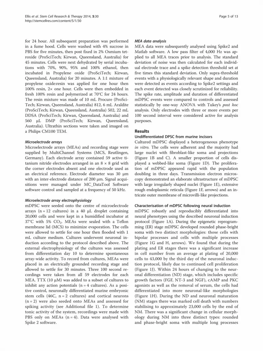
for 24 hour. All subsequent preparation was performedin a fume hood. Cells were washed with 4% sucrose inPBS for five minutes, then post fixed in 2% Osmium tet-roxide (ProSciTech; Kirwan, Queensland, Australia) for45 minutes. Cells were next dehydrated by serial incuba-tions with 70%, 90%, 95% and 100% ethanol, thenincubated in Propylene oxide (ProSciTech; Kirwan,Queensland, Australia) for 20 minutes. A 1:1 mixture ofpropylene oxide:resin was applied for one hour then100% resin, 2× one hour. Cells were then embedded infresh 100% resin and polymerized at 70°C for 24 hours.The resin mixture was made of 10 mL Procure (ProSci-Tech; Kirwan, Queensland, Australia) 812, 6 mL Araldite(ProSciTech; Kirwan, Queensland, Australia) 502, 22 mLDDSA (ProSciTech; Kirwan, Queensland, Australia) and560 μL DMP (ProSciTech; Kirwan, Queensland,Australia). Ultrathin sections were taken and imaged ona Philips CM100 TEM.
Microelectrode arraysMicroelectrode arrays (MEAs) and recording stage weresupplied by MultiChannel Systems (MCS, Reutlingen,Germany). Each electrode array contained 59 active ti-tanium nitride electrodes arranged in an 8 × 8 grid withthe corner electrodes absent and one electrode used asan electrical reference. Electrode diameter was 30 μmwith an inter-electrode distance of 200 μm. Signal acqui-sitions were managed under MC_DataTool Softwaresoftware control and sampled at a frequency of 50 kHz.
Microelectrode array electrophysiologymDPSC were seeded onto the centre of microelectrodearrays (n = 12 cultures) in a 40 μL droplet containing20,000 cells and were kept in a humidified incubator at37°C with 5% CO2. MEAs were sealed with a Teflonmembrane lid (MCS) to minimize evaporation. The cellswere allowed to settle for one hour then flooded with 1mL culture medium. Cultures underwent neuronal in-duction according to the protocol described above. Theexternal electrophysiology of the cultures was assessedfrom differentiation day 10 to determine spontaneousarray-wide activity. To record from cultures, MEAs wereplaced in an electrically grounded recording stage andallowed to settle for 30 minutes. Three 100 second re-cordings were taken from all 59 electrodes for eachMEA. TTX (10 μM) was added to a subset of cultures toinhibit any action potentials (n = 4 cultures). As a posi-tive control, neuronally differentiated murine embryonicstem cells (46C, n = 2 cultures) and cortical neurons(n = 2) were also seeded onto MEAs and assessed forspiking activity (see Additional file 1). To determinetonic activity of the system, recordings were made withPBS only on MEAs (n = 4). Data were analysed withSpike 2 software.
MEA data analysisMEA data were subsequently analysed using Spike2 andMatlab software. A low pass filter of 4,000 Hz was ap-plied to all MEA traces prior to analysis. The standarddeviation of noise was then calculated for each individ-ual electrode trace and a spike detection threshold set atfive times this standard deviation. Only supra-thresholdevents with a physiologically relevant shape and durationwere detected as events according to Spike2 settings andeach event detected was closely scrutinized for reliability.The spike rate, amplitude and duration of differentiatedmDPSC events were compared to controls and assessedstatistically by one-way ANOVA with Tukey’s post hocanalysis. Only electrodes with three or more events per100 second interval were considered active for analysispurposes.
ResultsUndifferentiated DPSC from murine incisorsCultured mDPSC displayed a heterogeneous phenotypein vitro. The cells were adherent and the majority hadlarge nuclei with fibroblast-like soma and projections(Figure 1B and C). A smaller proportion of cells dis-played a webbed-like soma (Figure 1D). The prolifera-tion of mDPSC appeared rapid with the populationdoubling in three days. Transmission electron micros-copy demonstrated an elaborate ultrastructure of mDPSCwith large irregularly shaped nuclei (Figure 1E), extensiverough endoplasmic reticula (Figure 1F, arrows) and an in-tricate outer membrane of microvilli-like projections.
Characterisation of mDPSC following neural inductionmDPSC robustly and reproducibly differentiated intoneural phenotypes using the described neuronal inductionprotocol (Figure 1A). During the epigenetic reprogram-ming (ER) stage mDPSC developed rounded phase-brightsoma with two distinct morphologies: those cells withbipolar processes and cells with multiple processes(Figure 1G and H, arrows). We found that during theplating and ER stages there was a significant increasein cell number from an average at plating of 20,000cells to 43,000 by the third day of the neuronal induc-tion protocol, likely due to continued cell proliferation(Figure 1J). Within 24 hours of changing to the neur-onal differentiation (ND) stage, which includes specificgrowth factors (FGF, NT-3 and NGF), cAMP and PKCagonists as well as the removal of serum, the cells haddifferentiated into more neuronal-like morphologies(Figure 1H). During the ND and neuronal maturation(NM) stages there was marked cell death with numbersstabilising to approximately 23,000 cells by the end ofNM. There was a significant change in cellular morph-ology during NM into three distinct types: roundedand phase-bright soma with multiple long processes
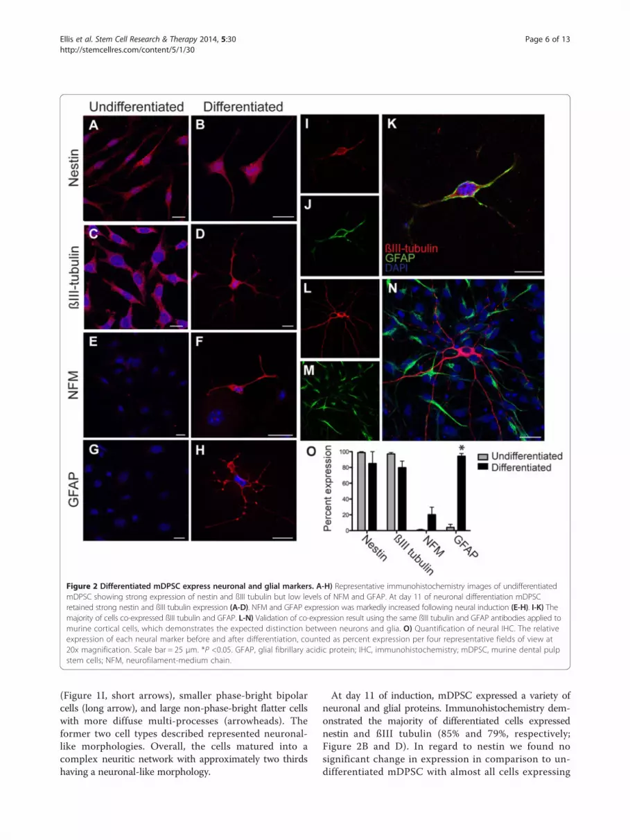
Figure 2 Differentiated mDPSC express neuronal and glial markers. A-H) Representative immunohistochemistry images of undifferentiatedmDPSC showing strong expression of nestin and ßIII tubulin but low levels of NFM and GFAP. At day 11 of neuronal differentiation mDPSCretained strong nestin and ßIII tubulin expression (A-D). NFM and GFAP expression was markedly increased following neural induction (E-H). I-K) Themajority of cells co-expressed ßIII tubulin and GFAP. L-N) Validation of co-expression result using the same ßIII tubulin and GFAP antibodies applied tomurine cortical cells, which demonstrates the expected distinction between neurons and glia. O) Quantification of neural IHC. The relativeexpression of each neural marker before and after differentiation, counted as percent expression per four representative fields of view at20x magnification. Scale bar = 25 μm. *P <0.05. GFAP, glial fibrillary acidic protein; IHC, immunohistochemistry; mDPSC, murine dental pulpstem cells; NFM, neurofilament-medium chain.
Ellis et al. Stem Cell Research & Therapy 2014, 5:30 Page 6 of 13http://stemcellres.com/content/5/1/30
(Figure 1I, short arrows), smaller phase-bright bipolarcells (long arrow), and large non-phase-bright flatter cellswith more diffuse multi-processes (arrowheads). Theformer two cell types described represented neuronal-like morphologies. Overall, the cells matured into acomplex neuritic network with approximately two thirdshaving a neuronal-like morphology.
At day 11 of induction, mDPSC expressed a variety ofneuronal and glial proteins. Immunohistochemistry dem-onstrated the majority of differentiated cells expressednestin and ßIII tubulin (85% and 79%, respectively;Figure 2B and D). In regard to nestin we found nosignificant change in expression in comparison to un-differentiated mDPSC with almost all cells expressing

Ellis et al. Stem Cell Research & Therapy 2014, 5:30 Page 7 of 13http://stemcellres.com/content/5/1/30
this neural precursor protein (Figure 2A, B and O). ßIIItubulin expression reduced from 97% in undifferentiatedmDPSC (Figure 2C) to 79% following induction (Figure 2Dand O). There was a marked co-expression of nestin andßIII tubulin in undifferentiated and differentiated mDPSC.Using a mature neuronal marker, neurofilament-mediumchain (NFM) [20], expression increased considerably from2% in the undifferentiated mDPSC up to 20% following in-duction (Figure 2E and F). GFAP expression was seen in4% of undifferentiated mDPSC and increased significantlyto 94% in differentiated cells (Figure 2G, H and O). Unex-pectedly, the majority of differentiated cells that expressedßIII-tubulin also co-expressed GFAP (Figure 2I-K). To val-idate this finding of co-expression the same antibodiesagainst ßIII-tubulin and GFAP were used in cultures ofprimary E19 murine cortical cells and clearly indicateddiscrete expression of these proteins with respect to theirneural cell types (Figure 2L-N).We next investigated neurally differentiated mDPSC
for a range of antigens expressed by mature neuralcells to determine whether the expression patternswere consistent with peripheral or central nervoussystem cell types. We found that 5% of mDPSC at in-duction day 11 expressed the central and peripheralglial marker, S100, and 87% were positive for acetyl-choline specific neurons, ChAT (Figure 3A and B, re-spectively). There was also specific expression ofmarkers for GABAergic (GAD65/67, 5%) and glutamater-gic neurons (vGlut2, 15%), but not dopaminergic neurons
Figure 3 Differentiated mDPSC produce central and peripheral nervocentral and peripheral nervous system glial marker, S100 (A), and acetylchoof GAD 65/67 (C) and vGlut2 (D) but not TH (E). Scale bar = 25 μm. ChAT, cmDPSC, murine dental pulp stem cells; TH, tyrosine hydroxylase; vGlut2, vesic
with a lack of tyrosine hydroxylase immunoreactivity(Figure 3C, D and E, respectively).
Neural network properties of differentiated mDPSCDifferentiated mDPSC demonstrated variable propertiesindicative of neural networks. Cells were subject to wholecell patch clamp analysis and the results were grouped intoneuronal-like cells that were either clustered (Figure 4A)or isolated from each other (Figure 4B). Amplifier-reportedcapacitance of clustered cells (M = 26.64 ± 12, n = 23 cells)was significantly greater than that of isolated cells (M =14.32 ± 7.2, n = 12; P <0.01, Figure 4C). However, the re-ported capacitance of clustered cells was likely a grossunderestimation of actual values due to the amplifier limi-tations in compensating for such large current dissipation.Nevertheless, the reported capacitance of isolated cells wasaccurate and demonstrated the distinction between theclasses of differentiated mDPSC.The nature of cell-cell contacts within clusters was in-
vestigated further using immunohistochemistry. Con-nexin 43 (Cx43), a common gap junction protein, wasexpressed widely in differentiated (Figure 4D) but notundifferentiated mDPSC (Figure 4E). Cx43 was moreabundantly expressed in cell clusters, as would be ex-pected (Figure 4F). We did not find synapsin 1 expres-sion in differentiated cells (data not shown).To assess the functionality of these gap junctions, Lucifer
yellow and neurobiotin tracer dye were injected into a sin-gle cell through a patch pipette and allowed to spread
us system markers. At day 11 of differentiation mDPSC expressline-specific enzyme, ChAT (B). mDPSC also show positive expressionholine acetyltransferase; GAD65/57, glutamic acid decarboxylase 65/67;ular glutamate transporter 2.

Figure 4 Network connectivity of differentiated mDPSC. Representative images of differentiated mDPSC in a cluster of many cells (A) and asan isolated unit (B). C) Capacitance of clustered cells (n = 23) was significantly greater than isolated cells (n = 12) measured by whole cell patchclamp analysis. IHC shows mDPSC expression of connexin 43 (Cx43) in differentiated (D) but not undifferentiated cells (E). F) Cx43 (yellow) ismost highly expressed within clusters of ßIII-tubulin (red) positive differentiated mDPSC (iii). G) Image of a clustered mDPSC injected with Luciferyellow and neurobiotin does not show any dye coupling with adjacent cells. Lucifer yellow injection into a clustered differentiated human DPSCdoes show dye coupling through numerous adjacent cells observed under bright field (H) and ultraviolet light (I). Scale bar = 25 μm. **P <0.01.IHC, immunohistochemistry; mDPSC, murine dental pulp stem cells.
Ellis et al. Stem Cell Research & Therapy 2014, 5:30 Page 8 of 13http://stemcellres.com/content/5/1/30
through membrane pores or gap junctions of sufficient size(n = 9 patched cells). Surprisingly, there was no evidence ofLucifer yellow or neurobiotin spread to adjacent cells indifferentiated mDPSC cultures indicating that gap junc-tions were not permeable to small molecules (Figure 4G).In contrast, extensive Lucifer yellow dye spread was seenfollowing injection into a single human DPSC after 14 daysof the same neuronal differentiation protocol (n = 3 humanDPSCs injected, Figure 4H and I). These data indicate aphysiological distinction between species.
Differentiated mDPSC express L-type voltage gated Ca2+
channelsTo determine the presence of neuron-specific ion chan-nels, whole cell patch clamp analysis was performed onundifferentiated mDPSC and mDPSC following neuralinduction. We found voltage-gated L-type calcium chan-nels in 21 of 27 cells with neuronal-like morphology.Figure 5 shows current traces from a representativeneuronal-like differentiated cell recorded in response to500 ms voltage steps in the presence of 10 mM Ca2+

Figure 5 Differentiated mDPSC express voltage-gated Ca2+ currents. A) Representative whole cell patch clamp recording of an isolateddifferentiated mDPSC with multiprocessor neuronal morphology showing typical voltage-gated Ca2+ current in response to increasing voltagesteps; 500 ms voltage steps were applied from −50 to +50 mV in 10 mV increments. B) There is a two-fold increase in current amplitude in responseto the addition of 10 mM Ba2+. C) The current–voltage relationship of voltage-dependent current from one representative cell with neuronal morphologyshowing sensitivity to 10 mM Ba2+ addition. The amplitude of the current was normalised to cell capacitance. D) Representative L-type Ca2+ currentsrecorded from undifferentiated mDPSC (UD, green) and differentiated DPSC (D, black) in the presence of 10 mM Ca2+ and following theaddition of Ba2+ (D, red), in response to 100 ms voltage ramps from −120 to +120 mV. mDPSC, murine dental pulp stem cells.
Ellis et al. Stem Cell Research & Therapy 2014, 5:30 Page 9 of 13http://stemcellres.com/content/5/1/30
(Figure 5A) or 10 mM Ba2+ (Figure 5B) in the bath solu-tion. Figure 5C shows the current–voltage relationshipof the Ca2+ current from the same cell. The current amp-litude increased more than two-fold upon Ba2+ addition.To normalise the measured currents for variable cell size,the changes in amplitude were expressed as changes incurrent densities (pA/pF). In contrast, undifferentiatedmDPSC produced only small L-type Ca2+ currents inresponse to 100 ms voltage ramps from −120 to +120mV with an amplitude much lower than that fromdifferentiated mDPSC (Figure 5D). No evidence ofTTX-sensitive voltage-gated Na+ channels (n = 6) orTEA-sensitive K+ channels (n = 6) was observed indifferentiated cells.
Networks of neuronal-like differentiated mDPSC do notdemonstrate action potentialsTo investigate the network electrophysiology of differen-tiated mDPSC, cells underwent neuronal induction onMEAs (n = 12 cultures). Each MEA was assessed forextracellular electrical activity between days 10 and 20with one culture surviving to 34 days. Measurementswere taken a maximum of once every two days as in-creased use led to cell death and infection. Standardnoise levels were recorded at 5 to 8 μV due to the small30 μm electrodes of MEAs. Numerous spike events wereidentified in all 12 differentiated mDPSC cultures that
satisfied the parameters of an action potential accordingto predetermined settings of amplitude, duration andshape. Figure 6Ai shows a representative singe electrodetrace from one mDPSC-derived culture at differentiationday 16, such as that seen in Figure 6B. It shows oneevent that passed the spike detection threshold over thefour second period. These events were common acrossmany electrodes and had an average 1 ms duration,however they were insensitive to 10 μM TTX adminis-tration (n = 4 TTX controls, data not shown). Figure6Aii and 6Aiii show representative traces of neuronallydifferentiated murine embryonic stem cells (mESC) andmurine cortical cultures, respectively, each of which hadlarger and more frequent spike events. Average max-imum spike rate per 100-second recording for mDPSC-derived cultures was 2.26 (± 0.751) spikes and was notsignificantly different from control PBS-only cultures(2.12 ± 0.335, P >0.05, Figure 6C). Cohen’s d demon-strated a small effect size between groups; d = 0.019(−0.61,1.01). Comparatively, the maximum spike rates ofdifferentiated mESC and cortical cultures were both higherat 25.75 ± 17.31 (P >0.05) and 1,028 ± 229 (P <0.001) spikesper 100 seconds, respectively. The effect size of each com-parison was very large (mESC; d = 2.52 (1.64,3.51); corticalcells; d = 10.12). Furthermore, as can be seen in Figure 6A,event amplitude of mDPSC cultures was far smaller thanthat of the other cell types. Events from mDPSC cultures

Figure 6 Extracellular electrophysiology of differentiated mDPSC. Twelve differentiated mDPSC cultures were assayed on MEAs and assessedfor network activity. Ai) Differentiated mDPSC cultures exhibited numerous short, low amplitude events that surpassed a threshold of detection.These events were smaller and less numerous than those seen in mESC (Aii) and cortical (Aiii) cultures. B) A representative image of a centralregion of nine electrodes of a MEA with mDPSC at day 11 of differentiation. Electrode diameter is 30 μm. C) Maximum spike rate of mDPSCevents within a 100 second bin was significantly lower than mESC and cortical signals and not distinguishable from PBS-only events. D) Boxplotsof the mean amplitude of spike events. Spike amplitides were significantly lower in mDPSC cultures than control mESC or cortical cultures andwere not significantly different from PBS-only control traces. E) A representative trace of a single oscillation event observed in a mDPSC MEAculture from day 32 of differentiation. The epicenter of oscillatory activity occurred at electrode 13 (e13, black) and was repeated at numerousadjacent electrodes, such as e23 (red), in phasic synchrony. A distant electrode, e75 (ii, blue), did not show any obvious oscillatory activity. F) Thespectral power density of oscillatory activity in 6E reveals its broad frequency range peaking at 95 Hz that is consistent across the three representativeelectrodes shown. e13 (red) has the greatest intensity as observed visually followed by e23 and also e75 whose oscillations could not be detected by eye.**P <0.01, ***P <0.0001. ESC, muring embryonic stem cells; mDPSC, murine dental pulp stem cells; MEA, microelectrode array.
Ellis et al. Stem Cell Research & Therapy 2014, 5:30 Page 10 of 13http://stemcellres.com/content/5/1/30
had an average amplitude of −8.38 ± 1.77 μV, which wasnot distinguishable from PBS-only cultures (−8.12 ± 0.59μV, P >0.05, d = 0.16 (−1.0,0.68)). By contrast, the averageamplitude of mESC events was greater at −12.08 ± 1.1 μV(P >0.05, d = 2.52 (1.78,3.33)) and cortical cultures signifi-cantly greater at −25.75 ± 8.57 μV (P <0.001, d = 2.45(1.82,3.13)) per 100 second bin (Figure 6D). From thesedata we conclude that the events observed in mDPSC-derived neural cultures were not spontaneous actionpotentials.We recorded oscillatory-like electrical activity over a
population of local electrodes on day 32 (n = 1 culture).
Each oscillation event lasted approximately 400 ms andoccurred up to five times per 100 second recording. Theepicenter of the oscillations occurred at a single electrode(e13), which had the greatest amplitude of activity(Figure 6E). Noise levels between oscillation periodson this electrode (e13) were similar to those of the sur-rounding electrodes. Numerous nearby electrodes sup-ported the same oscillatory patterns but with reducedamplitude. Figure 6E shows representative traces ofone oscillation event from e13, an adjacent electrode,e23, and a distant electrode over 1 mm away, e75,which does not display an obvious oscillatory pattern.
Ellis et al. Stem Cell Research & Therapy 2014, 5:30 Page 11 of 13http://stemcellres.com/content/5/1/30
The overlay of e13 (black) with adjacent e23 (red)showed that the electrical oscillations from both elec-trodes were in phase. The power spectral density dem-onstrated a broad frequency peak at 95Hz in both e13and e23 (Figure 6F). Interestingly, the control tracefrom e75 also showed a smaller peak at the same fre-quency, indicative of weak electrical spread across theentire MEA.
DiscussionWe have shown that DPSC derived from mouse incisorsgive rise to immature neuronal-like cells. Following neuronalinduction, mDPSC expressed neuronal cytoplasmic proteins,neurotransmitter-specific markers and functional voltage-gated L-type Ca2+ channels. The majority of mDPSCdeveloped over time into networks with high gap junc-tion protein expression and did not demonstrate spon-taneous action potentials. These data suggest thatmDPSC undergo neuronal differentiation in vitro, butto a limited maturity with properties more consistentwith early neuronal development.We identified that the majority of undifferentiated
mDPSC expressed nestin and ßIII-tubulin which sug-gests a neurogenic potential; however, maintenance ofthe expression of these proteins in the differentiated cul-tures suggested continued immature phenotype as sup-ported by electrophysiological data. This study, to thebest of our knowledge, presents the first evidence ofneuronal differentiation of DPSC from murine incisors.Following neural induction, mDPSC expressed the moremature neural markers NFM and GFAP. Consistent withother published findings, we also observed that mDPSC-derived neural cells co-expressed neuronal and glialmarkers [21]. It is not uncommon for these seeminglydistinct markers to be co-expressed in the developing,but not the mature, CNS [22,23], thereby providing fur-ther evidence for the early stage of neural developmentof these cells. In summary, the immunophenotype ofneural cells derived from mDPSC indicated a mixture ofcentral and peripheral nervous system cell types. Thiswas supported by expression of GFAP and S100, amarker of central and peripheral glial cells. In addition,we found that neuronal-like cells expressed cholinergic,GABAergic and glutaminergic markers. The differenti-ation of stem cells from human deciduous tooth dentalpulp into dopaminergic neuron-like cells in vitro haspreviously been reported [24], indicating the potentialfor directing the differentiation of DPSC toward a cen-tral lineage.From the neural inductive protocol used throughout
these experiments we propose that mDPSC undergoneuronal differentiation in vitro but to a limited maturity.This is supported by high expression of a gap junctionprotein, Cx43, lack of synapsin expression, functional Na+
and K+ ion channels and a lack of spontaneous action po-tentials. We found clusters of cellular networks dominatedby gap junctions rather than synapse protein expression.In vivo nervous system development proceeds from primi-tive gap junction signalling and later, during maturation, issuperseded by synaptic signalling [25]. Cx43, in particular,is involved with neural precursor proliferation, neural dif-ferentiation and neurite outgrowth [26-29]. Moreover,Cx43 blockade decreases the rate of mature neuronal de-velopment in the mouse P19 carcinoma cell line, indicat-ing its central role in nervous system maturation [27]. It isunclear what role is retained by connexins expressed inmDPSC-derived neural networks, as we saw no evidenceof dye coupling, as would be expected if functional gapjunctions were present. Non-channel functions of connexinsare also of central importance to neural development. Theyare involved in cell-to-cell adhesion [30] and small moleculerelease through hemichannels, which is important forfunctions such as cell migration and neurite outgrowth[29,31]. In particular, hemichannel-mediated ATP re-lease stimulates Ca2+ waves in early neural develop-ment, which has been linked with motoneuron, axonand dendritic development [32,33].Differentiated mDPSC did not produce spontaneous
action potentials or express the ion channels necessaryto support them. Rather, we found an abundance ofL-type voltage-gated Ca2+ channels in differentiatedcells, in contrast to low levels of Ca2+ currents in un-differentiated mDPSC. Ca2+ channels are known to beabundant in developing cortical neurons [34,35], andcalcium transients have been shown to be an import-ant regulator of neurogenesis and neurite extension[36,37], often dependent on L-type calcium channelsignaling [34]. In contrast, human DPSCs reliably de-veloped voltage-gated Na+ and K+ currents in vitrousing the same and another differentiation protocol[2,5], highlighting an important distinction betweenDPSC from different species. We made an isolated in-teresting observation that in a more mature mDPSCculture there were oscillatory electrical patterns inthe gamma frequency range. This suggests the pos-sible development of spontaneous network activity asfound in vivo. It is well understood that oscillationsare central to the development of neural networksduring embryogenesis and early postnatal develop-ment; however, these are typically of a lower beta fre-quency range [25,38]. Gap junctions are intrinsic tosustaining such neural oscillations, as gap junctionblockade causes a reduction or cessation of oscillatoryactivity [25,39]. Gap junction signalling may be re-sponsible for the oscillations seen in this study butthis was not empirically tested. We suggest that theseoscillations may demonstrate the emergence of earlyelectrical activity within networks of developing

Ellis et al. Stem Cell Research & Therapy 2014, 5:30 Page 12 of 13http://stemcellres.com/content/5/1/30
mDPSC. We made multiple attempts to pursue thisin vitro observation but there were technical difficul-ties with maintaining these cultures long-term onMEA surfaces.In contrast to that reported during human DPSC
neuronal differentiation [5], we found that mDPSC num-bers initially increased during the plating and epigeneticreprogramming stages of differentiation, likely due tocontinued progenitor cell proliferation. Following the re-moval of media serum and the addition of specific PKCagonists and neural growth factors, cell numbers de-clined. We suggest this may be due to the death of cellsthat did not have the intrinsic potential to respond toneuronal induction as well as the arrest of cell prolifera-tion caused by long term PKC activation and the removalof serum [40]. Our observation of a stable cell numberduring the maturation stage of induction suggests thatremaining cells showed neither a net loss nor gain duringthis stage of the protocol.Alternative methods of neural induction may be con-
sidered for more efficient differentiation of mDPSC.Neurosphere generation, for example, may provide amicroenvironment more reminiscent of in vivo develop-ment to support mature neural differentiation as seenpreviously with rat incisor and human DPSC [16,41].
ConclusionsIn conclusion, we have successfully generated neuronal-like cells from murine incisor DPSC to an immaturestage of development. Our findings encourage the use ofmDPSC to develop mouse models of autologous neuraltherapeutic transplantations for pre-clinical studies.
Additional file
Additional file 1: Supplementary methods. Preparation of murinecortical cultures and murine embryonic stem cells.
Abbreviationsα-MEM: alpha-modified Eagle’s medium; ANOVA: analysis of variance;ChAT: choline acetyltransferase; CsGlutamine: caesium glutamine;Cx43: connexin 43; DAPI: 4’,6-diamidino-2-phenylindole; DPSC: dental pulpstem cell; (D)MEM: (Dulbecco’s) modified Eagle’s medium; EGTA: ethyleneglycol tetraacetic acid; ER: epigenetic reprogramming; ESC: embryonic stemcell; FBS: fetal bovine serum; FGF: fibroblast growth factor; GAD65/67: glutamic acid decarboxylase 65/67; GFAP: glial fibrillary acidic protein;GTP: guanosine triphosphate; hDPSC: human dental pulp stem cell;hFF: human foreskin fibroblasts; IBMX: 3-isobutyl-1-methylxanthine;IHC: immunohistochemistry; MCS: multi channel systems; mDPSC: murinedental pulp stem cell; MEA: microelectrode array; mESC: murine embryonicstem cells; ND: neuronal differentiation; NFM: neurofilament-medium chain;NGF: nerve growth factor; NM: neuronal maturation; NT-3: neurotrophin 3;PBS: phosphate-buffered saline; PFA: paraformaldehyde; PKC: Protein KinaseC; TEA: Tetraethylammonium; TEM: transmission electron microscopy;TH: tyrosine hydroxylase; TPA: phorbol 12-myristate13-acetate; TTX: tetrodotoxin; vGlut2: vesicular glutamate transporter 2.
Competing interestsSAK currently receives funding from Mesoblast. All experiments wereperformed prior to the establishment of this partnership and Mesoblast hadno input into experimental conceptualisation, interpretation of data ormanuscript review. All other authors declare that they have no competinginterests.
Authors’ contributionsKME contributed to experimental design, performed all experiments,collected, analysed and interpreted data and wrote the manuscript.DCO was involved in experimental conceptualisation and design,supervision of work, data interpretation and manuscript review. MDL wasinvolved in experimental conceptualisation and design, isolation ofmurine dental pulp stem cells, supervision of work and manuscriptreview. GYR assisted in experimental design, provided intracellularelectrophysiology expertise and assisted in manuscript review. SAKcontributed to experimental conceptualisation and design, datainterpretation and considerable manuscript review. All authors read andapproved the final manuscript.
AcknowledgementsThe authors would like to acknowledge the work by Lauren Sandeman withmouse embryonic stem cells and the patch clamp technical assistanceprovided by Nathan Scrimgeour.
Author details1Adelaide Centre for Neuroscience Research, University of Adelaide, Adelaide,South Australia, Australia. 2School of Medical Sciences, University of Adelaide,Adelaide, South Australia, Australia. 3School of Molecular and BiomedicalScience, University of Adelaide, Adelaide, South Australia, Australia. 4Schoolof Medicine, University of Adelaide, Adelaide, South Australia 5005, Australia.5Stroke Research Programme, University of Adelaide, Adelaide, SouthAustralia, Australia.
Received: 1 November 2013 Revised: 3 February 2014Accepted: 19 February 2014 Published: 27 February 2014
References1. Gronthos S, Mankani M, Brahim J, Robey PG, Shi S: Postnatal human dental
pulp stem cells (DPSCs) in vitro and in vivo. Proc Natl Acad Sci U S A 2000,97:13625–13630.
2. Arthur A, Rychkov G, Shi S, Koblar SA, Gronthos S: Adult human dentalpulp stem cells differentiate toward functionally active neurons underappropriate environmental cues. Stem Cells 2008, 26:1787–1795.
3. Karbanová J, Soukup T, Suchánek J, Pytlík R, Corbeil D, Mokry J:Characterization of dental pulp stem cells from impacted third molarscultured in low serum-containing medium. Cells Tissues Organs 2011,193:344–365.
4. Karaöz E, Dogan BN, Aksoy A, Gacar G, Akyüz S, Ayhan S, Genç ZS, YürükerS, Duruksu G, Demircan PC, Sariboyaci AE: Isolation and in vitrocharacterisation of dental pulp stem cells from natal teeth. Histochem CellBiol 2010, 133:95–112.
5. Kiraly M, Porcsalmy B, Pataki A, Kadar K, Jelitai M, Molnar B, Hermann P, GeraI, Grimm WD, Ganss B, Zsembery A, Varga G: Simultaneous PKC and cAMPactivation induces differentiation of human dental pulp stem cells intofunctionally active neurons. Neurochem Int 2009, 55:323–332.
6. Arthur A, Shi S, Zannettino AC, Fujii N, Gronthos S, Koblar SA: Implantedadult human dental pulp stem cells induce endogenous axon guidance.Stem Cells 2009, 27:2229–2237.
7. Kiraly M, Kadar K, Horvathy DB, Nardai P, Racz GZ, Lacza Z, Varga G, GerberG: Integration of neuronally predifferentiated human dental pulp stemcells into rat brain in vivo. Neurochem Int 2011, 59:371–381.
8. Leong WK, Henshall TL, Arthur A, Kremer KL, Lewis MD, Helps SC, Field J,Hamilton-Bruce MA, Warming S, Manavis J, Vink R, Gronthos S, Koblar S:Human adult dental pulp stem cells enhance poststroke functional recoverythrough non-neural replacement mechanisms. Stem Cells Trans Med 2012,1:177–187.
9. Huang AH, Snyder BR, Cheng PH, Chan AW: Putative dental pulp-derivedstem/stromal cells promote proliferation and differentiation of endogenousneural cells in the hippocampus of mice. Stem Cells 2008, 26:2654–2663.

Ellis et al. Stem Cell Research & Therapy 2014, 5:30 Page 13 of 13http://stemcellres.com/content/5/1/30
10. Nosrat IV, Smith CA, Mullally P, Olson L, Nosrat CA: Dental pulp cellsprovide neurotrophic support for dopaminergic neurons anddifferentiate into neurons in vitro; implications for tissue engineeringand repair in the nervous system. Euro J Neurosci 2004, 19:2388–2398.
11. Balic A, Aguila HL, Caimano MJ, Francone VP, Mina M: Characterization ofstem and progenitor cells in the dental pulp of erupted and uneruptedmurine molars. Bone 2010, 46:1639–1651.
12. Balic A, Mina M: Characterization of progenitor cells in pulps of murineincisors. J Dental Res 2010, 89:1287–1292.
13. Janebodin K, Horst OV, Ieronimakis N, Balasundaram G, Reesukumal K,Pratumvinit B, Reyes M: Isolation and characterization of neural crest-derivedstem cells from dental pulp of neonatal mice. PLoS One 2011, 6:e27526.
14. Guimarães ET, Cruz GS, de Jesus AA, Lacerda De Carvalho AF, Rogatto SR,Pereira LDV, Ribeiro-Dos-Santos R, Soares MBP: Mesenchymal and embryoniccharacteristics of stem cells obtained from mouse dental pulp. Arch Oral Biol2011, 56:1247–1255.
15. Nozaki T, Ohura K: Gene expression profile of dental pulp cells duringdifferentiation into an adipocyte lineage. J Pharmacol Sci 2011, 115:354–363.
16. Sasaki R, Aoki S, Yamato M, Uchiyama H, Wada K, Okano T, Ogiuchi H:Neurosphere generation from dental pulp of adult rat incisor. Eur JNeurosci 2008, 27:538–548.
17. Varga G, Bori E, Kallo K, Nagy K, Tarjan I, Racz GZ: Novel possiblepharmaceutical research tools: stem cells, gene delivery and theircombination. Curr Pharm Des 2013, 19:133–141.
18. Sasaki R, Aoki S, Yamato M, Uchiyama H, Wada K, Okano T, Ogiuchi H:Tubulation with dental pulp cells promotes facial nerve regeneration inrats. Tissue Eng Part A 2008, 14:1141–1147.
19. Gronthos S, Arthur A, Bartold PM, Shi S: A method to isolate and cultureexpand human dental pulp stem cells. Methods Mol Biol 2011, 698:107–121.
20. Carden MJ, Trojanowski JQ, Schlaepfer WW, Lee VMY: Two-stageexpression of neurofilament polypeptides during rat neurogenesis withearly establishment of adult phosphorylation patterns. J Neurosci 1987,7:3489–3504.
21. Sakai K, Yamamoto A, Matsubara K, Nakamura S, Naruse M, Yamagata M,Sakamoto K, Tauchi R, Wakao N, Imagama S, Hibi H, Kadomatsu K, IshiguroN, Ueda M: Human dental pulp-derived stem cells promote locomotorrecovery after complete transection of the rat spinal cord by multipleneuro-regenerative mechanisms. J Clin Invest 2012, 122:80–90.
22. Messam CA, Hou J, Major EO: Coexpression of nestin in neural and glialcells in the developing human CNS defined by a human-specific anti-nestinantibody. Exp Neurol 2000, 161:585–596.
23. Draberova E, Del Valle L, Gordon J, Markova V, Smejkalova B, Bertrand L, deChadarevian JP, Agamanolis DP, Legido A, Khalili K, Draber P, Katsetos CD:Class III beta-tubulin is constitutively coexpressed with glial fibrillaryacidic protein and nestin in midgestational human fetal astrocytes:implications for phenotypic identity. J Neuropathol Exp Neurol 2008,67:341–354.
24. Wang J, Wang X, Sun Z, Yang H, Shi S, Wang S: Stem cells fromhuman-exfoliated deciduous teeth can differentiate into dopaminergicneuron-like cells. Stem Cells Dev 2010, 19:1375–1383.
25. Dupont E, Hanganu IL, Kilb W, Hirsch S, Luhmann HJ: Rapid developmentalswitch in the mechanisms driving early cortical columnar networks.Nature 2006, 439:79–83.
26. Todorova MG, Soria B, Quesada I: Gap junctional intercellularcommunication is required to maintain embryonic stem cells in anon-differentiated and proliferative state. J Cell Physiol 2008, 214:354–362.
27. Bani-Yaghoub M, Bechberger JF, Underhill TM, Naus CC: The effects of gapjunction blockage on neuronal differentiation of human NTera2/cloneD1 cells. Exp Neurol 1999, 156:16–32.
28. Bani-Yaghoub M, Underhill TM, Naus CC: Gap junction blockage interfereswith neuronal and astroglial differentiation of mouse p19 embryonalcarcinoma cells. Dev Gen 1999, 24:69–81.
29. Belliveau DJ, Bani-Yaghoub M, McGirr B, Naus CC, Rushlow WJ: Enhancedneurite outgrowth in PC12 cells mediated by connexin hemichannelsand ATP. J Biol Chem 2006, 281:20920–20931.
30. Butkevich E, Hulsmann S, Wenzel D, Shirao T, Duden R, Majoul I: Drebrin isa novel connexin-43 binding partner that links gap junctions to thesubmembrane cytoskeleton. Curr Biol 2004, 14:650–658.
31. Elias LA, Wang DD, Kriegstein AR: Gap junction adhesion is necessary forradial migration in the neocortex. Nature 2007, 448:901–907.
32. Konur S, Ghosh A: Calcium signaling and the control of dendriticdevelopment. Neuron 2005, 46:401–405.
33. Webb SE, Miller AL: Calcium signalling during embryonic development.Nat Rev Mol Cell Biol 2003, 4:539–551.
34. Tang F, Dent EW, Kalil K: Spontaneous calcium transients in developingcortical neurons regulate axon outgrowth. J Neurosci 2003, 23:927–936.
35. Dolmetsch RE, Pajvani U, Fife K, Spotts JM, Greenberg ME: Signaling to thenucleus by an L-type calcium channel- calmodulin complex through theMAP kinase pathway. Science 2001, 294:333–339.
36. Owens DF, Kriegstein AR: Patterns of intracellular calcium fluctuation inprecursor cells of the neocortical ventricular zone. J Neurosci 1998,18:5374–5388.
37. Gomez TM, Spitzer NC: In vivo regulation of axon extension andpathfinding by growth-cone calcium transients. Nature 1999, 397:350–355.
38. Khazipov R, Luhmann HJ: Early patterns of electrical activity in thedeveloping cerebral cortex of humans and rodents. Trends Neurosci 2006,29:414–418.
39. Peinado A: Immature neocortical neurons exist as extensive syncitialnetworks linked by dendrodendritic electrical connections. J Neurophysiol2001, 85:620–629.
40. Racz GZ, Szucs A, Szlavik V, Vag J, Burghardt B, Elliott AC, Varga G: Possiblerole of duration of PKC-induced ERK activation in the effects of agonistsand phorbol esters on DNA synthesis in Panc-1 cells. J Cell Biochem 2006,98:1667–1680.
41. Widera D, Grimm WD, Moebius JM, Mikenberg I, Piechaczek C, Gassmann G,Wolff NA, Thevenod F, Kaltschmidt C, Kaltschmidt B: Highly efficient neuraldifferentiation of human somatic stem cells, isolated by minimallyinvasive periodontal surgery. Stem Cells Dev 2007, 16:447–460.
doi:10.1186/scrt419Cite this article as: Ellis et al.: Neurogenic potential of dental pulp stemcells isolated from murine incisors. Stem Cell Research & Therapy2014 5:30.
Submit your next manuscript to BioMed Centraland take full advantage of:
• Convenient online submission
• Thorough peer review
• No space constraints or color figure charges
• Immediate publication on acceptance
• Inclusion in PubMed, CAS, Scopus and Google Scholar
• Research which is freely available for redistribution
Submit your manuscript at www.biomedcentral.com/submit