Embed Size (px)
Citation preview

NEUROIMAGING AND COGNITIVE CHANGES
DURING DÉJÀ VU
Norbert Kovacs1, Tibor Auer1,2,3,4, Istvan Balas2,
Kazmer Karadi1,5, Katalin Zambo6, Attila Schwarcz2,4,
Peter Klivenyi7, Hennric Jokeit8, Krisztina Horvath1,
Ferenc Nagy1, and Jozsef Janszky1,4
This is a preprint of an article published in Epilepsy & Bahavior Epilepsy Behav 2009;14(1):190-196
1University of Pecs, Department of Neurology, Pecs, Hungary 2University of Pecs, Department of Neurosurgery, Pecs, Hungary 3Biomedizinische NMR Forschungs GmbH am Max-Planck-Institut für Biophysikalische Chemie, Göttingen, Germany 4Pécs Diagnostic Institute, University of Pecs, Pecs, Hungary
5University of Pecs, Institute of Behavioral Sciences, Pecs, Hungary 6University of Pecs, Department of Nuclear Medicine, Pecs, Hungary 7University of Szeged, Department of Neurology, Szeged, Hungary 8Swiss Epilepsy Center, Zurich, Switzerland
Correspondence to: Dr. Norbert Kovacs University of Pecs Department of Neurology 7623, Pecs, Ret utca 2, Tel: +36 70 222-1178 Fax: +36 72 535-911 Email: [email protected]
Running title: Déjà vu elicited by pallidal DBS
Number of text pages: 14, number of figures 2 (color: 1), number of tables: 1.
Supplementary data attached to the manuscript!
2
Abstract
Purpose: The cause or the physiological role of déjà vu (DV) in healthy people is unknown.
The pathophysiology of DV-type epileptic aura is also unresolved. Here we present a 22-year-old
woman treated by deep brain stimulation (DBS) of the left internal globus pallidus due to
hemidystonia. On a certain stimulations settings, DBS elicited reproducible DVs.
Methods: Neuropsychological tests and SPECT were performed during DBS-evoked DV
and during normal DBS stimulation without DV experiences.
Results: SPECT during DBS-evoked DV revealed hyperperfusions in the right side
(contralateral to the electrode) hippocampus and other limbic structures. Neuropsychological
examinations performed during several evoked DV experiences revealed disturbances in the non-
verbal memory.
Conclusion: Our results confirm the role of mesiotemporal structures in the pathogenesis of
DV. We hypothesize that the individual neuroanatomy, disturbance in gamma oscillations or in
dopaminerg system can play a role in DBS-elicited DV experiences in our patient.
Keywords: déjà vu, deep brain stimulation, pallidum, SPECT, SISCOM, adverse reaction,
epilepsy, dystonia, MRI, aura

3
Introduction
Déjà vu (DV) is “any subjectively inappropriate impression of familiarity of present experience
with an undefined past” [1]. Although 60-80% of the healthy population has experienced déjà vu
[2], DV aura is one of the leading symptoms of temporal lobe epilepsy [3] occurring in 10% of all
epileptic auras [4]. DV aura is the most characteristic symptom of familial mesial temporal lobe
epilepsy reported in about one-third of these patients [5, 6]. DV experiences occurring in other
brain disorders (e.g. depression [7] and schizophrenia [8]) were also analyzed in more details.
Studying DV is difficult because of its rarity, unpredictable appearance and heterogenity.
Contrary to spontaneous DV experiences, the induced DV can be examined objectively during
presurgical evaluation of epilepsy [3]. The stimulation of the temporal structures [9] or the rhinal
cortex [10] often, but not always [11] could elicit DV in temporal lobe epilepsy (TLE) patients. Most
studies found that DV was confined to the non-dominant temporal lobe and accompanied by
hallucinations or illusions [3, 4, 9, 11]. Furthermore, DV experience could also be provoked by
electrical stimulation of brain structures contralateral to the epileptic focus suggesting that DV can
also be elicited in normal brain tissue [12].
Despite numerous investigations, the pathomechanism of DV experiences in healthy people
is still unknown. The “small seizure” theory is based on the clinical findings that DV is an aura
type in TLE. It is hypothesized that in non-epileptic population a “small temporal lobe seizure” may
elicit déjà vu episodes without producing clinical seizures [13, 14]. However, there are several
counter-arguments about this theory: the DV is much more common than TLE [15, 16] and only a
portion of TLE patients have DV auras [17].
‘Tape-recorder’ theory [18] is one of the most known DV theories applying the dual-
processing approach. It assumes that two different memory-related processes that normally
synchronously work become asynchronous or one process becomes activated in the absence of
the other. Under normal condition, the memory encoding (“recording head”) and the retrieval
(“playing head”) work with appropriate timing and synchronization. According this speculation if the
new sensory information is simultaneously encoded and retrieved, the sensory input is
accompanied by familiarity resulting in déjà vu feeling. Based on the clinical evaluation of

4
electrically evoked déjà vu experiences of 16 TLE patients underwent presurgical depth electrode
implantation, Bancaud and colleagues [9] postulated the neuroanatomical bases for the ‘tape-
recorder’ theory. Because association cortical and limbic areas encode the holistic memory of an
event, and perceptual information is encoded by the temporal neocortex and stored in the
hippocampus, the inappropriate activation of centers can lead to the experience of déjà vu. Similar
electrophysiological results [19] expanded Bancaud’s theory by the complementary assumption of
parallel neuronal networks underlying encoding and retrieval [20].
Interestingly, a recent case-study described a “drug-induced” DV, where a patient
experienced recurrent DV after receiving a combination of amantadine and phenylpropanolamine
[21]. Because both drugs can facilitate dopaminergic neurotransmission and recent animal studies
proved that hippocampal dopaminerg systems are involved in the spatial memory processes [22],
this case suggests that an increased dopaminerg activity probably might play a crucial role in the
development of DV [21].
In a very recent case report, hypothalamic deep brain simulation (DBS) was found to evoke
detailed autobiographic memories, but not déjà vu [23].
These data inspired us to systematically analyze the pathophysiology of DV by applying
functional neuroimaging (SPECT) and neuropsychological batteries in a case where the DBS of left
internal globus pallidum (GPi) elicited DV. As far as the authors are aware, this is the first study
utilizing direct, reproducible and integrative neuropsychological and neuroimaging investigations
during DV.
Methods
The patient
The 22-year-old female university student was born with a right-sided spastic hemiparesis
due to a perinatal injury. Although the strength of the right limbs normalized, the abnormal posture
of the right upper limb was noticed at the age of 2 months, developing to a drug-refractory and
painful secondary hemidystonia. The locomotive and intellectual development was otherwise
normal.

5
Brain MRI revealed a 4x15x18mm lesion in the left GP. At age 22, she underwent a
microelectrode-guided Medtronic quadripolar 3389 DBS electrode implantation into the left
posteroventral GPi without perioperative complications. The patient has given written informed
consent to the whole surgical procedure, pre- and postsurgical examinations and publication of the
present case report, whereas the study was also approved by the Local Ethical Committee.
Stimulation settings
On the first postoperative day, contact 1 was activated in a monopolar mode (C+1-, 120µs,
130Hz, 3.2V) without any adverse reactions. The patient was admitted to the Neurological ward on
the 3rd postoperative week to learn the use of patient controller. During the testing of electrodes,
we noticed that the monopolar stimulation of contact 0 with the amplitude exceeding 2.7V could
elicit several DV episodes. As the turning on or off the stimulation had an immediate effect on this
experience, we assumed it to be a stimulation-related adverse reaction. The impedance of contact
0 (C+0-, 3.2V, 120µs, 130Hz) was 562 Ohms.
SPECT
The current safety regulations do not permit using functional MRI during DBS [24].
Therefore, we performed 99mTc-hexamethylpropyleneamineoxime SPECT to study the
pathophysiology of DV because 99mTc HMPAO has faster binding (2-10 minutes) compared to
PET tracers [25].
The SPECT acquisition was performed 1 month postoperatively. To exclude the long-term
duration effect of DBS, the ‘baseline’ SPECT was obtained during the normal stimulation of
contact 1 (C+1-, 3.2V, 120µs, 130Hz). For studying the pathophysiology behind DV, three days
later we stimulated simultaneously both contact 0 and 1 (C+0-1-, 120µs, 130Hz, 3.2V referred to
as DV-inducing stimulation). Analogously to epilepsy studies we defined this setting as ‘ictal’
SPECT.
As the 99m-TcHMPAO tracer (750MBq) was administered immediately after starting the DV-
inducing stimulation and the patient experienced numerous DV experiences during the first 5
minutes of stimulation, we assumed that the tracer binding in the ‘ictal’ SPECT represented the

6
combination of acute DV induction and normal pallidal stimulation. Therefore, the subtraction of
baseline from the ‘ictal’ SPECT images theoretically corresponded only to the areas activated
during DV experience. The comparison of baseline and ‘ictal’ SPECT was performed by the
subtraction ictal SPECT co-registered to MRI (SISCOM) method, which is also applied in the
presurgical evaluation of epilepsy [26].
Neuropsychological tests
The subject underwent neuropsychological examinations three times: 9 months
preoperatively, 2 months postoperatively with and without DV eliciting stimulation. There was 1 day
difference between the postoperative examinations, where Rey and Medical College of Georgia
Complex Figure, Rey 8/64, Benton Visual Retention, Boston Naming and Rey Auditory Verbal
Learning Tests were obtained [27, 28]. (Supplementary data).
Results
The appearance of DV
Preoperatively the patient had never experienced any DVs. Immediately after turning on the
DV-inducing stimulation; an unusual and obscure feeling appeared. Besides discomfort and slight
disturbance, the subject had intact reality sense; she was able to observe what is going on around
her and maintain verbal and behavioral responsiveness. We defined this experience as ‘standby-
state for DV (SSDV)’. The SSDV persisted until the stimulation of contact 0 was turned off or the
amplitude of stimulation was lowered below 2.7 Volts.
During SSDV, sudden, impulse DV experiences appeared lasting 4-5 seconds. At these
occasions she felt that the situation seemed familiar. No visual, auditory illusions or hallucinations
accompanied the DV. Besides, the patient felt neither the ability to predict the future nor unreality
about the current circumstances.
DV appeared more frequent immediately after turning the stimulation on (approximately 2-5
DV during the first 5-10 minutes) and became rarer as the time went by (approximately another 3-5
DV in the first hour and 2-3 in the second hour). Interestingly, DV appeared only if her eyes were
7
open and questions were addressed directly to the patient (e.g. “What is the name of your
physiotherapist?”). However, not all of the direct questioning of the patient elicited DV. Standard
digital EEG recording showed physiologic activity and there were no clinical signs of epilepsy
either.
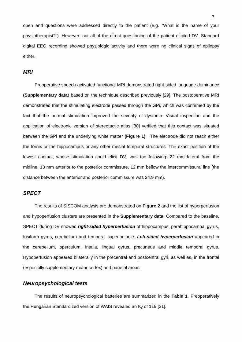
MRI
Preoperative speech-activated functional MRI demonstrated right-sided language dominance
(Supplementary data) based on the technique described previously [29]. The postoperative MRI
demonstrated that the stimulating electrode passed through the GPi, which was confirmed by the
fact that the normal stimulation improved the severity of dystonia. Visual inspection and the
application of electronic version of stereotactic atlas [30] verified that this contact was situated
between the GPi and the underlying white matter (Figure 1). The electrode did not reach either
the fornix or the hippocampus or any other mesial temporal structures. The exact position of the
lowest contact, whose stimulation could elicit DV, was the following: 22 mm lateral from the
midline, 13 mm anterior to the posterior commissure, 12 mm bellow the intercommissural line (the
distance between the anterior and posterior commissure was 24.9 mm).
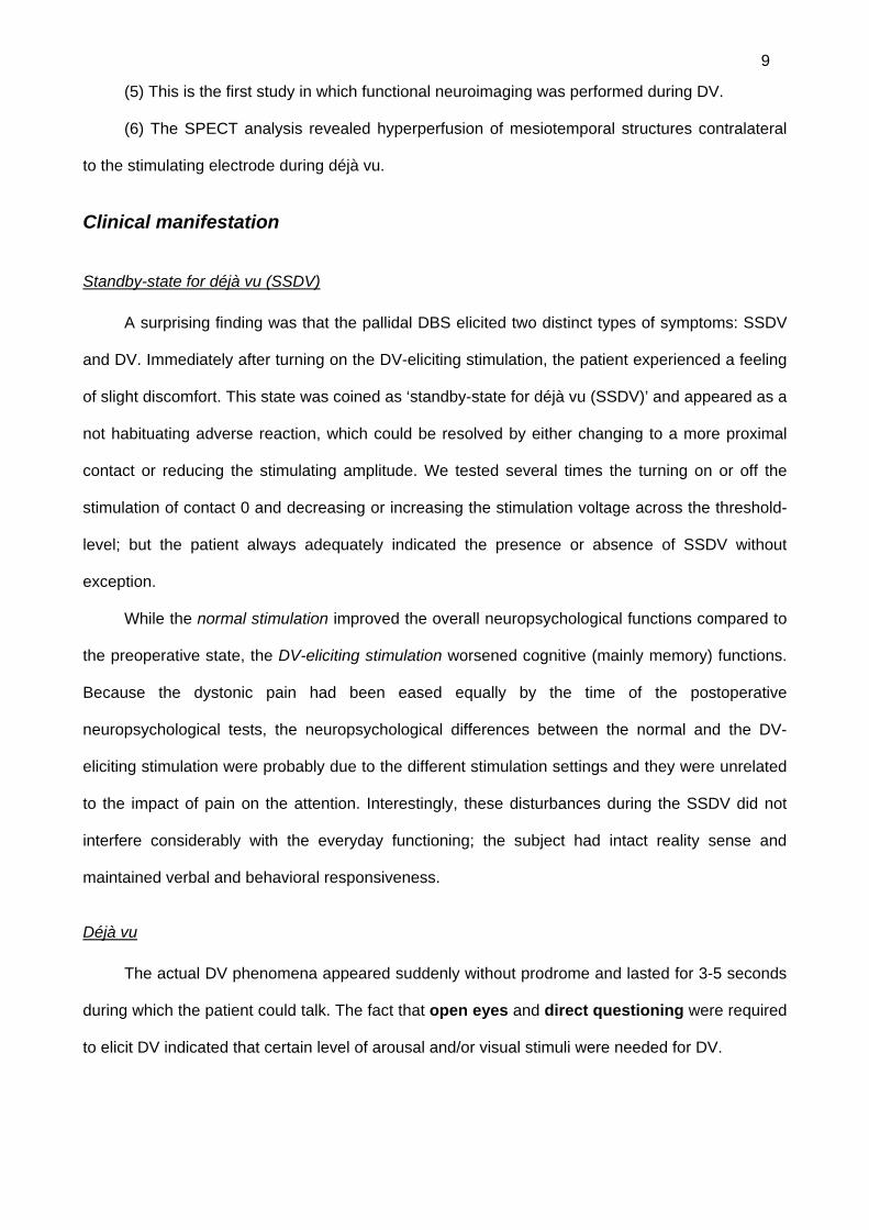
SPECT
The results of SISCOM analysis are demonstrated on Figure 2 and the list of hyperperfusion
and hypoperfusion clusters are presented in the Supplementary data. Compared to the baseline,
SPECT during DV showed right-sided hyperperfusion of hippocampus, parahippocampal gyrus,
fusiform gyrus, cerebellum and temporal superior pole. Left-sided hyperperfusion appeared in
the cerebellum, operculum, insula, lingual gyrus, precuneus and middle temporal gyrus.
Hypoperfusion appeared bilaterally in the precentral and postcentral gyri, as well as, in the frontal
(especially supplementary motor cortex) and parietal areas.
Neuropsychological tests
The results of neuropsychological batteries are summarized in the Table 1. Preoperatively
the Hungarian Standardized version of WAIS revealed an IQ of 119 [31].

8
During normal stimulation there was better performance in verbal fluency, copy score of
Complex Figure, delayed memory of Benton test and Trail Making A and B version compared to
the preoperative state. However, the recall memory of Complex figure became worsened during
the normal stimulation.
During DV-eliciting stimulation there was some deterioration in verbal fluency and Boston
Naming tests. Furthermore, severe non-verbal memory impairment could be demonstrated by
Complex Figure Test, and Rey 8/64 visual learning test compared to either the normal stimulation
or the preoperative state.
Discussion
In our patient the electrically evoked déjà vu phenomenon could be easily studied because of
several reasons: (1) it could be repeated without any restraints; (2) déjà vu could be elicited
several times; (3) no other neurological phenomenon disturbed the evaluation (e.g. altered
consciousness during an epileptic seizure); (4) the anatomical site of electrical stimulation could be
determined by a high-resolution MRI-examination; (5) the functional changes in the brain during
DV could be identified by functional neuroimaging and (6) the examinations did not bother or harm
the patient.
The main findings of our study:
(1) This case report demonstrates that pallidal DBS may evoke déjà vu as an adverse
reaction, which can be resolved by changing to a more proximal contact or reducing the stimulation
amplitude.
(2) The elicited déjà vu can be characterized as the ‘non-pathological’ form [15].
(3) Contrary to the unreliable results of electrical stimulation in epileptic patients [12], in our
case the setting of certain stimulation parameters reliably and reproducibly elicited déjà vu.
(4) Neuropsychological examinations indicate prominent alterations of visual learning and
retrieval during déjà vu. During normal stimulation the performance of most neuropsychological
tests improved compared to the preoperative state, which might be associated with dystonic pain-
assuaging effect of normal stimulation. Conversely, déjà vu-eliciting stimulation slightly worsened
the verbal and severely impaired the non-verbal memory performance.
9
(5) This is the first study in which functional neuroimaging was performed during DV.
(6) The SPECT analysis revealed hyperperfusion of mesiotemporal structures contralateral
to the stimulating electrode during déjà vu.
Clinical manifestation
Standby-state for déjà vu (SSDV)
A surprising finding was that the pallidal DBS elicited two distinct types of symptoms: SSDV
and DV. Immediately after turning on the DV-eliciting stimulation, the patient experienced a feeling
of slight discomfort. This state was coined as ‘standby-state for déjà vu (SSDV)’ and appeared as a
not habituating adverse reaction, which could be resolved by either changing to a more proximal
contact or reducing the stimulating amplitude. We tested several times the turning on or off the
stimulation of contact 0 and decreasing or increasing the stimulation voltage across the threshold-
level; but the patient always adequately indicated the presence or absence of SSDV without
exception.
While the normal stimulation improved the overall neuropsychological functions compared to
the preoperative state, the DV-eliciting stimulation worsened cognitive (mainly memory) functions.
Because the dystonic pain had been eased equally by the time of the postoperative
neuropsychological tests, the neuropsychological differences between the normal and the DV-
eliciting stimulation were probably due to the different stimulation settings and they were unrelated
to the impact of pain on the attention. Interestingly, these disturbances during the SSDV did not
interfere considerably with the everyday functioning; the subject had intact reality sense and
maintained verbal and behavioral responsiveness.
Déjà vu
The actual DV phenomena appeared suddenly without prodrome and lasted for 3-5 seconds
during which the patient could talk. The fact that open eyes and direct questioning were required
to elicit DV indicated that certain level of arousal and/or visual stimuli were needed for DV.

10
The occurrence of DV seemed to have a habituating feature, i.e. in the first 5 minutes
starting the stimulation of contact 0 produced more frequent DV appearance (2-5 during the first 5
minutes), which became rarer with time.
Because DV is a transitory experience lasting for a few seconds, obviously we could not
apply neuropsychological tests targeting directly it. However, on the ‘ictal’ SPECT scan we could
identify the brain structures involved in DV, since the patient experienced several DVs during the
interval of tracer binding. Therefore, the subtraction of ictal and baseline images presumably
corresponds to only those areas responsible for both DV.
Neuroanatomical considerations
Based on the position of the stimulation electrode, we might speculate the anatomical target
responsible for DV. The postoperative MRI scans demonstrated that contact 0 was situated in the
border of GPi and the underlying white matter. The spreading of electrical current is roughly
spherical around the activated contact and in no case extends underneath the electrode [32]. We
can also presume that the electricity can diffuse approximately up to 4 millimeters in a low
impedance tissue from the surface of the contact [33]. Because mesial temporal structures (e.g.
hippocampus and fornix) are situated bellow the lowest contact, the direct stimulation of these
mesiotemporal structures was unlikely.
We performed subtraction SPECT analysis comparing ‘ictal’ and baseline SPECT images.
The subtracted picture revealed hyperperfusion in the right mesial structures contralateral to the
stimulation. There was also hyperperfusion of ipsilateral (left) operculum, insula, precuneus and
lingual gyrus. This finding is in harmony with TLE studies [9] demonstrating that the elicitation of
DV involves mainly mesiotemporal structures.
Proposed theories on pathophysiology on DV
We cannot explain the pathophysiology of DBS-evoked DV. However, we can provide some
possible theories concluding from our results.
As far as the authors are aware, there is not even a single case published describing the
occurrence of déjà vu after pallidotomies, despite several ten thousands were performed worldwide

11
[34, 35]. Because ablative procedures could not evoke DVs and certain stimulation settings were
required to produce DV, we may presume the importance of high-frequency stimulation in the
background.
(1) We can hypothesize that several independent constellations together led to DV: (i) the
altered memory functions of the SSDV and a certain combination of (ii) visual and (iii) direct verbal
stimuli requiring memory matching processes. This hypothesis is based on the fact that the DV-
inducing stimulation itself was unable to produce DV in the lack of simultaneous visual and verbal
stimuli; it elicited “only” the SSDV with non-verbal memory disturbances. Clinically, DV appeared
only when the patient was addressed with questions and her eyes were open. The type of visual
stimuli seemed to be irrelevant in the elicitation, because DV appeared in both dim and bright
rooms, either with or without persons in the visual field. On the contrary, only direct questioning of
the patient was able to elicit DV, other auditory stimuli (e.g. environmental noises, conversation
between other persons not involving the patient) never did so. However, not all questioning elicited
DV. One potential explanation for this phenomenon might be that direct questioning requires high-
level attention, interpretation and memory processing.
(2) Probably not only the left GPi stimulation itself, but also the individual (atypical)
neuroanatomy might play a role in the development of DV. The atypical language dominance
suggests that due to the perinatal brain injury our patient developed an atypical brain anatomy [36-
39]. Because the reorganization of the injured brain is not always complete, similar modalities
might be present bilaterally and the division of labor between dominant and non-dominant
hemisphere functions might not be completed. Normally, language-dominant hemisphere is more
strongly engaged in memory processing of verbal material [40]. However, in our case the individual
neuroanatomy might have produced that non-verbal functions are confined to both hemispheres. If
this speculation is true, the disturbing effect of left GPi DBS might have greater impact on one
hemisphere and less on the other one producing DV in a similar way described in the dual-
processing theories. The elicitation of DV seemed to be a habituating phenomenon. An explanation
for this habituation may be that the memory processing system(s) noticed the DVs as errors.
Possibly, this error recognition enabled the system to adapt to the SSDV resulting in less DV
experiences over time.

12
(3) Dopaminerg system might also take part in the elicitation of DV. Previously a case study
demonstrated that the concomitant use of amantadine and phenylpropanolamine could produce
recurring DVs [21]. Based on PET studies carried out on Parkinson’s disease patients, the pallidal
DBS might interfere with endogenous dopamine release and/or dopamine-receptor functions [41].
Recent study investigating drug effects on schizophrenic patients found that the dosage of
antipsychotic (anti-dopaminerg) drugs positively correlated with the frequency of DV experiences
[42], which contradicts that DV is a result of an elevated dopaminerg activity, but underline the role
of dopaminerg system in the pathophysiology of DV. Therefore, we cannot rule out the possibility
that not the stimulation itself, but the disturbances in the dopaminerg system were responsible for
DV. However, one could expect that considerably more time would be needed to alter the
dopaminergic system.
(4) Another hypothesis might be that DV is caused by separation of two main memory
systems: familiarity and recollection. Recent neuropsychological data suggest that recognition
memory applies of two different mechanisms: recollection and familiarity discrimination [43, 44].
Therefore, in certain situations it is possible to recognize that a person or a subject is familiar even
without the ability to recollect any particulars about it. Electrophysiological studies in monkeys
demonstrated that the neurons in the rhinal cortex responded differently on the basis of the relative
familiarity or novelty of presentation of the stimuli, which occurred much rarely in the hippocampus
[45]. Therefore, possibly two separate memory compartments may co-exist: one, including the
hippocampus, enables recall and conscious recollection of contextual elements, while the other
system, including the structures of the parahippocampal gyrus, is important for familiarity
judgments [46, 47]. In our case the DV-eliciting stimulation altered the non-verbal memory
processes, including recollection as demonstrated by neuropsychological tests. Thus, during déjà
vu both recollection and familiarity discrimination was affected, which contradicts that déjà vu is a
result of a separation of these two memory systems. We might presume that the elicitation of DV
might be the result of the altered dual-processing due to atypical functional localization of
recollection and familiarity systems. Possibly, the heavy memory-related load induced by the visual
and auditory stimuli produced a delay in memory processing between the hemispheres resulting in
false familiarity recognition.

13
(5) The non-dominant localization of the GPi stimulating electrode might have also had
an impact on DV-elicitation. Previous electrophysiological studies confined the DV to the non-
dominant hemisphere. However, these studies were limited to the TLE [9, 19].
(6) Furthermore, experimental studies demonstrated a functional relationship between the
hippocampus and the contralateral basal ganglia [48]. In rats, the electrical stimulation of GP
altered the contralateral hippocampal theta field activity presumably via a septohippocampal
pathway. This relationship between the GPi and hippocampal formation has not been verified in
humans yet. However, there are some indirect data suggesting this hypotheis, for example the
transitory unilateral ictal dystonia in TLE could be associated with hyperperfusion of the basal
ganglia ipsilateral to the seizure focus [49]. Alternatively, GPi stimulation could be accompanied by
hypoperfusion of the mesiotemporal structures [50].
(7) Analogously we can assume that high-frequency deep brain stimulation can interfere
with high-frequency (gamma) oscillations of the contralateral mesiotemporal structures,
which in turn play a crucial role in memory functions. Phase synchronization of gamma oscillations
of around 40-50 Hz is a general mechanism underlying transient functional coupling between
different neuroanatomical structures playing an important role in every aspect of memory functions
[51, 52]. Thus, one may expect that high-frequency DBS can interfere with the gamma oscillation in
the brain independent of the stimulating site. Moreover, novel studies hypothesize that déjà vu may
be related to the alteration of gamma-oscillations of mesiotemporal structures [51].
(8) Since the major output from GPi is to thalamus and from there to cortex, its role also
should also be considered for the development of déjà vu. However, several thousands of GPi
DBS had been implanted worldwide either for Parkinson’s disease or dystonia, and not even a
single case report mentioned déjà vu as a stimulation-related side-effect. Therefore, the sole
alteration of pallido-thalamico-cortical pathways is unlikely to produce DV experiences.
In a recent case report, hypothalamic DBS could evoke detailed autobiographic memories
[23]. The stimulation increased recollection, but not familiarity-based recognition, nor déjà vu. EEG
source localization suggested an activity in the mesiotemporal structures [23]. Summing up, our
case with Hamani’s case, we may assume that in the case of certain constellations (e.g. during
specific electrode localizations, stimulation parameters and individual neuroanatomy), the deep

14
brain stimulation can interfere with some memory-related processes. However, these types of
memory alterations due to deep brain stimulation are rather rare.
Open questions and limitations
Several questions remain unanswered in our case. We only compared the functional
neuroimages between the DV-eliciting and the “normal” pallidal stimulation. Because during the
interval of tracer binding four déjà vu episodes occurred, the difference of these scans probably
identifies the anatomical structures responsible for DV. However, we should have obtained a third
SPECT scan during SSDV without DV during tracer binding to compare the different activations
between the DV and SSDV and between the SSDV and the normal stimulation. This way, we could
have identified those structures, which are directly responsible for the DV experience but does not
contribute to the SSDV. Moreover, the SPECT scan methodology (SISCOM) was adapted from
epilepsy studies, which is usually useful for seizures lasting more than 20-30 seconds. The single
observation in this patient of a DV lasting less than 10 seconds is of uncertain significance. To test
reproducibility, we planned wanted to repeat the baseline (normal stimulation) and ictal (DV-
eliciting stimulation) SPECT images, but respecting the request of our patient, we have forborne
from these acquisitions.
Acknowledgements
Hungarian State Research Funds (OTKA T043005 to N.K and FN., ETT 219/2006 and OTKA
F68720 to J.J., ETT 176/2006 to A.S.). Hungarian Neuroimaging Foundation (JJ), JJ and KK were
awarded by the Bolyai Fellowship (Republic of Hungary).
Disclosure of conflicts of interests
None of the authors has conflict of interest to disclosure.

15
References
[1] Neppe VM. The concept of déjá vu. Parapsychological Journal of South Africa 1983;4:1-10. [2] Brown AS. A review of the deja vu experience. Psychol Bull 2003;129:394-413. [3] Mullan S, Penfield W. Illusions of comparative interpretation and emotion. Arch Neurol
Psychiatry 1959;81:269-284. [4] Palmini A, Gloor P. The localizing value of auras in partial seizures: a prospective and
retrospective study. Neurology 1992;42:801-8. [5] Berkovic SF, McIntosh A, Howell RA, Mitchell A, Sheffield LJ, Hopper JL. Familial temporal
lobe epilepsy: a common disorder identified in twins. Ann Neurol 1996;40:227-35. [6] Striano P, Gambardella A, Coppola A, Di Bonaventura C, Bovo G, Diani E, Boaretto F,
Egeo G, Ciampa C, Labate A, Testoni S, Passarelli D, Manna I, Sferro C, Aguglia U, Caranci F, Giallonardo AT, Striano S, Nobile C, Michelucci R. Familial mesial temporal lobe epilepsy (FMTLE) : a clinical and genetic study of 15 Italian families. J Neurol 2008;255:16-23.
[7] Sno HN, Linszen DH. The deja vu experience: remembrance of things past? Am J Psychiatry 1990;147:1587-95.
[8] Adachi T, Adachi N, Takekawa Y, Akanuma N, Ito M, Matsubara R, Ikeda H, Kimura M, Arai H. Deja vu experiences in patients with schizophrenia. Compr Psychiatry 2006;47:389-93.
[9] Bancaud J, Brunet-Bourgin F, Chauvel P, Halgren E. Anatomical origin of deja vu and vivid 'memories' in human temporal lobe epilepsy. Brain 1994;117 ( Pt 1):71-90.
[10] Bartolomei F, Barbeau E, Gavaret M, Guye M, McGonigal A, Regis J, Chauvel P. Cortical stimulation study of the role of rhinal cortex in deja vu and reminiscence of memories. Neurology 2004;63:858-64.
[11] Gloor P, Olivier A, Quesney LF, Andermann F, Horowitz S. The role of the limbic system in experiential phenomena of temporal lobe epilepsy. Ann Neurol 1982;12:129-44.
[12] Halgren E, Walter RD, Cherlow DG, Crandall PH. Mental phenomena evoked by electrical stimulation of the human hippocampal formation and amygdala. Brain 1978;101:83-117.
[13] Stevens JR. Psychiatric consequences of temporal lobectomy for intractable seizures: a 20-30-year follow-up of 14 cases. Psychol Med 1990;20:529-45.
[14] Penfield W. The twenty-ninth Maudsley lecture: the role of the temporal cortex in certain psychical phenomena. J Ment Sci 1955;101:451-65.
[15] Neppe VM. The psychology of déjá vu. Have I been here before? Johannesburg: Witwaterstrand University Press; 1983.
[16] Neppe VM. Is deja vu a symptom of temporal lobe epilepsy? S Afr Med J 1981;60:907-8. [17] Sengoku A, Toichi M, Murai T. Dreamy states and psychoses in temporal lobe epilepsy:
mediating role of affect. Psychiatry Clin Neurosci 1997;51:23-6. [18] de Nayer A. [Deja vu: elaboration of a hypothetical model of explanation]. Psychiatr Clin
(Basel) 1979;12:92-6. [19] Gloor P. Experiential phenomena of temporal lobe epilepsy. Facts and hypotheses. Brain
1990;113 ( Pt 6):1673-94. [20] Wild E. Deja vu in neurology. J Neurol 2005;252:1-7. [21] Taiminen T, Jaaskelainen SK. Intense and recurrent deja vu experiences related to
amantadine and phenylpropanolamine in a healthy male. J Clin Neurosci 2001;8:460-2. [22] Wilkerson A, Levin ED. Ventral hippocampal dopamine D1 and D2 systems and spatial
working memory in rats. Neuroscience 1999;89:743-9. [23] Hamani C, McAndrews MP, Cohn M, Oh M, Zumsteg D, Shapiro CM, Wennberg RA,
Lozano AM. Memory enhancement induced by hypothalamic/fornix deep brain stimulation. Ann Neurol 2008;63:119-23.
[24] Kovacs N, Nagy F, Kover F, Feldmann A, Llumiguano C, Janszky J, Kotek G, Doczi T, Balas I. Implanted deep brain stimulator and 1.0-Tesla magnetic resonance imaging. J Magn Reson Imaging 2006;24:1409-12.

16
[25] Andersen AR. 99mTc-D,L-hexamethylene-propyleneamine oxime (99mTc-HMPAO): basic kinetic studies of a tracer of cerebral blood flow. Cerebrovasc Brain Metab Rev 1989;1:288-318.
[26] O'Brien B, O'Connor M, Mullan BP. Subtraction ictal SPECT co-registered to MRI in partial epilepsy: description and technical validation of the method with phantom and patient studies. Nucl Med Commun 1998;19:31-45.
[27] Hodges JR. Cognitive assessment for clinicians. Oxford: Oxford University Press; 1994. [28] Lezak M. Neuropsychological assesment. fourth edition ed. New York, USA: Oxford
University Press; 2004. [29] Janszky J, Mertens M, Janszky I, Ebner A, Woermann FG. Left-sided interictal epileptic
activity induces shift of language lateralization in temporal lobe epilepsy: an fMRI study. Epilepsia 2006;47:921-7.
[30] Schaltenbrand G, Wahren B. Atlas for the sterotaxy of the human brain. 2nd edition ed. Stuttgart, Germany: Thieme; 1977.
[31] Degrell I, Pek G, Nagy D, Nagy E, Hoyer S. Dementias, psychological tests, and neurotransmitters. Arch Gerontol Geriatr 1985;4:365-71.
[32] Butson CR, Cooper SE, Henderson JM, McIntyre CC. Patient-specific analysis of the volume of tissue activated during deep brain stimulation. Neuroimage 2007;34:661-70.
[33] Butson CR, Maks CB, McIntyre CC. Sources and effects of electrode impedance during deep brain stimulation. Clin Neurophysiol 2006;117:447-54.
[34] Guridi J, Lozano AM. A brief history of pallidotomy. Neurosurgery 1997;41:1169-80; discussion 1180-3.
[35] Gross CE, Boraud T, Guehl D, Bioulac B, Bezard E. From experimentation to the surgical treatment of Parkinson's disease: prelude or suite in basal ganglia research? Prog Neurobiol 1999;59:509-32.
[36] Jacola LM, Schapiro MB, Schmithorst VJ, Byars AW, Strawsburg RH, Szaflarski JP, Plante E, Holland SK. Functional magnetic resonance imaging reveals atypical language organization in children following perinatal left middle cerebral artery stroke. Neuropediatrics 2006;37:46-52.
[37] Muller RA, Watson CE, Muzik O, Chakraborty PK, Chugani HT. Motor organization after early middle cerebral artery stroke: a PET study. Pediatr Neurol 1998;19:294-8.
[38] Rasmussen T, Milner B. The role of early left-brain injury in determining lateralization of cerebral speech functions. Ann N Y Acad Sci 1977;299:355-69.
[39] Janszky J, Jokeit H, Heinemann D, Schulz R, Woermann FG, Ebner A. Epileptic activity influences the speech organization in medial temporal lobe epilepsy. Brain 2003;126:2043-51.
[40] Weber B, Fliessbach K, Lange N, Kugler F, Elger CE. Material-specific memory processing is related to language dominance. Neuroimage 2007;37:611-617.
[41] Nakajima T, Nimura T, Yamaguchi K, Ando T, Itoh M, Yoshimoto T, Shirane R. The impact of stereotactic pallidal surgery on the dopamine D2 receptor in Parkinson disease: a positron emission tomography study. J Neurosurg 2003;98:57-63.
[42] Adachi N, Adachi T, Akanuma N, Matsubara R, Ito M, Takekawa Y, Ikeda H, Arai H. Deja vu experiences in schizophrenia: relations with psychopathology and antipsychotic medication. Compr Psychiatry 2007;48:592-6.
[43] Hintzman DL, Caulton DA, Levitin DJ. Retrieval dynamics in recognition and list discrimination: further evidence of separate processes of familiarity and recall. Mem Cognit 1998;26:449-62.
[44] Bowles B, Crupi C, Mirsattari SM, Pigott SE, Parrent AG, Pruessner JC, Yonelinas AP, Kohler S. Impaired familiarity with preserved recollection after anterior temporal-lobe resection that spares the hippocampus. Proc Natl Acad Sci U S A 2007;104:16382-7.
[45] Xiang JZ, Brown MW. Differential neuronal encoding of novelty, familiarity and recency in regions of the anterior temporal lobe. Neuropharmacology 1998;37:657-76.
[46] Aggleton JP, Brown MW. Episodic memory, amnesia, and the hippocampal-anterior thalamic axis. Behav Brain Sci 1999;22:425-44; discussion 444-89.
[47] Bogacz R, Brown MW. Comparison of computational models of familiarity discrimination in the perirhinal cortex. Hippocampus 2003;13:494-524.

17
[48] Hallworth NE, Bland BH. Basal ganglia--hippocampal interactions support the role of the hippocampal formation in sensorimotor integration. Exp Neurol 2004;188:430-43.
[49] Newton MR, Berkovic SF, Austin MC, Reutens DC, McKay WJ, Bladin PF. Dystonia, clinical lateralization, and regional blood flow changes in temporal lobe seizures. Neurology 1992;42:371-7.
[50] Miyawaki E, Perlmutter JS, Troster AI, Videen TO, Koller WC. The behavioral complications of pallidal stimulation: a case report. Brain Cogn 2000;42:417-34.
[51] Herrmann CS, Demiralp T. Human EEG gamma oscillations in neuropsychiatric disorders. Clin Neurophysiol 2005;116:2719-33.
[52] Bruns A, Eckhorn R, Jokeit H, Ebner A. Amplitude envelope correlation detects coupling among incoherent brain signals. Neuroreport 2000;11:1509-14.
[53] Jokeit H, Makeig S. Different event-related patterns of gamma-band power in brain waves of fast- and slow-reacting subjects. Proc Natl Acad Sci U S A. 1994;91:6339-43.

18
Figure legends
Figure 1 The localisation of the stimulating electrode (A coronal MP-RAGE, B
coronal FLAIR and C sagittal MP-RAGE). Visual inspection and the application of
electronic version of the Schaltenbrand stereotactic atlas verified that the contact
responsible for DV was situated between the GPi and the underlying white matter. The
electrode did not hit the mesial temporal structures.

19
Figure 2. SISCOM analysis comparing déjà vu-eliciting stimulation (ictal) the normal
(interictal) SPECT superimposed on axial MRI scans. (yellow: hyperperfusion, blue:
hypoperfusion). Compared to the baseline, SPECT during DV showed right-sided
hyperperfusion of hippocampus, parahippocampal gyrus, fusiform gyrus, cerebellum and
temporal superior pole left-sided hyperperfusion appeared in the cerebellum, operculum,
insula, lingual gyrus, precuneus and middle temporal gyrus. Hypoperfusion appeared
bilaterally in the precentral and postcentral gyri, as well as, in the frontal (especially
supplementary motor cortex) and parietal areas.

20
Table
Table 1. Neuropsychological test results preoperatively and postoperatively with standard
stimulation (C+1-, 120µs, 130Hz, 3.2V) and déjà vu-eliciting stimulation settings (C+0-1-, 120µs,
130Hz, 3.2V).
Preoperative “Standard stimulation” “Déjà vu stimulation”
Digit span forward 5 4 5
Corsi 4 4 4
Trail making (A/B) 35 / 71 sec 24/44 27/48
Verbal fluency F:12, A:8, S:9,
category:26
P:17, E: 16, M: 13,
category: 17
F:9, A:9, S:7,
category: 20
Boston naming test Not obtained 60 57†
AVLT language
learning
Learning phase:
47 recalled words
New information:7,
Interferency:10
Delayed Recall: 10
Recognition: 100%
Learning phase:
49 recalled words
New information:4
Interferency:9
Delayed Recall: 11
Recognition: 100%
Learning phase:
54 recalled words
New information: 3†
Interferency:12
Delayed Recall: 14
Recognition: 100%
Rey and Medical
College of Georgia
Complex Figure
(copy/recall)
30/26 35/18 36/12
Benton Visual
retention test:
C-figure memory: 6/10
D-figure copy: 10/10
E-figure delayed
recall: 8/10
C-figure: 7/10
D-figure delayed
recall: 6/10†
Rey 8/64 visual
learning test Not obtained Learnt by the 6th trial No learning
†While the subject was performing these tests, she experienced a sudden déjà vu feeling
lasting for several seconds.

21
SUPPLEMENTARY DATA
Applied neuropsychological tests [27, 28]
Visual memory and Learning
1. Complex Figure Tests: Rey Complex Figure test was assessed by subject before
surgery. After copying the Rey Complex Figure, the patient has to draw it by memory in
immediate recall. Postoperatively (during both normal and déjà vu-inducing stimulation) two
figures of Medical College of Georgia Complex Figure test were applied in repeated
visual memory testing. In every Complex Figure Test standard scoring system was
administered with the maximum of 36 points. Each figure was divided into 18 different
blocks. When subject drawn properly placed, correct blocks, she got 2 points. In case of
properly paced and distorted or poorly placed and correct blocks 1 point was given.
Distorted, poorly placed blocks were scored with half point. Absent or not recognizable
blocks gave no points.
2. Rey 8/64 test is the visual pair of Rey Auditory Verbal Learning Test. During testing period
the neuropsychologist touch 8 squares one after the other in 8x8 square grids. Subject
must touch same squares in this grid. Ten trials were used to measure the visual learning
during testing. The result shows the trials when the visual learning was correct.
3. Benton Visual Retention Test is a widely applied method to measure the visual
perception, memory and construction abilities. Test consists of three forms: each C, D, and
E forms have 10 figure series. D form was used in perceptual session, where the subject
had to copy the given figure. In preoperatively C form the patient saw each figures in 5
seconds, after presentation she draw the figure immediately by memory. C form was
applied in the copying session of postoperative déjà vu elicitation trial. Figures of D forms
were used in 5 seconds delayed recall after 5 seconds presentation in postoperative
session with déjà vu elicitation. Figures of the E form were solely used during normal

22
stimulation period with above mentioned delayed recall trial. Each drawing was worth 1
point when the figures were drawn in correct mode. If drawing had error it did not get point.
Verbal Learning and Skills Tests:
1. Boston Naming Test consists of 60 line-drawing from high familiar to low familiar items.
Stimulus cues (e.g. this is a sea animal) and phonetic cues (e.g. first syllable) are
presented if items were unnamed.
2. Rey Auditory Verbal Learning Test measures verbal series learning ability using 15
common nouns (A and B list). Five presentation of A list was given. After each presentation
the subject had to recall the words from list. Learning over five trials was evaluated. After
fifth trial a B list was read and subject had to remember this new list. In 7th trial the subject
recalled the A list without auditory presentation. 8th trial presented the delayed recall after
20 minutes.
Intelligence quotient: The Hungarian version of WAIS was obtained [31].

23
Figure of supplementary data
Preoperative fMRI demonstrated right hemispherial language dominance during word
generation task.

24
Table of supplementary data
Hyperperfusion and hypoperfusion clusters and their size revealed by the SISCOM method.
Anatomical localization Number of voxels
Hyperperfusion Cluster: 1 168 Vermis_R
Cerebellum_R
Hyperperfusion Cluster: 2 20 Frontal_Inf_Orb_R
Hyperperfusion Cluster: 3 12 Temporal_Pole_Sup_R
Hyperperfusion Cluster: 4 36 Fusiform_R
Temporal_Inf_R
Hyperperfusion Cluster: 5 648 Fusiform_R
Hippocampal_R
ParaHippocampal_R
Cerebellum_R
Hyperperfusion Cluster: 6 60 Cerebelum_L
Hyperperfusion Cluster: 7 196 Frontal_Inf_Oper_L
Insula_L
Hyperperfusion Cluster: 8 436 Calcarine_L
Lingual_L
Precuneus_L
Cerebellum_L
Vermis_L
Hyperperfusion Cluster: 9 28 Temporal_Mid_L

25
Hypoperfusion Cluster 1 104 Cingulum_Ant_L
Caudate_L
Hypoperfusion Cluster: 2 44 Insula_L
Putamen_L
Hypoperfusion Cluster: 3 208 Cuneus_L
Precuneus_L
Hypoperfusion Cluster: 4 3056 Precentral_L
Precentral_R
Frontal_Sup_L
Frontal_Sup_R
Frontal_Mid_L
Frontal_Mid_R
Supp_Motor_Area_L
Supp_Motor_Area_R
Frontal_Sup_Medial_L
Occipital_Sup_R
Postcentral_L
Postcentral_R
Parietal_Sup_L
Parietal_Sup_R
Parietal_Inf_R
Angular_R
Precuneus_L
Precuneus_R
Paracentral_Lobule_L
Paracentral_Lobule_R
Hypoperfusion Cluster: 5 44 Postcentral_R
SupraMarginal_R
Hypoperfusion Cluster: 6 380 Postcentral_L
Parietal_Inf_L
SupraMarginal_L


