Embed Size (px)
Citation preview

Saadoun et al. Acta Neuropathologica Communications 2014, 2:35http://www.actaneurocomms.org/content/2/1/35
RESEARCH Open Access
Neuromyelitis optica MOG-IgG causes reversiblelesions in mouse brainSamira Saadoun1, Patrick Waters2, Gregory P Owens3, Jeffrey L Bennett3,4, Angela Vincent2
and Marios C Papadopoulos1*
Abstract
Introduction: Antibodies against myelin oligodendrocyte glycoprotein (MOG-IgG) are present in some neuromyelitisoptica patients who lack antibodies against aquaporin-4 (AQP4-IgG). The effects of neuromyelitis optica MOG-IgG inthe central nervous system have not been investigated in vivo. We microinjected MOG-IgG, obtained from patientswith neuromyelitis optica, into mouse brains and compared the results with AQP4-IgG.
Results: MOG-IgG caused myelin changes and altered the expression of axonal proteins that are essential for actionpotential firing, but did not produce inflammation, axonal loss, neuronal or astrocyte death. These changes wereindependent of complement and recovered within two weeks. By contrast, AQP4-IgG produced complement-mediatedmyelin loss, neuronal and astrocyte death with limited recovery at two weeks.
Conclusions: These differences mirror the better outcomes for MOG-IgG compared with AQP4-IgG patients and raisethe possibility that MOG-IgG contributes to pathology in some neuromyelitis optica patients.
Keywords: Antibody, Demyelination, Myelin oligodendrocyte glycoprotein, Neuromyelitis optica
IntroductionMost neuromyelitis optica (NMO) patients have IgGagainst aquaporin-4 (AQP4), here termed AQP4-IgG[1,2]. AQP4 is a water channel protein found in astro-cytes throughout the central nervous system (CNS), es-pecially in perivascular astrocyte foot processes [3]. Incultured cells, AQP4-IgG binds extracellular conform-ational domains of AQP4 and activates complement,causing cell lysis [4]. In mice, intracerebral injection ofAQP4-IgG activates co-injected human complement(Chu) and damages the astrocytes [5,6]. Inflammatorycells then enter the lesion causing further tissue injuryincluding demyelination and axonal damage. In AQP4-IgG NMO patients, recovery after an attack is usuallylimited [7-9].A few NMO patients without AQP4-IgG have IgG
against myelin oligodendrocyte glycoprotein (MOG-IgG),which recognize extracellular conformational domains ofMOG [10-13]. MOG is expressed on the outer surface ofCNS myelin sheaths and comprises about 0.05% of total
* Correspondence: [email protected] Neurosurgery Unit, St. George’s, University of London, London, UKFull list of author information is available at the end of the article
© 2014 Saadoun et al.; licensee BioMed CentrCommons Attribution License (http://creativecreproduction in any medium, provided the orDedication waiver (http://creativecommons.orunless otherwise stated.
myelin protein [14]. There is mounting evidence thatMOG-IgG NMO has more favorable clinical outcomethan AQP4-IgG NMO, with resolution of imaging abnor-malities [10,11,15,16]. It is currently unclear whetherMOG-IgG plays any role in NMO by causing lesions inthe CNS in vivo. Here we compared the effects of MOG-IgG with those of AQP4-IgG in the intracerebral injectionmouse model. We used total IgG from a normal subject(IgGCON) and from NMO patients with AQP4-IgG(IgGAQP4) or MOG-IgG (IgGMOG).
Materials and methodsIgG and complementNMO patients with MOG-IgG or AQP4-IgG were identi-fied using live cell-based assays. Briefly, AQP4-IgG andMOG-IgG positivity was determined by visualization ofbinding to human embryonic kidney cells, transfected withthe extracellular and trans-membrane domains of MOG orwith full-length M23-AQP4. Details of the assays are givenelsewhere [5,10,11,15]. IgG was purified using Protein Gfrom sera or plasmas of five patients with AQP4-IgGNMO, five MOG-IgG NMO or one healthy volunteer. Theeffect of injecting IgG and Chu from healthy volunteers intomouse brain was extensively investigated in our earlier
al Ltd. This is an Open Access article distributed under the terms of the Creativeommons.org/licenses/by/4.0), which permits unrestricted use, distribution, andiginal work is properly credited. The Creative Commons Public Domaing/publicdomain/zero/1.0/) applies to the data made available in this article,

Saadoun et al. Acta Neuropathologica Communications 2014, 2:35 Page 2 of 9http://www.actaneurocomms.org/content/2/1/35
studies [5,17,18]. The purified, dialysed and pooled totalIgG preparations (6 – 38 mg/ml) are termed IgGAQP4,IgGMOG and IgGCON. Clinical details of the 5 AQP4-IgG +[5] and 5 MOG-IgG+ [11,15] patients are given elsewhere.To deplete MOG-IgG, the IgGMOG was adsorbed by in-cubation with MOG-HEK cells until MOG-IgG becameundetectable (IgGMOG(AdsMOG-HEK)). IgGMOG adsorbedagainst untransfected HEK cells (IgGMOG(AdsHEK)) wasused as control. A chimeric mouse-human recombinantmonoclonal anti-mouse MOG antibody, MOG-IgG2B7,was produced as described [19]. Human recombinantmonoclonal anti-AQP4 IgG1, termed AQP4-IgG53, wasalso generated [20]. A measles virus-specific antibodytermed CON-IgG2B4 was used as isotype control [20].The source of Chu was fresh serum from healthyvolunteers [5].
MiceExperiments were performed at St. George’s, University ofLondon using CD1 mice 8 – 12 30 – 35 g, 8 – 12w old.Protocols were approved by the British Home Office(Project Licence, PPL 70/7081). After administering 2,2,2-tribromoethanol i.p., mice were mounted onto a stereotac-tic frame (Benchmark, Neurolab, St Louis, MO, USA).Four burrholes were made on the right side using a highspeed drill (0.7 mm burr, Foredom, Bethel, CT, USA) atthe following coordinates in millimetres from the bregma(lateral, anterior): (1, 0), (1, −1), (1, −2), (2, −1). Mice wereallocated to the different experimental groups by a personunaware of the aim of the study. A 30 g needle attached to50 ml gas-tight glass syringe (Hamilton, Reno, NV, USA)was inserted 3 mm deep to micro-infuse (1 μL/min) intothe right hemisphere 16.8 μL IgGMOG, IgGAQP4 or IgGCON
or 16.8 μL (20 μg) MOG-IgG2B7 or AQP4-IgG53 + 11.2 μLChu (or normal saline) as described [5]. Rectal temperaturewas kept 37 – 38°C with a heating lamp. After regainingthe righting reflex, mice were returned to their cages, keptin 12 hour light/dark cycle and given water and normalchow ad libitum. Mice (5 per group) were killed at24 hours, seven days or two weeks. Investigators were un-aware of which antibody was injected.
Mouse brain histology and immunohistologyMice were anaesthetized and perfused-fixed by injecting4% formaldehyde through the left cardiac ventricle.Brains were removed, post-fixed in 4% formaldehydeovernight and processed into paraffin. Coronal tissuesections (7 μm thick) through the injection tract werestained with H + E, Luxol Fast Blue (LFB) [5] orimmunostained.For diaminobenzidine immunostaining, the sections
were unmasked in citrate, incubated with primary anti-body (one hour, 25°C), biotinylated secondary antibody(1:500, one hour, 25°C) and visualized using the Vectastain
HRP kit (Vector Labs, Peterborough, UK). We counter-stained nuclei with haematoxylin. Primary antibodieswere rabbit anti-AQP4 (1:100), rabbit anti-glial fibrillaryacidic protein (GFAP, 1:200), mouse anti-NeuN (1:200),(Millipore, Livingstone, UK), mouse anti-myelin basicprotein (MBP, 1:400, Leica, Newcastle, UK), mouse anti-neurofilament-70 (1:600, DAKO, Ely, UK), rabbit anti-C5b-9 (1:100, Abcam, Cambridge, UK) and rat anti-CD45(1:200, BD Bioscience, Oxford, UK). Samples were thenincubated with the appropriate species biotinylated sec-ondary antibody (1:500, Vector Laboratories). Immuno-staining was visualized brown using the Vectastainhorseradish peroxidase kit (Vector Laboratories) followedby diaminobenzidine/H2O2. Nuclei were counterstainedblue with haematoxylin.For immunofluorescence staining, we used rabbit anti-
Ankyrin G (AnkG, InsightBio, Wembley, UK) or rabbitanti-Contactin associated protein (Caspr) from Abcam(1:200, 12 hours, 25°C) followed by Alexafluor-linkedanti-rabbit antibody (1:200, one hour, 25°C, Invitrogen,Paisley, UK).To determine MOG-IgG binding to mouse brain sec-
tions, brains were removed, immersed in 30% sucroseovernight, embedded in OCT and cut into 7 μm sec-tions. These were fixed in acetone and exposed toIgGMOG(AdsHEK), IgGMOG(AdsMOG-HEK), IgGCON (1:100)or MOG-IgG2b7 (1 mg/mL) ± rabbit anti-MOG (1:100,InsightBio) for one hour at 25°C followed by Alexafluor-linked anti-human ± anti-rabbit IgG (1:200, one hour,25°C, Invitrogen) and DAPI.Photomicrographs were taken using an Olympus BX-
51 microscope.
Data analysisCoded photomicrographs were analysed with ImageJ(v1.45S, NIH). Neurofilament immunoreactivity in theinjected hemisphere was quantified as mean staining in-tensity minus background. AnkG and Caspr expressionwas the number of fluorescent spots/mm2 in four photo-micrographs, 90 μm × 67 μm, taken from the injectedhemisphere 0.5 mm from the needle tract. After sub-tracting background, formatting images to 8-bit, adjust-ing threshold, the ‘analyse particles’ function of Image Jwas used. Spots < 0.01 μm2 were excluded as noise.
StatisticsData are mean ± standard error. We used Student t-testor ANOVA with Student-Newman-Keuls post-hoc ana-lysis. Significance is P < 0.05*, 0.01**, 0.001***.
ResultsLesions induced by IgGMOG compared to IgGAQP4
IgGMOG + Chu caused brain edema at 24 hours, but byseven days and two weeks the brain appeared normal

Saadoun et al. Acta Neuropathologica Communications 2014, 2:35 Page 3 of 9http://www.actaneurocomms.org/content/2/1/35
(Figure 1A). Although IgGAQP4 + Chu also caused edemaat 24 hours, at seven days there was marked leukocyteinfiltration and by two weeks reactive gliosis (Figure 1A).IgGMOG + Chu caused loss of Luxol Fast Blue (LFB)staining at 24 hours, but this had reversed by two weeks,while the IgGAQP4 + Chu – injected tissue showed in-creased loss of LFB staining at seven days and onlypartially recovered at two weeks (Figure 1B). The re-cruitment of inflammatory cells also differed markedlybetween the two preparations. IgGMOG + Chu did notproduce inflammation while IgGAQP4 + Chu caused in-flammation at 24 hours (perivascular neutrophils) andseven days (mostly macrophages) (Figure 1C).We immunostained for two astrocyte markers, AQP4
and GFAP. Loss of AQP4 and GFAP was seen in
Figure 1 Brain lesions caused by MOG-IgG and AQP4-IgG. Mice receivwere killed at 24 hours (d1), seven days (d7) or two weeks (d14) and coronEd, edema; L, lumen; leuko, leukocytes; RG, reactive glia; wm, white matter.LFB stain (area without LFB/ipsilateral hemispheric area) vs. days since injecmagnified below. Arrows, CD45+ cells; L, vessel lumen; mϕ, macrophages.vs. days since injection. Mean ± SEM, 5 mice per group. P < 0.01**, 0.001***(B, C d7 top), 20 μm (C d7 bottom).
IgGAQP4 + Chu – injected brains (at 24 hours and sevendays) but IgGMOG + Chu did not reduce AQP4 and GFAP(Figure 2). At two weeks there was marked gliosis (in-creased AQP4 and GFAP) in brains injected withIgGAQP4 + Chu, compared to little gliosis in brains thatreceived IgGMOG + Chu (Figure 2).
MOG-IgG binds mouse MOG and causes loss of LFBstainingTo confirm that IgGMOG binds mouse myelin, it wasapplied to brain sections. IgGMOG bound the corpuscallosum; binding co-localized with a commercial anti-MOG antibody (Figure 3A). IgGMOG adsorbed by incu-bation with MOG-expressing human embryonic kidney(MOG-HEK) cells until MOG-IgG became undetectable
ed IgGCON + Chu (purple), IgGMOG + Chu (green) or IgGAQP4 + Chu (blue),al brain sections were cut through the injection site. A. H + E staining.B. (Top) LFB staining. Red line, loss of LFB staining. (Bottom) % Loss oftion. C. (Top) CD45 immunostain. Each boxed area in d7 top is shown(Bottom) % CD45+ area (CD45+ area/ipsilateral hemispheric area)(compared with each of the other two groups). Bar 50 μm (A), 1 mm

Figure 2 Effect of MOG-IgG and AQP4-IgG on astrocytes. Micereceived IgGCON + Chu (purple), IgGMOG + Chu (green) or IgGAQP4 +Chu (blue), were killed at 24 hours (d1), seven days (d7) or twoweeks (d14) and coronal brain sections were cut through theinjection site. A. (Top) AQP4 immunostain. Arrows, perivascularimmunostain; red line, lesion border; ↑AQP4, area with high AQP4(reactive astrocytes). (Bottom) % AQP4 loss (AQP4 immunonegativearea/ipsilateral hemispheric area) vs. days since injection. B. GFAPimmunostain. Arrows, GFAP+ processes; red line, lesion border;↑GFAP, area with high GFAP (reactive astrocytes). Mean ± SEM, 5mice per group. P < 0.01**, 0.001*** (compared with each of theother two groups). Bar 50 μm (A, B).
Saadoun et al. Acta Neuropathologica Communications 2014, 2:35 Page 4 of 9http://www.actaneurocomms.org/content/2/1/35
(IgGMOG(AdsMOG-HEK)) did not bind the corpus callosum,unlike IgGMOG adsorbed against untransfected HEKcells (IgGMOG(AdsHEK)) (Figure 3B). To confirm that theMOG-IgG was responsible for the loss of LFB staining,the two adsorbed preparations were injected with Chu
and mice were killed at seven days. Loss of LFB stainingin the injected hemisphere was only found whenIgGMOG(AdsHEK) + Chu was used (Figure 3C).
MOG-IgG2B7 causes loss of LFB staining largelyindependent of immune cells or complement activationIn case the amount of MOG-IgG in the patient prepara-tions was insufficient to cause inflammatory cell infiltra-tion, a large amount (20 μg) of the humanized anti-mouseMOG-IgG2B7 was co-injected with Chu. At seven days,MOG-IgG2B7 + Chu caused loss of LFB staining, but with-out inflammatory cell infiltration (Figure 4A). At 24 hoursafter injecting MOG-IgG2B7 + Chu there was faint C5b-9immunoreactivity in white matter tracts suggesting slightcomplement activation, whereas injection of a monoclonalrecombinant anti-AQP4 (AQP4-IgG53) + Chu caused strongperivascular C5b-9 immunoreactivity (Figure 4B). More-over, intracerebral injection of MOG-IgG2B7 without Chu
produced loss of LFB staining at 24 hours similar toMOG-IgG2B7 + Chu (Figure 4C).
MOG-IgG causes reversible damage to myelinated axonsAt two weeks there was marked neuronal loss inIgGAQP4 + Chu lesions compared to little neuronal loss inbrains injected with IgGMOG + Chu (Figure 5A). We in-vestigated the effect of IgGMOG + Chu on myelin andaxonal proteins including myelin basic protein (MBP),neurofilament, ankyrin G (AnkG) and contactin associ-ated protein (Caspr) (Figure 5B). MBP adheres adjacentcytoplasmic faces of myelin together, neurofilament pro-vides structural support for axons, AnkG clustersvoltage-gated Na+ channels at nodes of Ranvier [21] andCaspr attaches paranodal myelin loops to the axons [22].At 24 hours after IgGMOG + Chu injection, MBP expres-sion appeared abnormal (Figure 5C) and there was sig-nificant reduction in AnkG (Figure 5D) and Caspr(Figure 5E) immunoreactivities. At two weeks, theMOG-IgG + Chu – induced changes in MBP, AnkG andCaspr had recovered and neurofilament expression wasnormal (Figure 5F), indicating intact axons.
DiscussionAlthough there is growing interest in the potentialpathogenicity of MOG antibodies in NMO, the effectsof NMO MOG-IgG have not been explored in vivo.Our results indicate that MOG-IgG directly damagesmyelin. The detrimental effects of MOG-IgG markedlydiffer from those of AQP4-IgG and are reversible (seeTable 1).
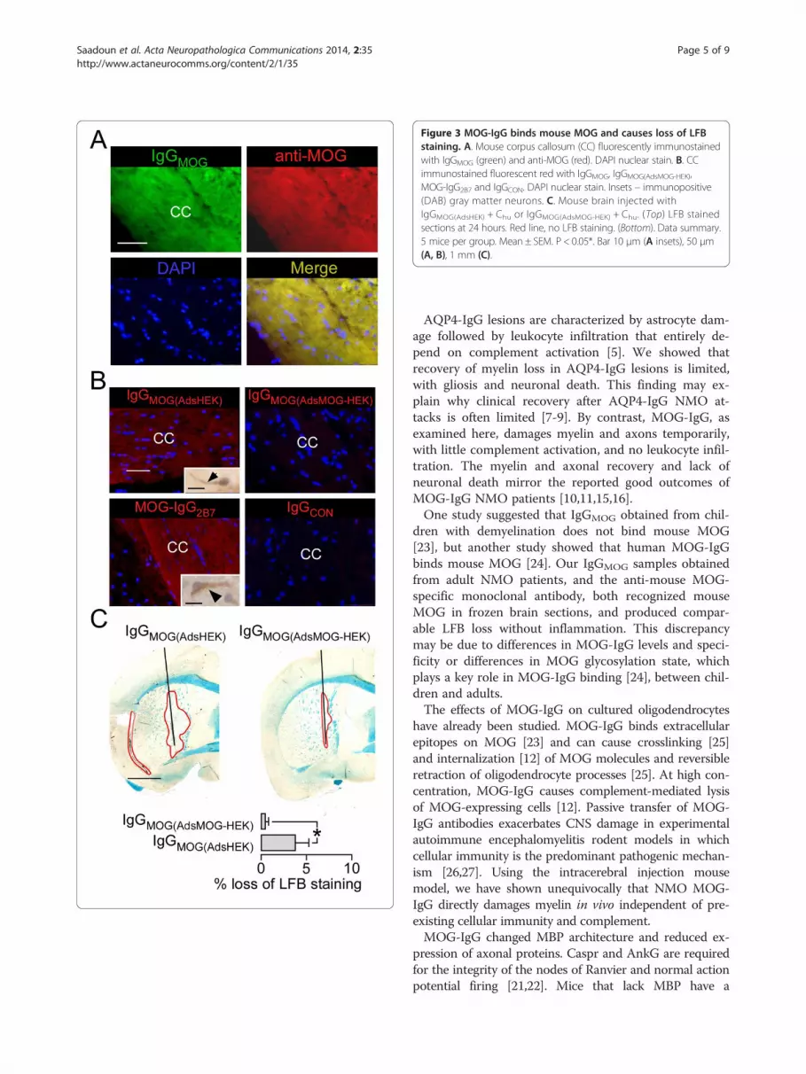
Figure 3 MOG-IgG binds mouse MOG and causes loss of LFBstaining. A. Mouse corpus callosum (CC) fluorescently immunostainedwith IgGMOG (green) and anti-MOG (red). DAPI nuclear stain. B. CCimmunostained fluorescent red with IgGMOG, IgGMOG(AdsMOG-HEK),MOG-IgG2B7 and IgGCON. DAPI nuclear stain. Insets – immunopositive(DAB) gray matter neurons. C. Mouse brain injected withIgGMOG(AdsHEK) + Chu or IgGMOG(AdsMOG-HEK) + Chu. (Top) LFB stainedsections at 24 hours. Red line, no LFB staining. (Bottom). Data summary.5 mice per group. Mean ± SEM. P < 0.05*. Bar 10 μm (A insets), 50 μm(A, B), 1 mm (C).
Saadoun et al. Acta Neuropathologica Communications 2014, 2:35 Page 5 of 9http://www.actaneurocomms.org/content/2/1/35
AQP4-IgG lesions are characterized by astrocyte dam-age followed by leukocyte infiltration that entirely de-pend on complement activation [5]. We showed thatrecovery of myelin loss in AQP4-IgG lesions is limited,with gliosis and neuronal death. This finding may ex-plain why clinical recovery after AQP4-IgG NMO at-tacks is often limited [7-9]. By contrast, MOG-IgG, asexamined here, damages myelin and axons temporarily,with little complement activation, and no leukocyte infil-tration. The myelin and axonal recovery and lack ofneuronal death mirror the reported good outcomes ofMOG-IgG NMO patients [10,11,15,16].One study suggested that IgGMOG obtained from chil-
dren with demyelination does not bind mouse MOG[23], but another study showed that human MOG-IgGbinds mouse MOG [24]. Our IgGMOG samples obtainedfrom adult NMO patients, and the anti-mouse MOG-specific monoclonal antibody, both recognized mouseMOG in frozen brain sections, and produced compar-able LFB loss without inflammation. This discrepancymay be due to differences in MOG-IgG levels and speci-ficity or differences in MOG glycosylation state, whichplays a key role in MOG-IgG binding [24], between chil-dren and adults.The effects of MOG-IgG on cultured oligodendrocytes
have already been studied. MOG-IgG binds extracellularepitopes on MOG [23] and can cause crosslinking [25]and internalization [12] of MOG molecules and reversibleretraction of oligodendrocyte processes [25]. At high con-centration, MOG-IgG causes complement-mediated lysisof MOG-expressing cells [12]. Passive transfer of MOG-IgG antibodies exacerbates CNS damage in experimentalautoimmune encephalomyelitis rodent models in whichcellular immunity is the predominant pathogenic mechan-ism [26,27]. Using the intracerebral injection mousemodel, we have shown unequivocally that NMO MOG-IgG directly damages myelin in vivo independent of pre-existing cellular immunity and complement.MOG-IgG changed MBP architecture and reduced ex-
pression of axonal proteins. Caspr and AnkG are requiredfor the integrity of the nodes of Ranvier and normal actionpotential firing [21,22]. Mice that lack MBP have a

Figure 4 MOG-IgG2B7 causes loss of LFB staining largelyindependent of immune cells or complement activation. A.Adjacent sections stained with H + E and LFB at seven days afterinjecting MOG-IgG2B7 + Chu. Green line shows loss of LFB staining.Rectangles show sites of Sections. B. Mouse brain immunostaned forC5b-9 at 24 hours after injection of IgGCON + Chu, IgGMOG + Chu, orIgGAQP4 + Chu. Lu, lumen; wm, white matter. Weak (gray arrows) andstrong (black arrows) immunoreactivity. C. Loss of LFB staining at24 hours after injection of MOG-IgG2B7, MOG-IgG2B7 + Chu, orisotype control (CON-IgG2B4). 5 mice per group. Mean ± SEM.P < 0.05*, < 0.01**. Bar 50 μm (A bottom right, B), 200 μm(A bottom left), 1 mm (A top).
Saadoun et al. Acta Neuropathologica Communications 2014, 2:35 Page 6 of 9http://www.actaneurocomms.org/content/2/1/35
characteristic motor dysfunction including tremor and sei-zures [28], mice that lack Caspr have severe motor paresis[22] whereas mice lacking cerebellar ankG develop pro-gressive ataxia [21]. Therefore, the altered MBP expressionand reduced Caspr and AnkG expression produced byMOG-IgG are predicted to produce a neurological deficitif the NMO lesion is in an eloquent region of the CNS.Unlike AQP4-IgG, MOG-IgG did not produce axonal dis-integration or neuronal death. Given the 96% homologybetween mouse and human MOG [14], our findings raisethe possibility that MOG-IgG may also cause similar re-versible lesions in the human CNS.MOG-IgG has been reported in other non-NMO dis-
eases including multiple sclerosis, acute disseminatedencephalomyelitis and even some normal subjects [29].Does MOG-IgG from these non-NMO subjects alsocause the same reversible CNS changes, as describedhere for NMO MOG-IgG? This question is difficult toanswer at present because of the variety of assays usedto detect MOG-IgG. For example, the assay used here,which employs C-terminal truncated rather than full-length MOG, did not detect MOG-IgG in adult multiplesclerosis patients and normal individuals [11], whichsuggests that different assays detect different subpopula-tions of MOG-IgG. It is important to first standardizethe assays before determining which subpopulations ofMOG-IgG can cause CNS damage and in whichdiseases.The mechanism of MOG-IgG-induced myelin damage
in vivo is unknown. Our data show that MOG-IgG –mediated myelin damage is a direct effect of MOG-IgGand that complement activation is not necessary. MOG-IgG binding may cause MOG conformational changes orinternalization that disrupts the myelin structure andsecondarily alters axonal protein expression. To explainthe lack of complement involvement, we hypothesizethat, after MOG-IgG binding, MOG might not aggregate(because of its low abundance) or MOG might becomeinternalized (thus prohibiting C1q activation). The fullrecovery within two weeks of the MOG-IgG-inducedLFB, MBP, ankG and Caspr changes suggests thatMOG-IgG does not kill the oligodendrocytes, but causesa reversible damage.Our findings raise the possibility that MOG-IgG con-
tributes to pathology in some NMO patients. If MOG-IgG is pathogenic, antibody depletion (plasmapheresis)or suppression with steroids should be effective, as in-deed appears to be the case [10,11,15,16]. Conversely,some of the newly proposed therapies for AQP4-IgGNMO, such as sivelestat for inhibiting neutrophils [17],or eculizumab for inhibiting complement [30], are lesslikely to be needed in MOG-IgG NMO. Examining le-sions from MOG-IgG NMO patients may help elucidatethe pathogenicity of MOG-IgG in the human CNS.

Figure 5 Effect of MOG-IgG on neurons. A. (Left) NeuN immunoreactivity at 2w. Area lacking neurons outlined red. (Right) % NeuN loss (NeuNimmunonegative area/ipsilatral hemispheric area). B. Node of Ranvier: ankG, ankyrin G; Caspr, contactin associated protein; MBP, myelin basicprotein; MOG, myelin oligodendrocyte glycoprotein; Nav, voltage-gated Na+ channel; Nfil, neurofilament. C. MBP immunoreactivity. (Top) Normaland abnormal white matter tracts. (Bottom) % abnormal tracts in injected hemisphere. D. Caspr immunoreactivity within white matter tracts. (Top)Hemispheres injected with IgGCON + Chu and IgGMOG + Chu. (Bottom) Data summary. E. AnkG immunoreactivity within white matter tracts. (Top)Hemispheres injected with IgGCON + Chu and IgGMOG + Chu. (Bottom) Data summary. F. Nfil immunoreactivity. (Top) Hemispheres injected withIgGCON + Chu and IgGMOG + Chu. Arrowheads: white matter tracts. (Bottom) Data summary (a.u. arbitrary units). 5 mice per group. P <0.05*, <0.01**.Bar 0.5 mm (A), 20 μm (C), 10 μm (D, E), 50 μm (F).
Saadoun et al. Acta Neuropathologica Communications 2014, 2:35 Page 7 of 9http://www.actaneurocomms.org/content/2/1/35
ConclusionsMOG-IgG obtained from neuromyelitis optica patientscauses myelin changes and alters the expression of axonalproteins when injected in mouse brain. These effects arenot associated with inflammatory cell infiltration, are largelyindependent of complement and recover within two weeks.AQP4-IgG obtained from neuromyelitis optica patientscauses complement-mediated myelin loss, inflammatory
Table 1 Comparison of MOG-IgG with AQP4-IgG lesions in mo
Characteristic MOG-IgG
Target cell Oligodendrocyte (myelin)
Lesion onset Within hours of exposure to MOG-Ig
Effect on astrocytes No major effect (normal AQP4 and G
Effect on neurons No major effect
Effect on oligodendrocytes Change in myelin (loss of LFB)
Effect on axons Myelin (transient change in MBP)
Intact axons (normal nfil)
Node of Ranvier (transient change in
Inflammatory cell infiltration No
Complement activation Slight (in white matter tracts)
Not required for lesion to develop
Recovery Yes, within 2 weeks
β-APP, beta amyloid precursor protein; FJ-C, fluorojade C; GFAP, glial fibrillary acidic prmarker; nfil, neurofilament.
cell infiltration, neuronal and astrocyte death with limitedrecovery at two weeks. These findings raise the possibilitythat MOG-IgG contributes to pathology in some neuromy-elitis optica patients.
Availability of supporting dataNo supporting data.
use brain
AQP4-IgG
Astrocyte (foot process)
G Within hours of exposure to AQP4-IgG
FAP) Astrocyte death (loss of AQP4 and GFAP)
Neuronal death (loss of NeuN, FJ-C staining [5])
Loss of myelin (loss of LFB)
Permanent loss of myelin
Axonal degeneration (β-APP expression [5])
casp and ankG)
Yes
Marked (perivascular)
Essential for lesion to develop
No, pan-necrosis followed by glial scarring
otein; LFB, Luxol fast blue; MBP, myelin basic protein; NeuN, neuronal nuclear

Saadoun et al. Acta Neuropathologica Communications 2014, 2:35 Page 8 of 9http://www.actaneurocomms.org/content/2/1/35
AbbreviationsAnkG: Ankyrin G; AQP4: Aquaporin-4; AQP4-IgG: Aquaporin-4 IgG found inmost neuromyelitis optica patients; C5b-9: Complement membrane attackcomplex; Caspr: Contactin associated protein; Chu: Human complement;CNS: Central nervous system; CON-IgG2B4: Monoclonal (2B4) control IgG;GFAP: Glial fibrillary acidic protein; H + E: Hematoxylin and eosin; IgGAQP4:IgG fraction of serum from neuromyelitis optica patients containing AQP4-IgG;IgGCON: IgG fraction of serum from normal subjects; IgGMOG: IgG fraction ofserum from neuromyelitis optica patients containing MOG-IgG; IgGMOG(AdsHEK):IgGMOG adsorbed against untransfected HEK cells; IgGMOG(AdsMOG-HEK):IgGMOG adsorbed against MOG-HEK to deplete MOG-IgG; LFB: Luxol fast blue;MBP: Myelin basic protein; MOG: Myelin oligodendrocyte glycoprotein;MOG-IgG2B7: Chimeric anti-mouse MOG recombinant IgG in which theconstant mouse regions of the heavy and light chains were substituted withthe human IgG1 constant regions, CH and Cκ.; NeuN: Neuronal nuclei;Nfil: Neurofilament; NMO: Neuromyelitis optica.
Competing interestsThe authors declare that they have no competing interests.
Authors’ contributionsSS designed and carried out the animal experiments, analyzed the data,produced the figures, contributed key ideas and helped draft themanuscript. PW prepared the IgG patient samples and contributed key ideas.GPO produced the recombinant MOG-IgG antibody. JLB produced therecombinant AQP4-IgG antibody and contributed key ideas. AV contributedkey ideas and helped to draft the manuscript. MCP participated in the designand coordination of the study and wrote manuscript. All authors read andapproved the final manuscript.
AcknowledgementsFunded by a research grant from the Guthy Jackson Charitable Foundationto MCP. PW and AV are supported by the Oxford NIHR Biomedical ResearchCentre and the NHS Specialised Services for NMO. JLB is supported by theGuthy-Jackson Charitable Foundation and the NIH (EY022936). GPO issupported by the NIH (NS072141).
Author details1Academic Neurosurgery Unit, St. George’s, University of London, London,UK. 2Nuffield Department of Clinical Neurosciences, University of Oxford,Oxford, UK. 3Department of Neurology, University of Colorado Denver,Aurora, Colorado, USA. 4Department of Ophthalmology, University ofColorado Denver, Aurora, Colorado, USA.
Received: 9 March 2014 Accepted: 9 March 2014Published: 31 March 2014
References1. Waters PJ, McKeon A, Leite MI, Rajasekharan S, Lennon VA, Villalobos A,
Palace J, Mandrekar JN, Vincent A, Bar-Or A, Pittock SJ: Serologic diagnosisof NMO: a multicenter comparison of aquaporin-4-IgG assays. Neurology2012, 78(9):665–671. discussion 669. doi:10.1212/WNL.0b013e318248dec1.
2. Lennon VA, Kryzer TJ, Pittock SJ, Verkman AS, Hinson SR: IgG marker ofoptic-spinal multiple sclerosis binds to the aquaporin-4 water channel.J Exp Med 2005, 202(4):473–477. doi:10.1084/jem.20050304.
3. Papadopoulos MC, Verkman AS: Aquaporin water channels in the nervoussystem. Nat Rev Neurosci 2013, 14(4):265–277. doi:10.1038/nrn3468.
4. Hinson SR, Pittock SJ, Lucchinetti CF, Roemer SF, Fryer JP, Kryzer TJ, Lennon VA:Pathogenic potential of IgG binding to water channel extracellular domainin neuromyelitis optica. Neurology 2007, 69(24):2221–2231. doi:10.1212/01.WNL.0000289761.64862.ce.
5. Saadoun S, Waters P, Bell BA, Vincent A, Verkman AS, Papadopoulos MC:Intra-cerebral injection of neuromyelitis optica immunoglobulin G andhuman complement produces neuromyelitis optica lesions in mice.Brain 2010, 133(Pt 2):349–361.
6. Papadopoulos MC, Verkman AS: Aquaporin 4 and neuromyelitis optica.Lancet Neurol 2012, 11(6):535–544. doi:10.1016/S1474-4422(12)70133-3.
7. Matiello M, Lennon VA, Jacob A, Pittock SJ, Lucchinetti CF, Wingerchuk DM,Weinshenker BG: NMO-IgG predicts the outcome of recurrent opticneuritis. Neurology 2008, 70(23):2197–2200.
8. Jarius S, Ruprecht K, Wildemann B, Kuempfel T, Ringelstein M, Geis C, KleiterI, Kleinschnitz C, Berthele A, Brettschneider J, Hellwig K, Hemmer B, LinkerRA, Lauda F, Mayer CA, Tumani H, Melms A, Trebst C, Stangel M, MarziniakM, Hoffmann F, Schippling S, Faiss JH, Neuhaus O, Ettrich B, Zentner C,Guthke K, Hofstadt-van Oy U, Reuss R, Pellkofer H, et al: Contrasting diseasepatterns in seropositive and seronegative neuromyelitis optica: amulticentre study of 175 patients. J Neuroinflammation 2012,9(1):14. doi:10.1186/1742-2094-9-14.
9. Akman-Demir G, Tuzun E, Waters P, Icoz S, Kurtuncu M, Jarius S, Yapici Z,Mutlu M, Yesilot N, Vincent A, Eraksoy M: Prognostic implications ofaquaporin-4 antibody status in neuromyelitis optica patients. J Neurol2011, 258(3):464–470. doi:10.1007/s00415-010-5780-4.
10. Kitley J, Leite MI, Kuker W, Quaghebeur G, George J, Waters P, Woodhall M,Vincent A, Palace J: Longitudinally extensive transverse myelitis with andwithout aquaporin 4 antibodies. JAMA Neurol 2013. EPub ahead of print.doi:10.1001/jamaneurol.2013.3890.
11. Kitley J, Woodhall M, Waters P, Leite MI, Devenney E, Craig J, Palace J,Vincent A: Myelin-oligodendrocyte glycoprotein antibodies in adults witha neuromyelitis optica phenotype. Neurology 2012, 79(12):1273–1277.doi:10.1212/WNL.0b013e31826aac4e.
12. Mader S, Gredler V, Schanda K, Rostasy K, Dujmovic I, Pfaller K, Lutterotti A,Jarius S, Di Pauli F, Kuenz B, Ehling R, Hegen H, Deisenhammer F,Aboul-Enein F, Storch MK, Koson P, Drulovic J, Kristoferitsch W, Berger T,Reindl M: Complement activating antibodies to myelin oligodendrocyteglycoprotein in neuromyelitis optica and related disorders.J Neuroinflammation 2011, 8:184. doi:10.1186/1742-2094-8-184.
13. Rostasy K, Mader S, Hennes EM, Schanda K, Gredler V, Guenther A, BlaschekA, Korenke C, Pritsch M, Pohl D, Maier O, Kuchukhidze G, Brunner-Krainz M,Berger T, Reindl M: Persisting myelin oligodendrocyte glycoproteinantibodies in aquaporin-4 antibody negative pediatric neuromyelitisoptica. Mult Scler 2013, 19(8):1052–1059. doi:10.1177/1352458512470310.
14. Vourc’h P, Andres C: Oligodendrocyte myelin glycoprotein (OMgp):evolution, structure and function. Brain Res Brain Res Rev 2004,45(2):115–124. doi:10.1016/j.brainresrev.2004.01.003.
15. Kitley J, Waters P, Woodhall M, Leite MI, Murchison A, George J, Kuker W,Chandratre S, Vincent A, Palace J: Neuromyelitis optica spectrum disorderswith aquaporin-4 and myelin-oligodendrocyte glycoprotein antibodies: acomparative study. JAMA Neurol 2014. doi:10.1001/jamaneurol.2013.5857.
16. Sato DK, Callegaro D, Lana-Peixoto MA, Waters PJ, de Haidar Jorge FM,Takahashi T, Nakashima I, Apostolos-Pereira SL, Talim N, Simm RF, MartinsLino AM, Misu T, Leite MI, Aoki M, Fujihara K: Distinction between MOGantibody-positive and AQP4 antibody-positive NMO spectrum disorders.Neurology 2014. doi:10.1212/WNL.0000000000000101.
17. Saadoun S, Waters P, MacDonald C, Bell BA, Vincent A, Verkman AS,Papadopoulos MC: Neutrophil protease inhibition reduces NMO-IgG-induced damage in mouse brain. Ann Neurol 2012, 71(3):323–333.doi:10.1002/ana.22686.
18. Saadoun S, Waters P, Macdonald C, Bridges LR, Bell BA, Vincent A, VerkmanAS, Papadopoulos MC: T cell deficiency does not reduce lesions in miceproduced by intracerebral injection of NMO-IgG and complement.J Neuroimmunol 2011, 235(1–2):27–32. doi:10.1016/j.jneuroim.2011.03.007.
19. Owens GP, Bennett JL, Lassmann H, O’Connor KC, Ritchie AM, Shearer A,Lam C, Yu X, Birlea M, DuPree C, Williamson RA, Hafler DA, Burgoon MP,Gilden D: Antibodies produced by clonally expanded plasma cells inmultiple sclerosis cerebrospinal fluid. Ann Neurol 2009, 65(6):639–649.doi:10.1002/ana.21641.
20. Bennett JL, Lam C, Kalluri SR, Saikali P, Bautista K, Dupree C, Glogowska M,Case D, Antel JP, Owens GP, Gilden D, Nessler S, Stadelmann C, Hemmer B:Intrathecal pathogenic anti-aquaporin-4 antibodies in early neuromyelitisoptica. Ann Neurol 2009, 66(5):617–629. doi: 10.1002/ana.21802.
21. Zhou D, Lambert S, Malen PL, Carpenter S, Boland LM, Bennett V: AnkyrinGis required for clustering of voltage-gated Na channels at axon initialsegments and for normal action potential firing. J Cell Biol 1998,143(5):1295–1304.
22. Bhat MA, Rios JC, Lu Y, Garcia-Fresco GP, Ching W, St Martin M, Li J,Einheber S, Chesler M, Rosenbluth J, Salzer JL, Bellen HJ: Axon-gliainteractions and the domain organization of myelinated axons requiresneurexin IV/Caspr/Paranodin. Neuron 2001, 30(2):369–383.
23. Mayer MC, Breithaupt C, Reindl M, Schanda K, Rostasy K, Berger T, Dale RC,Brilot F, Olsson T, Jenne D, Probstel AK, Dornmair K, Wekerle H, Hohlfeld R,Banwell B, Bar-Or A, Meinl E: Distinction and temporal stability of

Saadoun et al. Acta Neuropathologica Communications 2014, 2:35 Page 9 of 9http://www.actaneurocomms.org/content/2/1/35
conformational epitopes on myelin oligodendrocyte glycoproteinrecognized by patients with different inflammatory central nervous systemdiseases. J Immunol 2013, 191(7):3594–3604. doi:10.4049/jimmunol.1301296.
24. Marta CB, Oliver AR, Sweet RA, Pfeiffer SE, Ruddle NH: Pathogenic myelinoligodendrocyte glycoprotein antibodies recognize glycosylatedepitopes and perturb oligodendrocyte physiology. Proc Natl Acad SciU S A 2005, 102(39):13992–13997. doi:10.1073/pnas.0504979102.
25. Marta CB, Taylor CM, Coetzee T, Kim T, Winkler S, Bansal R, Pfeiffer SE:Antibody cross-linking of myelin oligodendrocyte glycoprotein leads toits rapid repartitioning into detergent-insoluble fractions, and alteredprotein phosphorylation and cell morphology. J Neurosci 2003,23(13):5461–5471.
26. Lee DH, Linker RA: The role of myelin oligodendrocyte glycoprotein inautoimmune demyelination: a target for multiple sclerosis therapy? ExpertOpin Ther Targets 2012, 16(5):451–462. doi: 10.1517/14728222.2012.677438.
27. Zhou D, Srivastava R, Nessler S, Grummel V, Sommer N, Bruck W, HartungHP, Stadelmann C, Hemmer B: Identification of a pathogenic antibodyresponse to native myelin oligodendrocyte glycoprotein in multiplesclerosis. Proc Natl Acad Sci U S A 2006, 103(50):19057–19062.doi:10.1073/pnas.0607242103.
28. Chernoff GF: Shiverer: an autosomal recessive mutant mouse with myelindeficiency. J Hered 1981, 72(2):128.
29. Reindl M, Di Pauli F, Rostasy K, Berger T: The spectrum of MOGautoantibody-associated demyelinating diseases. Nat Rev Neurol 2013,9(8):455–461. doi: 10.1038/nrneurol.2013.118.
30. Pittock SJ, Lennon VA, McKeon A, Mandrekar J, Weinshenker BG, LucchinettiCF, O’Toole O, Wingerchuk DM: Eculizumab in AQP4-IgG-positive relapsingneuromyelitis optica spectrum disorders: an open-label pilot study.Lancet Neurol 2013, 12(6):554–562. doi:10.1016/S1474-4422(13)70076-0.
doi:10.1186/2051-5960-2-35Cite this article as: Saadoun et al.: Neuromyelitis optica MOG-IgG causesreversible lesions in mouse brain. Acta Neuropathologica Communications2014 2:35.
Submit your next manuscript to BioMed Centraland take full advantage of:
• Convenient online submission
• Thorough peer review
• No space constraints or color figure charges
• Immediate publication on acceptance
• Inclusion in PubMed, CAS, Scopus and Google Scholar
• Research which is freely available for redistribution
Submit your manuscript at www.biomedcentral.com/submit


