Embed Size (px)
Citation preview

TECHNIQUE ASSESSMENTS
Neuronavigation by Intraoperative Three-dimensionalUltrasound: Initial Experience during Brain Tumor Resection
Geirmund Unsgaard, Ph.D., M.D.,Steinar Ommedal, B.Sc., Tomm Muller, Ph.D., M.D.,
Aage Gronningsaeter, Ph.D.,Toril A. Nagelhus Hernes, Ph.D.
Department of Neurosurgery (GU, TM), University Hospital of Trondheim; MedicalFaculty (GU), Norwegian University of Science and Technology; SINTEF Unimed
(SO, TANH); and MISON AS (AG), Trondheim, Norway
OBJECTIVE: Three-dimensional (3-D) ultrasound is an intraoperative imaging modality used in neuronavigation as analternative to magnetic resonance imaging (MRI). This article summarizes 4 years of clinical experience in the useof intraoperative 3-D ultrasound integrated into neuronavigation for guidance in brain tumor resection.
METHODS: Patients were selected for inclusion in the study on the basis of the size and location of their lesion.Preoperative 3-D MRI data were registered and used for planning as in other conventional neuronavigationsystems. Intraoperative 3-D ultrasound images were acquired three to six times, and tumor resection was guidedon the basis of these updated 3-D images.
RESULTS: Intraoperative 3-D ultrasound represents a good solution to the problem of brain shift in neuronavigationbecause it easily provides an updated, and hence more accurate, map of the patient’s true anatomy in all phasesof the operation. Ultrasound makes it possible to follow the progression of the operation, and it improves theradicality of tumor resection by detecting tumor tissue that would remain if the imaging technology had not beenused (in 53% of the cases). Integration of 3-D ultrasound with navigation technology solves the orientationproblem experienced previously with two-dimensional ultrasound in neurosurgery. The technology makes itpossible to directly compare intraoperative ultrasound and MRI data regarding visualization of the lesion.Ultrasound image quality is useful for guiding surgical procedures.
CONCLUSION: Intraoperative 3-D ultrasound seems to provide a time- and cost-effective way to update high-quality 3-D maps used in neuronavigation. (Neurosurgery 50:804–812, 2002)
Key words: Brain shift, Brain tumor surgery, Intraoperative imaging, Minimally invasive surgery, Neuronavigation, Sonography, Three-dimensionalultrasound
Neuronavigation systems have been demonstrated toconvey several advantages in improved planning andperformance of image-guided surgery (16, 40, 41).
However, the conventional systems still have practical limi-tations owing to the lack of an intraoperative imaging modal-ity to provide the surgeon with information regarding dy-namic changes that occur during surgery. Intraoperativeimaging technologies such as magnetic resonance imaging(MRI) and ultrasound have been demonstrated to be benefi-cial for monitoring the progression of the operation and forresection control (17, 24, 31, 38), as well as in coping with thebrain shift that occurs during surgery (19, 22, 30). The optimalsolution for the neurosurgeon would be a navigation systemwith high-quality, real-time, three-dimensional (3-D) imaging
capabilities. Although this is not yet a reality, different ap-proaches toward this ultimate goal have been presented byvarious companies and research groups in recent years.
Intraoperative MRI and intraoperative computed tomogra-phy (CT) give the surgeon an opportunity to obtain scans ofthe patient one or more times during surgery by transportingthe patient in and out of the scanner. The advantage is anupdated, high-quality, 3-D map that may be used for furtherguidance and resection control (23, 28, 34, 39). An importantdrawback is the relatively long image acquisition procedure(typically, a total of 20–60 min), which limits the practicalnumber of acquired 3-D scans allowed during surgery. Aregistration technique is also required to calibrate these intra-operative 3-D scans to the patient.
804 Neurosurgery, Vol. 50, No. 4, April 2002

Intraoperative MRI, in which the surgeon stands inside themagnet during surgery, provides the surgeon access tonearly-real-time two-dimensional (2-D) images as well as theopportunity to update the 3-D map in minutes without mov-ing the patient (5, 6, 35). Drawbacks of this technique includehigh investment and running costs, limited working space,and special surgical equipment and system requirements.These systems do not, however, require a patient registrationalgorithm, because the images as well as the surgical instru-ments can be handled from the same reference system.
Preoperative MRI has been combined with intraoperativeultrasound in an attempt to provide the surgeon with anupdated 3-D map (7, 18, 22, 33, 37). The preoperative MRIscans are modified during surgery according to landmarkmovements that are registered through ultrasound imaging.The ultrasound images are used in an indirect manner, i.e.,only to provide information so that the MRI data set canundergo an elastic warping procedure.
Ultrasound has been used directly for guidance. In thesestudies, ultrasound seems to provide valuable information interms of updated 2-D images several times per second, butimage quality varies (3, 9, 11, 13, 15, 25, 43). However, effortshave been made to improve the image quality in terms oftechnical adjustments of parameters (13) and optimal clinicalsetup (G Unsgaard, A Gronningsaeter, S Ommedal, TANHernes, submitted for publication). To enable guidance ofsurgical instruments by means of real-time 2-D images, thescan plane must be aligned so that the instrument is observedproperly at all times. This is challenging and time consumingeven for an experienced user. This orientation problem hasbeen solved by combining 3-D ultrasound and navigationtechnology (13, 38). The surgeon may update the 3-D map inseconds during surgery and navigate directly on a map thatreflects the patient’s true anatomy without a requirement forpatient registration and probe adjustment procedures. All ofthese improvements, including the future capabilities of real-time 3-D imaging and the relatively lower costs of the equip-ment, as compared with present alternatives, may establishultrasound in future neuronavigation. This article describesthe initial clinical results and experiences from 1997 to 2001 inthe use of 3-D ultrasound integrated with navigation technol-ogy for guiding brain tumor resections.
PATIENTS AND METHODS
Patients
Patients (n � 91) who were expected to benefit from the useof ultrasound-based neuronavigation during their operationswere selected on the basis of the size and location of theirtumors. All patients were informed regarding the methodol-ogy and agreed to be included in the study. The lesions werelocated in the supratentorial region of the brain and wereprimarily deep-seated parenchymal tumors with diameters
ranging from 1 to 5 cm. The tumors included glioblastomas,anaplastic astrocytomas, low-grade astrocytomas, metastases,meningiomas, and some other tumors. The patients are listedin Table 1.
Ultrasound equipment
Different solutions and equipment were used in the presentstudy because of the continuous development of the technol-ogy. From 1997 to 2000, we used a two-rack prototype con-sisting of a high-end System FiVe ultrasound scanner (GEVingmed Ultrasound, Horten, Norway) and navigation soft-ware (developed in our group) integrated to an optical track-ing system. Beginning in 2000, a high-end ultrasound scannerand a Polaris optical tracking system (Northern Digital, Wa-terloo, ON, Canada) were integrated with the navigation soft-ware into one single-rack navigation system, a prereleaseversion of the final product, SonoWand (MISON AS, Trond-heim, Norway) (13) (Fig. 1). Since the study began in 1997, thesystem-user interface and the speed of data transfer have beenimproved considerably. The most recent prototype requiresapproximately 30 seconds for 3-D data transfer andreconstruction.
This combined system may be used as an ultrasound scanner,a conventional neuronavigation system, or an integrated,ultrasound-based neuronavigation system that uses features ofboth technologies. This makes it possible to present updated 3-Dimage volumes. The camera reads the position of the patientreference frame, the ultrasound probe, and surgical instrumentssuch as a Cavitron ultrasonic surgical aspirator (CUSA) (Valley-lab, Boulder, CO) and biopsy forceps (Fig. 2). A 4- to 8-MHz flatphased-array probe (Fig. 2C) with optimal focusing propertiesat 3 to 6 cm was used in all cases. The ultrasound probewas covered with a sterile condom containing sterile gel. Thescanner factory and clinical setups were optimized for brainsurgery applications as described previously (13; G Unsgaard, AGronningsaeter, S Ommedal, TAN Hernes, submitted forpublication).
TABLE 1. Patient Data
LesionTotal No. of Image-guided Procedures
SecondMinicraniotomy
(%)
Glioblastoma 19 52%
Anaplastic astrocytoma 11 54%
Low-grade astrocytoma 17 47%
Metastasis 17 65%
Meningioma 6 0%
Other tumors 21 24%
Total 91 45%
Three-dimensional Ultrasound in Neuronavigation
805Neurosurgery, Vol. 50, No. 4, April 2002

Preoperative preparation and planning
Five fiducial markers were placed at the following posi-tions on the patient’s head: behind right ear, behind left ear,right forehead, left forehead, and parietal region. The patientswere scanned using Picker (Picker International, Inc., Cleve-land, OH) or Siemens (Siemens Medical Systems, Inc., Erlan-
gen, Germany) 1.5-T MRI technology, and a high-resolution3-D MRI volume with a slice thickness of 1.5 mm was ac-quired (Fig. 3A). The 3-D MRI volume was registered to thepatient (Fig. 3B), and the surgical procedure then was plannedin the operating room. The position of the craniotomy, and insome cases an additional minicraniotomy for the ultrasoundprobe, was planned on the basis of the preoperative images.In 45% of the cases (Table 1), a second minicraniotomy wasused to obtain optimal image quality and surgical setup, asdescribed previously (G Unsgaard, A Gronningsaeter, SOmmedal, TAN Hernes, submitted for publication).
3-D ultrasound acquisition andneuronavigational guidance
Immediately after exposure of the dura, sterile ultrasound gelwas applied and the first 3-D ultrasound volume was acquired(Fig. 3C). The probe was tilted at angles of approximately 80degrees over the anatomic area of interest by free-hand move-ment for 15 seconds. The pyramid-shaped 3-D data sets (Fig. 4A)were transferred to the navigation computer and reconstructedto a 3-D volume as described (13). However, no patient registra-tion was needed for the 3-D ultrasound volume. When neces-sary, the maximum depth of the ultrasound image and the focuspositions of the ultrasound beams were adjusted to obtain opti-mal image quality at the tumor location. In some of the moresuperficial lesions, a gelatin stand-off pad with a thickness of 1 to2 cm was used to increase the distance between the probe andthe lesion. This improved focusing conditions and ensured thata larger part of the tumor and surrounding anatomy was cov-ered by the ultrasound sector. After 3-D acquisition, the ultra-sound probe could be removed from the working area andimage guidance could be performed on the basis of the acquired3-D volume.
Tumor resection was performed using a CUSA (Fig. 3D) orbiopsy forceps with positioning devices attached. Hence, theposition of the surgical tool determined the images to be dis-played on the navigation monitor. This made it possible to steerthe tools down to the lesion as guided by the 3-D images.Preoperative MRI and intraoperative ultrasound images weredisplayed simultaneously. Slices from the 3-D volumes were dis-played as ordinary orthogonal slices (Fig. 4B) or as single slices(any plane) from the 3-D volumes (Fig. 4C). The any-plane slicefrom the ultrasound 3-D volume was displayed according to theorientation of the surgical tool and not limited by the scan planeof the real-time 2-D ultrasound probe. Image slices that werelying parallel to the brain surface in a slice perpendicular to theultrasound image scan plane then could be displayed easily andused for surgery guidance as needed (Fig. 4D). As in any othernavigation system, the position of the pointer or surgical tool tipwas displayed as lines and crosshairs in the corresponding im-ages. Another 3-D update was acquired whenever tissuechanges made it unsafe to proceed; typically, updates wereobtained three to six times during a surgical procedure. At theend of each operation, the surgeon evaluated whether ultra-sound had been essential to detect tumor tissue that would nothave been found without this technology.
FIGURE 1. The single-rack, ultrasound-based SonoWandneuronavigation system in the operating room. A Polarisadjustable camera and screen positions (forward, backward,sideways) make the instrument suitable and practical toplace in relation to other instruments and the setup in theroom.
FIGURE 2. The integrated positioning system (Polaris) withthe camera (A) tracks the position of the patient referenceframe (B), the ultrasound probe (C), the biopsy forceps (D),the sterile pointer (E), or the CUSA (F). The position of theultrasound image planes and the tip of the pointers or surgi-cal tools can then be calculated, and the instruments may benavigated deep into the brain on the basis of updated 3-Dimage information displayed on the screen.
806 Unsgaard et al.
Neurosurgery, Vol. 50, No. 4, April 2002

RESULTS
Interpretation of ultrasound imaging modality
Most neurosurgeons are more familiar with MRI and CTthan with ultrasound images. In the present study, we foundthat it was important to be familiar with ultrasound imagingof the lesion early in the operation. After exposure of the dura,but before the resection was started, an initial 3-D ultrasoundimage was acquired. Corresponding slices from MRI and
ultrasound 3-D volumes (similarly scaled and oriented) werethen displayed simultaneously (Fig. 5). Preoperative MRIscans were useful, both for presenting an overview of theanatomy in areas where ultrasound images were not acquiredand for learning to interpret information in the correspondingultrasound images. In many cases, however, our experiencewas that ultrasound image quality was comparable to or evenbetter than the corresponding MRI scans when tumor andlandmark visualization were considered.
FIGURE 3. Typical procedures per-formed when using 3-D ultrasound inneuronavigation. The day before sur-gery, a high-resolution 3-D MRI mapof the patient is acquired (A). The3-D volume is then registered to thepatient, and the preoperative imagesare used for planning the procedure(B). A 3-D ultrasound volume of thebrain is acquired (C) and recon-structed for use in navigation. Noregistration of 3-D ultrasound imagevolumes is required. The tumorresection may be performed directlyby navigating the CUSA down to thelesion (D). Image information fromboth MRI and ultrasound is presentedon the screen. When the surgeonrequires another 3-D update becausetissue changes have occurred, the3-D acquisition procedure isrepeated and resection continues onan updated 3-D map (C and D).
FIGURE 4. A pyramid-shaped 3-D ultrasoundvolume is acquired by tilting the 2-D probeover the anatomic area of interest (A). The3-D data set is reconstructed and used directlyfor navigation. The ultrasound probe may beremoved from the working area, and the posi-tion and orientation of the surgical tool deter-mines which images from the 3-D volume aredisplayed on the monitor. The slices from bothMRI and ultrasound volumes may be displayedsimultaneously. Display techniques may beconventional orthogonal slices (B) oriented tothe patient (axial, sagittal, coronal), from thesurgeon’s view, or only defined by the positionand orientation of the surgical tool. In any-plane slicing (C), only one slice defined by theposition and orientation of the surgical tool isdisplayed from each 3-D volume. Because a3-D ultrasound volume is acquired, an ultra-sound slice not limited to the ultrasound scanplane may be used for navigation (D).
Three-dimensional Ultrasound in Neuronavigation 807
Neurosurgery, Vol. 50, No. 4, April 2002
Faster and more precise image-guided resection
A sterile pointer is one of the most frequently used instru-ments in neuronavigation. We found, however, that a biopsyforceps or CUSA with an attached tracking device represented asafer and more effective way to perform image-guided surgeryand navigation (Fig. 3D). The CUSA was calibrated in the oper-ating room, and the images to be displayed on the monitor weredetermined. However, the conventional orthogonal patient-oriented slicing of the 3-D volume, as shown in Figure 6, made itquite challenging and time consuming to guide the proceduredirectly and follow the progression of the operation. Therefore, amore intuitive and user-friendly display technique frequentlywas used, i.e., the any-plane display technique, in which theslices displayed on the screen were selected by the surgical toolposition and orientation (Fig. 4C). This made it possible to nav-igate the surgical tool more easily down to the lesion on the basisof a single slice from each 3-D volume without concentrating onthe orientation of the patient on the operating table. This noveldisplay technique also made it possible to interpret informationfrom two or three image volumes simultaneously and thuseasily follow the progression of the operation (Fig. 7). This user-friendly display technique also made it possible to effectivelyremove the central part of the tumor with no other visualizationthan 3-D images, thus reducing the pressure on surroundingbrain tissue. This blinded procedure was performed initiallyonly during the portion of the operation when the surgical toolcould be navigated easily by safe margins to the normal brain. Acavity for direct sight was then established.
After some resection, when the resection cavity came closer tothe tumor border, updates of the 3-D volumes were acquired
and the resection was continued on the basis of updated imageinformation. The microscope was used for work that demandedmore precision closer to the tumor border. Resection proceededon the basis of visual control until no tumor tissue was detectedby the naked eye or through the microscope.
Removal of remaining tumor tissue
Toward the conclusion of the operation, another 3-D ultra-sound volume was acquired. In our experience, ultrasound wasimportant for detecting remaining tumor tissue that was notdiscovered through the microscope or by the naked eye (Fig. 7).Tumor tissue that was present behind the normal tissue, whichtherefore could not be observed with the microscope, often wasdetected on ultrasound images. The process of localizing resid-ual tumor tissue required some experience, but the progressioninformation, which was available in the preceding 3-D ultra-sound volumes, simplified this task considerably. Position ofanatomic landmarks and tumor border in relation to the resec-tion cavity could be followed via images throughout the opera-tion. Our subjective experience was that residual tumor tissuewas discovered through the last 3-D ultrasound scan in 53% ofthe cases in which the resection otherwise was considered com-plete. Therefore, a more radical resection was achieved in thesecases because of ultrasound imaging.
Practical considerations in the operating room
The SonoWand prototype is simple to place in the operat-ing room, and it has adjustable cameras and a monitor arm,which makes it easy to optimize the ergonomic situation for
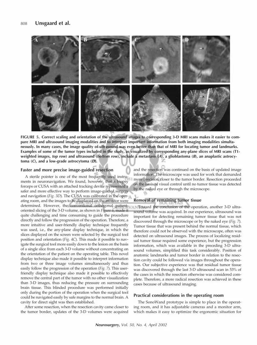
FIGURE 5. Correct scaling and orientation of the ultrasound images to corresponding 3-D MRI scans makes it easier to com-pare MRI and ultrasound imaging modalities and to interpret important information from both imaging modalities simulta-neously. In many cases, the image quality of ultrasound was even better than that of MRI for locating tumor and landmarks.Examples of some of the tumor types included in the study, as visualized by corresponding any-plane slices of MRI scans (T1-weighted images, top row) and ultrasound (bottom row), include a metastasis (A), a glioblastoma (B), an anaplastic astrocy-toma (C), and a low-grade astrocytoma (D).
808 Unsgaard et al.
Neurosurgery, Vol. 50, No. 4, April 2002

the surgeon. In our experience, adjustments were occasionallynecessary, however, to obtain visual contact between the cam-era and the surgical instruments. The system is easily trans-ported in and out of the operating room (Fig. 1) and repre-sents a compact and powerful system for surgical planningand intraoperative navigation. In our experience, the tightintegration of ultrasound and neuronavigation represents animprovement as compared with conventional neuronaviga-tion systems presently available.
DISCUSSION
The use of computer-assisted systems in neurosurgery hasevolved since the 1980s. Most of these systems have beendemonstrated to be useful, especially for planning the surgicalprocedure. The patient outcome of surgery, however, seemsto be based on several factors; the extent of tumor resection isimportant, especially for a variety of gliomas as well as forpediatric tumors (1, 2, 4, 8–10, 12, 20, 21, 26, 29, 32, 36).Although conventional navigation systems seem to enhance
the extent of tumor resection (41), these systems may haveeven more value when used in combination with an intraop-erative imaging modality. This makes it possible to followsurgery progression via images as well as to control resectionradicality toward the conclusion of surgery (38, 39, 42, 43).This also was demonstrated in the present study.
Intraoperative imaging technologies are now emerging in clin-ics and are being tested by research groups around the world.The attempts of research groups to explore alternative intraop-erative imaging solutions demonstrates the need for an imagingmodality that potentially can monitor tumor tissue at the con-clusion of surgery, which may increase the extent of tumorresection and patient outcome, including survival time.
Intraoperative images from open MRI systems are easilyintegrated into navigation systems and have been demon-strated to increase the extent of tumor resection and prolongpatient survival (42). These systems are, however, time-consuming and costly alternatives. CT usually is not the in-
FIGURE 6. As the operation progresses, orthogonal slicing ofa metastasis may be visualized with preoperative MRI (toprow), intraoperative ultrasound before opening of the dura(middle row), and 3-D ultrasound close to the end of theoperation (bottom row). The MRI scans provide an overviewof the anatomic area and help inexperienced ultrasoundusers to interpret ultrasound image information. Ultrasoundprovides updated image information in the important areawhere resection is performed. Axial, sagittal, and coronalslices from the 3-D ultrasound volume all show an imagequality useful for interpreting tumor border and for detectingremaining tumor tissue toward the conclusion of surgery.
FIGURE 7. Because the corresponding slices from all 3-Dscans are displayed on the screen simultaneously, the progres-sion of the resection may be followed easily. Correspondingsingle slices from a preoperative MRI volume (A), initial 3-Dultrasound volume (B), a 3-D ultrasound volume in the middleof the operation (C), and a 3-D ultrasound volume close to thecompletion of the resection (D) in a patient with a glioblas-toma. Arrows indicate remaining tumor tissue. In this operation,some tumor tissue was purposely left behind because of itslocation in eloquent areas. In other operations, we found thattumor tissue not detected by the microscope or by the nakedeye could be detected by use of 3-D ultrasound and removed,thus increasing the radicality of the tumor resection.
Three-dimensional Ultrasound in Neuronavigation 809
Neurosurgery, Vol. 50, No. 4, April 2002

traoperative imaging modality of choice because of ionizingradiation and limited tumor definition. The most discussedissues and objections regarding the use of ultrasound to guidesurgical procedures so far have involved the variable imagequality achieved by different users. In addition, most sur-geons are more familiar with MRI or CT and may need sometime to become familiar with the nature and interpretation ofinformation in the ultrasound images. Several researchgroups have demonstrated that the image quality of ultra-sound has improved considerably and is good enough tovisualize and guide tumor resections (3, 13, 43).
We have described various clinical and technical adjustmentsand arrangements for improving ultrasound image quality inpractical neurosurgery (13; G Unsgaard, A Gronningsaeter, SOmmedal, TAN Hernes, submitted for publication). One of theclinical arrangements we have developed is to perform a secondminicraniotomy for the ultrasound probe, which ensures opti-mal imaging conditions. We have not detected complicationsbecause of this special clinical setup, the image quality seemsimproved, and the flexibility of use of additional real-time im-aging is available. However, the orientation, scaling, and inter-pretation of information from real-time 2-D ultrasound imagesalso have made it difficult for experienced users to benefit fromthis imaging modality in guided neurosurgery procedures (GUnsgaard, A Gronningsaeter, S Ommedal, TAN Hernes, submit-ted for publication). This situation is soon to change, becausenew developments that result in tighter integration of 3-D ultra-sound imaging technology with navigation technology willsolve the orientation problems experienced with 2-D ultrasound(13). Ultrasound may now be used like any other 3-D imagingmodality in neuronavigation.
Access to high-quality intraoperative 3-D ultrasound hasenabled us to perform open tumor surgery through a slightlynarrower channel than would be possible without imageguidance. Tumor structures can be easily identified and lo-cated by use of the navigation system, and the surgeon canremove the structures with less visual control of the resectioncavity. In our patients, this minimally invasive approach wasespecially useful at the beginning of the operation, when theCUSA could be navigated down to the lesion and positionedcentrally in the tumor with safe margins between the tumorborder and normal tissue. In these cases, the opening in thenormal brain was limited to the size of the surgical instrument(2–8 mm). This blinded image-guided resection has been ap-plied experimentally to some deep-seated, low, vascularizedtumors with additional guidance from real-time 2-D ultra-sound. However, this method is challenging and time con-suming, and it will probably be more convenient and relevantwhen real-time 3-D ultrasound becomes available. We expectsuch techniques to be feasible and valuable for patients evenwith tumors located in eloquent areas of the brain.
A simultaneous display of corresponding MRI and ultrasoundslices from 3-D images enables the surgeon to more easily inter-pret information in the updated ultrasound images. We think,however, that there are still several display techniques with thepotential to improve the user-friendliness of image-guided sur-gery. One example may be multimodal imaging by fusion of 3-Dultrasound and MRI scans together in one scene (27). An alter-
native display technique may be stereoscopic interfaces used incombination with ordinary slicing techniques to improve theunderstanding and perception of complex 3-D structures duringsurgery (14). All available and needed preoperative MRI datasuch as functional MRI and various MRI scans may be fusedwith intraoperative real-time 3-D ultrasound and integrated infuture neuronavigation systems, because of both the increasedperceptibility of available image information modalities as wellas the relatively low costs of such systems as compared withavailable alternatives.
3-D ultrasonography has the potential to become an alter-native to open MRI as an intraoperative imaging modality inneuronavigation. Future real-time 3-D capabilities of ultra-sound make this imaging modality especially attractive. How-ever, the unsolved issue common to all of the alternativeintraoperative imaging technologies is their sensitivity andspecificity in detecting remaining tumor tissue at the conclu-sion of surgery, which will affect patient survival time. There-fore, studies initiated by Tronnier et al. (38) to compare theintraoperative imaging modalities, as well investigations tocompare intraoperative imaging modalities with histopatho-logical evaluation (3, 43), must continue. These studies shouldalso include an evaluation of the time and cost effectiveness ofthe systems in practical, daily hospital function to fully com-pare and evaluate the usefulness of the alternative navigationtechnologies for guiding neurosurgical procedures.
CONCLUSION
The introduction of 3-D ultrasound has increased the valueof neuronavigation substantially in our clinic, making it pos-sible to update 3-D maps several times during surgery andthereby minimize the problem of brain shift. The surgical toolmay be navigated down to the lesion with a high level ofprecision. Novel and user-friendly display techniques make itpossible to perform faster and more intuitive image-guidedsurgery as compared with conventional neuronavigation sys-tems and real-time 2-D ultrasound. These features allow theneurosurgeon to follow the progression of the resection andidentify, localize, and remove residual tumor tissue. Althoughour initial experience is promising, more research will berequired to scientifically explore the potential future value ofultrasound in neuronavigation.
ACKNOWLEDGMENTS
This work was supported by the Research Council of Norway,the Ministry of Health and Social Affairs of Norway, GE VingmedUltrasound (Horten, Norway), Norwegian Industrial and RegionalDevelopment Fund, and the Norwegian Cancer Society.
DISCLOSURE
At the time this study was initiated, all authors were re-search scientists and had no financial interest in the outcomeof the study. AG currently is employed by MISON AS andmay benefit from future success of the company resultingfrom the use of ultrasound in neurosurgery. However, AG
810 Unsgaard et al.
Neurosurgery, Vol. 50, No. 4, April 2002

was not involved in the study after he became Chief ExecutiveOfficer of MISON AS in 1998.
Received, July 27, 2001.Accepted, November 2, 2001.Reprint requests: Toril A. Nagelhus Hernes, Ph.D., SINTEF Unimed,Ultrasound, Medical Technical Research Centre, 7034 Trondheim,Norway.Email: [email protected]
REFERENCES
1. Albert FK, Forsting M, Sartor K, Adams HP, Kunze S: Early postoper-ative magnetic resonance imaging after resection of malignant glioma:Objective evaluation of residual tumor and its influence on regrowthand prognosis. Neurosurgery 34:45–60, 1994.
2. Albright AL, Wisoff JH, Zeltzer PM, Boyett JM, Rorke LB, Stanley P:Effects of medulloblastoma resection on outcome in children: A reportfrom the Children’s Cancer Group. Neurosurgery 38:265–271, 1996.
3. Auer LM, van Velthoven V: Intraoperative ultrasound (US) im-aging: Comparison of pathomorphological findings in US and CT.Acta Neurochir (Wien) 104:84–95, 1990.
4. Berger MS, Deliganis AV, Dobbins J, Keles GE: The effect of extentof resection on recurrence in patients with low grade cerebralhemisphere gliomas. Cancer 74:1784–1791, 1994.
5. Black PMcL, Alexander E III, Martin C, Moriarty T, Nabavi A,Wong TZ, Schwartz RB, Jolesz F: Craniotomy for tumor treatmentin an intraoperative magnetic resonance imaging unit. Neuro-surgery 45:423–433, 1999.
6. Black PMcL, Moriarty T, Alexander E III, Stieg P, Woodard EJ, GleasonPL, Martin CH, Kikinis R, Schwartz RB, Jolesz FA: Development andimplementation of intraoperative magnetic resonance imaging and itsneurosurgical applications. Neurosurgery 41:831–845, 1997.
7. Bucholz RD, Yeh DD, Trobaugh J, McDurmont LL, Sturm CD,Baumann C, Henderson JM, Levy A, Kessman P: The correction ofstereotactic inaccuracy caused by brain shift using an intraoperativeultrasound device. Presented in Lecture Notes in Computer Science,Proceedings of the 1997 1st Joint Conference of Computer Vision, Vir-tual Reality and Robotics in Medicine and Medical Robotics andComputer-Assisted Surgery, Grenoble, France, March 19–22, 1977.
8. Campbell JW, Pollack IF, Martinez AJ, Shultz B: High-grade astrocyto-mas in children: Radiologically complete resection is associated with anexcellent long-term prognosis. Neurosurgery 38:258–264, 1996.
9. Chandler WF, Knake JE, McGillicuddy JE, Lillehei KO, Silver TM:Intraoperative use of real-time ultrasonography in neurosurgery.J Neurosurg 57:157–163, 1982.
10. Fischbach AJ, Martz KL, Nelson JS, Griffin TW, Chang CH,Horton J, Nelson DF: Long-term survival in treated anaplasticastrocytomas: A report of combined RTOG/ECOG studies. Am JClin Oncol 14:365–370, 1991.
11. Gaab MR: Intraoperative ultrasound imaging in neurosurgery [inGerman]. Ultraschall Med 11:62–71, 1990.
12. Ganju V, Jenkis RB, O’Fallon JR, Scheithauer BW, Ransom DT,Katzman JA, Kimmel DW: Prognostic factors in gliomas: A mul-tivariate analysis of clinical, pathological, flow cytometric,cytogenic, and molecular markers. Cancer 74:920–927, 1994.
13. Gronningsaeter A, Kleven A, Ommedal S, Aarseth TE, Lie T,Lindseth F, Langø T, Unsgård G: SonoWand, an ultrasound-basedneuronavigation system. Neurosurgery 47:1373–1379, 2000.
14. Gronningsaeter A, Lie T, Kleven A, Mørland T, Langø, T,Unsgård G, Myhre HO, Mårvik R: Initial experience with stereo-scopic visualization of three-dimensional ultrasound data in sur-gery. Surg Endosc 14:1074–1078, 2000.
15. Gronningsaeter A, Unsgård G, Ommedal S, Angelsen BAJ:Ultrasound-guided neurosurgery: A feasibility study in the 3–30MHz frequency range. Br J Neurosurg 10:161–168, 1996.
16. Gumprecht H, Darius CW, Lumenta CB: BrainLab VectorVisionNeuronavigation System: Technology and clinical experiences in131 cases. Neurosurgery 44:97–105, 1999.
17. Hammoud MA, Ligon BL, elSouki R, Shi WM, Schomer DF,Sawaya R: Use of intraoperative ultrasound for localizing tumorsand determining the extent of resection: A comparative studywith magnetic resonance imaging. J Neurosurg 84:737–741, 1996.
18. Hata N, Dohi T, Iseki H, Takakura K: Development of a framelessand armless stereotactic neuronavigation system withultrasonographic registration. Neurosurgery 41:608–614, 2000.
19. Hill DLG, Maurer CR, Maciunas RJ, Barwise JA, Fitzpatrick JM,Wang MY: Measurement of intraoperative brain surface deforma-tion under a craniotomy. Neurosurgery 43:514–526.
20. Huber A, Beran H, Becherer A, Prosenc N, Witzmann A: Supra-tentorial glioma: Analysis of clinical and temporal parameters in163 cases [in German]. Neurochirurgia (Stuttg) 36:189–193, 1993.
21. Iacoangeli M, Roselli R, Prezioso A, Scerrati M, Rossi GF: Stagingof supratentoral hemispheric glioma using tumor extension, his-topathological grade, and extent of surgical resection. Br J Surg80:1130–1133, 1993.
22. Jödicke A, Deinsberger W, Erbe H, Kriete A, Böker DK: Intraop-erative three-dimensional ultrasonography: An approach to reg-ister brain shift using multidimensional image processing.Minim Invasive Neurosurg 41:13–19, 1998.
23. Kaibara T, Saunders JK, Sutherland GR: Advances in mobileintraoperative magnetic resonance imaging. Neurosurgery 47:131–138, 2000.
24. Knauth M, Wirtz CR, Tronnier VM, Aras N, Kunze S, Sartor K:Intraoperative MR imaging increases the extent of tumor resec-tion in patients with high-grade gliomas. AJNR Am JNeuroradiol 20:1642–1646, 1999.
25. Koivukangas J, Louhisalmi Y, Alakuijala J, Oikarinen J:Ultrasound-controlled neuronavigator-guided brain surgery.J Neurosurg 79:36–42, 1993.
26. Kowalczuk A, Macdonald RL, Amidei C, Dohrmann G III, EricksonRK, Hekmatpanah J, Krauss S, Krishnasamy S, Masters G, MullanSF, Mundt AJ, Sweeney P, Vokes EE, Weir BKA, Wollman RL:Quantitative imaging study of extent of surgical resection and prog-nosis of malignant astrocytomas. Neurosurgery 41:1028–1038, 1997.
27. Lindseth F, Ommedal S, Bang J, Unsgård G, Hernes TAN: Imagefusion of ultrasound and MRI as an aid for assessing anatomicalshifts and for improving overview and interpretation in ultra-sound guided neurosurgery, in Lemke HU, Inamura K, Doi K,Vannier MW, Farman AG (eds): CARS 2001: Proceedings of the 15thInternational Congress and Exhibition in Computer Assisted Radiologyand Surgery, Berlin, June 27–30, 2001. Amsterdam, Elsevier, 2001.
28. Martin AJ, Hall WA, Liu H, Pozza CH, Michel E, Casey SO,Maxwell RE, Truwit CL: Brain tumor resection: Intraoperativemonitoring with high-field-strength MR imaging—Initial results.Radiology 215:221–228, 2000.
29. Medical Research Council Brain Tumour Working Party: Prog-nostic factors for high-grade malignant glioma: Development of aprognostic index—A report of the Medical Research CouncilBrain Tumour Working Party. J Neurooncol 9:47–55, 1990.
30. Nimsky C, Ganslandt O, Cerny S, Hastreiter P, Greiner G,Fahlbusch R: Quantification of, visualization of, and compensa-tion for brain shift using intraoperative magnetic resonance im-aging. Neurosurgery 47:1070–1080, 2000.
Three-dimensional Ultrasound in Neuronavigation 811
Neurosurgery, Vol. 50, No. 4, April 2002

31. Paleologos TS, Wadley JP, Kitchen ND, Thomas DGT: Clinicalutility and cost-effectiveness of intraoperative image-guided cra-niotomy: Clinical comparison between conventional and image-guided meningioma surgery. Neurosurgery 47:40–48, 2000.
32. Prados MD, Gutin PH, Phillips TL, Wara WM, Larson DA, SneedPK, Davis RL, Ahn DK, Lamborn K, Wilson CB: Highly anaplasticastrocytoma: A review of 357 patients treated between 1977 and1989. Int J Radiat Oncol Biol Phys 23:3–8, 1992.
33. Roberts DW, Miga MI, Hartov A, Eisner S, Lemery JM, Kennedy FE,Paulsen KD: Intraoperatively updated neuroimaging using brain mod-eling and sparse data. Neurosurgery 45:1199–1207, 1999.
34. Rubino GJ, Farahani K, McGill D, Wiele B, Villablanca JP, Wang-Mathieson A: Magnetic resonance imaging-guided neurosurgeryin the magnetic fringe fields: The next step in neuronavigation.Neurosurgery 46:643–654, 2000.
35. Seifert V, Zimmermann M, Trantakis C, Vitzthum HE, Kuhnel K, RaabeA, Bootz F, Schneider JP, Schmidt F, Dietrich J: Open MRI-guidedneurosurgery. Acta Neurochir (Wien) 141:455–464, 1999.
36. Simpson JR, Horton J, Scott C, Curran WJ, Rubin P, Fischbach J,Isaacson S, Rotman M, Asbell SO, Nelson JS, Weinstein AS,Nelson DF: Influence of location and extent of surgical resectionon survival of patients with glioblastomas multiforme: Results ofthree consecutive Radiation Therapy Oncology Group (RTOG)clinical trials. Int J Radiat Oncol Biol Phys 26:239–244, 1993.
37. Trobaugh JW, Richard WD, Smith KR, Bucholz RD: Framelessstereotactic ultrasonography: Methods and applications. ComputMed Imaging Graph 18:235–246, 1994.
38. Tronnier VM, Bonsanto MM, Staubert A, Knauth M, Kunze S,Wirtz CR: Comparison of intraoperative MR imaging and 3D-navigated ultrasonography in the detection and resection controlof lesions. Neurosurg Focus 10:1–5, 2001.
39. Tronnier VM, Wirtz CR, Knauth M, Lenz G, Pastyr O, BonsantoMM, Albert FK, Kuth R, Staubert A, Sartor K, Kunze S: Intraop-erative diagnostic and interventional magnetic resonance imagingin neurosurgery. Neurosurgery 40:891–902, 1997.
40. Wadley J, Dorward N, Kitchen N, Thomas D: Preoperative plan-ning and intraoperative guidance in modern neurosurgery: Areview of 300 cases. Ann R Coll Surg Engl 81:217–225, 1999.
41. Wirtz CR, Albert FK, Schwaderer M, Heuer C, Staubert A, Tron-nier VM, Knauth M, Kunze S: The benefit of neuronavigation forneurosurgery analyzed by its impact on glioblastoma surgery.Neurol Res 22:354–360, 2000.
42. Wirtz CR, Kanuth M, Staubert A, Bonsanto MM, Sartor K, KunzeS, Tronnier VM: Clinical evaluation and follow-up results forintraoperative magnetic resonance imaging in neurosurgery.Neurosurgery 46:1112–1122, 2000.
43. Woydt M, Krone A, Becker G, Schmidt K, Roggendorf W, RoosenK: Correlation of intra-operative ultrasound with histopathologicfindings after tumour resection in supratentorial gliomas: Amethod to improve gross total tumour resection. Acta Neurochir(Wien) 138:1391–1398, 1996.
COMMENTS
In 1985, our group performed a few imaging-based volu-metric stereotactic tumor resections using a two-dimensionalultrasonic transducer mounted on the arc-quadrant of anarc-quadrant stereotactic frame (Compass system; CompassInternational, Inc., Rochester, MN). We used a separate tre-phine craniotomy for the ultrasonic transducer, and the ste-reotactic frame aligned the transducers to provide a constantview of the tumor, which had been centered in the focal pointof the stereotactic arc-quadrant. The ultrasonic transducer
could be rotated to provide many views, but all of these werecentered on the tumor. In this way, we reasoned, we couldobserve the surgical field during resection of the tumor.
We thought this was a great idea. Why did we not report it?Why did we not continue to use it? Because the images wereterrible! They became worse with the introduction of surgicalinstruments and uninterpretable with the presence of blood,instrument artifact, and air. In addition, it was not clear at thetime whether the ultrasonic images represented the samevolume as defined by computed tomographic and early-generation magnetic resonance imaging (MRI) units. The useof this methodology was not worth the difficulty, so weabandoned the effort after only a few operations.
Now, it seems that the Trondheim group may have solvedmany of the problems that discouraged us. The images thataccompany the present article are certainly of much better qual-ity than those we had, and slices from a three-dimensional dataset seem more versatile than a fixed two-dimensional system.The authors also have solved the problem of cross-registrationamong ultrasonic images, computed tomographic scans, andMRI scans with a frameless optical digitizing technology. None-theless, the successful use of ultrasonic imaging requires famil-iarity with the modality beyond that of the average circulatingnurse spinning dials and pushing buttons at the behest of asurgeon who does not understand the contraption any betterthan she does.
The technique, as presented in this article and in the handsof a moderately experienced team, clearly is useful. It cer-tainly provides a more convenient and less expensive alter-native for intraoperative imaging.
Patrick J. KellyNew York, New York
The authors have achieved an important integration of anavigation system driven by preoperative imaging with real-time, three-dimensional, intraoperative ultrasound imaging.Modern ultrasound imaging of the brain has become ex-tremely detailed, and in many instances it provides moreuseful information regarding a lesion than the preoperativeMRI scan. The authors have integrated the ultrasound imag-ing device into the navigation system by allowing the navi-gation system camera to recognize the exact location andposition of the ultrasound probe. This allows the constructedthree-dimensional MRI scan of the lesion to be corrected inspace when brain shifts occur during surgery.
The authors also have integrated surgical tools, such as the ul-trasonic aspirator, into the navigation system. This allows pre-cise localization of the instrument relative to the preoperative MRIscan and the intraoperative ultrasound imaging of the lesion.
The authors conclude that in 53% of cases, residual tumorwas identified by intraoperative ultrasound after it wasthought by the surgeon that the tumor had been maximallyresected. This marriage of ultrasound and frameless ster-eotaxy is an important step forward and will lead to moreprecise lesion localization.
William F. ChandlerAnn Arbor, Michigan
812 Unsgaard et al.
Neurosurgery, Vol. 50, No. 4, April 2002


