Embed Size (px)
Citation preview
1
Neutralizing antibody resistant hepatitis C virus cell-to-cell transmission.
Claire L. Brimacombe1†
, Joe Grove1†
, Luke W. Meredith1, Ke Hu
1, Andrew J. Syder
2, Maria
Victoria Flores3, Jennifer M. Timpe
1, Sophie E. Krieger
4, Thomas F. Baumert
4, Timothy L.
Tellinghuisen5, Flossie Wong-Staal
2, Peter Balfe
1* and Jane A. McKeating
1
† These authors contributed equally to this work.
1. Hepatitis C Research Group, Institute For Biomedical Research, University of Birmingham,
Birmingham, B15 2TT, UK.
2. iTherX Pharmaceuticals, Inc., P.O. Box 910530, San Diego, CA 92191-0530, USA
3. Pfizer Ltd�Ramsgate Road, Sandwich, Kent CT13 9NJ.�UK
4. Inserm U748, Université de Strasbourg and Pôle Hépato-digestif, Hôpitaux Universitaires de
Strasbourg, F-67000 Strasbourg, France.
5. Department of Infectology, The Scripps Research Institute, Jupiter, FL 33458, USA
* Corresponding author Peter Balfe, contact information: [email protected]
Tel: (44) 121 414 8174, fax: (44) 121 414 3599
Key words: Hepatitis C; transmission, CD81 and Scavenger receptor BI.
Copyright © 2010, American Society for Microbiology and/or the Listed Authors/Institutions. All Rights Reserved.J. Virol. doi:10.1128/JVI.01592-10 JVI Accepts, published online ahead of print on 20 October 2010
on October 5, 2016 by guest
http://jvi.asm.org/
Dow
nloaded from
2
Abstract. Hepatitis C virus (HCV) can initiate infection by cell-free particle and cell-cell contact
dependent transmission, in this study we use a novel infectious co-culture system to examine
these alternative modes of infection. Cell-to-cell transmission is relatively resistant to anti-HCV
glycoprotein monoclonal antibodies and polyclonal immunoglobulin isolated from infected
individuals, providing an effective strategy to escape host humoral immune responses. Chimeric
viruses expressing the structural proteins representing the seven major HCV genotypes
demonstrate neutralizing antibody resistant cell-to-cell transmission. HCV entry is a multi-step
process involving numerous receptors. In this study we demonstrate that, in contrast to earlier
reports, CD81 and the tight junction components Claudin-1 and Occludin are all essential for both
cell-free and cell-to-cell viral transmission. However, scavenger receptor BI (SR-BI) has a more
prominent role in virus cell-to-cell transmission, with SR-BI specific antibodies and small molecule
inhibitors showing preferential inhibition of this infection route. These observations highlight the
importance of targeting host cell receptors, in particular SR-BI, to control viral infection and
spread in the liver.
on October 5, 2016 by guest
http://jvi.asm.org/
Dow
nloaded from
3
Introduction. Hepatitis C virus (HCV) establishes chronic infection in 3% of the world’s
population, resulting in a progressive liver disease that is one of the leading indications for liver
transplantation. HCV has evolved several immune evasion strategies to persist within the infected
host (15, 20, 40), including genetic escape from humoral immune responses (25, 46). However,
functional constraints may restrict antigenic change in some regions of the viral encoded E1E2
envelope glycoproteins, such as the CD81 receptor binding site (9, 11, 33). The observation that
glycoprotein specific antibodies from chronically infected subjects neutralize the infectivity of
laboratory prototype HCV strains and yet demonstrate limited ability to control HCV replication in
vivo (40) suggest that additional means of evading antibody responses may exist.
How virus particles disseminate within an immune competent host has been a relatively
neglected area of study, however it is becoming increasingly clear that viruses employ multiple
strategies to infect new target cells. Diffusion through the pericellular environment or the vascular
circulation introduces a rate-limiting step in virus entry and exposes particles to the humoral
immune system. Consequently a number of viruses have evolved direct cell-to-cell modes of
transmission that maximize particle delivery, often in a neutralizing antibody (nAb) resistant
manner (reviewed in (30)).
We (44) and others (48) previously reported that HCV strain JFH-1 could transmit via cell-free
and cell-to-cell routes in vitro. We extend these observations and show that disruption of HCV
particle assembly or physical separation of target and producer cells ablates transmission,
demonstrating that intact virions transfer via cell-cell contacts. HCV readily transmits in the
presence of patient derived antibodies that are able to neutralize cell-free virus infectivity.
However, HCV cell-to-cell transmission was sensitive to some glycoprotein specific monoclonal
antibodies, notably those targeting the first hypervariable region in E2 (HVR-1). A diverse panel of
chimeric HCVcc viruses representing the seven major genotypes (12) infect via cell-to-cell
contact, demonstrating that this route of transmission is a universal property of HCV.
HCV entry is a complex process that is dependent on host cell molecules: scavenger receptor BI
(SR-BI), tetraspanin CD81 and the tight junction proteins Claudin-1 and Occludin (5, 29, 43). Co-
expression of human SR-BI, CD81, Claudin-1 and Occludin renders non-liver cells permissive for
HCV entry, suggesting that these four proteins constitute the minimal receptor requirement (34).
We demonstrate that CD81 and both tight junction protein entry factors were required for cell-free
and cell-to-cell transmission. However, antibodies and small molecule entry inhibitors targeting
SR-BI (41) preferentially inhibit cell-to-cell transmission. Furthermore, increased SR-BI
expression in the target cell augments nAb resistant infection, suggesting that SR-BI expression
levels limit cell-to-cell transmission. These findings shed new light on the strategies employed by
on October 5, 2016 by guest
http://jvi.asm.org/
Dow
nloaded from
4
HCV to evade the humoral immune response and have major implications for the development of
targeted anti-glycoprotein immune therapies and highlight the importance of targeting virus
receptors, in particular SR-BI, as a method to curtail HCV transmission and immune evasion.
Materials and Methods.
Cells lines and antibodies. Huh-7.5 cells (C. Rice, Rockefeller University, NY), and Huh-7 Lunet
cells (T. Pietschmann, TWINCORE, Hanover (4)) were propagated in DMEM supplemented with
10% fetal bovine serum (FBS) and 1% nonessential amino acids. Huh-7.5 cells were transduced
to over express SR-BI as previously described (13). Rat anti-E2 mAbs (9/27, 3/11 and 11/20) and
control (10/76b) were generated as previously described (16), IgG was isolated from the serum of
six chronically infected HCV patients by protein G-conjugated Sepharose beads and pooled (GE
Healthcare, United Kingdom). Anti-CD81 mAbs were generated by immunizing mice with full-
length purified CD81, anti-SR-BI mAbs were a gift from Pfizer Ltd. Anti-CLDN1 serum was raised
by genetic immunization of Wistar rats using a human CLDN1 complementary DNA expression
vector, as previously described (22). Anti-Occludin was purchased from Invitrogen. Lentiviral
shRNA vectors (pLK01) specific for Occludin were purchased from Open Biosystems (Alabama,
US). The anti-SRBI ligands ITX5061 and ITX7650 were the kind gift of Flossie Wong-Staal
(iTherx, San Diego, CA USA).
Infectious co-culture assay. Huh-7.5 cells were electroporated with in vitro transcribed full-
length HCV RNA 72h prior to their use in the assay (24, 47). Unlabelled naïve target cells were
seeded in a collagen coated 12 well plate (1.25x10⁵ cells/well) and allowed to rest for 1h at 37ºC
in the presence of either control or neutralizing anti-glycoprotein specific antibodies. HCV infected
producer cells were labeled with CMFDA (Invitrogen, CA USA) by incubating the cells at 37°C
with 5µM CMFDA (DMEM/3%FBS) for 30 minutes. Cells were then washed and trypsinized. An
equal number of CMFDA-labeled producer cells were seeded into co-culture with the naïve target
cells (total of 2.5x10⁵ cells in 1ml DMEM/3% FBS). ‘Indirect’ co-culture assays were performed by
seeding 2.5x10⁵ cells in 6 well plates on either side of a 0.1µm transwell insert (BD Falcon, CA
USA). After 48h co-cultured cells were trypsinized, harvested and fixed, and the culture medium
collected to allow quantification of infectious cell-free virus. De novo transmission events were
determined by staining for HCV non-structural protein NS5A and were quantified by flow
cytometry (Supplementary Fig.1). To investigate the role of receptors in HCV co-culture
transmission, receptor antagonists were added to co-cultures alongside anti-glycoprotein nAbs.
Inhibition by each antagonist was calculated by comparison of transmission in treated and control
cells. For confocal imaging of viral transmission the target and producer cells were seeded onto
on October 5, 2016 by guest
http://jvi.asm.org/
Dow
nloaded from
5
collagen coated 13 mm glass coverslips at a 1:20 ratio at 0.75x standard seeding density (1.75 x
105 cells/well).
Cell-free infectivity. To assess the infectivity of cell free particles generated in the co-culture
assay the culture supernatant was titrated in a standard infectious assay. Briefly, Huh-7.5 cells
were seeded at 0.75x10⁴ cells/well of a 96 well plate and the following day infected with a serially
diluted sample under test. After 48h the cells were stained for NS5A, foci counted and infectivity
expressed as the number of foci forming units/ml (FFU/ml).
Flow cytometry. For CD81 staining, 2x10⁵ cells were incubated in PBS containing 1% BSA and
0.01% sodium azide (PBA) for 20 minutes at 37°C. The CD81 specific mAb 2.s131 or an
irrelevant IgG control was incubated with cells in PBS for 30 minutes (2µg/ml) at RT and unbound
antibody removed by washing. Secondary anti-mouse Alexa-488 conjugated antibody (1/1000
dilution, Invitrogen, CA USA) was incubated for a further 30 minutes at RT, the cells washed and
fixed in 1% paraformaldehyde. To detect infection, cells were fixed with 1% paraformaldehyde,
permeabilized in buffer containing PBS + 1% BSA and 0.5% saponin and an anti-NS5A 9E10
primary antibody (C. Rice, Rockefeller University, NY) or an irrelevant IgG control added for 30
minutes at RT. Unbound antibody was removed by washing and the cells incubated for a further
30 minutes at RT with a secondary anti-mouse IgG2a isotype specific Alexa-fluor RPE
conjugated antibody (1/1000 dilution, Invitrogen, CA USA), followed by a buffer wash. Bound
antibody was detected by flow cytometry using a FACSCalibur (BD Biosciences) and analyzed
with FlowJo software (TreeStar, Ashland, OR).
Laser scanning confocal microscopy. Control and shRNA transduced Huh-7.5 cells were
grown on glass cover slips and fixed with ice-cold methanol (Occludin and Claudin-1) or 3%
paraformaldehyde (CD81) 24h post seeding. Primary antibodies were applied for 1h at room
temperature. After washing twice with PBS, anti-mouse, rabbit or rat Alexa Fluor 488 (Invitrogen,
CA) secondary antibody was applied for 1h at room temperature. For imaging infectious co-
culture transmission, cells were fixed with ice-cold methanol and stained for NS5A using 9E10
primary antibody and anti-mouse IgG2a Alexa-fluor 594 secondary antibody. Cells were
counterstained with 4’,6-diamidino-2-phenylindole (DAPI) (Invitrogen) for nuclei visualization and
mounted with ProLong Gold antifade (Invitrogen). Cells were viewed by laser-scanning confocal
microscopy on a Zeiss META head confocal microscope with a 40x (co-culture) or 60x (receptor
expression) water-immersion objective.
on October 5, 2016 by guest
http://jvi.asm.org/
Dow
nloaded from
6
Results.
Co-culture HCV transmission is resistant to the neutralizing effects of anti-glycoprotein
antibodies. We assessed the sensitivity of HCV strain H77/JFH co-culture transmission to a
panel of anti-E2 glycoprotein antibodies with diverse specificities: rodent mAb 9/27 is specific for
amino acids 396-407 within the first hypervariable region (HVR1); rodent mAbs 3/11 and 11/20
recognize linear amino acid 412-423 and 436-447 epitopes, respectively, within the discontinuous
CD81 binding site (16) and human mAbs CBH-4G, HC-1, HC-11 and CBH-23 are specific for
conformation-dependent epitopes (19). In the majority of cases, cell-free virus infectivity
decreased with increasing nAb concentration, with >95% of infectious cell-free particles being
neutralized with rodent mAbs and polyclonal HCV+ IgG (Fig.1). In contrast, the frequency of newly
infected target cells was only modestly reduced by the mAbs and polyclonal IgG. Although mAbs
9/27 and 11/20 exhibited the greatest activity against co-culture transmission, neither treatment
was able to completely ablate transmission at the maximum concentration tested, reaching ~80
and ~70% inhibition respectively (Fig.1). Similar results were obtained with the human mAbs
(Supplementary Fig.2). These data demonstrate that HCV co-culture transmission is relatively
resistant to a wide variety of glycoprotein-specific antibodies, consistent with a role for direct cell-
to-cell transmission in nAb evasion and persistence in vivo.
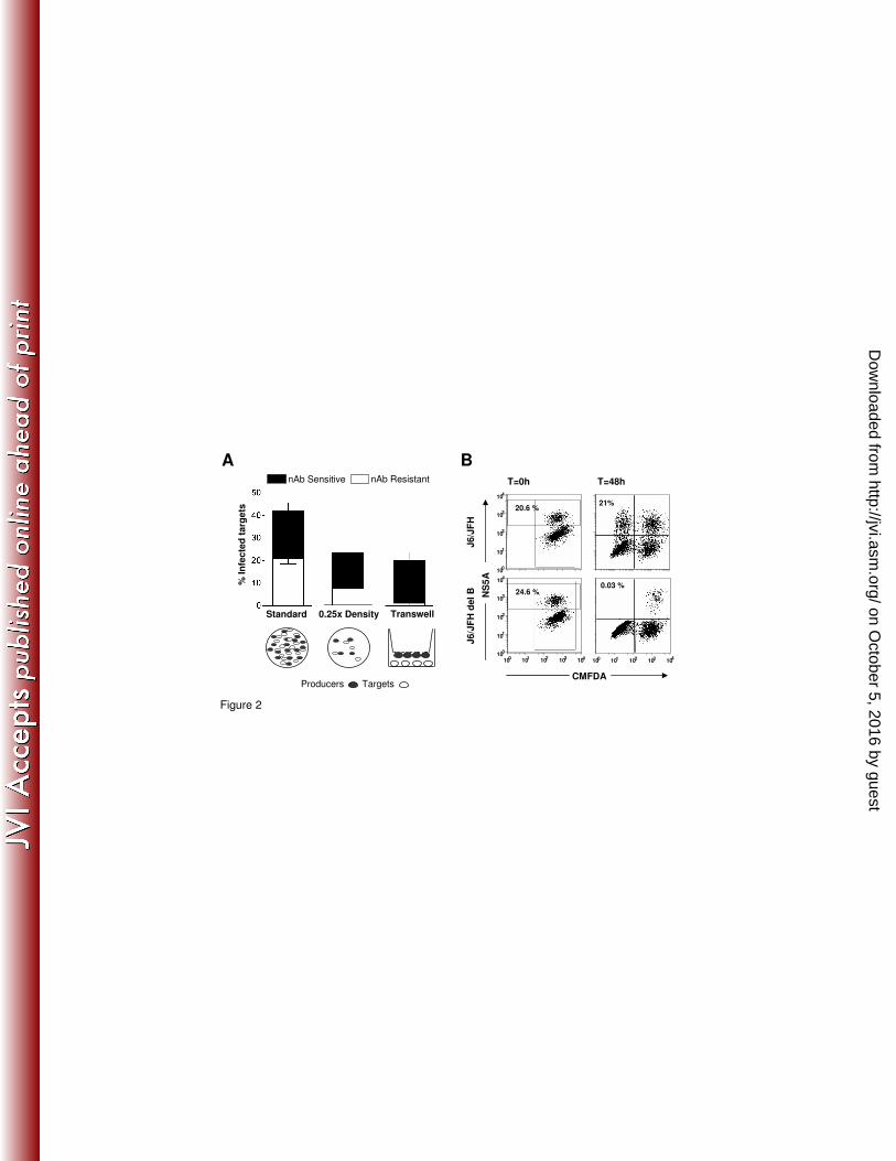
nAb resistant transmission requires cell contact and particle assembly. To examine the
processes of transmission we used two methods to segregate target and producer cells within the
co-culture (Fig.2A). Assays were performed where the cell seeding density was lowered to
reduce the number of cell-cell contacts. Alternatively producer cells were grown on the lower face
of a transwell insert directly above the target cells to prevent cell contact and to optimize the
diffusion of cell-free particles (Fig.2A). In direct co-culture both cell-free and cell-cell modes of
transmission can occur, however upon segregation of producer and target cells HCV can only
transmit via the extracellular medium. In the standard assay ~40% of target cells became infected
with an approximately equal ratio of nAb resistant and sensitive routes of viral transmission
(Fig.2A). However, nAb resistant transmission was significantly reduced at the lower seeding
density and was abrogated when the cells were separated by a trans-well insert (Fig.2A). These
observations suggest that cell-free virus does not contribute to nAb resistant transmission,
consistent with a direct cell-to-cell mechanism of nAb evasion.
It is possible that HCV may evade anti-glycoprotein antibody responses via the direct transfer of
RNA genomes between infected and naïve cells, thus negating the role of virions in transmission.
Indeed, the exosome secretion pathway represents an attractive target for localized spread of
intracellular pathogens (37). To test this model we used a J6/JFH virus encoding a deleted NS5A
(domain III - del B) (42) that expresses all of the viral proteins, but lacks a critical phosphorylation
on October 5, 2016 by guest
http://jvi.asm.org/
Dow
nloaded from
7
event in NS5A required for particle assembly, providing an ideal tool to investigate whether non-
encapsidated HCV genome is capable of cell-to-cell spread. For both genomic constructs the
number of infected (NS5A+) producer cells was comparable (J6/JFH 20.6% and J6/JFH/(del B)
24.6%) (Fig.2B). J6/JFH co-cultures efficiently transmitted infection to 21% of target cells after
48h, however, no target cells became infected when co-cultured with J6/JFH/(del B) expressing
cells, showing that non-encapsidated HCV genomes are not transferred between cells (Fig.2B).
These observations suggest that particle assembly is essential for HCV co-culture transmission.
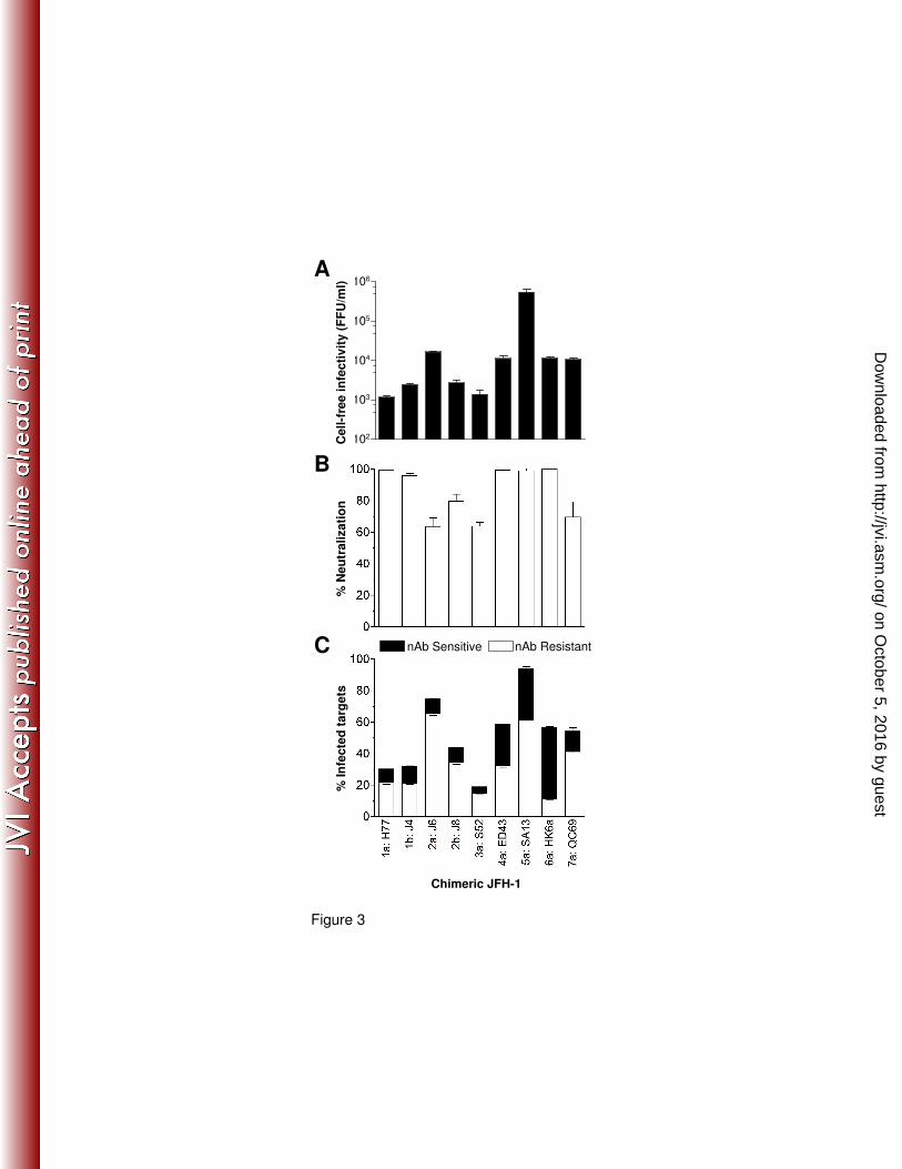
nAb resistant cell-to-cell transmission of diverse HCV genotypes. We performed infectious
co-culture assays with a panel of chimeric JFH viruses bearing the structural proteins of genotype
1a-7a viruses (12). The different viral strains generated a range of cell-free infectious virus
(Fig.3A) that was generally predictive of transmission efficiency (Fig.3C), in agreement with our
earlier results demonstrating that co-culture transmission is dependent on infectious particles. To
neutralize cell-free particle infectivity, cross-reactive pooled HCV patient IgG (300µg/ml) was
added to the co-cultures (Fig.3B). All of the viral strains demonstrated nAb resistant and sensitive
co-culture transmission (Fig.3C), however, differences were noted between viral strains in their
relative mode of transmission. These data suggest that nAb evasion by direct cell-to-cell transfer
of virions is a feature common to all HCV genotypes.
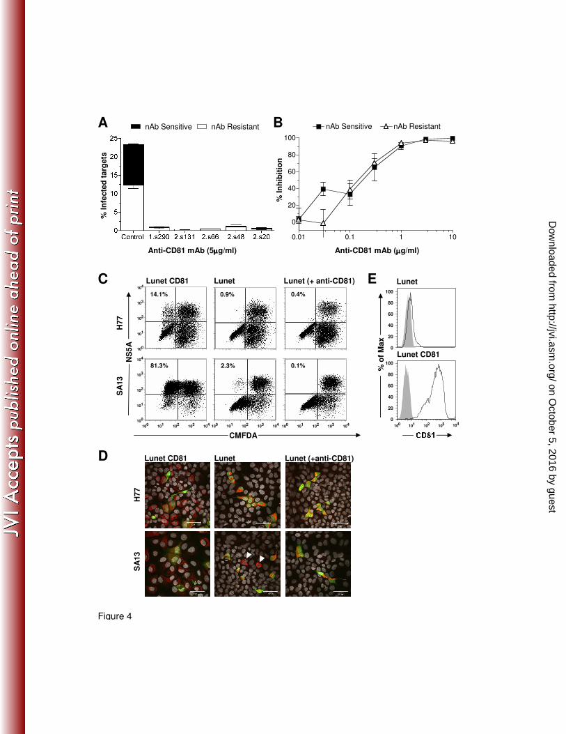
nAb resistant cell-to-cell transmission is CD81 dependent. We have shown that cell-free
virus infectivity is sensitive to the neutralizing effect(s) of antibodies targeting the viral
glycoproteins (Fig.1), consequently the addition of nAb to HCV co-cultures enables us to monitor
cell-to-cell transmission. We employed a H77/JFH co-culture assay in the presence or absence of
neutralizing anti-E2 mAb 9/27 to assess the receptor dependency of cell-to-cell transmission. We
first examined CD81 dependency using a panel of anti-receptor antibodies. Prior to co-culture
target cells were treated for 1h with anti-CD81 mAbs specific for a range of conformation-
dependent epitopes or a control antibody. A ll of the anti-CD81 mAbs inhibited nAb sensitive and
nAb resistant transmission by more than 90%, suggesting a similar CD81 dependency for both
routes of infection (Fig.4A). Indeed, titration of anti-CD81 mAb 2s131 demonstrated comparable
inhibition of nAb sensitive and nAb resistant transmission (Fig.4B). To confirm that HCV co-
culture transmission is CD81 dependent we used a Huh-7 derived Lunet cell line that expresses
low levels of CD81 as measured by flow cytometry (Fig.4E). Parental Lunet cells were resistant to
HCV pseudoparticle and cell-free HCVcc infection (data not shown), as previously reported (21).
H77/JFH and SA13/JFH infected producer cells were co-cultured with parental Huh-7 Lunet cells
or those transduced to express human CD81. Robust target cell infection was only detected in
Huh-7 Lunet-CD81 cells, consistent with CD81 being a critical HCV entry factor (Fig.4C).
However, a low number of SA13/JFH infected Huh-7 Lunet cells were detected, that may
on October 5, 2016 by guest
http://jvi.asm.org/
Dow
nloaded from
8
represent CD81 independent infection or could be attributed to the small number of Huh-7 Lunet
cells expressing low-level CD81 noted by flow cytometry (Fig.4E). Importantly, addition of anti-
CD81 mAb 2.s131 ablated SA13/JFH infection of Huh-7 Lunet cells, indicating that CD81
independent infection did not occur. The ability to detect SA13/JFH infected Huh-7 Lunet cells
may represent an increased affinity of SA13 glycoproteins for CD81 or may simply reflect the
higher infectivity of this chimeric virus (Fig.3A). We previously reported that HCV cell-to-cell
transmission could occur in the absence of CD81 (44), however this is most likely attributable to
the previous experimental design. In our earlier study naïve target cells rather than the producer
cells were CMFDA labeled, consequently multi-cell aggregates of infected producer and naïve
target cells can be wrongly registered as positive transmission events, an example of this can be
found in Supplementary Fig.3. The current experimental design eliminates these false positives.
To corroborate these findings we imaged HCV infected producer cells co-cultured with Huh-7
Lunet cells and found no evidence for CD81-independent transmission (Fig.4D).
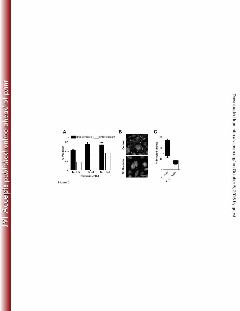
nAb resistant cell-to-cell transmission is dependent on tight junction proteins Claudin-1
and Occludin. The tight junction proteins Claudin-1 and Occludin are thought to act during the
late stages of HCV entry and there is limited evidence for direct glycoprotein interaction(s) (10).
We assessed the dependency of HCV cell-to-cell transmission on Claudin-1 using a using a
recently reported rat polyclonal antiserum that can inhibit cell-free virus infectivity (22). Target
cells were treated for 1hr with anti-Claudin-1 or control antiserum prior to co-culturing with Huh-
7.5 cells infected by chimeric viruses bearing the structural proteins of genotypes 1a, 1b and 4a
(H77/JFH, J4/JFH and ED43/JFH respectively) (Fig.5A). Anti-Claudin-1 inhibited nAb sensitive
and resistant cell-to-cell transmission of all three viruses, demonstrating a somewhat higher
efficiency for nAb sensitive transmission, suggesting that both modes of transmission are
Claudin-1 dependent. Due to the lack of antibodies targeting extracellular Occludin epitopes we
shRNA silenced protein expression in Huh-7.5 cells (Fig.5B). Confocal imaging of transduced
cells showed a significant reduction in Occludin expression with no observable effects on
Claudin-1, CD81 or SR-BI expression levels (data not shown). Silencing Occludin in Huh-7.5
target cells reduced both nAb sensitive and resistant routes of transmission (Fig.5C). Thus, both
tight junction proteins have a comparable role in cell-free and cell-to-cell modes of HCV
transmission.
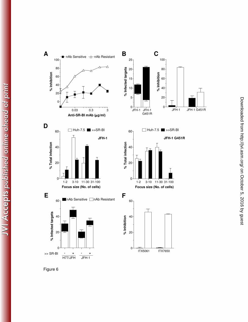
nAb resistant cell-to-cell transmission is SR-BI dependent. SR-BI is a receptor for high
density lipoprotein and is reported to be involved in the early stages of HCV attachment and entry
(35, 39). We (14) and others (6, 49) have reported that anti-SRBI antibodies can inhibit cell-free
HCV infectivity. To assess the role of SR-BI in H77/JFH cell-free and cell-to-cell transmission we
on October 5, 2016 by guest
http://jvi.asm.org/
Dow
nloaded from
9
treated Huh-7.5 target cells with increasing concentrations of anti-SR-BI mAb. Interestingly, anti-
SRBI demonstrated significantly greater inhibition of nAb resistant cell-to-cell transmission than
nAb sensitive transmission (Fig.6A). To further understand the role of SR-BI in cell-to-cell
transmission we employed a cell culture adapted JFH-1 mutant (G451R) that is relatively SR-BI-
independent (14, 51). Co-culture of JFH-1 and JFH-1 G451R infected producer and target cells
confirmed the adapted phenotype of the G451R virus with a greater capacity to transmit than the
parental virus (Fig.6B). However, G451R virus demonstrated a significantly reduced nAb resistant
cell-to-cell mode of transmission compared to wild type JFH-1 (Fig.6B). Furthermore, anti-SR-BI
significantly reduced JFH-1 cell-to-cell transmission compared to nAb sensitive cell-free infection
and yet had minimal effect on JFH G451R transmission (Fig.6C). We previously reported that SR-
BI over-expression increased the susceptibility of Huh-7.5 cells to HCVcc infection (14); this
phenotype is accompanied by an increase in the size of JFH-1 infected cell foci, indicative of
enhanced cell-to-cell transmission, whereas JFH-1 G451R foci remain unaltered (Fig.6D) (13).
Given this observation we employed these cells as targets in an infectious co-culture assay. HCV
strains H77/JFH and JFH-1 demonstrate a significant increase in nAb resistant cell-to-cell
transmission to Huh-7 cells expressing 2-fold greater levels of SR-BI (Fig.6E), suggesting that
SR-BI levels in Huh-7.5 hepatoma cells limit HCV cell-to-cell transmission. To further investigate
the role of SR-BI in HCV cell-to-cell transmission we studied the efficacy of two recently reported
entry inhibitors that bind SR-BI (41). Both compounds ITX5061 and ITX7650 significantly reduced
H77/JFH nAb resistant cell-to-cell transmission and had minimal effect on nAb sensitive cell-free
transmission (Fig.6F).
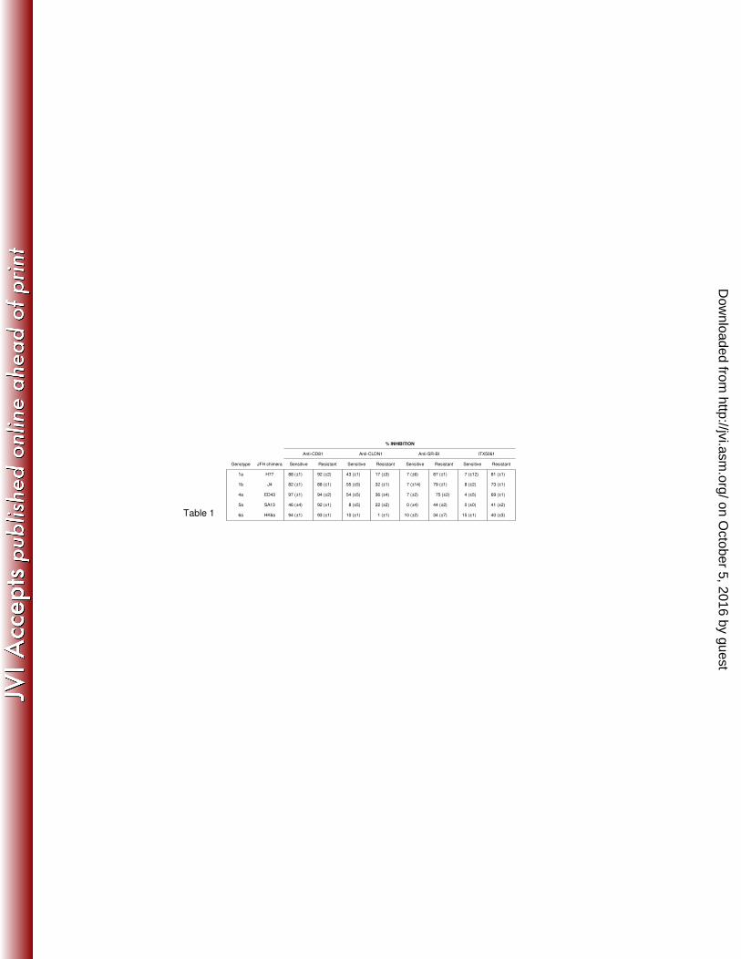
Receptor dependency of diverse HCV cell-to-cell transmission. We selected the chimeric
JFH-1 viruses in which robust (>90%) cell-free neutralization was achieved (Fig.4B) and
examined their sensitivity to receptor antagonists (Table 1). Anti-CD81 mAb 2s131 inhibited nAb
sensitive and resistant transmission of all HCV strains tested with comparable efficiency, with the
exception of SA13/JFH. Targeting SR-BI by receptor specific antibody or ITX5061 displayed a
broader range of efficiencies, but with excellent agreement between the two treatments. Notably
for all viruses, SR-BI antagonists were more effective at reducing nAb resistant cell-to-cell
transmission. In contrast, anti-CLDN1 demonstrated greater inhibition of nAb sensitive routes of
viral transmission, with minimal effect on either route of SA13/JFH or HK6a/JFH transmission
(Table 1). Although these findings suggest a spectrum of receptor dependencies between viral
strains, SR-BI remains an attractive therapeutic target, with ITX5061 displaying cross-genotype
inhibition of cell-to-cell transmission.
on October 5, 2016 by guest
http://jvi.asm.org/
Dow
nloaded from
10
Discussion. HCV persists in the face of a robust nAb response (40). Indeed, the ability to evade
the host immune responses is a common feature of many viruses capable of establishing chronic
infection. In vitro model systems suggest that HCV is a relatively sensitive target for antibody-
mediated neutralization (3, 24, 32, 50). We (44) and others (48)
reported efficient HCV
transmission in the presence of polyclonal patient IgG and a limited number of anti-E2 mAbs,
suggestive of direct cell-to-cell transfer.
There are various mechanisms by which a virus particle may transmit directly between cells, for
instance HIV and herpes simplex virus (HSV) spread across specific cellular contacts, in the case
of HIV a virally induced structure termed the virological synapse is formed (17). In contrast, HSV
exploits pre-existing cellular contacts (45). Other viruses remain tethered to the infected cell
surface, allowing directed lateral movement to adjacent naïve target cells (8, 36, 38). While these
varied modes of cell-to-cell transmission are believed to provide an efficient alternative to cell-free
infection, they may also confer resistance to the humoral immune response (27, 30).
The observation that HCV entry is dependent on two tight junction components (10, 34) offers a
tantalizing possibility that HCV may exploit such junctional contacts in transmission. However, the
Huh-7.5 cell line used in this study does not form functional tight junctions (28) and further studies
are required to investigate the mechanism(s) of HCV transmission in polarized cell systems. Our
observation that HCV E2 glycoprotein and receptor specific antibodies reduce cell-to-cell
transmission, suggests that spread is not occurring across a sealed cellular junction, which would
exclude such antibodies (27). HCV may be exploiting a contact structure similar to the virological
synapse of HIV (17), which has recently been shown to be permeable to antibodies (27).
We find co-culture transmission to be relatively insensitive to neutralization by a panel of diverse
anti-glycoprotein antibodies, whereas cell-free infectivity was readily eliminated (Fig.1 &
Supplementary Fig.1). nAb resistant transmission was prevented by separation of the target and
producer cells (Fig.2A), providing compelling evidence that this transmission occurs via direct
cell-cell contacts. Cell-to-cell transmission required particle assembly and did not occur via the
transfer of non-encapsidated HCV genomes (Fig.2B). Furthermore, we found that HCVcc
chimeras representative of seven genotypes transmitted via cell-to-cell transmission (Fig.3),
implying that this phenotype is common to all viral strains.
We investigated the receptor dependency of HCV transmission and found that CD81 mAbs
inhibited both cell-free and cell-to-cell infection, consistent with both routes of transmission being
CD81-dependent (Fig.4). Huh-7 Lunet cells were resistant to infection by both H77/JFH and
SA13/JFH. Furthermore, the low level SA13/JFH infection of Huh-7 Lunet cells was blocked by
on October 5, 2016 by guest
http://jvi.asm.org/
Dow
nloaded from
11
anti-CD81 mAb 2s131, suggesting that infection occurred in the minority of Huh-7 Lunet cells
expressing low levels of CD81. This observation is in contrast to previous findings by ourselves
and others of CD81 independent co-culture transmission (18, 44, 48). Further investigation into
our observation that HCV could transmit to CD81 negative HepG2 cells suggest that our earlier
finding was due to cell aggregates between labeled target and producer cells resulting in false
positives, as described in Supplementary Fig.3.
The tight junction components Claudin-1 and Occludin were recently identified as entry factors for
HCV (10, 34). Their elevated expression at points of cellular contact make them interesting
candidates in directing cell-to-cell transmission. We used an antiserum directed against the
Claudin-1 extracellular domain that disrupts Claudin-1/CD81 co-receptor association(s) (22), to
inhibit cell-free and cell-to-cell transmission (Fig.5). The antiserum exhibited moderate activity
against HCVcc chimeric viruses bearing the structural proteins of genotypes 1a, 1b and 4a and
demonstrated greater efficacy against nAb sensitive viral transmission. shRNA silencing Occludin
expression in Huh-7.5 target cells reduced both nAb sensitive and resistant routes of HCV
transmission (Fig.5). Thus, both tight junction proteins have a role in cell-to-cell HCV
transmission.
HCV particles are known to associate with host lipoproteins (2, 7) and we previously reported that
this association confers nAb resistance (14). This may have implications for localized spread by
allowing HCV to remain associated with cells via lipoprotein moieties (26). Interestingly, of all the
anti-glycoprotein mAbs tested, the anti-HVR1 mAb 9/27 demonstrated the greatest potential to
reduce cell-to-cell transmission, suggesting that antibodies targeting HVR1 may limit cell-cell
transfer of infection. However, in vivo HCV can escape anti-HVR1 responses by conventional
genetic escape (25, 46).
SR-BI is thought to play an important role in HCV attachment and entry via its interaction with the
E2 glycoprotein (35). SR-BI antagonists display preferential inhibition of cell-to-cell transmitted
HCV (Fig.6 and Table 1). A mutant JFH-1 G451R virus with limited SR-BI dependence (14)
demonstrated minimal cell-to-cell transmission (Fig.6B). Furthermore, over-expression of SR-BI in
Huh-7.5 cells promoted cell-to-cell transmission (Fig.6D and E). Taken together, these data
demonstrate that SR-BI plays an essential role in cell-to-cell transmission and targeting this
receptor may limit nAb resistant modes of HCV transmission. SR-BI specific compounds have
been developed to treat atherosclerosis (1, 31) and, unlike the other HCV receptors is
predominantly expressed in the liver (39), reducing the possibility for off-target effects.
on October 5, 2016 by guest
http://jvi.asm.org/
Dow
nloaded from
12
It is clear that cell-to-cell transmission of pathogens facilitates immune evasion and persistence.
The localized spread of HCV may be an adaptation to exploit the compact and highly ordered
environment of the liver. This conclusion is supported by the recent observation of HCV infected
foci in liver biopsy samples from HCV infected patients (23). It is important to remember that
extracellular forms of HCV are most likely responsible for spread between hosts and for liver
allograft re-infection following transplantation. However, cell-to-cell transmission may represent
the dominant route of virus dissemination within chronically infected individuals. Our findings raise
a number of issues that will require further consideration for the design and pre-clinical evaluation
of HCV glycoprotein-specific antibody and therapeutic B-cell vaccines, as it would appear that
HCV has evolved methods to evade such immune surveillance. In contrast, targeting the viral
receptors or entry factors, and in particular SR-BI, may provide a means of augmenting the host
immune response by inhibiting cell-to-cell transmission.
Acknowledgements. We would like to thank: Michelle J. Farquhar for critical reading of our
manuscript; David Mutimer for access to patient sera; Steven Foung for anti-glycoprotein mAbs;
Thomas Pietschmann for Lunet Huh-7 cells; Takaji Wakita, Brett Lindenbach, Charles Rice, Jin
Zhong, Frank V. Chisari and Jens Bukh for infectious HCVcc constructs. This work was
supported by MRC grant G0400802, Pfizer Ltd, the Wellcome Trust and PHS grants R01
DA024565, AI50798 and AI40034.
on October 5, 2016 by guest
http://jvi.asm.org/
Dow
nloaded from
13
Figure legends
Fig.1: Effect of anti-glycoprotein antibodies on HCV H77/JFH cell-free infectivity and co-
culture transmission. Anti-E2 mAbs 9/27, 3/11 and 11/20 (A) and pooled IgG isolated from 6
HCV-infected individuals (B) were titrated in a HCV strain H77/JFH co-culture assay. Co-culture
transmission (white bars) expressed as the percentage of infected target cells (left Y-axis) and
the infectivity of cell-free virus (black bars) defined as focus forming units per ml (FFU/ml, right Y-
axis) were measured. All titrations were performed in duplicate and the error bars indicate the
standard deviation. The data shown is representative of 3 experiments.
Fig.2: nAb resistant HCV transmission requires cell contact and infectious HCV particles.
(A) The standard H77/JFH co-culture assay was modified either by performing the co-culture at
low (0.25x) seeding density or by seeding the target and producer cells on opposing faces of the
culture well using a transwell insert. In both cases a 1:1 target:producer ratio was maintained.
The frequency of infected target cells in the absence (nAb sensitive) or presence (nAb resistant)
of mAb 9/27 at 4µg/ml is shown. The experiments were performed in duplicate and error bars
indicate the standard deviation. The data set is representative of 4 experiments. (B) J6/JFH and
J6/JFH del B RNA were electroporated into Huh-7.5 cells, after 72h the cells were CMFDA
labeled and NS5A expression measured immediately (T=0hr, left) or 48h after co-culture with
Huh-7.5 target cells (T=48hr, right).
Fig.3: Diverse HCVcc transmission. Huh-7.5 cells were electroporated with a panel of chimeric
JFH-1 HCV RNAs, where the infecting genotype is depicted prior to the strain nomenclature. 72h
post electroporation the cells were labeled with CMFDA and co-cultured with Huh-7.5 target cells
in the presence of control or cross reactive patient pooled HCV+ IgG at 300µg/ml for 48h.
Extracellular media was collected and the levels of infectious virus quantified (A) and the %
neutralization by patient HCV+ IgG determined (B). The stacked histogram displays nAb resistant
cell-to-cell (white bars) and nAb sensitive transmission (black bars) for each virus (C). All
treatments were performed in duplicate and the error bars indicate the standard deviation. The
data set is representative of 3 experiments.
Fig.4: HCV co-culture transmission is CD81 dependent. (A) Anti-CD81 inhibition of H77/JFH
co-culture transmission. The stacked histogram displays nAb resistant cell-to-cell (white bars) and
nAb sensitive (black bars) transmission in the presence of control or five anti-CD81 mAbs
(5µg/ml) specific for distinct epitopes. (B) Anti-CD81 mAb 2.s131 was titrated in a standard
H77/JFH co-culture assay and nAb sensitive (�) and resistant (∆) transmission is shown. All
treatments were performed in duplicate and error bars indicate the standard deviation. (C)
H77/JFH and SA13/JFH infected producer cells were co-cultured with Huh-7 Lunet cells or cells
on October 5, 2016 by guest
http://jvi.asm.org/
Dow
nloaded from
14
transduced to express human CD81 ± anti-CD81 (10µg/ml mAb 2s131), representative flow
cytometry plots are annotated with the percentage of infected target cells. (D) To corroborate the
infectious co-culture assay cells were fixed, stained for NS5A and imaged by confocal
microscopy. HCV infected target cells (red), uninfected producer (green) and infected producer
cells (orange) are clearly visible, with cell nuclei shown in grey. White arrows depict rare
SA13/JFH infected Lunet target cells. (E) Flow cytometric analysis of CD81 expression in
parental Huh-7 Lunet cells and those transduced to express human CD81.
Fig.5: A role for Claudin-1 and Occludin in HCV co-culture transmission. Infectious co-
culture assays were performed with chimeric HCV infected producer cells bearing the structural
proteins of genotypes 1a (H77), 1b (J4) and 4a (ED43). (A) Target cells were incubated with
polyclonal rat anti-Claudin-1 serum (1/100 dilution) prior to co-culture. Inhibition of nAb sensitive
(black bars) and nAb resistant (white bars) transmission is shown. (B) Huh-7.5 cells were
transduced with irrelevant and shRNA-Occludin lentiviruses and 120h later the cells were fixed,
permeabilised and stained for Occludin expression. (C) Irrelevant control and shRNA-Occludin
silenced Huh-7.5 target cells were co-cultured with H77/JFH infected producer cells and the
frequency of nAb sensitive (black) and resistant (white) transmission quantified by flow cytometry.
All treatments were performed in duplicate, the error bars indicate the standard deviation and this
data set is representative of two experiments.
Fig.6: HCV co-culture transmission is SR-BI dependent. (A) Anti-SR-BI mAb was titrated in a
standard H77/JFH co-culture assay and inhibition of nAb sensitive (�) and nAb resistant (∆)
infection is shown. (B) JFH-1 and JFH-1 G451R infected producer cells were co-cultured with
Huh-7.5 target cells in the presence or absence of nAb patient IgG (300µg/ml). The stacked
histogram displays nAb resistant cell-to-cell transmission (white bars) and nAb sensitive
transmission (black bars) for each virus. (C) The effect of anti-SR-BI mAb (1µg/ml) on JFH-1 and
JFH-1 G451 nAb resistant (white bars) and sensitive (black bars) transmission. (D) Parental Huh-
7.5 cells (white bars) or those over-expressing SR-BI (black) were infected with JFH-1 or
JFH/G451R for 72h in a standard cell-free infectious assay. HCV positive cells were enumerated
by immunofluorescent microscopy allowing quantification of infected cell focus size. The
histogram displays the percentage of total infection residing in small (1-2 cells), medium (3-10),
large (11-30) or very large (31-100) infected cell foci. (E) H77/JFH and JFH-1 infected producer
cells were co-cultured with parental Huh-7.5 cells or Huh-7.5 cells transduced to over-express
SR-BI (>>), in the presence or absence of neutralizing patient IgG (300µg/ml), and infected target
cells quantified. H77/JFH and JFH-1 nAb resistant infection of cells over expressing SR-BI was
significantly increased (p=0.0096 and p=0.0037 respectively). (F) Effect of the SR-BI specific
small molecules ITX5061 and ITX7650 (1µM) on H77/JFH nAb sensitive (black bars) and
on October 5, 2016 by guest
http://jvi.asm.org/
Dow
nloaded from
15
resistant (white bars) transmission. Treatments were performed in duplicate and error bars
indicate the standard deviation.
on October 5, 2016 by guest
http://jvi.asm.org/
Dow
nloaded from
16
References:
1. Acton, S. L., K. F. Kozarsky, and A. Rigotti. 1999. The HDL receptor SR-BI: a new therapeutic target for atherosclerosis? Mol Med Today 5:518-24.
2. Andre, P., F. Komurian-Pradel, S. Deforges, M. Perret, J. L. Berland, M. Sodoyer, S. Pol, C. Brechot, G. Paranhos-Baccala, and V. Lotteau. 2002. Characterization of low- and very-low-density hepatitis C virus RNA-containing particles. J Virol 76:6919-28.
3. Bartosch, B., J. Dubuisson, and F. L. Cosset. 2003. Infectious hepatitis C virus pseudo-particles containing functional E1-E2 envelope protein complexes. J Exp Med 197:633-42.
4. Bitzegeio, J., D. Bankwitz, K. Hueging, S. Haid, C. Brohm, M. B. Zeisel, E. Herrmann, M. Iken, M. Ott, T. F. Baumert, and T. Pietschmann. 2010. Adaptation of hepatitis C virus to mouse CD81 permits infection of mouse cells in the absence of human entry factors. PLoS Pathog 6:e1000978.
5. Burlone, M. E., and A. Budkowska. 2009. Hepatitis C virus cell entry: role of lipoproteins and cellular receptors. J Gen Virol 90:1055-70.
6. Catanese, M. T., R. Graziani, T. von Hahn, M. Moreau, T. Huby, G. Paonessa, C. Santini, A. Luzzago, C. M. Rice, R. Cortese, A. Vitelli, and A. Nicosia. 2007. High-avidity monoclonal antibodies against the human scavenger class B type I receptor efficiently block hepatitis C virus infection in the presence of high-density lipoprotein. J Virol 81:8063-71.
7. Chang, K. S., J. Jiang, Z. Cai, and G. Luo. 2007. Human apolipoprotein e is required for infectivity and production of hepatitis C virus in cell culture. J Virol 81:13783-93.
8. Doceul, V., M. Hollinshead, L. van der Linden, and G. L. Smith. 2010. Repulsion of superinfecting virions: a mechanism for rapid virus spread. Science 327:873-6.
9. Drummer, H. E., I. Boo, A. L. Maerz, and P. Poumbourios. 2006. A conserved Gly436-Trp-Leu-Ala-Gly-Leu-Phe-Tyr motif in hepatitis C virus glycoprotein E2 is a determinant of CD81 binding and viral entry. J Virol 80:7844-53.
10. Evans, M. J., T. von Hahn, D. M. Tscherne, A. J. Syder, M. Panis, B. Wolk, T. Hatziioannou, J. A. McKeating, P. D. Bieniasz, and C. M. Rice. 2007. Claudin-1 is a hepatitis C virus co-receptor required for a late step in entry. Nature 446:801-5.
11. Flint, M., C. Maidens, L. D. Loomis-Price, C. Shotton, J. Dubuisson, P. Monk, A. Higginbottom, S. Levy, and J. A. McKeating. 1999. Characterization of hepatitis C virus E2 glycoprotein interaction with a putative cellular receptor, CD81. J Virol 73:6235-44.
12. Gottwein, J. M., T. K. Scheel, T. B. Jensen, J. B. Lademann, J. C. Prentoe, M. L. Knudsen, A. M. Hoegh, and J. Bukh. 2009. Development and characterization of hepatitis C virus genotype 1-7 cell culture systems: role of CD81 and scavenger receptor class B type I and effect of antiviral drugs. Hepatology 49:364-77.
13. Grove, J., T. Huby, Z. Stamataki, T. Vanwolleghem, P. Meuleman, M. Farquhar, A. Schwarz, M. Moreau, J. S. Owen, G. Leroux-Roels, P. Balfe, and J. A. McKeating. 2007. Scavenger receptor BI and BII expression levels modulate hepatitis C virus infectivity. J Virol 81:3162-9.
14. Grove, J., S. Nielsen, J. Zhong, M. F. Bassendine, H. E. Drummer, P. Balfe, and J. A. McKeating. 2008. Identification of a residue in hepatitis C virus E2 glycoprotein that determines scavenger receptor BI and CD81 receptor dependency and sensitivity to neutralizing antibodies. J Virol 82:12020-9.
15. Horner, S. M., and M. Gale, Jr. 2009. Intracellular innate immune cascades and interferon defenses that control hepatitis C virus. J Interferon Cytokine Res 29:489-98.
16. Hsu, M., J. Zhang, M. Flint, C. Logvinoff, C. Cheng-Mayer, C. M. Rice, and J. A. McKeating. 2003. Hepatitis C virus glycoproteins mediate pH-dependent cell entry of pseudotyped retroviral particles. Proc Natl Acad Sci U S A 100:7271-6.
17. Jolly, C., K. Kashefi, M. Hollinshead, and Q. J. Sattentau. 2004. HIV-1 cell to cell transfer across an Env-induced, actin-dependent synapse. J Exp Med 199:283-93.
18. Jones, C. T., M. T. Catanese, L. M. Law, S. R. Khetani, A. J. Syder, A. Ploss, T. S. Oh, J. W. Schoggins, M. R. MacDonald, S. N. Bhatia, and C. M. Rice. 2010. Real-time imaging of hepatitis C virus infection using a fluorescent cell-based reporter system. Nat Biotechnol 28:167-71.
on October 5, 2016 by guest
http://jvi.asm.org/
Dow
nloaded from
17
19. Keck, Z. Y., T. K. Li, J. Xia, M. Gal-Tanamy, O. Olson, S. H. Li, A. H. Patel, J. K. Ball, S. M. Lemon, and S. K. Foung. 2008. Definition of a conserved immunodominant domain on hepatitis C virus E2 glycoprotein by neutralizing human monoclonal antibodies. J Virol 82:6061-6.
20. Klenerman, P., M. Lucas, E. Barnes, and G. Harcourt. 2002. Immunity to hepatitis C virus: stunned but not defeated. Microbes Infect 4:57-65.
21. Koutsoudakis, G., E. Herrmann, S. Kallis, R. Bartenschlager, and T. Pietschmann. 2007. The level of CD81 cell surface expression is a key determinant for productive entry of hepatitis C virus into host cells. J Virol 81:588-98.
22. Krieger, S. E., M. B. Zeisel, C. Davis, C. Thumann, H. J. Harris, E. K. Schnober, C. Mee, E. Soulier, C. Royer, M. Lambotin, F. Grunert, V. L. Dao Thi, M. Dreux, F. L. Cosset, J. A. McKeating, C. Schuster, and T. F. Baumert. 2010. Inhibition of hepatitis C virus infection by anti-claudin-1 antibodies is mediated by neutralization of E2-CD81-claudin-1 associations. Hepatology 51:1144-57.
23. Liang, Y., T. Shilagard, S. Y. Xiao, N. Snyder, D. Lau, L. Cicalese, H. Weiss, G. Vargas, and S. M. Lemon. 2009. Visualizing hepatitis C virus infections in human liver by two-photon microscopy. Gastroenterology 137:1448-58.
24. Lindenbach, B. D., M. J. Evans, A. J. Syder, B. Wolk, T. L. Tellinghuisen, C. C. Liu, T. Maruyama, R. O. Hynes, D. R. Burton, J. A. McKeating, and C. M. Rice. 2005. Complete replication of hepatitis C virus in cell culture. Science 309:623-6.
25. Liu, S., W. Yang, L. Shen, J. R. Turner, C. B. Coyne, and T. Wang. 2009. Tight junction proteins claudin-1 and occludin control hepatitis C virus entry and are downregulated during infection to prevent superinfection. J Virol 83:2011-4.
26. Maillard, P., T. Huby, U. Andreo, M. Moreau, J. Chapman, and A. Budkowska. 2006. The interaction of natural hepatitis C virus with human scavenger receptor SR-BI/Cla1 is mediated by ApoB-containing lipoproteins. Faseb J 20:735-7.
27. Martin, N., S. Welsch, C. Jolly, J. A. Briggs, D. Vaux, and Q. J. Sattentau. 2010. Virological synapse-mediated spread of human immunodeficiency virus type 1 between T cells is sensitive to entry inhibition. J Virol 84:3516-27.
28. Mee, C. J., J. Grove, H. J. Harris, K. Hu, P. Balfe, and J. A. McKeating. 2008. Effect of cell polarization on hepatitis C virus entry. J Virol 82:461-70.
29. Meertens, L., C. Bertaux, L. Cukierman, E. Cormier, D. Lavillette, F. L. Cosset, and T. Dragic. 2008. The tight junction proteins claudin-1, -6, and -9 are entry cofactors for hepatitis C virus. J Virol 82:3555-60.
30. Mothes, W., N. M. Sherer, J. Jin, and P. Zhong. 2010. Virus cell-to-cell transmission. J Virol.
31. Nieland, T. J., M. Penman, L. Dori, M. Krieger, and T. Kirchhausen. 2002. Discovery of chemical inhibitors of the selective transfer of lipids mediated by the HDL receptor SR-BI. Proc Natl Acad Sci U S A 99:15422-7.
32. Owsianka, A. M., A. W. Tarr, Z. Y. Keck, T. K. Li, J. Witteveldt, R. Adair, S. K. Foung, J. K. Ball, and A. H. Patel. 2008. Broadly neutralizing human monoclonal antibodies to the hepatitis C virus E2 glycoprotein. J Gen Virol 89:653-9.
33. Owsianka, A. M., J. M. Timms, A. W. Tarr, R. J. Brown, T. P. Hickling, A. Szwejk, K. Bienkowska-Szewczyk, B. J. Thomson, A. H. Patel, and J. K. Ball. 2006. Identification of conserved residues in the E2 envelope glycoprotein of the hepatitis C virus that are critical for CD81 binding. J Virol 80:8695-704.
34. Ploss, A., M. J. Evans, V. A. Gaysinskaya, M. Panis, H. You, Y. P. de Jong, and C. M. Rice. 2009. Human occludin is a hepatitis C virus entry factor required for infection of mouse cells. Nature 457:882-6.
35. Scarselli, E., H. Ansuini, R. Cerino, R. M. Roccasecca, S. Acali, G. Filocamo, C. Traboni, A. Nicosia, R. Cortese, and A. Vitelli. 2002. The human scavenger receptor class B type I is a novel candidate receptor for the hepatitis C virus. Embo J 21:5017-25.
36. Schelhaas, M., H. Ewers, M. L. Rajamaki, P. M. Day, J. T. Schiller, and A. Helenius. 2008. Human papillomavirus type 16 entry: retrograde cell surface transport along actin-rich protrusions. PLoS Pathog 4:e1000148.
on October 5, 2016 by guest
http://jvi.asm.org/
Dow
nloaded from
18
37. Schorey, J. S., and S. Bhatnagar. 2008. Exosome function: from tumor immunology to pathogen biology. Traffic 9:871-81.
38. Sherer, N. M., J. Jin, and W. Mothes. 2010. Directional spread of surface-associated retroviruses regulated by differential virus-cell interactions. J Virol 84:3248-58.
39. Silver, D. L., and A. R. Tall. 2001. The cellular biology of scavenger receptor class B type I. Curr Opin Lipidol 12:497-504.
40. Stamataki, Z., J. Grove, P. Balfe, and J. A. McKeating. 2008. Hepatitis C virus entry and neutralization. Clin Liver Dis 12:693-712.
41. Syder, A., H. Lee, M. Zeisel, J. Grove, E. Soulier, J. Macdonald, S. Chow, J. Chang, T. Baumert, J. McKeating, J. McKelvy, and F. Wong-Staal. 2010. Small molecule scavenger receptor BI antagonists are potent HCV entry inhibitors. J Hepatology in press.
42. Tellinghuisen, T. L., K. L. Foss, and J. Treadaway. 2008. Regulation of hepatitis C virion production via phosphorylation of the NS5A protein. PLoS Pathog 4:e1000032.
43. Thorley, J. A., J. A. McKeating, and J. Z. Rappoport. 2010. Mechanisms of viral entry: sneaking in the front door. Protoplasma 244:15-24.
44. Timpe, J. M., Z. Stamataki, A. Jennings, K. Hu, M. J. Farquhar, H. J. Harris, A. Schwarz, I. Desombere, G. L. Roels, P. Balfe, and J. A. McKeating. 2008. Hepatitis C virus cell-cell transmission in hepatoma cells in the presence of neutralizing antibodies. Hepatology 47:17-24.
45. Tomishima, M. J., G. A. Smith, and L. W. Enquist. 2001. Sorting and transport of alpha herpesviruses in axons. Traffic 2:429-36.
46. von Hahn, T., J. C. Yoon, H. Alter, C. M. Rice, B. Rehermann, P. Balfe, and J. A. McKeating. 2007. Hepatitis C virus continuously escapes from neutralizing antibody and T-cell responses during chronic infection in vivo. Gastroenterology 132:667-78.
47. Wakita, T., T. Pietschmann, T. Kato, T. Date, M. Miyamoto, Z. Zhao, K. Murthy, A. Habermann, H. G. Krausslich, M. Mizokami, R. Bartenschlager, and T. J. Liang. 2005. Production of infectious hepatitis C virus in tissue culture from a cloned viral genome. Nat Med 11:791-6.
48. Witteveldt, J., M. J. Evans, J. Bitzegeio, G. Koutsoudakis, A. M. Owsianka, A. G. Angus, Z. Y. Keck, S. K. Foung, T. Pietschmann, C. M. Rice, and A. H. Patel. 2009. CD81 is dispensable for hepatitis C virus cell-to-cell transmission in hepatoma cells. J Gen Virol 90:48-58.
49. Zeisel, M. B., G. Koutsoudakis, E. K. Schnober, A. Haberstroh, H. E. Blum, F. L. Cosset, T. Wakita, D. Jaeck, M. Doffoel, C. Royer, E. Soulier, E. Schvoerer, C. Schuster, F. Stoll-Keller, R. Bartenschlager, T. Pietschmann, H. Barth, and T. F. Baumert. 2007. Scavenger receptor class B type I is a key host factor for hepatitis C virus infection required for an entry step closely linked to CD81. Hepatology 46:1722-31.
50. Zhong, J., P. Gastaminza, G. Cheng, S. Kapadia, T. Kato, D. R. Burton, S. F. Wieland, S. L. Uprichard, T. Wakita, and F. V. Chisari. 2005. Robust hepatitis C virus infection in vitro. Proc Natl Acad Sci U S A 102:9294-9.
51. Zhong, J., P. Gastaminza, J. Chung, Z. Stamataki, M. Isogawa, G. Cheng, J. A. McKeating, and F. V. Chisari. 2006. Persistent hepatitis C virus infection in vitro: coevolution of virus and host. J Virol 80:11082-93.
on October 5, 2016 by guest
http://jvi.asm.org/
Dow
nloaded from
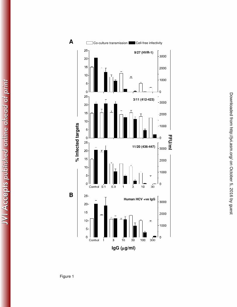
Figure 1
A
9/27 (HVR-1)
B
3/11 (412-423)
11/20 (436-447)
Human HCV +ve IgG
% I
nfe
cte
d t
arg
ets
FF
U/m
l
IgG (µµµµg/ml)
Co-culture transmission Cell-free infectivity
on October 5, 2016 by guest
http://jvi.asm.org/
Dow
nloaded from
A
Producers Targets
Standard 0.25x Density Transwell
B
CMFDA
NS
5A
J6/J
FH
J6/J
FH
del B
21%
100 101 102 103 104
0.03 %
100
101
102
103
104
20.6 %
100 101 102 103 10410
0
101
102
103
104
24.6 %
T=0h T=48hnAb Sensitive nAb Resistant
% In
fecte
d t
arg
ets
Figure 2
on October 5, 2016 by guest
http://jvi.asm.org/
Dow
nloaded from
A
B
C
102
103
104
105
106
% N
eu
tralizati
on
Cell-f
ree in
fecti
vit
y (
FF
U/m
l)%
In
fecte
d t
arg
ets
Figure 3
nAb Sensitive nAb Resistant
Chimeric JFH-1
on October 5, 2016 by guest
http://jvi.asm.org/
Dow
nloaded from
100 101 102 103 104100 101 102 103 104100
101
102
103
104
100
101
102
103
104
100 101 102 103 104
A
E
100 101 102 103 104
% o
f M
ax
Lunet CD81
H77
SA
13
NS
5A
Lunet CD81 Lunet Lunet (+ anti-CD81)
14.1%
81.3%
0.9%
2.3%
0.4%
0.1%
C
CMFDA
nAb Sensitive nAb Resistant
Anti-CD81 mAb (5µµµµg/ml)
% In
fecte
d t
arg
ets
Anti-CD81 mAb (µµµµg/ml)
% In
hib
itio
n
Figure 4
100
80
60
40
20
0
B
D
SA
13
H77
LunetLunet CD81 Lunet (+anti-CD81)
Lunet
100
80
60
40
20
0
0.01 0.1 1 10
0
20
40
60
80
100
nAb Sensitive nAb Resistant
on October 5, 2016 by guest
http://jvi.asm.org/
Dow
nloaded from
% In
hib
itio
n
Chimeric JFH-1
nAb Sensitive nAb Resistant
Sh
Occlu
din
Co
ntr
ol
A B C
Figure 5
% In
fecte
d t
arg
ets
on October 5, 2016 by guest
http://jvi.asm.org/
Dow
nloaded from
- + - +
0
20
40
60
H77/JFH JFH-1
>> SR-BI
1-2 3-10 11-30 31-1000
20
40
60
ITX5061 ITX76500
20
40
60
0.03 0.3 3
0
20
40
60
80
100nAb Sensitive nAb Resistant
JFH-1 JFH-1 G451R
0
5
10
15
20
25
JFH-1 JFH-1G451R
JFH-1 JFH-1 G451R0
20
40
60
80
100
nAb Sensitive nAb Resistant
Figure 6
DHuh-7.5 >>SR-BI
JFH-1 JFH-1 G451R
% T
ota
l in
fecti
on
% In
fecte
d t
arg
ets
% In
hib
itio
n
% In
hib
itio
nCA B
E F
Anti-SR-BI mAb (µµµµg/ml)
% In
hib
itio
n
% In
fecte
d t
arg
ets
Focus size (No. of cells)
1-2 3-10 11-30 31-1000
20
40
60
% T
ota
l in
fecti
on
Focus size (No. of cells)
Huh-7.5 >>SR-BI
on October 5, 2016 by guest
http://jvi.asm.org/
Dow
nloaded from
Genotype JFH chimera Sensitive Resistant Sensitive Resistant Sensitive Resistant Sensitive Resistant
1a H77 88 (±1) 92 (±2) 43 (±1) 17 (±3) 7 (±6) 87 (±1) 7 (±12) 81 (±1)
1b J4 82 (±1) 88 (±1) 55 (±5) 32 (±1) 7 (±14) 79 (±1) 8 (±2) 73 (±1)
4a ED43 97 (±1) 94 (±2) 54 (±5) 36 (±4) 7 (±2) 75 (±2) 4 (±5) 69 (±1)
5a SA13 46 (±4) 92 (±1) 8 (±5) 22 (±2) 0 (±4) 44 (±2) 0 (±0) 41 (±2)
6a HK6a 94 (±1) 93 (±1) 10 (±1) 1 (±1) 10 (±2) 34 (±7) 15 (±1) 40 (±3)
% INHIBITION
Anti-CD81 Anti-CLDN1 Anti-SR-BI ITX5061
Table 1
on October 5, 2016 by guest
http://jvi.asm.org/
Dow
nloaded from


