Embed Size (px)
Citation preview

A Crucial Role for Kupffer Cell-Derived Galectin-9 inRegulation of T Cell Immunity in Hepatitis C InfectionJohn A. Mengshol1,2, Lucy Golden-Mason1,3, Tomohiro Arikawa5, Maxwell Smith4, Toshiro Niki6, Ryan
McWilliams1, Jessica A. Randall1, Rachel McMahan1, Michael A. Zimmerman7, Manu Rangachari8,
Evgenia Dobrinskikh1, Pierre Busson9, Stephen J. Polyak10, Mitsuomi Hirashima5, Hugo R. Rosen1,2,3*
1 Department of Medicine, Division of Gastroenterology & Hepatology, University of Colorado School of Medicine, Aurora, Colorado, United States of America, 2 Denver
Veterans Affairs Medical Center, Denver, Colorado, United States of America, 3 Division of Clinical Immunology, University of Colorado School of Medicine and National
Jewish Hospital, Denver, Colorado, United States of America, 4 Department of Pathology, University of Colorado School of Medicine, Denver, Colorado, United States of
America, 5 Department of Immunology and Immunopathology, Kagawa Medical University, Kagawa, Japan, 6 GalPharma, Kagawa, Japan, 7 Division of Transplant Surgery,
University of Colorado School of Medicine, Aurora, Colorado, United States of America, 8 Center for Neurologic Diseases, Brigham and Women’s Hospital, Harvard Medical
School, Boston, Massachusetts, United States of America, 9 Institut de Cancerologie Gustave Roussy, Villejuif, France, 10 Division of Laboratory Medicine, University of
Washington, Seattle, Washington, United States of America
Abstract
Approximately 200 million people throughout the world are infected with hepatitis C virus (HCV). One of the most strikingfeatures of HCV infection is its high propensity to establish persistence (,70–80%) and progressive liver injury. Galectins areevolutionarily conserved glycan-binding proteins with diverse roles in innate and adaptive immune responses. Here, wedemonstrate that galectin-9, the natural ligand for the T cell immunoglobulin domain and mucin domain protein 3 (Tim-3),circulates at very high levels in the serum and its hepatic expression (particularly on Kupffer cells) is significantly increased inpatients with chronic HCV as compared to normal controls. Galectin-9 production from monocytes and macrophages isinduced by IFN-c, which has been shown to be elevated in chronic HCV infection. In turn, galectin-9 induces pro-inflammatory cytokines in liver-derived and peripheral mononuclear cells; galectin-9 also induces anti-inflammatorycytokines from peripheral but not hepatic mononuclear cells. Galectin-9 results in expansion of CD4+CD25+FoxP3+CD127low
regulatory T cells, contraction of CD4+ effector T cells, and apoptosis of HCV-specific CTLs. In conclusion, galectin-9production by Kupffer cells links the innate and adaptive immune response, providing a potential novel immunotherapeutictarget in this common viral infection.
Citation: Mengshol JA, Golden-Mason L, Arikawa T, Smith M, Niki T, et al. (2010) A Crucial Role for Kupffer Cell-Derived Galectin-9 in Regulation of T Cell Immunityin Hepatitis C Infection. PLoS ONE 5(3): e9504. doi:10.1371/journal.pone.0009504
Editor: Derya Unutmaz, New York University, United States of America
Received December 9, 2009; Accepted February 9, 2010; Published March 4, 2010
This is an open-access article distributed under the terms of the Creative Commons Public Domain declaration which stipulates that, once placed in the publicdomain, this work may be freely reproduced, distributed, transmitted, modified, built upon, or otherwise used by anyone for any lawful purpose.
Funding: This research is supported by National Institutes of Health (NIH) grants RO1 DK060590 and U19 A 1066328-01 (HCV center grant) to HRR. The fundershad no role in study design, data collection and analysis, decision to publish, or preparation of the manuscript. Dr Niki from Galpharma provided antibodies.
Competing Interests: Dr. Niki works for GalPharma and supplied antibodies for this study. This does not alter adherence to all the PLoS ONE policies on sharingdata and materials, as detailed online.
* E-mail: [email protected]
Introduction
Approximately 200 million people throughout the world are
infected with hepatitis C virus (HCV) [1]. HCV infection
establishes chronicity in the vast majority of patients [2], and is
a leading etiology of cirrhosis, liver cancer and indication for liver
transplantation [3]. Although new therapies have improved the
rates of sustained response, a large proportion of patients still fail
to respond to antiviral treatment or develop significant drug
toxicity [4], thus remaining at risk for disease progression.
Enhanced understanding of HCV-host interactions is required to
combat this virus and to develop improved therapies.
Galectins are evolutionarily conserved glycan-binding proteins
with diverse roles in innate and adaptive immune responses [5].
Galectins demonstrate a shared structural fold and at least one
conserved carbohydrate-recognition domain (CRD) of about 130
amino acids that recognizes glycans and mediates carbohydrate
binding. Moreover, at least 15 galectins have been identified in
mammals [6]http://www.nature.com/ni/journal/v9/n6/full/ni.
f.203.html - B8#B8. ‘‘Prototype’’ galectins have CRDs that can
dimerize, whereas so-called ‘‘tandem-repeat galectins’’ contain two
distinct CRDs in tandem in a single polypeptide chain, separated
by a linker of up to 70 amino acids. Accumulating evidence
indicates a critical role of the tandem repeat-type galectin-9 in
regulating the fate of effector T cells, specifically by binding to the
T cell immunoglobulin domain and mucin domain protein 3
(Tim-3) to induce apoptosis of some T cell subsets [5] [7], as well
as promoting the differentiation of regulatory T cells (Treg)
expressing FoxP3 [8]. Recently we found that Tim-3 was
increased on exhausted T cells in chronic hepatitis C infection
and may provide a novel therapeutic target [9].
As the natural ligand for Tim-3, galectin-9 is widely distributed
throughout various tissues, being particularly abundant in the liver
[10]. The liver is among the most interesting and potent
immunologic organs, rivaled only by what are considered the
true immune organs, such as thymus and lymph nodes [11]. The
liver is exposed continuously to a diverse and large antigenic load,
including pathogens, toxins, and tumor cells, as well as dietary and
PLoS ONE | www.plosone.org 1 March 2010 | Volume 5 | Issue 3 | e9504

commensal proteins. The liver must be actively immunocompetent
and simultaneously control inappropriate inflammatory responses
to harmless antigens encountered in the portal circulation, thus
being able to selectively induce immunity or tolerance to antigens
[12]. Moreover, numerous studies have identified the liver as a
major site for apoptosis and removal of peripheral CD8+ T cells
undergoing antigen induced cell death (AICD) [13].
We speculated that the dysfunctional antiviral T cell responses
characteristic of HCV infection–expansion of suppressive Tregs
[14,15] and impaired HCV-specific cytotoxic T lymphocytes (CTL)
[16] [17] [9], particularly within the intrahepatic compartment–
might be related to increased expression of galectin-9. In the current
study, we determined the circulating and hepatic expression of
galectin-9 in chronic HCV infection, finding that Kupffer cells (KC)
are the predominant source of galectin-9 within the liver, and that
its expression is upregulated in HCV-infected livers relative to
normal livers. Furthermore, we find that macrophage-derived
galectin-9 production is induced by IFN-c, and that galectin-9, in
turn, leads to the production of pro-inflammatory cytokines. In
addition, galectin-9 induces the expansion of regulatory T cells in
chronic HCV infection in a TGF-b-dependent manner, as well as
apoptosis of HCV-specific CTLs via activation of caspase 8. We
propose a novel paradigm whereby Kupffer cell-derived galectin-9
regulates T cell responses within the liver, providing novel insights
into the bridge between innate and adaptive immunity, mechanisms
of viral persistence, and potential immunotherapeutic targets for
HCV infection. Moreover, given the broad array of responses
induced by galectin-9, including potentially detrimental processes,
this pathway may be important in mediating immunopathology in
other hepatic diseases.
Results
Circulating Galectin-9 Levels Elevated in Chronic HCVPatients
Given the recent demonstration that expression of negative
immune regulator Tim-3 is increased on exhausted T cells in
patients with HCV [9] and that galectin-9 is a ligand for Tim-3
[7], we analyzed the circulating levels of galectin-9 in the plasma
from patients with chronic HCV, non-viral liver disease and
normal controls. We found that galectin-9 levels were strikingly
elevated in HCV patients (median 841 pg/ml in HCV vs. 0 in
normal controls, p = 0.0005, Figure 1). Importantly, HCV-
positive patients with all stages of fibrosis demonstrated elevated
levels of circulating galectin-9 (data not shown). Moreover, HCV-
infected patients with hepatocellular carcinoma had even higher
levels when compared to HCV alone (n = 7, median 1376 vs.
n = 15 median 715 pg/ml; p = 0.0288). Levels in 11 patients with
non-viral liver disease were elevated (median 340 pg/ml) but to
lower levels than HCV patients and were not significantly different
from normal patients (p = 0.06) or HCV patients.
Galectin-9 Expression Up-Regulated on Kupffer Cells inLivers of HCV-Infected Patients
In order to determine the cell types producing galectin-9 in
chronic HCV, we analyzed paraffin-embedded liver biopsy and
liver resection specimens for galectin-9 protein by immunohisto-
chemistry (IHC). Using CD68 as a marker for KC, we found that
KC had the highest staining for galectin-9 (Figure 2a–e). KC
constitute a cellular component of the hepatic sinusoids, anchored
to the luminal site of the endothelium and, thus, exposed to the
bloodstream [18]. HCV patients had higher intensity and
frequency of KC staining compared to normal controls
(Figure 2g and h, p,0.0001 for both). The majority of KC
are located in periportal regions where they have greater
phagocytic activity and are larger than those found in the
perilobular region [18], resulting in a zonal distribution with
differential KC function. We observed that 93% of normal control
livers demonstrated no galectin-9 staining in the periportal regions,
whereas virtually all the patients with HCV infection, regardless of
grade of inflammation or stage of fibrosis, stained positively
(Figure 2f). To confirm our staining, we also performed
immunofluorescence and confocal microscopy on paraffin-em-
bedded liver biopsy sections from HCV patients using antibodies
to galectin-9, albumin and CD68 (Figure S1). We did not see
staining of galectin-9 in hepatocytes (albumin positive) (FigureS1A); we again saw co-staining of CD68 and galectin-9,
confirming that KC are galectin-9 positive (Figure S1B).
IFN-c Induces Galectin-9 Production by HumanMonocytes and Macrophages
Next, based on the IHC data, we determined which factors
might stimulate production of galectin-9 by monocytes/macro-
Figure 1. Galectin-9 is elevated in the plasma of patients withchronic HCV and higher levels are seen in hepatocellularcarcinoma (HCC). Galectin-9 levels were analyzed in the plasma of 10normal controls, 22 patients with HCV and 11 patients with non-viralcauses of liver disease using a sandwich ELISA [50]. Seven patients withHCV and HCC are denoted by open squares; they had significantly highergalectin-9 levels compared to HCV patients without HCC, p = 0.0334.Plasma from 11 patients with non-viral liver disease was analyzed. Threepatients had alcoholic liver disease, 3 patients had Primary BiliaryCirrhosis, 3 patients had autoimmune hepatitis and 2 patients had non-alcoholic steatohepatitis. P- values were calculated using the two tailedMann-Whitney test. NS denotes non-significant, p values.0.05.doi:10.1371/journal.pone.0009504.g001
Gal-9 Regulates HCV Immunity
PLoS ONE | www.plosone.org 2 March 2010 | Volume 5 | Issue 3 | e9504
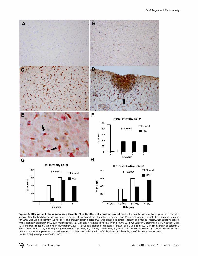
Figure 2. HCV patients have increased Galectin-9 in Kupffer cells and periportal areas. Immunohistochemistry of paraffin embeddedsamples (see Methods for details) was used to analyze 39 samples from HCV-infected patients and 15 normal subjects for galectin-9 staining. Stainingfor CD68 was used to identify Kupffer cells. The analyzing pathologist (M.S.) was blinded to patient identity and medical history. (A) Negative controlwith secondary antibody only, 206magnification. (B) Galectin-9 staining in normal liver (brown) 206; (C) Galectin-9 staining in a HCV patient 206,(D) Periportal galectin-9 staining in HCV patient, 2006. (E) Co-localization of galectin-9 (brown) and CD68 (red) 6006. (F–H) Intensity of galectin-9was scored from 0 to 3, and frequency was scored 0 (,10%), 1 (10–40%), 2 (40–70%), 3 (.70%). Distribution of scores by category expressed as apercent of the total patients comparing normal patients to patients with HCV. P-values calculated by the Chi-square test for trend.doi:10.1371/journal.pone.0009504.g002
Gal-9 Regulates HCV Immunity
PLoS ONE | www.plosone.org 3 March 2010 | Volume 5 | Issue 3 | e9504

phages. Prior work has indicated that galectin-9 expression can be
induced in various cells such as endothelial cells, fibroblasts and
astrocytes [19] [20] [21] by IFN-c or IL-1b. There are three
isoforms of galectin-9 according to the size of the linker peptide
connecting two CRDs [22]: long (Gal-9L, MW 39.5 kD), medium
(Gal-9M, 35.9 kD), and short (Gal-9S, 34.7 kD) isoforms.
Although these isoforms have shown comparable eosinophil
chemo-attractant activity [23], little is known about their
differences in other biological functions. Specifically, among the
three isoforms, the medium and long gal proteins were
upregulated by IFN- c in human fibroblasts [21].
We found that peripherally derived macrophages differentiated
with mCSF produced a basal level of galectin-9 and that IFN-cwas a strong inducer of galectin-9 at 48 hours (Figure 3a). The
medium and long isoforms of galectin-9 were induced preferen-
tially. The fold induction of galectin-9 was higher in patients with
chronic hepatitis C (7 fold) compared to normal controls (1.8 fold).
Production of galectin-9 from macrophages was not significantly
stimulated by IL-1b, LPS or HCV core protein.
Galectin-9 Induces Pro-Inflammatory Cytokines in Liver-Derived and Peripheral Mononuclear Cells
Having demonstrated that HCV-infected patients have higher
circulating and intrahepatic levels of galectin-9, we next explored
the effects of recombinant galectin-9 on whole peripheral blood
mononuclear cells (PBMCs) and liver-derived mononuclear cells
from normal healthy subjects and HCV-infected patients. As
shown in Figure 4, 48 hours of galectin-9 treatment induced an
array of pro-inflammatory mediators, such as TNF-a, IL-1b, and
IFN-c from peripheral and liver derived mononuclear cells of
study subjects. Intracellular cytokine staining demonstrated that
TNF-a was produced by CD14+ monocytes, T and NK cells, but
not B cells following stimulation with galectin-9 (data not shown).
Moreover, galectin-9-induced cytokine production was not due to
LPS contamination and was TLR4 independent, since a TLR4
blocking antibody had no effect on cytokine production (data not
shown).
Production of the anti-inflammatory cytokines IL-4, IL-10, and
IL-13 was induced in galectin-9-treated PBMCs but not hepatic
mononuclear cells (Figure 4). Liver-derived mononuclear cells
produced higher basal levels of IL-10 than PBMC, consistent with
prior studies showing more IL-10 production from KC [24–25].
Taken together, these data implicate galectin-9 as a key regulator
of hepatic immunity by preferentially inducing pro-inflammatory
cytokines instead of hepatoprotective factors such as IL-4 and IL-
13 in the liver.
Galectin-9 Expands Regulatory T Cells (Tregs)CD4+ CD25+ Foxp3+ regulatory T cells are crucial for
negatively regulating immune responses, and their immunosup-
pressive effects are mediated by both direct cell-to-cell contact and
secretion of anti-inflammatory cytokines [26]. There is ample
evidence that Treg cells are increased in frequency in HCV
infection [27] [14]; however, the precise mechanism by which this
occurs is unclear. In other model systems, it has been shown that
blocking Tim-3 results in a significant reduction of Treg
suppressive activity in vitro [28], and Galectin-9 up-regulates
both Foxp3 mRNA expression and Treg differentiation induced
by TGF-b1 [8]. Administration of galectin-9 improves two model
autoimmune conditions, experimental allergic encephalitis and
collagen-induced arthritis [7] [8]. Moreover, galectin-9-deficient
mice demonstrate significantly decreased Tregs, indicating that
Galectin-9, in part, regulates differentiation, maintenance or
expansion of Tregs expressing Foxp3 [8].
To elucidate a role for galectin-9 in chronic HCV, we
stimulated whole PBMCs from patients with chronic infection
and normal controls, and the percentage of CD4+CD25+FoxP3+
CD127low regulatory T cells was compared after 5 days in culture.
PBMCs used in these experiments were not separated into
subgroups in order to reflect physiological conditions most closely.
Prior work has shown that low levels of CD127 expression can
identify more than 95% of FoxP3+ T cells that have highly
immunosuppressive activity [29]. Galectin-9 consistently expand-
ed CD4+CD25+FoxP3+CD127low Tregs in both normal and
HCV-infected patients (Figure 5a). In keeping with data
indicating that Treg cells can suppress the proliferation of effector
T cells [30], we found that CD4+CD252 effector cells contracted
in culture with galectin-9 (Figure 5b).
Because it has been shown that TGF-b1 can convert
CD4+CD252 naıve T cells into Tregs in vitro [31], we explored
whether it mediated the galectin-9 effect. Galectin-9 increased the
Figure 3. Interferon gamma increases galectin-9 levels inmacrophages. CD14 positive cells from five normal controls and fiveHCV patients were bead selected from PBMCs and cultured for 48 hourswith 25 ng/ml of MCSF to differentiate them into macrophages. Cellswere stimulated for 48 hours with the stimuli shown at 26106/ml. IFN-cwas used at 25 ng/ml, HCV core and b-gal were used at 10 ug/ml, LPSwas used at 100 ng/ml, and IL-1b was used at 10 ng/ml. Cell lysateswere prepared and analyzed by Western Blot. (A) Western blot fromone representative normal patient and one representative HCV patientto galectin-9 and b-Actin. (B) Fold induction was determined bydensitometry using b-Actin as a loading control (n = 4 normal, and 3HCV-positive patients).doi:10.1371/journal.pone.0009504.g003
Gal-9 Regulates HCV Immunity
PLoS ONE | www.plosone.org 4 March 2010 | Volume 5 | Issue 3 | e9504

relative expression of TGF-b1 mRNA in CD14+ monocytes
(Figure 5c), and CD14-depleted PBMCs treated with galectin-9
showed decreased expansion of Tregs (Figure 5d). Moreover,
blockade of the 5 day whole PBMC cultures by anti-TGF–battenuated the ability of galectin-9 to expand Tregs (Figure 5e).
Because THP-1 cells show properties of human monocyte-
derived macrophages, we characterized their expression of
galectin-9 protein and mRNA which we found to be constitutive,
as well as induced by IFN-c (Figure 5f, Figure S2).Additionally, galectin-9 treatment increased TGF-b expression
on the surface of THP-1 cells (Figure 5g). IFN-c–treated THP-1
cells were able to expand CD4+CD25+FoxP3+CD127low Tregs
from two normal patients (Figure 5h).
Galectin-9 Induces Apoptosis of HCV-Specific CTLsBinding of galectin-9 to Tim-3 has been shown to induce
apoptosis of Th1 and alloreactive CD8+ T cells [32], leading to
attenuation of autoimmune disorders and prolongation of allograft
survival [5].
Chronic HCV infection is characterized by viral-specific CTLs
that demonstrate significant deficits in cytokine production and
proliferation, as well as high susceptibility to spontaneous apoptosis
and very high expression of PD-1 [33]. We cultured HCV-specific
CTL clones with galectin-9 and found that the level of annexin V
positivity increased significantly at 6 hrs (Figure 6a–6c), and these
results were similar in whole PBMCs containing high frequency
HLA class I pentamer-positive, HCV-specific T cells (Figure 6e).
Activation-induced cell death (AICD) is triggered by persistent
antigen stimulation and death receptor signaling and is mediated by
activation of caspase-8 [34]. We found that 1 hour of galectin-9
treatment activates caspase-8 to induce AICD of HCV-specific
CTLs (Figure 6f).
Discussion
Galectin-9 was first described as a T cell–derived factor with
eosinophil-specific chemotactic activity, and subsequent work has
demonstrated that it plays important roles in the control of effector
cells during various phases of the immune response in mammals
[35]. On one hand, galectin-9 is a proinflammatory factor that
promotes tissue inflammation [36], induces maturation of mono-
cyte-derived dendritic cells [37], and through this process, enhances
Th1 immune responses [35]. On the other hand, galectin-9 has a
major role in limiting the immune response [38]. Interferon-creleased by Th1/Tc1 cells induces various cell types to produce
galectin-9 that, in turn, creates a negative feedback by triggering
apoptosis of mature T cells through engagement and stimulation of
their Tim-3 receptor [7]. Our findings that galectin-9 triggers pro-
but not anti-inflammatory cytokines from hepatic mononuclear cells
implicates galectin-9 as a key regulator in inflammatory pathways
within the liver that lead to injury. In many models of hepatic
Figure 4. Galectin-9 induces cytokine secretion from liver-derived and peripheral mononuclear cells. Hepatic (n = 8) or peripheral (n = 8)mononuclear cells (26106/ml) were cultured in 96-well plates (200 ul/well) for 48 hours in media alone (RPMI+10% human serum) 6 galectin-9 (5 ug/ml). After the culture period supernatants were collected and cytokine levels were measured using cytokine multiplex (LuminexTM) technology asdescribed in materials and methods. P-values were calculated using the Wilcoxan matched-pairs signed rank test.doi:10.1371/journal.pone.0009504.g004
Gal-9 Regulates HCV Immunity
PLoS ONE | www.plosone.org 5 March 2010 | Volume 5 | Issue 3 | e9504

087pathogenesis, levels of TNF-a, the so-called ‘‘first’’ cytokine
upon LPS activation of Kupffer cells, are elevated and correlate with
extent of injury [18]. Production of proinflammatory cytokines such
as TNF-a by immune cells may initially contribute to the control of
invading pathogens, including HCV [39]. However, excessive and
uncontrolled production of TNF-a may lead to systemic chronic
inflammation, induction of hepatic apoptosis, and increased liver
fibrosis [40] [41,42] [43]. We have found galectin-9 to be circulating
at significantly higher levels in the serum of HCV-infected patients
as compared to normal healthy controls. Patients with non-viral
liver disease had intermediate levels of galectin-9 suggesting that it
may play an important role in the hepatic immune response.
Moreover, we demonstrated that the predominant source of hepatic
galectin-9 was Kupffer cells which are liver-resident macrophages.
Kupffer cells constitute the first macrophage population with which
pathogens, bacterial endotoxins, and microbial debris derived from
the gastrointestinal tract come into contact, and together with the
sinusoidal endothelial cells, comprise the reticuloendothelial system
of the liver [18].
We propose the paradigm shown in Figure 7. Previous work has
shown that IFN-c is elevated in chronic HCV infection [42] and
associated with progressive liver injury [44]. Immune cells (e.g.,
activated T cells, natural killer and natural killer T cells, which are
particularly enriched in the liver) produce IFN-c which then
stimulates galectin-9 production by hepatic KCs. The strategic
location of KCs within hepatic sinusoids [45] allows secreted and cell
surface-associated galectin-9 to interact with the Tim-3 receptor on T
cells to dampen Th1/Tc1 immunity. KC-derived galectin-9 induces
Figure 5. Galectin-9 induces regulatory T cells (Tregs) via TGF-b. Peripheral mononuclear cells (26106/ml) isolated from 5 chronic HCV patientsand 6 normal controls were cultured for 5 days in media alone (RPMI+10% human serum) 6 galectin-9 (5 ug/ml). After the culture period Treg levelswere estimated using flow cytometry. Tregs were defined as CD4+CD25+FoxP3+CD1272 T cells. Culture in the presence of galectin-9 induced Tregs(A) and a concomitant reduction in the CD4+CD252FoxP32 effector population was observed (B). (C) CD14+ monocytes were isolated from 3 chronicHCV patients and cultured in for 48 hours in media alone (RPMI+10% human serum) 6 Galectin-9 (5 ug/ml). Galectin-9 increased the relativeexpression of TGF-b1 mRNA as analyzed by real time PCR. (D) Depletion of CD14+ cells from PBMC (8 normal patients) cultures treated with Galectin-9attenuated the induction of Tregs at 5 days. (E) The addition of anti-TGF-b antibody at day 0 and day 2 of culture with PBMC and galectin-9 blockedinduction of the Treg population. Representative flow plots are shown; the bar graphs are derived from three separate experiments. (F) THP-1monocyte cell line constitutively expresses Galectin-9 which is upregulated by IFN-c (25 ng/ml for 48 hours). Both the western blot and thedensitometry analysis are shown, samples analyzed in triplicate. (G) Galectin-9 treatment induces TGF-b production from THP-1 cells. THP-1 cells werecultured with galectin-9 at 2.5 ug/ml for 24 hours then incubated with the latency associated peptide (LAP) then anti-LAP-PE to detect TGF-b. (H)IFN-c treated THP-1 cells induce Treg from CD4+ T cells. Cells were cultured at a 1:1 ratio for 5 days. Tregs were defined as CD4+CD25+FoxP3+CD1272
T cells and graphed as a % of total CD4+ cells. P-values were calculated using the Wilcoxan matched-pairs signed rank test.doi:10.1371/journal.pone.0009504.g005
Gal-9 Regulates HCV Immunity
PLoS ONE | www.plosone.org 6 March 2010 | Volume 5 | Issue 3 | e9504

Figure 6. Galectin-9 induces apoptosis of HCV-specific CTLs through caspase-8 activation. CD8+ T cells clones specific for NS3:1406,NS3:1436 and NS5:2594 were incubated for 6 hours in media alone (A) or with 5 ug/ml of galectin-9 (B) followed by staining with Annexin V and 7-AAD. Representative histograms showing the percent of HCV-specific T cells staining with Annexin V following the indicated treatment. (C)Combined data for 5 CTL clones from different patients demonstrated an increase in Annexin V/7-AAD with galectin-9 treatment. P-values werecalculated using the Wilcoxan matched-pairs signed rank test. (D) Plot showing NS3:1436-specific CD8+ T cells by pentamer staining ex vivo. (E)Annexin V staining of NS3:1436- specific CD8+ T cells from two HCV patients cultured for 6 hours with media (grey shading) or galectin-9 (solid line,no shading). (F). Galectin-9 induces T cell apoptosis through caspase-8 activation. Shown are PBMC from a HCV-positive patient treated for 1 hourwith 5 ug/ml galectin-9 or media alone. A FITC-conjugated caspase-8 inhibitor that binds specifically to activated caspase-8 was added for the lasthour of culture. Cells were then stained with anti-CD8 and 1436-pentamer and the percentage of HCV-specific T cells with activated caspase-8 wasdetermined by flow cytometric analysis.doi:10.1371/journal.pone.0009504.g006
Gal-9 Regulates HCV Immunity
PLoS ONE | www.plosone.org 7 March 2010 | Volume 5 | Issue 3 | e9504

the robust secretion of an array of pro-inflammatory mediators (TNF-
a, IL-1-b, IFN-c) that can further amplify the immunopathology
associated with HCV. As a counter-effect, Galectin-9 is found to
expand Tregs in a TGF-b dependent manner and induce rapid
apoptosis of HCV-specific CTLs (recently shown to express the
highest levels of Tim-3) [9], attenuating the adaptive effector immune
response. Prior work has demonstrated that caspase-1 is involved in
galectin-9-mediated apoptosis of the MOLT-4 lymphoblastic
leukemia cell line [46]; we found that caspase 8 mediated antigen
induced cell death of HCV-specific CTLs.
Secreted galectin-9 has also been implicated in direct Th1
immunosupression by nasopharyngeal cancer cells [35]. We found
the highest levels of plasma galectin-9 in HCV-infected patients
with HCC suggesting that a similar mechanism may be operating.
Secreted galectin-9 from tumor cells or KC may explain the
increase in Tregs and decrease in CD8 T cells seen in the
peritumor region of HCC [47,48].
In conclusion, our data provide several new insights into the
immunobiology of HCV, offering a possible explanation for the
observation that HCV infection is associated with generation and
expansion of regulatory T cells, functional impairment and
apoptosis of HCV-specific CTLs, and ultimately, the development
of viral persistence in the majority of patients. Galectin-9 has
pleiotropic roles and may represent a novel therapeutic target in
patients with viral or inflammatory diseases of the liver.
Methods
Ethics StatementThe study protocol was approved by the Institutional Review
Boards at the University of Colorado Health Sciences Center,
Denver; and the Oregon Health Sciences University, Portland.
Both written and oral consent was obtained before samples were
collected.
Cell IsolationPBMC and plasma were prepared from whole blood using
CPT tubes from Becton Dickinson (Franklin lakes, NJ) per the
Figure 7. Paradigm for central role of galectin-9 in regulation of hepatitis C immunity. Liver sinusoids are lined by a fenestrated layer ofsinusoidal endothelial cells. The unique architecture of the liver allows interaction of antigen-presenting cells with T cells. a) Chronic HCV infection ischaracterized by hepatic infiltration of Th1, Tc1, NK and NKT cells that secrete IFN-c which stimulates KCs to produce galectin-9. b) Galectin-9 (largebrown arrows) expands CD4+CD25+FoxP3+CD127low regulatory T cells via TGF-b dependent mechanisms. c) HCV-specific CTLs expressing high levelsof the Tim-3 receptor are engaged and become apoptotic. d) Galectin-9 also induces production of pro-inflammatory cytokines (TNF-a, IL-1b, IFN-c)and pro-fibrotic cytokines (TGF-b) that can act on hepatocytes and hepatic stellate cells (HSC). The space of Disse contains the HSC. Hepatocytes (HC),liver sinusoidal endothelial cells (LSEC), Kupffer cells (KC), Th1 (CD4+ T cells), Tc1 (CD8+ T cells), natural killer (NK) and natural killer T (NKT) cells,hepatic stellate cells (HSC). Inset, the strategic location of KCs and the slow blood flow through the sinusoids allows contact with infiltratinglymphocytes (structural relationship of hepatic cells adapted from [18,45]).doi:10.1371/journal.pone.0009504.g007
Gal-9 Regulates HCV Immunity
PLoS ONE | www.plosone.org 8 March 2010 | Volume 5 | Issue 3 | e9504

manufacturer’s protocol. PBMCs were also isolated from whole
blood using Ficoll (Amersham Biosciences; Piscataway, NJ).
PBMCs were viably frozen in 80% fetal bovine serum (BioWhit-
taker, Walkersville, MD), 10% dimethyl sulfoxide (DMSO), and
10% RPMI 1640 medium (Life Technologies, Grand Island, NY)
in liquid nitrogen for subsequent analyses. Plasma was frozen at
280uC until analysis. Hepatic perfusate mononuclear cells were
isolated from cadaveric liver transplant donors using centrifuga-
tion Ficoll separation as previously described [25,49]. Hepatic
mononuclear cells (HMNCs) were isolated from explanted liver
tissue at the time of liver transplantation for HCV-related liver
disease. Tissue samples were dissected into 1-mm3 pieces and
added to complete RPMI 1640 medium and 0.05%collagenase
type IV (312 U/mg), and the mixture was incubated at 37uC for
60 min. The supernatant was removed, and cell pellets were
diluted in complete RPMI 1640 medium and centrifuged at 1256g
for 10 min. HMNCs were viably frozen in 80% fetal bovine serum
(as described above) for subsequent analysis.
Galectin-9 ELISAUsing a previously described sandwich ELISA assay [50,51], we
analyzed plasma from 10 normal controls, average age 37; 30%
were male. We also analyzed 22 patients with chronic HCV, 7 also
had hepatocellular carcinoma (HCC). The average age of these
patients was 50 years and 59% were male. In the HCV group that
included HCC patients, 67% of patients had cirrhosis (stage IV)
clinically or by liver biopsy, 4.5% were stage III, 9.5% were stage
II and 19% were stage I.
In brief, 96-well plates (Nunc, Naperville, IL) were coated with
an anti-human Galectin-9 MoAb (9S2-3, GalPharma, Japan),
blocked with 3% fetal bovine serum containing 0.05% Tween 20
in PBS, then incubated for 1 hour at 37uC with 8-fold-diluted
plasma. After several washings, Galectin-9 remaining in the wells
was recognized by polyclonal anti-human Galectin-9 antibody
conjugated with biotin using EZ-Link Sulfo-NHS-Biotin reagent
(Pierce). Quantification was performed using streptavidin-conju-
gated horseradish peroxidase (Invitrogen, Tokyo, Japan) and the
colorimetric substrate tetramethylbenzidine (KPL, Gaithersburg,
MD), and the optical density was read with a microplate
spectrophotometer (Bio-Rad).
Galectin-9 ImmunohistochemistryParaffin-embedded liver biopsy and liver resection specimens
were stained for Galectin-9 and CD68. Normal tissue was
obtained from patients being evaluated by liver biopsy for live
donor transplantation (n = 5) or patients undergoing resection of
metastatic adenocarcinoma (n = 10). The average age of these
patients was 51, and 53% were male. Thirty nine patients with
chronic hepatitis C were analyzed with an average age of 52, 64%
were men. Forty eight percent were fibrosis stage I, 36 percent
were stage II, 8 percent were stage III and eight percent had stage
IV fibrosis (cirrhosis).
The goat polyclonal galectin-9 antibody (R&D Systems;
Minneapolis, MN) was used at a dilution of 1:100 for large tissues
and 1:200 for biopsies. The antibody was diluted in Background
Reducing Diluent (Biocare Medical; Concord, CA) to reduce
background staining of non-specific proteins. The CD68 antibody
(clone KP-1; DAKO, Carpenteria, California) was diluted to
1:2000 in PBS pH 7.4 + 1% BSA + 0.05% Sodium Azide. Antigen
retrieval was performed using pH 9.5 BORG solution (Biocare
Medical; Concord, CA) in a pressure cooker for 5 minutes at
125uC, (22 psi). The slides were cooled on the benchtop for 10
minutes after retrieval. All of the staining steps were performed
in the Ventana NexES autostainer (Ventana Medical Systems;
Tucson, AZ) at 37uC. An I-VIEW DAB detection kit (Ventana)
was used for antibody detection and included endogenous
peroxidase block, secondary antibody, streptavidin horseradish
peroxidase, DAB substrate with hydrogen peroxide activator and
copper enhancer. A biotinylated rabbit anti-goat secondary
antibody (Jackson ImmunoResearch; West Grove, PA) used at a
1:50 dilution in PBS, pH 7.6 was used in place of the secondary
antibody included in the I-View kit. Following the 16 minute
primary antibody incubation, the secondary and SA-HRP
antibodies were applied for 8 minutes respectively. Visualization
was achieved with DAB plus hydrogen peroxide activator for 5
minutes. The process was completed with the addition of copper
sulfate for 4 minutes for final color enhancement. The slides were
then counterstained with Mayer’s hematoxylin for 2 minutes at
room temperature and the nuclei stained blue in 1% ammonium
hydroxide in water. The slides were dehydrated, cleared and
mounted with synthetic resin for microscopic evaluation.
Staining for galectin-9 and CD68 was analyzed and scored by a
single pathologist (M.S.) in a blinded fashion. Intensity was graded
on a scale from 0 to 3+ (4 total values) and frequency of staining
was graded as 0 (,10%); 1 (10–40%); 2 (40–70%); 3 (.70%).
Immunofluorescence and Confocal MicroscopyImmunostaining of 5 mm sections from paraffin-embeded liver
biopsies was performed using a combination of the following
markers, Galectin-9 (goat-anti-human Galectin-9, 1:100, R&D
Systems), CD-68 or Albumin (mouse-IgG-anti CD68 or mouse-
IgG-anti Albumin, 1:100, Abcam) in combination with DAPI for
nuclear staining (1:20,000, Invitrogen). Sections were cleared in
xylene and blocked for 1 h with 10% Normal Goat Serum and 1%
bovine serum albumin in phosphate-buffered saline (block solution)
and then incubated over- night at 4uC with primary antibody diluted
in 10% block solution. Sections were washed with phosphate-
buffered saline and stained with the appropriate secondary
antibodies: rabbit anti-goat Alexa 546 and donkey anti-mouse Alexa
488 (1:500; Invitrogen). Sections were mounted using Mowiol 4–88
(Calbiochem) containing 2,5% of 1,4-diazabicyclo[2.2.2]octane
(DABCO, Sigma). Immunoreactivity was not detected in the
absence of primary antibodies. Images were captured using a Zeiss
LSM 510 META system on Axiovert 200 M microscope with Zeiss
plan-apochromat 40x/1.2 objective. Images were assembled in
Adobe Photoshop.
Monocyte Purification, Cell Culture and Western BlottingCD14+ cells were isolated by positive selection using magnetic
beads (Miltenyi Biotech; Auburn, CA) according to the manufac-
turers protocol. CD14+ cells were greater than 95% pure. CD14+
cells were cultured at 16106/ml in 25 ng/ml MCSF (R&D
Systems; Minneapolis, MN) for 48 hours in RPMI (Invitrogen;
Carlsbad, CA) + 10% fetal bovine serum in to differentiate them
into macrophages. IFN-c, IL-1b, and LPS were from R&D
Systems. Core protein and the b-gal control were from Virogen
(Boston, Massachusetts). Galectin-9 protein was purified and
confirmed to be LPS free as described previously. [37]
Cells were stimulated for 48 hours and cell lysates were
collected in 5XSDS-PAGE sample buffer (Sigma-Aldrich, St.
Louis, MO). Protein was quantitated using a BCA Assay Kit
(Pierce; Rockford, IL) and 10 ug per lane was run on a 10% SDS-
PAGE gel. Western blotting was performed using BioRad
(Hercules, CA) mini gel protocol and reagents. Protein was
transferred to PVDF membranes (Bio-Rad; Hercules, CA).
Chemiluminescent reagents (Pierce; Rockford, IL) and the
following antibodies were used for detection; a polyclonal goat
anti-human galectin-9 antibody (R&D Systems) used at 1:1000,
Gal-9 Regulates HCV Immunity
PLoS ONE | www.plosone.org 9 March 2010 | Volume 5 | Issue 3 | e9504

followed by a donkey anti-goat HRP secondary (R&D Systems).
Densitometry analysis was performed using Image J software from
the NIH.
Galectin-9 Real Time PCRTHP-1 cells were cultured at 106/ml with and without
Interferon-c (20 ng/ml) for the times indicated. RNA was isolated
using Trizol (Invitrogen; Carlsbad, CA) and 1 mg RNA was used for
reverse transcription. Validated primers and probes for gal-9
TaqMan RT-PCR were obtained from Applied Biosystems (Foster
City, CA) Gene Expression Assays (LGALS9, #HS_00247135) and
used according to the manufacturer’s recommendations with 1/20th
of the RT reaction [36]. GAPDH was used as the reference control
and relative expression was calculated using the DDCt method.
Induction of Cytokines by Galectin-9Hepatic or peripheral mononuclear cells (26106/ml) were
cultured in 96 well plates (200 ul/well) for 48 hours in media alone
(RPMI+10% human serum) 6 Galectin-9 (5 ug/ml). After the
incubation period, supernatant was collected and frozen for
subsequent cytokine analysis. Thawed supernatants were trans-
ferred to MultiScreen filter plates (Millipore; Billerica, MA) and
assayed using the human cytokine/chemokine MilliplexTM MAP
Kit 96-well plate assay (Millipore) using a Luminex100 IS System
(Luminex Corp; Austin, TX) to determine the quantities of pro
(IFN-c, IL-1b, TNF-a) and anti-inflammatory (IL-4, IL-10 and IL-
13) cytokines. Duplicate samples and standards were processed
according to the manufacturer’s protocol. Results were analyzed
using 4-parameter logistic curves (fluorescence intensity vs. pg/ml)
generated by Luminex100 IS Software (versions 2.2 and 2.3).
Treg Induction and AnalysesPeripheral mononuclear cells (26106/ml) or CD14-depleted
mononuclear cells were cultured for 5 days in media alone
(RPMI+10% human serum) 6 Galectin-9 (5 ug/ml). CD14+ cells
were depleted using anti-CD14 magnetic isolation beads (Miltenyi
Biotech; Auburn, CA). For some cultures anti-TGF-b antibody
(anti-human LAP, 10 ug/ml) (R&D Systems; Minneapolis, MN)
was added at day 0 and day 2 of culture. Co-culture experiments
using the THP-1 monocyte cell line (ATCC; Manassas, VA) 6
IFN-c (20 ng/ml for 48 hours) and bead-purified CD4+ T cells
(Miltenyi Biotech), .90% purity) were carried out at a 1:1 ratio for
5 days. Treg levels were assessed by flow cytometic analysis. Multi-
color multiparameter flow cytometry was performed using a
FACSCanto II instrument (BD Biosciences) compensated with
single fluorochromes and analyzed using DivaTM software (BD
Biosciences). Fluorochrome-labeled monoclonal antibodies (MAb)
specific for CD3, CD4, CD8 and CD25 were obtained from BD
Biosciences (San Jose, CA). Anti-CD127 was supplied by R&D
Systems (Minneapolis, MN). Cryopreserved PBMCs (1–26106)
were stained for cell surface antigen expression by incubating with
antibody at 4uC for 30 minutes in the dark. Cells were, washed
twice in 2 ml phosphate-buffered saline (PBS) containing 1%
bovine serum albumin and 0.01% sodium azide (FACS Wash).
Intracellular staining for FoxP3 was carried out using the APC
anti-human FoxP3 staining set (eBioscience; San Diego, CA)
according to the manufacturer’s instructions. Fluorescence minus
one (FMO) controls were used to determine background levels of
staining.
Quantification of TGF-b Transcripts in MonocytesMonocytes were isolated from peripheral blood mononuclear
cells using anti-CD14 magnetic beads (Miltenyi Biotech), .90%
purity). CD14+ monocytes (26106/ml) were cultured in 96-well
plates (200 ul/well) for 48 hours in media alone (RPMI+10%
human serum) 6 Galectin-9 (1 ug/ml). After the culture period,
cell pellets were harvested and frozen for subsequent PCR
analysis. RNA was isolated using the RNeasy Mini Kit and
converted to cDNA using the QuantiTect RT kit (both from
Qiagen, Valencia, CA, standard protocols). Real Time Quantita-
tive PCR was carried out on a 7300 Real Time PCR system
(Applied Biosystems, Carlsbad, CA). The TGF-b primer set was
purchased from Qiagen (Hs_TGFB1_1_SG, QT00000728).
Samples were run in duplicate in a 25 ul reaction volume
consisting of 12.5 ul of SYBR Green PCR Mix (Qiagen), 2.5 ul of
the primer set, 2.5 ul of cDNA and 7 ul of H2O. Cycling
conditions consisted of 40 cycles (94u for 15 seconds, 50u for 30
seconds, 72u for 30 seconds). Each individual sample was
normalized to b-actin and TGF-b in Galectin-9 treated samples
was compared to the matched media alone sample. Fold change in
TGF-b transcripts was estimated using the DDCT method.
T Cell CloningPBMCs from HCV-positive patients were stained with anti-
CD8 and HLA-A2 Pro5 MHC pentamers with the NS3:1406–
1415 or NS5:2594–2603 peptide or an HLA-A1 pentamer with
the NS3:1426–1444 (ProImmune; Springfield, VA). Pentamer
positive cells were single cell sorted into 96-well round bottom
plates containing 16105 irradiated autologous feeder cells and
56103 LCL using a FACSAria multi-color high-speed sorter (BD
Biosciences). Wells were stimulated with 0.05 ng/ml anti-CD3
antibody (eBioscience) and 3 ng/ml IL-2 and wells were stained
with pentamer after 3–5 weeks. HCV-specific T cell clones were
maintained by stimulating with anti-CD3 and IL-2 every 14 days
in RPMI + 10% human serum (Gemini Bio-Products, Sacra-
mento, CA).
T Cell Apoptosis AssayPBMCs from chronic HCV patients or HCV-specific T cell
clones were stimulated with 5 ug/ml recombinant galectin-9 for
6 hours at 37uC. Cells were stained with fluorochrome-labeled
monoclonal antibodies (MAb) specific for human CD3- Pacific
Blue, CD4- APC, CD8-PerCp (BD Biosciences) and Pro5 MHC
pentamer for 1 hour at 4uC in the dark. Cells were washed twice
with 2 ml FACS Wash and subsequently resuspended in 1X
Annexin V Binding Buffer (BD Biosciences) at a concentration of
16106 cells/ml. Cells were stained with Annexin V-FITC and the
vital dye 7-AAD for 15 minutes at room temperature in the dark
and analyzed by Multiparameter flow cytometry using a BD
FACSCanto II instrument (BD Biosciences). For analysis of active
caspase 8 and caspase 9 cells were stimulated for 1 hour with
5 ug/ml galectin 9 and incubated with FITC-IETD-FMK
(caspase 8) or RED-LEHD-FMK (Caspase 9) according to the
caspase detection kit protocol (Calbiochem).
Detection of TGF-b on the Surface of THP-1 CellsTHP-1 cells were cultured +/2 Galectin-9 (2.5 ug/ml) for
various times (overnight to 72 hours). For the detection of active
TGF-b1, cultured THP-1 cells were incubated with and without
LAP (10 ug/ml) for two hours at 4uC followed by staining for anti-
LAP-PE (TGF-b1) or isotype matched control (R&D Systems)
[52].
Supporting Information
Figure S1 Galectin-9 Immunofluorescence in HCV liver. Paraffin
embedded liver biopsy specimens were stained with antibodies to the
Gal-9 Regulates HCV Immunity
PLoS ONE | www.plosone.org 10 March 2010 | Volume 5 | Issue 3 | e9504

proteins indicated and analyzed by confocal microscopy. A.
Galectin-9/Albumin/DAPI staining. B. Galectin-9/CD68/DAPI
staining. Double positive staining (yellow) indicated by white arrows.
The white bar denotes 10 mm.
Found at: doi:10.1371/journal.pone.0009504.s001 (1.99 MB
TIF)
Figure S2 Interferon-c induces Galectin-9 mRNA in THP-1
cells. 106 THP-1 cells/ml were cultured in 20 ng/ml IFN-cfor the
times indicated in triplicate. RNA was isolated, reverse transcribed
and analyzed by TaqMan real time PCR using GAPDH as a
control. P-values were calculated using a t-test.
Found at: doi:10.1371/journal.pone.0009504.s002 (4.24 MB
TIF)
Acknowledgments
The authors would like to thank the patients that donated blood and tissue
samples for this study. We would also like to thank Lin ling Cheng for
performing western blot and real-time PCR analysis.
Author Contributions
Conceived and designed the experiments: JAM LGM TA MS RM MZ SJP
MH HRR. Performed the experiments: JAM LGM TA MS TN RM JAR
RM ED SJP MH. Analyzed the data: JAM LGM TA MS RM JAR RM
MR ED PB SJP MH HRR. Contributed reagents/materials/analysis tools:
JAM LGM TA MS TN RM MZ MR ED PB SJP MH. Wrote the paper:
JAM LGM RM HRR.
References
1. Shepard CW, Finelli L, Alter MJ (2005) Global epidemiology of hepatitis C virusinfection. Lancet Infect Dis 5: 558–567.
2. Wang CC, Krantz E, Klarquist J, Krows M, McBride L, et al. (2007) Acutehepatitis C in a contemporary US cohort: modes of acquisition and factors
influencing viral clearance. J Infect Dis 196: 1474–1482.
3. Kim WR, Brown RS, Jr., Terrault NA, El-Serag H (2002) Burden of liver
disease in the United States: summary of a workshop. Hepatology 36: 227–242.
4. Fried MW, Shiffman ML, Reddy KR, Smith C, Marinos G, et al. (2002)Peginterferon alfa-2a plus ribavirin for chronic hepatitis C virus infection.
N Engl J Med 347: 975–982.
5. Rabinovich GA, Toscano MA (2009) Turning ‘sweet’ on immunity: galectin-
glycan interactions in immune tolerance and inflammation. Nat Rev Immunol 9:338–352.
6. van Kooyk Y, Rabinovich GA (2008) Protein-glycan interactions in the controlof innate and adaptive immune responses. Nat Immunol 9: 593–601.
7. Zhu C, Anderson AC, Schubart A, Xiong H, Imitola J, et al. (2005) The Tim-3ligand galectin-9 negatively regulates T helper type 1 immunity. Nat Immunol 6:
1245–1252.
8. Seki M, Oomizu S, Sakata KM, Sakata A, Arikawa T, et al. (2008) Galectin-9
suppresses the generation of Th17, promotes the induction of regulatory T cells,and regulates experimental autoimmune arthritis. Clin Immunol 127: 78–88.
9. Golden-Mason L, Palmer BE, Kassam N, Townshend-Bulson L, Livingston S,et al. (2009) Negative immune regulator Tim-3 is overexpressed on T cells in
hepatitis C virus infection and its blockade rescues dysfunctional CD4+ andCD8+ T cells. J Virol 83: 9122–9130.
10. Wada J, Kanwar YS (1997) Identification and characterization of galectin-9, anovel beta-galactoside-binding mammalian lectin. J Biol Chem 272: 6078–6086.
11. Rosen HR (2008) Transplantation immunology: what the clinician needs to
know for immunotherapy. Gastroenterology 134: 1789–1801.
12. Calne RY (2000) Immunological tolerance–the liver effect. Immunol Rev 174:
280–282.
13. Keating R, Yue W, Rutigliano JA, So J, Olivas E, et al. (2007) Virus-specific
CD8+ T cells in the liver: armed and ready to kill. J Immunol 178: 2737–2745.
14. Smyk-Pearson S, Golden-Mason L, Klarquist J, Burton JR, Jr., Tester IA, et al.
(2008) Functional suppression by FoxP3+CD4+CD25(high) regulatory T cellsduring acute hepatitis C virus infection. J Infect Dis 197: 46–57.
15. Franceschini D, Paroli M, Francavilla V, Videtta M, Morrone S, et al. (2009)
PD-L1 negatively regulates CD4+CD25+Foxp3+ Tregs by limiting STAT-5
phosphorylation in patients chronically infected with HCV. J Clin Invest 119:551–564.
16. Golden-Mason L, Palmer B, Klarquist J, Mengshol JA, Castelblanco N, et al.
(2007) Upregulation of PD-1 expression on circulating and intrahepatic hepatitis
C virus-specific CD8+ T cells associated with reversible immune dysfunction.J Virol 81: 9249–9258.
17. Radziewicz H, Ibegbu CC, Fernandez ML, Workowski KA, Obideen K, et al.
(2007) Liver-infiltrating lymphocytes in chronic human hepatitis C virus
infection display an exhausted phenotype with high levels of PD-1 and lowlevels of CD127 expression. J Virol 81: 2545–2553.
18. Vollmar B, Menger MD (2009) The hepatic microcirculation: mechanisticcontributions and therapeutic targets in liver injury and repair. Physiol Rev 89:
1269–1339.
19. Yoshida H, Imaizumi T, Kumagai M, Kimura K, Satoh C, et al. (2001)
Interleukin-1beta stimulates galectin-9 expression in human astrocytes. Neu-roreport 12: 3755–3758.
20. Imaizumi T, Kumagai M, Sasaki N, Kurotaki H, Mori F, et al. (2002)
Interferon-gamma stimulates the expression of galectin-9 in cultured human
endothelial cells. J Leukoc Biol 72: 486–491.
21. Asakura H, Kashio Y, Nakamura K, Seki M, Dai S, et al. (2002) Selectiveeosinophil adhesion to fibroblast via IFN-gamma-induced galectin-9. J Immunol
169: 5912–5918.
22. Zhang F, Zheng M, Qu Y, Li J, Ji J, et al. (2009) Different roles of galectin-9
isoforms in modulating E-selectin expression and adhesion function in LoVocolon carcinoma cells. Mol Biol Rep 36: 823–830.
23. Sato M, Nishi N, Shoji H, Seki M, Hashidate T, et al. (2002) Functional analysis
of the carbohydrate recognition domains and a linker peptide of galectin-9 as to
eosinophil chemoattractant activity. Glycobiology 12: 191–197.
24. Knolle P, Schlaak J, Uhrig A, Kempf P, Meyer zum Buschenfelde KH, et al.
(1995) Human Kupffer cells secrete IL-10 in response to lipopolysaccharide
(LPS) challenge. J Hepatol 22: 226–229.
25. Tu Z, Bozorgzadeh A, Pierce RH, Kurtis J, Crispe IN, et al. (2008) TLR-
dependent cross talk between human Kupffer cells and NK cells. J Exp Med
205: 233–244.
26. O’Garra A, Vieira P (2004) Regulatory T cells and mechanisms of immune
system control. Nat Med 10: 801–805.
27. Cabrera R, Tu Z, Xu Y, Firpi RJ, Rosen HR, et al. (2004) An
immunomodulatory role for CD4(+)CD25(+) regulatory T lymphocytes in
hepatitis C virus infection. Hepatology 40: 1062–1071.
28. Wang F, Wan L, Zhang C, Zheng X, Li J, et al. (2009) Tim-3-Galectin-9
pathway involves the suppression induced by CD4+CD25+ regulatory T cells.
Immunobiology 214: 342–349.
29. Liu W, Putnam AL, Xu-Yu Z, Szot GL, Lee MR, et al. (2006) CD127
expression inversely correlates with FoxP3 and suppressive function of human
CD4+ T reg cells. J Exp Med 203: 1701–1711.
30. Tang Q, Bluestone JA (2008) The Foxp3+ regulatory T cell: a jack of all trades,
master of regulation. Nat Immunol 9: 239–244.
31. Li MO, Wan YY, Sanjabi S, Robertson AK, Flavell RA (2006) Transforming
growth factor-beta regulation of immune responses. Annu Rev Immunol 24:
99–146.
32. Wang F, He W, Yuan J, Wu K, Zhou H, et al. (2008) Activation of Tim-3-
Galectin-9 pathway improves survival of fully allogeneic skin grafts. Transpl
Immunol 19: 12–19.
33. Radziewicz H, Ibegbu CC, Hon H, Osborn MK, Obideen K, et al. (2008)
Impaired hepatitis C virus (HCV)-specific effector CD8+ T cells undergo
massive apoptosis in the peripheral blood during acute HCV infection and in the
liver during the chronic phase of infection. J Virol 82: 9808–9822.
34. Krammer PH, Arnold R, Lavrik IN (2007) Life and death in peripheral T cells.
Nat Rev Immunol 7: 532–542.
35. Klibi J, Niki T, Riedel A, Pioche-Durieu C, Souquere S, et al. (2009) Blood
diffusion and Th1-suppressive effects of galectin-9-containing exosomes released
by Epstein-Barr virus-infected nasopharyngeal carcinoma cells. Blood 113:
1957–1966.
36. Anderson AC, Anderson DE, Bregoli L, Hastings WD, Kassam N, et al. (2007)
Promotion of tissue inflammation by the immune receptor Tim-3 expressed on
innate immune cells. Science 318: 1141–1143.
37. Dai SY, Nakagawa R, Itoh A, Murakami H, Kashio Y, et al. (2005) Galectin-9
induces maturation of human monocyte-derived dendritic cells. J Immunol 175:
2974–2981.
38. Anderson AC, Anderson DE (2006) TIM-3 in autoimmunity. Curr Opin
Immunol 18: 665–669.
39. Rehermann B (1999) Cellular immune response to the hepatitis C virus. J Viral
Hepat 6 Suppl 1: 31–35.
40. Neuman MG, Benhamou JP, Bourliere M, Ibrahim A, Malkiewicz I, et al.
(2002) Serum tumour necrosis factor-alpha and transforming growth factor-beta
levels in chronic hepatitis C patients are immunomodulated by therapy.
Cytokine 17: 108–117.
41. (2004) Hepatitis E fact sheet (revised August 2004). Wkly Epidemiol Rec 79:
314–316.
42. Dolganiuc A, Norkina O, Kodys K, Catalano D, Bakis G, et al. (2007) Viral and
host factors induce macrophage activation and loss of toll-like receptor tolerance
in chronic HCV infection. Gastroenterology 133: 1627–1636.
43. Mengshol JA, Golden-Mason L, Rosen HR (2007) Mechanisms of Disease:
HCV-induced liver injury. Nat Clin Pract Gastroenterol Hepatol 4: 622–634.
44. Napoli J, Bishop GA, McGuinness PH, Painter DM, McCaughan GW (1996)
Progressive liver injury in chronic hepatitis C infection correlates with increased
intrahepatic expression of Th1-associated cytokines. Hepatology 24: 759–765.
Gal-9 Regulates HCV Immunity
PLoS ONE | www.plosone.org 11 March 2010 | Volume 5 | Issue 3 | e9504
45. Racanelli V, Manigold T (2007) Presentation of HCV antigens to naive CD8+T
cells: why the where, when, what and how are important for virus control andinfection outcome. Clin Immunol 124: 5–12.
46. Kashio Y, Nakamura K, Abedin MJ, Seki M, Nishi N, et al. (2003) Galectin-9
induces apoptosis through the calcium-calpain-caspase-1 pathway. J Immunol170: 3631–3636.
47. Fu J, Xu D, Liu Z, Shi M, Zhao P, et al. (2007) Increased regulatory T cellscorrelate with CD8 T-cell impairment and poor survival in hepatocellular
carcinoma patients. Gastroenterology 132: 2328–2339.
48. Unitt E, Rushbrook SM, Marshall A, Davies S, Gibbs P, et al. (2005)Compromised lymphocytes infiltrate hepatocellular carcinoma: the role of
T-regulatory cells. Hepatology 41: 722–730.
49. Tu Z, Bozorgzadeh A, Crispe IN, Orloff MS (2007) The activation state of
human intrahepatic lymphocytes. Clin Exp Immunol 149: 186–193.
50. Seki M, Sakata KM, Oomizu S, Arikawa T, Sakata A, et al. (2007) Beneficial
effect of galectin 9 on rheumatoid arthritis by induction of apoptosis of synovial
fibroblasts. Arthritis Rheum 56: 3968–3976.
51. Chagan-Yasutan H, Saitoh H, Ashino Y, Arikawa T, Hirashima M, et al. (2009)
Persistent elevation of plasma osteopontin levels in HIV patients despite highly
active antiretroviral therapy. Tohoku J Exp Med 218: 285–292.
52. Andersson J, Tran DQ, Pesu M, Davidson TS, Ramsey H, et al. (2008) CD4+FoxP3+ regulatory T cells confer infectious tolerance in a TGF-beta-dependent
manner. J Exp Med 205: 1975–1981.
Gal-9 Regulates HCV Immunity
PLoS ONE | www.plosone.org 12 March 2010 | Volume 5 | Issue 3 | e9504


