Embed Size (px)
Citation preview

J Physiol 588.12 (2010) pp 2205–2218 2205
New autosomal recessive mutations in aquaporin-2causing nephrogenic diabetes insipidus through deficienttargeting display normal expression in Xenopus oocytes
Alexandre Leduc-Nadeau1, Yoann Lussier1, Marie-Francoise Arthus2, Michele Lonergan2,Alejandro Martinez-Aguayo3, Eva Riveira-Munoz4, Olivier Devuyst4, Pierre Bissonnette1
and Daniel G. Bichet1,2
1Groupe d’Etude des Proteines Membranaires (GEPROM), departement de Physiologie, Universite de Montreal, Montreal, Quebec, Canada2Centre de Recherche, Hopital du Sacre-Cœur de Montreal, Montreal, Quebec, Canada3Pediatrics Universidad Catolica of Chile, Santiago, Chile4Division of Nephrology, Universite Catholique de Louvain Medical School, Brussels, Belgium
Aquaporin-2 (AQP2), located at the luminal side of the collecting duct principal cells, is awater channel responsible for the final concentration of urine. Lack of function, often occurringthrough mistargeting of mutated proteins, induces nephrogenic diabetes insipidus (NDI), acondition characterized by large urinary volumes. In the present study, two new mutations(K228E and V24A) identified in NDI-affected individuals from distinct families along withthe already reported R187C were analysed in comparison to the wild-type protein (AQP2-wt)using Xenopus laevis oocytes and a mouse collecting duct cell-line (mIMCD-3). Initial data inoocytes showed that all mutations were adequately expressed at reduced levels when comparedto AQP2-wt. K228E and V24A were found to be properly targeted at the plasma membraneand exhibited adequate functionality similar to AQP2-wt, as opposed to R187C which wasretained in internal stores and was thus inactive. In coexpression studies using oocytes, R187Cimpeded the functionality of all other AQP2 variants while combinations with K228E, V24A andAQP2-wt only showed additive functionalities. When expressed in mIMCD-3 cells, forskolintreatment efficiently promoted the targeting of AQP2-wt at the plasma membrane (>90%)while K228E only weakly responded to the same treatment (∼20%) and both V24A and R187Cremained completely insensitive to the treatment. We concluded that both V24A and K228E areintrinsically functional water channels that lack a proper response to vasopressin, which leadsto NDI as found in both compound mutations studied (K228E + R187C and V24A + R187C).The discrepancies in plasma membrane targeting response found in both expression systemsstress the need to evaluate such data using mammalian cell systems.
(Received 21 January 2010; accepted after revision 19 April 2010; first published online 19 April 2010)Corresponding author P. Bissonnette: Dep. Physiologie, Universite de Montreal, C.P. 6128, Succ. Centre-Ville, Montreal,Quebec, Canada, H3C 3J7. Email: [email protected]
Abbreviations AQP2, aquaporin-2; ER, endoplasmic reticulum; HA, haemagglutinin tag; NDI, nephrogenic diabetesinsipidus; PDI, protein disulphide isomerase.
Introduction
Aquaporin-2 (AQP2) is a water channel of homo-tetrameric structure specifically expressed in principalcells of the kidney collecting duct. Found within intra-cellular vesicles, AQP2s are shuttled and fused to theluminal membrane in response to basolateral signallingfrom vasopressin, thus opening water permeability andallowing for water reabsorption from urinary filtrate(Robben et al. 2006). As the final water reabsorption
step the activity of AQP2 is paramount to body waterhomeostasis, and lack of function, as found in individualsbearing incapacitating mutations, induces nephrogenicdiabetes insipidus (NDI), a condition where normal waterreabsorption is impeded. Affected individuals are unableto concentrate ultrafiltrate, thus producing large urinaryvolumes.
Extensive biochemical evaluations of NDI-causingmutations (http://www.medicine.mcgill.ca/nephros/aqp2.html) performed using diverse ‘expression’
C© 2010 The Authors. Journal compilation C© 2010 The Physiological Society DOI: 10.1113/jphysiol.2010.187674

2206 A. Leduc-Nadeau and others J Physiol 588.12
systems have enabled the elucidation of key aspects ofAQP2-related NDI mechanisms. Since most AQP2-relatedNDI are found to be recessive hereditary traits (Robbenet al. 2006), heterozygotes bearing one wild-type form ofthe protein are usually non-symptomatic as two defectivealleles are required to induce the disease. Even thoughaltered forms of the AQP2 protein were often foundto be less efficiently synthesized, as shown in studiesperformed using Xenopus oocytes (Marr et al. 2002a;Guyon et al. 2009b), the lack of function usually originatesfrom their inadequate synthesis and routing (Deen et al.1995; Lin et al. 2002; Marr et al. 2002a). The mutatedprotein, believed to be misfolded, is usually retained inintracellular stores (endoplasmic reticulum, ER) andeventually routed for degradation (class II mutations)(Robben et al. 2006). In some cases, mutated forms ofAQP2 may also display weakened functionality (Marret al. 2001, 2002a). In other cases, NDI is inherited as adominant trait (Robben et al. 2006). These mutationsare believed to be restricted to the C-terminal end of theAQP2 protein and usually operate through a dominantnegative effect. In this mode of action, the altered proteinassociates and retains functional AQP2 counterpartswithin intracellular stores thus preventing normal apicaltargeting and function (Mulders et al. 1998; Kuwaharaet al. 2001).
Past studies using the Xenopus laevis oocyteexpression system have been very useful in definingthe pathophysiological aspect of AQP2 enabling bothfunctional and biochemical analysis of the protein (Deenet al. 1995; Marr et al. 2001). Water flux measurements arereadily accessible through volume evaluations of oocyteschallenged with osmotic gradients and most biochemicalfeatures including protein synthesis, post-translationalmodification and targeting can easily be studied in suchlarge cells. Also, the oocyte enables highly controlledcoexpression studies (Kamsteeg & Deen, 2000) that allowthe unveiling of possible multimeric associations withinAQP2 variants (mutants) that may be responsible forthe dominant negative effect suspected with some AQP2mutations. Other expression systems such as yeast (Shinboet al. 1999) and cell lines (Levin et al. 2001; Marret al. 2002a; Kamsteeg et al. 2003) have also been usedwith success, and normally confirm data determined inoocytes. Cell studies were most valuable in decipheringthe regulatory means to APQ2 targeting and overallmanagement in its natural mammalian cell environment(Kamsteeg et al. 2003; Umenishi et al. 2005).
In this study, we present the analysis of two newmutations (K228E and V24A) identified in two distinctfamilies along the already reported mutation R187C. Inthe first family, two individuals are affected, while inthe second family, only one individual is affected, allof which were determined to be compound mutations(K228E + R187C and V24A + R187C, respectively). All
AQP2-wt-bearing heterozygotes related to these familiespresented no NDI-related symptoms. Using X. laevisoocytes and the mIMCD-3 cell-line as expression systems,we have performed a complete functional analysis andbiochemical characterization for each mutation, probingboth pure and coexpression conditions to determine themeans by which these mutations induce NDI.
Methods
Ethical approval
All procedures regarding Xenopus laevis treatmentsand manipulations were performed in accordance withthe Canadian guidelines and ethics committee of theUniversite de Montreal.
Oocyte preparation, injection and maintenance
Ovary nodes were surgically removed from gravid Xenopuslaevis frogs anaesthetized with 2-aminobenzoic acid ethylester (1.3 g l−1) and bicarbonate (4 g l−1), and oocytes(stage V–VI) were dissected by hand. When required,animals were killed by prolonged anaesthesia. Follicularlayers were removed using a collagenase treatment(17.5 mg ml−1, Type 1A, Sigma-Aldrich) for 1 h undermild agitation in a Ca2+ free Barth’s solution (in mM: 90NaCl, 3 KCl, 0.82 MgSO4, and 5 Hepes, pH 7.6) followedby overnight incubation at 18◦C in normal Barth’ssolution (same as above with 0.4 mM CaCl2 and 0.33Ca(NO3)2) supplemented with horse serum (5%), sodiumpyruvate (2.5 mM) and antibiotics (100 U ml−1 penicillin,0.1 mg ml−1 streptomycin and 0.1 mg ml−1 kanamycin).The oocytes were then injected with water (controls) orthe same volume of AQP2 cRNAs in varying (Figs 2, 6 and7) or fixed quantities for wild-type (1 ng), mutated (5 ng)or a combination of both (as specified in figure legends)and further incubated for 1–3 days before proceeding toexperimentation. cRNA preparations were diluted in waterand injected with 46 nl per oocyte using a microinjectionapparatus (Drummond Scientific, Broomall, PA, USA).
Vectors and cRNA
All constructs used for expression in oocytes wereelaborated using the pT7Ts vector. The three pointmutations, K228E, V24A and R187C, were insertedusing site-directed mutagenesis within the wild-typeAPQ2 vector (pT7Ts-AQP2-wt) and validity of constructswas confirmed through sequencing. Primers used were5′ CCGCCAGCCGAGAGCCTGTC 3′ (forward) and 5′
GACAGGCTCTCGGCTGGCGG 3′ (reverse) for K228E,5′ CTCCTCTTCGCCTTCTTTGGC 3′ (forward) and 5′
GCCAAAGAAGGCGAAGAGGAG 3′ (reverse) for V24A
C© 2010 The Authors. Journal compilation C© 2010 The Physiological Society

J Physiol 588.12 New AQP2 mutations responsible for nephrogenic diabetes insipidus 2207
and 5′ AATCCTGCCTGCTCCCTGGC 3′ (forward) and 5′
GCCAGGGAGCAGGCAGGATT 3′ (reverse) for R187C.For coexpression studies, a haemagglutinin (HA) tag wasadded to pT7Ts-AQP2-wt at the N-terminal position usingSpeI and BglII restriction sites. For preparation of cRNAs,all vectors were linearized with SalI and cRNAs synthesizedusing the mMessage mMachine T7 kit (Ambion, Austin,TX, USA). pcDNA6-AQP2-wt vector used for expressionin mIMCD-3 cells was generated by subcloning the fulllength AQP2 from pT7TS-AQP2-wt using BamH1 andXba1 unique sites and all three mutations were inserted inpcDNA6-AQP2-wt using the same strategy as describedabove for pT7Ts-AQP2-wt.
Cell culture maintenance and transfection
mIMCD-3 cells were routinely cultured in DMEM-F12media supplemented with 10% FBS and antibiotics(100 U ml−1 penicillin and 100 μg ml−1 streptomycin)and maintained in a 95% air, 5% CO2 atmosphere.For transfection with pcDNA6-AQP2 variants, cellswere seeded on coverslips and transfection performedusing Lipofectamine 2000 (Invitrogen) on 90% confluentmonolayers (8 μg per 60 mm Petri dish). After 16–24 hincubation, coverslips were fixed at −20◦C for 20 min withformaldehyde (1%) diluted in methanol. For evaluation ofplasma membrane targeting, the coverslips were treatedwith forskolin (50 μM, 45 min) prior to fixation.
Preparation of total and plasma membrane fractionsof oocytes
For descriptive methodology, see (Leduc-Nadeau et al.2006). Briefly, total membranes were prepared by homo-genizing 10 oocytes in 1 ml homogenizing buffer (HbA,in mM: 5 MgCl2, 5NaH2PO4, 1 EDTA, 80 sucrose, and20 Tris pH 7.4) followed by low (250 g for 10 min)and high (16,000 g for 20 min) speed centrifugations.Pellets were resuspended in 20 μl HbA (2 μl solutionper oocyte) and frozen until use. For plasma membranes,40 oocytes were treated for 10 min at room temperaturewith 0.005% subtilisin A (Sigma-Aldrich) diluted in MESbuffered saline for silica (MBSS) (80 mM NaCl, 20 mM
Mes, pH 6.0) followed by two 1 h polymerizing steps, firstwith 1% ludox (Sigma-Aldrich), then with 0.1% poly-acrylic acid (Sigma-Aldrich, MW 30,000) again dilutedin MBSS and performed at 4◦C. The oocytes were thenhomogenized in cold HbA by hand with a P200 pipettorfollowed by successive low speed centrifugations (16, 16,25 and 35 g , all for 30 s at 4◦C) keeping the bottom 75 μland replacing the supernatant with equivalent volumes offresh HbA. A final centrifugation at 16,000 g for 20 minwas performed to pellet the purified plasma membraneswhich were resuspended in 10 μl HbA and frozen untiluse.
Western blots
Western blots were performed as described previously(Bissonnette et al. 1999) using total membranes andpurified plasma membranes as described above. Samplesrepresenting either 2 oocytes (total membranes) or 40oocytes (purified plasma membranes) were run on a12% gel and transferred onto a nitrocellulose membrane.The efficiency of the overall procedure was monitored byPonceau red staining. To prevent non-specific binding ofantibodies, the membranes were first blocked with 5%non-fat milk in TBS-T (TBS + Tween 20, 0.1%) and thenprobed with the specific antibody (α-AQP2 (N-20) 1:100,Santa Cruz Biotech, CA, USA) followed by incubationwith secondary antibody (HRP-linked chicken anti-goat1:25,000, Santa Cruz Biotech). For specific determinationof AQP2-wt in coexpression studies using HA-AQP2-wt(Fig. 5), blots were probed using HRP-linked mouseanti-HA antibody (1:500 dilution, Roche, Laval, QC,Canada). Validation of plasma membrane purification wasperformed by probing Western blots for protein disulphideisomerase (PDI), an endoplasmic reticulum residentenzyme (α-PDI 1:500 followed by HRP-linked donkeyanti-rabbit 1:10,000; Santa Cruz Biotech). Blots wererevealed using enhanced chemiluminescence detection(Phototope-HRP, New England Biolabs, Pickering, ON,Canada).
Immunofluorescence
Oocytes. Immunofluorescence detection in oocytes wasperformed according to the protocol described earlier forfixed oocytes (Bissonnette et al. 1999). Control, wild-type(1 ng) and mutant (10 ng) injected oocytes were incubatedfor 72 h before the immunofluorescence assay. Oocyteswere rinsed 3 times with serum-free Barth’s solution andfixed for 15 min in ice-cold methanol solution containing1% formaldehyde. Fixed oocytes were rinsed again threetimes and incubated overnight in a 30% sucrose Barth’ssolution. Oocytes were embedded in Tissue-tek mediumdiluted 1:7 in water (Sakura Finetek, USA), frozen, sliced(10 μm thickness) on a cryostat and mounted on slides.Slices were blocked for 30 min at room temperature using a2% BSA solution in PBS. Incubation with anti-AQP2 anti-body at a dilution of 1:20 in PBS + BSA was performedin a wet chamber at room temperature for one hour,followed by three rinses in PBS. Incubation with secondaryantibody (Alexa Fluor 488-conjugated anti-goat anti-body, 1:1000 in blocking solution, Molecular Probes,Eugene, OR, USA) was performed as for the first anti-body and rinsed accordingly. Slides were mounted usingan anti-quenching agent (Prolong antifade, MolecularProbes) prior to observation.
C© 2010 The Authors. Journal compilation C© 2010 The Physiological Society
2208 A. Leduc-Nadeau and others J Physiol 588.12
Cells. Immunofluorescence of cell monolayers wasperformed on confluent mIMCD-3 cells grown and trans-fected on coverslips (see above) as described previously(Bissonnette et al. 1999). AQP2 proteins (green) werevisualized using the same protocol as for oocytes. Priorto fixation, some coverslips were treated with forskolin(50 μM, 45 min, de Mattia et al. 2005) to evaluate thecAMP-dependent plasma membrane insertion of all AQP2variants through positive identification (fluorescence)of membrane ruffles at the plasma membrane. PDIwas used as ER marker and visualized (red) using aspecific antibody (mouse α-PDI, 1:50 dilution, SantaCruz Biotech) with corresponding secondary antibody(Alexa 568-labelled α-mouse, 1:1000 dilution, MolecularProbes). Nuclei were stained by incubating slidesin 4′,6-diamidino-2-phenylindole (DAPI) for 30 min(Sigma, 1 μg ml−1 in PBS). As for the oocytes, slideswere mounted using an anti-quenching agent (Prolongantifade) prior to visualization (×60) and micrographtreatments were performed using an Olympus IX-81microscope and Image-Pro Plus v.5.0 software.
Volume measurements
Functionality of AQP2 was assessed by water fluxmeasurements in water-injected and AQP2-injectedoocytes. Briefly, the oocytes were placed in a 0.07 ml
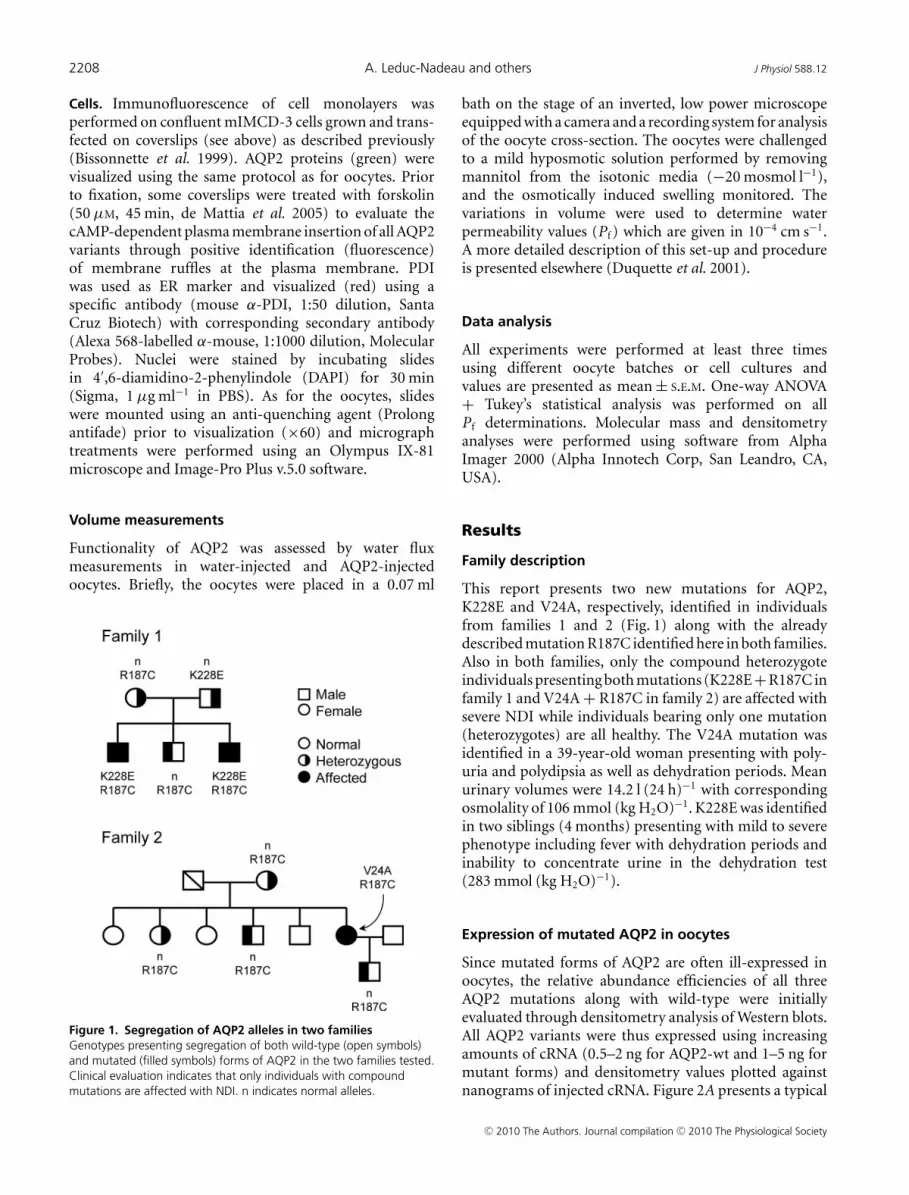
Figure 1. Segregation of AQP2 alleles in two familiesGenotypes presenting segregation of both wild-type (open symbols)and mutated (filled symbols) forms of AQP2 in the two families tested.Clinical evaluation indicates that only individuals with compoundmutations are affected with NDI. n indicates normal alleles.
bath on the stage of an inverted, low power microscopeequipped with a camera and a recording system for analysisof the oocyte cross-section. The oocytes were challengedto a mild hyposmotic solution performed by removingmannitol from the isotonic media (−20 mosmol l−1),and the osmotically induced swelling monitored. Thevariations in volume were used to determine waterpermeability values (Pf ) which are given in 10−4 cm s−1.A more detailed description of this set-up and procedureis presented elsewhere (Duquette et al. 2001).
Data analysis
All experiments were performed at least three timesusing different oocyte batches or cell cultures andvalues are presented as mean ± S.E.M. One-way ANOVA+ Tukey’s statistical analysis was performed on allPf determinations. Molecular mass and densitometryanalyses were performed using software from AlphaImager 2000 (Alpha Innotech Corp, San Leandro, CA,USA).
Results
Family description
This report presents two new mutations for AQP2,K228E and V24A, respectively, identified in individualsfrom families 1 and 2 (Fig. 1) along with the alreadydescribed mutation R187C identified here in both families.Also in both families, only the compound heterozygoteindividuals presenting both mutations (K228E+R187C infamily 1 and V24A + R187C in family 2) are affected withsevere NDI while individuals bearing only one mutation(heterozygotes) are all healthy. The V24A mutation wasidentified in a 39-year-old woman presenting with poly-uria and polydipsia as well as dehydration periods. Meanurinary volumes were 14.2 l (24 h)−1 with correspondingosmolality of 106 mmol (kg H2O)−1. K228E was identifiedin two siblings (4 months) presenting with mild to severephenotype including fever with dehydration periods andinability to concentrate urine in the dehydration test(283 mmol (kg H2O)−1).
Expression of mutated AQP2 in oocytes
Since mutated forms of AQP2 are often ill-expressed inoocytes, the relative abundance efficiencies of all threeAQP2 mutations along with wild-type were initiallyevaluated through densitometry analysis of Western blots.All AQP2 variants were thus expressed using increasingamounts of cRNA (0.5–2 ng for AQP2-wt and 1–5 ng formutant forms) and densitometry values plotted againstnanograms of injected cRNA. Figure 2A presents a typical
C© 2010 The Authors. Journal compilation C© 2010 The Physiological Society

J Physiol 588.12 New AQP2 mutations responsible for nephrogenic diabetes insipidus 2209
Western blot with increasing AQP2 labelling from whichmean densitometry values of five individual experimentswere used to evaluate their relative expression efficiencies(Fig. 2B). The Western blot profile found for wild-typeprotein essentially depicts a major 29 kDa band alonga very faint band at 31 kDa. In comparison, mutatedforms of AQP2 usually display a more intense 31 kDaband of varying intensity which may even be equivalentto that of 29 kDa (R187C). This heavier form of AQP2corresponds to an N-glycosylation intermediate whosefate remains unclear. As shown, the wild-type form ofAQP2 was expressed more efficiently so that 1 or 2 ngof AQP2-wt cRNA globally compares to 5 ng cRNA ofany mutant. Since the 31 kDa form is not expected toactually participate in functional channels, a ratio of1:5 was thus chosen in coexpression studies to evaluateinteractions between wild-type and mutant forms ofAQP2. In the following set of experiments, all three
Figure 2. Expression of AQP2 variants in X. laevis oocytesA, Western blot of oocytes injected with increasing amounts of cRNAfor wild-type (0.5, 1 and 2 ng) and mutated forms (1, 2 and 5 ng) ofAQP2. Each lane represents total membranes from 4 oocytes. B,densitometry evaluation of AQP2 bands (combination of both 29 and31 kDa bands) extracted from A correlated against cRNA quantities.Data represent mean ± S.D. from triplicates and fitted tomonoexponential equation.
mutants were compared to AQP2-wt looking at bothfunctionality (Fig. 3A) and plasma membrane targeting(Fig. 3C and D). For this analysis, oocytes were injectedwith either 1 ng AQP2-wt cRNA or 5 ng cRNA codingfor each mutation. As seen in Fig. 3B, where total AQP2proteins are evaluated (total membrane preparations),abundance levels of all APQ2 variants tested were found
Figure 3. Expression of AQP2 variants in X. laevis oocytesA, functional evaluation of all AQP2 variants. Oocytes were injectedwith cRNA coding for either wild-type (WT, 1 ng) or mutated (V24A,R187C and K228E, 5 ng) AQP2 along with controls (Ctrl) andincubated for 72 h prior to testing for water permeability capacities(see Methods). Pf values are in 10−4 cm s−1 and represent 7–8determinations per condition. Data are representative of 5–7 individualassays. The same oocytes were tested in Western blot using eithertotal membranes (B, 1 oocyte per lane) or purified plasma membranes(C, 40 oocytes per lane) fractions. PDI detection was performed on thesame material to confirm the quality of the purification procedure forplasma membrane in C. D, immunofluorescence labelling of all AQP2in the same samples showing retention of R187C within intracellularstores as opposed to AQP2-wt, V24A and K228E.
C© 2010 The Authors. Journal compilation C© 2010 The Physiological Society

2210 A. Leduc-Nadeau and others J Physiol 588.12
to be equivalent thus validating the 1:5 cRNA ratiodetermined previously in Fig. 2. In all assays performedon mutants, the 29 kDa band was always found tobe predominant with the exception of R187C whichusually displays equivalent 29 and 31 kDa bands. Whenpurified plasma membranes from the same oocytes wereprobed for AQP2 (Fig. 3C), all variants except R187Cwere found to be adequately targeted at the plasma
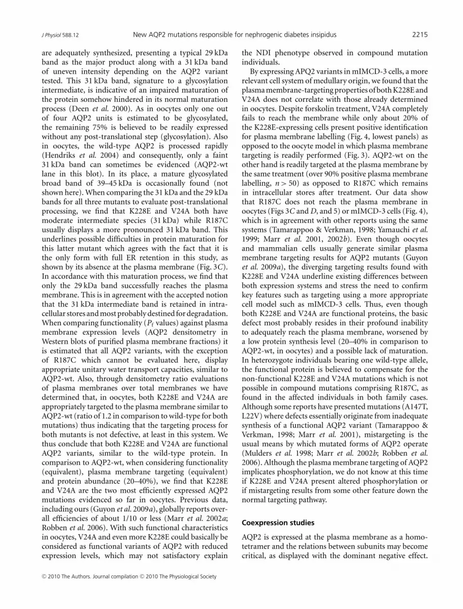
Figure 4. Immunofluorescence analysis of AQP2 in transfectedIMCD-3 cellsmIMCD-3 cells were transfected with pcDNA6-AQP2-wt, -V24A,-R187C or -K228E, incubated for 16–24 h and treated (+forskolin) ornot (–forskolin) with forskolin (50 μM, 45 min) prior to fixation. PDIwas used as an ER marker (red) and DAPI was used as a nuclear stain(blue). Plasma membrane staining is found for both AQP2-wt and-K228E (arrows) but not for R187C or V24A. Magnification ×60. Scalebar, 10 μm.
membrane. It is also noteworthy that the 31 kDa bandsare absent from the plasma membrane fractions. Theabsence of ER-resident protein PDI staining in the lowerblot of Fig. 3C, as opposed to Fig. 3B, demonstratesthe absence of intracellular membrane contaminantsin the purified plasma membrane fractions tested, thusvalidating the purification procedure (Leduc-Nadeauet al. 2006). Similarly, immunofluorescence labellingof AQP2 performed on oocyte slices (Fig. 3D) alsodemonstrated the plasma membrane localization for allAQP2 variants under study with the exception of R187Cwhich is retained in intracellular stores, most probablythe ER. These plasma membrane targeting results alsocorrelate with functionality assays (Fig. 3A) as all variantsadequately targeted also display appropriate waterpermeability values (in this experiment, Pf values are(14.4 ± 5.5), (16.3 ± 3.5) and (20 ± 7) × 10−4 cm s−1, forAQP2-wt, V24A and K228E, respectively). As expected, theonly mistargeted mutant, R187C, displayed no specificactivity (Pf = (2.6 ± 0.6) × 10−4 cm s−1) similar towater-injected oocytes (Pf = (2.8 ± 1.0) × 10−4 cm s−1).Overall statistical analysis of functionality for allAQP2 variants (7 independent assays) comparingexpressions of 1 ng wild-type against 5 ng for mutantforms gave Pf values of (14.2 ± 4.5) × 10−4 cm s−1 forAQP2-wt (n = 64), (16 ± 6) × 10−4 cm s−1 for V24A(n = 24), (22 ± 5) × 10−4 cm s−1 for K228E (n = 24) and(2.4 ± 0.7) × 10−4 cm s−1 for R187C (n = 24), which issimilar to controls ((2.8 ± 1.1) × 10−4 cm s−1, n = 64).
Studies were also performed to determine the relativeplasma membrane targeting efficacy for all AQP2 speciesunder study. To do so, Western blots from both total andplasma membranes samples were probed for AQP2 andrelative densities of specific bands for each mutant wereevaluated and compared to AQP2-wt, which served asreference, i.e. plasma over total membrane densities ratiofor AQP2-wt = 1. Results averaging five assays indicatedthat both V24A (1.2 ± 0.3) and K228E (1.2 ± 0.4)have similar plasma membrane targeting efficacies incomparison to AQP2-wt. Also, relative single channelactivity levels were assessed by comparing Western blotdensitometry values of each AQP2 variant, as found atthe plasma membrane, against their respective activitylevels (Pf values). Once again taking AQP2-wt as reference(Pf over blot density = 1), both V24A (ratio = 1.0 ± 0.2)and K228E (ratio = 1.3 ± 0.2) displayed single channelfunctionalities similar to AQP2-wt.
More relevant targeting studies were performed usingthe renal mIMCD-3 cell line. To do so, cells transfectedwith all three mutant AQP2 forms were compared toAQP2-wt for their ability to reach the plasma membranein response to forskolin treatment. As seen in the top panelof Fig. 4, AQP2-wt responded adequately and showedpositive plasma membrane localization (identification ofplasma membrane ruffles) of the protein after forskolin
C© 2010 The Authors. Journal compilation C© 2010 The Physiological Society

J Physiol 588.12 New AQP2 mutations responsible for nephrogenic diabetes insipidus 2211
treatment (arrow) in over 90% of observations (n > 50)while R187C remained sequestered within intracellularstores with the same treatment (n > 30). These tworesults are in agreement with those previously foundin oocytes. For the two remaining mutants, plasmamembrane targeting induced by forskolin was either oflow occurrence for K228E (20%, n > 50) or completelyabsent for V24A (n > 30).
Coexpression studies
As mentioned above, only the two compound mutationindividuals (K228E + R187C in family 1 and V24A+ R187C in family 2) are affected. In that context, astudy was performed where oocytes were injected with
APQ2 variants in accordance with the reported familygenotypes presented in Fig. 1. The goal of this studywas to correlate functionality results, as determinedby water permeability evaluations, with their respectiveplasma membrane expression levels, as determined byWestern blot. Oocytes were thus injected either withpure AQP2 variants or with a mixture of cRNAs toallow an equivalent coexpression of AQP2 products (1 ngHA-tagged AQP2-wt + 5 ng mutant cRNAs or 5 ngof each mutant). The functionality results presented inFig. 5A and B are mean ± S.D. (as % of AQP2-wt alone)of 22–30 determinations per condition collected withinsix separate experiments. As seen in Fig. 5A representingfamily 1, K228E displays a functionality superior to thatof the wild-type (162 ± 37%) while R187C is withoutactivity, similar to water-injected oocytes (22 ± 9 and
Figure 5. Expression of AQP2 in relation to familygenotypesOocytes were injected with AQP2 cRNA combinations inaccordance with Fig. 1 reproducing genotypes presented forfamily 1 (A) and family 2 (B). With regard to translationefficiency, cRNAs were injected at 1 ng (HA-AQP2-wt) or5 ng (mutant) with the exception of double mutantconditions (K228E + R187C and V24A + R187C) whereboth mutants were injected at 5 ng each. Functionality wasevaluated using a mild hyposmotic shock (−20 mosmol l−1).Pf values are expressed as percentage activity of pureAQP2-wt and represent 7–8 determinations for eachcondition. Western blots of purified plasma membrane werefirst probed using α-AQP2 for total AQP2 staining thenstripped and probed again using α-HA to specifically identifywt-AQP2. Data are representative of three individual assays.
C© 2010 The Authors. Journal compilation C© 2010 The Physiological Society

2212 A. Leduc-Nadeau and others J Physiol 588.12
22 ± 13%, respectively). Co-injecting mutant forms alongwith AQP2-wt either increased (+K228E, 169 ± 58%)or partially inhibited (+R187C, 52 ± 24%) the levelof activity in comparison to AQP2-wt alone. Lastly,co-injecting K228E along with R187C also generated inter-mediate activity levels (107 ± 45%). Asterisks indicate astatistical difference from pure AQP2-wt (P < 0.05). Inaddition, Western blots were also performed to evaluatethe impact of these expression conditions on the presenceof both total and wild-type forms of AQP2 at the plasmamembrane. For that purpose, an HA-tagged version ofAQP2-wt was used to allow its discrimination againstmutant forms of the protein within a same sample.Care was taken to confirm that the presence of theHA-tag at the C-terminus did not impede the normal
Figure 6. Functionality of AQP2-wt in coexpression studiesA standard 1 ng AQP2-wt cRNA was injected along with increasingconcentrations (0–10 ng) of cRNA for K228E (A), V24A (B) or R187C(C) and water permeability was evaluated using a 20 mosmol l−1
hyposmotic shock. Pf values are given in 10−4 cm s−1 and represent7–8 determinations for each condition. Asterisks indicate statisticalsignificance (P < 0.05) in comparison to pure 1 ng AQP2-wt valuewhile # indicates non-significance compared to controls (P < 0.05).Data are representative of three individual assays.
functionality of the protein (data not shown). Westernblots were thus performed on purified plasma membranesusing either an anti-AQP2 antibody to evaluate totalAQP2 (upper blot) or an anti-HA antibody to specificallyevaluate the wild-type form of AQP2 (lower blot). Asseen in the blots presented in Fig. 5A, all AQP2-injectedoocytes with the exception of R187C displayed appropriatelabelling at plasma membranes, which correspond to thefunctionality data shown in the graph above. The lowerblot specifically presenting HA-AQP2-wt indicates thatthe wild-type protein was present in plasma membranesof all conditions, including when coexpressed withK228E or R187C. The higher molecular mass foundwith HA-AQP2-wt (∼2 kDa) is due to the addition ofthe HA-tag. Also of interest, we find that staining forHA-AQP2-wt is more intense in the presence of K228Eand reduced in the presence of R187C in comparison toHA-AQP2-wt alone (α-HA blot).
For the second family case study (Fig. 5B), similarresults were found; functionality of V24A was foundto be equivalent to AQP2-wt (109 ± 29%) while R187Cremained completely inactive, similar to control oocytes(15 ± 5 and 14 ± 5%, respectively). Also, co-injectingAQP2-wt was additive with V24A (136 ± 26%) andinhibitory with R187C (36 ± 17%), similar to when bothmutations were co-injected together (43 ± 24%). Onceagain, the corresponding Western blot presents AQP2labelling at the plasma membrane that is compatiblewith activity, i.e. adequate labelling for AQP2-wt andV24A but not R187C (α-AQP2 blot). The presence ofAQP2-wt at the plasma membrane, as detected usinganti-HA antibody (α-HA blot), was also found in bothcoexpression conditions and again seemed to be increasedin the presence of V24A and reduced in presence of R187C.
A distinctive feature of some AQP2 mutations, knownas the dominant negative effect, is found when a mutatedform of the protein combines with and sequesters itswild-type counterpart within intracellular stores. Thisassociation between wild-type and mutant forms of AQP2results in a reduction of properly targeted AQP2 channelswith concomitant lack of function. In order to furtherinvestigate interrelations within AQP2 variants, we havemeasured the water permeability of oocytes injected witha fixed 1 ng cRNA coding for AQP2-wt with increasingcRNA concentrations (0–10 ng) coding for each mutationin the study (Fig. 6). The profiles found for both K228E(panel A) and V24A (panel B) are similar; AQP2-wtgenerates an increase in water permeability over controloocytes which is gradually enhanced by the addition ofincreasing cRNA coding for either mutation. Additionof 10 ng mutant cRNA increased the wild-type-inducedpermeability by (14.9 ± 2.9) × 10−4 cm s−1 and(14.4 ± 1.0) × 10−4 cm s−1 for K228E and V24A,respectively. These inductions are compatible withPf values determined previously with independent
C© 2010 The Authors. Journal compilation C© 2010 The Physiological Society

J Physiol 588.12 New AQP2 mutations responsible for nephrogenic diabetes insipidus 2213
expressions of the same mutants (Fig. 3A). On the otherhand, results found when co-injecting R187C (Fig. 3C) arecompletely opposite to AQP2-wt-dependent Pf graduallydecreasing to control levels (water-injected oocytes)within 10 ng R187C cRNA ((−8.1 ± 1.1) × 10−4 cm s−1).While coexpressions of wild-type and mutant AQP2 reflectthe prevailing conditions found for AQP2-wt-bearingheterozygote members in both families studied, Fig. 7presents analyses of coexpressions representing the twoaffected conditions. In both assays, a standard 5 ngcRNA for either K228E (panel A) or V24A (panel B) ischallenged against increasing concentrations of R187C(0–10 ng). Once again, R187C drastically diminishes thePf values of both K228E ((−9.0 ± 1.8) × 10−4 cm s−1),and V24A ((−4.1 ± 0.4) × 10−4 cm s−1). Although theeffect of R187C on the three forms of AQP2 tested issimilar, its inhibitory action against both mutants ismore pronounced with Pf reductions evidenced at a1:1 ratio for both K228E and V24A (Fig. 7A and B)while a 1:5 ratio is necessary to reach a similar effectwith AQP2-wt (Fig. 6C). These functional assays werealso tested through Western blot evaluations, probing
Figure 7. Functional analysis of mutant AQP2 in coexpressionstudiescRNA (5 ng) for either K228E (A) or V24A (B) was injected along withincreasing concentrations of R187C cRNA (0–10 ng) and waterpermeability was evaluated using a 20 mosmol l−1 hyposmotic shock.Pf values are given in 10−4 cm s−1 and represent 7–8 determinationsfor each condition. Asterisks indicate statistical significance (P < 0.05)in comparison to pure K228E or V24A values while # indicatesnon-significance compared to controls (P < 0.05). Data arerepresentative of three individual assays.
key conditions found in this study (HA-AQP2-wt alone(1 ng) or in combination with untagged mutants (5 ng)),in order to evaluate the impact of all mutations on theexpression and targeting of the AQP2-wt counterpart.To do so, total membranes were probed with α-AQP2 toidentify all AQP2 forms (evaluation of AQP2 abundances)while plasma membrane fractions purified from thesame samples were probed using α-HA (evaluation ofHA-AQP2-wt targeting at the plasma membrane; Fig. 8).As shown in Fig. 8A, AQP2-wt is equally expressed in allconditions (AQP2-wt) while its presence at the plasmamembrane (Fig. 8B, α-HA) seems to be modulatedby expression conditions with both V24A and K228Epromoting the presence of AQP2-wt at the plasmamembrane while R187C reduces it. These results are inagreement with those presented in Fig. 5A and B (α-HAblots).
Discussion
Many mutations responsible for NDI have been identifiedso far and expression in Xenopus oocytes has proven tobe a tool of choice for both biochemical and functionalstudies. Key pathophysiological data related to NDI, suchas misrouting or inadequate processing of altered AQP2proteins, were identified using this approach (Tamarappoo& Verkman, 1998; Marr et al. 2002a). Most frequently,NDI is transmitted to lineage as a recessive trait, whichimplies that two mutated alleles are required to induce thedisease. Although expressing AQP2 mutations in oocytes isnot expected to reproduce actual phenotypes reported byaffected individuals, coexpression studies have permittedsome important features related to this condition to behighlighted. One important trait sometimes found inAQP2-related NDI is the dominant negative behaviourin which a defective subunit associates with its functionalcounterpart(s) and impedes the normal processing and/or
Figure 8. Effects of mutant’s coexpression on AQP2-wtabundance and plasma membrane targetingOocytes injected with water (Ctrl), HA-AQP2-wt alone (WT, 1 ng) orwith untagged mutants (+, 5 ng) were probed for overall AQP2abundances in total membranes using α-AQP2 (A) or specifically forAQP2-wt in purified plasma membranes using α-HA (B).
C© 2010 The Authors. Journal compilation C© 2010 The Physiological Society

2214 A. Leduc-Nadeau and others J Physiol 588.12
targeting of the oligomer, thus sequestering the complexin internal stores and preventing the normal activityof the whole (Robben et al. 2006). These mutations,essentially located at the C-terminal end of the protein,are responsible for the autosomal dominant form of trans-mission of NDI. A few reports in the past have presentedsuch interactions between wild-type and mutated formsof AQP2, usually through co-immunoprecipitation assays(Kamsteeg et al. 1999; Kamsteeg & Deen, 2001; Marr et al.2002b).
The present study focuses on two new mutationsidentified from distinct families, K228E and V24A, as wellas R187C found in both families and already reportedin the literature (Mulders et al. 1998; Tamarappoo &Verkman, 1998; Shinbo et al. 1999). As indicated inclinical data, only the individuals presenting compoundmutations (K228E + R187C and V24A + R187C)are affected with NDI and since heterozygotes are notaffected, no dominant negative behaviour of pathologicalimpact is suspected for these mutations. The schematicrepresentation of the AQP2 protein in Fig. 9 showsthe positions of all three mutations under study.V24A, situated within the first transmembrane domain,represents a modest modification with the replacementof valine with a similar non-polar amino acid of smallersize. Functionality for that mutant form is thus notsurprising. On the other hand, the R187C mutation islocated immediately after the second NPA motif which
is implicated in the binding and transfer of watermolecules. The arginine in position 187 is most probablya constituent of the water channel structure, similar toR181 in the closely related AQP1 channel (Wu et al.2009). Moreover, replacing a large basic amino acid(arginine) by a small neutral one (cysteine) at such asensitive location represents a profound modificationmost inclined to alter the protein’s functionality. Thelast mutant, K228E, is located at the C-terminal end ofthe protein, near the last transmembrane domain. Eventhough this mutation implies changing a bulkier basiclysine for a smaller glutamic acid, and thus changingthe charge of the residue, this amino acid is foundin a region of low secondary structure which may bemore compliant to such modifications. We thus set tocharacterize these new mutations, looking at functionality,maturation and targeting properties through a series ofAQP2 expressions in oocytes and mammalian cells withthe goal of identifying the means by which each mutationoperates.
Processing, functionality and targeting of mutatedAQP2
Figure 3 presents an overall characterization of AQP2expression in oocytes and describes both the abundanceand targeting of the protein as well as its functionality asa water channel. As seen in Figure 3B, all AQP2 variants
Figure 9. Schematic representation ofAQP2 proteinDepiction of the human AQP2 proteinshowing the positions of V24A, R187C andK228E mutations. Brackets indicateconserved NPA motifs.
C© 2010 The Authors. Journal compilation C© 2010 The Physiological Society
J Physiol 588.12 New AQP2 mutations responsible for nephrogenic diabetes insipidus 2215
are adequately synthesized, presenting a typical 29 kDaband as the major product along with a 31 kDa bandof uneven intensity depending on the AQP2 varianttested. This 31 kDa band, signature to a glycosylationintermediate, is indicative of an impaired maturation ofthe protein somehow hindered in its normal maturationprocess (Deen et al. 2000). As in oocytes only one outof four AQP2 units is estimated to be glycosylated,the remaining 75% is believed to be readily expressedwithout any post-translational step (glycosylation). Alsoin oocytes, the wild-type AQP2 is processed rapidly(Hendriks et al. 2004) and consequently, only a faint31 kDa band can sometimes be evidenced (AQP2-wtlane in this blot). In its place, a mature glycosylatedbroad band of 39–45 kDa is occasionally found (notshown here). When comparing the 31 kDa and the 29 kDabands for all three mutants to evaluate post-translationalprocessing, we find that K228E and V24A both havemoderate intermediate species (31 kDa) while R187Cusually displays a more pronounced 31 kDa band. Thisunderlines possible difficulties in protein maturation forthis latter mutant which agrees with the fact that it isthe only form with full ER retention in this study, asshown by its absence at the plasma membrane (Fig. 3C).In accordance with this maturation process, we find thatonly the 29 kDa band successfully reaches the plasmamembrane. This is in agreement with the accepted notionthat the 31 kDa intermediate band is retained in intra-cellular stores and most probably destined for degradation.When comparing functionality (Pf values) against plasmamembrane expression levels (AQP2 densitometry inWestern blots of purified plasma membrane fractions) itis estimated that all AQP2 variants, with the exceptionof R187C which cannot be evaluated here, displayappropriate unitary water transport capacities, similar toAQP2-wt. Also, through densitometry ratio evaluationsof plasma membranes over total membranes we havedetermined that, in oocytes, both K228E and V24A areappropriately targeted to the plasma membrane similar toAQP2-wt (ratio of 1.2 in comparison to wild-type for bothmutations) thus indicating that the targeting process forboth mutants is not defective, at least in this system. Wethus conclude that both K228E and V24A are functionalAQP2 variants, similar to the wild-type protein. Incomparison to AQP2-wt, when considering functionality(equivalent), plasma membrane targeting (equivalent)and protein abundance (20–40%), we find that K228Eand V24A are the two most efficiently expressed AQP2mutations evidenced so far in oocytes. Previous data,including ours (Guyon et al. 2009a), globally reports over-all efficiencies of about 1/10 or less (Marr et al. 2002a;Robben et al. 2006). With such functional characteristicsin oocytes, V24A and even more K228E could basically beconsidered as functional variants of AQP2 with reducedexpression levels, which may not satisfactory explain
the NDI phenotype observed in compound mutationindividuals.
By expressing APQ2 variants in mIMCD-3 cells, a morerelevant cell system of medullary origin, we found that theplasma membrane-targeting properties of both K228E andV24A does not correlate with those already determinedin oocytes. Despite forskolin treatment, V24A completelyfails to reach the membrane while only about 20% ofthe K228E-expressing cells present positive identificationfor plasma membrane labelling (Fig. 4, lowest panels) asopposed to the oocyte model in which plasma membranetargeting is readily performed (Fig. 3). AQP2-wt on theother hand is readily targeted at the plasma membrane bythe same treatment (over 90% positive plasma membranelabelling, n > 50) as opposed to R187C which remainsin intracellular stores after treatment. Our data showthat R187C does not reach the plasma membrane inoocytes (Figs 3C and D, and 5) or mIMCD-3 cells (Fig. 4),which is in agreement with other reports using the samesystems (Tamarappoo & Verkman, 1998; Yamauchi et al.1999; Marr et al. 2001, 2002b). Even though oocytesand mammalian cells usually generate similar plasmamembrane targeting results for AQP2 mutants (Guyonet al. 2009a), the diverging targeting results found withK228E and V24A underline existing differences betweenboth expression systems and stress the need to confirmkey features such as targeting using a more appropriatecell model such as mIMCD-3 cells. Thus, even thoughboth K228E and V24A are functional proteins, the basicdefect most probably resides in their profound inabilityto adequately reach the plasma membrane, worsened bya low protein synthesis level (20–40% in comparison toAQP2-wt, in oocytes) and a possible lack of maturation.In heterozygote individuals bearing one wild-type allele,the functional protein is believed to compensate for thenon-functional K228E and V24A mutations which is notpossible in compound mutations comprising R187C, asfound in the affected individuals in both family cases.Although some reports have presented mutations (A147T,L22V) where defects essentially originate from inadequatesynthesis of a functional AQP2 variant (Tamarappoo &Verkman, 1998; Marr et al. 2001), mistargeting is theusual means by which mutated forms of AQP2 operate(Mulders et al. 1998; Marr et al. 2002b; Robben et al.2006). Although the plasma membrane targeting of AQP2implicates phosphorylation, we do not know at this timeif K228E and V24A present altered phosphorylation orif mistargeting results from some other feature down thenormal targeting pathway.
Coexpression studies
AQP2 is expressed at the plasma membrane as a homo-tetramer and the relations between subunits may becomecritical, as displayed with the dominant negative effect.
C© 2010 The Authors. Journal compilation C© 2010 The Physiological Society

2216 A. Leduc-Nadeau and others J Physiol 588.12
Hence, coexpression analyses combining wild-type andmutant elements may highlight some of the molecularaspects of the protein’s functionality and even relate toactual clinical situations. For that reason, coexpressionanalyses were performed by combining the different APQ2species to investigate conditions depicted for both thefamilies described here. In a first study, combinationsof cRNA were performed based upon respective trans-lation efficiencies (1 unit for AQP2-wt and 5 units for allmutants) in order to evaluate the possible interactionsbetween AQP2 variants, as was done previously in asimilar case study (Guyon et al. 2009a). Although a 2:5ratio could have been considered with respect to results inFig. 2, choosing a 1:5 ratio seemed more adequate since the31 kDa high-mannose forms of AQP2 are usually believedto be trapped non-functional forms of the protein andshould thus not be considered when designing functionalassays. Also, we consider this approach only as indicativesince we can merely speculate on the actual expressionproperties (both quantitatively and qualitatively) of theseAQP2 forms, as found in the actual individuals, in pureor combined expressions. The Western blots performedon purified plasma membrane fractions (Fig. 5) indicatethat the wild-type protein is adequately synthesized androuted to the plasma membrane in all conditions ofexpression tested. Through visualization of AQP2-wtusing an HA tag (lower blots in Figs 5A and B, and8B), it is shown that both K228E and V24A not onlyenable but even seem to promote the plasma membraneinsertion of the wild-type protein. These data confirmthat the chosen ratio of 1:5 for wild-type over mutantcRNA did not impede the normal expression level ofAQP2-wt through synthesis competition, as cautionedpreviously (Kamsteeg & Deen, 2000). In coexpressionanalyses where 1 ng AQP2-wt was co-injected withincreasing amounts of all three mutants (Fig. 6A andB), we find a gradual increase in water permeability forboth K228E and V24A (mean Pf increase = (14.9 ± 2.8)and (14.4 ± 1.0) × 10−4 cm s−1, respectively) which iscompatible with the sum of individual activities, indicatingagain that there is no effective competition for synthesisbetween wild-type and mutant forms of the protein. Themechanism by which both V24A and K228E promotethe plasma membrane insertion of the wild-type formof AQP2 is not known but may implicate heteromericassociations between mutant and wild-type forms.
On the other hand, under the same conditions, R187Cseems to impede or restrain AQP2-wt targeting, as seenin both functionality and Western blot studies (Figs 5Aand B, and 8B). This behaviour was not expectedsince this mutant, which does not impede AQP2-wtexpression levels under our working conditions (Fig. 8A),is not believed to directly interact with AQP2-wt dueto its monomeric nature (Kamsteeg et al. 1999). Inthe subsequent studies, increasing R187C cRNA loads
against 1 ng AQP2-wt was shown to gradually diminishits functionality down to background level (Fig. 6C, meanPf decrease = (7.0 ± 5.1) × 10−4 cm s−1), a behaviour alsofound with both K228E and V24A (Fig. 7A and B). Again,this loss of function was not believed to be secondary todirect competition in protein synthesis from overloadedmachinery since K228E and V24A fail to do so in similarconditions (Fig. 6A and B). These results indicate thatR187C, which is not targeted to the plasma membrane,can impede the functionality of any of the other AQP2species tested here. Again, the inability of R187C tomultimerize basically prevents the dominant negativeeffect usually proposed to explain the retention of thewild-type protein and, since the coexpression conditionsdo not seem to significantly impede wild-type proteinsynthesis (Fig. 8A), other mechanisms leading to inter-ference in the normal processing of AQP2-wt are to beexplored, possibly implicating protein maturation andtargeting features.
In this study, the two new AQP2 mutations, K228Eand V24A, display high levels of functional expressionfor mutant forms (between 20 and 40% of wild-type)with adequate plasma membrane targeting in oocytes butnot in mIMCD-3 cells where they are largely (K228E)or completely (V24A) restrained within internal stores.The lack of response following proper signalling (vaso-pressin) is thus believed to be responsible for the lackof functionality found with these mutants as well asreduced synthesis levels. With regards to heterozygotefamily members, we believe that an effective dominantnegative effect could have been possible since bothmutations can modulate the activity of the wild-type formof AQP2 (Figs 5 and 8), but prevented by low synthesislevels and/or insufficient multimeric associations withAQP2-wt. In that regard, both mutations may be valuablecandidates for recovery assays using strategies such aschemical chaperones to promote or restore adequatefunctionality (Tamarappoo & Verkman, 1998). R187Cshows mistargeting in both expression models tested andmay also interfere with the normal processing of bothmutant and wild-type proteins. Further investigationsregarding the synthesis and cellular management of R187Ccould provide key information in relation to the normalexpression of AQP2-wt. Nevertheless heterozygotes ofboth families are not affected, we can only concludethat the deleterious effects of these mutations, includingR187C, do not supersede the basic functionality ofAQP2-wt.
References
Bissonnette P, Noel J, Coady MJ & Lapointe JY (1999).Functional expression of tagged human Na+-glucosecotransporter in Xenopus laevis oocytes. J Physiol 520,359–371.
C© 2010 The Authors. Journal compilation C© 2010 The Physiological Society
J Physiol 588.12 New AQP2 mutations responsible for nephrogenic diabetes insipidus 2217
de Mattia F, Savelkoul PJ, Kamsteeg EJ, Konings IB, Van DerSluijs P, Mallmann R, Oksche A & Deen PM (2005). Lack ofarginine vasopressin-induced phosphorylation ofaquaporin-2 mutant AQP2-R254L explains dominantnephrogenic diabetes insipidus. J Am Soc Nephrol 16,2872–2880.
Deen PM, Croes H, van Aubel RA, Ginsel LA & van Os CH(1995). Water channels encoded by mutant aquaporin-2genes in nephrogenic diabetes insipidus are impaired in theircellular routing. J Clin Invest 95, 2291–2296.
Deen PM, van Balkom BW & Kamsteeg EJ (2000). Routing ofthe aquaporin-2 water channel in health and disease. Eur JCell Biol 79, 523–530.
Duquette PP, Bissonnette P & Lapointe JY (2001). Localosmotic gradients drive the water flux associated withNa+/glucose cotransport. Proc Natl Acad Sci U S A 98,3796–3801.
Guyon C, Lussier Y, Bissonnette P, Leduc-Nadeau A, LonerganM, Arthus MF, Perez RB, Tiulpakov A, Lapointe JY & BichetDG (2009a). Characterization of D150E and G196Daquaporin-2 mutations responsible for nephrogenic diabetesinsipidus: importance of a mild phenotype. Am J PhysiolRenal Physiol.
Guyon C, Lussier Y, Bissonnette P, Leduc-Nadeau A, LonerganM, Arthus MF, Perez RB, Tiulpakov A, Lapointe JY & BichetDG (2009b). Characterization of D150E and G196Daquaporin-2 mutations responsible for nephrogenic diabetesinsipidus: importance of a mild phenotype. Am J PhysiolRenal Physiol 297, F489–F498.
Hendriks G, Koudijs M, van Balkom BW, Oorschot V,Klumperman J, Deen PM & Van Der Sluijs P (2004).Glycosylation is important for cell surface expression of thewater channel aquaporin-2 but is not essential fortetramerization in the endoplasmic reticulum. J Biol Chem279, 2975–2983.
Kamsteeg EJ, Bichet DG, Konings IB, Nivet H, Lonergan M,Arthus MF, van Os CH & Deen PM (2003). Reversedpolarized delivery of an aquaporin-2 mutant causesdominant nephrogenic diabetes insipidus. J Cell Biol 163,1099–1109.
Kamsteeg EJ & Deen PM (2000). Importance of aquaporin-2expression levels in genotype-phenotype studies innephrogenic diabetes insipidus. Am J Physiol Renal Physiol279, F778–F784.
Kamsteeg EJ & Deen PM (2001). Detection of aquaporin-2 inthe plasma membranes of oocytes: a novel isolation methodwith improved yield and purity. Biochem Biophys ResCommun 282, 683–690.
Kamsteeg EJ, Wormhoudt TA, Rijss JP, van Os CH & Deen PM(1999). An impaired routing of wild-type aquaporin-2 aftertetramerization with an aquaporin-2 mutant explainsdominant nephrogenic diabetes insipidus. EMBO J 18,2394–2400.
Kuwahara M, Iwai K, Ooeda T, Igarashi T, Ogawa E,Katsushima Y, Shinbo I, Uchida S, Terada Y, Arthus MF,Lonergan M, Fujiwara TM, Bichet DG, Marumo F & Sasaki S(2001). Three families with autosomal dominantnephrogenic diabetes insipidus caused by aquaporin-2mutations in the C-terminus. Am J Hum Genet 69, 738–748.
Leduc-Nadeau A, Lahjouji K, Bissonnette P, Lapointe JY &Bichet DG (2006). Elaboration of a novel technique for thepurification of plasma membranes from Xenopus laevisoocytes. Am J Physiol Cell Physiol 292, C1132–C1136.
Levin MH, Haggie PM, Vetrivel L & Verkman AS (2001).Diffusion in the endoplasmic reticulum of an aquaporin-2mutant causing human nephrogenic diabetes insipidus.J Biol Chem 276, 21331–21336.
Lin SH, Bichet DG, Sasaki S, Kuwahara M, Arthus MF,Lonergan M & Lin YF (2002). Two novel aquaporin-2mutations responsible for congenital nephrogenic diabetesinsipidus in Chinese families. J Clin Endocrinol Metab 87,2694–2700.
Marr N, Bichet DG, Hoefs S, Savelkoul PJ, Konings IB, DeMattia F, Graat MP, Arthus MF, Lonergan M, Fujiwara TM,Knoers NV, Landau D, Balfe WJ, Oksche A, Rosenthal W,Muller D, van Os CH & Deen PM (2002a). Cell-biologic andfunctional analyses of five new aquaporin-2 missensemutations that cause recessive nephrogenic diabetesinsipidus. J Am Soc Nephrol 13, 2267–2277.
Marr N, Bichet DG, Lonergan M, Arthus MF, Jeck N, SeyberthHW, Rosenthal W, van Os CH, Oksche A & Deen PM(2002b). Heteroligomerization of an aquaporin-2 mutantwith wild-type aquaporin-2 and their misrouting to lateendosomes/lysosomes explains dominant nephrogenicdiabetes insipidus. Hum Mol Genet 11, 779–789.
Marr N, Kamsteeg EJ, van Raak M, van Os CH & Deen PM(2001). Functionality of aquaporin-2 missense mutants inrecessive nephrogenic diabetes insipidus. Pflugers Arch 442,73–77.
Mulders SM, Bichet DG, Rijss JP, Kamsteeg EJ, Arthus MF,Lonergan M, Fujiwara M, Morgan K, Leijendekker R, VanDer Sluijs P, van Os CH & Deen PM (1998). An aquaporin-2water channel mutant which causes autosomal dominantnephrogenic diabetes insipidus is retained in the Golgicomplex. J Clin Invest 102, 57–66.
Robben JH, Knoers NV & Deen PM (2006). Cell biologicalaspects of the vasopressin type-2 receptor and aquaporin 2water channel in nephrogenic diabetes insipidus. Am JPhysiol Renal Physiol 291, F257–F270.
Shinbo I, Fushimi K, Kasahara M, Yamauchi K, Sasaki S &Marumo F (1999). Functional analysis of aquaporin-2mutants associated with nephrogenic diabetes insipidus byyeast expression. Am J Physiol Renal Physiol 277, F734–F741.
Tamarappoo BK & Verkman AS (1998). Defective aquaporin-2trafficking in nephrogenic diabetes insipidus and correctionby chemical chaperones. J Clin Invest 101, 2257–2267.
Umenishi F, Narikiyo T & Schrier RW (2005). Effect onstability, degradation, expression, and targeting ofaquaporin-2 water channel by hyperosmolality in renalepithelial cells. Biochem Biophys Res Commun 338,1593–1599.
Wu B, Steinbronn C, Alsterfjord M, Zeuthen T & Beitz E(2009). Concerted action of two cation filters in theaquaporin water channel. EMBO J 28, 2188–2194.
Yamauchi K, Fushimi K, Yamashita Y, Shinbo I, Sasaki S &Marumo F (1999). Effects of missense mutations on rataquaporin-2 in LLC-PK1 porcine kidney cells. Kidney Int 56,164–171.
C© 2010 The Authors. Journal compilation C© 2010 The Physiological Society

2218 A. Leduc-Nadeau and others J Physiol 588.12
Author contributions
Identification, clinical data and initial evaluations (genotyping,Western) for family members 1 and 2 were performed by A.M.-A.and Devuyst laboratories, respectively. D.G.B.’s laboratory wasresponsible for confirmation of AQP2 mutations (AVPR2 andAQP2 genotyping), expressions in oocytes and mIMCD-3 cells.All authors contributed equally to the elaboration of themanuscript and approved the final version.
Acknowledgements
This work was supported by the Canadian Institutes of HealthResearch (CIHR), grant no. MOP-10580, Chair in Genetics ofRenal Disease (D.G.B.) and by the FRSQ infrastructure programno. 5252.
C© 2010 The Authors. Journal compilation C© 2010 The Physiological Society


