Embed Size (px)
Citation preview
Biosensors and Bioelectronics 65 (2015) 427–434
Contents lists available at ScienceDirect
Biosensors and Bioelectronics
http://d0956-56
n CorrE-m
journal homepage: www.elsevier.com/locate/bios
New redox-active layer create via epoxy–amine reaction – The base ofgenosensor for the detection of specific DNA and RNA sequences ofavian influenza virus H5N1
Kamila Malecka a, Anna Stachyra b, Anna Góra-Sochacka b, Agnieszka Sirko b,Włodzimierz Zagórski-Ostoja b, Wim Dehaen c, Hanna Radecka a, Jerzy Radecki a,n
a Institute of Animal Reproduction and Food Research, Polish Academy of Sciences, Tuwima 10, 10-748 Olsztyn, Polandb Institute of Biochemistry and Biophysics, Polish Academy of Sciences, Pawińskiego 5A, 02-106 Warsaw, Polandc University of Leuven, Department of Chemistry, Celestijnenlaan 200F, B-3001 Leuven, Belgium
a r t i c l e i n f o
Article history:Received 4 August 2014Received in revised form14 October 2014Accepted 29 October 2014Available online 4 November 2014
Keywords:Epoxide–amine “click”Phenanthroline-Epoxy–Fe(III)–(Phenan-throline Epoxy–NH2-ssDNA)2 redox activelayerElectrochemical genosensorRNA transcriptsAvian influenza virus
x.doi.org/10.1016/j.bios.2014.10.06963/& Elsevier B.V. All rights reserved.
esponding author. Fax: þ48 89 524 0124.ail address: [email protected] (J. Radeck
a b s t r a c t
This paper concerns the development of a redox-active monolayer and its application for the con-struction of an electrochemical genosensor designed for the detection of specific DNA and RNA oligo-nucleotide sequences related to the avian influenza virus (AIV) type H5N1. This new redox layer wascreated on a gold electrode surface step by step. Cyclic Voltammetry, Osteryoung Square-Wave Vol-tammetry and Differential Pulse Voltammetry were used for its characterization. This new redox-activelayer was applied for the construction of the DNA biosensor. The NH2-NC3 probe (20-mer) was covalentlyattached to the gold electrode surface via a “click” reaction between the amine and an epoxide group.The hybridization process was monitored using the Osteryoung Square-Wave Voltammetry. The 20-merDNA and ca. 280-mer RNA oligonucleotides were used as the targets. The constructed genosensor wascapable to determine complementary oligonucleotide sequences with a detection limit in the pM range.It is able to distinguish the different position of the part RNA complementary to the DNA probe. Thegenosensor was very selective. The 20-mer DNA as well as the 280-mer RNA oligonucleotides without acomplementary sequence generated a weak signal.
& Elsevier B.V. All rights reserved.
1. Introduction
In the past two decades, much progress has been made in thedevelopment of methods of preparing electrodes with controllablesurface properties. The research on self-assembled monolayers(SAMs) of organic molecules created via chemisorption of “headgroups” on the surfaces of solid electrodes has become a boomingfield (Ulman, 1996; Smith et al., 2004; Vericat et al., 2010; Sto-biecka and Hepel, 2011; Hepel and Zhong, 2012; Iost and Cre-spilho, 2012). Redox-active layers are an excellent platform forresearch including for example the kinetics of electron transfer(Albrecht et al., 2005; Zhang et al., 2008; Pobelov et al., 2008;Eckermann et al., 2010; Li et al., 2010). This very promising di-rection of current research is associated with electrochemicalbiosensors based on a redox active layer immobilized on theelectrode surface. Electrochemical sensors based on redox activelayers represent a relatively new trend in sensing tools
i).
development. The main advantage of this type of layers is thatthere is no need of using external redox active markers. The ap-plication of redox active centers for the construction of sensorsprovides many interesting possibilities. The redox centers locatedinside the active layers are responsible for converting signals fromintermolecular recognition processes to an analytically useful one(Jargiło et al., 2013; Mielecki et al., 2013; Mikuła et al., 2013;Wojtasik et al., 2014; Zborowska et al., 2014).
Avian influenza (AI) is an infectious disease spreading amongwild and domestic birds. It is caused by type A influenza viruses ofthe family Orthomyxoviridae. Highly pathogenic avian influenzavirus (HPAIV) H5N1 is not only lethal to birds but can also pose arisk to mammals, including humans (Neumann et al., 2010; Kii-lerich-Pedersen et al., 2013). Therefore, there is an urgent need forthe development of rapid detection methods of the H5N1. Bio-sensors are cost-effective analytical devices, which do not demandsophisticated procedures and offer fast and simple analysis.
The clue of the work presented is the development of a newredox-active layer and its application for the construction of anelectrochemical genosensor for the detection of specific DNA andRNA sequences of AIV H5N1. The literature describes several types
K. Malecka et al. / Biosensors and Bioelectronics 65 (2015) 427–434428
of genosensors for detecting H5N1 (Table S1-Supplementary data).The first example was based on oxidation/reduction of the peakcurrent of electroactive oligonucleotides bases treated as an ana-lytical signal. This concept has been originally developed by Pa-lecek and Bartosik (2012). Electroactivity of nucleotides, such ascytosine, adenine and guanine (Zhu et al., 2009) changes uponhybridization. Any external indicator was not necessary. However,in case of ion-channel mimetic sensors, an external redox activemarker is required. This type of biosensors was originally devel-oped by Umezawa and Aoki (2004). The binding of analytes (targetssDNA) to receptors (ssDNA probe) immobilized on the electrodesurfaces facilitates or suppresses the access of anionic (cationic)marker ions, present in the sample solution, to the modified sur-face due to electrostatic attraction or repulsion of the marker and/or distortion of the modification layer organization. This leads tochanges of the electron transfer rate between the marker andelectrode surface through the sensing layer (Aoki and Umezawa,2002; Aoki and Umezawa, 2003; Umezawa and Aoki, 2004). Thistype of sensor based on a self-assembled layer with [Fe(CN)6]3�/4�
as the redox-active marker was successfully used for the detectionof specific DNA sequences characteristic for the H5N1 (Kukol et al.,2008; Chung et al., 2011; Malecka et al., 2012, 2013). Intercalationsof redox active compounds into DNA helix have been used foranother type of genosensors. The most frequently used inter-calators for detection of specific oligonucleotides sequences of theAI viruses are methylene blue (Fan et al., 2010), doxorubicin (Tinget al., 2009) or tris(1,10-phenanthroline cobalt (III) perchlorate)(Liu et al., 2011). H5N1 specific sequences were also detected usinggenosensors attached to the electrode surface DNA probes deco-rated with redox-active labels (Grabowska et al., 2013, 2014a,2014b). Electroactive molecules such as 3-iron bis(dicarbollide),ferrocene, methylene blue or Co-porphyrin were covalently at-tached to the DNA probes.
Joining to this intense research area, in this work we present anovel method of forming redox active layer on the gold electrodesurface using the spontaneous reaction between the amine andepoxy groups. This type of reaction belongs to the larger family of“click” chemistry and it is called the nucleophilic opening of theoxirane ring (Kolb et al., 2001). The main advantages of clickchemistry are good yield, the availability of a wide range ofstarting materials and that the process is carried out under mildreaction conditions, insensitive to oxygen and water. Therefore,these type of reactions have been recently applied for coupling ofmolecules or macromolecules to surfaces (Aiello et al., 2013;Nguyen et al., 2014). The way of attaching substrates to the elec-trode surface to form the complex via epoxy-amine reaction isnovelty. In the literature, activation of EDC/NHS of a carboxylgroup for attachment to the amino group are mainly used (Mal-ecka et al., 2013, 2014; Jarocka et al., 2014; Im et al., 2010).
We have designed the aminoethanethiol, 5,6-epoxy-5,6-dihy-dro-[1,10]-phenanthroline and iron (III) (AET–Phen-Epoxy–Fe(III)–(Phen-Epoxy)2) redox active sensing layer and character-ized it by electrochemical methods – Cyclic Voltammetry (CV),Oster Young Square Wave Voltammetry (OSWV), Differential PulseVoltammetry (DPV). This layer was applied to the genosensorconstruction for the detection of the specific DNA and RNA se-quences of H5N1. The NH2-NC3 probe was attached toPhen-Epoxy–Fe(III)–(Phen-Epoxy)2 redox active layer by epoxy–amine “click” reaction. The signals generated as a result of hy-bridization processes have been registered by OSWV. The geno-sensor sensitivity and selectivity were tested with two types oftargets, a short (20-mer) DNA and long (ca. 280-mer) RNA se-quences of oligonucleotides.
2. Materials and methods
2.1. Reagents and materials
Iron chloride (III), 2-aminoethanethiol hydrochloride (AET),potassium chloride, ethanolamine (EA), acetonitrile (AN) andphosphate buffer saline (PBS) components (137 mM NaCl, 2.7 mMKCl, 10 mM KH2PO4, 1.8 mM Na2HPO4) were obtained from Sigma-Aldrich (Poznań, Poland). Alumina slurry 0.3 and 0.05 μm waspurchased from Buehler (USA). Sulphuric acid, potassium hydro-xide, hydrogen peroxide, methanol (MeOH) and ethanol weresupplied by POCh (Poland). 5,6-epoxy-5,6-dihydro-[1,10]-phenan-throline (Phen-Epoxy) was obtained from the University of Leuven(Belgium).
The modified oligonucleotide NH2-ssDNA (5′-NH2–(CH2)6–CCTCAA GGA GAG AGA AGA AG-3′) was used as a probe (namedNH2-NC3) for immobilization on a gold electrode surface, whiletwo unmodified oligonucleotides, c-NC3 (5′-CTT CTT CTC TCT CCTTGA GG-3′) and nc-NC3 (5′-GGA GTT CCT CTC TCA TCA TC-3′)served as complementary and non-complementary hybridizationtargets, respectively. The oligonucleotides were supplied by Bio-mers (Germany). The region complementary to the probe is lo-cated in the region of 83–102 nt and 160–170 nt from the 5′-ter-minus of the RNA1 and RNA2, respectively. The RNA3 has nocomplementary sequence to the probe at all and was used fordemonstration of the genosensor selectivity. DNA and RNA oligo-nucleotides were derived from the HA gene of Polish isolate of theHPAIV H5N1 (A/swan/Poland/305-135V08/2006) – Supplementarydata.
The immobilization of the NH2-NC3 probe was performed inAN. The hybridization processes were carried out in the presenceof 0.1 M PBS buffer (137 mM NaCl, 2.7 mM KCl, 10 mM KH2PO4,1.8 mM Na2HPO4), pH 7.4. RNA hybridization buffer was the same(0.1 M PBS) and prepared with sterile, nuclease free water fromSigma-Aldrich (Poznań, Poland). All aqueous solutions were pre-pared using autoclaved Milli-Q water, with resistivity 18.2 MΩ·cm(Millipore Corporation, USA). Reagents and solvents were of ana-lytical grade and used without further purification. All experi-ments were carried out at room temperature.
2.2. Successive steps of genosensor fabrication
The gold electrodes with a diameter of 2 mm (BioanalyticalSystems (BAS), West Lafayette, IN) were applied to genosensorpreparation. They were initially cleaned mechanically by polishingwith 0.3 and 0.05 μm alumina slurries (Alpha and Gamma Mi-cropolish; Buehler, Lake Bluff, IL) on a flat pad (BAS) for 5 mineach. Afterwards, they were carefully rinsed with Milli-Q water.The polished electrodes were further cleaned electrochemically byCV. At the first they were dipped in 0.5 M potassium hydroxidesolution and swept with the potential between –0.4 V and –1.2 V(versus the Ag/AgCl reference electrode and the platinum wirecounter electrode) with scan rate of 0.1 V s–1, number of cycles: 3,50 and 10. Subsequently, the electrodes were cleaned in 0.5 Msulphuric acid in the potential window between –0.3 V andþ1.5 V, number of cycles: 3, 10 and 3. Before modification, thesurfaces of the electrodes were refreshed in 0.5 M potassium hy-droxide solution for 10 cycles. After finishing of the electro-chemical cleaning, each electrode was washed, and next stored inwith Milli-Q water, until the next step of modification, to avoidcontaminations from the air. All solutions were deoxygenated bypurging with nitrogen (ultra pure 6.0, Air Products, Poland) for15 min.
Directly after cleaning, the electrodes were rinsed repeatedlywith water, MeOH and mixture of MeOH and AN (1:1) and dipped
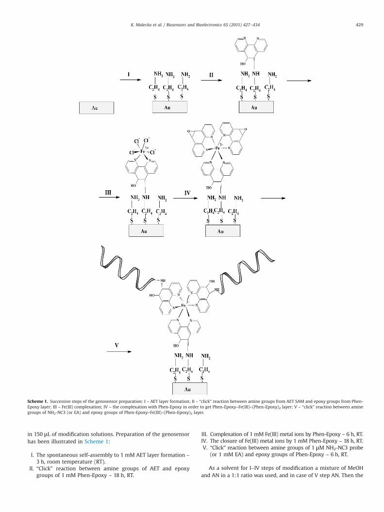
Scheme 1. Successive steps of the genosensor preparation: I – AET layer formation; II – “click” reaction between amine groups from AET SAM and epoxy groups from Phen-Epoxy layer; III – Fe(III) complexation; IV – the complexation with Phen-Epoxy in order to get Phen-Epoxy–Fe(III)–(Phen-Epoxy)2 layer; V – “click” reaction between aminegroups of NH2-NC3 (or EA) and epoxy groups of Phen-Epoxy–Fe(III)–(Phen-Epoxy)2 layer.
K. Malecka et al. / Biosensors and Bioelectronics 65 (2015) 427–434 429
in 150 μL of modification solutions. Preparation of the genosensorhas been illustrated in Scheme 1:
I.
The spontaneous self-assembly to 1 mM AET layer formation –3 h, room temperature (RT).
II. “Click” reaction between amine groups of AET and epoxygroups of 1 mM Phen-Epoxy – 18 h, RT.
III.
Complexation of 1 mM Fe(III) metal ions by Phen-Epoxy – 6 h, RT. IV. The closure of Fe(III) metal ions by 1 mM Phen-Epoxy – 18 h, RT. V. “Click” reaction between amine groups of 1 μM NH2-NC3 probe(or 1 mM EA) and epoxy groups of Phen-Epoxy – 6 h, RT.
As a solvent for I–IV steps of modification a mixture of MeOHand AN in a 1:1 ratio was used, and in case of V step AN. Then the
K. Malecka et al. / Biosensors and Bioelectronics 65 (2015) 427–434430
electrodes were rinsed with 5 mL of AN and next with 0.1 M PBS.After modification the electrodes were conditioned in 0.1 M PBSovernight.
2.3. Electrochemical measurements
All electrochemical measurements were performed with a po-tentiostat–galvanostat AutoLab (Eco Chemie, Utrecht, Nether-lands) with a three electrode configuration. Potentials were mea-sured versus the Ag/AgCl electrode, and a Pt wire was used as theauxiliary electrode. The voltammetric experiments were carriedout in an electrochemical cell of 5 mL volume.
The AET/Phen-Epoxy/Fe(III)/(Phen-Epoxy)2 redox activemonolayer was characterized by electrochemical methods – CV,OSWV and DPV. In CV the potential was cycled from þ0.6 V to –
0.2 V with a scan rate of 0.05, 0.1, 0.2, 0.3, 0.4, 0.5, 0.6, 0.7, 0.8,0.9 and 1 V s–1. In OSWV, a potential window from þ0.4 V to�0.4 V, a step potential of 0.001 V, square-wave frequency of25 Hz and amplitude of 0.05 V were applied. DPV measurementswere performed in two cycles: with the potential from þ0.5 V to –
0.4 V for reduction of Fe(III) ions and from –0.4 V to þ0.5 V foroxidation of Fe(II) ions. The values of the modulation amplitudeand step potential were 0.05 V and 0.001 V, respectively. Allmeasurements were carried out in the presence of 0.1 M KClpurged with nitrogen for 15 min. A gentle nitrogen flow was ap-plied over the sample solution during all measurements.
For characterization of the AET/Phen-Epoxy/Fe(III)/(Phen-Epoxy)2 electroactive monolayer, at the last step of the modifica-tion the epoxy groups were deactivated with 1 mM solution of EAfor 3 h. Finally, electrodes were rinsed with AN, MeOH and 0.1 MKCl and then conditioned in 0.1 M KCl overnight.
2.4. Hybridization processes
The gold electrodes surfaces modified with a AET/Phen-Epoxy/Fe(III)/(Phen-Epoxy–NH2-NC3)2 redox active layerwere covered with 10 μL of a solution of variable concentration ofthe target oligonucleotides (c-NC3, nc-NC3, RNA1, RNA2 or RNA3)in 0.1 M PBS buffer (137 mM NaCl, 2.7 mM KCl, 10 mM KH2PO4,1.8 mM Na2HPO4) pH 7.4, for 30 min at room temperature. Thenthe electrodes were rinsed with 5 mL of PBS, pH 7.4 in order toremove the unbound targets.
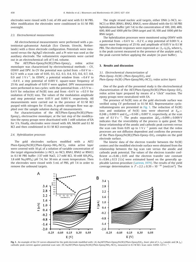
Fig. 1. An example of the CV curves obtained for the gold electrode modified with: (A) Acathodic peak current against potential scan rate; (B) Au/AET/Phen-Epoxy/Fe(III)/(Phen-
The single strand nucleic acid targets, either DNA (c-NC3, nc-NC3) or RNA (RNA1, RNA2, RNA3), were diluted with the 0.1 M PBShybridization buffer (pH 7.4) to the concentration of 100, 200, 400,600, 800 and 1000 pM for DNA target and 10, 100 and 1000 pM forRNA target.
The hybridization processes were monitored using OSWV witha potential from þ0.4 V to �0.2 V, a step potential of 0.001 V, asquare-wave frequency of 25 Hz and amplitude of 0.05 V in 0.1 MPBS. The electrode responses were expressed as: (In–I0)/I0, where Inis the peak current measured in the presence of the analyte and I0the peak current before applying the analyte (in pure buffer).
3. Results and discussion
3.1. Electrochemical characterization ofPhen-Epoxy–Fe(III)–(Phen-Epoxy/EA)2 andPhen-Epoxy–Fe(III)-(Phen-Epoxy/NH2-NC3)2 redox active layers
One of the goals of the presented study is the electrochemicalcharacterization of the AET/Phen-Epoxy/Fe(III)/(Phen-Epoxy–EA)2redox active layer prepared by means of a “click” reaction. Theepoxy groups were neutralized with EA.
The presence of Fe(III) ions at the gold electrode surface wasverified using CV performed in 0.1 M KCl. Representative cyclo-voltammograms are presented in Fig. 1. The reduction of Fe(III)ions and oxidation of Fe(II) ions were observed at Epc¼0.24870.009 V and Epa¼0.14970.007 V, respectively, at the scanrate of 0.1 V s�1. The peaks separation ΔEp¼0.09970.009 Vindicates that the reversibility of the process is quite good. Thelinear relationship of the anodic and cathodic peak currents versusthe scan rate from 0.05 up to 1 V s�1 points out that the redoxprocesses are not diffusion dependent and confirms the presenceof the Phen-Epoxy/Fe(III)/(Phen-Epoxy–EA)2 complex on the goldelectrode surface.
The kinetic data of the electron transfer between the Fe(III)centers and the modified electrode surface were obtained from therelationship between the log scan rate versus the anodic andcathodic peak potential. The values of the electron transfer coef-ficient α¼0.4670.05 and the electron transfer rate constantk¼0.8470.13 [1/s] were estimated based on the generally ap-plicable Laviron procedure (Laviron, 1979). The results of the yieldcoverage determination is Γ¼2.270.39�10�10 [mol/cm2]. The
u/AET/Phen-Epoxy/Fe(III)/(Phen-Epoxy/EA)2, Inset: plot of (▲, Ipa) anodic and (■, Ipc)Epoxy/NH2-NC3)2 measured in 0.1 M KCl. Scan rates: 0.050–1.0 V s�1.
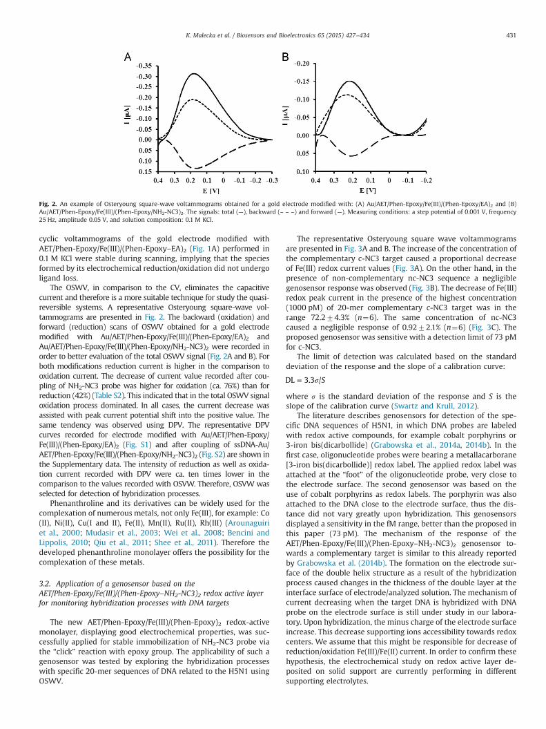
Fig. 2. An example of Osteryoung square-wave voltammograms obtained for a gold electrode modified with: (A) Au/AET/Phen-Epoxy/Fe(III)/(Phen-Epoxy/EA)2 and (B)Au/AET/Phen-Epoxy/Fe(III)/(Phen-Epoxy/NH2-NC3)2. The signals: total (—), backward (– – –) and forward (—). Measuring conditions: a step potential of 0.001 V, frequency25 Hz, amplitude 0.05 V, and solution composition: 0.1 M KCl.
K. Malecka et al. / Biosensors and Bioelectronics 65 (2015) 427–434 431
cyclic voltammograms of the gold electrode modified withAET/Phen-Epoxy/Fe(III)/(Phen-Epoxy–EA)2 (Fig. 1A) performed in0.1 M KCl were stable during scanning, implying that the speciesformed by its electrochemical reduction/oxidation did not undergoligand loss.
The OSWV, in comparison to the CV, eliminates the capacitivecurrent and therefore is a more suitable technique for study the quasi-reversible systems. A representative Osteryoung square-wave vol-tammograms are presented in Fig. 2. The backward (oxidation) andforward (reduction) scans of OSWV obtained for a gold electrodemodified with Au/AET/Phen-Epoxy/Fe(III)/(Phen-Epoxy/EA)2 andAu/AET/Phen-Epoxy/Fe(III)/(Phen-Epoxy/NH2-NC3)2 were recorded inorder to better evaluation of the total OSWV signal (Fig. 2A and B). Forboth modifications reduction current is higher in the comparison tooxidation current. The decrease of current value recorded after cou-pling of NH2-NC3 probe was higher for oxidation (ca. 76%) than forreduction (42%) (Table S2). This indicated that in the total OSWV signaloxidation process dominated. In all cases, the current decrease wasassisted with peak current potential shift into the positive value. Thesame tendency was observed using DPV. The representative DPVcurves recorded for electrode modified with Au/AET/Phen-Epoxy/Fe(III)/(Phen-Epoxy/EA)2 (Fig. S1) and after coupling of ssDNA-Au/AET/Phen-Epoxy/Fe(III)/(Phen-Epoxy/NH2-NC3)2 (Fig. S2) are shown inthe Supplementary data. The intensity of reduction as well as oxida-tion current recorded with DPV were ca. ten times lower in thecomparison to the values recorded with OSVW. Therefore, OSVW wasselected for detection of hybridization processes.
Phenanthroline and its derivatives can be widely used for thecomplexation of numerous metals, not only Fe(III), for example: Co(II), Ni(II), Cu(I and II), Fe(II), Mn(II), Ru(II), Rh(III) (Arounaguiriet al., 2000; Mudasir et al., 2003; Wei et al., 2008; Bencini andLippolis, 2010; Qiu et al., 2011; Shee et al., 2011). Therefore thedeveloped phenanthroline monolayer offers the possibility for thecomplexation of these metals.
3.2. Application of a genosensor based on theAET/Phen-Epoxy/Fe(III)/(Phen-Epoxy–NH2-NC3)2 redox active layerfor monitoring hybridization processes with DNA targets
The new AET/Phen-Epoxy/Fe(III)/(Phen-Epoxy)2 redox-activemonolayer, displaying good electrochemical properties, was suc-cessfully applied for stable immobilization of NH2-NC3 probe viathe “click” reaction with epoxy group. The applicability of such agenosensor was tested by exploring the hybridization processeswith specific 20-mer sequences of DNA related to the H5N1 usingOSWV.
The representative Osteryoung square wave voltammogramsare presented in Fig. 3A and B. The increase of the concentration ofthe complementary c-NC3 target caused a proportional decreaseof Fe(III) redox current values (Fig. 3A). On the other hand, in thepresence of non-complementary nc-NC3 sequence a negligiblegenosensor response was observed (Fig. 3B). The decrease of Fe(III)redox peak current in the presence of the highest concentration(1000 pM) of 20-mer complementary c-NC3 target was in therange 72.274.3% (n¼6). The same concentration of nc-NC3caused a negligible response of 0.9272.1% (n¼6) (Fig. 3C). Theproposed genosensor was sensitive with a detection limit of 73 pMfor c-NC3.
The limit of detection was calculated based on the standarddeviation of the response and the slope of a calibration curve:
σ= SDL 3.3 /
where s is the standard deviation of the response and S is theslope of the calibration curve (Swartz and Krull, 2012).
The literature describes genosensors for detection of the spe-cific DNA sequences of H5N1, in which DNA probes are labeledwith redox active compounds, for example cobalt porphyrins or3-iron bis(dicarbollide) (Grabowska et al., 2014a, 2014b). In thefirst case, oligonucleotide probes were bearing a metallacarborane[3-iron bis(dicarbollide)] redox label. The applied redox label wasattached at the “foot” of the oligonucleotide probe, very close tothe electrode surface. The second genosensor was based on theuse of cobalt porphyrins as redox labels. The porphyrin was alsoattached to the DNA close to the electrode surface, thus the dis-tance did not vary greatly upon hybridization. This genosensorsdisplayed a sensitivity in the fM range, better than the proposed inthis paper (73 pM). The mechanism of the response of theAET/Phen-Epoxy/Fe(III)/(Phen-Epoxy–NH2-NC3)2 genosensor to-wards a complementary target is similar to this already reportedby Grabowska et al. (2014b). The formation on the electrode sur-face of the double helix structure as a result of the hybridizationprocess caused changes in the thickness of the double layer at theinterface surface of electrode/analyzed solution. The mechanism ofcurrent decreasing when the target DNA is hybridized with DNAprobe on the electrode surface is still under study in our labora-tory. Upon hybridization, the minus charge of the electrode surfaceincrease. This decrease supporting ions accessibility towards redoxcenters. We assume that this might be responsible for decrease ofreduction/oxidation Fe(III)/Fe(II) current. In order to confirm thesehypothesis, the electrochemical study on redox active layer de-posited on solid support are currently performing in differentsupporting electrolytes.
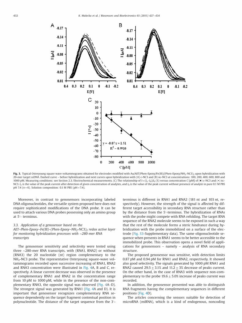
Fig. 3. Typical Osteryoung square wave voltammograms obtained for electrodes modified with Au/AET/Phen-Epoxy/Fe(III)/(Phen-Epoxy/NH2-NC3)2 upon hybridization with20-mer target ssDNA. Dashed curve – before hybridization and next curves upon hybridization with (A) c-NC3 and (B) nc-NC3 at concentrations: 100, 200, 400, 600, 800 and1000 pM. Measuring conditions: see Section 2.3. Electrochemical measurements. (C) The relationship of I¼(In–I0)/I0 (%) versus concentration C [pM] of (♦) c-NC3 and (▲) nc-NC3. In is the value of the peak current after detection of given concentration of analytes, and I0 is the value of the peak current without presence of analyte in pure 0.1 M PBSpH 7.4 (n¼6). Solution composition: 0.1 M PBS (pH¼7.4).
K. Malecka et al. / Biosensors and Bioelectronics 65 (2015) 427–434432
Moreover, in contrast to genosensors incorporating labeledDNA oligonucleotides, the versatile system proposed here does notrequire sophisticated modifications of the DNA probe. It can beused to attach various DNA probes possessing only an amino groupat 5′- terminus.
3.3. Application of a genosensor based on theAET–Phen-Epoxy–Fe(III)–(Phen-Epoxy–NH2-NC3)2 redox active layerfor monitoring hybridization processes with ∼280-mer RNAtranscripts
The genosensor sensitivity and selectivity were tested usingthree ∼280-mer RNA transcripts, with (RNA1, RNA2) or without(RNA3) the 20 nucleotide (nt) region complementary to theNH2-NC3 probe. The representative Osteryoung square-wave vol-tammograms recorded upon successive increasing of RNA1, RNA2and RNA3 concentration were illustrated in Fig. 4A, B and C, re-spectively. A linear current decrease was observed in the presenceof complementary RNA1 and RNA2 in the concentration rangefrom 10 pM to 1000 pM, while in the presence of the non-com-plementary RNA3, the opposite signal was observed (Fig. 4A–D).The strongest signal was generated by RNA1 (Fig. 4A and D). It isimportant that genosensor recognizes complementary RNA se-quence dependently on the target fragment contextual position inpolynucleotide. The distance of the target sequence from the 3′-
terminus is different in RNA1 and RNA2 (181 nt and 103 nt, re-spectively). However, the strength of the signal is affected by dif-ferent target accessibility in secondary RNA structure rather thanby the distance from the 5′-terminus. The hybridization of RNAswith the probe might compete with RNA refolding. The target RNAsequence of the RNA2 molecule seems to be exposed in such a waythat the rest of the molecule forms a steric hindrance during hy-bridization with the probe immobilized on a surface of the elec-trode (Fig. S3-Supplementary data). The same oligonucleotide se-quence when presents in RNA1 seems to be better accessible to theimmobilized probe. This observation opens a novel field of appli-cations for genosensors – namely – analysis of RNA secondarystructure.
The proposed genosensor was sensitive, with detection limits0.87 pM and 0.94 pM for RNA1 and RNA2, respectively. It showedalso good selectivity. The signals generated by 1000 pM RNA1 andRNA2 caused 29.573.1% and 11.273% decrease of peaks current.On the other hand, in the case of RNA3 with sequence non-com-plementary to the probe 19.675.0% increase of peaks current wasrecorded.
In addition, the genosensor presented was able to distinguishRNA fragments having the complementary sequences in differentpositions (Fig. 4D).
The articles concerning the sensors suitable for detection ofmicroRNA (miRNA), which is a kind of endogenous, noncoding
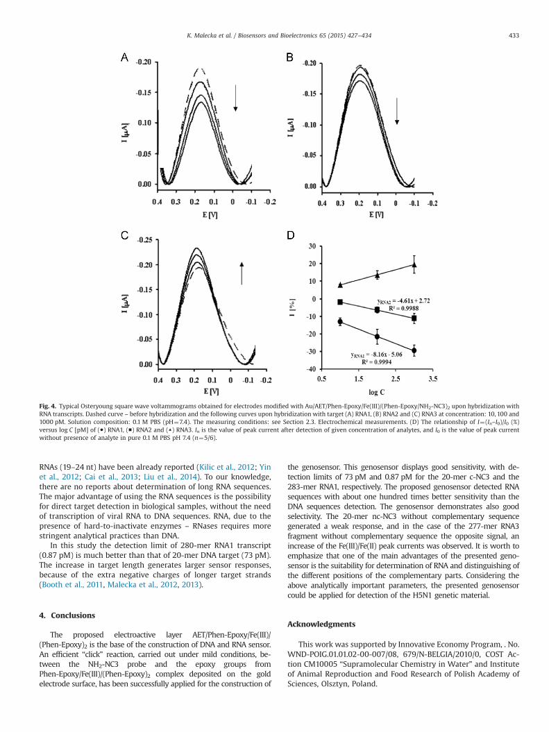
Fig. 4. Typical Osteryoung square wave voltammograms obtained for electrodes modified with Au/AET/Phen-Epoxy/Fe(III)/(Phen-Epoxy/NH2-NC3)2 upon hybridization withRNA transcripts. Dashed curve – before hybridization and the following curves upon hybridization with target (A) RNA1, (B) RNA2 and (C) RNA3 at concentration: 10, 100 and1000 pM. Solution composition: 0.1 M PBS (pH¼7.4). The measuring conditions: see Section 2.3. Electrochemical measurements. (D) The relationship of I¼(In–I0)/I0 (%)versus log C [pM] of (●) RNA1, (■) RNA2 and (▲) RNA3. In is the value of peak current after detection of given concentration of analytes, and I0 is the value of peak currentwithout presence of analyte in pure 0.1 M PBS pH 7.4 (n¼5/6).
K. Malecka et al. / Biosensors and Bioelectronics 65 (2015) 427–434 433
RNAs (19–24 nt) have been already reported (Kilic et al., 2012; Yinet al., 2012; Cai et al., 2013; Liu et al., 2014). To our knowledge,there are no reports about determination of long RNA sequences.The major advantage of using the RNA sequences is the possibilityfor direct target detection in biological samples, without the needof transcription of viral RNA to DNA sequences. RNA, due to thepresence of hard-to-inactivate enzymes – RNases requires morestringent analytical practices than DNA.
In this study the detection limit of 280-mer RNA1 transcript(0.87 pM) is much better than that of 20-mer DNA target (73 pM).The increase in target length generates larger sensor responses,because of the extra negative charges of longer target strands(Booth et al., 2011, Malecka et al., 2012, 2013).
4. Conclusions
The proposed electroactive layer AET/Phen-Epoxy/Fe(III)/(Phen-Epoxy)2 is the base of the construction of DNA and RNA sensor.An efficient “click” reaction, carried out under mild conditions, be-tween the NH2-NC3 probe and the epoxy groups fromPhen-Epoxy/Fe(III)/(Phen-Epoxy)2 complex deposited on the goldelectrode surface, has been successfully applied for the construction of
the genosensor. This genosensor displays good sensitivity, with de-tection limits of 73 pM and 0.87 pM for the 20-mer c-NC3 and the283-mer RNA1, respectively. The proposed genosensor detected RNAsequences with about one hundred times better sensitivity than theDNA sequences detection. The genosensor demonstrates also goodselectivity. The 20-mer nc-NC3 without complementary sequencegenerated a weak response, and in the case of the 277-mer RNA3fragment without complementary sequence the opposite signal, anincrease of the Fe(III)/Fe(II) peak currents was observed. It is worth toemphasize that one of the main advantages of the presented geno-sensor is the suitability for determination of RNA and distinguishing ofthe different positions of the complementary parts. Considering theabove analytically important parameters, the presented genosensorcould be applied for detection of the H5N1 genetic material.
Acknowledgments
This work was supported by Innovative Economy Program, . No.WND-POIG.01.01.02-00-007/08, 679/N-BELGIA/2010/0, COST Ac-tion CM10005 “Supramolecular Chemistry in Water” and Instituteof Animal Reproduction and Food Research of Polish Academy ofSciences, Olsztyn, Poland.
K. Malecka et al. / Biosensors and Bioelectronics 65 (2015) 427–434434
Appendix A. Supplementary material
Supplementary data associated with this article can be found inthe online version at http://dx.doi.org/10.1016/j.bios.2014.10.069.
References
Aiello, V., Joo, N., Buckley, J., Nonglaton, G., Duclairoir, F., Dubois, L., Marchon, J.C.,Gely, M., Chevalier, N., De Salvo, B., 2013. Surf. Sci 612, 57–62.
Albrecht, T., Guckian, A., Ulstrup, J., Vos, J.G., 2005. Nano Lett. 5, 1451–1455.Aoki, H., Umezawa, Y., 2002. Electroanalysis 14, 1–6.Aoki, H., Umezawa, Y., 2003. Analyst 128, 681–685.Arounaguiri, S., Easwaramoorthy, D., Ashokkumar, A., Dattagupta, A., Maiya, B.G.,
2000. Proc. Indian Acad. Sci. (Chem. Sci.) 112, 1–17.Bencini, A., Lippolis, V., 2010. Coord. Chem. Rev. 254, 2096–2180.Cai, Z., Song, Y., Wu, Y., Zhu, Z., Yang, C.J., Chen, X., 2013. Biosens. Bioelectron. 41,
783–788.Booth, M.A., Harbison, S.A., Travas-Sejdic, J., 2011. Biosens. Bioelectron. 28,
362–367.Chung, D.–J., Kim, K.–Ch, Choi, S.–H., 2011. Appl. Surf. Sci. 257, 9390–9396.Eckermann, A.L., Feld, D.J., Shaw, J.A., Meade, T.J., 2010. Coord. Chem. Rev. 254,
1769–1802.Fan, H., Ju, P., Ai, S., 2010. Sens. Actuators B – Chem 149, 98–104.Grabowska, I., Malecka, K., Stachyra, A., Góra-Sochacka, A., Sirko, A., Zagórski-Os-
toja, W., Radecka, H., Radecki, J., 2013. Anal. Chem. 85, 10167–10173.Grabowska, I., Singleton, D.G., Stachyra, A., Góra-Sochacka, A., Sirko, A., Zagórski-
Ostoja, W., Radecka, H., Stulz, E., Radecki, J., 2014a. Chem. Commun. 50,4196–4199.
Grabowska, I., Stachyra, A., Góra-Sochacka, A., Sirko, A., Olejniczak, A.B., Leśni-kowski, Z.J., Radecka, H., Radecki, J., 2014b. Biosens. Bioelectron. 51, 170–176.
Hepel M. and Zhong Ch.-J., 2012. Functional Nanoparticles for Bioanalysis, Nano-medicine, and Bioelectronic Devices, vol. 1, ACS Symposium series 1112.
Im, J.-E., Han, J.-A., Kim, B.K., Han, J.H., Park, T.S., Hwang, S., Cho, S.I., Lee, W.-Y., Kim,Y.-R., 2010. Surf. Coat. Technol 205, S275–S278.
Iost, R.M., Crespilho, F.N., 2012. Biosens. Bioelectron 31, 1–10.Jargiło, A., Grabowska, I., Radecka, H., Sulima, M., Marszałek, I., Wysłouch- Cies-
zyńska, A., Dehaen, W., Radecki, J., 2013. Electroanalysis 25, 1185–1193.Jarocka, U., Sawicka, R., Góra-Sochacka, A., Sirko, A., Zagórski-Ostoja, W., Radecki, J.,
Radecka, H., 2014. Biosens. Bioelectron. 55, 301–306.Kiilerich-Pedersen, K., Daprà, J., Cherré, S., Rozlosnik, N., 2013. Biosens. Bioelectron.
49, 374–379.Kilic, T., Nur Topkaya, S., Ozkan Ariksoysal, D., Ozsoz, M., Ballar, P., Erac, Y., Gozen,
O., 2012. Biosens. Bioelectron. 38, 195–201.Kolb, H.C., Finn, M.G., Sharpless, K.B., 2001. Angew. Chem. Int. Ed. 40, 2004–2021.
Kukol, A., Li, P., Estrela, P., Ko-Ferrigno, P., Migliorato, P., 2008. Anal. Biochem. 374,143–153.
Laviron, E.J., 1979. J. Electroanal. Chem. 101, 19–28.Li, Z., Liu, Y., Mertens, S.F.L., Pobelov, I.V., Wandlowski, T., 2010. J. Am. Chem. Soc.
132, 8187–8193.Liu, X., Cheng, Z., Fan, H., Ai, S., Han, R., 2011. Electrochim. Acta 56, 6266–6270.Liu, L., Xia, N., Liu, H., Kang, X., Liu, X., Xue, Ch, He, X., 2014. Biosens. Bioelectron. 53,
399–405.Malecka, K., Grabowska, I., Radecki, J., Stachyra, A., Góra-Sochacka, A., Sirko, A.,
Radecka, H., 2012. Electroanalysis 24, 439–446.Malecka, K., Grabowska, I., Radecki, J., Stachyra, A., Góra-Sochacka, A., Sirko, A.,
Radecka, H., 2013. Electroanalysis 25, 1871–1878.Malecka, K., Michalczuk, L., Radecka, H., Radecki, J., 2014. Sensors 14, 18611–18624.Mielecki, M., Wojtasik, J., Zborowska, M., Kurzątkowska, K., Radecki, J., Grzelak, K.,
Dehaen, W., Radecka, H., 2013. Electrochim. Acta 96, 147–154.Mikuła, E., Sulima, M., Marszałek, I., Wysłouch-Cieszyńska, A., Verwilst, P., Dehaen,
W., Radecki, J., Radecka, H., 2013. Sensors 13, 11586–11602.Mudasir, Wijaya, K., Yoshioka, N., Inoue, H., 2003. J. Inorg. Biochem. 94, 263–271.Neumann, G., Chen, H., Gao, G.F., Shu, Y., Kawaoka, Y., 2010. Cell. Res. 20 (1), 51–61.Nguyen, N.T., Hofkens, J., Scheblykin, I.G., Kruk, M., Dehaen, W., 2014. Eur. J. Org.
Chem. 8, 1766–1777.Palecek, E., Bartosik, M., 2012. Chem. Rev. 112, 3427–3481.Pobelov, I.V., Li, Z., Wandlowski, T., 2008. J. Am. Chem. Soc. 130, 16045–16054.Qiu, B., Guo, L., Guo, Ch, Guo, Z., Lin, Z., Chen, G., 2011. Biosens. Bioelectron. 26,
2270–2274.Shee, N.K., Das, D., Oluwafunmilayo Adekunle, F.A., Drew, M.G.B., Datta, D., 2011.
Inorg. Chim. Acta 366, 198–202.Smith, R.K., Lewis, P.A., Weiss, P.S., 2004. Prog. Surf. Sci. 75, 1–68.Stobiecka, M., Hepel, M., 2011. Biosens. Bioelectron. 26, 3524–3530.Swartz M.E. and Krull I.S., 2012. Handbook of Analytical Validation, CRC Press Taylor
& Francis Group.Ting, B.P., Zhang, J., Gao, Z., Ying, J.Y., 2009. Biosens. Bioelectron. 25, 282–287.Ulman, A., 1996. Chem. Rev. 96, 1533–1554.Umezawa, Y., Aoki, H., 2004. Anal. Chem. 76, 320A–326A.Vericat, C., Vela, M.E., Benitez, G., Carro, P., Salvarezza, R.C., 2010. Chem. Soc. Rev. 39,
1805–1834.Wei, H., Yin, J., Wang, E., 2008. Anal. Chem. 80, 5635–5639.Wojtasik, J., Mielecki, M., Kurzątkowska, K., Grzelak, K., Verwilst, P., Dehaen, W.,
Radecki, J., Radecka, H., 2014. Sens. Actuators, B - Chem 196, 223–230.Yin, H., Zhou, Y., Zhang, H., Meng, X., Ai, S., 2012. Biosens. Bioelectron. 33, 247–253.Zborowska, M., Sulima, M., Marszałek, I., Wysłouch-Cieszyńska, A., Radecka, H.,
Radecki, J., 2014. Anal. Lett. 47, 1375–1391.Zhang, J., Kuznetsov, A.M., Medvedev, I.G., Chi, Q., Albrecht, T., Jensen, P.S., Ulstrup, J.,
2008. Chem. Rev. 108, 2737–2791.Zhu, X., Ai, S., Chen, Q., Yin, H., Xu, J., 2009. Electrochem. Commun. 11, 1543–1546.


