Embed Size (px)
Citation preview

Elbow
☐ Cubital Tunnel Syndrome
☐ Elbow Bursitis
☐ Lateral Epicondylitis (Tennis Elbow)
☐ Medial Ulnar Collateral Ligament Injury
☐ Throwing Injuries of the Elbow
☐ Cubital Tunnel Release at the Elbow
☐ Medial Ulnar Collateral Ligament Reconstruction (Tommy John Surgery)
☐ Ulnar Nerve Transposition at the Elbow
☐ Basal Joint Surgery
☐ Carpal Tunnel Release (Open Technique)
☐ Distal Radius Fracture Repair With Volar Plate
☐ Electromyography (EMG)
☐ Endoscopic Carpal Tunnel Release
☐ Endoscopic Carpal Tunnel Release (Lins)
☐ Finger Fracture Fixation
☐ Nerve Conduction Study (NCS)
☐ Scaphoid Fracture Open Reduction and Internal Fixation (ORIF)
☐ Tendon Repair
☐ Trigger Digit Release
☐ Basal Joint Osteoarthritis
☐ Boxer’s Fracture
☐ Carpal Tunnel Syndrome
☐ Dupuytren’s Disease
☐ Finger Dislocation
☐ Fingertip Injuries
☐ Flexor Tendon Injuries
☐ Fractures of the Finger
☐ Fractures of the Hand (Metacarpal Fractures)
☐ Mallet Finger
☐ Nerve Injuries of the Hand
☐ Osteoarthritis of the Hand
☐ Paronychia
☐ Thumb Ulnar Collateral Ligament (UCL) Injury
☐ Trigger Digit
☐ Bankart Repair
☐ ORIF Surgery for Proximal Humerus Fracture
☐ Reverse Total Shoulder Replacement
☐ Rotator Cuff Repair (Arthroscopic)
☐ SLAP Repair
☐ Subacromial Injection
☐ Total Shoulder Replacement
☐ De Quervain’s Release
☐ Ganglion Cyst Removal
☐ Wrist Arthroscopy
Dr. Muhammad has recommended you watch the following video(s):Hand Shoulder Wrist
Other
oakortho.com
Conditions: Conditions: Conditions: Conditions:
Treatments:
Treatments:
Treatments:
Treatments:
☐ Fracture of the Collarbone (Clavicle)
☐ De Quervain’s Tenosynovitis
☐ Distal Radius Fracture (Broken Wrist)
☐ Kienbock’s Disease
☐ Scaphoid Fractures
☐ Triangular Fibrocartilage Complex (TFCC) Tears
☐ Anesthesia (Conscious Sedation)
☐ Anesthesia (General)
☐ Anesthesia (Monitored Anesthesia Care, MAC)
☐ Anesthesia (Peripheral Nerve Block)
oakortho.com

OAK Sports Medicine Update Fall 2018 Page 2
BRADLEY400 S. Kennedy Dr., Suite 100, Bradley, IL 60915
Phone (815) 928-8050
FRANKFORT19552 S. Harlem Ave. , Frankfort, IL 60423
Phone (815) 469-3452
WATSEKA200 Fairman, Watseka, IL 60970
Phone (815) 432-7722 ADVERTISINGSPACE is AVAILABLE!
This is the perfect newsletter to feature your business, products or services. Over 500 newsletters distributed to physicians,
hospitals and physical therapy centers in your area and viewed by thousands of
decision makers!
FOR ADVERTISEMENT INFORMATION CONTACT:[email protected]
Don’t miss out on one of the HOTTESTSports Medicine newsletters around!
Saturday Sports Injury Clinics OAK ORTHOPEDICS will once again offer its Saturday morning Sports Clinic to area athletes. The Bradley and Frankfort offices will be staffed by an orthopedic physician, medical assistant and x-ray technician. We will be able to do x-rays, braces, MRI, physical therapy and other tests that may be rendered by the physician. The sports clinic is offered to all athletes, all ages. It begins at 9:00 a.m. on Saturday mornings. The clinic in Bradley will run year round and the clinic in Frankfort will run through the fall sports season. The clinic will be held at the Bradley and Frankfort offices listed below.
BRADLEY: 400 S. Kennedy Dr., Suite 100Bradley, IL 60915 Phone (815) 928-8050
FRANKFORT: 19552 S. Harlem Ave. Frankfort, IL 60423 Phone (815) 469-3452
EDITORCarey E. Ellis, M.D.
PUBLICATIONS COMMITTEECarey E. Ellis, M.D.
Mark N. Sutton MA, ATC/R, CAA
CONTRIBUTORSKermit Muhammad, M.D.
Eric Lee, M.D.Timothy Friedrich, D.P.M.Jeff Weber, MS, CSCS
Mark N. Sutton MA, ATC/R, CAA
PRINTINGPrintwerk Graphics & Design
OAK Sports Medicine Update is a publication of OAK Orthopedics. This newsletter is intended for those
healthcare professionals, coaches, and athletic directors who are interested in the diagnosis, prevention, treatment and
rehabilitation of sports injuries.
cover photo courtesy of: profootballweekly.com
Kermit Muhammad, M.D.
ORTHOPEDIC SURGEON
E: [email protected]: (815) 928-8050, ext. 221F: (815) 928-8932
SPECIALIZING IN WHAT MOVES YOU
Kermit Muhammad, M.D.
ORTHOPEDIC SURGEON
E: [email protected]: (815) 928-8050, ext. 221F: (815) 928-8932
SPECIALIZING IN WHAT MOVES YOU
400 S. Kennedy Dr., Ste. 100Bradley, IL 60915
Phone (815) 928-8050
19552 S. Harlem Ave. Frankfort, IL 60423
Phone (815) 469-3452
200 FairmanWatseka, IL 60970
Phone (815) 432-7874
We take care of the Best,Why not you!
OAKORTHOPEDICS
www.oakortho.com
400 S. Kennedy Dr., Ste. 100Bradley, IL 60915
Phone (815) 928-8050
19552 S. Harlem Ave.Frankfort, IL 60423
Phone (815) 469-3452
200 FairmanWatseka, IL 60970
Phone (815) 432-7722
Page 2
Elbow
☐ Cubital Tunnel Syndrome
☐ Elbow Bursitis
☐ Lateral Epicondylitis (Tennis Elbow)
☐ Medial Ulnar Collateral Ligament Injury
☐ Throwing Injuries of the Elbow
☐ Cubital Tunnel Release at the Elbow
☐ Medial Ulnar Collateral Ligament Reconstruction (Tommy John Surgery)
☐ Ulnar Nerve Transposition at the Elbow
☐ Basal Joint Surgery
☐ Carpal Tunnel Release (Open Technique)
☐ Distal Radius Fracture Repair With Volar Plate
☐ Electromyography (EMG)
☐ Endoscopic Carpal Tunnel Release
☐ Endoscopic Carpal Tunnel Release (Lins)
☐ Finger Fracture Fixation
☐ Nerve Conduction Study (NCS)
☐ Scaphoid Fracture Open Reduction and Internal Fixation (ORIF)
☐ Tendon Repair
☐ Trigger Digit Release
☐ Basal Joint Osteoarthritis
☐ Boxer’s Fracture
☐ Carpal Tunnel Syndrome
☐ Dupuytren’s Disease
☐ Finger Dislocation
☐ Fingertip Injuries
☐ Flexor Tendon Injuries
☐ Fractures of the Finger
☐ Fractures of the Hand (Metacarpal Fractures)
☐ Mallet Finger
☐ Nerve Injuries of the Hand
☐ Osteoarthritis of the Hand
☐ Paronychia
☐ Thumb Ulnar Collateral Ligament (UCL) Injury
☐ Trigger Digit
☐ Bankart Repair
☐ ORIF Surgery for Proximal Humerus Fracture
☐ Reverse Total Shoulder Replacement
☐ Rotator Cuff Repair (Arthroscopic)
☐ SLAP Repair
☐ Subacromial Injection
☐ Total Shoulder Replacement
☐ De Quervain’s Release
☐ Ganglion Cyst Removal
☐ Wrist Arthroscopy
Dr. Muhammad has recommended you watch the following video(s):HandShoulderWrist
Other
oakortho.com
Conditions:Conditions:Conditions:Conditions:
Treatments:
Treatments:
Treatments:
Treatments:
☐ Fracture of the Collarbone (Clavicle)
☐ De Quervain’s Tenosynovitis
☐ Distal Radius Fracture (Broken Wrist)
☐ Kienbock’s Disease
☐ Scaphoid Fractures
☐ Triangular Fibrocartilage Complex (TFCC) Tears
☐ Anesthesia (Conscious Sedation)
☐ Anesthesia (General)
☐ Anesthesia (Monitored Anesthesia Care, MAC)
☐ Anesthesia (Peripheral Nerve Block)
oakortho.com

OAK Sports Medicine Update Fall 2018 Page 3
Kermit Muhammad, M.D.
ORTHOPEDIC SURGEON
E: [email protected]: (815) 928-8050, ext. 221F: (815) 928-8932
SPECIALIZING IN WHAT MOVES YOU
continued on the following page
Elbow
☐ Cubital Tunnel Syndrome
☐ Elbow Bursitis
☐ Lateral Epicondylitis (Tennis Elbow)
☐ Medial Ulnar Collateral Ligament Injury
☐ Throwing Injuries of the Elbow
☐ Cubital Tunnel Release at the Elbow
☐ Medial Ulnar Collateral Ligament Reconstruction (Tommy John Surgery)
☐ Ulnar Nerve Transposition at the Elbow
☐ Basal Joint Surgery
☐ Carpal Tunnel Release (Open Technique)
☐ Distal Radius Fracture Repair With Volar Plate
☐ Electromyography (EMG)
☐ Endoscopic Carpal Tunnel Release
☐ Endoscopic Carpal Tunnel Release (Lins)
☐ Finger Fracture Fixation
☐ Nerve Conduction Study (NCS)
☐ Scaphoid Fracture Open Reduction and Internal Fixation (ORIF)
☐ Tendon Repair
☐ Trigger Digit Release
☐ Basal Joint Osteoarthritis
☐ Boxer’s Fracture
☐ Carpal Tunnel Syndrome
☐ Dupuytren’s Disease
☐ Finger Dislocation
☐ Fingertip Injuries
☐ Flexor Tendon Injuries
☐ Fractures of the Finger
☐ Fractures of the Hand (Metacarpal Fractures)
☐ Mallet Finger
☐ Nerve Injuries of the Hand
☐ Osteoarthritis of the Hand
☐ Paronychia
☐ Thumb Ulnar Collateral Ligament (UCL) Injury
☐ Trigger Digit
☐ Bankart Repair
☐ ORIF Surgery for Proximal Humerus Fracture
☐ Reverse Total Shoulder Replacement
☐ Rotator Cuff Repair (Arthroscopic)
☐ SLAP Repair
☐ Subacromial Injection
☐ Total Shoulder Replacement
☐ De Quervain’s Release
☐ Ganglion Cyst Removal
☐ Wrist Arthroscopy
Dr. Muhammad has recommended you watch the following video(s):Hand Shoulder Wrist
Other
oakortho.com
Conditions: Conditions: Conditions: Conditions:
Treatments:
Treatments:
Treatments:
Treatments:
☐ Fracture of the Collarbone (Clavicle)
☐ De Quervain’s Tenosynovitis
☐ Distal Radius Fracture (Broken Wrist)
☐ Kienbock’s Disease
☐ Scaphoid Fractures
☐ Triangular Fibrocartilage Complex (TFCC) Tears
☐ Anesthesia (Conscious Sedation)
☐ Anesthesia (General)
☐ Anesthesia (Monitored Anesthesia Care, MAC)
☐ Anesthesia (Peripheral Nerve Block)
oakortho.com
Kermit Muhammad, M.D.
How to Understand Your Orthopedic SurgeonSubmitted by: Kermit Muhammad, M.D.OAK Orthopedics
Many times I find that miscommunication between patients and their orthopedic surgeon leads to unfulfilled expectations and unnecessary confusion. At the root of this problem is the fact that medical training is focused and based on learning an entirely new language, let’s call it “medicalese” or “doctor speak”. It’s not a foreign tongue but it might as well be. It’s so technical and specialized that even doctors in different subspecialties can have a hard time talking to each other sometimes. The anatomy terms are Latin and the shop talk of the orthopedic surgeon is highly technical. It is inevitable that something gets lost in translation when the surgeon is explaining the problem and the solution to the patient. To illustrate this consider when a patient goes to the ER and the ER doc says that you have a hairline fracture, which sounds pretty innocuous. Then the patient shows up to the orthopedic surgeon who says that it is an intra-articular (in the joint) fracture which is highly unstable. This doesn’t give the patient a sense of confidence in the whole scenario but in reality the The ER doctor and Orthopedic surgeon were describing the same thing using different terminology. Here is another example; I will very often explain to the parents that their child has a broken wrist. In response they may look at me with a great sigh of relief and say “I’m so glad it’s not fractured”. Meanwhile I might have to convey that I need to take the child to surgery to properly set the bone and this can lead to a big confusion. The interpretation of the parents is that it’s not that serious and in the same breath I’m recommending surgery. To a surgeon a fracture and a break mean the same thing with a wide gradient between simple and complex. I have observed that to most patients these terms are not synonymous and an extra effort is needed to give a clear picture of the problem. To solve this problem there are several avenues of approach. As a patient it’s important that if the doctor uses a word that you are not familiar with just stop and ask for a definition that you can easily understand. If that doesn’t help, maybe have the surgeon use diagram or plastic model if it’s available for demonstration. Surgeons have a tendency to revert to technical language but most will respond readily if you clearly state that
you don’t understand. An example I’ll give is that I will tell the patient that I have to fix their fracture with a plate and screws, that can mean a lot of different things to the patient but it only means one thing to the surgeon. So it’s the job of the surgeon to find the right language to duplicate the mental picture that he has in his mind for the patient. What is the solution to the problem of miscommunication in medicine and in the doctor patient relationship? It’s definitely good practice on the surgeons part to use common language and not technical language in explaining procedures to patients. But when the natural tendency comes to start using the big words, as a patient you can stop your doctor and either ask “what does that mean” or get the explanation in a different way. The good news is that technology has given us a way to circumvent this problem. The best education that I have found is the surgical educational videos that we have on the internet and on our website (oakortho.com). Of course you can look anything up on the web but most practices offer a select group of standard videos which can give an easy understanding of the orthopedic problem and the treatment. As well as surgical videos without all the blood and gore. The success of any procedure depends on the technical skill of the surgeon but it also depends on the understanding and expectations the patient has about his condition. It’s one thing for a surgeon to tell a patient that he has a wrist fracture. What mental picture does the patient get when he hears those words? He may see a little crack in a bone, or maybe a bone shattered into a million pieces. Either one may be correct. The severity has an important implication on prognosis so If the patient is thinking that it’s a pretty straight forward injury and they really have a shattered bone they may have unrealistic expectations. So the goal here is to match the expectations of the surgeon and patient through proper understanding which leads to happiness on both sides. In sum, the combination of visual aids including diagrams, brochures with pictures, and videos can help bridge the gap in communication between you, the patient, and your orthopedic surgeon. It’s important that as a patient you have a complete understanding of your condition, procedure or treatment plan and it does not require medical knowledge to get these concepts, just a little extra translation.

OAK Sports Medicine Update Fall 2018 Page 4
Compassionate Orthopedic Care Right Here!
Presence St. Mary’s Hospital is proud to offer you compassionate, high-quality care in a welcoming setting that puts you and your family first. Our Orthopedic Program provides a patient centered approach to all orthopedic events. We have assembled a dedicated, multidisciplinary team charged with meeting the expectations of all the patients and families we serve.
+ E�cient and rapid surgery scheduling
+ Orthopedic Nurse Navigator to guide patients
+ Joint Camp educational session for patients and families
+ Board Certi�ed and specialty trained Orthopedic Surgeons
+ Trained and certi�ed surgical team
+ Discharge team to assist in post-surgical needs
+ Physical therapist for inpatient and outpatient therapy
+ Geriatric Hip Fracture Program
+ Pain Management
+ Newly renovated orthopedic inpatient unit
Comprehensive Program | Renovated Unit | Dedicated Surgeons
Learn more. Call 877-737-4636 or visit presencehealth.org
Page 4
Elbow
☐ Cubital Tunnel Syndrome
☐ Elbow Bursitis
☐ Lateral Epicondylitis (Tennis Elbow)
☐ Medial Ulnar Collateral Ligament Injury
☐ Throwing Injuries of the Elbow
☐ Cubital Tunnel Release at the Elbow
☐ Medial Ulnar Collateral Ligament Reconstruction (Tommy John Surgery)
☐ Ulnar Nerve Transposition at the Elbow
☐ Basal Joint Surgery
☐ Carpal Tunnel Release (Open Technique)
☐ Distal Radius Fracture Repair With Volar Plate
☐ Electromyography (EMG)
☐ Endoscopic Carpal Tunnel Release
☐ Endoscopic Carpal Tunnel Release (Lins)
☐ Finger Fracture Fixation
☐ Nerve Conduction Study (NCS)
☐ Scaphoid Fracture Open Reduction and Internal Fixation (ORIF)
☐ Tendon Repair
☐ Trigger Digit Release
☐ Basal Joint Osteoarthritis
☐ Boxer’s Fracture
☐ Carpal Tunnel Syndrome
☐ Dupuytren’s Disease
☐ Finger Dislocation
☐ Fingertip Injuries
☐ Flexor Tendon Injuries
☐ Fractures of the Finger
☐ Fractures of the Hand (Metacarpal Fractures)
☐ Mallet Finger
☐ Nerve Injuries of the Hand
☐ Osteoarthritis of the Hand
☐ Paronychia
☐ Thumb Ulnar Collateral Ligament (UCL) Injury
☐ Trigger Digit
☐ Bankart Repair
☐ ORIF Surgery for Proximal Humerus Fracture
☐ Reverse Total Shoulder Replacement
☐ Rotator Cuff Repair (Arthroscopic)
☐ SLAP Repair
☐ Subacromial Injection
☐ Total Shoulder Replacement
☐ De Quervain’s Release
☐ Ganglion Cyst Removal
☐ Wrist Arthroscopy
Dr. Muhammad has recommended you watch the following video(s):HandShoulderWrist
Other
oakortho.com
Conditions:Conditions:Conditions:Conditions:
Treatments:
Treatments:
Treatments:
Treatments:
☐ Fracture of the Collarbone (Clavicle)
☐ De Quervain’s Tenosynovitis
☐ Distal Radius Fracture (Broken Wrist)
☐ Kienbock’s Disease
☐ Scaphoid Fractures
☐ Triangular Fibrocartilage Complex (TFCC) Tears
☐ Anesthesia (Conscious Sedation)
☐ Anesthesia (General)
☐ Anesthesia (Monitored Anesthesia Care, MAC)
☐ Anesthesia (Peripheral Nerve Block)
oakortho.com

OAK Sports Medicine Update Fall 2018 Page 5
Elbow
☐ Cubital Tunnel Syndrome
☐ Elbow Bursitis
☐ Lateral Epicondylitis (Tennis Elbow)
☐ Medial Ulnar Collateral Ligament Injury
☐ Throwing Injuries of the Elbow
☐ Cubital Tunnel Release at the Elbow
☐ Medial Ulnar Collateral Ligament Reconstruction (Tommy John Surgery)
☐ Ulnar Nerve Transposition at the Elbow
☐ Basal Joint Surgery
☐ Carpal Tunnel Release (Open Technique)
☐ Distal Radius Fracture Repair With Volar Plate
☐ Electromyography (EMG)
☐ Endoscopic Carpal Tunnel Release
☐ Endoscopic Carpal Tunnel Release (Lins)
☐ Finger Fracture Fixation
☐ Nerve Conduction Study (NCS)
☐ Scaphoid Fracture Open Reduction and Internal Fixation (ORIF)
☐ Tendon Repair
☐ Trigger Digit Release
☐ Basal Joint Osteoarthritis
☐ Boxer’s Fracture
☐ Carpal Tunnel Syndrome
☐ Dupuytren’s Disease
☐ Finger Dislocation
☐ Fingertip Injuries
☐ Flexor Tendon Injuries
☐ Fractures of the Finger
☐ Fractures of the Hand (Metacarpal Fractures)
☐ Mallet Finger
☐ Nerve Injuries of the Hand
☐ Osteoarthritis of the Hand
☐ Paronychia
☐ Thumb Ulnar Collateral Ligament (UCL) Injury
☐ Trigger Digit
☐ Bankart Repair
☐ ORIF Surgery for Proximal Humerus Fracture
☐ Reverse Total Shoulder Replacement
☐ Rotator Cuff Repair (Arthroscopic)
☐ SLAP Repair
☐ Subacromial Injection
☐ Total Shoulder Replacement
☐ De Quervain’s Release
☐ Ganglion Cyst Removal
☐ Wrist Arthroscopy
Dr. Muhammad has recommended you watch the following video(s):Hand Shoulder Wrist
Other
oakortho.com
Conditions: Conditions: Conditions: Conditions:
Treatments:
Treatments:
Treatments:
Treatments:
☐ Fracture of the Collarbone (Clavicle)
☐ De Quervain’s Tenosynovitis
☐ Distal Radius Fracture (Broken Wrist)
☐ Kienbock’s Disease
☐ Scaphoid Fractures
☐ Triangular Fibrocartilage Complex (TFCC) Tears
☐ Anesthesia (Conscious Sedation)
☐ Anesthesia (General)
☐ Anesthesia (Monitored Anesthesia Care, MAC)
☐ Anesthesia (Peripheral Nerve Block)
oakortho.com
Minimally Invasive Treatment for Plantar Fasciitis and Achilles TendinitisSubmitted by: Timothy Friedrich, D.P.M.OAK Orthopedics
Most people will likely develop plantar fasciitis and/or Achilles tendinitis at some point in their life. Conservative treatments for these problems range from the usual suspects like rest, ice, non-steroidal anti-inflammatory drugs (nsaids), orthotics, steroid injection, night splints, walking boots, and physical therapy to the more exotic like platelet rich plasma (PRP) injections, shockwave therapy, and amniotic stem cell injections. The last three typically not covered by insurance. Once a patient has failed many of these treatments there are invasive or surgical procedures that can be done. For plantar fasciitis there is an endoscopic plantar fascia release or open release and for Achilles tendinitis there is an open debridement and repair of the tendon. Fortunately with the development of technology there is a new minimally invasive treatment for chronic pain to these areas by utilization of the TENEX (precutanous tenotomy).
Developed in collaboration with the world-renown MAYO Clinic, TENEX Health TX ™ developed this technology as an effective and innovative breakthrough from the treatment of chronic tendon pain. Clinic studies have shown that patient recovery times are 6-8 weeks compared to 7-8 months for these problems when doing more conventional surgery. The TENEX is able to remove the source of the problem which is the chronically damaged tissue while sparing normal tissue (Figure 1). This also stimulates a renewed healing process.
This procedure is also covered by most insurance and can be done in an office setting or in the operative room. Having the procedure done in the office setting does cut down on the total cost of the procedure significantly for those who may not have the best insurance coverage or a higher deductable. Here’s what to expect if you were to have this done. First, you will have an ultrasound done by your doctor to identify the damaged tissue and plan the best point to have an incision. Second, you will have a local anesthetic injection to the area of the procedure to numb up the area from the procedure. Third, an incision is made through the skin in order get the TENEX probe down the damaged tissue. Fourth, the TENEX probe is inserted through the incision and down into the damaged tissue using ultrasound guidance (Figure 2 plantar fascia, Figure 3 Achilles tendon). The probe is used on the damaged tissue for usually 2-3 minutes depending on how much there is. Fifth, the probe is removed and the wound is closed via steri-strips or sutures followed by a dry sterile bandage. Once the patient is done with the procedure they are given a handout with instructions on how to care for their bandage, pain medications to take, and activity restrictions. Patients will typically be placed in a walking boot for the first 2 weeks of their recovery and then gradually wean themselves into tennis shoes after that.
Timothy Friedrich, D.P.M.
figure 1
figure 2
figure 3

OAK Sports Medicine Update Fall 2018 Page 6
Elbow
☐ Cubital Tunnel Syndrome
☐ Elbow Bursitis
☐ Lateral Epicondylitis (Tennis Elbow)
☐ Medial Ulnar Collateral Ligament Injury
☐ Throwing Injuries of the Elbow
☐ Cubital Tunnel Release at the Elbow
☐ Medial Ulnar Collateral Ligament Reconstruction (Tommy John Surgery)
☐ Ulnar Nerve Transposition at the Elbow
☐ Basal Joint Surgery
☐ Carpal Tunnel Release (Open Technique)
☐ Distal Radius Fracture Repair With Volar Plate
☐ Electromyography (EMG)
☐ Endoscopic Carpal Tunnel Release
☐ Endoscopic Carpal Tunnel Release (Lins)
☐ Finger Fracture Fixation
☐ Nerve Conduction Study (NCS)
☐ Scaphoid Fracture Open Reduction and Internal Fixation (ORIF)
☐ Tendon Repair
☐ Trigger Digit Release
☐ Basal Joint Osteoarthritis
☐ Boxer’s Fracture
☐ Carpal Tunnel Syndrome
☐ Dupuytren’s Disease
☐ Finger Dislocation
☐ Fingertip Injuries
☐ Flexor Tendon Injuries
☐ Fractures of the Finger
☐ Fractures of the Hand (Metacarpal Fractures)
☐ Mallet Finger
☐ Nerve Injuries of the Hand
☐ Osteoarthritis of the Hand
☐ Paronychia
☐ Thumb Ulnar Collateral Ligament (UCL) Injury
☐ Trigger Digit
☐ Bankart Repair
☐ ORIF Surgery for Proximal Humerus Fracture
☐ Reverse Total Shoulder Replacement
☐ Rotator Cuff Repair (Arthroscopic)
☐ SLAP Repair
☐ Subacromial Injection
☐ Total Shoulder Replacement
☐ De Quervain’s Release
☐ Ganglion Cyst Removal
☐ Wrist Arthroscopy
Dr. Muhammad has recommended you watch the following video(s):HandShoulderWrist
Other
oakortho.com
Conditions:Conditions:Conditions:Conditions:
Treatments:
Treatments:
Treatments:
Treatments:
☐ Fracture of the Collarbone (Clavicle)
☐ De Quervain’s Tenosynovitis
☐ Distal Radius Fracture (Broken Wrist)
☐ Kienbock’s Disease
☐ Scaphoid Fractures
☐ Triangular Fibrocartilage Complex (TFCC) Tears
☐ Anesthesia (Conscious Sedation)
☐ Anesthesia (General)
☐ Anesthesia (Monitored Anesthesia Care, MAC)
☐ Anesthesia (Peripheral Nerve Block)
oakortho.com
Immediate Orthopedic Access with New Extended Hours OAK Orthopedics now offers Immediate Orthopedic Access at both Bradley and Frankfort locations. These locations welcome patients of all ages who experience an acute orthopedic injury. Immediate Orthopedic Access allows the patient to walk in during designated hours to be treated by a physician or physician assistant, saving the patient time and money by avoiding the hospital emergency room. A variety of acute orthopedic injuries can be treated through Immediate Orthopedic Access from job/work related injuries, sprains & strains, fractures/broken bones, dislocations as well as activity and sports injuries. No appointment is necessary! Insurance network rules apply. Immediate Orthopedic Access hours • Monday-Thursday:10:00A.M–7:00P.M. • Friday:10:00A.M.–5:00P.M. • Saturday:9:00A.M.–12:00P.M.
Immediate Orthopedic Access allows the patient the fastest path to the expertise and knowledge that OAK Orthopedics brings in treating these types of injuries. Please note that Immediate Orthopedic Access is designed for new, acute injuries, as chronic or on-going orthopedic issues will require an appointment for a more extensive work-up. OAK Orthopedics’ Immediate Orthopedic Access is offered at the Bradley Office located at 400 S. Kennedy Drive, Bradley, IL or the Frankfort Office at 19552 S. Harlem Ave., Frankfort, IL.
Department Spotlight - Our Appointment Team The Appointment Team at OAK Orthopedics has instituted new procedures to better serve the patient during the registration and scheduling process. The OAK Appointment Team obtains all required information needed to secure the appointment during the initial phone call. By combining the appointment and registration teams into one, OAK has doubled the number of team members to meet the demands of the high-volume call center. Increasing the size of the team has allowed OAK to streamline the process by having one team member schedule and register the patient in a few short minutes. The Appointment Team’s goal is to schedule all patients for same day or next day appointments. If the patient specifically requests a certain physician the appointment timeframe will be based on that physician’s clinic schedule, as he may be in surgery or at another OAK location. To ensure patients’ scheduling experience is quick yet complete, all patients should have the following information readily available: patient demographics (address, phone, DOB, etc.) and any applicable health insurance cards. In the event the patient is being referred by a primary care or family physician that information will also be needed to complete the process. Contact with the Appointment Team is the vital first step in accessing the physician services at OAK Orthopedics. We thought you would enjoy seeing (below) these extremely dedicated individuals who handle your phone calls. They may be behind the scenes, but these individuals work extremely hard so that the patient will be seen in a timely manner to receive the world class care that OAK Orthopedics provides.
Front row left to right: Lori, Cathy, Jill, ShannonBack row left to right: Cindy, Bree, Lonnie, Dawn
P.O. Box 652 • Clifton, IL 60927www.orthomotiontech.com
Ph. 888.697.2210Cell 815.383.3156Fax 815.694.2022Email [email protected]
Provider of Orthopedic Rehab Equipment & Supplies
Todd Mathy

OAK Sports Medicine Update Fall 2018 Page 7
oakortho.com
Monday – Thursday: 10:00 a.m. – 7:00 p.m.Friday: 10:00 a.m. – 5:00 p.m.
Saturday: 9:00 a.m. – 12:00 p.m.
400 S. Kennedy Drive, Suite 100Bradley, IL 60915(815) 928-8058
19552 S. Harlem AvenueFrankfort, IL 60423
(815) 469-3452
NEWEXTENDED
HOURS
Walk-ins welcome. No appointment necessary.
At OAK Orthopedics, we know that injuries don’t �t into anyone’s schedule.
With our new Immediate Orthopedic Access clinic, you can be seen the same day you sustain an injury, which saves you on cost and time by skipping the ER.
We treat a variety of orthopedic injuries, including:
• Dislocations
• Fractures/broken bones
• Sports injuries
• Sprains and strains
• Work injuries
continued on the following page
Elbow
☐ Cubital Tunnel Syndrome
☐ Elbow Bursitis
☐ Lateral Epicondylitis (Tennis Elbow)
☐ Medial Ulnar Collateral Ligament Injury
☐ Throwing Injuries of the Elbow
☐ Cubital Tunnel Release at the Elbow
☐ Medial Ulnar Collateral Ligament Reconstruction (Tommy John Surgery)
☐ Ulnar Nerve Transposition at the Elbow
☐ Basal Joint Surgery
☐ Carpal Tunnel Release (Open Technique)
☐ Distal Radius Fracture Repair With Volar Plate
☐ Electromyography (EMG)
☐ Endoscopic Carpal Tunnel Release
☐ Endoscopic Carpal Tunnel Release (Lins)
☐ Finger Fracture Fixation
☐ Nerve Conduction Study (NCS)
☐ Scaphoid Fracture Open Reduction and Internal Fixation (ORIF)
☐ Tendon Repair
☐ Trigger Digit Release
☐ Basal Joint Osteoarthritis
☐ Boxer’s Fracture
☐ Carpal Tunnel Syndrome
☐ Dupuytren’s Disease
☐ Finger Dislocation
☐ Fingertip Injuries
☐ Flexor Tendon Injuries
☐ Fractures of the Finger
☐ Fractures of the Hand (Metacarpal Fractures)
☐ Mallet Finger
☐ Nerve Injuries of the Hand
☐ Osteoarthritis of the Hand
☐ Paronychia
☐ Thumb Ulnar Collateral Ligament (UCL) Injury
☐ Trigger Digit
☐ Bankart Repair
☐ ORIF Surgery for Proximal Humerus Fracture
☐ Reverse Total Shoulder Replacement
☐ Rotator Cuff Repair (Arthroscopic)
☐ SLAP Repair
☐ Subacromial Injection
☐ Total Shoulder Replacement
☐ De Quervain’s Release
☐ Ganglion Cyst Removal
☐ Wrist Arthroscopy
Dr. Muhammad has recommended you watch the following video(s):Hand Shoulder Wrist
Other
oakortho.com
Conditions: Conditions: Conditions: Conditions:
Treatments:
Treatments:
Treatments:
Treatments:
☐ Fracture of the Collarbone (Clavicle)
☐ De Quervain’s Tenosynovitis
☐ Distal Radius Fracture (Broken Wrist)
☐ Kienbock’s Disease
☐ Scaphoid Fractures
☐ Triangular Fibrocartilage Complex (TFCC) Tears
☐ Anesthesia (Conscious Sedation)
☐ Anesthesia (General)
☐ Anesthesia (Monitored Anesthesia Care, MAC)
☐ Anesthesia (Peripheral Nerve Block)
oakortho.com

OAK Sports Medicine Update Fall 2018 Page 8
Elbow
☐ Cubital Tunnel Syndrome
☐ Elbow Bursitis
☐ Lateral Epicondylitis (Tennis Elbow)
☐ Medial Ulnar Collateral Ligament Injury
☐ Throwing Injuries of the Elbow
☐ Cubital Tunnel Release at the Elbow
☐ Medial Ulnar Collateral Ligament Reconstruction (Tommy John Surgery)
☐ Ulnar Nerve Transposition at the Elbow
☐ Basal Joint Surgery
☐ Carpal Tunnel Release (Open Technique)
☐ Distal Radius Fracture Repair With Volar Plate
☐ Electromyography (EMG)
☐ Endoscopic Carpal Tunnel Release
☐ Endoscopic Carpal Tunnel Release (Lins)
☐ Finger Fracture Fixation
☐ Nerve Conduction Study (NCS)
☐ Scaphoid Fracture Open Reduction and Internal Fixation (ORIF)
☐ Tendon Repair
☐ Trigger Digit Release
☐ Basal Joint Osteoarthritis
☐ Boxer’s Fracture
☐ Carpal Tunnel Syndrome
☐ Dupuytren’s Disease
☐ Finger Dislocation
☐ Fingertip Injuries
☐ Flexor Tendon Injuries
☐ Fractures of the Finger
☐ Fractures of the Hand (Metacarpal Fractures)
☐ Mallet Finger
☐ Nerve Injuries of the Hand
☐ Osteoarthritis of the Hand
☐ Paronychia
☐ Thumb Ulnar Collateral Ligament (UCL) Injury
☐ Trigger Digit
☐ Bankart Repair
☐ ORIF Surgery for Proximal Humerus Fracture
☐ Reverse Total Shoulder Replacement
☐ Rotator Cuff Repair (Arthroscopic)
☐ SLAP Repair
☐ Subacromial Injection
☐ Total Shoulder Replacement
☐ De Quervain’s Release
☐ Ganglion Cyst Removal
☐ Wrist Arthroscopy
Dr. Muhammad has recommended you watch the following video(s):HandShoulderWrist
Other
oakortho.com
Conditions:Conditions:Conditions:Conditions:
Treatments:
Treatments:
Treatments:
Treatments:
☐ Fracture of the Collarbone (Clavicle)
☐ De Quervain’s Tenosynovitis
☐ Distal Radius Fracture (Broken Wrist)
☐ Kienbock’s Disease
☐ Scaphoid Fractures
☐ Triangular Fibrocartilage Complex (TFCC) Tears
☐ Anesthesia (Conscious Sedation)
☐ Anesthesia (General)
☐ Anesthesia (Monitored Anesthesia Care, MAC)
☐ Anesthesia (Peripheral Nerve Block)
oakortho.com
ClinicSpotlight–RyanSullivan,M.D.&Staff Dr. Ryan Sullivan along with clinical coordinator Tonya and first assistant Kelci certainly are on the go and their schedule only confirms this. With just over a year at OAK Orthopedics Dr. Sullivan and his staff have become experienced veterans with more and more patients requesting services. The week starts out with seeing patients in clinic at the OAK Bradley Office in a morning block of time, then traveling to Silver Cross Hospital in New Lenox to see more patients in the afternoon. Tuesday is a surgical day for Dr. Sullivan with Tonya organizing and preparing for those busy clinic days, and then it’s back in clinic Wednesday seeing patients at the OAK Frankfort Office. Thursday becomes another surgical day followed by a full clinic day on Friday in the OAK Bradley Office. With Dr. Sullivan’s fellowship training in total joints and joint revisions his surgical expertise becomes in great demand to help keep an aging population in a functional active life style. Not to mention his clinic also sees general orthopedic issues. Then after a long week and day in clinic on Friday Dr. Sullivan serves as a volunteer team physician for Lincoln-Way Central High School traveling to both home and away football games. Dr. Sullivan, Tonya and Kelci’s professionalism, dedication and commitment are what make’s OAK Orthopedics special.
left to right: Kelci C., Tonya W. and Dr. Sullivan
AthleticTrainerSpotlight–Autum Korosic, ATC Autum Korosic was always involved in playing competitive sports growing up and found herself always wanting to know every detail about every injury that happened during the game’s she played in. As a former athlete at Lincoln-Way East High School and playing basketball for Griffins she got to ask numerous questions over her competitive years. These questions led her to help out as a student athletic trainer at LW East where Autum realized her passion for athletic training and wanting to help injured athletes. Following her passion Autum enrolled at Western Illinois University where she received her Bachelor’s in Athletic Training in 2013 and was privileged to do a semester long internship with the Los Angeles Galaxy professional soccer team in the last semester of her senior year. After graduating from Western Illinois University Autum became a graduate assistant athletic trainer at Lindenwood University while pursuing a master’s degree in human performance. During her time at Lindenwood University she was the head athletic trainer for the women’s lacrosse team and the universities competitive dance team. After graduating from Lindenwood University Autum accepted a position with ATI Physical Therapy and was assigned as an athletic trainer to Lincoln-Way West High School and has served in that position for the last three years. Autum’s three years has been very rewarding at LW West as she gets to help people in many different ways. Whether it’s being there when someone has a season ending injury and they need that calming hand to get them through the news or being there the day an athlete is ready to step back on the field for the first time since recovering from an injury to help provide that extra boost of confidence it all has become very rewarding. Autum loves learning something new each and every day and being part of such a great community and school. Her passion for athletic training comes from the people around her which allows Autum to do the best job she can. Autum enjoys working out, running, photography and cooking, and always catching up with family and friends and playing with her nephews.
Autum Korosic, ATC

OAK Sports Medicine Update Fall 2018 Page 9
Jeff Weber, MS, CSCS
continued on the following page
Elbow
☐ Cubital Tunnel Syndrome
☐ Elbow Bursitis
☐ Lateral Epicondylitis (Tennis Elbow)
☐ Medial Ulnar Collateral Ligament Injury
☐ Throwing Injuries of the Elbow
☐ Cubital Tunnel Release at the Elbow
☐ Medial Ulnar Collateral Ligament Reconstruction (Tommy John Surgery)
☐ Ulnar Nerve Transposition at the Elbow
☐ Basal Joint Surgery
☐ Carpal Tunnel Release (Open Technique)
☐ Distal Radius Fracture Repair With Volar Plate
☐ Electromyography (EMG)
☐ Endoscopic Carpal Tunnel Release
☐ Endoscopic Carpal Tunnel Release (Lins)
☐ Finger Fracture Fixation
☐ Nerve Conduction Study (NCS)
☐ Scaphoid Fracture Open Reduction and Internal Fixation (ORIF)
☐ Tendon Repair
☐ Trigger Digit Release
☐ Basal Joint Osteoarthritis
☐ Boxer’s Fracture
☐ Carpal Tunnel Syndrome
☐ Dupuytren’s Disease
☐ Finger Dislocation
☐ Fingertip Injuries
☐ Flexor Tendon Injuries
☐ Fractures of the Finger
☐ Fractures of the Hand (Metacarpal Fractures)
☐ Mallet Finger
☐ Nerve Injuries of the Hand
☐ Osteoarthritis of the Hand
☐ Paronychia
☐ Thumb Ulnar Collateral Ligament (UCL) Injury
☐ Trigger Digit
☐ Bankart Repair
☐ ORIF Surgery for Proximal Humerus Fracture
☐ Reverse Total Shoulder Replacement
☐ Rotator Cuff Repair (Arthroscopic)
☐ SLAP Repair
☐ Subacromial Injection
☐ Total Shoulder Replacement
☐ De Quervain’s Release
☐ Ganglion Cyst Removal
☐ Wrist Arthroscopy
Dr. Muhammad has recommended you watch the following video(s):Hand Shoulder Wrist
Other
oakortho.com
Conditions: Conditions: Conditions: Conditions:
Treatments:
Treatments:
Treatments:
Treatments:
☐ Fracture of the Collarbone (Clavicle)
☐ De Quervain’s Tenosynovitis
☐ Distal Radius Fracture (Broken Wrist)
☐ Kienbock’s Disease
☐ Scaphoid Fractures
☐ Triangular Fibrocartilage Complex (TFCC) Tears
☐ Anesthesia (Conscious Sedation)
☐ Anesthesia (General)
☐ Anesthesia (Monitored Anesthesia Care, MAC)
☐ Anesthesia (Peripheral Nerve Block)
oakortho.com
What Age Should Kids Start Lifting Weights?Submitted by: Jeff Weber MS, CSCSOAK Athletic Development
I get this question all of the time from parents. There are multiple factors which go into this answer. In this article we are going to help you understand the considerations which go into deciding whether your child is ready to start weight lifting. To begin, we must make a very important distinction between resistance/strength training and weight lifting or lifting weights. The term strength training or resistance training is an all encompassing description for the type of training being conducted. It describes the goal of the training but does not give us a specific means. It is a very general descriptor of what we are training for. Meanwhile, weightlifting can be seen as a sub-category of strength training, as it is a more specific method to train for strength but at the same time is most certainly a type of resistance or strength training. The point of this separation is simple; there are multiple methods to train for strength. It can be with a barbell or a dumbbell but it doesn’t have to be if the stimulus is appropriate to induce a training adaptation. This consideration between the two is very important to understand. Resistance training helps athletes either produce force to create a movement (overcoming force) or absorb a force to control for or resist a movement (yielding to a force). Both abilities are essential, not just for youth athletes, but for literally anyone who moves. Resistance training develops athlete’s abilities to produce forceful movements such as: pushing, pulling, squatting, jumping, sprinting, turning, and swinging among many other movements. While the ability to absorb force allows athletes to land, stop, cut, and decelerate after throwing a pitch or running a sprint among many other tasks. So to be clear, all movements, especially athletic, require force, either to create a movement or resist a movement. The ability to create force or control for force can be trained and improved upon with resistance training, otherwise known as strength training. Now that we understand strength training as simply resistance training and resistance training is simply overcoming or yielding to a force or resistance (force training) we can start to better understand when and how we actually develop safe and effective strength for a youth population. So when does it become appropriate to start adding a dumbbell or barbell to a child’s strength training?
Groundbreakinganswer…Whentheyneedit. In other words, once they have shown the movement competency and fitness capacity to advance and progress
in their training. If a young athlete can’t perform a sound bodyweight squat, what good will putting a bar on their back do? If a child can’t perform a proper hip hinge pattern why would we load up a barbell and tell them to do heavy deadlifts? If a child can’t load jump and land with appropriate coordination and explosiveness why in the world would we be trying to teach them a clean and jerk exercise? Besides skill competence we know chronological age and training age are not always the same among groups of athletes or even within individual athletes. In other words, all 12 year olds don’t move the same. All 14 year olds don’t move the same. In addition, it is quite possible a well trained 12 year old has better movement skills and fitness than a 14 year old. The point being made is simple. There is not set age to start lifting weights. Timing is based on the developmental readiness of an individual athlete, not what year they were born. So how do we know when it’s time to advance to weightlifting for a young athlete? Here is a menu to utilize. When all of the boxes can be checked, then maybe it’s time to consider advancing to more advanced methods of strength training.
1) Can the athlete demonstrate competency in all movements which would be trained with external loading?
Can the athlete squat, lunge, hinge, pull, push with sound • technique, in other words, do they know how to move and do they do it right every time? Don’t advance them until they can.
2) Does the athlete have fitness capacity in all of the movements which they would be training with external loading?
Maybe the athlete has great form but lacks fitness. If an • athlete is struggling with finishing a set of 10 pushups there is really no reason to rush to a bench press exercise, they don’t need it yet. Get them strong with body weight movements first.
3) Is the athlete mentally mature enough to advance to the weight room setting?
Can you trust the athlete to take it seriously, be aware • of their environment and have the focus needed to be successful and safe in the weight room? If you can’t, they aren’t ready.
4) Will the addition of weightlifting bring them closer to the goal?
Not every athlete needs to be a weight room hero. Yes, all • athletes should strength train but for some, getting really good at body weight movements might be about all of the resistance work they need. Focus on needs and goals of the athlete and then determine the dosage of types of training.
To wrap up, context matters most when making sound decisions about training methods and techniques for young athletes. The better you understand the readiness of your athletes the better chance you will have to develop long-term success with them. Don’t force square pegs into round holes. Find the right fit, be patient, have a plan, execute and adapt as needed and you will have great success.

OAK Sports Medicine Update Fall 2018 Page 10
Elbow
☐ Cubital Tunnel Syndrome
☐ Elbow Bursitis
☐ Lateral Epicondylitis (Tennis Elbow)
☐ Medial Ulnar Collateral Ligament Injury
☐ Throwing Injuries of the Elbow
☐ Cubital Tunnel Release at the Elbow
☐ Medial Ulnar Collateral Ligament Reconstruction (Tommy John Surgery)
☐ Ulnar Nerve Transposition at the Elbow
☐ Basal Joint Surgery
☐ Carpal Tunnel Release (Open Technique)
☐ Distal Radius Fracture Repair With Volar Plate
☐ Electromyography (EMG)
☐ Endoscopic Carpal Tunnel Release
☐ Endoscopic Carpal Tunnel Release (Lins)
☐ Finger Fracture Fixation
☐ Nerve Conduction Study (NCS)
☐ Scaphoid Fracture Open Reduction and Internal Fixation (ORIF)
☐ Tendon Repair
☐ Trigger Digit Release
☐ Basal Joint Osteoarthritis
☐ Boxer’s Fracture
☐ Carpal Tunnel Syndrome
☐ Dupuytren’s Disease
☐ Finger Dislocation
☐ Fingertip Injuries
☐ Flexor Tendon Injuries
☐ Fractures of the Finger
☐ Fractures of the Hand (Metacarpal Fractures)
☐ Mallet Finger
☐ Nerve Injuries of the Hand
☐ Osteoarthritis of the Hand
☐ Paronychia
☐ Thumb Ulnar Collateral Ligament (UCL) Injury
☐ Trigger Digit
☐ Bankart Repair
☐ ORIF Surgery for Proximal Humerus Fracture
☐ Reverse Total Shoulder Replacement
☐ Rotator Cuff Repair (Arthroscopic)
☐ SLAP Repair
☐ Subacromial Injection
☐ Total Shoulder Replacement
☐ De Quervain’s Release
☐ Ganglion Cyst Removal
☐ Wrist Arthroscopy
Dr. Muhammad has recommended you watch the following video(s):HandShoulderWrist
Other
oakortho.com
Conditions:Conditions:Conditions:Conditions:
Treatments:
Treatments:
Treatments:
Treatments:
☐ Fracture of the Collarbone (Clavicle)
☐ De Quervain’s Tenosynovitis
☐ Distal Radius Fracture (Broken Wrist)
☐ Kienbock’s Disease
☐ Scaphoid Fractures
☐ Triangular Fibrocartilage Complex (TFCC) Tears
☐ Anesthesia (Conscious Sedation)
☐ Anesthesia (General)
☐ Anesthesia (Monitored Anesthesia Care, MAC)
☐ Anesthesia (Peripheral Nerve Block)
oakortho.com
HighAnkleSprain:ADifficultAthletic Injury Submitted by Eric Lee, M.D. OAK Orthopedics
Ankle sprains are among the most common musculoskeletal injuries in athletes, both competitive and non-competitive, accounting for anywhere between 10-30% of injuries. Indeed, the NCAA Injury Surveillance Survey demonstrated that it was the most common injury in college athletes, accounting for 15% of reported injuries. The medical cost of treating ankle sprains in athletes and the general population is extremely large, estimated five years ago to be greater than one billion dollars per year (US Consumer Products Safety Commission). Most people have some knowledge of the “classic” low, or inversion, ankle sprain and appropriate diagnostic, treatment, and rehabilitative measures. However, less is understood about the high, or syndesmotic, ankle sprain even though multiple studies have demonstrated that high ankle sprains cause more morbidity, dysfunction, and greater time to return to sport or activity. Epidemiological studies point out that between 11-17% of all ankle sprains are high/syndesmotic sprains. Due to the different mechanism of injury than low ankle sprains, they are found to be most common in high velocity collision sports, such as football, hockey, rugby, and lacrosse. Wrestling, due to the nature of the moves required to compete, also has a higher rate of high ankle sprains. There is not a clear consensus on the best approach to treating high ankle sprains, which also lends confusion to the overall management of these injuries. Because of this, it is important for the health care provider to distinguish between low and high ankle sprains with a careful history, examination, and appropriate imaging. In this article, we will take a look at the high ankle sprain - the anatomy of the syndesmosis, the mechanisms behind the most common causes of syndesmotic injuries, the diagnosis, and finally the treatment/management of high ankle sprains.
Figure 1. Anatomy of Ankle Syndesmosis
continued on the following page
jury, as injury to the deltoid ligament usually indicates a more serious injury.
MECHANISMOFINJURY Because the syndesmosis is so stable, it takes a large force to cause a significant injury; therefore it is not surprising that many syndesmotic injuries are accompanied by a fracture. The most common mechanism of injury is forceful external rotation of the foot and ankle, most often when the foot is dorsiflexed and slightly pronated, as is seen when 1) an ath-lete rapidly pivots internally off of a foot planted in external rotation, 2) when contact with another player applies a valgus load to the leg while the foot is planted, or 3) when a direct blow to the lateral aspect of the heel forces the foot and ankle of a kneeling or fallen athlete into external rotation (toward the ground). Less commonly hyperdorsiflexion, as is seen for example, when a gymnast underrotates on a landing or dis-mount, can cause syndesmotic injury by forcing the talus up into the syndemosis, driving the tibia and fibula apart. A num-ber of other mechanisms of injury have been reported, includ-ing eversion of the ankle, inversion of the ankle with foot in plantarflexion (more commonly seen with low ankle sprains), and internal rotation of the foot and ankle.
HISTORY/EXAM As with all injuries, a careful history is important as it can yield many clues to a diagnosis of syndesmotic injury. The pa-tient’s or another observer’s report of the mechanism of injury and description of the symptoms is important, as is whether any risk factors, such as rigid ski boots or skates, participa-tion in high velocity collision sports, or history of flat foot, were involved. Most athletes are not able to ambulate imme-diately after the injury. If they can ambulate, it is usually with a protected gait pattern avoiding both full heel strike and full push off. The exam of the ankle may or may not demonstrate swelling and/or bruising. Isolated sprains to the distal tibio-fibular ligaments (AITFL, PITFL) often have a small amount of swelling directly over the area, whereas more extensive inju-ries involving tears of the interosseus membrane and/or del-toid ligament will often have more substantial swelling of the ankle and distal leg. Most patients will have tenderness over the anterior syndesmosis, just above the ankle joint line, and it may extend proximally between the tibia and fibula as much as six inches. Studies have shown that there is a significant as-sociation between how far tenderness extends up the leg and how much time is lost from competitive sports. There may be tenderness posteriorly over the PITFL as well. The deltoid ligament should be checked for tenderness, and depending
Eric Lee, M.D.
ANATOMY The syndesmosis plays an important role in maintaining the structural stability of the ankle joint, both between the tibia and talus (ankle bone), as well as between the tibia and fibula. It is made of up both bony and soft tissue structures. For the purpose of this article, we will focus on the soft tissue complex, which in-cludes an interosseus membrane and four ligaments: the anterior inferior tibiofibular ligament (AITFL), posterior inferior tibiofibu-lar ligament (PITFL), inferior transverse (tibiofibular) ligament (ITL), and the interosseus ligament (IOL). Fig. 1 The deltoid ligament, on the medial side of the lower ankle, also plays a role in helping to stabilize the ankle syndesmosis in addition to its role as the primary ligamentous stabilizer of the entire ankle joint. It is important to include examination of this ligament whenever you suspect a syndesmotic ankle in-
on the mechanism of injury, tenderness may be noted over the lat-eral ligaments. Palpat-ing the bony structures about the ankle as well the entire fibular shaft is important to help rule out fracture.

OAK Sports Medicine Update Fall 2018 Page 11
Elbow
☐ Cubital Tunnel Syndrome
☐ Elbow Bursitis
☐ Lateral Epicondylitis (Tennis Elbow)
☐ Medial Ulnar Collateral Ligament Injury
☐ Throwing Injuries of the Elbow
☐ Cubital Tunnel Release at the Elbow
☐ Medial Ulnar Collateral Ligament Reconstruction (Tommy John Surgery)
☐ Ulnar Nerve Transposition at the Elbow
☐ Basal Joint Surgery
☐ Carpal Tunnel Release (Open Technique)
☐ Distal Radius Fracture Repair With Volar Plate
☐ Electromyography (EMG)
☐ Endoscopic Carpal Tunnel Release
☐ Endoscopic Carpal Tunnel Release (Lins)
☐ Finger Fracture Fixation
☐ Nerve Conduction Study (NCS)
☐ Scaphoid Fracture Open Reduction and Internal Fixation (ORIF)
☐ Tendon Repair
☐ Trigger Digit Release
☐ Basal Joint Osteoarthritis
☐ Boxer’s Fracture
☐ Carpal Tunnel Syndrome
☐ Dupuytren’s Disease
☐ Finger Dislocation
☐ Fingertip Injuries
☐ Flexor Tendon Injuries
☐ Fractures of the Finger
☐ Fractures of the Hand (Metacarpal Fractures)
☐ Mallet Finger
☐ Nerve Injuries of the Hand
☐ Osteoarthritis of the Hand
☐ Paronychia
☐ Thumb Ulnar Collateral Ligament (UCL) Injury
☐ Trigger Digit
☐ Bankart Repair
☐ ORIF Surgery for Proximal Humerus Fracture
☐ Reverse Total Shoulder Replacement
☐ Rotator Cuff Repair (Arthroscopic)
☐ SLAP Repair
☐ Subacromial Injection
☐ Total Shoulder Replacement
☐ De Quervain’s Release
☐ Ganglion Cyst Removal
☐ Wrist Arthroscopy
Dr. Muhammad has recommended you watch the following video(s):Hand Shoulder Wrist
Other
oakortho.com
Conditions: Conditions: Conditions: Conditions:
Treatments:
Treatments:
Treatments:
Treatments:
☐ Fracture of the Collarbone (Clavicle)
☐ De Quervain’s Tenosynovitis
☐ Distal Radius Fracture (Broken Wrist)
☐ Kienbock’s Disease
☐ Scaphoid Fractures
☐ Triangular Fibrocartilage Complex (TFCC) Tears
☐ Anesthesia (Conscious Sedation)
☐ Anesthesia (General)
☐ Anesthesia (Monitored Anesthesia Care, MAC)
☐ Anesthesia (Peripheral Nerve Block)
oakortho.com
A number of tests have been used to assess the syndesmosis including the Cotton test, crossed-leg test, fibular translation test, stabilization test, squeeze test, and external rotation stress test. All of these tests aim to apply a stress to the syndesmosis. However, subsequent reviews and studies have demonstrated that none of the tests accurately predict the degree of instabili-ty of the syndesmosis. Only the external rotation test has good reliability for diagnosing an actual sprain and can be predictive of the time it takes to return to activity. In my practice, I use the external rotation test to gauge for pain, and have found it to be useful in following the athlete’s progress in rehabilitation. It is administered by having the patient sit on a table with knee flexed to 90 degrees and grasping the lower leg to stabilize. The ankle is slightly dorsiflexed and externally rotated, with-out inverting the foot. Fig. 2. It is positive if it causes pain in the syndesmotic area. However, in the acute setting, the external rotation test is often too painful to attempt.
IMAGING
High Ankle Sprain continued
Figure 2. External Rotation Test
If there is a suspected syndesmotic injury, xrays of the ankle and of the entire tibia and fibula are obtained to rule out frac-tures and to look for any abnormalities in ankle joint spacing. Tradition-ally, if there has been a question of ankle joint in-stability, stress xrays were recommended. However, recent studies have dem-
operative treatment (as early as six weeks vs. twelve to fourteen weeks). Anecdotally, I have found this to be the case and will usu-ally obtain a surgical consult in this instance. When the ankle syndesmosis is sprained, but stable, as con-firmed by exam and imaging studies, I follow a three phase progression of rehabilitation. The goal of the first phase is to reduce swelling and pain and protect the ankle from further injury, usually by limiting external rotation. If the patient is having pain or trouble with weightbearing, or the injury is suspected to be on the more severe end of the spectrum, he is placed in a CAM boot to allow the ankle to rest. Crutches are used in addition to the boot if weightbearing is painful. Ice, compressive wrap, elevation, and occasionally anti-inflamma-tories are used to control swelling. If the patient does not have a limp or painful gait, or the injury appears to be quite mild, he may be placed in a lace up tri-lock ankle brace for support and allowed to weightbear. The ability of the patient to protect the ankle within the gait cycle also plays a role in the extent of ini-tial immobilization. As swelling and pain subside, I will have the patient start gentle, pain free range of motion exercises about the ankle.
onstrated that they may not add much to the normal xrays, es-pecially in the acute setting where pain and swelling may limit the ability to stress the ankle. MRI has been shown to be very sensitive and specific in identifying injuries to the soft tissues of the syndesmosis. In my practice, I always obtain xrays to rule out a fracture. If there is a question of syndesmotic dis-ruption which may lead to instability, I obtain an MRI, as treat-ment can be surgical in these cases. MRI is also very helpful in ruling out injuries to the deltoid and lateral ligaments.
MANAGEMENT/TREATMENT One of the difficulties in treating syndesmotic ankle injuries is that there is not universal agreement on one treatment approach, and there are not scientifically validated studies which detail the optimal rehabilitative program. In addition, despite following a logical treatment plan, patients often are left with residual pain or feelings of instability following a return to activity after a high an-kle sprain. Even with a logical rehabilitative program, experienced therapist, and motivated patient/athlete, the time to return can still vary from between 10 to 60 days. Frank disruption of the ankle joint, displaced fracture, or Maissoneuve fracture (fracture of the proximal fibula due to ro-tational stress at the ankle joint at time of injury) are indications for operative repair. Recent research has suggested that operative screw fixation of the distal syndesmosis in complete tears of the interosseus ligament, even without disruption of the ankle joint, can allow athletes to return to their sport much quicker than non-
The goal of the second phase of treatment is to restore nor-mal range of motion to the ankle and build strength for simple daily tasks such as walking or doing stairs. The goal is to nor-malize gait, and allow the ankle to begin to take some light stress. Strengthening progresses from low-intensity, high repetition sets to higher intensity, low repetition sets. Closed chain functional tasks and light balancing exercises are helpful as well as the patient progresses. If there is access to a pool, aquatic therapy is a useful adjunct as well in this phase. Some authors have suggested a time based progression through phases in return. However because of the variability of heal-ing time with these injuries, I prefer a functional capacity pro-gression. The patient can progress out of phase two when functional strength for normal tasks is achieved, such as walk-ing normally or hopping on one foot without pain or signifi-cant dysfunction. The goal of the third phase is return to sport and increased activity. As the patient progresses to full functional strength in his every day activities, higher level neuromuscular and sport specific activities are added in. These will focus on neuro-muscular control, higher level functional strength, and sport specific stresses. As stated earlier, the time to return is quite variable, which is why progression through the phases is de-termined by functional capacity of the ankle rather than by strict time measures. Throughout the process, good commu-nication between the athlete, trainers, physical therapist, and physician is essential. High ankle sprains can be a discouraging injury for an ath-lete and a frustrating injury to manage for physicians, train-ers, and therapists. Managing the athlete’s expectations regarding return to play is important, as is regular commu-nication between all involved in the patient’s care. With a prompt diagnosis, early initiation of treatment, and a logi-cal rehabilitation program which progresses as the athlete proves he is ready, we can provide the athlete the best chance at resuming his activity as quickly as possible.

OAK Sports Medicine Update Fall 2018 Page 12
Medwest Cartiform Ad 8.5x11.indd 1 10/1/18 12:04 PM
Elbow
☐ Cubital Tunnel Syndrome
☐ Elbow Bursitis
☐ Lateral Epicondylitis (Tennis Elbow)
☐ Medial Ulnar Collateral Ligament Injury
☐ Throwing Injuries of the Elbow
☐ Cubital Tunnel Release at the Elbow
☐ Medial Ulnar Collateral Ligament Reconstruction (Tommy John Surgery)
☐ Ulnar Nerve Transposition at the Elbow
☐ Basal Joint Surgery
☐ Carpal Tunnel Release (Open Technique)
☐ Distal Radius Fracture Repair With Volar Plate
☐ Electromyography (EMG)
☐ Endoscopic Carpal Tunnel Release
☐ Endoscopic Carpal Tunnel Release (Lins)
☐ Finger Fracture Fixation
☐ Nerve Conduction Study (NCS)
☐ Scaphoid Fracture Open Reduction and Internal Fixation (ORIF)
☐ Tendon Repair
☐ Trigger Digit Release
☐ Basal Joint Osteoarthritis
☐ Boxer’s Fracture
☐ Carpal Tunnel Syndrome
☐ Dupuytren’s Disease
☐ Finger Dislocation
☐ Fingertip Injuries
☐ Flexor Tendon Injuries
☐ Fractures of the Finger
☐ Fractures of the Hand (Metacarpal Fractures)
☐ Mallet Finger
☐ Nerve Injuries of the Hand
☐ Osteoarthritis of the Hand
☐ Paronychia
☐ Thumb Ulnar Collateral Ligament (UCL) Injury
☐ Trigger Digit
☐ Bankart Repair
☐ ORIF Surgery for Proximal Humerus Fracture
☐ Reverse Total Shoulder Replacement
☐ Rotator Cuff Repair (Arthroscopic)
☐ SLAP Repair
☐ Subacromial Injection
☐ Total Shoulder Replacement
☐ De Quervain’s Release
☐ Ganglion Cyst Removal
☐ Wrist Arthroscopy
Dr. Muhammad has recommended you watch the following video(s):HandShoulderWrist
Other
oakortho.com
Conditions:Conditions:Conditions:Conditions:
Treatments:
Treatments:
Treatments:
Treatments:
☐ Fracture of the Collarbone (Clavicle)
☐ De Quervain’s Tenosynovitis
☐ Distal Radius Fracture (Broken Wrist)
☐ Kienbock’s Disease
☐ Scaphoid Fractures
☐ Triangular Fibrocartilage Complex (TFCC) Tears
☐ Anesthesia (Conscious Sedation)
☐ Anesthesia (General)
☐ Anesthesia (Monitored Anesthesia Care, MAC)
☐ Anesthesia (Peripheral Nerve Block)
oakortho.com

OAK Sports Medicine Update Fall 2018 Page 13
Less Pain.More Glory.
That’s ATI
For a complimentary injury screening or to find a location near you, call 855-MY-ATIPT or visit ATIpt.com
For more than 20 years, ATI Physical Therapy has been a trusted leader for millions of patients nationwide.
Local ATI Clinic Locations
Bourbonnais - N Convent St1275 N Convent, Ste 3Bourbonnais, IL 60914
815-936-1855
Bourbonnais - Mooney Dr110 Mooney Dr, Ste 5Bourbonnais, IL 60914
815-936-0611
Bradley Hand400 South Kennedy Drive, Ste 500
Bradley, IL 60915815-936-0400
Frankfort - S Harlem Ave19552 S Harlem AveFrankfort, IL 60423
815-464-3525
Frankfort - W Bankview Dr43 W. Bankview DriveFrankfort, IL 60423
815-469-6676
Matteson - Lincoln Hwy4555 Lincoln HighwayMatteson, IL 60443
708-283-0021
New Lenox – E Lincoln Hwy2534 E Lincoln HighwayNew Lenox, IL 60451
815-462-9420
New Lenox – Laraway Rd806 Laraway Rd
New Lenox, IL 60451815-462-8416
Orland Park – 15127 S La Grange Rd15127 S LaGrange RdOrland Park, IL 60462
708-403-4497
Orland Park – 13125 S La Grange Rd13125 S LaGrange RdOrland Park, IL 60462
708-671-1971
Tinley Park – S Harlem Ave16651 S Harlem AvenueTinley Park, IL 60477
708-444-2467
* ATI reserves the right to change the time or day of the screening based on the location’s schedule. Your screening will be confirmed within 24-48 hours. There is no cost associated with a complimentary screening. The screening process takes 15-20 minutes. Screenings are provided by a licensed provider, however are not inclusive of a comprehensive physical or occupational therapy evaluation or treatment.
© 2018 ATI Holdings, LLC and its subsidiaries. All rights reserved.
Corporate Office790 Remington Blvd | Bolingbrook, IL 60440855-MY-ATIPT• ATIpt.com
Elbow
☐ Cubital Tunnel Syndrome
☐ Elbow Bursitis
☐ Lateral Epicondylitis (Tennis Elbow)
☐ Medial Ulnar Collateral Ligament Injury
☐ Throwing Injuries of the Elbow
☐ Cubital Tunnel Release at the Elbow
☐ Medial Ulnar Collateral Ligament Reconstruction (Tommy John Surgery)
☐ Ulnar Nerve Transposition at the Elbow
☐ Basal Joint Surgery
☐ Carpal Tunnel Release (Open Technique)
☐ Distal Radius Fracture Repair With Volar Plate
☐ Electromyography (EMG)
☐ Endoscopic Carpal Tunnel Release
☐ Endoscopic Carpal Tunnel Release (Lins)
☐ Finger Fracture Fixation
☐ Nerve Conduction Study (NCS)
☐ Scaphoid Fracture Open Reduction and Internal Fixation (ORIF)
☐ Tendon Repair
☐ Trigger Digit Release
☐ Basal Joint Osteoarthritis
☐ Boxer’s Fracture
☐ Carpal Tunnel Syndrome
☐ Dupuytren’s Disease
☐ Finger Dislocation
☐ Fingertip Injuries
☐ Flexor Tendon Injuries
☐ Fractures of the Finger
☐ Fractures of the Hand (Metacarpal Fractures)
☐ Mallet Finger
☐ Nerve Injuries of the Hand
☐ Osteoarthritis of the Hand
☐ Paronychia
☐ Thumb Ulnar Collateral Ligament (UCL) Injury
☐ Trigger Digit
☐ Bankart Repair
☐ ORIF Surgery for Proximal Humerus Fracture
☐ Reverse Total Shoulder Replacement
☐ Rotator Cuff Repair (Arthroscopic)
☐ SLAP Repair
☐ Subacromial Injection
☐ Total Shoulder Replacement
☐ De Quervain’s Release
☐ Ganglion Cyst Removal
☐ Wrist Arthroscopy
Dr. Muhammad has recommended you watch the following video(s):Hand Shoulder Wrist
Other
oakortho.com
Conditions: Conditions: Conditions: Conditions:
Treatments:
Treatments:
Treatments:
Treatments:
☐ Fracture of the Collarbone (Clavicle)
☐ De Quervain’s Tenosynovitis
☐ Distal Radius Fracture (Broken Wrist)
☐ Kienbock’s Disease
☐ Scaphoid Fractures
☐ Triangular Fibrocartilage Complex (TFCC) Tears
☐ Anesthesia (Conscious Sedation)
☐ Anesthesia (General)
☐ Anesthesia (Monitored Anesthesia Care, MAC)
☐ Anesthesia (Peripheral Nerve Block)
oakortho.com

OAK Sports Medicine Update Fall 2018 Page 14
Athletes often associate appearance and weight with performance. Sports such as wrestling, youth football, rowing and boxing use weight classifications to ensure healthy, safe and fair participation. With activities such as dance, distance
running, gymnastics and cycling, the athlete’s body composition is believed to influence their performance physically and aesthetically. While there are performance and health benefits associated with lean body mass and lower levels of body fat,
there are negative outcomes associated with excessive weight loss.
Unsafe weight management practicescan compromise athletic performance and negatively affect health.
What is Unsafe?: Engaging in problematic weight-control behaviors such as not eating, binge eating, purging, limiting caloric intake and restricting fluids.
Athletes can feel pressured to control their weight from various sources, such as parents, coaches, society and judging criteria, which can place them at risk of developing unrealistic weight goals and problematic weight-control behaviors.
Athletes who take unhealthy steps to manage their weight can experience reductions in energy, aerobic performance, recall, visual understanding, reaction time and planning time.
Extremely low- caloric diets can negatively impact the:
There are healthy ways athletes can achieve their ideal weight and body composition.
Athletes should talk to their athletic trainer, family physician or a registered dietitian about creating reasonable, individualized weight and body composition goals.
A proper weight management plan should include diet and exercise, and should be designed to meet the athlete’s specific needs.
Diet: Caloric intake should be based on lean body mass, desired body composition, goal weight and sports or activity requirements.• A healthy meal plan should
include essential energy-producing nutrients—protein, carbohydrates and fats—and non-energy-producing nutrients—vitamins, minerals and water.
• A healthy diet should be maintained throughout the year.
Exercise:
Weight and body composition adjustments ideally should occur before competitive seasons. • During competitive
seasons, focus on performance, strength, power and training intensity.
• During off-season preparatory periods, focus on physical conditioning, developing lean body mass, aerobic capacity and muscular endurance.
Any body composition adjustmentsshould be gradual and shouldn’t include any excessive restrictions or use of unsafe behaviors or products.
The goal should be to lose one to two pounds and no more than 1.5 percent body weight a week—a higher rate can indicate unsafe behaviors that can negatively affect performance and health.
Cardiovascular system
Immune system
Endocrine system
Healthful Ways to
MANAGE WEIGHT
Sour
ce: N
ATA
|
Info
grap
hic p
rovi
ded
by th
e N
atio
nal A
thle
tic T
rain
ers’
Ass
ocia
tion
Elbow
☐ Cubital Tunnel Syndrome
☐ Elbow Bursitis
☐ Lateral Epicondylitis (Tennis Elbow)
☐ Medial Ulnar Collateral Ligament Injury
☐ Throwing Injuries of the Elbow
☐ Cubital Tunnel Release at the Elbow
☐ Medial Ulnar Collateral Ligament Reconstruction (Tommy John Surgery)
☐ Ulnar Nerve Transposition at the Elbow
☐ Basal Joint Surgery
☐ Carpal Tunnel Release (Open Technique)
☐ Distal Radius Fracture Repair With Volar Plate
☐ Electromyography (EMG)
☐ Endoscopic Carpal Tunnel Release
☐ Endoscopic Carpal Tunnel Release (Lins)
☐ Finger Fracture Fixation
☐ Nerve Conduction Study (NCS)
☐ Scaphoid Fracture Open Reduction and Internal Fixation (ORIF)
☐ Tendon Repair
☐ Trigger Digit Release
☐ Basal Joint Osteoarthritis
☐ Boxer’s Fracture
☐ Carpal Tunnel Syndrome
☐ Dupuytren’s Disease
☐ Finger Dislocation
☐ Fingertip Injuries
☐ Flexor Tendon Injuries
☐ Fractures of the Finger
☐ Fractures of the Hand (Metacarpal Fractures)
☐ Mallet Finger
☐ Nerve Injuries of the Hand
☐ Osteoarthritis of the Hand
☐ Paronychia
☐ Thumb Ulnar Collateral Ligament (UCL) Injury
☐ Trigger Digit
☐ Bankart Repair
☐ ORIF Surgery for Proximal Humerus Fracture
☐ Reverse Total Shoulder Replacement
☐ Rotator Cuff Repair (Arthroscopic)
☐ SLAP Repair
☐ Subacromial Injection
☐ Total Shoulder Replacement
☐ De Quervain’s Release
☐ Ganglion Cyst Removal
☐ Wrist Arthroscopy
Dr. Muhammad has recommended you watch the following video(s):HandShoulderWrist
Other
oakortho.com
Conditions:Conditions:Conditions:Conditions:
Treatments:
Treatments:
Treatments:
Treatments:
☐ Fracture of the Collarbone (Clavicle)
☐ De Quervain’s Tenosynovitis
☐ Distal Radius Fracture (Broken Wrist)
☐ Kienbock’s Disease
☐ Scaphoid Fractures
☐ Triangular Fibrocartilage Complex (TFCC) Tears
☐ Anesthesia (Conscious Sedation)
☐ Anesthesia (General)
☐ Anesthesia (Monitored Anesthesia Care, MAC)
☐ Anesthesia (Peripheral Nerve Block)
oakortho.com

OAK Sports Medicine Update Fall 2018 Page 15
OPTIMIZE CARBOHYDRATE status to prevent fatigue and restore glycogen content. Consume a carb-rich meal that is low-fat, low-fiber and low- to moderate-protein to avoid GI discomfort.
FOODS: Pancakes or waffles with syrup, fruit and milk; yogurt with granola and fruit; bagel with cream cheese; 1 to 2 eggs with toast and fruit; sandwich with fruit and milk
CONSUME MORE LOW-GI, Carb-rich foods to ensure optimal fueling.
FOODS: Sport gels, bars or drinks; pretzels; crackers; high-carb granola bars
GIVE ADDITIONAL FUEL TO EXERCISING
MUSCLES during prolonged exercise. If physical activity
consists of high intensity in a short duration–around one hour–carb
consumption can improve performance. If physical activity lasts longer than two hours,
carb consumption can help prevent or delay hypoglycemia and increase endurance.
FOODS: Sport gels, bars and drinks; small piece of fruit such as clementine, half a banana,
small apple, handful of grapes
CARB AND PROTEIN CONSUMPTION is needed immediately after physical activity to assist in glycogen restoration and prevent fatigue.
FOODS: Low-fat chocolate milk; smoothie; yogurt; bagel; piece of fruit with peanut butter
RECOVERY IS NECESSARY FOR
RESTORATION OF MUSCLE AND LIVER GLYCOGEN STORAGE
(refueling), replacement of fluid and electrolytes lost in sweat (rehydration),
protein synthesis for repair and adaptation (rebuilding) and care of other systems
such as immune, inflammatory and antioxidant. Consume carb-rich and protein-rich foods to
aid in muscle repair and improve muscle glycogen storage.
FOODS: Whole grain or brown rice with grilled chicken/fish; whole wheat pasta with meat sauce or
meatballs; hoagie/wrap; sweet potato and steak
Timing is everything, even when it comes to nutrition. Consuming the proper foods at the right time will help with endurance and performance during workouts, practices and games.
CLOCKING NUTRITION
Sources: SCAN, "Clinical Sports Nutrition" 5th edition, "Sports Nutrition: An Introduction to Energy Production and Performance," Journal of the International Society of Sports Nutrition
Contributors: Jennifer Doane, MS, RDN, CSSD, ATC, and Allison Vinciguerra, MS
Infographic handout provided by the National Athletic Trainers' Association
3-4 Hours Before Less than 1 Hour before During
I
mmediat
ely a
fter
1-2 Hours after
Elbow
☐ Cubital Tunnel Syndrome
☐ Elbow Bursitis
☐ Lateral Epicondylitis (Tennis Elbow)
☐ Medial Ulnar Collateral Ligament Injury
☐ Throwing Injuries of the Elbow
☐ Cubital Tunnel Release at the Elbow
☐ Medial Ulnar Collateral Ligament Reconstruction (Tommy John Surgery)
☐ Ulnar Nerve Transposition at the Elbow
☐ Basal Joint Surgery
☐ Carpal Tunnel Release (Open Technique)
☐ Distal Radius Fracture Repair With Volar Plate
☐ Electromyography (EMG)
☐ Endoscopic Carpal Tunnel Release
☐ Endoscopic Carpal Tunnel Release (Lins)
☐ Finger Fracture Fixation
☐ Nerve Conduction Study (NCS)
☐ Scaphoid Fracture Open Reduction and Internal Fixation (ORIF)
☐ Tendon Repair
☐ Trigger Digit Release
☐ Basal Joint Osteoarthritis
☐ Boxer’s Fracture
☐ Carpal Tunnel Syndrome
☐ Dupuytren’s Disease
☐ Finger Dislocation
☐ Fingertip Injuries
☐ Flexor Tendon Injuries
☐ Fractures of the Finger
☐ Fractures of the Hand (Metacarpal Fractures)
☐ Mallet Finger
☐ Nerve Injuries of the Hand
☐ Osteoarthritis of the Hand
☐ Paronychia
☐ Thumb Ulnar Collateral Ligament (UCL) Injury
☐ Trigger Digit
☐ Bankart Repair
☐ ORIF Surgery for Proximal Humerus Fracture
☐ Reverse Total Shoulder Replacement
☐ Rotator Cuff Repair (Arthroscopic)
☐ SLAP Repair
☐ Subacromial Injection
☐ Total Shoulder Replacement
☐ De Quervain’s Release
☐ Ganglion Cyst Removal
☐ Wrist Arthroscopy
Dr. Muhammad has recommended you watch the following video(s):Hand Shoulder Wrist
Other
oakortho.com
Conditions: Conditions: Conditions: Conditions:
Treatments:
Treatments:
Treatments:
Treatments:
☐ Fracture of the Collarbone (Clavicle)
☐ De Quervain’s Tenosynovitis
☐ Distal Radius Fracture (Broken Wrist)
☐ Kienbock’s Disease
☐ Scaphoid Fractures
☐ Triangular Fibrocartilage Complex (TFCC) Tears
☐ Anesthesia (Conscious Sedation)
☐ Anesthesia (General)
☐ Anesthesia (Monitored Anesthesia Care, MAC)
☐ Anesthesia (Peripheral Nerve Block)
oakortho.com
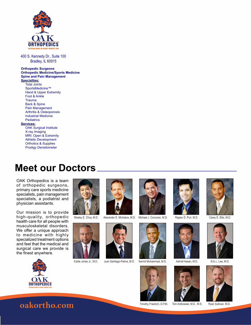
OAK Sports Medicine Update Fall 2018 Page 16
OAK Orthopedics is a team of orthopedic surgeons, primary care sports medicine specialists, pain management specialists, a podiatrist and physician assistants.
Our mission is to provide high-qual i ty, orthopedic health care for all people with musculoskeletal disorders. We offer a unique approach to medicine with highly specialized treatment options and feel that the medical and surgical care we provide is the finest anywhere.
Orthopedic SurgeonsOrthopedic Medicine/Sports MedicineSpine and Pain ManagementSpecialties: Total Joints SportsMedicine™ Hand & Upper Extremity Foot & Ankle Trauma Back & Spine Pain Management Arthritis & Osteoporosis Industrial Medicine PediatricsServices: OAK Surgical Institute X-ray Imaging MRI; Open & Extremity Athletic Development Orthotics & Supplies Prodigy Densitometer
Meet our Doctors
400 S. Kennedy Dr., Suite 100Bradley, IL 60915
Kermit Muhammad, M.D.
ORTHOPEDIC SURGEON
E: [email protected]: (815) 928-8050, ext. 221F: (815) 928-8932
SPECIALIZING IN WHAT MOVES YOU
Kermit Muhammad, M.D.
ORTHOPEDIC SURGEON
E: [email protected]: (815) 928-8050, ext. 221F: (815) 928-8932
SPECIALIZING IN WHAT MOVES YOU
Elbow
☐ Cubital Tunnel Syndrome
☐ Elbow Bursitis
☐ Lateral Epicondylitis (Tennis Elbow)
☐ Medial Ulnar Collateral Ligament Injury
☐ Throwing Injuries of the Elbow
☐ Cubital Tunnel Release at the Elbow
☐ Medial Ulnar Collateral Ligament Reconstruction (Tommy John Surgery)
☐ Ulnar Nerve Transposition at the Elbow
☐ Basal Joint Surgery
☐ Carpal Tunnel Release (Open Technique)
☐ Distal Radius Fracture Repair With Volar Plate
☐ Electromyography (EMG)
☐ Endoscopic Carpal Tunnel Release
☐ Endoscopic Carpal Tunnel Release (Lins)
☐ Finger Fracture Fixation
☐ Nerve Conduction Study (NCS)
☐ Scaphoid Fracture Open Reduction and Internal Fixation (ORIF)
☐ Tendon Repair
☐ Trigger Digit Release
☐ Basal Joint Osteoarthritis
☐ Boxer’s Fracture
☐ Carpal Tunnel Syndrome
☐ Dupuytren’s Disease
☐ Finger Dislocation
☐ Fingertip Injuries
☐ Flexor Tendon Injuries
☐ Fractures of the Finger
☐ Fractures of the Hand (Metacarpal Fractures)
☐ Mallet Finger
☐ Nerve Injuries of the Hand
☐ Osteoarthritis of the Hand
☐ Paronychia
☐ Thumb Ulnar Collateral Ligament (UCL) Injury
☐ Trigger Digit
☐ Bankart Repair
☐ ORIF Surgery for Proximal Humerus Fracture
☐ Reverse Total Shoulder Replacement
☐ Rotator Cuff Repair (Arthroscopic)
☐ SLAP Repair
☐ Subacromial Injection
☐ Total Shoulder Replacement
☐ De Quervain’s Release
☐ Ganglion Cyst Removal
☐ Wrist Arthroscopy
Dr. Muhammad has recommended you watch the following video(s):HandShoulderWrist
Other
oakortho.com
Conditions:Conditions:Conditions:Conditions:
Treatments:
Treatments:
Treatments:
Treatments:
☐ Fracture of the Collarbone (Clavicle)
☐ De Quervain’s Tenosynovitis
☐ Distal Radius Fracture (Broken Wrist)
☐ Kienbock’s Disease
☐ Scaphoid Fractures
☐ Triangular Fibrocartilage Complex (TFCC) Tears
☐ Anesthesia (Conscious Sedation)
☐ Anesthesia (General)
☐ Anesthesia (Monitored Anesthesia Care, MAC)
☐ Anesthesia (Peripheral Nerve Block)
oakortho.com
Wesley E. Choy, M.D. Alexander E. Michalow, M.D. Michael J. Corcoran, M.D. Rajeev D. Puri, M.D. Carey E. Ellis, M.D.
Eddie Jones Jr., M.D. Juan Santiago-Palma, M.D. Kermit Muhammad, M.D. Ashraf Hasan, M.D. Eric L. Lee, M.D.
Timothy Friedrich, D.P.M. Tom Antkowiak, M.D., M.S. Ryan Sullivan, M.D.


