Embed Size (px)
Citation preview

Article
NFIL3 Orchestrates the Emergence of CommonHelper Innate Lymphoid Cell Precursors
Graphical Abstract
Highlights
d Cell-intrinsicNfil3 ablation results in impaired development of
ILC subsets
d NFIL3 deficiency leads to loss of common helper-like ILC
progenitors (CHILPs)
d NFIL3 is controlled by mesenchyme-derived IL-7 in lymphoid
precursors
d NFIL3 exerts its function in CHILP via direct regulation of Id2
Authors
Wei Xu, Rita G. Domingues, ...,
James P. Di Santo,
Henrique Veiga-Fernandes
Correspondence
[email protected] (J.P.D.),
[email protected] (H.V.-
F.)
In Brief
Innate lymphoid cells (ILCs) originate
from a common innate lymphoid cell
progenitor. However, the transcriptional
program that sets the identity of the ILC
lineage remains elusive. Xu et al. show
that NFIL3 acts downstream of IL-7
signaling, regulating the emergence of
common helper ILC progenitors via direct
regulation of Id2.
Xu et al., 2015, Cell Reports 10, 2043–2054
March 31, 2015 ª2015 The Authors
http://dx.doi.org/10.1016/j.celrep.2015.02.057

Cell Reports
Article
NFIL3 Orchestrates the Emergence of Common HelperInnate Lymphoid Cell Precursors
Wei Xu,1,9 Rita G. Domingues,2,9 Diogo Fonseca-Pereira,2,9 Manuela Ferreira,2 Helder Ribeiro,2 Silvia Lopez-Lastra,1
YasutakaMotomura,3 LaraMoreira-Santos,2 FranckBihl,4VeroniqueBraud,4Barbara Kee,5HughBrady,6MarkC. Coles,7
Christian Vosshenrich,1 Masato Kubo,3,8 James P. Di Santo,1,10,* and Henrique Veiga-Fernandes2,10,*1Innate Immunity Unit, Inserm U668, Institut Pasteur, 25 Rue du Docteur Roux, 75724 Paris, France2Instituto de Medicina Molecular, Faculdade de Medicina de Lisboa, Av. Prof. Egas Moniz, Edifıcio Egas Moniz, 1649-028 Lisboa, Portugal3Laboratory for Cytokine Regulation, Research Center for IntegrativeMedical Science (IMS), RIKEN Yokohama Institute, Suehiro-cho 1-7-22,
Tsurumi, Yokohama, Kanagawa 230-0045, Japan4Centre National de la Recherche Scientifique - UMR 7275 Institut de Pharmacologie Moleculaire et Cellulaire, 660 Route des Luciole, 06560
Valbonne, France5Department of Pathology, University of Chicago, Chicago, IL 60637, USA6Department of Life Sciences, Imperial College, London SW7 2AZ, UK7Centre for Immunology and Infection, Department of Biology and Hull York Medical School, University of York, York YO10 5DD, UK8Division of Molecular Pathology, Research Institute for Biomedical Science, Tokyo University of Science, Chiba 278-0022, Japan9Co-first author10Co-senior author
*Correspondence: [email protected] (J.P.D.), [email protected] (H.V.-F.)
http://dx.doi.org/10.1016/j.celrep.2015.02.057
This is an open access article under the CC BY-NC-ND license (http://creativecommons.org/licenses/by-nc-nd/3.0/).
SUMMARY
Innate lymphoid cells (ILCs) are a family of effectors
that originate from a common innate lymphoid cell
progenitor. However, the transcriptional program
that sets the identity of the ILC lineage remains
elusive. Here, we show that NFIL3 is a critical regu-
lator of the common helper-like innate lymphoid
cell progenitor (CHILP). Cell-intrinsic Nfil3 ablation
led to variably impaired development of fetal and
adult ILC subsets. Conditional gene targeting de-
monstrated that NFIL3 exerted its function prior to
ILC subset commitment. Accordingly, NFIL3 ablation
resulted in loss of ID2+CHILP and PLZF+ ILC progen-
itors. Nfil3 expression in lymphoid progenitors was
under the control of the mesenchyme-derived hema-
topoietin IL-7, and NFIL3 exerted its function via
direct Id2 regulation in the CHILP. Moreover, ectopic
Id2 expression in Nfil3-null precursors rescued
defective ILC lineage development in vivo. Our data
establish NFIL3 as a key regulator of common help-
er-like ILC progenitors as they emerge during early
lymphopoiesis.
INTRODUCTION
The immune system is composed by myriads of cell types and
lymphoid organs that ensure immune surveillance and protective
immunity. The adaptive immune system arose late in evolution
and consists of B and T lymphocytes that express recombining
antigen-specific receptors. Naive T and B cells are activated
by their cognate antigen in secondary lymphoid organs and un-
dergo significant cell division and differentiation before exerting
their effector function. In contrast, innate lymphocytes display
rapid effector functions despite their set of limited germ-line-en-
coded receptors. Formore than three decades, natural killer (NK)
cells were the only recognized innate lymphocytes (Diefenbach
et al., 2014; McKenzie et al., 2014; Spits et al., 2013). More
recently, additional innate lymphocytes have been discovered
andwere considered to be part of a family of effector cells collec-
tively named innate lymphoid cells (ILCs) (Diefenbach et al.,
2014; McKenzie et al., 2014; Spits et al., 2013).
ILCs have a lymphoid morphology, lack rearranged antigen
receptors, and are abundantly present at mucosal surfaces,
such as the enteric lamina propria. The expression of lineage-
specific transcription factors and discrete cytokine profiles
led to the identification of three distinct ILC subsets that have
striking parallels with T helper (Th) cell subsets. Group 1 ILCs
(ILC1) resemble Th1 cells and include NK cells and other
IFNg-producing innate effectors ILC1 (Bernink et al., 2013; Die-
fenbach et al., 2014; Fuchs et al., 2013; Klose et al., 2014;
McKenzie et al., 2014; Spits et al., 2013; Vonarbourg et al.,
2010). ILC1s were shown to depend on TBX21 (T-bet), IL-7,
and IL-15 (Diefenbach et al., 2014; McKenzie et al., 2014; Spits
et al., 2013). Group 2 ILCs are similar to Th2 cells. ILC2s are
RORa (Halim et al., 2012; Wong et al., 2012) and GATA3 (Hoyler
et al., 2012; Klein Wolterink et al., 2013; Mjosberg et al., 2012)
dependent, IL-7 dependent, and produce IL-5 and IL-13 (Moro
et al., 2010; Neill et al., 2010; Spits et al., 2013). ILC2s have
been shown to play important roles in helminth infections,
asthma, and allergy contexts (McKenzie et al., 2014; Spits
et al., 2013). Group 3 ILCs (ILC3) are RORgt and partly AhR
dependent, rely on IL-7, and similarly to Th17 cells produce
IL-17 and IL-22 (Diefenbach et al., 2014; Kiss et al., 2011;
Lee et al., 2012; McKenzie et al., 2014; Qiu et al., 2012; Spits
et al., 2013). ILC3s were also shown to mediate inflammatory
Cell Reports 10, 2043–2054, March 31, 2015 ª2015 The Authors 2043

ce
lln
um
be
r(1
03)
0
820
E
** *
ce
lln
um
be
r(1
03)
0
35 *
F
Nfil3+/+
Nfil3-/-
ce
lln
um
be
r(1
04)
0
9 ** *
D
Nfil3+/+ Nfil3+/- Nfil3-/-
ce
lln
um
be
r(1
02)
α4β7highRORγt+
0
5 ***
*
CD4+ LTiCD4- LTi
ce
ll fr
eq
ue
ncy
****
******
0
12
6
0
28
14
***
fetal liver fetal gutG
B
cell
num
ber
TH
Y1
IL-7Rα
NK
p46
CD4
ILC
3
CLPBMlung
control
FLT
3
CD
25
SCA1
BM
Nfil3GFP
ILC
2
preNKPrNKPmNK
control
FLT
3
NK
1.1
CD122
BM
Nfil3GFP
NK
DX5
BM
NK
p46
NK1.1
CD
49
a
IL-7Rα
ILC
1
Nfil3GFP
controlNKp46+CD4+
controlBMgut
Nfil3GFP
Lin IL-7Rα Nfil3GFP
TH
Y1
CD
4
control
CD4-
CD4+
gut
Are
lative
exp
ressio
n
0
0.3
Nfil3GFP reporterNfil3 transcripts
fetal liver
fetal gut
Lin IL-7Rα Nfil3GFP
TH
Y1
CD
4
n.s.n.s.n.s.80
C
ce
lln
um
be
r(1
04)
0
12n.s. n.s.
cell
num
ber
cell
num
ber
cell
num
ber
cell
num
ber
cell
num
ber
adult embryo
Figure 1. Nfil3 Deficiency Results in Reduced Adult and Fetal ILCs
(A) Fetal liver (FL) CLP, BMCLP and NK, BM and liver ILC1, BM and lung ILC2, gut ILC3, fetal gut (FG) CD4� LTi, FG CD4+ LTi, and B and T cells were analyzed by
quantitative RT-PCR for Nfil3 expression.
(B) Flow cytometry analysis of Nfil3GFP expression in developing BM NK cells, spleen mature NK cells, ILC1s from BM and gut, CLPs, ILC2s from BM and lung,
enteric CD4+ and NKp46+ ILC3s from Nfil3GFP mice, and FL and FG CD4� and CD4+ LTi cells from E15.5 Nfil3GFP embryos.
(C) Number of FL and BM CLPs from Nfil3+/+ and Nfil3�/� mice. BM: Nfil3+/+ n = 6; Nfil3�/� n = 6.
(D) Number of BM and gut ILC1s from Nfil3+/+ and Nfil3�/� mice. BM: Nfil3+/+ n = 9; Nfil3�/� n = 11; gut: Nfil3+/+ n = 6; Nfil3�/� n = 5.
(E) Number of BM, lung, and gut ILC2s fromNfil3+/+ andNfil3�/�mice. BM:Nfil3+/+ n = 6;Nfil3�/� n = 6, lung:Nfil3+/+ n = 8;Nfil3�/� n = 8; gut:Nfil3+/+ n = 3;Nfil3�/�
n = 3.
(legend continued on next page)
2044 Cell Reports 10, 2043–2054, March 31, 2015 ª2015 The Authors

bowel diseases (Buonocore et al., 2010; Vonarbourg et al.,
2010) and to control immune responses to attaching and
effacing enteric pathogens such as Escherichia coli and Citro-
bacter rodentium (Sonnenberg et al., 2011; Zheng et al.,
2008). Lymphoid tissue inducer (LTi) cells are the prototypical
member of ILC3s, are dependent on retinoic acid signaling,
and were shown to play a critical role in secondary lymphoid
organ (SLO) development and tissue homeostasis (Diefenbach
et al., 2014; Spencer et al., 2014; Spits et al., 2013; van de Pa-
vert et al., 2014).
ILCs express the transcriptional regulator inhibitor of DNA
binding 2 (ID2), and Id2 deficiency leads to developmental block
of ILCs (Boos et al., 2007; Moro et al., 2010). ID2 is a helix-loop-
helix factor that was shown to sequester E proteins from their
target gene promoters. Interestingly, EBF1 has been shown to
counter Id2 expression (Thal et al., 2009), and conditional dele-
tion of Ebf1 in committed pro-B cells leads to their conversion
into different ILC subsets (Nechanitzky et al., 2013), suggesting
the existence of a common ILC precursor. Interestingly, a
a4b7+PLZF+ cell was recently identified as a committed precur-
sor to ILCs with the exception of NK and LTi cells (Constanti-
nides et al., 2014) and another study demonstrated that a
a4b7+ID2high cell was the common helper-like innate lymphoid
precursor (CHILP) to all helper-like ILCs (Klose et al., 2014).
Nevertheless, the factors that control the emergence of these
recently described ILC precursors from common lymphoid
progenitors (CLPs) remain unknown. Altogether, these data
suggest the existence of additional, yet unrecognized, tran-
scriptional regulators that set the identity of the CHILP (Klose
et al., 2014).
NFIL3 (also known as E4BP4) is a basic leucine zipper tran-
scription factor that was identified by its DNA-binding activity
(Zhang et al., 1995). NFIL3 coordinates signals from several
regulatory pathways, including the circadian clock. More
recently, NFIL3 was shown to mediate several immune pro-
cesses. NFIL3 controls pro-B cell survival (Ikushima et al.,
1997), IgE class-switch (Kashiwada et al., 2010), Th2 and
Th17 cytokine expression (Kashiwada et al., 2011a; Motomura
et al., 2011; Yu et al., 2013), IL-12 regulation (Kobayashi et al.,
2011; Smith et al., 2011), and CD8a dendritic cell develop-
ment (Kashiwada et al., 2011b). Interestingly, Nfil3-null mice
have an early block in NK cell development that perturbs
ID2, GATA3, EOMES, and TBX21 expression in hematopoietic
precursors (Gascoyne et al., 2009; Kamizono et al., 2009;
Male et al., 2014); these different transcription factors critically
control NK cell differentiation and homeostasis (Spits et al.,
2013).
In this report, we show that cytokine-dependent expression
of NFIL3 promotes the development of theCHILP via direct regu-
lation of Id2. Based on our results, NFIL3 emerges as a key tran-
scription factor that orchestrates the emergence of ILC precur-
sors from CLPs.
RESULTS
Nfil3 Is Expressed byAll Helper-like ILCs and Is Required
for ILC Homeostasis
NFIL3 is an essential transcription factor for NK cell commitment
from lymphoid progenitors. Nfil3-deficient mice have a profound
defect in peripheral NK cell homeostasis, which arises from an
early block in NK cell maturation in the bone marrow (Crotta
et al., 2014; Gascoyne et al., 2009; Male et al., 2014; Seillet
et al., 2014a). As diverse ILCs are thought to arise from lymphoid
precursors via common intermediates (Constantinides et al.,
2014; Klose et al., 2014; Spits et al., 2013), we hypothesized
that Nfil3 might have generalized roles in ILC development.
We first assessedNfil3 expression in lymphoid precursors and
distinct ILC subsets from fetal and adult tissues. We found high
levels of Nfil3 transcripts in all ILC subsets, whereas adaptive B
and T lymphocytes expressed very low levels ofNfil3 (Figure 1A).
In agreement, Nfil3GFP reporter mice revealed that GFP was
clearly expressed byNK cells and their immature BMprecursors,
whereas B and T cells were GFPlo (Figures 1B and S1A). Interest-
ingly, helper ILC1, ILC2, and ILC3 subsets from diverse fetal and
adult tissue sites also expressed high levels of GFP (Figures 1B
and S1A), a finding also confirmed at the protein level using intra-
cellular staining for NFIL3 in adult NK, ILC1, ILC2, and ILC3 sub-
sets (Figure S1B). These data demonstrate that NK cells are not
the only lymphoid cell subset that strongly expresses Nfil3 but
that high constitutive Nfil3 expression is a common character-
istic of all ILC subsets.
To assess the role of NFIL3 on the development and homeo-
stasis of helper-like ILC1, ILC2, and ILC3, we analyzed mice
with a germ-line deletion of Nfil3 (Gascoyne et al., 2009). In the
absence of Nfil3, ILC1 in the BM and gut, ILC2 in the BM and
lung, and CD4+ ILC3 in the gut were all clearly reduced, whereas
CLPs were not affected (Figures 1C–1F). Germ-line deletion of
Nfil3 also resulted in a decrease of RORgt+ ILC3 subsets in the
fetal liver, gut, and lymph nodes with a clear dose-dependent ef-
fect (Figures 1G and S1C–S1F). Consequently, Nfil3 ablation re-
sulted in reduced number of minute PPs and severely diminished
fetal LN size (Figures S1G–S1I). Of note, PPs were also reduced
in Nfil3�/� adult animals, arguing against a putative PP develop-
mental delay (Figure S1G). Taken together, these data confirm
recent reports on the broad role for Nfil3 in controlling the ho-
meostasis of helper ILC subsets and NK cells in both fetal and
adult life (Geiger et al., 2014; Seillet et al., 2014b; Yu et al., 2014).
Hematopoietic-Autonomous NFIL3 Controls ILC
Development before Commitment into Discrete ILC
Lineages
Nfil3 is widely expressed by tissues from different germ layers
having significant pleiotropic effects. In order to determine
whether hematopoietic cell-intrinsic Nfil3 expression is required
for ILC homeostasis, we ablated Nfil3 in a lineage-specific
(F) Number of gut NCR+ (NKp46+), NCR� (NKp46�CD4�), and CD4+ ILC3 populations from Nfil3+/+ and Nfil3�/� mice. Nfil3+/+ n = 6; Nfil3�/� n = 7.
(G) Left: number of a4b7highRORgt+ FL cells. Nfil3+/+ n = 5; Nfil3+/� n = 9; Nfil3�/� n = 8. Right: percentage of FG CD4� and CD4+ LTi cells. Nfil3+/+ n = 5; Nfil3+/�
n = 9; Nfil3�/� n = 8.
Error bars show SE. *, **, and *** p values for Student’s t test lower than 0.05, 0.01, and 0.001, respectively. See also Figure S1.
Cell Reports 10, 2043–2054, March 31, 2015 ª2015 The Authors 2045

E
gut
RORγt
TH
Y1
38.6
Nfil3fl/fl
Nfil3Δ/Δ
21.8
ce
lln
um
be
r(1
04)
0
8
RORγt+
Rorc-Cre
ce
ll fr
eq
ue
ncy
0 0
36 30
Rorc-CreF
CD4+ LTiCD4- LTi
Nfil3fl/fl
Nfil3Δ/Δ
Nfil3fl/fl Nfil3Δ/Δ
x
Nfil3fl/fl
Cre+Nfil3Δ/Δ
Vav1-iCre
or
Rorc-Cre
Cre-Nfil3fl/fl
A BVav1-iCre
IL-7
Rα
CD
25
FLT3
BM
21.1
1.75
26.5
1.97
2.44
0.12
SCA1
liver
EO
ME
S
CD49a
72.9
72.9
21.6
12.1
NK1.1
NK
p46
2.68
11.6
Nfil3fl/fl
Nfil3Δ/Δ
ILC1
ILC2NK
CLP
n.s. n.s.n.s.
C
Nfil3fl/fl Nfil3Δ/Δ
*** **
ce
ll fr
eq
ue
ncy
0 0
30 14
Vav1-iCreD
28.4
17.8
49.6
41.8
RO
Rγt
LTi
IL-7Rα
CD4+ LTiCD4- LTi
fetal gut
NK
p46
CD4
12.3
2.64
13.1
32.9
RORγt
GA
TA
3 8.22
8.7
11.1
42.7
gut
Nfil3fl/fl
Nfil3Δ/Δ
ILC3
ILC2
Vav1-iCre
41.3
45.6CD
4
RORγt
59.8
54.9
46.2
47.9
RO
Rγt
LTi
IL-7Rα
fetal gut
41.6
47.9CD
4
RORγt
HG
FACS purified
RORγt+ CD4-LTi29.2
47.1
27.2
52.3
RorcGFP
CD
4
Nfil3+/+
Nfil3+/- ce
ll fr
eq
ue
ncy
0
30
15
CD4+LTi
FACS purified
RORγt- ILC precursors
RorcGFP
IL7
Rα
10.6
Nfil3+/+
Nfil3+/- ce
ll fr
eq
ue
ncy
0
10
5
2.5
Nfil3+/+
Nfil3+/-
CD4-LTi
*
Nfil3+/+
Nfil3+/-
n.s.
OP9
6 days
+RANK-L
6 days
+RANK-L
OP9
(legend on next page)
2046 Cell Reports 10, 2043–2054, March 31, 2015 ª2015 The Authors

fashion. Nfil3fl/fl mice were bred to Vav1-iCre mice, ensuring Cre
activity in the hematopoietic lineage (de Boer et al., 2003; Moto-
mura et al., 2011; Figure 2A). Analysis of adult Vav1-iCre.Nfil3D
mice revealed unperturbed BM CLP development, whereas
helper ILC1, ILC2, and ILC3 subsets were reduced when
compared to their Nfil3fl/fl littermate controls (Figures 2B and
2C). Consistent with observations in germ-line Nfil3�/� mice,
fetal gut ILC3 subsets were significantly reduced in E15.5
Vav1-iCre.Nfil3D/D embryos when compared to their Nfil3fl/fl
littermate controls (Figures 2D, S2A, and S2B).
Having established the important function of NFIL3 in overall
ILC homeostasis, we next assessed whether NFIL3 is still
required upon commitment into mature ILC subsets. In order
to test this hypothesis, we analyzed mice in which Nfil3 was ab-
lated after commitment into the ILC3 lineage using Rorc-Cre
mice (Eberl and Littman, 2004; Figure 2A). Strikingly, analysis
of adult and fetal Rorc-Cre.Nfil3D/D mice demonstrated normal
enteric ILC3 development (Figures 2E, 2F, and S2B), indicating
that NFIL3 exerts its hematopoietic cell-intrinsic function before
Rorc acquisition but appears dispensable once ILC3s become
lineage committed.
Further evidence that NFIL3 is required before RORgt acquisi-
tion was provided by in vitro differentiation assays. The fetal gut
harbors ILC precursors Lin�IL7Ra+a4b7+ID2+RORgt� and CD4�
Rorc-GFP+ LTi that can further differentiate to CD4+ LTi cells
when co-cultured with OP9 cells (van de Pavert et al., 2014).
Whereas these ILC precursors from Nfil3+/� failed to give rise
to committed ILC3s (Rorc-GFP+), Nfil3+/� CD4� LTi (Rorc-
GFP+) could efficiently differentiate into CD4+ LTi cells under
the same conditions (Figures 2G and 2H). Thus, hematopoiet-
ic-autonomous NFIL3 expression in uncommitted fetal ILC pre-
cursors is critical for their further maturation.
NFIL3 Regulates the Emergence of Common Helper-ILC
Precursors
Given the broad impact of NFIL3 on ILC homeostasis and the ev-
idence that NFIL3 exerted its role prior to ILC lineage commit-
ment, we hypothesize that NFIL3 may be required during the
generation of committed ILC precursors (Constantinides et al.,
2014; Klose et al., 2014). Common-helper-like ILC precursors
(CHILPs) have been defined as Lin�IL-7Ra+a4b7highID2+ cells
that express variable amounts of PLZF (Constantinides et al.,
2014; Klose et al., 2014). Using Id2 reporter mice (Rawlins
et al., 2009), we found that Nfil3 has a critical, dose-dependent
role in the development of fetal and adult ID2+ CHILPs (Figures
3A and 3B). Similarly, a4b7highPLZF+ progenitors were severely
reduced in the BM and fetal liver of Nfil3 germ-line-deficient
and Vav1-iCre.Nfil3D/D mice (Figures 3C, 3D, S3A, and S3B). In
line with these findings, a4b7+ ILC precursors expressed higher
levels of Nfil3 transcripts, Nfil3GFP, and NFIL3 protein when
compared to CLPs (Figures 3E and 3F).
The observation that CHILPs were strongly reduced in the
absence of Nfil3 provides an explanation for the broad effects
of Nfil3 in ILC homeostasis. Nevertheless, despite the apparent
lack of CHILPs inNfil3-deficient mousemodels, some peripheral
ILC2s and ILC3s were still present in the gut and lung (Figure 1).
This finding could suggest a CHILP-independent pathway of
ILC2 and ILC3 development. Alternatively, despite strongly
reduced CHILPs in Nfil3�/� mice, these rare CHILPs further
develop into ILC2s and/or ILC3s and expand in the periphery.
In order to address these possibilities, we performed competitive
BM reconstitution experiments using lethally irradiated hosts
(CD45.1), which received WT (CD45.2) or Nfil3�/� (CD45.2) BM
against a WT competitor (CD45.1/2) in a 1:1 ratio (Figure 4A).
Analysis of such chimeras 8 weeks after transplantation revealed
that, despite normal CLP development, a4b7high PLZF� and
PLZF+ CHILPs derived from Nfil3�/� precursors were signifi-
cantly reduced when compared to their WT counterparts (Fig-
ures 4B and S4). In line with the normal CLP development in
the absence of NFIL3, thymic T cell development from Nfil3�/�
precursors was indistinguishable from WT (Figure 4C). In
contrast, all mature ILC subsets (ILC1, ILC2, and ILC3) that
derived from Nfil3�/� precursors were consistently and severely
reduced in these chimeras (Figure 4D). Altogether, these data
further confirm that Nfil3 acts in a hematopoietic cell-intrinsic
fashion to drive ILC development. In addition, these results pro-
vide compelling evidence that NFIL3 is a critical regulator of ILC
progenitors in early lymphopoiesis as they emerge from CLPs.
Nfil3 Expression Is Modulated by the gc-Dependent
Cytokine IL7
Early studies of NFIL3 implicated its role in regulating the survival
of lymphoid cells in response to IL-3 (Ikushima et al., 1997).
Therefore, we interrogated whether NFIL3 acts downstream of
critical cytokines required for early stages of ILC development.
Lymphoid precursors were isolated from Nfil3GFP mice and
Figure 2. NFIL3 Acts in a Cell-Autonomous Fashion before Commitment into Mature ILCs
(A) Conditional Nfil3-deficient animals breeding scheme.
(B and C) Conditional Nfil3-deficient animals were bred with Vav1-iCre animals. Flow cytometry analysis of liver NK and ILC1; BM CLP and ILC2; enteric ILC2
and total ILC3; and NCR+, NCR�, CD4+ ILC3 sub-populations from adult conditional Nfil3-deficient animals and their littermate controls. Frequency of NK,
ILC1, and BM ILC2 from Lin� cells: NK Nfil3+/+ = 8.5%, Nfil3�/� = 2%; ILC1 Nfil3+/+ = 2.5%, Nfil3�/� = 0.3%; ILC2 Nfil3+/+ = 0.5%, Nfil3�/� = 0.03%. Frequency
of NCR+, NCR�, and CD4+ ILC3 from Lin�THY1+ cells: NCR+ ILC3 Nfil3+/+ = 5.3%, Nfil3�/� = 0.2%; NCR� ILC3 Nfil3+/+ = 31.9%, Nfil3�/� = 5.6%; CD4+ ILC3
Nfil3+/+ = 5.6%, Nfil3�/� = 2.9%.
(D) Flow cytometry analysis and percentage of CD4� and CD4+ LTi cells in E15.5 FG. Nfil3fl/fl n = 8; Nfil3fl/D n = 7; Nfil3D/D n = 5.
(E) Conditional Nfil3-deficient animals were bred with Rorc-Cre animals. Flow cytometry analysis and number of ILC3 in gut. Nfil3fl/fl n = 3; Nfil3D/D n = 3.
(F) Conditional Nfil3-deficient animals were bred with Rorc-Cre animals. Flow cytometry analysis and percentage of FG CD4� and CD4+ LTi cells. Nfil3fl/fl n = 7;
Nfil3 D/D n = 5.
(G and H)Nfil3+/�mice were bred toRorcGFP animals. E15.5 CD4�LTi cells and RORgt� ILC precursors fromNfil3+/+and Nfil3+/�were co-cultured with OP9. Flow
cytometry analysis and percentage of ILC3 RORgt+ (G) and CD4+ LTi (H) after 6 days of culture. Nfil3 +/+ n = 4; Nfil3 +/� n = 8.
Error bars show SE. *, **, and *** p values for Student’s t test lower than 0.05, 0.01, and 0.001, respectively. See also Figure S2.
Cell Reports 10, 2043–2054, March 31, 2015 ª2015 The Authors 2047

stimulated with IL-7.We found that GFP expression was strongly
induced by this cytokine in BM FLT3�a4b7high ILC precursors as
well as in BM CLP (Figure 5A). In line with these findings, lung
ILC2 and gut NKp46+ ILC3 also upregulatedNfil3GFP expression,
whereas mature NK cells and intestinal ILC1 did not modulate
Nfil3GFP expression (Figures 5B and 5C). Binding of IL-7 to its
cognate receptor triggers a signaling cascade, resulting in the
activation of the JAK/STAT and PI3K/Akt pathways (Demoulin
and Renauld, 1998). Thus, we asked whether IL-7 regulates
Nfil3 in a STAT5-dependent fashion. We found that GFP levels
were reduced in ILC that were cultured with STAT5 inhibitor, indi-
cating that NFIL3 functions downstream of IL-7 and that the acti-
vation of STAT5 is required for IL-7-induced Nfil3GFP expression
(Figure 5D).
As IL-7 could enhance NFIL3 expression in early lymphoid cell
precursors and especially a4b7high ILC precursors (Figure 5A),
we hypothesized that cytokine-driven NFIL3 expression might
regulate these cells as they emerge during early lymphopoiesis.
Thus, we examined ILC2 generation in vitro from single Nfil3+/+
and Nfil3�/� BM CLP. We observed that the average clone
D
0
14
7
PLZF+CHILP
***
fetal liver
Nfil3+/+
Nfil3-/-
Germ line mutant
ce
lln
um
be
r(1
03)
Nfil3fl/fl
Nfil3Δ/Δ
0
8
4
PLZF+CHILP
*
BM
Vav1-iCre deleted
C BM
0
4
Nfil3+/+
Nfil3-/-2
PLZF+CHILP
***
Germ line mutant
ce
lln
um
be
r(1
02)
Nfil3+/+
Nfil3-/-
ce
lln
um
be
r(1
03)
0
3 *
CHILPCHILP
0
1.2ce
lln
um
be
r(1
03)
E
rela
tive
exp
ressio
n
0
Nfil30.3
***
F
controlCLPFLT3-α4β7high
cell
num
ber
α4β7
Nfil3GFP
FLT
3
α4β7
FLT
3
Nfil3GFP
feta
lliv
er
BM
NFIL3
NFIL3
A
Nfil3+/+
Nfil3-/-
BM
Id2GFP
α4β7
B
1.242.90
12.47.19
Nfil3+/+
Nfil3-/-
fetal liver
Id2GFPα4β7
***
0.28
4.21
Figure 3. NFIL3 Is Required for the Emer-
gence of CHILP and a4b7highPLZF+ ILC Pro-
genitors
(A and B) Flow cytometry analysis and number of
CHILP cells in BM and FL fromNfil3+/+ andNfil3�/�
mice. BM: Nfil3+/+ n = 4 and Nfil3�/� n = 5; FL:
Nfil3+/+ n = 5 and Nfil3�/� n = 6.
(C) Number of a4b7highPLZF+ ILC progenitors in
BM from Nfil3+/+ and Nfil3�/� mice and conditional
deficient animals and their littermate controls. BM:
Nfil3+/+ n = 6; Nfil3�/� n = 3; BM: Nfil3fl/fl n = 3;
Nfil3D/D n = 3.
(D) Number of FL CHILP PLZF+ progenitors in
E15.5 Nfil3+/+ and Nfil3�/� embryos. FL: Nfil3+/+
n = 5; Nfil3+/� n = 5.
(E) CLP and FLT3�a4b7high cells were analyzed by
quantitative RT-PCR for Nfil3 expression.
(F) Flow cytometry analysis of Nfil3GFP expression
and NFIL3 in CLPs and FLT3�a4b7high from BM
and E15.5 FL cells.
Error bars show SE. * and *** p values for Student’s
t test lower than 0.05 and 0.001, respectively. See
also Figure S3.
size of the resultant ILC2 colonies from
Nfil3�/� CLPs were significantly smaller
compared to that obtained from WT
CLP, which could not be explained by
increased apoptosis rates or defective
proliferation capacity (Figures 5E, 5F,
and S5A–S5C). Noteworthy, this finding
was also in agreement with normal pro-
liferation and cytokine production of
Nfil3�/� ILC2 (Figures S5D and S5E).
Interestingly, T cell colony sizes from
Nfil3+/+ and Nfil3�/� CLPs were similar
(Figure 5E), suggesting an ILC-specific
target for NFIL3 action. Taken together,
these data suggest a model whereby the
gc cytokine IL-7 regulates Nfil3 expression in common lymphoid
precursors that impacts on these developing progenitors.
NFIL3 Directly Regulates Id2 in the CHILP
Because Id2+ ILC precursors were strongly reduced inNfil3-defi-
cient mice (Figure 3), we hypothesized that Id2 was a relevant
downstream target of Nfil3 in CHILP. Strikingly, Id2 transcripts
were strongly decreased in both fetal and adult Nfil3�/� ILC pre-
cursors and NFIL3 impacted on Id2 levels in FLT3�a4b7high pre-
cursors, whereas Id2 levels were only moderately modulated in
CLPs and mature ILC subsets (Figures 6A, 6B, and S6A). To
gain additional insight into the regulatory mechanisms of Nfil3,
we performed chromatin immunoprecipitation with a specific
anti-NFIL3 antibody followed by quantitative PCR analysis
(ChIP) in biologically relevant ILC progenitors ex vivo. NFIL3
bound the Id2 locus in CHILP, which also displayed active dime-
thylated H3K4 in the NFIL3-binding region (Figure 6C). Interest-
ingly, whereas enrichment of NFIL3 binding was found at a re-
gion ‘‘D’’ close to the Id2 promoter in CHILP, NFIL3 bound to
the distinct region ‘‘H’’ in more mature ILC2 and ILC3 (Figures
2048 Cell Reports 10, 2043–2054, March 31, 2015 ª2015 The Authors

6C, S6B, and S6C), which also displayed active dimethylated
H3K4 upstream of the NFIL3-binding region (Figure 6C). Note-
worthy, NFIL3 did not bind to important genes to ILC2 and IL3
development, notably Ahr, Tox, Notch2, and Rora (Figure S6D).
Despite this specific NFIL3 binding to the Id2 locus in mature
ILC subsets, Nfil3 ablation did not affect Id2 expression in these
cells (Figure S6A), in agreement with themodel thatNfil3 expres-
sion is critical prior to, but not after, ILC commitment.
To further test whether NFIL3 controls CHILP generation via
Id2, we determined whether differentiation of Nfil3-deficient
ILC in vivo could be restored by enforced Id2 expression. We
transduced Nfil3+/+ and Nfil3�/� fetal liver progenitors with retro-
viral particles expressing Id2 (pMig-Id2) or GFP only (empty vec-
tor) and generated bone marrow chimeras (Figure 6D). Retroviral
transduction of Id2 (pMig-Id2) allowed Nfil3�/� progenitors to re-
acquire their potential to differentiate into ILC1, ILC2, and ILC3
in vivo, whereas ectopic expression of Id2 in WT progenitors
had no impact in ILC lineages (Figure 6D). In agreement, retro-
viral transduction of Id2 (pMig-Id2) also allowed Nfil3�/� CLP
to develop in vitro into PLZF+ CHILP when compared to their
counterparts transduced with retroviral particles containing
GFP only (Figure S6E). Collectively, these experiments demon-
strate that NFIL3 directly regulates Id2 expression in ILC progen-
itors and orchestrates their emergence from CLPs (Figure 6E).
DISCUSSION
The development of multiple and distinct hematopoietic cell
lineages relies on tightly controlled expression of transcription
A
Nfil3+/+ Nfil3-/-
0
60
BM
%o
fC
D4
5.2
ce
lls 60***
NK
0
60
liver
***
ILC1
0
60
lung
0
60
gut
******
ILC2
0
20
gut
0
20
spleen
****
ILC3
0
60
BM
***
0
liver
***
D
thymus
0
100
DP
%o
fC
D4
5.2
ce
lls 100
0
100
CD8
0
CD4
C
n.s. n.s. n.s.
%o
fC
D4
5.2
ce
lls
0
60
PLZF-
FLT3-α4β7high
0
60
PLZF+
*****
0
60
ILC2
**
0
60
CLP
BMB
n.s.
5x10⁵ BM cells
5x10⁵ BM cellsor
CD45.1/2
CD45.2
CD45.1
Nfil3+/+
Nfil3-/-
CD
45
.1
CD45.2
host competitor
test
Figure 4. NFIL3 Regulates All Helper-like
ILCs In Vivo through Regulation of the
CHILP
(A) Competitive transplantation assays scheme.
BM of Nfil3+/+and Nfil3�/� littermate controls were
co-transplanted with equal numbers of third-party
WT progenitors.
(B–D) Percentage of donor BM CLP, PLZF�, and
PLZF+ progenitors and BM ILC2s (B); thymic DP,
CD4, and CD8 cells (C); and BM and liver NK cells,
BM and liver ILC1s, lung and gut ILC2s, and gut
and spleen ILC3s (D). Nfil3+/+ n = 8; Nfil3�/� n = 9.
Error bars show SE. ** and *** p values for Stu-
dent’s t test lower than 0.01 and 0.001, respec-
tively. See also Figure S4.
factors that promote lineage specification
and commitment while suppressing alter-
native cell fates. As an example, several
regulators induce the development of un-
committed hematopoietic progenitors
into the B or T cell lineage. In contrast to
the transcription factors that promote
generation of adaptive lymphocytes, the
factors that control ILC development are
less well understood.
Diverse ILC subsets can be generated
from CLPs, and ID2 has emerged as
a central regulator of ILC fate (Hoyler
et al., 2012;Moro et al., 2010; Yokota et al., 1999). More recently,
committed precursors to all helper ILCs (CHILPs) were identified
within the fetal and adult Lin�IL-7Ra+a4b7high progenitor cell
population (Klose et al., 2014). Committed ILC precursors
strongly express ID2 and harbor both PLZF+ and PLZF� frac-
tions (Klose et al., 2014), and NOTCH triggering could induce
PLZF expression on PLZF�a4b7high cells, suggesting a precur-
sor-product relation between these subsets (Constantinides
et al., 2014). PLZF-expressing ILC progenitors could give rise
to ILC1, ILC2, and ILC3, but not LTi cells (Constantinides et al.,
2014), whereas Id2+a4b7high ILC precursors originate all help-
er-ILC subsets, suggesting that a4b7highID2+PLZF� cells may
harbor LTi potential. Nevertheless, how the emergence of CHILP
from CLP is regulated remains elusive.
Whereas the transcriptional repressor ID2 is essential for
development of all known ILC subsets, it is not clear how Id2
expression is regulated in lymphoid progenitors (CLP) or how
titration and reduction of E-protein activity allows for emergence
of CHILP from these cells. Previous studies demonstrated that
the transcription factor NFIL3 is broad regulator of ILC homeo-
stasis (Gascoyne et al., 2009; Geiger et al., 2014; Male et al.,
2014; Seillet et al., 2014b), although the molecular basis for the
NFIL3 effect remained unclear. In this report, we demonstrate
that NFIL3 is a critical regulator of the common-helper-like ILC
progenitor (CHILP), while being dispensable for overall helper-
like ILC fate and maintenance of discrete mature ILC subsets
(Geiger et al., 2014; Seillet et al., 2014b). We demonstrate
that IL-7 regulated NFIL3 expression in the CHILP and that
NFIL3 operated via direct regulation of Id2 expression in ILC
Cell Reports 10, 2043–2054, March 31, 2015 ª2015 The Authors 2049

precursors. In agreement, ectopic Id2 expression in vivo rescued
developmental defects of ILC1, ILC2, and ILC3 from Nfil3-null
lymphoid precursors. We further demonstrated that Id2 expres-
sion could rescue PLZF expression in Nfil3�/� CHILP, suggest-
ing a transcription factor cascade that links Nfil3, Id2, and
Zbtb16. Based on our results, NFIL3 emerges as a central regu-
lator of the common helper ILC precursor in early lymphopoiesis.
Interestingly, it was recently shown that NFIL3 could act in CLP
upstream of Tox, also directing the development of a CXCR6+
common cytotoxic and helper ILC precursor (aLP; Yu et al.,
2014). The relationship between aLP and CHILP is unclear, but
it is possible that NFIL3 may act by distinct mechanisms in
different ILC precursors. This notion is also in line with our own
findings showing that NFIL3 binding occurred at different Id2
genomic regions in the CHILP and mature ILC3 and the absence
of NFIL3 binding to Tox in ILC3 (Figures 6C and S6D).
A potent cell-intrinsic role for NFIL3 in the generation of all
recognized ILC subsets, including NK cells (Gascoyne et al.,
2009; Kamizono et al., 2009) and ILC1, ILC2, and ILC3 (this
study; Geiger et al., 2014; Seillet et al., 2014b) has recently
been reported. Our observation that NFIL3 is required for the
generation of helper-ILC precursors, namely a4b7highPLZF�
and a4b7highPLZF+ precursors (Constantinides et al., 2014;
A
Nfil3GFP
CLP CHILP
ce
ll n
um
be
r
B
Nfil3GFP
ILC2 ILC3
ce
ll n
um
be
r
ILC2
medium
IL7
IL7+100μM
IL7+200μM
D
ce
ll n
um
be
r
Nfil3GFP
C
Nfil3GFP
NK ILC1
ce
ll n
um
be
r
iCD3
NK
1.1
CD
44
ICO
S
IL-7Rα GATA3
Nfil3+/+
Nfil3-/-
FE
ILC2
Nfil3+/+ Nfil3-/-
ILC2 T cells
**
ce
lln
um
be
r(1
03)
0
0.5
1.5
1
0
12
24
18
6
n.s.
medium IL7
Figure 5. Nfil3 Expression Is Modulated by
the gc-Dependent Cytokine IL-7
(A–C) BM CLP and FLT3�a4b7high (A), BM ILC2
and gut ILC3 (B), and spleen NK and liver ILC1
cells (C) fromNfil3GFPmice were stimulated in vitro
with IL-7 and analyzed by flow cytometry.
(D) BM ILC2s from Nfil3GFP mice were stimulated
with IL-7 or IL-7 and STAT5 inhibitors and analyzed
by flow cytometry.
(E) Flow cytometry analysis of ILC2 differentiated
in vitro from Nfil3+/+ and Nfil3�/� BM CLPs. iCD3,
intracellular CD3.
(F) ILC2 and T cell number per colony. ILC2:
Nfil3+/+ n = 37; Nfil3�/� n = 46; T cells: Nfil3+/+ n =
49; Nfil3�/� n = 49.
Error bars show SE. **p values for Student’s t test
lower than 0.01. See also Figure S5.
Klose et al., 2014), confirms a cellular
mechanism for the broad effect of NFIL3
on multiple ILC subsets (Geiger et al.,
2014). On a molecular level, we found
that NFIL3 directly regulated Id2 in
CHILP. ID2 is a transcriptional repressor
that is critically required for NK cell and
ILC development from hematopoietic
precursors (Boos et al., 2007; Moro
et al., 2010; Satoh-Takayama et al.,
2010). We found that Id2 expression in
Nfil3�/� CHILP was severely compro-
mised and that NFIL3 bound specifically
to the Id2 promoter in CHILP-remodeling
chromatin configuration as revealed
by specific enrichment of dimethylated
H3K4. Moreover, ID2 appeared upstream
of Zbtb16 expression in ILC precursors, allowing us to propose a
NFIL3 > ID2 > PLZF transcription factor cascade that regulates
ILC emergence from CLPs.
Our data indicate that cellular expansion of Nfil3�/� ILC pre-
cursors toward ILC2was less robust, although it could not be ex-
plained by reduced proliferation or increased apoptosis, and that
Nfil3-deficient fetal ILC precursors were unable to further mature
in vitro. In addition, we found that Nfil3-dependent CHILP
strongly upregulated NFIL3 expression in response to IL-7.
Thus, we propose that cytokine-dependent signalsmay promote
stabilization and/or enhancement of NFIL3, which in turn orches-
trates the emergence of CHILP via direct Id2 regulation.
Whereas our results clearly indicate an essential role for NFIL3
in early ILC precursors, NFIL3 may also play additional context-
dependent roles at later stages of ILC differentiation and for
maintenance of effector functions in mature ILC subsets. Recent
studies of conditional ablation ofNfil3 in NK cells and NCR+ ILC3
(using Ncr1-Cre mice) failed to identify a major role for NFIL3 in
NK cell proliferation following MCMV infection and maintenance
of mature ILC3, respectively (Firth et al., 2013; Geiger et al.,
2014). Similarly, our results using Rorc-Cre mice likewise sug-
gest that many critical functions associated with ILC3 subsets
(LTi and lymphoid tissue organogenesis) are intact despite
2050 Cell Reports 10, 2043–2054, March 31, 2015 ª2015 The Authors

Nfil3 ablation after ILC subset commitment (Figure S2B). In addi-
tion, Nfil3-deficient ILC2 displayed normal cytokine secretion
and expansion in response to IL-33 (Figures S5D–S5F). Finally,
whereas NFIL3 binding to the Id2 locus was a common charac-
teristic of ILC progenitors and mature ILC2 and ILC3, NFIL3
binding occurred at different Id2 genomic regions in the CHILP
and mature ILC, suggesting differential transcriptional activity
of NFIL3 in ILC progenitors and committed cells. Nevertheless,
detailed studies on conditional Nfil3 deletion in mature ILC sub-
sets need to be performed in order to fully address this question.
Our current data demonstrating that NFIL3 is a key regulator of
the common-helper-like innate lymphoid precursor and previous
studies establishing that cytotoxic ILC development, notably NK
cells, also rely on NFIL3 suggest that NFIL3 may be required for
the early establishment of a common helper- and cytotoxic-ILC
lineage progenitor (Figure 6E). In line with this idea, it was
recently shown that NFIL3 can direct the development of a com-
mon cytotoxic and helper ILC precursor (Yu et al., 2014). Genetic
fate-mapping studies, multiparametric reporter lines, and line-
age-targeted strategies will be central to further elucidate the ex-
istence and the fate of such global innate lymphoid progenitor
(GILP) to helper and cytotoxic ILCs (Figure 6E).
EXPERIMENTAL PROCEDURES
Mice
C57Bl/6 mice were purchased from Charles River. Nfil3GFP transgenic mice
were generated using bacterial artificial chromosomes (BACs) (obtained
from Gene Expression Nervous System Atlas) that comprise the Nfil3 gene
with upstream and downstream regulatory regions. NSG and C57Bl/6 Ly5.1
(CD45.1) mice were maintained in house at Instituto de Medicina Molecular
A-8441
-7783
-940
1745
2208
2805
5845
13864
14108
7130
A B C ED F G H I J
Id2 locusC
10
fold
en
rich
me
nt
0
**
*
0
16
10
0
0
80
**
Anti-NFIL3
Anti-H3K4me2
fold
en
rich
me
nt
**
A B CD E F G H I J A B CD E F G H I J
CHILP CD4- LTi
n.s.
pMig.EmptyN
um
be
r(N
orm
aliz
ed
to
tra
nsd
uctio
ne
ffic
ien
cy)
0
900
NK/ILC1
*
0
400
ILC2
0
3000
ILC3
pMig.Id2
or
Nfil3+/+
Nfil3-/-
overnight
pMig.GFP
or
pMig.Id2.GFP NSG
**n.s. n.s.
DB
Nfil3+/+
Nfil3+/-
Nfil3-/-
Id2
GF
PM
FI(1
02)
0
5
ce
ll n
um
be
rId2GFP
Nfil3+/+
Nfil3+/-
Nfil3-/-
***
FLT3-α4β7highId2
0
1
fold
incre
ase
toW
T
FL BM
3x
decrease
CD45.2
CD45.1
FLT3-α4β7high
E
Figure 6. NFIL3 Acts in the CHILP via Direct Control of Id2 Expression
(A) Nfil3+/+ and Nfil3�/� FLT3�a4b7high cells were analyzed by quantitative RT-PCR for Id2 expression.
(B) Mean fluorescence intensity of Id2GFP in E15.5 FL FLT3�a4b7high cells from Nfil3+/+, Nfil3+/�, and Nfil3�/� littermate controls.
(C) Id2 locus scheme (top). ChIP analysis for NFIL3 and H3K4me2 binding in the Id2 locus of BM CHILP cells (left panel) and E15.5 CD4� LTi cells (right panel).
(D) In vivo Id2 rescue scheme (top). Hematopoietic progenitor cells from Nfil3+/+ and Nfil3�/� (CD45.2) mice were transduced with pMig.Id2-IRES-GFP retroviral
vector and control, and 53 105 total cells were injected into irradiated NSGhosts (CD45.1). GFP-positive cells were analyzed by flow cytometry, and cell numbers
normalized to the transduction efficiency are displayed. Nfil3+/+ pMig-Empty n = 4; Nfil3+/+ pMig-Id2 n = 4; Nfil3�/� pMig-Empty n = 4; Nfil3�/� pMig-Id2 n = 5.
(E) NFIL3 controls helper-like innate lymphoid cell generation through regulation of the CHILP.
Error bars show SE. * and ** p values for Student’s t test (B), ANOVA test (C), and Mann-Whitney (D) lower than 0.05 and 0.01, respectively. See also Figure S6.
Cell Reports 10, 2043–2054, March 31, 2015 ª2015 The Authors 2051
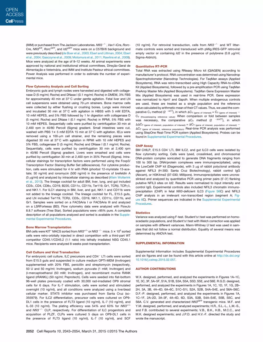
(IMM) or purchased from The Jackson Laboratories. Nfil3�/�, Vav1-iCre,Rorc-
Cre, Nfil3fl/fl, RorcGFP, and Id2GFP mice were on a C57Bl/6 background and
were previously described (de Boer et al., 2003; Eberl and Littman, 2004; Eberl
et al., 2004; Gascoyne et al., 2009; Motomura et al., 2011; Rawlins et al., 2009).
Mice were analyzed at the age of 8–12 weeks. All animal experiments were
approved by national and institutional ethical committees, Direcao-Geral de
Alimentacao e Veterinaria, and IMM and Institute Pasteur ethical committees.
Power Analysis was performed in order to estimate the number of experi-
mental mice.
Flow Cytometry Analysis and Cell Sorting
Embryonic guts and lymph nodes were harvested and digested with collage-
nase D (5 mg/ml; Roche) and DNase I (0.1 mg/ml; Roche) in DMEM, 3% FBS
for approximately 40 min at 37�C under gentle agitation. Fetal liver and LN
cell suspensions were obtained using 70-mm strainers. Bone marrow cells
were collected by either flushing or crushing bones. Lungs were minced
and incubated 30 min at 37�C with agitation in HBSS with 5 mM EDTA,
10 mM HEPES, and 5% FBS followed by 1 hr digestion with collagenase D
(5 mg/ml; Roche) and DNase I (0.1 mg/ml; Roche) in RPMI, 5% FBS with
10 mM HEPES. Sequentially cells were purified by centrifugation 30 min at
2,400 rpm in 40/80 Percoll (Sigma) gradient. Small intestines were cut,
washed with PBS 13 5 mM EDTA 15 min at 37�C with agitation. IELs were
removed using a 100-mm cell strainer, and the remaining pieces were
digested 30 min at 37�C with agitation in RPMI with 10 mM HEPES and
5% FBS, collagenase D (5 mg/ml; Roche) and DNase I (0.1 mg/ml; Roche).
Sequentially, cells were purified by centrifugation 30 min at 2,400 rpm
in 40/80 Percoll (Sigma) gradient. Livers were smashed and cells were
purified by centrifugation 30 min at 2,400 rpm in 35% Percoll (Sigma). Intra-
cellular stainings for transcription factors were performed using the Foxp3/
Transcription Factor Staining Buffer Set (eBioscience). For cytokine produc-
tion, cells were stimulated for 4 hr with PMA (phorbol 12-myristate 13-ace-
tate; 50 ng/ml) and ionomycin (500 ng/ml) in the presence of brefeldin A
(3 mg/ml) and analyzed by intracellular staining as described (Klein Wolterink
et al., 2013). The lineage cocktail for adult BM, liver, lung, and gut included
CD3e, CD4, CD8a, CD19, B220, CD11c, CD11b, Ter119, Gr1, TCRb, TCRgd,
and NK1.1. For ILC1 staining in BM, liver, and gut, NK1.1 and CD11b were
not added to the lineage cocktail. The lineage cocktail for FL, E15.5 guts,
and LN included Ter119, TCRb, CD3e, CD19, NK1.1, CD11c, CD11b, and
Gr1. Samples were sorted on a FACSAria I or FACSAria III and analyzed
on a LSRFortessa (BD). Flow cytometry data were analyzed with FlowJo
8.8.7 software (Tree Star). Sorted populations were >95% pure. A complete
description of all populations analyzed and sorted is available in the Supple-
mental Experimental Procedures.
Bone Marrow Transplantation
BM cells were KIT+MACS sorted from Nfil3+/+ or Nfil3�/� mice. 53 105 sorted
cells were retro-orbitally injected in direct competition with a third-part WT
competitor CD45.1/CD45.2 (1:1 ratio) into lethally irradiated NSG CD45.1
mice. Recipients were analyzed 8 weeks post-transplantation.
Cell Culture and Viral Transduction
For embryonic cell culture, ILC precursors and CD4� LTi cells were sorted
from E15.5 guts and suspended in culture medium OPTI-MEM (Invitrogen)
supplemented with 20% FBS, penicillin and streptomycin (respectively,
50 U and 50 mg/ml; Invitrogen), sodium pyruvate (1 mM; Invitrogen) and
b-mercaptoethanol (50 mM; Invitrogen), and recombinant murine RANK
ligand (rRANKL) (50 ng/ml; Peprotech). Cells were seeded into flat-bottom
96-well plates previously coated with 30,000 rad-irradiated OP9 stromal
cells for 6 days. For IL-7 stimulation, cells were sorted and stimulated
overnight (10 ng/ml), and all conditions were analyzed using a live/dead
cellular marker. STAT5 inhibitor was purchased from Santa Cruz (sc-
355979). For ILC2 differentiation, precursor cells were cultured on OP9-
DL1 cells in the presence of FLT3 ligand (10 ng/ml), IL-7 (10 ng/ml), and
IL-33 (10 ng/ml). The plating efficiency was 54% and 63% for Nfil3+/+
and Nfil3�/� CLP, respectively. For differentiation of ILC progenitors and
acquisition of PLZF, CLPs were cultured 5 days on OP9-DL1 cells in
the presence of FLT3 ligand (10 ng/ml), IL-7 (10 ng/ml), and SCF
(10 ng/ml). For retroviral transduction, cells from Nfil3�/� and WT litter-
mate controls were sorted and transduced with pMig.IRES-GFP retroviral
empty vector or containing Id2 in the presence of polybrene (0.8 mg/ml;
Sigma-Aldrich).
Quantitative RT-PCR
Total RNA was extracted using RNeasy Micro kit (QIAGEN) according to
manufacturer’s protocol. RNA concentration was determined using Nanodrop
Spectrophotometer (Nanodrop Technologies). For TaqMan assays (Applied
Biosystems), RNA was retro-transcribed using High Capacity RNA-to-cDNA
Kit (Applied Biosystems), followed by a pre-amplification PCR using TaqMan
PreAmp Master Mix (Applied Biosystems). TaqMan Gene Expression Master
Mix (Applied Biosystems) was used in real-time PCR. Gene expression
was normalized to Hprt1 and Gapdh. When multiple endogenous controls
are used, these are treated as a single population and the reference
value calculated by arithmeticmean of their CT values. Thus, we used the com-
parative CT method (2 �DCT), in which DCT (gene of interest) = CT (gene of interest) �
CT (Housekeeping reference value). When comparison or fold between samples
was necessary, the comparative DCT method (2 �DDCT), in which
DDCT (gene of interest, population of interest) = DCT (gene of interest, population of interest) �
DCT (gene of interest, reference population). Real-time PCR analysis was performed
using StepOne Real-Time PCR system (Applied Biosystems). Probes can be
found in Supplemental Experimental Procedures.
ChIP Assay
BM CHILP, E15.5 CD4�LTi, BM ILC2, and gut ILC3 cells were isolated by
flow cytometry sorting. Cells were lysed, crosslinked, and chromosomal
DNA-protein complex sonicated to generate DNA fragments ranging from
100 to 300 bp. DNA/protein complexes were immunoprecipitated, using
the LowCell# ChIP kit (Diagenode), with 3 mg of rabbit polyclonal antibody
against NFIL3 (H-300; Santa Cruz Biotechnology), rabbit control IgG
(Abcam), or H3K4me2 (07-030; Millipore). Immunoprecipitates were uncros-
slinked and analyzed by quantitative PCR using primer pairs (50-30) flanking
putative NFIL3 sites on Id2. Results were normalized to input intensity and
control IgG. Experimental controls also included NFIL3 chromatin immuno-
precipitation (ChIP) in fetal Nfil3-deficient ILC3 (Figure S6C) and NFIL3
ChIP analysis in an irrelevant non-transcribed region (segment A; Fig-
ure 6C). Primer sequences are indicated in the Supplemental Experimental
Procedures.
Statistics
Variance was analyzed using F-test. Student’s t test was performed on homo-
scedastic populations, and Student’s t test with Welch correction was applied
on samples with different variances. Mann-Whitney U test was used in sam-
ples that did not follow a normal distribution. Equality of several means was
determined by ANOVA test.
SUPPLEMENTAL INFORMATION
Supplemental Information includes Supplemental Experimental Procedures
and six figures and can be found with this article online at http://dx.doi.org/
10.1016/j.celrep.2015.02.057.
AUTHOR CONTRIBUTIONS
W.X. designed, performed, and analyzed the experiments in Figures 1A–1C,
1E, 3C, 3F, 5A–5F, S1A, S1B, S3A, S5A, S5D, S5E, and S6E. R.G.D. designed,
performed, and analyzed the experiments in Figures 1A, 1C, 1D, 1F, 1G, 2B–
2H, 3A, 3B, 4A–4D, 6A–6C, S1C–S1I, S2A, S2B, S4A–S4E, and S6A–S6C.
D.F.-P. designed, performed, and analyzed the experiments in Figures 1A,
1C–1F, 2A–2D, 3A–3F, 4A–4D, 6D, S3A, S3B, S4A–S4E, S5B, S5C, and
S6A. C.V. generated and characterized Nfil3GFP transgenic mice. M.F. and
Y.M. designed, performed, and analyzed experiments; H.R., S.L.-L., L.M.-S.,
and F.B. contributed to several experiments; V.B., B.K., H.B., M.C.C., and
M.K. designed experiments; and J.P.D. and H.V.-F. directed the study and
wrote the manuscript.
2052 Cell Reports 10, 2043–2054, March 31, 2015 ª2015 The Authors

ACKNOWLEDGMENTS
We thank the Bioimaging, Rodent, and Flow Cytometry facilities at IMM and
Institut Pasteur for technical assistance. We thank Sreerama Chaitanya Srid-
hara and Sergio Almeida for technical help and Francina Langa Vives (CIGM)
forNfil3BACmicroinjection. R.G.D., D.F.-P., andM.F. were supported by Fun-
dacao para a Ciencia e Tecnologia, Portugal; H.V.-F. by Fundacao para a
Ciencia e Tecnologia, Portugal, EMBO (1648) and ERC (207057); and J.P.D.
by grants from the Institut Pasteur, Inserm, LNCC (Equipe Labellisee Ligue
Contre le Cancer) and the Agence National pour la Recherche (Program
‘‘Blanc’’ Gut_ILC). J.P.D. is a founder and stakeholder in the biotechnology
company AXENIS (Paris, France).
Received: May 28, 2014
Revised: January 8, 2015
Accepted: February 23, 2015
Published: March 19, 2015
REFERENCES
Bernink, J.H., Peters, C.P., Munneke, M., te Velde, A.A., Meijer, S.L., Weijer,
K., Hreggvidsdottir, H.S., Heinsbroek, S.E., Legrand, N., Buskens, C.J.,
et al. (2013). Human type 1 innate lymphoid cells accumulate in inflamed
mucosal tissues. Nat. Immunol. 14, 221–229.
Boos, M.D., Yokota, Y., Eberl, G., and Kee, B.L. (2007). Mature natural killer
cell and lymphoid tissue-inducing cell development requires Id2-mediated
suppression of E protein activity. J. Exp. Med. 204, 1119–1130.
Buonocore, S., Ahern, P.P., Uhlig, H.H., Ivanov, I.I., Littman, D.R., Maloy, K.J.,
and Powrie, F. (2010). Innate lymphoid cells drive interleukin-23-dependent
innate intestinal pathology. Nature 464, 1371–1375.
Constantinides,M.G., McDonald, B.D., Verhoef, P.A., and Bendelac, A. (2014).
A committed precursor to innate lymphoid cells. Nature 508, 397–401.
Crotta, S., Gkioka, A., Male, V., Duarte, J.H., Davidson, S., Nisoli, I., Brady,
H.J., and Wack, A. (2014). The transcription factor E4BP4 is not required for
extramedullary pathways of NK cell development. J. Immunol. 192, 2677–
2688.
de Boer, J., Williams, A., Skavdis, G., Harker, N., Coles, M., Tolaini, M., Norton,
T., Williams, K., Roderick, K., Potocnik, A.J., and Kioussis, D. (2003). Trans-
genic mice with hematopoietic and lymphoid specific expression of Cre.
Eur. J. Immunol. 33, 314–325.
Demoulin, J.B., and Renauld, J.C. (1998). Signalling by cytokines interacting
with the interleukin-2 receptor gamma chain. Cytokines Cell. Mol. Ther. 4,
243–256.
Diefenbach, A., Colonna, M., and Koyasu, S. (2014). Development, differenti-
ation, and diversity of innate lymphoid cells. Immunity 41, 354–365.
Eberl, G., and Littman, D.R. (2004). Thymic origin of intestinal alphabeta T cells
revealed by fate mapping of RORgammat+ cells. Science 305, 248–251.
Eberl, G., Marmon, S., Sunshine, M.J., Rennert, P.D., Choi, Y., and Littman,
D.R. (2004). An essential function for the nuclear receptor RORgamma(t) in
the generation of fetal lymphoid tissue inducer cells. Nat. Immunol. 5, 64–73.
Firth, M.A., Madera, S., Beaulieu, A.M., Gasteiger, G., Castillo, E.F., Schluns,
K.S., Kubo, M., Rothman, P.B., Vivier, E., and Sun, J.C. (2013). Nfil3-indepen-
dent lineage maintenance and antiviral response of natural killer cells. J. Exp.
Med. 210, 2981–2990.
Fuchs, A., Vermi, W., Lee, J.S., Lonardi, S., Gilfillan, S., Newberry, R.D., Cella,
M., and Colonna, M. (2013). Intraepithelial type 1 innate lymphoid cells are a
unique subset of IL-12- and IL-15-responsive IFN-g-producing cells. Immunity
38, 769–781.
Gascoyne, D.M., Long, E., Veiga-Fernandes, H., de Boer, J.,Williams, O., Sed-
don, B., Coles, M., Kioussis, D., and Brady, H.J. (2009). The basic leucine
zipper transcription factor E4BP4 is essential for natural killer cell develop-
ment. Nat. Immunol. 10, 1118–1124.
Geiger, T.L., Abt, M.C., Gasteiger, G., Firth, M.A., O’Connor, M.H., Geary,
C.D., O’Sullivan, T.E., van den Brink, M.R., Pamer, E.G., Hanash, A.M., and
Sun, J.C. (2014). Nfil3 is crucial for development of innate lymphoid cells
and host protection against intestinal pathogens. J. Exp. Med. 211, 1723–
1731.
Halim, T.Y., MacLaren, A., Romanish, M.T., Gold, M.J., McNagny, K.M., and
Takei, F. (2012). Retinoic-acid-receptor-related orphan nuclear receptor alpha
is required for natural helper cell development and allergic inflammation. Im-
munity 37, 463–474.
Hoyler, T., Klose, C.S., Souabni, A., Turqueti-Neves, A., Pfeifer, D., Rawlins,
E.L., Voehringer, D., Busslinger, M., and Diefenbach, A. (2012). The transcrip-
tion factor GATA-3 controls cell fate and maintenance of type 2 innate
lymphoid cells. Immunity 37, 634–648.
Ikushima, S., Inukai, T., Inaba, T., Nimer, S.D., Cleveland, J.L., and Look, A.T.
(1997). Pivotal role for the NFIL3/E4BP4 transcription factor in interleukin 3-
mediated survival of pro-B lymphocytes. Proc. Natl. Acad. Sci. USA 94,
2609–2614.
Kamizono, S., Duncan, G.S., Seidel, M.G., Morimoto, A., Hamada, K., Gros-
veld, G., Akashi, K., Lind, E.F., Haight, J.P., Ohashi, P.S., et al. (2009). Nfil3/
E4bp4 is required for the development and maturation of NK cells in vivo.
J. Exp. Med. 206, 2977–2986.
Kashiwada, M., Levy, D.M., McKeag, L., Murray, K., Schroder, A.J., Canfield,
S.M., Traver, G., and Rothman, P.B. (2010). IL-4-induced transcription factor
NFIL3/E4BP4 controls IgE class switching. Proc. Natl. Acad. Sci. USA 107,
821–826.
Kashiwada, M., Cassel, S.L., Colgan, J.D., and Rothman, P.B. (2011a). NFIL3/
E4BP4 controls type 2 T helper cell cytokine expression. EMBO J. 30, 2071–
2082.
Kashiwada, M., Pham, N.L., Pewe, L.L., Harty, J.T., and Rothman, P.B.
(2011b). NFIL3/E4BP4 is a key transcription factor for CD8a⁺ dendritic cell
development. Blood 117, 6193–6197.
Kiss, E.A., Vonarbourg, C., Kopfmann, S., Hobeika, E., Finke, D., Esser, C., and
Diefenbach, A. (2011). Natural aryl hydrocarbon receptor ligands control
organogenesis of intestinal lymphoid follicles. Science 334, 1561–1565.
Klein Wolterink, R.G.J., Serafini, N., van Nimwegen, M., Vosshenrich, C.A.J.,
de Bruijn, M.J.W., Fonseca Pereira, D., Veiga Fernandes, H., Hendriks,
R.W., and Di Santo, J.P. (2013). Essential, dose-dependent role for the tran-
scription factor Gata3 in the development of IL-5+ and IL-13+ type 2 innate
lymphoid cells. Proc. Natl. Acad. Sci. USA 110, 10240–10245.
Klose, C.S., Flach, M., Mohle, L., Rogell, L., Hoyler, T., Ebert, K., Fabiunke, C.,
Pfeifer, D., Sexl, V., Fonseca-Pereira, D., et al. (2014). Differentiation of type 1
ILCs from a common progenitor to all helper-like innate lymphoid cell lineages.
Cell 157, 340–356.
Kobayashi, T., Matsuoka, K., Sheikh, S.Z., Elloumi, H.Z., Kamada, N., Hisa-
matsu, T., Hansen, J.J., Doty, K.R., Pope, S.D., Smale, S.T., et al. (2011).
NFIL3 is a regulator of IL-12 p40 in macrophages and mucosal immunity.
J. Immunol. 186, 4649–4655.
Lee, J.S., Cella, M., McDonald, K.G., Garlanda, C., Kennedy, G.D., Nukaya,
M., Mantovani, A., Kopan, R., Bradfield, C.A., Newberry, R.D., and Colonna,
M. (2012). AHR drives the development of gut ILC22 cells and postnatal
lymphoid tissues via pathways dependent on and independent of Notch.
Nat. Immunol. 13, 144–151.
Male, V., Nisoli, I., Kostrzewski, T., Allan, D.S., Carlyle, J.R., Lord, G.M., Wack,
A., and Brady, H.J. (2014). The transcription factor E4bp4/Nfil3 controls
commitment to the NK lineage and directly regulates Eomes and Id2 expres-
sion. J. Exp. Med. 211, 635–642.
McKenzie, A.N., Spits, H., and Eberl, G. (2014). Innate lymphoid cells in inflam-
mation and immunity. Immunity 41, 366–374.
Mjosberg, J., Bernink, J., Golebski, K., Karrich, J.J., Peters, C.P., Blom, B., te
Velde, A.A., Fokkens, W.J., van Drunen, C.M., and Spits, H. (2012). The tran-
scription factor GATA3 is essential for the function of human type 2 innate
lymphoid cells. Immunity 37, 649–659.
Moro, K., Yamada, T., Tanabe, M., Takeuchi, T., Ikawa, T., Kawamoto, H., Fur-
usawa, J., Ohtani, M., Fujii, H., and Koyasu, S. (2010). Innate production of T(H)
Cell Reports 10, 2043–2054, March 31, 2015 ª2015 The Authors 2053

2 cytokines by adipose tissue-associated c-Kit(+)Sca-1(+) lymphoid cells. Na-
ture 463, 540–544.
Motomura, Y., Kitamura, H., Hijikata, A., Matsunaga, Y., Matsumoto, K., Inoue,
H., Atarashi, K., Hori, S., Watarai, H., Zhu, J., et al. (2011). The transcription
factor E4BP4 regulates the production of IL-10 and IL-13 in CD4+ T cells.
Nat. Immunol. 12, 450–459.
Nechanitzky, R., Akbas, D., Scherer, S., Gyory, I., Hoyler, T., Ramamoorthy, S.,
Diefenbach, A., and Grosschedl, R. (2013). Transcription factor EBF1 is essen-
tial for the maintenance of B cell identity and prevention of alternative fates in
committed cells. Nat. Immunol. 14, 867–875.
Neill, D.R., Wong, S.H., Bellosi, A., Flynn, R.J., Daly, M., Langford, T.K.A.,
Bucks, C., Kane, C.M., Fallon, P.G., Pannell, R., et al. (2010). Nuocytes repre-
sent a new innate effector leukocyte that mediates type-2 immunity. Nature
464, 1367–1370.
Qiu, J., Heller, J.J., Guo, X., Chen, Z.M., Fish, K., Fu, Y.X., and Zhou, L. (2012).
The aryl hydrocarbon receptor regulates gut immunity through modulation of
innate lymphoid cells. Immunity 36, 92–104.
Rawlins, E.L., Clark, C.P., Xue, Y., and Hogan, B.L. (2009). The Id2+ distal tip
lung epithelium contains individual multipotent embryonic progenitor cells.
Development 136, 3741–3745.
Satoh-Takayama, N., Lesjean-Pottier, S., Vieira, P., Sawa, S., Eberl, G., Vos-
shenrich, C.A., and Di Santo, J.P. (2010). IL-7 and IL-15 independently pro-
gram the differentiation of intestinal CD3-NKp46+ cell subsets from Id2-
dependent precursors. J. Exp. Med. 207, 273–280.
Seillet, C., Huntington, N.D., Gangatirkar, P., Axelsson, E., Minnich, M., Brady,
H.J., Busslinger, M., Smyth, M.J., Belz, G.T., and Carotta, S. (2014a). Differen-
tial requirement for Nfil3 during NK cell development. J. Immunol. 192, 2667–
2676.
Seillet, C., Rankin, L.C., Groom, J.R., Mielke, L.A., Tellier, J., Chopin, M., Hun-
tington, N.D., Belz, G.T., and Carotta, S. (2014b). Nfil3 is required for the devel-
opment of all innate lymphoid cell subsets. J. Exp. Med. 211, 1733–1740.
Smith, A.M., Qualls, J.E., O’Brien, K., Balouzian, L., Johnson, P.F., Schultz-
Cherry, S., Smale, S.T., and Murray, P.J. (2011). A distal enhancer in Il12b is
the target of transcriptional repression by the STAT3 pathway and requires
the basic leucine zipper (B-ZIP) protein NFIL3. J. Biol. Chem. 286, 23582–
23590.
Sonnenberg, G.F., Monticelli, L.A., Elloso, M.M., Fouser, L.A., and Artis, D.
(2011). CD4(+) lymphoid tissue-inducer cells promote innate immunity in the
gut. Immunity 34, 122–134.
Spencer, S.P., Wilhelm, C., Yang, Q., Hall, J.A., Bouladoux, N., Boyd, A., Nut-
man, T.B., Urban, J.F., Jr., Wang, J., Ramalingam, T.R., et al. (2014). Adapta-
tion of innate lymphoid cells to amicronutrient deficiency promotes type 2 bar-
rier immunity. Science 343, 432–437.
Spits, H., Artis, D., Colonna, M., Diefenbach, A., Di Santo, J.P., Eberl, G.,
Koyasu, S., Locksley, R.M., McKenzie, A.N., Mebius, R.E., et al. (2013). Innate
lymphoid cells—a proposal for uniform nomenclature. Nat. Rev. Immunol. 13,
145–149.
Thal, M.A., Carvalho, T.L., He, T., Kim, H.G., Gao, H., Hagman, J., and Klug,
C.A. (2009). Ebf1-mediated down-regulation of Id2 and Id3 is essential for
specification of the B cell lineage. Proc. Natl. Acad. Sci. USA 106, 552–557.
van de Pavert, S.A., Ferreira, M., Domingues, R.G., Ribeiro, H., Molenaar, R.,
Moreira-Santos, L., Almeida, F.F., Ibiza, S., Barbosa, I., Goverse, G., et al.
(2014). Maternal retinoids control type 3 innate lymphoid cells and set the
offspring immunity. Nature 508, 123–127.
Vonarbourg, C., Mortha, A., Bui, V.L., Hernandez, P.P., Kiss, E.A., Hoyler, T.,
Flach, M., Bengsch, B., Thimme, R., Holscher, C., et al. (2010). Regulated
expression of nuclear receptor RORgt confers distinct functional fates to
NK cell receptor-expressing RORgt(+) innate lymphocytes. Immunity 33,
736–751.
Wong, S.H., Walker, J.A., Jolin, H.E., Drynan, L.F., Hams, E., Camelo, A.,
Barlow, J.L., Neill, D.R., Panova, V., Koch, U., et al. (2012). Transcription factor
RORa is critical for nuocyte development. Nat. Immunol. 13, 229–236.
Yokota, Y., Mansouri, A., Mori, S., Sugawara, S., Adachi, S., Nishikawa, S.,
and Gruss, P. (1999). Development of peripheral lymphoid organs and natural
killer cells depends on the helix-loop-helix inhibitor Id2. Nature 397, 702–706.
Yu, X., Rollins, D., Ruhn, K.A., Stubblefield, J.J., Green, C.B., Kashiwada, M.,
Rothman, P.B., Takahashi, J.S., and Hooper, L.V. (2013). TH17 cell differenti-
ation is regulated by the circadian clock. Science 342, 727–730.
Yu, X., Wang, Y., Deng, M., Li, Y., Ruhn, K.A., Zhang, C.C., and Hooper, L.V.
(2014). The basic leucine zipper transcription factor NFIL3 directs the develop-
ment of a common innate lymphoid cell precursor. eLife 3, e04406.
Zhang, W., Zhang, J., Kornuc, M., Kwan, K., Frank, R., and Nimer, S.D. (1995).
Molecular cloning and characterization of NF-IL3A, a transcriptional activator
of the human interleukin-3 promoter. Mol. Cell. Biol. 15, 6055–6063.
Zheng, Y., Valdez, P.A., Danilenko, D.M., Hu, Y., Sa, S.M., Gong, Q., Abbas,
A.R., Modrusan, Z., Ghilardi, N., de Sauvage, F.J., and Ouyang, W. (2008).
Interleukin-22mediates early host defense against attaching and effacing bac-
terial pathogens. Nat. Med. 14, 282–289.
2054 Cell Reports 10, 2043–2054, March 31, 2015 ª2015 The Authors