Embed Size (px)
Citation preview

Type 2 innate lymphoid cells control eosinophil homeostasis
Jesse C. Nussbaum1, Steven J. Van Dyken1, Jakob von Moltke1, Laurence E. Cheng2,Alexander Mohapatra3, Ari B. Molofsky4, Emily E. Thornton5, Matthew F. Krummel5, AjayChawla1,6,7, Hong-Erh Liang1, and Richard M. Locksley1,3,8
1Department of Medicine, University of California San Francisco, San Francisco, CA, USA94143-07952Department of Pediatrics, University of California San Francisco, San Francisco, CA, USA94143-07953Department of Microbiology & Immunology, University of California San Francisco, SanFrancisco, CA, USA 94143-07954Department of Laboratory Medicine, University of California San Francisco, San Francisco, CA,USA 94143-07955Department of Pathology, University of California San Francisco, San Francisco, CA, USA94143-07956Department of Physiology, University of California San Francisco, San Francisco, CA, USA94143-07957Department of Cardiovascular Research Institute, University of California San Francisco, SanFrancisco, CA, USA 94143-07958Department of Howard Hughes Medical Institute, University of California San Francisco, SanFrancisco, CA, USA 94143-0795
AbstractEosinophils are specialized myeloid cells associated with allergy and helminth infections. Bloodeosinophils demonstrate circadian cycling, as described over 80 years ago,1 and are abundant inthe healthy gastrointestinal tract. Although a cytokine, interleukin (IL)-5, and chemokines such aseotaxins, mediate eosinophil development and survival,2 and tissue recruitment,3 respectively, theprocesses underlying the basal regulation of these signals remain unknown. Here, we show thatserum IL-5 is maintained by long-lived type 2 innate lymphoid cells (ILC2) resident in peripheraltissues. ILC2 secrete IL-5 constitutively and are induced to co-express IL-13 during type 2inflammation, resulting in localized eotaxin production and eosinophil accumulation. In the smallintestine where eosinophils and eotaxin are constitutive,4 ILC2 co-express IL-5 and IL-13, whichis enhanced after caloric intake. The circadian synchronizer vasoactive intestinal peptide (VIP)also stimulates ILC2 through the VPAC2 receptor to release IL-5, linking eosinophil levels withmetabolic cycling. Tissue ILC2 regulate basal eosinophilopoiesis and tissue eosinophil
Correspondence and requests for materials should be addressed to [email protected].
Supplementary Information is linked to the online version of the paper at www.nature.com/nature.
Author Contributions J.C.N. performed experiments, interpreted data and wrote the manuscript; L.E.C. provided experimental andimaging assistance; S.V.D., A.M., A.B.M. and J.v.M. provided experimental assistance; E.E.T. performed imaging assays; M.F.K.provided reagents and expertise; H.E.L. generated mouse cytokine reporter strains; A.C. discussed experiments and provided oversightfor metabolic studies; R.M.L. directed the studies and wrote the paper with J.C.N.
Author Information The authors declare no competing financial interests.
NIH Public AccessAuthor ManuscriptNature. Author manuscript; available in PMC 2014 April 10.
Published in final edited form as:Nature. 2013 October 10; 502(7470): 245–248. doi:10.1038/nature12526.
NIH
-PA Author Manuscript
NIH
-PA Author Manuscript
NIH
-PA Author Manuscript

accumulation through constitutive and stimulated cytokine expression, and this dissociatedregulation can be tuned by nutrient intake and central circadian rhythms.
Eosinophils require survival signals delivered through the common beta-receptor chain (βc)shared by interleukins (IL)-3, IL-5 and granulocyte-macrophage colony stimulating factor(GM-CSF).1 IL-5 is particularly important, as supported by studies in IL-5-5 and IL-5receptor α chain (IL-5Rα)-deficient6 mice, and in humans using anti-IL-5 and anti-IL-5Rαthat target eosinophilis in disease. Without IL-5 signaling, residual eosinophils have beenattributed to IL-3 and GM-CSF, as well as eosinophil chemokines, such as eotaxins, thatsequester these cells into tissues.1,3
To identify cells that support eosinophils, we generated IL-5 reporter mice, designated Red5(recombinase-expressing detector for IL-5; R5).7 Cells from these mice contain a tandemdimer red fluorescent protein (tdTomato) linked by an internal ribosomal entry site (IRES)to a Cre element replacing the translation initiation site of the endogenous Il5 gene,facilitating function-marking, fate-mapping and deletion based on IL-5 expression (Fig. 1a).We validated that the construct disrupts the endogenous Il5 gene and that R5 fluorescencecorrelates with IL-5 production using CD4+ T cells examined after Th1 or Th2 polarization(Extended Data Fig. 1a-c).
Consistent with prior observations,8 CD45+R5+CD4− cells were present in non-lymphoidtissues, including the brain, heart, lung, kidney, skin, intestine and uterus while few R5+cells were in lymphoid organs, including spleen, bone marrow, lymph nodes or thymus, orin the liver (Fig. 1b and c). In all tissues, the vast majority of R5+ cells were small cells(forward-/side-scatter low) that lacked lineage markers for T, B, NK and myeloid cells, andexpressed markers for innate lymphoid type 2 cells (ILC2),9 including CD90.2 (Thy1),CD127 (IL7Rα), KLRG1 and ICOS; T1/ST2 and CD25 expression varied among tissues butwere present on all lung R5+ ILC2 (Extended Data Fig. 2a). Most (75–80%) CD90.2+T1/ST2+ lung ILC2 were R5+, and these cells had the highest reporter expression compared tothe few ILC2 from bone marrow and lymph nodes (Extended Data Fig. 2b). Sorted R5/+ILC2 spontaneously secreted IL-5 in culture, confirming that the reporter marks IL-5production (Extended Data Fig. 2c). Consistent with prior reports,9 numbers of ILC2 andR5+ ILC2 were similar in Recombinase Activating Gene (RAG)-deficient mice but werenearly absent in CD127-deficient mice (Extended Data Fig. 3a-c). Correspondingly, serumIL-5 was comparable in wild-type and RAG-deficient mice, reduced to about half-normallevels in heterozygous R5/+ mice, and was not detected in CD127-deficient mice (Fig. 1d).
Few ILC2 were present in lungs of newborns, but within the first week CD90.2+T1/ST2+cells increased, and the percent that were R5+ reached adult levels (Fig. 2a, Extended DataFig. 3d-e). We administered BrdU in drinking water of adult mice for two weeks to labeldividing cells and found that fewer than 10% of lung ILC2 were labeled (Extended Data Fig.3f). Pulse-chase labeling indicated that the decay of labeled ILC2 was substantially slowerthan CD4+ T cells (Fig. 2b). As assessed using multiphoton microscopy, lung R5+ ILC2were embedded in collagen-rich regions near the confluence of medium-sized blood vesselsand airways but absent from alveoli (Fig. 2c and data not shown).
Lung eosinophilia is a hallmark of allergic lung disease and helminth migration, buteosinophils are rare in the lung at baseline1 despite constitutive local IL-5. In prior studies,ILC2 stimulated with cytokines or helminth infection upregulated IL-13,10,11 which isgenetically linked to IL-5 in mice and humans and induces epithelial eotaxins (includingCCL11) and endothelial adhesins necessary for eosinophil trafficking.1,3 We crossed R5mice to Smart13 (S13) reporter mice, in which non-signaling human CD4 marks cellsproducing IL-13.11 In contrast to resting ILC2, lung ILC2 expressed the IL-13 reporter after
Nussbaum et al. Page 2
Nature. Author manuscript; available in PMC 2014 April 10.
NIH
-PA Author Manuscript
NIH
-PA Author Manuscript
NIH
-PA Author Manuscript

infection with the helminth Nippostrongylus brasiliensis. All IL-13+ ILC2 in the lung wereR5+, whereas CD4+ T cells expressed IL-5, IL-13, or both cytokines, consistent with priorobservations (Fig. 3a).11
We crossed R5/R5 mice to mice carrying a ROSA26-flox stop-YFP allele to fate-map cellsthat expressed the IL-5-linked Cre recombinase and infected the mice with N. brasiliensis toelicit a type 2 immune response. After infection, YFP was present only in ILC2 and CD4+ Tcells, and all YFP+ cells were also R5+ (Extended Data Fig. 4a-b). We also crossed R5/R5mice to mice carrying a ROSA26-flox stop-diphtheria toxin A allele to delete IL-5-producing cells. The R5 allele was designed such that expression of the tdTomato reporterprecedes Cre-mediated loxP recombination. Therefore, in R5/R5 Deleter mice, a populationof R5-lo cells may be detectable before they express the ROSA26-diphtheria toxin. Atbaseline, R5/R5 and R5/R5 Deleter mice had comparable numbers of total cells and CD4+ Tcells in the bone marrow, spleen, lung and small intestine lamina propria (Extended DataFig. 4c), but R5+ ILC2 were deleted in the lung (Fig. 3b) and small intestine (Extended DataFig. 4d).
To study the activity of lung ILC2 in the absence of Th2 cells, we crossed R5/R5 and R5/R5Deleter mice onto a RAG-deficient background and administered IL-2 and IL-33.10,12,13 Asexpected, cytokine-activated ILC2 in R5/R5 RAG-deficient mice showed increased surfaceKLRG1 expression and R5 MFI (Extended Data Fig. 5a). Cytokine administration increasedthe ILC2 population and induced eotaxin-1 (CCL11) in lungs of R5/R5 RAG-deficient micebut not in RAG-deficient R5/R5 Deleter mice (Fig. 3c). ILC2 deficiency was bypassed byadministering IL-13, which partially restored eotaxin levels.
Whereas eosinophils are rare in the lung at baseline, they are abundant in other tissues, suchas the small intestine lamina propria, where they depend on CCL11,4 and are absent in micethat lack ILC2.13,14 Our finding that ILC2 in the lung can control eosinophil recruitmentthrough dissociated expression of IL-5 and IL-13 led us to explore the role of ILC2-derivedcytokines in the basal regulation of peripheral eosinophils. We measured serum IL-5 at10:00 and at 22:00 and found that the levels correlated with the circadian variation in bloodeosinophils.15 Although influenced by the adrenal-cortical axis,15 blood eosinophils can bedominantly synchronized by meal timing.16 Mice fasted for 16 hours exhibited suppressedserum IL-5 and blood eosinophils at 10:00 (Fig. 4a-b). To minimize effects of altered light-dark cycle or stress induced by fasting, we restricted two groups of mice to feeding at nightonly or day only, allowed both groups to acclimate for 9 days, and analyzed them at 8:00(Extended Data Fig. 6a). Unlike lung ILC2, lamina propria ILC2 express IL-13constitutively, and this was increased in the morning if mice had just been in a fed asopposed to fasted cycle (Fig. 4c and Extended Data Fig. 6b); the numbers of ILC2 remainedconstant (Extended Data Fig. 6c). IL-13 reporter expression by intestinal ILC2 at 8:00 wasdiminished after a 16-hr overnight fast (Extended Data Fig. 6d) and restored byadministering an evening food (but not water) gavage at 22:00 (Fig. 4d).
The response of small intestine ILC2 to caloric input raised the possibility that these cellscould respond to hormonal cues induced by feeding. Indeed, purified intestinal ILC2, mostof which were R5+ (Extended Data Fig. 7a), released detectable IL-5 when incubated withIL-7 alone, but increased IL-5 production with addition of vasoactive intestinal peptide(VIP) but not ghrelin or enterostatin (Fig. 4e and Extended Data Fig. 7b). The effect on lungILC2 was similar (Extended Data Fig. 7c). VIP is a member of the secretin family ofneuropeptides, which are expressed throughout the nervous system. They are highlyexpressed in intestinal neurons, coordinating pancreatic secretion with smooth musclerelaxation in response to feeding,17 and in neurons of the suprachiasmatic nucleus (SCN),relaying environmental cues necessary to synchronize central circadian oscillators.18
Nussbaum et al. Page 3
Nature. Author manuscript; available in PMC 2014 April 10.
NIH
-PA Author Manuscript
NIH
-PA Author Manuscript
NIH
-PA Author Manuscript

VPAC2-deficient and VIP-deficient mice exhibit similar defects in circadian behavior,19,20
and rhythms mediated by VPAC2 are entrained by feeding.21
We hypothesized that VIP might signal through VPAC2 on ILC2. VIP and its receptors arealso expressed by immune cells, and signals through VPAC2 have been implicated in Th2cell expansion, survival and cytokine production.22 Mice deficient in VPAC2 trend towarddecreased circulating eosinophils at rest and delayed infiltrating eosinophils with allergicchallenge.23 We detected both VIP receptor type 1 (VPAC1) and type 2 (VPAC2) mRNA inintestinal and lung ILC2, whereas VPAC2 expression was undetectable in eosinophils andlow in macrophages and CD4+ T cells (Fig. 4f). Comparable levels of IL-5 were induced inculture with VIP or with a VPAC2-specific agonist (Fig. 4e and Extended Data Fig. 7c).24
VPAC2 is a G protein-coupled receptor that can associate with Gαs to activate adenylatecyclase.25 Consistent with this, we also induced comparable IL-5 levels bypassing thereceptor with dibutyryl-cAMP (Fig. 4e).
First noted in humans over 80 years ago,26 circadian variation of blood eosinophils has beenlinked to neuroendocrine15 and metabolic16 cycling. As shown here, long-lived ILC2 inperipheral tissues are the predominant source of circulating IL-5, and their close associationwith vasculature positions these cells for eosinophil recruitment. After stimulation byepithelial and/or Th2 cytokines, lung ILC2 increase IL-5 and co-express IL-13, leading tolocal eosinophil accumulation, a process that mimics the post-prandial response of intestinalILC2 to caloric intake. Further, ILC2 express functional VPAC2 receptors, providing apotential mechanism linking these dispersed tissue resident cells with central circadian andmetabolic rhythms. Intestinal eosinophils are normal in germ-free animals,4 and IL-13+ILC2 are found in human fetal gut,27 suggesting that these biologic pathways areindependent of intestinal microbiota. Although further study is needed, our findings suggestthat eosinophils are linked to basal circadian oscillations through ILC2 activation and raisethe possibility that helminthic parasites may have co-opted these fundamental pathways ofhost metabolic homeostasis.
METHODS (ONLINE ONLY)IL-5 reporter mice
R5 IL-5 reporter mice were generated by homologous gene targeting in C57BL/6 embryonicstem cells. The previously published plasmid pKO915-DT (Lexicon) containing theBasoph8 reporter17 was modified to express tdTomato in place of YFP, such that thecassette now contained (in order from 5’ to 3’) genomic sequence of the rabbit β-globin genepartial exon 2–3, the gene encoding tdTomato (Clontech), encephalomyocarditis virus IRES,humanized Cre recombinase, bovine growth hormone poly(a), and a loxP-flanked neomycinresistance cassette. Homologous arms straddling the Il5 translation intiation site (3.8 kbtoward 5’, containing the promoter and 5’ UTR and 3.0 kb toward 3’, containing the startATG halfway through exon 3) were amplified from C57BL/6 genomic DNA using Phusionpolymerase (Finnzymes) and cloned into the cassette by standard methods. The constructwas linearized with NotI and transfected by electroporation into C57BL/6 embryonic stemcells. Cells were grown on irradiated feeders with the aminoglycoside G418 in the media,and neomycin-resistant clones were screened for 5’ and 3’ homologous recombination byPCR. Eleven positive clones were subsequently tested (and all eleven confirmed) by 5’ and3’ Southern Blot. Two clones were selected for injection into albino C57BL/6 blastocysts togenerate chimeras, and the male pups with highest ratios of black-to-white coat color from asingle clone were selected to breed with homozygous CMV-Cre transgenic C57BL/6females (B6.C-Tg(CMV-cre)1Cgn/J; 006054, obtained from The Jackson Laboratory) toexcise the neomycin resistance cassette. The CMV-Cre transgene is X-linked and the males
Nussbaum et al. Page 4
Nature. Author manuscript; available in PMC 2014 April 10.
NIH
-PA Author Manuscript
NIH
-PA Author Manuscript
NIH
-PA Author Manuscript

from this cross were bred to wild-type C57BL/6 females to remove the CMV-Cre allele.Male and female R5/+ offspring were intercrossed to yield R5/R5 homozygotes.
Miceβ-actin-cyan fluorescent protein mice (B6.129(ICR)-Tg(CAG-ECFP)CK6Nagy/J; 004218),Rag1−/− mice (B6.129S7-Rag1tm1Mom/J; 002216), Il7rα−/− mice (B6.129S7-Il7rtm1Imx/J;002295), and ROSA-YFP mice (B6.129X1-Gt(ROSA)26Sortm1(EYFP)Cos/J; 006148) werefrom The Jackson Laboratory. ROSA-DTα and Smart13 mice have been described.11
Rag1−/− mice were maintained on SCIDS MD’s “Breeders Formula” antibiotic tablets (Bio-Serv). Mice were fed ad lib except when feeding was restricted to 12 hours daily (7:00 to19:00 or vice versa) or during 16-hour fasting. For gavage experiments, mice previously fedstandard chow ad lib were fasted at 16:00 before receiving water or a 1:1 mixture of high-fatchow (Research Diets) and 20% dextrose by oral gavage, representing 13% of ad lib caloricintake at 22:00. Mice used in experiments were mixed gender, between 6 and 10 weeks old,on the C57BL/6 background and were maintained according to institutional guidelines inspecific pathogen-free facilities at the University of California, San Francisco (SanFrancisco, CA).
Lung imagingLung slices were prepared using a modification of established methods.29 After euthanasia,lungs were inflated with 1 ml of 2% low-melt agarose (Type VII, Sigma-Aldrich), excisedand placed in 5 ml cold PBS, and 600 µm sections were cut on a vibratome (Model G,Oxford Laboratories). Lung sections were maintained in PBS at room temperature untilmounting. All sections were mounted with PBS and imaged on a multiphoton microscopewith data collected in three channels (CFP, GFP, RFP). Images were analyzed with Imarissoftware (Bitplane). Software spot detection algorithms were used to identify cells.
Nippostrongylus brasiliensis infection and cytokine administrationMice were infected with 500 N. brasiliensis third-stage larvae (L3) and were killed at theindicated timepoints for analysis of the mediastinal and mesenteric lymph nodes, lungs, andbone marrow. Procedures for maintaining as described.11 Rag1−/− mice were given IL-2,IL-33, and IL-13 as follows: IL-2 complexes were generated by incubating 0.5 µg mouseIL-2 (R&D Systems) with 5 µg anti-IL2 (JES6-A12, R&D Systems), and then administeredintraperitoneally in 200 ml PBS on day 0; IL-33 was given in two daily doses of 500 ng in30 µl PBS intranasally on days 0 and 1; some animals additionally received 1 µg of IL-13intranasally with the daily doses of IL-33 on days 0 and 1. On day 2, the lungs wereharvested. The left lobe was treated as above and cells were isolated for flow cytometry. Theright lung was homogenized in 1 ml of PBS using GentleMACS C tubes (Miltenyi Biotec),pelleted, and the supernatant was filtered through a 0.8 µm strainer and used for CCL11ELISA (R&D Systems).
In vitro CD4+ T cell polarizationCD4+ T cells were isolated from the lymph nodes of R5/R5, R5/+ and wild-type C57BL/6mice using negative selection MACS beads (Miltenyi Biotec) and cultured in plates pre-coated with anti-CD3ε and anti-CD28 (BD Pharmingen) under standard Th2 polarizationconditions for four days, as described11. On day 4 the cells were washed and re-plated with50 U/ml recombinant human IL-2 (R&D Systems) and then split at day 6 and day 8. On day9 or 10, the cells were plated at 2×10⁶/ml in plates pre-coated with anti-CD3ε. One well wasused for intracellular cytokine staining: 3 µM monensin was added at 18 hours, and at 24hours the cells were stained with phycoerythrin-cyanine 7 (PE-Cy7) anti-CD3 (17A2,eBioscience) and peridinin chlorophyll protein-cyanine 5.5 (PerCP-Cy5.5) anti-CD4 (RM4–
Nussbaum et al. Page 5
Nature. Author manuscript; available in PMC 2014 April 10.
NIH
-PA Author Manuscript
NIH
-PA Author Manuscript
NIH
-PA Author Manuscript

5, eBiosciences) and with Violet LIVE/DEAD (Invitrogen) prior to fixation in 2%paraformaldehyde (PFA, Electron Microscopy Sciences) in PBS, permeabilization with0.5% saponin/3% fetal calf serum (FCS) in PBS, and staining with allophycocyanin (APC)anti-IL-5 (TRFK5, BD Pharmingen), fluorescein (FITC) anti-IFN-γ (XMG1.2, BDPharmingen), eFluor 660 anti-IL-13 (50-7133-80, eBioscience), or PE anti-IL-4 (11B11, BDPharmingen). For the remaining wells, restimulation on anti-CD3ε was continued for 4 daysand each day supernatant was collected and stored at −20° C, and one well was harvested forflow cytometry.
Cell preparation from tissuesWe performed transcardiac perfusion with 20 ml of PBS prior to harvesting organs. Single-cell suspensions were prepared as follows: spleen, lymph nodes and thymus weremechanically dissociated through 70 µm filters and bone marrow was processed by crushinga single femur with a mortar and pestle prior to 70-µm filtration. Whole lungs, heart, kidneyand uterus were minced, digested by gentle shaking in 5 ml HBSS with 0.1 WU/ml LiberaseTM (Roche) and 25 µg/ml DNase I (Roche) for 30 minutes at 37° C, and then mechanicallydissociated using GentleMACS C tubes (Miltenyi Biotec) followed by a 70-µm filter. Brainand skeletal muscle were similarly digested in Liberase/DNase, but were resuspended in40% Percoll (GE Healthcare), underlaid with 90% Percoll and centrifuged at 2200 rpm for20 minutes at 20° C to isolate the hematopoietic cells from the interphase. Liver wasminced, passed through a 70 µm filter and separated using a 90/40 Percoll gradient withoutenzymatic digestion. Skin and small intestinal lamina propria were prepared as described.28
Peyer’s patches were treated like lymph nodes (see above). Cells from all tissues werewashed with PBS containing 3% (v/v) FCS and 1 mg/L sodium azide.
Flow cytometryThe single-cell suspensions prepared above were pelleted and incubated with anti-CD16/CD32 monoclonal antibodies (UCSF Antibody Core Facility) for 10 minutes at 4° C. Thecells were stained with antibodies to surface markers for 25 minutes at 4° C and, ifnecessary, were washed and incubated with secondary antibodies for an additional 25minutes at 4° C. After a final wash, cells were resuspended in 1 µg/ml 4',6-diamidino-2-phenylindole (DAPI, Roche) for dead cell exclusion. Monoclonal antibodies from Biolegendincluded: Pacific Blue (PB) anti-Ly-6G/Ly-6C (Gr-1), PB anti-CD3 (17A2), PB anti-CD8α(53-6.7), PB anti-CD11b (M1/70), PB anti-CD11c (N418), PB anti-NK1.1 (PK136), AlexaFluor 488 anti-CD3 (17A2); FITC anti-FcεRIα (MAR-1); PerCP-Cy5.5 anti-CD11c (N418),and anti-Gr-1 (RB6-8C5); Brilliant Violet (BV) 605 anti-CD4 (RM4-5) and anti-CD11b(M1/70); BV 711 anti-CD4 (RM4-5); Alexa Fluor 647 anti-FcεRIα (MAR-1); APC anti-KLRG1 (2F1), anti-ICOS (C398.4A), and anti-CD45R/B220 (RA3-6B2); APC-Cy7 anti-CD25 (PC-61) and anti-CD45 (30-F11); and biotinylated anti-ICOS (C398.44). Monoclonalantibodies from eBioscience included: Alexa Fluor 647 anti-CD19 (eBio1D3); APC anti-NK1.1 (PK136); PE-Cy7 anti-CD5 (53-7.3); APC-eFluor 780 anti-CD11b (M1/70) and anti-CD90.2 (53-2.1); PerCP-eFluor 710 anti-KLRG1 (2F1); PerCP-Cy5.5 anti-CD127 (A7R34);APC anti-human CD4 (RPA-T4) was used to detect human CD4 expressed in Smart13 mice.Monoclonal antibodies from BD Biosciences included: FITC anti-TCRβ (H57-597); PerCP-Cy5.5 anti-CD11b (M1/70), anti-CD19 (1D3), and anti-CD8α (53-6.7); Alexa Fluor 647anti-SiglecF (E50-2440); APC anti-CD11c (HL3); APC-Cy7 anti-Gr-1 (RB6-8C5); PE-Cy7anti-CD11c (HL3) and anti-NK1.1 (PK136); Horizon V500 anti-CD4 (RM4-5) and anti-CD45 (30-F11); and biotinylated anti-KLRG1 (2F1). Monoclonal antibodies fromInvitrogen included APC anti-Gr-1 (RB6-8C5) and APC Alexa Fluor 750 anti-CD45R/B220(RA3-6B2). FITC and biotinylated anti-T1/ST2 (DJ8) were from MD Bioproducts. AnAlexa Fluor 488-conjugated anti-SiglecF antibody was generated using purified anti-SiglecF(E50-2440, BD Pharmingen) with an Alexa Fluor 488 Monoclonal Antibody Labeling Kit
Nussbaum et al. Page 6
Nature. Author manuscript; available in PMC 2014 April 10.
NIH
-PA Author Manuscript
NIH
-PA Author Manuscript
NIH
-PA Author Manuscript

(Invitrogen). Secondary antibodies included streptavidin (SA) V500 (BD Horizon), SA BV605 and BV 650 (Biolegend). Cell counts were performed using CountBright beads(Invitrogen). Samples were analyzed on an LSR II (BD Biosciences) with four lasers (403nm, 488 nm, 535 nm, and 633 nm) and data was analyzed with FlowJo software (Treestar).
5-Bromo-2’-deoxyuridine (BrdU)Naïve mice received 300 µg BrdU (Sigma-Aldrich) in 300 µl PBS as an intraperitonealinjection on the day that their standard drinking water was exchanged for water containing800 µg/ml BrdU and 220 µg/ml sodium saccharin (Sigma-Aldrich). The water bottles werecovered in aluminum foil and water was changed every 3 days. On the indicated days, lungsand thymus were harvested. Single-cell suspensions were prepared as above and cells werestained with antibodies for surface markers followed by violet fixable LIVE/DEAD(Invitrogen). Cells were then fixed in 4% PFA in PBS at room temperature for 15 minutes,followed by staining for BrdU incorporation using the APC BrdU Flow Kit (BDBiosciences).
ILC2 cultureILC2 from lungs and small intestines of mice were sorted on a MoFlo XDP gating on cellsnegative for lineage markers (CD3, CD4, CD5, CD8, CD19, CD11b, CD11c, Gr-1, NK1.1),followed by CD90.2+R5+ selection (lung) or Lin-KLRG1+R5+ selection (intestine). Insome experiments, wild-type organs were prepared and gated on cells negative for lineagemarkers (as above), followed by CD90.2+CD25+ selection (lung) or CD45+KLRG1+selection (intestine). For Elispot, cells were cultured at 3000 per well in 10 ng/ml IL-7 for 48hours. For supernatant IL-5, cells were cultured at 5000 per well for 18 hours, or at 10,000per well for 6 hours, in 10 ng/ml IL-7 plus 1 µM VIP, VPAC2-specific agonist (BAY55-9837), ghrelin, or enterostatin or 100 µM dibutyryl cAMP.
IL-5 detectionSupernatant from T cell cultures was assayed for IL-5 by ELISA, performed in duplicateserial 2-fold dilutions using IL-5 Duoset (R&D Systems). For Elispot, ILC2 were plated at3000 per well in a 96-well Multiscreen filter plate (Millipore) pre-coated with anti-IL-5capture antibody (eBiosience). The cells were cultured in complete RPMI-10% FCS andafter 48 hours the wells were washed and treated according to manufacturer’s ELISPOTprotocol (eBioscience). IL-5 from serum and ILC2 culture supernatant was measured usingan Enhanced Sensitivity Flex Set with Enhanced Sensitivity Cytometric Bead Array kit(BD). Bead fluorescence was captured on an LSRII (BD) and analyzed using FlowCytometric Analysis Program (FCAP) Array software (BD).
Quantitative RT-PCRILC2 (see above), lung macrophages (CD11b+CD11c+), blood eosinophils (SiglecF+CD11b+SSC-hi), and blood and intestinal CD4+ cells were sorted on a MoFlo XDP and RNA wasisolated using the Micro RNeasy kit (Qiagen). The RNA was reverse transcribed withSuperScript III (Invitrogen), and the resulting cDNA was used as template for quantitativePCR with the Power SYBR Green kit on a StepOnePlus cycler (Applied Biosystems).Intron-spanning Vpac1 and Vpac2 primers were as described.30 Transcripts werenormalized to 40S ribosomal protein S17 (Rps17) (sense: CGCCATTATCCCCAGCAAG;antisense: TGTCGGGATCCACCTCAATG).
Experimental design and statisticsAll experiments comparing treatment groups were made using randomly assignedlittermates without investigator blinding. Comparisons among mice of different litters were
Nussbaum et al. Page 7
Nature. Author manuscript; available in PMC 2014 April 10.
NIH
-PA Author Manuscript
NIH
-PA Author Manuscript
NIH
-PA Author Manuscript

made using age- and gender-matched cohorts. Cohort sizes were chosen after estimatingeffect size and consulting power tables, and data were analyzed for statistical significanceafter at least two repeated experiments. Results from independent experiments performedsimilarly were pooled. All data points reflect biological replicates; technical replicates wereaveraged to yield a single value for analysis. No data were excluded. All data were analyzedusing Prism (GraphPad Software): to compare means in BrdU experiments and ILC2 culturesupernatants we used paired two-tailed Student’s t tests and significance was defined as p <0.05. Comparison across multiple groups in (Extended Data Fig. 3c) was performed usingKruskal-Wallis. Otherwise, all data were analyzed by comparison of means using unpairedtwo-tailed Student’s t tests. If the groups to be compared had significantly differentvariances (p < 0.05 by F test) then Welch’s post-test was performed. Figures display means+/− standard error of the mean (SEM) unless otherwise noted.
Supplementary MaterialRefer to Web version on PubMed Central for supplementary material.
AcknowledgmentsWe thank the NIH Tetramer Core Facility for reagents, B. Sullivan, N. Flores, M. Consengco, and Z. Wang fortechnical expertise, and M. Anderson, C. Lowell and M. McCune for comments on the manuscript. Supported byNIH (AI026918, AI030663, AI078869, HL107202), the Diabetes Endocrinology Research Center grant(DK063720), the Howard Hughes Medical Institute and the Sandler Asthma Basic Research Center at theUniversity of California San Francisco. J.C.N. supported by NIH Training Grants (AI007641 and AI007334).
REFERENCES1. Rothenberg ME, Hogan SP. The Eosinophil. Annual Review of Immunology. 2006; 24:147–174.
2. Takatsu K, Nakajima H. IL-5 and eosinophilia. Curr Opin Immunol. 2008; 20:288–294. [PubMed:18511250]
3. Pope S, et al. IL-13 induces eosinophil recruitment into the lung by an IL-5 and eotaxin-dependentmechanism. J Allergy Clin Immunol. 2001; 108:594–601. [PubMed: 11590387]
4. Mishra A, Hogan S, Lee J, Foster P, Rothenberg M. Fundamental signals that regulate eosinophilhoming to the gastrointestinal tract. J Clin Invest. 1999; 103:1719–1727. [PubMed: 10377178]
5. Kopf M, et al. IL-5-deficient mice have a developmental defect in CD5+ B-1 cells and lackeosinophilia but have normal antibody and cytotoxic T cell responses. Immunity. 1996; 4:15–24.[PubMed: 8574848]
6. Yoshida T, et al. Defective B-1 cell development and impaired immunity against Angiostrongyluscantonensis in IL-5R alpha-deficient mice. Immunity. 1996; 4:483–494. [PubMed: 8630733]
7. Molofsky AB, et al. Innate lymphoid type 2 cells sustain visceral adipose tissue eosinophils andalternatively activated macrophages. J Exp Med. 2013; 210:535–549. [PubMed: 23420878]
8. Ikutani M, et al. Identification of innate IL-5-producing cells and their role in lung eosinophilregulation and antitumor immunity. J Immunol. 2012; 188:703–713. [PubMed: 22174445]
9. Spits H, Cupedo T. Innate lymphoid cells: emerging insights in development, lineage relationships,and function. Annu Rev Immunol. 2012; 30:647–675. [PubMed: 22224763]
10. Price A, et al. Systemically dispersed innate IL-13-expressing cells in type 2 immunity. Proc NatlAcad Sci U S A. 2010; 107:11489–11494. [PubMed: 20534524]
11. Liang HE, et al. Divergent expression patterns of IL-4 and IL-13 define unique functions inallergic immunity. Nat Immunol. 2012; 13:58–66. [PubMed: 22138715]
12. Neill DR, et al. Nuocytes represent a new innate effector leukocyte that mediates type-2 immunity.Nature. 2010; 464:1367–1370. [PubMed: 20200518]
13. Moro K, et al. Innate production of TH2 cytokines by adipose tissue-associated c-Kit+Sca-1+lymphoid cells. Nature. 2010; 463:540–544. [PubMed: 20023630]
Nussbaum et al. Page 8
Nature. Author manuscript; available in PMC 2014 April 10.
NIH
-PA Author Manuscript
NIH
-PA Author Manuscript
NIH
-PA Author Manuscript

14. Carlens J, et al. Common gamma-chain-dependent signals confer selective survival of eosinophilsin the murine small intestine. J Immunol. 2009; 183:5600–5607. [PubMed: 19843944]
15. Halberg F, Visscher M, Bittner J. Eosinophil rhythm in mice: range of occurrence; effects ofillumination, feeding, and adrenalectomy. Am J Physiol. 1953; 174:109–122. [PubMed:13065505]
16. Pauly J, et al. Meal timing dominates the lighting regimen as a synchronizer of the eosinophilrhythm in mice. Acta Anat (Basel). 1975; 93:60–68. [PubMed: 1189901]
17. Lelievre V, et al. Gastrointestinal dysfunction in mice with a targeted mutation in the geneencoding vasoactive intestinal polypeptide: a model for the study of intestinal ileus andHirschsprung's disease. Peptides. 2007; 28:1688–1699. [PubMed: 17606312]
18. Maywood ES, et al. Analysis of core circadian feedback loop in suprachiasmatic nucleus ofmCry1-luc transgenic reporter mouse. Proc Natl Acad Sci U S A. 2013; 110:9547–9552.[PubMed: 23690615]
19. Harmar AJ, et al. The VPAC(2) receptor is essential for circadian function in the mousesuprachiasmatic nuclei. Cell. 2002; 109:497–508. [PubMed: 12086606]
20. Colwell CS, et al. Disrupted circadian rhythms in VIP- and PHI-deficient mice. Am J PhysiolRegul Integr Comp Physiol. 2003; 285:R939–R849. [PubMed: 12855416]
21. Sheward WJ, et al. Entrainment to feeding but not to light: circadian phenotype of VPAC2receptor-null mice. J Neurosci. 2007; 27:4351–4358. [PubMed: 17442819]
22. Voice J, et al. c-Maf and JunB mediation of Th2 differentiation induced by the type 2 G protein-coupled receptor (VPAC2) for vasoactive intestinal peptide. J Immunol. 2004; 172:7289–7296.[PubMed: 15187104]
23. Samarasinghe AE, Hoselton SA, Schuh JM. The absence of VPAC2 leads to aberrant antibodyproduction in Aspergillus fumigatus sensitized and challenged mice. Peptides. 2011; 32:131–137.[PubMed: 20923692]
24. Tsutsumi M, et al. A potent and highly selective VPAC2 agonist enhances glucose-induced insulinrelease and glucose disposal: a potential therapy for type 2 diabetes. Diabetes. 2002; 51:1453–1460. [PubMed: 11978642]
25. Dickson L, Finlayson K. VPAC and PAC receptors: From ligands to function. Pharmacol Ther.2009; 121:294–316. [PubMed: 19109992]
26. Domarus, Av. Die bedeutung der kammerzahlung der eosinophilen fur die klinik. Deutsch ArchKlin Med. 1931; 171:333–358.
27. Mjösberg JM, et al. Human IL-25- and IL-33-responsive type 2 innate lymphoid cells are definedby expression of CRTH2 and CD161. Nat Immunol. 2011; 12:1055–1062. [PubMed: 21909091]
28. Sullivan BM, et al. Genetic analysis of basophil function in vivo. Nat Immunol. 2011; 12:527–535.[PubMed: 21552267]
29. Thornton EE, et al. Spatiotemporally separated antigen uptake by alveolar dendritic cells andairway presentation to T cells in the lung. J Exp Med. 2012; 209:1183–1199. [PubMed: 22585735]
30. Vomhof-DeKrey EE, et al. Radical reversal of vasoactive intestinal peptide (VIP) receptors duringearly lymphopoiesis. Peptides. 2011; 32:2058–2066. [PubMed: 21878358]
Nussbaum et al. Page 9
Nature. Author manuscript; available in PMC 2014 April 10.
NIH
-PA Author Manuscript
NIH
-PA Author Manuscript
NIH
-PA Author Manuscript

Figure 1. Innate cells produce IL-5 in tissues at resta, Schematic of targeting construct. b-c, Flow cytometry of tissues, previously gated onCD45+CD90.2+ cells in wild-type and R5/+ (b) or CD90.2+ cells in R5/R5 (c) naïve mice.d, Serum IL-5. Data representative of two independent experiments with two mice per group(b-d) or pooled from three independent experiments for 7 (wild-type), 4 (Red5), or 8(others) mice per group (c). LN, lymph nodes; ND, none detected; NS not significant; *, p <0.05.
Nussbaum et al. Page 10
Nature. Author manuscript; available in PMC 2014 April 10.
NIH
-PA Author Manuscript
NIH
-PA Author Manuscript
NIH
-PA Author Manuscript

Figure 2. ILC2 expand after birth and persist in collagen-rich structuresa, Percent of lung Lin-CD90.2+ cells R5+T1/ST2 on day 1, day 8, or week 8. b, PercentBrdU+ of R5+ ILC2 and total CD4+ cells in lung after four weeks BrdU. c, Representativemultiphoton images of tdTomato fluorescence (red) in naïve R5/R5 actin-CFP mice; CFPand autofluorescence in blue and green, respectively. A=airway. V=vasculature. Collagensecond harmonic appears blue. Scale bars 100 µm. Data pooled from three independentexperiments for 5 (Day 1), 6 (Day 8), or 4 (Adult) mice per group (a); or pooled from twoindependent experiments for 5 (week 0), 6 (week 1), or 3 (others) mice per group (b),represented as mean +/− SEM. Images represent 8 regions taken from two mice. Lin,Lineage markers (B220, CD5, CD11b, CD11c, Ly6G, FcεRI, and NK1.1); ***, p < 0.01 byStudent’s t test.
Nussbaum et al. Page 11
Nature. Author manuscript; available in PMC 2014 April 10.
NIH
-PA Author Manuscript
NIH
-PA Author Manuscript
NIH
-PA Author Manuscript
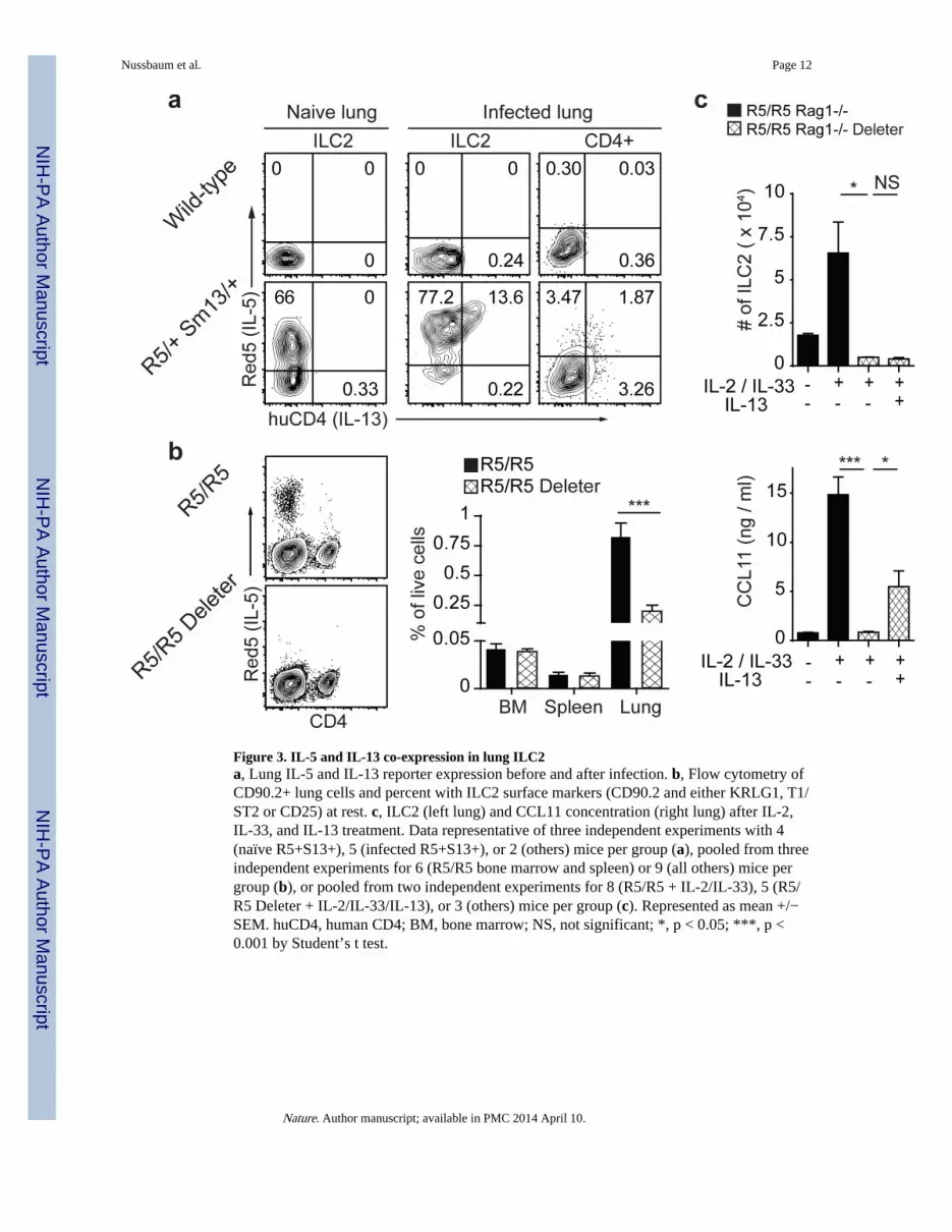
Figure 3. IL-5 and IL-13 co-expression in lung ILC2a, Lung IL-5 and IL-13 reporter expression before and after infection. b, Flow cytometry ofCD90.2+ lung cells and percent with ILC2 surface markers (CD90.2 and either KRLG1, T1/ST2 or CD25) at rest. c, ILC2 (left lung) and CCL11 concentration (right lung) after IL-2,IL-33, and IL-13 treatment. Data representative of three independent experiments with 4(naïve R5+S13+), 5 (infected R5+S13+), or 2 (others) mice per group (a), pooled from threeindependent experiments for 6 (R5/R5 bone marrow and spleen) or 9 (all others) mice pergroup (b), or pooled from two independent experiments for 8 (R5/R5 + IL-2/IL-33), 5 (R5/R5 Deleter + IL-2/IL-33/IL-13), or 3 (others) mice per group (c). Represented as mean +/−SEM. huCD4, human CD4; BM, bone marrow; NS, not significant; *, p < 0.05; ***, p <0.001 by Student’s t test.
Nussbaum et al. Page 12
Nature. Author manuscript; available in PMC 2014 April 10.
NIH
-PA Author Manuscript
NIH
-PA Author Manuscript
NIH
-PA Author Manuscript

Figure 4. ILC2 respond to circadian and metabolic cuesa-b, Serum IL-5 and blood eosinophils at 10:00, 22:00 or at 10:00 after fasting. c-d, Flowcytometry of small intestine ILC2 (Lin-CD127+ICOS+) and percent of R5-hi ILC2expressing S13 at 8:00 in mice on nighttime (black) or daytime (white) feeding (c) or infasted mice given food (black) or water (white) by oral gavage (d). e, Supernatant IL-5 fromintestinal Lin-CD45+KLRG1+ ILC2 cultured in IL-7 alone or with indicated reagents. f,Expression of Vpac1 and Vpac2 in sorted cells, relative to Rps17. Data pooled fromindependent experiments for 19 (AM), 6 (PM), or 5 (fasted) mice per group (a); 7 (AM), 4(PM), or 8 (fasted) mice per group (b); 8 mice per group (c); 6 mice per group (d); or pooled
Nussbaum et al. Page 13
Nature. Author manuscript; available in PMC 2014 April 10.
NIH
-PA Author Manuscript
NIH
-PA Author Manuscript
NIH
-PA Author Manuscript

averages of duplicate cultures from 6 (IL-7 alone, + VIP, + VPAC2 agonist) or 3 (all others)cell sorts from independent mice (e), or representative of two experiments of independentcell sorts (f). Represented as mean +/− SEM. Lin, Lineage markers (B220, CD11b, CD11c,Ly6G, FcεRI, and NK1.1); Rps17, 40S ribosomal protein S17; NS, not significant; *, p <0.05; **, p < 0.01; ***, p < 0.001 by Student’s t test.
Nussbaum et al. Page 14
Nature. Author manuscript; available in PMC 2014 April 10.
NIH
-PA Author Manuscript
NIH
-PA Author Manuscript
NIH
-PA Author Manuscript


