Embed Size (px)
Citation preview

FORUM ORIGINAL RESEARCH COMMUNICATION
Nitroso-Redox Status and Vascular Functionin Marginal and Severe Ascorbate Deficiency
Maria-Francisca Garcia-Saura,1 Fumito Saijo,1 Nathan S. Bryan,1 Selena Bauer,1
Juan Rodriguez,2 and Martin Feelisch1,3
Abstract
Marginal vitamin C (ascorbic acid) deficiency is a prevalent yet underappreciated risk factor for cardiovasculardisease. Along with glutathione, ascorbate plays important roles in antioxidant defense and redox signaling.Production of nitric oxide (NO) and reactive oxygen species and their interaction, giving rise to nitroso andnitrosyl product formation, are key components of the redox regulation/signaling network. Numerous in vitrostudies have demonstrated that these systems are interconnected via multiple chemical transformation reactions,but little is known about their dynamics and significance in vivo. Aims: We sought to investigate the time-courseof changes in NO/redox status and vascular function during ascorbate depletion in rats unable to synthesizevitamin C. Results: We here show that both redox and protein nitros(yl)ation status in blood and vital organsvary dynamically during development of ascorbate deficiency. Prolonged marginal ascorbate deficiency isassociated with cell/tissue-specific perturbations in ascorbate and glutathione redox and NO status. Scurvydevelops earlier in marginally deficient compared to adequately supplemented animals, with blunted com-pensatory NO production and a dissociation of biochemistry from clinical symptomology in the former. Para-doxically, aortic endothelial reactivity is enhanced rather than impaired, irrespective of ascorbate status.Innovation/Conclusion: Enhanced NO production and protein nitros(yl)ation are integral responses to the redoxstress of acute ascorbate deprivation. The elevated cardiovascular risk in marginal ascorbate deficiency is likelyto be associated with perturbations of NO/redox-sensitive signaling nodes unrelated to the regulation of vas-cular tone. This new model may have merit for the future study of redox-sensitive events in marginal ascorbatedeficiency. Antioxid. Redox Signal. 00, 000-000.
Introduction
Ascorbic acid (vitamin C) is the most potent water-soluble antioxidant in biological fluids and is widely
distributed across cells/tissues (36). At physiological pH, itexists largely in the form of its monovalent anion, ascorbate.In addition to its role as antioxidant, it is a cofactor of prolylhydroxylases and involved in several other cellular functions.Most mammals can synthesize ascorbate from glucose in apathway involving the enzyme L-gulonolactone oxidase (fora review on ascorbate biosynthesis, recycling and degrada-tion, see Ref. 26). Humans, along with primates, guinea pigs,and bats, lack a functional enzyme and therefore depend onan adequate dietary supply with this vitamin. Current re-commended dietary allowances are based on the absence ofobvious signs of deficiency, but this may be insufficient foroptimal health (12). Prolonged inadequate supply eventually
leads to depletion of bodily pools and development of scurvy,an almost forgotten disease historically known to affect sailorson long voyages.
Innovation
This is the first study that investigated the time-course ofchanges in NO/redox status in blood and tissues duringacute and marginal ascorbate deficiency in vivo. Our resultsindicate that NO is a key regulator of cellular redox ho-meostasis whose production and metabolism is intimatelylinked to local ascorbate status. The system is characterizedby a compensatory upregulation of NO production andprotein nitros(yl)ation as a response to the redox stresscaused by ascorbate deprivation, with a compromised re-activity following prolonged marginal deficiency.
1Whitaker Cardiovascular Institute, Boston University School of Medicine, Boston, Massachusetts.2Department of Physics, Centenary College of Louisiana, Shreveport, Louisiana.3Division of Vascular & Metabolic Health, University of Warwick Medical School, Coventry, United Kingdom.
ANTIOXIDANTS & REDOX SIGNALINGVolume 00, Number 00, 2012ª Mary Ann Liebert, Inc.DOI: 10.1089/ars.2011.4201
1
ARS-2011-4201-ver9-Garcia-Saura_1P
Type: Forum Original Research Communication
ARS-2011-4201-ver9-Garcia-Saura_1P.3d 02/21/12 3:28pm Page 1

In spite of occasional reports of its occurrence in recentyears (33, 49), scurvy has become rare in industrialized soci-eties. In contrast, marginal ascorbate deficiency, a conditioncaused by suboptimal vitamin C intake, is much more prev-alent. Estimates suggest that > 20% of the UK population and30%–40% of US adults are affected by subclinical ascorbatedeficiency (20, 35, 42, 51). Because this condition is neitherassociated with overt clinical symptoms nor routinely as-sessed, its health implications have long been underappreci-ated. However, low blood ascorbate levels are a strongpredictor of cardiovascular disease and all-cause mortality(10), and recent observations have linked ascorbate deficiencyto increased risk for cardiovascular (13, 14), respiratory (8),and metabolic disease (6, 39), warranting a re-evaluation of itsphysiological significance.
Several diseases linked to ascorbate deficiency are knownto be associated with endothelial dysfunction, an impairedvascular reactivity due to reduced formation and/or avail-ability of nitric oxide (NO). Endothelial dysfunction mayrepresent the vascular manifestation of systemic events af-fecting NO-related processes. NO is a pleiotropic cell signal-ing and effector molecule that controls vital bodily functionsacross all organ systems (32). NO is also an important anti-oxidant (50) and a key regulator of mitochondrial functionand redox signaling (40). Ascorbate has been shown to reverseendothelial dysfunction, but the underlying mechanism(s) ofthese effects remain unclear (30). The main mechanisms pro-posed include stimulation of nitric oxide synthase (NOS) ac-tivity by modulation of cofactor requirements, sparing ofintracellular thiols, and release of NO from circulating/tissuestorage forms. Irrespective of mechanism, these results areinconsistent with the reported lack of endothelial dysfunctionin ODS (Osteogenic, Disorder Shionogi) rats (45), a strainderived from the Wistar rat that carries a missense mutation ofL-gulonolactone oxidase, rendering these animals incapableof synthesizing ascorbate (23, 31). Interestingly, Gulo-/- mu-tant mice, a genetically engineered mouse lacking the sameenzyme, present with aortic wall damage on withdrawal ofascorbate supplementation (27), consistent with the associa-tion between low ascorbate levels and cardiovascular disease.
The notion that a perturbation of NO-dependent regulatoryredox circuits, coined the ‘‘nitroso-redox status’’ (16), maycontribute to cardiovascular disease has received consider-able attention lately. Together with glutathione, ascorbaterepresents the ‘‘heart of the redox hub’’ (11), and both anti-oxidants are linked to NO/nitrosation status via a multitudeof reactions. Studies in the 1970s identified ascorbate as a ni-trosation inhibitor (25), but more recent studies revealed thatit can also release NO from nitrosated proteins (21). Ascorbatealso reacts with nitrite (7) and plasma S-nitrosothiols (38) toproduce NO, and this reactivity is exploited for S-nitrosothioldetection using the ‘‘biotin switch’’ assay (18). With a fewexceptions (2, 22), many of these reactions have been studiedusing in vitro systems, and our knowledge about their sig-nificance in vivo is limited. Moreover, reaction dynamics tendto be complex, rendering network interrogation by a selectfew time points problematic. Reactions may also differ be-tween organs/compartments, depending on the expression oftransporters, local concentrations, and recycling mechanisms.Thus, investigations in relevant in vivo models, ideally withcomprehensive assessment of multiple organ systems, arerequired to put those reactions into context.
Much of the insight into the role of ascorbate in endothelialdysfunction has been gained using pharmacological doses;yet, the chemistry of such high ascorbate concentrations candiffer substantially from that prevailing at physiological lev-els (5). A complementary approach to studying the role ofascorbate in physiology uses inhibition of its production. Astandardized technique for inducing chronic latent ascorbatedeficiency has been developed in guinea pigs (14). However,the majority of published research has been carried out in ratsand mice, hampering direct comparison of results due topossible species differences. In the present study we usedODS rats, in combination with bioanalytical and functionaltechniques, to assess the nitroso-redox status in blood andtissue and investigate mechanisms of acute and chronicascorbate depletion. Using this integrated approach, wedemonstrate a complex pattern of compartmentalized re-sponses that suggest that NO upregulation is a crucial re-sponse to redox stress. This response is blunted followingprolonged marginal ascorbate deficiency and associated withperturbations in tissue NO/redox tone. Surprisingly, thesechanges do not impair the regulation of vascular tone.
Results
Changes in NO/redox status in response to acuteascorbate depletion in optimally supplemented animals
To study the effects of acute ascorbate depletion on bloodand tissue NO/redox status in animals adequately suppliedwith ascorbate before, ODS rats were placed on a diet lower invitamin C and regular tap water and monitored over thesubsequent 2 months. Blood and tissues were harvested justbefore switching from full supplementation to depletion(t = 0), at the end of weeks 1 and 2, and on appearance of overtsigns of scurvy (weeks 6–7). As shown in b F1Figure 1a, a rapidascorbate decline was apparent in most compartments withinthe first 2 weeks. This was accompanied by changes in totalglutathione in which each data point for every organ roseabove baseline at weeks 1 and 2, peaking at week 2 (Fig. 1c). Asimilar increase was observed in the GSH/GSSG ratio duringthat time (Fig. 1d). Although the glutathione data (n = 2 foreach tissue) may be insufficient to assess the significance of thechanges in individual organs, the consistency in direction ofthe change in all compartments allows us to state that sig-nificant global increases in glutathione content and redoxstatus took place within the first 2 weeks. These changes wereaccompanied by a dramatic yet transient improvement inascorbate redox (AA/DHA ratio) at week 1 (Fig. 1b). Thisincrease in AA/DHA ratio was mirrored by increases in theconcentrations of nitrite (Fig. 1e), nitrate (Fig. 1f), and totalnitroso (RXNO) and nitrosyl (NO-heme) products (Figs. 1gand 1h) in several compartments. By week 7, total ascorbateand AA/DHA ratios were significantly reduced in mostcompartments (Figs. 1a and 1b), while total glutathione andGSH/GSSG ratios returned to normal levels. Conversely, NO-related products (in particular, nitrite/nitrate in blood andtissue nitroso/nitrosyl species) showed dramatic elevations inmost compartments investigated (Figs. 1e–1h). These resultssuggest a compensatory increase in tissue antioxidant statusin response to progressively decreasing ascorbate levels, al-though this does not appear to be sustainable for longer pe-riods. NO production may be upregulated, as indicated by themassive increase in NO-related metabolites, to substitute for
2 GARCIA-SAURA ET AL.
ARS-2011-4201-ver9-Garcia-Saura_1P.3d 02/21/12 3:28pm Page 2

a b
c d
e f
g h
FIG. 1. Time dependence
4C c
of the effects of acute ascorbate depletion on NO/redox status in blood and tissues of ODSrats. Panels depict steady-state concentrations and corresponding redox status of (a) total ascorbate and (b) ratio of reduced(AA) over oxidized ascorbate (DHA); (c) total glutathione, and (d) ratio of reduced (GSH) over oxidized glutathione (GSSG);and concentrations of (e) nitrite, (f) nitrate, (g) total nitrosation (RXNO), and (h) heme nitrosylation products (NO-heme)immediately before (time = 0), and 1, 2, and 7 weeks after start of ascorbate depletion. Means – SEM of n = 3–5 animals/timepoint for ascorbate and NO measurements, and means of n = 2 animals/time point—normalized to control values—forglutathione determinations, with * denoting significance at p < 0.05 for the compartments indicated.
ARS-2011-4201-ver9-Garcia-Saura_1P.3d 02/21/12 3:28pm Page 3
3

the lower availability of classical antioxidants at later stages ofascorbate deficiency. These data also demonstrate that re-sponses are not uniform across organs but highly compart-mentalized.
NO/redox status during development of marginalascorbate deficiency
A subset of ODS rats was kept on a standard rodent chowcontaining ascorbate in amounts just sufficient to prevent thedevelopment of visible signs of scurvy (group 2). Directcomparison of controls on full supplementation to animalswith a suboptimal ascorbate supply revealed a significantdecline in ascorbate across most compartments (F2 c Fig. 2). Afterstabilization of ascorbate levels (around day 28), a similar andoften more reduced redox state was maintained compared tothe beginning (Fig. 2d), suggesting the involvement of com-pensatory mechanisms to maintain tissue redox status. Incontrast to the acute depletion study, NO-related metaboliteswere maintained within a rather tight concentrations range,with no further increases over time. In fact, several compart-ments exhibited significant decreases during this time (Figs.2g and 2h).
Optimal time window for study of marginalascorbate deficiency
Since none of the animals within group 2 revealed anyscorbutic signs within the first 6 weeks, we extended the ob-servation period to see what might happen over the followingmonths while animals were kept marginally ascorbate defi-cient. Animals were sacrificed at regular intervals for blood/tissue analysis. No further ascorbate loss or major changes inNO metabolites were observed in the majority of animals(data not shown). However, 25% of rats (5 out of 20) keptunder these conditions spontaneously developed scurvy be-tween months 5 and 7, narrowing the window of opportunityfor future studies of marginal ascorbate deficiency using thismodel to about 6 months. No sentinels optimally supple-mented with ascorbate developed clinical symptoms at anypoint of the study.
NO/redox status on acute ascorbate depletionfollowing prolonged marginal deficiency
After 7 months of asymptomatic marginal ascorbate defi-ciency, a subset of animals were subjected to completeascorbate deprivation (group 3) in order to compare the bio-chemical changes associated with scurvy development afterprolonged marginal deficiency to those of acute depletion inadequately supplemented animals (group 1). We hypothe-sized that animals maintained under suboptimal conditions(group 2) would develop scurvy faster than those optimallysupplemented with ascorbate. As in the acute study, animalswere sacrificed after 1 and 2 weeks, and following the ap-pearance of overt signs of scurvy.
The most obvious difference between animals of groups 1and 3 relates to the time required to develop scurvy. While inoptimally supplemented animals it took up to 7 weeks toreach a scorbutic score that justified sacrifice, this period wasreduced to only 4 weeks in marginally deficient animals. In-deed, 90% of the animals showed first signs of scurvy alreadyafter 1 week. In week 2 about half of the animals developed
severe symptoms, necessitating sacrifice of the entire groupbetween weeks 3 and 4 to avoid unnecessary suffering ( b F3Fig. 3).While animals in the acute study showed a gradual depletionin ascorbate over approximately 3 weeks, tissue ascorbatelevels in marginally deficient rats now exhibited changes(compare Fig. 1a with b F4Fig. 4a). As seen before, plasma con-tinued to experience a significant increase in total ascorbate.Except for the brain, absolute levels of ascorbate in marginallydeficient animals at the beginning of the depletion experimentwere comparable to those in animals of group 1 after 2–3weeks of acute ascorbate deprivation. Yet, signs of scurvybecame apparent almost immediately in the former (com-pared to *2 more weeks to develop mild to severe symptomsin optimally supplemented rats), indicative of a dissociationbetween tissue biochemistry and clinical symptomology.
Several other differences in outcome between these twogroups are worth mentioning. The transient increase inascorbate redox status seen in the first week of the acute studywas not observed in most tissues after prolonged marginaldeficiency (Fig. 4b). Moreover, nitrite levels experienced adrop rather than an increase in the first week of total ascorbatedeprivation, followed by a rebound in nitrite and nitrate inthe second week, to again drop near death. These changes arealmost the mirror image of what was observed in the acutestudy, suggesting tissues are struggling to adapt to the newsituation, perhaps due to differences in NO system operation.Differences do not only relate to the time-course of biochem-ical changes but also to response magnitudes, consistent witha lower regulatory reserve. At the time of sacrifice of opti-mally supplemented animals, the NO system seems to besystemically upregulated, resulting in a massive rebound inNO metabolite concentrations (43-fold increase in steady-state levels of nitrite + nitrate (NOx) in blood) ( b F5Figs. 5a and5b); in marginally deficient animals at the same point, thisincrease was not nearly as spectacular (6-fold increase forNOx in blood), although starting levels were comparable inmost tissues (Figs. 5c and 5d). This observation is consistentwith a specific impairment of the nitroso-redox system inmarginal ascorbate deficiency which translates into a com-promised response to further redox stress. Of note, the aortadiffered from this general pattern of responses, showing amarked up-regulation of nitrosation near the study end (Fig.5d), although this difference did not reach statistical signifi-cance. NO-heme levels in blood and tissues showed onlyminor variations (Fig. 4e), suggesting changes in overall NOavailability remained moderate. This contrasts with the re-sults obtained on acute ascorbate depletion in optimallysupplemented animals, where production (indicated by NOxformation) and availability of NO (indicated by NO-hemelevels in tissues and erythrocytes) were markedly upregu-lated (Figs. 1e, 1f, and 1h).
NO/redox status on ascorbate repletion
To test the reversibility of alterations in nitroso-redox statusat advanced stages of deficiency, a subset of animals kept for 7months marginally ascorbate deficient and subjected to fullascorbate deprivation thereafter (scorbutic score ‡ 14) wererepleted with vitamin C via their drinking water and followedfor another 3 weeks. Most (* 70%) animals recovered withindays of repletion, starting to gain weight with rapid behav-ioral improvements and gradual disappearance of scurvy
4 GARCIA-SAURA ET AL.
ARS-2011-4201-ver9-Garcia-Saura_1P.3d 02/21/12 3:28pm Page 4

a b
c d
e f
g h
FIG. 2. Development of mar-ginal ascorbate deficiency andeffects on NO/redox status inblood and
4C c
tissues of ODS rats.Time course of changes in (a, b)total ascorbate content; (c, d)ascorbate redox status, expressedas ratio of reduced (AA) over oxi-dized (DHA); (e, f) nitrite concen-tration; and (g, h) concentration oftotal nitros(yl)ation products(nitrosothiols + nitrosamines + NO-heme species). Left panels depictchanges in blood, right panelsthose in tissues. Means – SEM ofn = 3–5 animals/time point;*p < 0.05.
NO/REDOX STATUS IN VITAMIN C DEFICIENCY 5
ARS-2011-4201-ver9-Garcia-Saura_1P.3d 02/21/12 3:28pm Page 5

symptoms. As depicted inF6 c Figure 6a, tissues fully recoveredto their pre-depleted ascorbate levels. This was accompaniedby a strikingly enhanced AA/DHA redox ratio in all tissuesexcept the aorta (Fig. 6b), perhaps a result of improved re-cycling, and a normalization of NOx levels in all compart-ments (Fig. 6c). The latter suggests the increase in NOproduction in scorbutic animals was due to an upregulation ofconstitutive NOS activity rather than expression of inducibleNOS (which might be expected to further increase on ascor-bate repletion).
Changes in vascular function in responseto ascorbate alterations
As alluded to earlier, the aorta is the only compartmentbesides the brain that follows a trend for biochemical changesdistinct from that in other tissues. This observation, alongwith puzzling reports about a lack of endothelial dysfunctionin these animals (45), prompted us to investigate possiblechanges in vascular function in response to variations inascorbate levels.F7 c Figure 7a depicts an original tracing of theprotocol used for assessment of endothelium-dependent andindependent vascular responses (see Methods for details),with quantitative data depicted below. Control experimentsconfirmed the lack of endothelial dysfunction in ODS ratsoptimally supplemented with ascorbate (Fig. 7b). In fact, en-dothelial vascular reactivity of ODS rats was superior to thatof age-matched Wistar rats (pD2: 8.2 – 0.02 versus 6.94 – 0.01in ODS and Wistar rats, respectively). In contrast, vasorelax-ant responses to the endothelium-independent NO-donor,DEA/NO were comparable between both strains (Fig. 7c),indicating a specific enhancement of vascular endothelialreactivity. Unexpectedly, marginally ascorbate deficient ani-
mals retained their improved vascular endothelial reactivityand revealed no sign of alteration in endothelium-independentrelaxation to NO. Both potency and maximal relaxation toAch were greatly improved in ODS rats rendered marginallydeficient in ascorbate compared to age-matched Wistar rats(pD2: 8.13 – 0.04 vs. 6.94 – 0.01 in ODS and Wistar rats, re-spectively; maximal relaxation: 89 – 1% in ODS, 80 – 0.8%in Wistar controls; Figs. 7b and 7c). Remarkably, vascularfunction remained unimpaired even after induction of scurvyfollowing prolonged marginal deficiency. While calculatedpD2 values (a measure of vasorelaxant potency) for Ach werenearly identical in all ODS treatment groups, maximal relax-ation responses to Ach were significantly impaired in overtscurvy (71 – 3%; p = 0.0004 vs. optimally supplemented ODScontrols, p < 0.0001 vs. marginally ascorbate deficient rats),possibly secondary to enhanced oxidative stress. In conclu-sion, ODS rats show improved NO-dependent endothelialreactivity compared to control animals corresponding to theirgenetic background (Wistar rats), and this is independent oftheir ascorbate status. The lack of difference in NO-dependentbut endothelium-independent vasorelaxation clearly demon-strates that this improved vascular reactivity is not secondaryto enhanced downstream processes mediating vasorelaxa-tion, but the result of increased eNOS activity. This assump-tion is further corroborated by higher levels of basal NOrelease in ODS compared to Wistar rats, as evidenced by thedifference in maximal contractile tone following NOS inhibi-tion (Fig. 7c, inset).
Discussion
The key findings of the current study are that i) ascorbatedeficiency is associated with characteristic changes in NOproduction and metabolism, accompanied by a shift towardsa more reduced redox state; ii) the effects of acute ascorbatedepletion on cellular redox/nitros(yl)ation status differ be-tween well-supplemented and marginally deficient animals,with faster scurvy development in the latter; iii) changes inNO/redox status are complex and highly compartmenta-lized; iv) NO responses are blunted and dissociated fromphenotypical changes in marginal ascorbate deficiency; and v)vascular endothelial dilatation remains largely unaffected byascorbate deficiency. To the best of our knowledge, ours is thefirst study to comprehensively assess the dynamics and definethe role of ascorbate in modulating cellular redox status andblood and tissue nitros(yl)ation status in vivo. We believe thisnew rodent model has merit for the future study of redoxsignaling mechanisms related to ascorbate deficiency.
Marginal ascorbate deficiency is a growing problem inmost contemporary societies due to lifestyle-related changesin dietary habits with a lower than desirable intake of fruitsand vegetables (further compounded by physical inactivityand psychosocial stress). Marginal ascorbate deficiency isassociated with fatigue, increased prevalence for chronicdiseases, and a higher risk of developing scurvy (19). Thelatter is consistent with our finding that scurvy developedfaster in marginally deficient rats. That chronic latent ascor-bate deficiency is associated with changes distinct from thoseof acutely scorbutic animals is not new (41), but we are thefirst to report that those differences are associated with spe-cific alteration in NO production and metabolism. These re-sults may be relevant to subclinical micronutrient deficiency
FIG. 3. Scurvy
4C c
development following acute ascorbatedepletion in ODS rats with previously optimal vitamin Csupply compared to animals after prolonged marginal de-ficiency. Minimum score: 1 (no symptoms), maximum score:19 (overt scurvy). Animals were sacrificed at scores between14 and 17 for ethical reasons, dependent on severity of thesymptoms (see Methods for details). Each time point repre-sents the average – SEM of individual animal scores calcu-lated weekly (n = 4–20).
6 GARCIA-SAURA ET AL.
ARS-2011-4201-ver9-Garcia-Saura_1P.3d 02/21/12 3:28pm Page 6
in humans, particularly in the pediatric population and theelderly where unspecific ascorbate deficiency symptoms areeasily mistaken for something else. Moreover, while immi-nently treatable once recognized, the amelioration of symp-toms may not always lead to full restoration of health. Ourdata suggest that an adequate NO production combined witha change in NO metabolism are key elements in the counter-regulatory response to acute and chronic ascorbate deficiency,but there is a paucity of information on what other redox-sensitive processes might be affected in addition. Moreover,our results suggest a dissociation of tissue biochemistry andclinical symptomology at a stage of ongoing redox perturba-tion the long-term consequences of which remain unexplored.The relationship between low ascorbate levels and all-causemortality (10) suggests some of these changes are crucial forsurvival. The observation that endothelial vasoreactivity re-mains largely unaffected by ascorbate depletion suggest that
regulation of vascular tone (and thus convective oxygen andnutrient delivery) are unlikely to be involved. An alternativepossibility is that endothelial function is so vital that it ismaintained irrespective of the level of ascorbate supply. Thisinterpretation would be consistent with the finding that NOmetabolite levels are highest in overt illness.
Glutathione and NO as first-line of defense againstacute ascorbate depletion
In the acute study group, animals fully deprived of ascor-bate developed scurvy in the same fashion as originally de-scribed (31), with hindlimb disorders appearing after 3 weeks,body weight gain arrest at week 4, and death ensuing after 7–8weeks. These symptoms were accompanied by specificchanges in NO/redox status. Ascorbate depletion was asso-ciated with a compensatory increase in total glutathione
a b
c d
e f
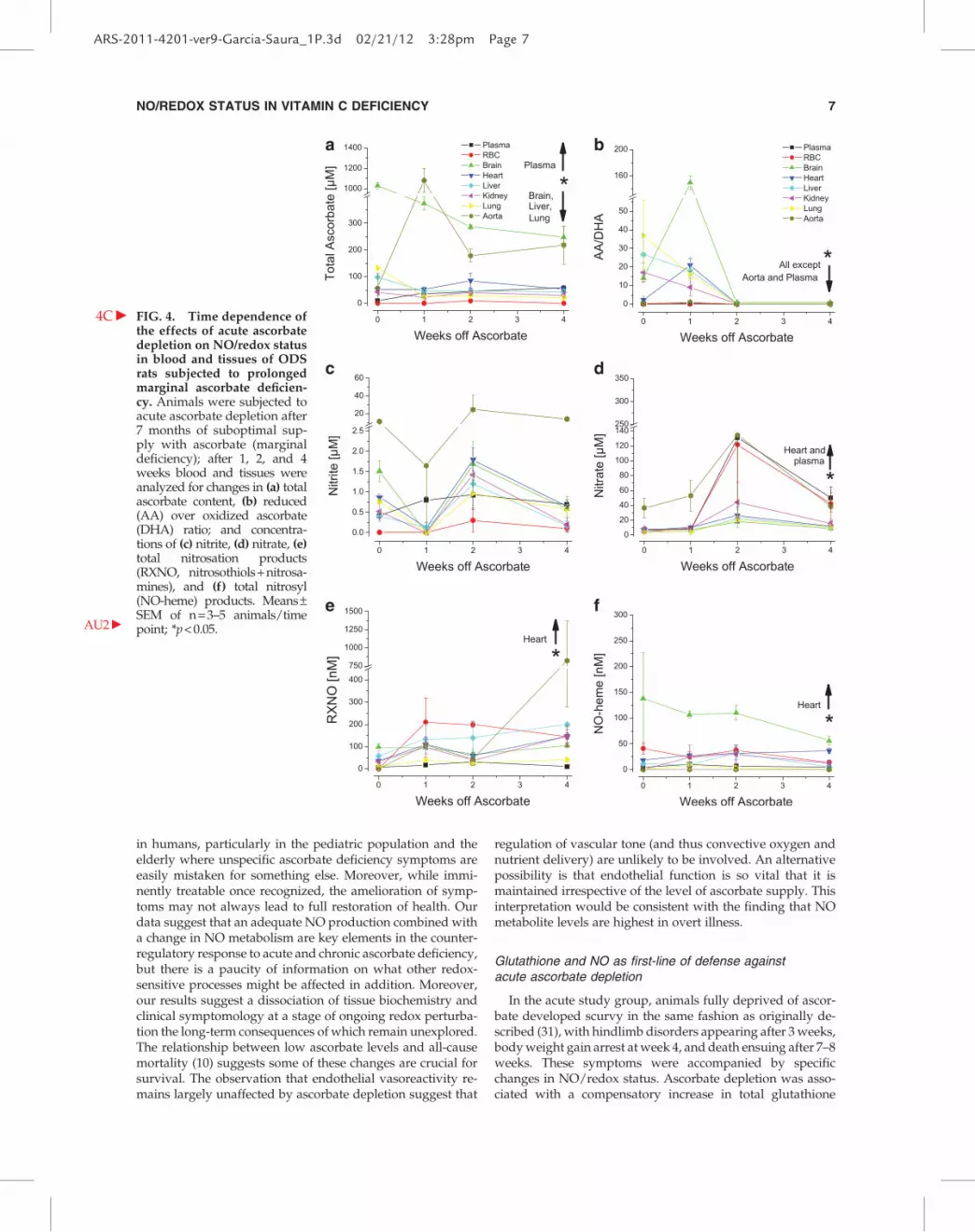
FIG. 4. Time dependence ofthe effects of acute ascorbatedepletion on NO/redox statusin blood and tissues of ODSrats subjected to prolongedmarginal ascorbate deficien-cy. Animals were
4C c
subjected toacute ascorbate depletion after7 months of suboptimal sup-ply with ascorbate (marginaldeficiency); after 1, 2, and 4weeks blood and tissues wereanalyzed for changes in (a) totalascorbate content, (b) reduced(AA) over oxidized ascorbate(DHA) ratio; and concentra-tions of (c) nitrite, (d) nitrate, (e)total nitrosation products(RXNO, nitrosothiols + nitrosa-mines), and (f) total nitrosyl(NO-heme) products. Means–SEM of n = 3–5 animals/timepoint; *p < 0.05.AU2 c
NO/REDOX STATUS IN VITAMIN C DEFICIENCY 7
ARS-2011-4201-ver9-Garcia-Saura_1P.3d 02/21/12 3:28pm Page 7

a b
c d
FIG. 5. Comparison of mag-nitudes in NO-related re-sponses between symptom-freeand overtly scorbutic animalsin adequately supplemented(a, b) and marginally deficientODS rats (c, d). Bar charts de-picting steady-state concentra-tions in blood and tissues of (a)NOx (nitrite + nitrate) and (b)total nitros(yl)ation products(nitrosothiols + nitrosamines +NO-heme species) in animalsduring acute induction ofscurvy (no symptoms = day 0;overt scurvy = 6–7 weeks afterfull ascorbate depletion); (c)NOx and (d) total ni-tros(yl)ation products in ani-mals with marginal ascorbatedeficiency and subsequent in-duction of scurvy (no symp-toms = day 0; overt scurvy = 3–4weeks of full ascorbate deple-tion). Means – SEM of n = 3–5animals/group); *p < 0.05.
a b
c d
FIG. 6. Effects of manipulations in ascorbate supply on nitroso-redox status in blood and tissues of ODS rats. Bar chartsdepicting steady-state concentrations of (a) total ascorbate, (b) reduced (AA) over oxidized ascorbate (DHA) ratio, (c) NOx(nitrite + nitrate), and (d) total nitros(yl)ation products (nitrosothiols + nitrosamines + NO-heme species). Open bars: ODS ratssupplemented with 1 mg/mL ascorbic acid in the drinking water. Light gray bars: ODS rats with marginal ascorbate deficiency(symptom-free). Gray bars: ODS rats with marginal ascorbate deficiency completely deprived of ascorbate for 4 weeks(average scorbutic score: 14). Dark gray bars: ODS rats with marginal ascorbate deficiency, completely deprived of ascorbatefor 4 weeks and then replenished with 1 mg/mL ascorbate with the drinking water for another 3 weeks (at which timeaverage scorbutic scores returned to values within healthy limits). Means – SEM of n = 3–5 animals/group; *p < 0.05.
8 GARCIA-SAURA ET AL.
ARS-2011-4201-ver9-Garcia-Saura_1P.3d 02/21/12 3:28pm Page 8

concentration and a shift towards a more reduced redox state.These results are consistent with the counter-regulatory na-ture of responses described by Wang et al. (48) and the notionthat ascorbate and glutathione form an inter-connected anti-oxidant network (28); they are also in line with the observationthat ODS rats are unable to compensate the oxidative stresselicited by inhibition of glutathione production (37). It is ap-parent from our results that the adjustments in glutathionemetabolism alone are not sufficient, as indicated by the con-
comitant increase in NO production during the same period.More importantly, glutathione redox changes cannot be sus-tained for long, and tissues become gradually more oxidizedagain. It appears whether the NO system compensates for theascorbate loss, in particular as the deficiency becomes moresevere. This interpretation is in agreement with the observationthat ODS rats develop alternative antioxidative defenses asascorbate level vanish (46). Our data would seem to suggest thatthis alternative defense system involves the production of NO.
FIG. 7. ODS rats exert improved vascular endothelial reactivity compared to normal Wistar rats, irrespective ofascorbate status. (a) Representative original tracing of organ bath experiments on the relaxation to acetylcholine (Ach) andDEA/NO in endothelium-intact thoracic aortic rings of ODS rats. (b) Endothelium-dependent relaxation induced by in-creasing concentrations of Ach in phenylephrine (PE, 0.2 lM) pre-contracted vascular rings, expressed as percentage ofcontractile tone induced by the first PE stimulus (PE-1). (c) Endothelium-independent relaxation induced by the NO-donor,DEA/NO in aortas washed and re-contracted with PE (PE-2), following addition of the NOS inhibitor L-NAME (100 lM).Results are expressed as percentage of contraction induced by PE + L-NAME. Inset: Contraction induced by L-NAME after PEre-contraction in Wistar rats vs. ODS rats fully supplemented with ascorbic acid; results are expressed as percentage ofcontraction elicited by first PE stimulus. Means – SEM of a minimum of two-paired rings from 3–6 individual animals. Groups:, age matched Wistar controls, C ODS rats supplemented with 1 mg/mL ascorbic acid in drinking water, : ODS rats withmarginal ascorbate deficiency, * ODS rats with marginal ascorbate deficiency subjected to acute ascorbate depletion (overtscurvy). Endothelium-dependent relaxant responses to Ach in ODS rats were significantly different from those in Wistar ratsunder all conditions ( p < 0.001); maximal vasorelaxant responses in overt scurvy also differed from those in fully supple-mented and marginally deficient ODS rats (see text for details).
NO/REDOX STATUS IN VITAMIN C DEFICIENCY 9
ARS-2011-4201-ver9-Garcia-Saura_1P.3d 02/21/12 3:28pm Page 9
NO responses differ in dependence of ascorbatesupplementation status
The consequences of acute ascorbate depletion differ be-tween optimally supplemented and marginally deficient ani-mals. Either group experiences an early upregulation of theNO system that coincides with the initial drop in ascorbate.While in marginal ascorbate deficiency levels of NO-relatedmetabolites and ascorbate tend to stabilize thereafter, NO up-regulation in optimally supplemented animals then translatesinto gradual increases in NO metabolites, which is most pro-nounced towards the study end. These findings are consistentwith earlier reports that nitrate levels in plasma and urine ofascorbate-deficient ODS rats are higher than in fully supple-mented animals (44). The concomitant increase in tissuenitros(yl)ation status, which is normally maintained withinrather tight limits (2) and may also rise as a result of the loweravailability of ascorbate to act as nitrosation scavenger, mayserve to protect critical protein sulfhydryl and tryptophangroups from irreversible oxidation. The same adjustments seemto prevent animals from developing scurvy during marginalascorbate deficiency. However, although ascorbate and NO/redox levels do not seem to deteriorate further, more subtlealterations of the NO system may render animals more sus-ceptible to scurvy development. In fact, 25% of animals sub-jected to prolonged marginal ascorbate deficiency developedscurvy without further reduction in supply, limiting the com-plication-free period for study of marginal ascorbate deficiencyusing this model to 5–6 months. When marginally deficientanimals were subjected to further depletion, the increases in NOmetabolite levels were not as obvious as under optimal sup-plementation, suggesting NO-dependent responses are com-promised following chronic marginal ascorbate deficiency.
Is increased NO production due to enhancedendothelial NOS activity?
While a quantification of NOS isoform expression profilesin different tissues was beyond the scope of the present study,our vascular function studies suggest that the increase in NO-related products observed in response to any form of ascor-bate deficiency is related to an upregulation of eNOS activityand/or altered cofactor association. This is at variance withmuch of the information gained from vitamin C supplemen-tation studies. If high doses of ascorbate augment cardiacfunction, confer cardioprotection, and enhance endothelium-dependent vasorelaxation (3), then its depletion might beexpected to cause the opposite. However, Vergely et al. (45)had reported that ODS rats reveal improved vascular reac-tivity compared to Wistar controls. We here confirm and ex-tend those results to show that endothelial reactivity isenhanced regardless of ascorbate supplementation status andassociated with an increase in basal and stimulated endothe-lial NO production without changes in sensitivity of the NOreceptor, soluble guanylyl cyclase, or downstream processesof vasorelaxation. Future studies should clarify whether this isa feature of this particular rat strain or perhaps a consequenceof ascorbate deficiency during embryonic development.
Is nitrite linked to ascorbate oxidation?
Following prolonged marginal deficiency, the develop-ment of overt signs of scurvy did neither seem to depend on
massive further ascorbate loss from a specific bodily com-partment nor did the drop in plasma ascorbate correlate withscurvy development, raising concern about the predictivepower of predefined concentration thresholds for ascorbate.Whether this reflects the crucial importance of a minimalbodily pool of ascorbate and is associated with alterna-tive pathways of ascorbate production independent of L-gulonolactone oxidase activity warrants further study.
An intriguing association between nitrite concentrationchanges and ascorbate oxidation status may be gleaned froma comparison of NO/redox status between tissues. Theseobservations may be a result of direct ascorbate oxidation bynitrite (6) and accompanied by the release of NO. The increasein NO-related metabolites in conjunction with a more reducedredox state observed in the first week of the acute study wasabsent in the majority of tissues following marginal ascorbatedeficiency. In fact, a dramatic drop in nitrite and AA/DHAratio was apparent instead. Likewise, during the developmentof scurvy in marginally deficient animals, AA/DHA ratiosdropped whenever nitrite levels fell. Brain and aorta werenotable exceptions to this rule, perhaps due to a heightenedreliance on ascorbate, which may provide an explanation forthe finding that levels in these tissues never drop below300 lM. Indeed, the aorta was the only organ that respondedwith an increase in ascorbate in the first week of acute de-pletion, demonstrating a distinct pattern of regulation in thevasculature compared to the rest of the body. These obser-vations are reminiscent of changes in tissue nitrite in the samecompartments, which follow trends opposite to all other or-gans (1), and may reflect a re-prioritization of tissue ascorbatepools. Whether these differences are due to tissue-specificvariations in ascorbate transporter expression or perhaps theirsusceptibility to redox regulation is unknown.
Antioxidant rebound following ascorbate repletion
Approximately 70% of animals suffering from advancedascorbate deficiency recovered upon repletion with vitaminC. This percentage of recovery is comparable to that seen inhuman studies (33). Even though NO-related metabolite lev-els had returned to control levels following repletion, tissueredox status tended to remain more reduced, consistent withearlier studies in guinea pigs that provided evidence for anantioxidant rebound in preparation for future oxidativechallenges (28).
Conclusions and Future Directions
Our results reveal a close relationship between the ascorbateand the NO system that is characterized by a compensatoryupregulation of NO production and protein nitros(yl)ation as aresponse to the redox stress of acute ascorbate deprivation, witha compromised reactivity following chronic marginal ascorbatedeficiency. The difference in response between acute ascorbatedepletion and chronic marginal deficiency is likely a conse-quence of adaptive changes to a prolonged inadequate supplywith vitamin C, consistent with changes in nutrient handlingupon prolonged alterations in intake (17). We believe that aparallel assessment of global protein nitrosation and tissue redoxstatus across multiple organ systems in vivo is key to gaining adeeper understanding of redox signaling/regulation; this maynot require a certain balance to be maintained for optimalfunctioning, as originally put forward as part of the ‘‘nitroso
10 GARCIA-SAURA ET AL.
ARS-2011-4201-ver9-Garcia-Saura_1P.3d 02/21/12 3:29pm Page 10
redox’’ concept (16). The current study did not address whichbiological targets are affected most by the variations in NO/redox status. Scurvy is associated with compromised collagenproduction due to the role of ascorbate as cofactor of prolylhydroxylase, and activity of the latter may be further inhibitedby enhanced NO production; perturbation of HIF-1a hydroxyl-ation with consecutive alterations in hypoxic signaling is anotherobvious possibility. Future studies are warranted to investigatethe possible involvement of other redox-sensitive signalingpathways affected by prolonged marginal ascorbate deficiency.
Methods
Materials, animals, diets, and assignmentto experimental groups
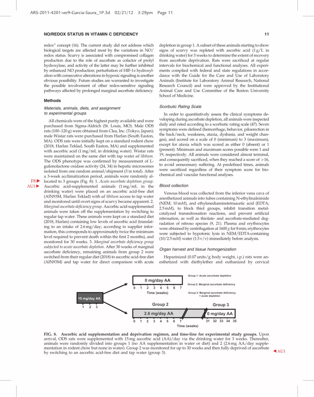
All chemicals were of the highest purity available and werepurchased from Sigma-Aldrich (St. Louis, MO). Male ODSrats (100–120 g) were obtained from Clea, Inc. (Tokyo, Japan);male Wistar rats were purchased from Harlan (South Easton,MA). ODS rats were initially kept on a standard rodent chow(2018, Harlan Teklad, South Easton, MA) and supplementedwith ascorbic acid (1 mg/mL in drinking water). Wistar ratswere maintained on the same diet with tap water ad libitum.The ODS phenotype was confirmed by measurement of L-gulonolactone oxidase activity (24, 34) in hepatic microsomesisolated from one random animal/shipment (3 in total). Aftera 3-week acclimatization period, animals were randomly al-located to 3 groups (F8 c Fig. 8): 1. Acute ascorbate depletion group.AscorbicAU1 c acid-supplemented animals (1 mg/mL in thedrinking water) were placed on an ascorbic acid-free diet(AIN93M, Harlan Teklad) with ad libitum access to tap waterand monitored until overt signs of scurvy became apparent. 2.Marginal ascorbate deficiency group. Ascorbic acid-supplementedanimals were taken off the supplementation by switching toregular tap water. These animals were kept on a standard diet(2018, Harlan) containing low levels of ascorbic acid (translat-ing to an intake of 2.6 mg/day; according to supplier infor-mation, this corresponds to approximately twice the minimumlevel required to prevent death within the first 2 months), andmonitored for 30 weeks. 3. Marginal ascorbate deficiency groupsubjected to acute ascorbate depletion. After 30 weeks of marginalascorbate deficiency, remaining animals from group 2 wereswitched from their regular diet (2018) to ascorbic acid-free diet(AIN93M) and tap water for direct comparison with acute
depletion in group 1. A subset of these animals starting to showsigns of scurvy was repleted with ascorbic acid (1 g/L indrinking water) for 3 weeks to determine the extent of recoveryfrom ascorbate deprivation. Rats were sacrificed at regularintervals for biochemical and functional analyses. All experi-ments complied with federal and state regulations in accor-dance with the Guide for the Care and Use of LaboratoryAnimals (Institute for Laboratory Animal Research, NationalResearch Council) and were approved by the InstitutionalAnimal Care and Use Committee of the Boston UniversitySchool of Medicine.
Scorbutic Rating Scale
In order to quantitatively assess the clinical symptoms de-veloping during ascorbate depletion, all animals were inspecteddaily and rated according to a scorbutic rating scale (47). Sevensymptoms were defined (hemorrhage, behavior, piloerection inthe back/neck, weakness, ataxia, dysbasia, and weight chan-ges), and scored on a scale of 0 (minimum) to 3 (maximum),except for ataxia which was scored as either 0 (absent) or 1(present). Minimum and maximum scores possible were 1 and19, respectively. All animals were considered almost terminal,and consequently sacrificed, when they reached a score of > 16,to avoid unnecessary suffering. At predefined times, animalswere sacrificed regardless of their symptom score for bio-chemical and vascular functional analyses.
Blood collection
Venous blood was collected from the inferior vena cava ofanesthetized animals into tubes containing N-ethylmaleimide(NEM; 10 mM), and ethylenediaminetetraacetic acid (EDTA;2.5 mM), to block thiol groups, inhibit transition metal-catalyzed transnitrosation reactions, and prevent artificialnitrosation, as well as thiolate- and ascorbate-mediated deg-radation of nitroso species (9, 21). Plasma and erythrocyteswere obtained by centrifugation at 1600 g for 8 min; erythrocyteswere subjected to hypotonic lysis in NEM/EDTA-containing(10/2.5 mM) water (1:3 v/v) immediately before analysis.
Organ harvest and tissue homogenization
Heparinized (0.07 units/g body weight, i.p.) rats were an-esthetized with diethylether and euthanized by cervical
FIG. 8. Ascorbic acid supplementation and deprivation regimen, and time-line for experimental study groups. Uponarrival, ODS rats were supplemented with 15 mg ascorbic acid (AA)/day via the drinking water for 3 weeks. Thereafter,animals were randomly divided into groups 1 (no AA supplementation in water or diet) and 2 (2.6 mg AA/day supple-mentation in rodent chow but none in water). Group 2 was monitored for up to 30 weeks and then fully deprived of ascorbateby switching to an ascorbic acid-free diet and tap water (group 3). b AU1
NO/REDOX STATUS IN VITAMIN C DEFICIENCY 11
ARS-2011-4201-ver9-Garcia-Saura_1P.3d 02/21/12 3:29pm Page 11

dislocation. After thoracotomy, a catheter was inserted intothe infrarenal part of the abdominal aorta, and organs wereflushed free of blood by retrograde in situ perfusion withair-equilibrated, NEM/EDTA (10/2.5 mM) supplementedphosphate buffered saline (PBS) at a rate of 10 ml/min, im-mediately followed by tissue harvest and homogenization (2).For glutathione measurements, separate animals from thesame treatment group/stage were used in which tissue per-fusion and homogenization was carried out using EDTA-containing buffer without NEM. All steps were carried outunder reduced ambient light to prevent photolytic decom-position of nitroso products.
Determination of tissue nitroso/nitrosyl and nitrite/nitrate concentrations
Tissue nitroso/nitrosyl compounds were quantified usinggroup-specific reductive denitrosation by iodine-iodide withsubsequent detection of the NO liberated by gas-phase che-miluminescence (9). Given that the identity of most nitrosospecies in the various biological compartments examinedremains unknown, the concentrations of S- and N-nitrosospecies (nitrosothiols and nitrosamines, respectively) are re-ported as total nitroso species (RXNO) in the present study.NO-heme levels were determined by parallel injection of ali-quots of tissue homogenates into 0.05 M ferricyanide in PBS(2). This method employs one-electron oxidation rather thanreduction to achieve denitrosation, with the liberated NO alsobeing quantified by gas-phase chemiluminescence. In somecases, the sum of nitroso and nitrosyl species are reported as‘‘total nitros(yl)ation’’ products. Nitrate and nitrite werequantified by ion chromatography with on-line reduction ofnitrate to nitrite and post-column derivatization (ENO20Analyzer; Eicom, Kyoto, Japan).
Redox measurements (ascorbate and glutathione)
Redox status was expressed as ratio of reduced over oxi-dized forms of ascorbate and glutathione, respectively, withdeterminations carried out in either duplicate or triplicate.
Blood and tissue ascorbate content was assayed essentiallyas described by Carr et al. (4). Tissues were homogenized inPBS supplemented with 4 mM EDTA at a ratio of 1:5. Afterseparation of plasma and RBCs, 100 lL of 50% metapho-sphoric acid was added to 900 lL of plasma, RBC lysate, ortissue homogenate, followed by vortexing and centrifugationat 14,000 rpm for 10 min at 4�C. Reduced ascorbic acid (AA)in the deproteinized samples was oxidized to dehy-droascorbic acid (DHA) by addition of 25 lL of 0.2% 2,6-dichlorophenolindophenol (DCIP) to 250 lL of sample.Following 1 h of incubation at room temperature, 250 lL eachof 2% thiourea (in 5% metaphosphoric acid) and 2% 2,4-dinitrophenylhydrazine (DNPH) (in 12 M H2SO4) wereadded, and the samples further incubated for 3 h at 60�C.Reactions were stopped by addition of ice-cold 18 M H2SO4
(500 lL) to each tube, and the content transferred to 96-wellplates for immediate reading at 524 nm using a plate reader(SpectraMax M5, Molecular Devices, Sunnyvale, CA). Totalascorbate (TA) (i.e., the sum of oxidized and reduced forms ofascorbate) was calculated from these readings. Vials con-taining water instead of DCIP were used to determine DHAconcentrations in each sample. An additional control was runin which DNPH was not added until after the addition of
H2SO4 to account for sample-specific background coloration.TA and DHA concentration in tissues and blood were deter-mined by the difference in readings of the DCIP/H2O-treatedsamples, after correction for background, by comparison to astandard curve with authentic ascorbate. AA concentrationswere determined by the difference in readings of the DCIP-treated and H2O-treated samples.
Tissue glutathione concentrations were determined using amodified Tietze recycling assay (43). Tissues were homoge-nized in PBS supplemented with 4 mM EDTA at a ratio of 1:5.After separation of plasma and RBCs, 300 lL of 5% meta-phosphoric acid were added immediately to 100 lL of plasma,RBC lysate, or tissue homogenate, followed by vortexing andcentrifugation at 14,000 rpm for 10 min at 4�C. Total gluta-thione content was determined in the supernatant solution bythe recycling assay. Briefly, reduced glutathione (GSH) isoxidized by 5,5’-dithiobis-2-nitrobenzoic acid (DTNB) to yieldoxidized glutathione (GSSG) with stoichiometric formation of5-thio-2-nitrobenzoic acid (TNB). Then GSSG is reduced toGSH by the action of GSSG reductase and NADPH. The rateof TNB formation is followed at 412 nm using a microplatereader and is proportional to the sum of GSH and GSSG in thesample. To determine GSSG, a parallel 100 lL sample, previ-ously derivatized with 2-vinylpiridine for 60 min, is assayedas described above, and GSH concentrations are calculatedfrom the difference between total glutathione and 2xGSSG.Calibration curves for GSH and GSSG were run daily to cal-culate final blood/tissue concentrations.
Assessment of vascular function
To assess vascular reactivity, duplicate animals were sac-rificed and their descending thoracic aorta (until just abovethe celiac trunk, excluding the aortic arch) was harvested fororgan bath studies (the same tissue segment was used in thebiochemical studies to allow for a direct comparison betweenvascular reactivity and ascorbate/NO-related metabolitestatus). The aorta was placed in ice-cold, oxygenated Krebs-Henseleit solution, carefully dissected free of adipose andconnective tissue, and cut into 4 mm-wide rings. Vascularrings were then mounted on tungsten hooks and suspendedin water-jacketed, 20 ml organ baths containing oxygenated(95% O2/5% CO2) Krebs-Henseleit buffer (pH 7.4) supple-mented with indomethacin (5 lM), and tissues were allowedto equilibrate for 90 min under a resting tension of 2.0 g at37�C. During this period, the bathing medium was exchangedevery 15 min. After final adjustment of the passive tension to2.0 g, vascular segments were contracted submaximally with0.2 lM L-phenylephrine (PE). To assess NOS-related endo-thelial reactivity, a concentration-response curve (CRC) toacetylcholine (Ach) was performed. Following completionand extensive wash-out vessels were re-contracted with0.2 lM PE. After reaching a stable contractile tone, NG-nitro-L-arginine methylester (L-NAME; 100 lM) was added to allowassessment of basal NO vasodilator tone under the conditionsof this study. The contribution of endothelial NO formationto overall vascular tone was judged by comparison of thecontraction in the absence and presence of complete NOSinhibition, and the NOS-inhibitable component was ex-pressed as percent increase from the contraction achieved inthe absence of L-NAME. Thereafter, vascular smooth musclesensitivity to NO was assessed by constructing a CRC for
12 GARCIA-SAURA ET AL.
ARS-2011-4201-ver9-Garcia-Saura_1P.3d 02/21/12 3:29pm Page 12

the NOS-independent NO-donor, 2-(N,N-diethylamino)-diazenolate-2-oxide (DEA/NO). Changes in isometric tensionwere measured by means of force displacement transducersand documented on a six-channel recorder, and relaxant re-sponses were expressed as a percentage of the contractionachieved with PE + L-NAME.
Statistic analyses
All graphical data is expressed as means – SEM from Nexperiments. Statistical analysis was performed using eitherOrigin or GraphPad Prism. Comparison between groups wasachieved by one-way ANOVA with statistical significance setat p < 0.05. Analysis of dose-response curves were performedthrough nonlinear regression to a sigmoidal function withthree parameters, including bottom, EC50, and top.
Acknowledgments
We thank Bernadette O. Fernandez for help with the fig-ures. This work was supported by grants from the NationalInstitute of Health (HL 69029 to M.F.) and the Strategic Ap-pointment Scheme of the Medical Research Council, UK (toM.F.).
Author Disclosure Statement
No competing financial interests exist for any of theauthors.
References
1. Bryan NS, Fernandez BO, Bauer SM, Garcia-Saura MF,Milsom AB, Rassaf T, Maloney RE, Bharti A, Rodriguez J,and Feelisch M. Nitrite is a signaling molecule and regulatorof gene expression in mammalian tissues. Nat Chem Biol 1:290–297, 2005.
2. Bryan NS, Rassaf T, Maloney RE, Rodriguez CM, Saijo F,Rodriguez JR, and Feelisch M. Cellular targets and mecha-nisms of nitros(yl)ation: An insight into their nature andkinetics in vivo. Proc Natl Acad Sci USA 101: 4308–4313, 2004.
3. Carr AC and Frei B. Toward a new recommended dietaryallowance for vitamin C based on antioxidant and healtheffects in humans. Am J Clin Nutr 69: 1086–1107, 1999.
4. Carr RS, Bally MB, Thomas P, and Neff JM. Comparison ofmethods for determination of ascorbic acid in animal tissues.Anal Chem 55: 1229–1232, 1983.
5. Chen Q, Espey MG, Sun AY, Lee JH, Krishna MC, Shacter E,Choyke PL, Pooput C, Kirk KL, Buettner GR, and Levine M.Ascorbate in pharmacologic concentrations selectively gener-ates ascorbate radical and hydrogen peroxide in extracellularfluid in vivo. Proc Natl Acad Sci USA 104: 8749–8754, 2007.
6. Czernichow S, Vergnaud AC, Galan P, Arnaud J, Favier A,Faure H, Huxley R, Hercberg S, and Ahluwalia N. Effects oflong-term antioxidant supplementation and association ofserum antioxidant concentrations with risk of metabolicsyndrome in adults. Am J Clin Nutr 90: 329–335, 2009.
7. Dahn H, Loewe L, and Bunton CA, Ueber die Oxidation vonAscorbinsaeure durch Salpetrige Saeure. Teil VI: Uebersichtund Diskussion der Ergebnisse. Helv Chim Acta 43: 320–333,1960.
8. de Luis DA, Armentia A, Aller R, Asensio A, Sedano E,Izaola O, and Cuellar L. Dietary intake in patients withasthma: A case control study. Nutrition 21: 320–324, 2005.
9. Feelisch M, Rassaf T, Mnaimneh S, Singh N, Bryan NS,Jourd’Heuil D, and Kelm M. Concomitant S-, N-, and heme-nitros(yl)ation in biological tissues and fluids: Implicationsfor the fate of NO in vivo. FASEB J 16: 1775–1785, 2002.
10. Fletcher AE, Breeze E, and Shetty PS. Antioxidant vitaminsand mortality in older persons: Findings from the nutritionadd-on study to the Medical Research Council Trial of As-sessment and Management of Older People in the Com-munity. Am J Clin Nutr 78: 999–1010, 2003.
11. Foyer CH and Noctor G. Ascorbate and glutathione: Theheart of the redox hub. Plant Physiol 155: 2–18, 2011.
12. Frei B and Traber MG. The new US dietary reference intakesfor vitamins C and E. Redox Re 6: 5–9, 2001.
13. Gey KF, Moser UK, Jordan P, Stahelin HB, Eichholzer M,and Ludin E. Increased risk of cardiovascular disease atsuboptimal plasma concentrations of essential antioxidants:An epidemiological update with special attention to caro-tene and vitamin C. Am J Clin Nutr 57: 787S–797S, 1993.
14. Ginter E, Bobek P, and Ovecka M. Model of chronic hypo-vitaminosis C in guinea-pigs. Int Z Vitaminforsch 38: 104–113,1968.
15. Ginter E. Chronic vitamin C deficiency increases the risk ofcardiovascular diseases. Bratisl Lek Listy 108: 417–421, 2007.
16. Hare JM. Nitroso-redox balance in the cardiovascular sys-tem. N Engl J Med 351: 2112–2114, 2004.
17. Holloway DE and Rivers JM. Long-term effects of inade-quate and excessive dietary ascorbate on bile acid metabo-lism in the guinea pig. J Nutr 114: 1370–1376, 1984.
18. Jaffrey SR and Snyder SH. The biotin switch method for thedetection of S-nitrosylated proteins. Sci STKE 2001: pl1, 2001.
19. Johnston CS and Corte C. People with marginal vitamin Cstatus are at high risk of developing vitamin C deficiency. JAm Diet Assoc 99: 854–856, 1999.
20. Johnston CS, Solomon RE, and Corte C. Vitamin C status ofa campus population: College students get a C minus. J AmColl Health 46: 209–213, 1998.
21. Jourd’heuil FL, Lowery AM, Melton EM, Mnaimneh S,Bryan NS, Fernandez BO, Park JH, Ha CE, Bhagavan NV,Feelisch M, and Jourd’heuil D. Redox-sensitivity and site-specificity of S- and N- denitrosation in proteins. PLoS One 5:e14400, 2010.
22. Kashiba-Iwatsuki M, Kitoh K, Kasahara E, Yu H, NisikawaM, Matsuo M, and Inoue M. Ascorbic acid and reducingagents regulate the fates and functions of S-nitrosothiols. JBiochem (Tokyo) 122: 1208–1214,1997.
23. Kawai T, Nishikimi M, Ozawa T, and Yagi K. A missensemutation of L-gulono-gamma-lactone oxidase causes theinability of scurvy-prone osteogenic disorder rats to syn-thesize L-ascorbic acid. J Biol Chem 267: 21973–21976, 1992.
24. Kito M, Ohishi N, and Kunio Y, Micro-determination of L-gulonolactone oxidase activity. Biochem Int 24: 131–135, 1991.
25. Licht WR, Tannenbaum SR, and Deen WM. Use of ascorbicacid to inhibit nitrosation: Kinetic and mass transfer con-siderations for an in vitro system. Carcinogenesis 9: 365–372,1988.
26. Linster CL and Van Schaftingen E. Vitamin C. Biosynthesis,recycling and degradation in mammals. FEBS J 274: 1–22, 2007.
27. Maeda N, Hagihara H, Nakata Y, Hiller S, Wilder J, andReddick R. Aortic wall damage in mice unable to synthesizeascorbic acid. Proc Natl Acad Sci USA 97: 841–846, 2000.
28. Margittai E, Banhegyi G, Kiss A, Nagy G, Mandl J, Schaff Z,and Csala M. Scurvy leads to endoplasmic reticulum stressand apoptosis in the liver of guinea pigs. J Nutr 135: 2530–2534, 2005.
NO/REDOX STATUS IN VITAMIN C DEFICIENCY 13
ARS-2011-4201-ver9-Garcia-Saura_1P.3d 02/21/12 3:29pm Page 13

29. Martensson J, Han J, Griffith OW, and Meister A.Glutathione ester delays the onset of scurvy in ascorbate-deficient guinea pigs. Proc Natl Acad Sci USA 90: 317–321,1993.
30. May JM. How does ascorbic acid prevent endothelial dys-function? Free Radic Biol Med 28: 1421–1429, 2000.
31. Mizushima Y, Harauchi T, Yoshizaki T, and Makino S. A ratmutant unable to synthesize vitamin C. Experientia 40: 359–361, 1984.
32. Moncada S, Palmer RM, and Higgs EA. Nitric oxide: Phy-siology, pathophysiology, and pharmacology. Pharmacol Rev43: 109–142, 1991.
33. Olmedo JM, Yiannias JA, Windgassen EB, and Gornet MK.Scurvy: A disease almost forgotten. Int J Dermatol 45: 909–913, 2006.
34. Omura T and Sato R. The carbon monoxide-binding pig-ment of liver microsomes. I. Evidence for its hemoproteinnature. J Biol Chem 239: 2370–2378, 1964.
35. Padayatty SJ and Levine M. New insights into the physiologyand pharmacology of vitamin C. CMAJ 164: 353–355, 2001.
36. Rose RC and Bode AM. Biology of free radical scavengers:An evaluation of ascorbate. FASEB J 7: 1135–1142, 1993.
37. Rougemont M, Do KQ, and Castagne V. New model ofglutathione deficit during development: Effect on lipid per-oxidation in the rat brain. J Neurosci Res 70: 774–783, 2002.
38. Scorza G, Pietraforte D, and Minetti M. Role of ascorbateand protein thiols in the release of nitric oxide from S-nitroso-albumin and S-nitroso-glutathione in human plas-ma. Free Radic Biol Med 22: 633–642, 1997.
39. Song Y, Xu Q, Park Y, Hollenbeck A, Schatzkin A, and ChenH. Multivitamins, individual vitamin and mineral supple-ments, and risk of diabetes among older US adults. DiabetesCare 34: 108–114, 2011.
40. Stamler JS, Lamas S, and Fang FC. Nitrosylation. The pro-totypic redox-based signaling mechanism. Cell 106: 675–683,2001.
41. Sulkin NM and Sulkin DF. Tissue changes induced bymarginal vitamin C deficiency. Ann NY Acad Sci 258: 317–328, 1975.
42. Taylor CA, Hampl JS, and Johnston CS. Low intakes ofvegetables and fruits, especially citrus fruits, lead to inade-quate vitamin C intakes among adults. Eur J Clin Nutr 54:573–578, 2000.
43. Tietze F. Enzymic method for quantitative determination ofnanogram amounts of total and oxidized glutathione: Ap-plications to mammalian blood and other tissues. Anal Bio-chem 27: 502–522, 1969.
44. Tsuda M, Kurashima Y, Kosaka H, Ohshima H, Sugimura T,and Esumi H. Marked increase in urinary excretion of nitrateand N-nitrosothioproline in the osteogenic disordered syn-drome rats, lacking ascorbic acid biosynthesis, by adminis-tration of lipopolysaccharide and thioproline. Carcinogenesis16: 2653–2657, 1995.
45. Vergely C, Goirand F, Ecarnot-Laubriet A, Renard C, MoreauD, Guilland JC, Dumas M, and Rochette L. Vitamin C defi-ciency exerts paradoxical cardiovascular effects in osteogenicdisorder Shionogi (ODS) rats. J Nutr 134: 729–735, 2004.
46. Vergely C, Perrin C, Laubriet A, Oudot A, Zeller M, Guil-land JC, and Rochette L. Postischemic myocardial recovery
and oxidative stress status of vitamin C deficient rat hearts.Cardiovasc Res 51: 89–99, 2001.
47. Verlangieri AJ, Fay MJ, and Bannon AW. Comparison of theanti-scorbutic activity of L-ascorbic acid and Ester C in thenon-ascorbate synthesizing Osteogenic Disorder Shionogi(ODS) rat. Life Sci 48: 2275–2281, 1991.
48. Wang Y, Kashiba M, Kasahara E, Tsuchiya M, Sato EF, Ut-sumi K, and Inoue M. Metabolic cooperation of ascorbic acidand glutathione in normal and vitamin C-deficient ODS rats.Physiol Chem Phys Med NMR 33: 29–39, 2001.
49. Weinstein M, Babyn P, and Zlotkin S. An orange a day keepsthe doctor away: Scurvy in the year 2000. Pediatrics 108: E55,2001.
50. Wink DA, Miranda KM, Espey MG, Pluta RM, Hewett SJ,Colton C, Vitek M, Feelisch M, and Grisham MB. Mechan-isms of the antioxidant effects of nitric oxide. Antioxid RedoxSignal 3: 203–213, 2001.
51. Wrieden WL, Hannah MK, Bolton-Smith C, Tavendale R,Morrison C, and Tunstall-Pedoe H. Plasma vitamin C andfood choice in the third Glasgow MONICA populationsurvey. J Epidemiol Community Health 54: 355–360, 2000.
Address correspondence to:Martin Feelisch, Ph.D.
Professor of Experimental Medicine and Integrative BiologyUniversity of Warwick Medical School
Gibbet Hill RoadCoventry CV4 7AL
United Kingdom
E-mail: [email protected]
Date of first submission to ARS Central, July 29, 2011; date offinal revised submission, January 10, 2012; date of acceptance,January 26, 2012.
Abbreviations used
AA¼ ascorbic acidAch¼ acetylcholineAsc¼ ascorbate
CRC¼ concentration-response curveDCIP¼ 2,6-dichlorophenolindophenol
DEA/NO¼ 2-(N,N-diethylamino)-diazenolate-2-oxideDHA¼dihydroascorbate (oxidized ascorbate)
DNPH¼ 2,4-dinitrophenylhydrazineEDTA¼ ethylenediaminetetraacetic acid
GSH¼ reduced glutathioneGSSG¼ oxidized glutathione
L-NAME¼NG-nitro-L-arginine methylesterNEM¼N-ethylmaleimide
NO¼nitric oxideNOS¼nitric oxide synthaseODS¼Osteogenic Disorder Shionogi
PE¼phenylephrineTA¼ total ascorbate (reduced+oxidized)
TNB¼ 5-thio-2-nitrobenzoic acid
14 GARCIA-SAURA ET AL.
ARS-2011-4201-ver9-Garcia-Saura_1P.3d 02/21/12 3:29pm Page 14

AUTHOR QUERY FOR ARS-2011-4201-VER9-GARCIA-SAURA_1P
AU1: Fig. 8 is not cited in the text and there is no legend for Fig. 8. Scheme 1 (which looks like a figure) is cited inthe text, and there is a legend called Scheme 1. Therefore, Scheme 1 has been changed to Figure 8. Pleaseconfirm that this is correct.
AU2: Please check part labels e and f in figure 4.
ARS-2011-4201-ver9-Garcia-Saura_1P.3d 02/21/12 3:29pm Page 15


