Embed Size (px)
Citation preview
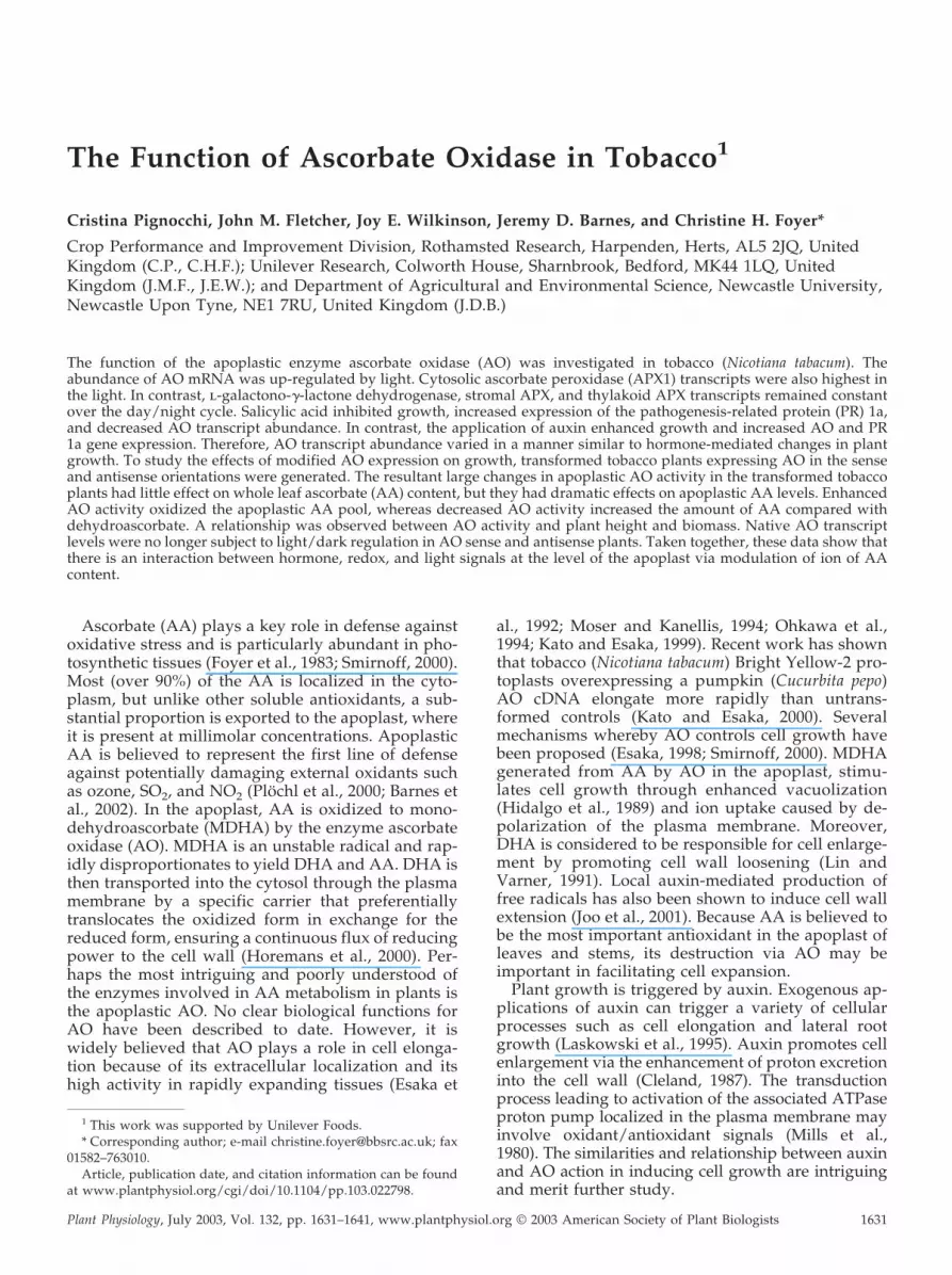
The Function of Ascorbate Oxidase in Tobacco1
Cristina Pignocchi, John M. Fletcher, Joy E. Wilkinson, Jeremy D. Barnes, and Christine H. Foyer*
Crop Performance and Improvement Division, Rothamsted Research, Harpenden, Herts, AL5 2JQ, UnitedKingdom (C.P., C.H.F.); Unilever Research, Colworth House, Sharnbrook, Bedford, MK44 1LQ, UnitedKingdom (J.M.F., J.E.W.); and Department of Agricultural and Environmental Science, Newcastle University,Newcastle Upon Tyne, NE1 7RU, United Kingdom (J.D.B.)
The function of the apoplastic enzyme ascorbate oxidase (AO) was investigated in tobacco (Nicotiana tabacum). Theabundance of AO mRNA was up-regulated by light. Cytosolic ascorbate peroxidase (APX1) transcripts were also highest inthe light. In contrast, l-galactono-�-lactone dehydrogenase, stromal APX, and thylakoid APX transcripts remained constantover the day/night cycle. Salicylic acid inhibited growth, increased expression of the pathogenesis-related protein (PR) 1a,and decreased AO transcript abundance. In contrast, the application of auxin enhanced growth and increased AO and PR1a gene expression. Therefore, AO transcript abundance varied in a manner similar to hormone-mediated changes in plantgrowth. To study the effects of modified AO expression on growth, transformed tobacco plants expressing AO in the senseand antisense orientations were generated. The resultant large changes in apoplastic AO activity in the transformed tobaccoplants had little effect on whole leaf ascorbate (AA) content, but they had dramatic effects on apoplastic AA levels. EnhancedAO activity oxidized the apoplastic AA pool, whereas decreased AO activity increased the amount of AA compared withdehydroascorbate. A relationship was observed between AO activity and plant height and biomass. Native AO transcriptlevels were no longer subject to light/dark regulation in AO sense and antisense plants. Taken together, these data show thatthere is an interaction between hormone, redox, and light signals at the level of the apoplast via modulation of ion of AAcontent.
Ascorbate (AA) plays a key role in defense againstoxidative stress and is particularly abundant in pho-tosynthetic tissues (Foyer et al., 1983; Smirnoff, 2000).Most (over 90%) of the AA is localized in the cyto-plasm, but unlike other soluble antioxidants, a sub-stantial proportion is exported to the apoplast, whereit is present at millimolar concentrations. ApoplasticAA is believed to represent the first line of defenseagainst potentially damaging external oxidants suchas ozone, SO2, and NO2 (Plochl et al., 2000; Barnes etal., 2002). In the apoplast, AA is oxidized to mono-dehydroascorbate (MDHA) by the enzyme ascorbateoxidase (AO). MDHA is an unstable radical and rap-idly disproportionates to yield DHA and AA. DHA isthen transported into the cytosol through the plasmamembrane by a specific carrier that preferentiallytranslocates the oxidized form in exchange for thereduced form, ensuring a continuous flux of reducingpower to the cell wall (Horemans et al., 2000). Per-haps the most intriguing and poorly understood ofthe enzymes involved in AA metabolism in plants isthe apoplastic AO. No clear biological functions forAO have been described to date. However, it iswidely believed that AO plays a role in cell elonga-tion because of its extracellular localization and itshigh activity in rapidly expanding tissues (Esaka et
al., 1992; Moser and Kanellis, 1994; Ohkawa et al.,1994; Kato and Esaka, 1999). Recent work has shownthat tobacco (Nicotiana tabacum) Bright Yellow-2 pro-toplasts overexpressing a pumpkin (Cucurbita pepo)AO cDNA elongate more rapidly than untrans-formed controls (Kato and Esaka, 2000). Severalmechanisms whereby AO controls cell growth havebeen proposed (Esaka, 1998; Smirnoff, 2000). MDHAgenerated from AA by AO in the apoplast, stimu-lates cell growth through enhanced vacuolization(Hidalgo et al., 1989) and ion uptake caused by de-polarization of the plasma membrane. Moreover,DHA is considered to be responsible for cell enlarge-ment by promoting cell wall loosening (Lin andVarner, 1991). Local auxin-mediated production offree radicals has also been shown to induce cell wallextension (Joo et al., 2001). Because AA is believed tobe the most important antioxidant in the apoplast ofleaves and stems, its destruction via AO may beimportant in facilitating cell expansion.
Plant growth is triggered by auxin. Exogenous ap-plications of auxin can trigger a variety of cellularprocesses such as cell elongation and lateral rootgrowth (Laskowski et al., 1995). Auxin promotes cellenlargement via the enhancement of proton excretioninto the cell wall (Cleland, 1987). The transductionprocess leading to activation of the associated ATPaseproton pump localized in the plasma membrane mayinvolve oxidant/antioxidant signals (Mills et al.,1980). The similarities and relationship between auxinand AO action in inducing cell growth are intriguingand merit further study.
1 This work was supported by Unilever Foods.* Corresponding author; e-mail [email protected]; fax
01582–763010.Article, publication date, and citation information can be found
at www.plantphysiol.org/cgi/doi/10.1104/pp.103.022798.
Plant Physiology, July 2003, Vol. 132, pp. 1631–1641, www.plantphysiol.org © 2003 American Society of Plant Biologists 1631
Relatively little information is available on otherenzymes involved in AA synthesis and metabolism.The last step of the pathway of AA synthesis in plantsis relatively well characterized and involves the cyto-chrome c-dependent oxidation of l-galactono-1,4-lactone by the mitochondrial enzyme l-galactono-�-lactone dehydrogenase (GLDH; Ostergaard et al.,1997; Bartoli et al., 2000). Similarly, the AA peroxidase(APX) family that is responsible for the scavenging ofH2O2 is well characterized. There are at least sevenAPX isoforms in plants, some of which are encoded bydiscrete genes with expression localized in differentsubcellular compartments: cytosol, mitochondria, per-oxisomes, apoplast, and chloroplasts (Asada, 1997).Two isoforms are located in the cytosol (cAPX1 andcAPX2), with cAPX2 expression restricted to high pho-tosynthetic photon flux densities in Arabidopsisleaves (Karpinski et al., 1999). In chloroplasts, APX islocated in the stroma (sAPX) and is bound to thethylakoids (tAPX).
The present study was undertaken to elucidate thefunction of AO in tobacco leaves. We show that ex-pression of AO is regulated by light and by planthormones that modulate growth. Moreover, we dem-onstrate that enhanced apoplastic AO activities intransformed tobacco plants favor increased growth.Taken together, these observations provide evidencethat AO has a role in the control of growth.
RESULTS
Diurnal Changes in Transcript Abundance of EnzymesInvolved in the AA Metabolism
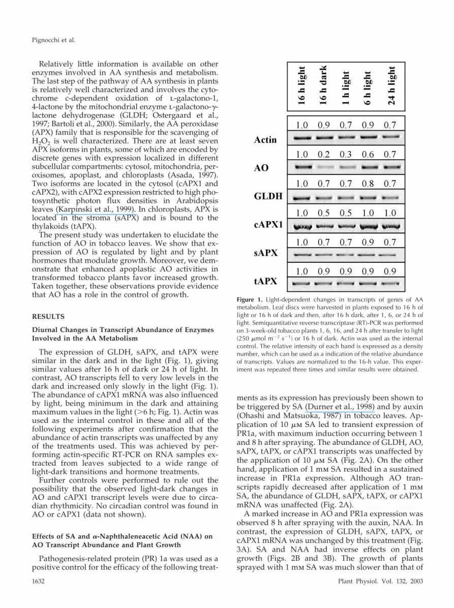
The expression of GLDH, sAPX, and tAPX weresimilar in the dark and in the light (Fig. 1), givingsimilar values after 16 h of dark or 24 h of light. Incontrast, AO transcripts fell to very low levels in thedark and increased only slowly in the light (Fig. 1).The abundance of cAPX1 mRNA was also influencedby light, being minimum in the dark and attainingmaximum values in the light (�6 h; Fig. 1). Actin wasused as the internal control in these and all of thefollowing experiments after confirmation that theabundance of actin transcripts was unaffected by anyof the treatments used. This was achieved by per-forming actin-specific RT-PCR on RNA samples ex-tracted from leaves subjected to a wide range oflight-dark transitions and hormone treatments.
Further controls were performed to rule out thepossibility that the observed light-dark changes inAO and cAPX1 transcript levels were due to circa-dian rhythmicity. No circadian control was found inAO or cAPX1 (data not shown).
Effects of SA and �-Naphthaleneacetic Acid (NAA) onAO Transcript Abundance and Plant Growth
Pathogenesis-related protein (PR) 1a was used as apositive control for the efficacy of the following treat-
ments as its expression has previously been shown tobe triggered by SA (Durner et al., 1998) and by auxin(Ohashi and Matsuoka, 1987) in tobacco leaves. Ap-plication of 10 �m SA led to transient expression ofPR1a, with maximum induction occurring between 1and 8 h after spraying. The abundance of GLDH, AO,sAPX, tAPX, or cAPX1 transcripts was unaffected bythe application of 10 �m SA (Fig. 2A). On the otherhand, application of 1 mm SA resulted in a sustainedincrease in PR1a expression. Although AO tran-scripts rapidly decreased after application of 1 mmSA, the abundance of GLDH, sAPX, tAPX, or cAPX1mRNA was unaffected (Fig. 2A).
A marked increase in AO and PR1a expression wasobserved 8 h after spraying with the auxin, NAA. Incontrast, the expression of GLDH, sAPX, tAPX, orcAPX1 mRNA was unchanged by this treatment (Fig.3A). SA and NAA had inverse effects on plantgrowth (Figs. 2B and 3B). The growth of plantssprayed with 1 mm SA was much slower than that of
Figure 1. Light-dependent changes in transcripts of genes of AAmetabolism. Leaf discs were harvested in plants exposed to 16 h oflight or 16 h of dark and then, after 16 h dark, after 1, 6, or 24 h oflight. Semiquantitative reverse transcriptase (RT)-PCR was performedon 3-week-old tobacco plants 1, 6, 16, and 24 h after transfer to light(250 �mol m�2 s�1) or 16 h of dark. Actin was used as the internalcontrol. The relative intensity of each band is expressed as a densitynumber, which can be used as a indication of the relative abundanceof transcripts. Values are normalized to the 16-h value. This exper-iment was repeated three times and similar results were obtained.
Pignocchi et al.
1632 Plant Physiol. Vol. 132, 2003

control plants sprayed with low SA concentrations(10 �m; Fig. 2B). In contrast, low concentrations (0.5�m) of NAA stimulated growth (Fig. 3B).
Transgenic Tobacco Overexpressing AO in Sense andAntisense Orientations
To further explore the function of AO in tobaccoleaves, two constructs for the expression of AO in thesense and antisense orientations were produced. Sixindependent primary transformants (T0 generation)were selected for each construct on the basis of theirability to survive growth on kanamycin, the presenceof the transgene, and their leaf AO activity. Leaf AOactivity was markedly different in all the transfor-mants compared with the wild type. AO activity wasfive to 16 times that of PAO leaves and 0.3 to 0.5times that of the TAO leaves than the wild type (Fig.4). Transgene-dependent differences in leaf AO weresimilar whether plants were grown in tissue cultureor in soil (data not shown). Genomic DNA fromeach of the lines was probed with 2X35S-PAO or2X35S-TAO DNA fragments to confirm the pres-ence of the transgene and check the copy number ofthe insertion. All lines tested positive for the trans-gene (data not shown). Three sense (PAO2-2-1,PAO1-7-2, PAO3-7-2) and two antisense (TAO2-7-1,TAO1-6-1) lines containing only one copy of theintroduced sequence were selected for further anal-ysis. These lines were grown for two additional
generations (T1 and T2) and the relationship be-tween AO and plant development was followed ineach generation.
The AO Over- and Underexpression Phenotypes
Plant growth, measured as biomass accumulationover time, was monitored in T1 and T2 populationsderived from each selected transgenic line. Each pop-ulation was composed of 25 individuals germinatedon nonselective media, allowing simultaneous mea-surements of individuals that had passed through thetransformation procedure but did not express thetransgene and AO-expressing plants. All plants weregrown under controlled environment conditions at thesame time and shoot fresh weights were recordedweekly from three plants per population. Additionalwild-type populations were not necessary in theseexperiments, given the presence of internal controlswithin each population. No statistically significant dif-ferences in shoot fresh weight were observed in 3-, 4-,or 5-week-old sense (PAO) or antisense (TAO) plants(Fig. 5A). However, between weeks 6 and 8, plantsexpressing the AO transgene in the sense orientation(PAO) accumulated greater biomass than plants ex-pressing the AO transgene in the antisense orientation(TAO; Fig. 5A). By the end of the experiment (8weeks), AO sense-expressing plants (PAO) showed astatistically significant increase in fresh weight (P �0.05 at 7 weeks, and P � 0.01 at 8 weeks) compared
Figure 2. Effects of SA on transcripts abundance and plant growth. A, Transcript abundance; whole shoot samples wereharvested 1, 8, and 24 h after spraying, which was performed halfway through the light period. Semiquantitative RT-PCR wasperformed on 3-week-old tobacco plants sprayed with 10 �M or 1 mM SA and then exposed to an extended photoperiod(24 h at 250 �mol m�2 s�1). Actin was used as the internal control. B, Plant growth; phenotype of SA-treated and -untreatedplants showing seedlings before treatment (a) and 1 week after the treatment (b). This experiment was repeated three timesand similar results were obtained.
The Function of Ascorbate Oxidase
Plant Physiol. Vol. 132, 2003 1633

with the AO antisense plants (TAO; Fig. 5A). Therelative growth (measured as weekly biomass accu-mulation) rate was linear (r2 � 0.95) between weeks 6and 8. At this point, the growth rate of AO senseplants (PAO) was 30% higher than that of AO anti-sense (TAO) plants. Values of 20.8 g fresh weight gainper week were measured in AO sense plants (PAO)
compared with 14.1 g fresh weight gain per week inantisense (TAO) plants (Fig. 5A).
Once established that AO sense plants (PAO)showed an increased growth rate when comparedwith antisense (TAO) plants, growth of sense plants(PAO), measured this time as plant height, was com-pared with that of wild-type plants. This experiment
Figure 3. Effects of auxin on transcript abundance and plant growth. A, Transcript abundance; whole shoot samples wereharvested 1, 8, and 24 h after spraying, which was performed halfway through the light period. Semiquantitative RT-PCR wasperformed on 3-week-old tobacco plants sprayed with 0.5 �M NAA and then exposed to an extended photoperiod (24 h at250 �mol m�2 s�1). B, Plant growth; phenotype of NAA-treated and -untreated plants showing seedlings before treatment(a) and 1 week after the treatment (b). This experiment was repeated three times and similar results were obtained.
Figure 4. Characterization of primary transfor-mants. AO activity in leaf samples of PAO sense(1) and TAO antisense (2) primary transgeniclines. Each value represents the mean � SD ofthree independent measurements.
Pignocchi et al.
1634 Plant Physiol. Vol. 132, 2003

was conducted to establish whether a correlationbetween AO activity and plant height could bedrawn. T1 and T2 seeds from two AO sense lines(PAO2-2-1 and PAO3-7-2) that had been selected onthe basis of high AO activity, were germinated onmedia containing kanamycin to select for plants con-taining the transgene. Once this was confirmed byPCR (data not shown), PAO plants were transferredto compost and were grown in controlled environ-ment chambers together with wild-type populations.After 6 weeks, AO activities were measured in youngfully developed leaves of four to five plants per line,and the height of the corresponding plants was re-corded. Overall, T2 plants exhibited higher AO activ-ity and a taller phenotype than their respective T1progenitors (Fig. 5B). A positive correlation wasdemonstrated between AO activity and plant height(Fig. 5B; r2 � 0.85). Analysis of variance of the dataconfirmed that T2 PAO lines grew significantly taller
than T1 PAO lines and that T1 and T2 PAO lines grewtaller than the wild type (P � 0.01; Fig. 5, B and C).
Relationships between AO Activity and AA in theLeaf Apoplast
Whole leaf AA, DHA contents, and AO activitieswere measured in extracts from discs excised fromyoung fully expanded leaves of 6-week-old T1 sense(PAO lines PAO2-2-1 and PAO3-7-2) and antisense(TAO lines TAO2-7-1 and TAO1-6-1) plants. PAOplants exhibited about a 40-fold higher leaf AO ac-tivity than the wild type (Table I), whereas TAO linesexhibited 0.4-fold less leaf AO activity. ApoplasticAO activity was measured in whole leaf extracts,soluble, and ionically bound fractions (Table I). TotalAO activity measured in whole leaf extracts wasfound to be entirely associated with the cell wall(ionically bound) and localized in the apoplast (Table
Figure 5. Phenotypes of AO sense and antisense transgenic plants. A, Effect of AO over- and underexpression on growthrates of T1 populations: three AO sense and two AO antisense. Growth rate of sense (F) and antisense (E) AO transgenicT1 populations measured as the increase in shoot fresh weight per week. All plants were grown under controlledenvironment conditions. B, Correlation between AO activity and height in 7-week-old AO sense (PAO) and wild-type plants.Squares, wild type; circles, T1 generation; triangles, T2 generation. Each activity value represents the mean of threeindependent measurements. Mean values for AO activities are 0.05 � 0.00 for wild type; 1.4 � 0.5 for T1 generation; 3.0 �0.4 for T2 generation. Mean values for plant heights are 33 � 5.0 for wild type; 42 � 9.8 for T1 generation; 50 � 8.0 forT2 generation. This experiment was repeated three times and similar results were obtained. C, Phenotype of wild-type and7-week-old AO sense tobacco plants (PAO).
The Function of Ascorbate Oxidase
Plant Physiol. Vol. 132, 2003 1635

I). AO activity was undetectable in soluble fractionsof leaf extracts. TAO leaves contained similaramounts of AA and DHA to the wild type, but PAOleaves contained less AA and total (AA plus DHA)AA (Table I). DHA contents were higher in PAOlines, but the leaf ascorbate pool was always largelyreduced, values being slightly higher for wild-type(92%) than PAO plants (85%). In contrast to wholeleaf AA, modified AO expression resulted in dra-matic changes in the apoplastic AA pool. The total(AA plus DHA) AA content of the apoplast was atleast double that of the wild type in sense and anti-sense plants (Table I). However, the greatest effectwas on the AA redox state (Table I). In the wild type,the AA pool in the apoplast was about 40% reduced,whereas it was 66% reduced in the antisense plantsand only 3% reduced in the sense plants. The DHAcontent in the apoplast of sense plants was 3.5-foldgreater than in wild-type or antisense plants. Forapoplastic AA and DHA measurements, cytosoliccontamination of the apoplast (monitored by measur-ing Glc-6-P in intercellular washing fluid (IWF) wasmaintained below 0.1% (data not shown). However,it should be noted that the presence of apoplastic acid
phosphatases may lead to underestimation of thecontamination of the apoplastic fraction as well.
Light/Dark Effects on AO Transcript Abundance in AOTransgenic Plants
AO transcript abundance was highest in the lightand showed a marked light/dark expression patternin wild-type plants (Fig. 1). The following experi-ments were performed to investigate whether suchmarked diurnal patterns of expression were main-tained in the native wild-type AO of PAO and TAOtransgenic plants. Due to the sequence similarity be-tween the tobacco and the pumpkin AO (used for thegeneration of the AO sense plants), careful design ofspecies-specific primers was necessary to allow ex-pression patterns of native and transgenic AO to bediscriminated. No expression patterns in the expres-sion of the transgenic AO (PAO) were expected giventhe presence of the strong constitutive promoter 35Scauliflower mosaic virus (CaMV). The specificity ofTAO (specific for tobacco AO) and PAO (specific forpumpkin AO) primers was confirmed by sequencingof the relative PCR products. They were then used to
Figure 6. Effects of light and dark on nativewild-type AO transcript abundance and trans-genic pumpkin AO in wild-type and transgenicplants by semiquantitative RT-PCR. Expressionanalysis of native wild-type tobacco in sense(PAO) and antisense (TAO) primary transgeniclines. L, after 16 h of light; D, after 16 h of dark.Sense lines: PAO 2-2-1, PAO 3-7-2, PAO 1-7-2;antisense lines: TAO 2-7-1, TAO 1-6-1. Thedensity numbers above each band represent therelative intensity of each band and can be usedas an indication of the relative abundance oftranscripts. Actin was used as the internal con-trol. Similar results were obtained in three otherindependent experiments.
Table I. AO activity and ascorbate content in whole leaves and apoplast of T1 and T2 PAO senseand TAO antisense transgenic lines and in wild-type plantsa
Whole-Leaf Parameters PAO Senseb TAO Antisensec Wild Type
AO activityd 1.92 � 0.1 0.02 � 0.0 0.05 � 0.0Total ascorbatee 218.7 � 7.8 282.1 � 8.6 270.3 � 34.4AAe 186.6 � 6.5 258.8 � 8.1 250.5 � 30.2DHAe 33.9 � 3.4 24.1 � 2.3 20.9 � 5.8
Apoplastic parameters
AO activityd (ionically bound) 1.92 � 0.1 0.02 � 0.0 0.05 � 0.0AO activityd (soluble) 0.00 0.00 0.00Total ascorbatee 32.6 � 2.9 26.0 � 6.2 13.7 � 3.7AAe 1.1 � 0.2 17.3 � 5.4 5.2 � 0.9DHAe 31.5 � 2.8 8.6 � 1.1 8.1 � 2.9a Leaf samples were taken from 6-week-old-plants grown in controlled environment. Transgenic
plants were generated from T1 seeds germinated in selective medium. Values are the mean of threesamples per leaf and 20 leaves per line. b Lines PAO 2-2-1 and PAO 3-7-2. c Lines TAO 2-7-1and TAO 1-6-1. d Given as units per milligram of total protein content. e Given as nanomolesper milligram of total protein content.
Pignocchi et al.
1636 Plant Physiol. Vol. 132, 2003

analyze by RT-PCR the expression of the native to-bacco AO and the introduced pumpkin AO (PAO) inthe leaves of T1 and T2 generations of transformantsgrown in soil under controlled environment condi-tions. Samples from wild type, three sense (PAO),and two antisense (TAO) lines were harvested after16 h of light or 16 h of dark (Fig. 6). PAO was highlyand equally expressed in all the sense PAO leaves inthe light and in the dark. This is consistent with theconstitutive expression of PAO under the control ofthe CaMV35S promoter. Moreover, PAO plantsshowed no cosuppression of the native wild-type AObecause the transcript abundance of the native wild-type tobacco AO transcripts was similar in the sensePAO lines and in the wild type. In contrast to thestrong dark/light pattern of AO expression observedin wild-type plants, no dark/light effects on nativewild-type tobacco AO transcript abundance was de-tected in the PAO sense lines. In this case, theamounts of native AO transcripts were similar in thelight and dark (Fig. 6). There was a large overalldecrease in native AO transcripts in TAO leaves com-pared with the wild type (Fig. 6), demonstrating theefficacy of the antisense suppression strategyadopted. However, in the same TAO plants, therewas also a decrease in the observed stimulation ofnative wild-type AO expression by light (Fig. 6).
DISCUSSION
Although AO activity was first described in plantsmany years ago, its biological function has remainedelusive. Much circumstantial evidence has linked tis-sue AA contents to growth (Chinoy, 1984), but todate, the mechanism has remained unknown andlargely unexplored. Recent evidence has provided anindication of the mechanism whereby AA is involvedin the regulation of growth (Pastori et al., 2003).Several lines of evidence also suggest AO may beinvolved in this process (for example, see Esaka,1998). The results presented here show that the ex-pression of native AO is regulated by light but thatthis control is lost when overall AO transcript abun-dance is manipulated in transgenic plants; AO tran-scripts are modified by auxin and SA in a mannerconsistent with effects on growth, and enhanced AOactivity oxidizes the apoplastic AA pool and this canstimulate growth in certain conditions. Taken to-gether, these observations demonstrate that AO ac-tivity can influence plant growth.
Light Regulation of Gene Expression in Enzymes of theAA Metabolism
We report here the effects of light and dark over a24-h period on the expression patterns of genes in-volved in AA metabolism. The expression of cAPX1is regulated by light in tobacco (Fig. 1), consistentwith earlier observations of APX1 mRNA abundance
in tobacco (Tabata et al., 2002), Arabidopsis (Kubo etal., 1995), and APX activity in mustard (Sinapis alba;Thomsen et al., 1992). The expression of GLDH, thelast enzyme of AA biosynthesis, was not induced bylight over a 24-h period in tobacco leaves in thegrowth conditions used here (Fig. 1). Therefore, weconclude that the light-dependent AA accumulationthat we observe under these conditions, increasingfrom 800 � 200 nmol g�1 fresh weight at the end ofa 16-h dark period to 3000 � 400 nmol g�1 freshweight after 16 h in the light is not the result ofmodulation of GLDH expression. Therefore, the lightdependency of AA accumulation observed over theday/night cycle in leaves must result from otherfactors such as the requirement for carbon skeletonsproduced by photosynthesis (Smirnoff and Pallanca,1996). Several of the genes encoding enzymes of theAA biosynthetic pathway increase in expression aftertransfer of plants to high light intensity (Tabata et al.,2002). GLDH transcripts were found to decrease aftera prolonged (5 d) dark treatment (Tabata et al., 2002).Although a recent study in Arabidopsis leaves hasreported that GLDH transcripts are lowest in themorning, increasing during the day, and decreasingin the subsequent night (Tamaoki et al., 2003), this isclearly not the case in tobacco.
Complex transcriptional and translational controlsmodulate AO expression (Esaka et al., 1992; Kato andEsaka, 1999). Phytochrome-dependent modulation ofAO activity by light has been reported (Leaper andNewbury, 1989; Hayashi and Morohashi, 1993). Al-though our data are not exhaustive in exploring theregulation of AO in response to light, they providecorroborative evidence that AO expression is modu-lated by light and is repressed in the dark (Fig. 1).Controls showed that this diurnal pattern of regula-tion was not due to circadian rhythmicity (data notshown). It is surprising that sense and antisense ma-nipulation of AO expression led to the elimination oflight-induced changes in abundance of native wild-type AO transcripts (Fig. 6). None of the PAO linesshowed a decrease in native wild-type AO transcriptabundance after a 16-h dark period (Fig. 6). It is ourhypothesis that the observed perturbation of the re-dox state of the apoplast, as discussed below, is re-sponsible for the loss the endogenous diurnal regu-lation of AO.
AO Transcript Abundance Is Regulated by PlantHormones in Wild-Type Tobacco
An induction of AO by auxin, similar to that ob-served here, was reported in pumpkin (Esaka et al.,1992). Moreover, the existence of a cis-acting regionin the AO promoter responsible for auxin regulationhas been suggested (Kisu et al., 1997). We demon-strate that AO expression is also induced by auxin intobacco and that the effect is associated with thepromotion of plant growth. AO has been shown to
The Function of Ascorbate Oxidase
Plant Physiol. Vol. 132, 2003 1637

catalyze the oxidative decarboxylation of auxin (Kerket al., 2000), suggesting a role for AO in the regula-tion of auxin levels within the quiescent centers ofmaize roots.
High concentrations of SA (1 mm) caused a pro-nounced inhibition of the growth of tobacco seed-lings (Fig. 2B). SA inhibition of plant growth anddevelopment is a well-known phenomenon (Petersenet al., 2000). SA can affect cell growth, positively ornegatively, depending on the context in which sig-naling occurs, as studies on Arabidopsis mutantshave illustrated (Vanacker et al., 2001). Of the AA-related enzymes analyzed, only the expression of AOwas changed by the application of SA and NAA(Figs. 2A and 3A).
Sense and Antisense Manipulation of AO
The modification of AO expression in tobacco bysense (PAO) and antisense (TAO) technologies hasallowed us to explore the relationship between AOand growth in planta. To minimize the risk of genesilencing by cosuppression, we used a pumpkin AOcDNA for the sense construct, which shares approx-imately 68% homology with the tobacco sequence.This choice proved to be successful, as no silencing ofthe native AO was found in the sense (PAO) lines.The native AO gene was expressed at wild-type lev-els in PAO lines (Fig. 6). For the antisense suppres-sion of AO, we used one-third of the 5� sequence ofthe tobacco cDNA, as in recent years, the use of onlypartial cDNA sequences proved to be very effectivein antisense suppression (Bourque, 1995). In anti-sense (TAO) lines, we obtained a dramatic reductionin AO transcript abundance (Fig. 6). However, noneof the TAO lines exhibited complete suppression ofAO transcripts, perhaps because of the limited effi-ciency of the chosen antisense fragment or because ofthe genomic position of the transgene.
The data presented here demonstrate that our ge-netic approach was successful in significantly modi-fying apoplastic AO activity in leaves, as increases ofup to 40-fold were measured in PAO sense trans-formed lines, whereas activities were halved in anti-sense TAO lines. We were unable to measure AOprotein abundance in this study, as the AO antibod-ies that we prepared did not have sufficient specific-ity. Nevertheless, the parallel changes in transcriptsand AO activities reported here allow us to assumethat our manipulation also modified amounts of AOprotein.
AA Content of the Apoplast Was Modulated byAO Activity
PAO sense lines contained less (22%–27%) total AAand AA and more (50%) DHA than TAO antisenselines or wild-type plants (Table I). No differences inleaf AA content were detected between TAO anti-
sense and wild-type plants. The effect of high AOactivity on the whole leaf AA pool is minimal, takinginto account the fact that AO activities in sense lineswere increased by up to 40-fold. The observation thatsuch large increases in AO activity did not lead tomajor changes in leaf AA may be explained by dif-ferential localization of the two components (Lin andVarner, 1991). AO is located in the apoplast, whereasmost of the AA is in the cytoplasm (Table I). MDHAand DHA generated in the apoplast have to be trans-located to the cytoplasm where they are efficientlyregenerated. The observation that the DHA contentwas doubled in sense lines suggests that the rate ofDHA transport or the rate of AA regeneration (orboth) were too slow to compensate for the enhancedAO activity in sense lines. We measured the AAcontent and redox state in the leaf apoplast in thesame plants as those used for total leaf AA determi-nations. The AA pool was virtually all oxidized (97%)in the PAO sense lines and was more reduced thanthe wild type in TAO antisense lines (66% as opposedto 40%; Table I). These data, together with the obser-vation that the sense lines accumulated almost fourtimes more DHA than the wild type in the apoplast,confirmed two points. First, our sense construct suc-cessfully targeted AO to the apoplast. Second, ma-nipulation of AO produced a marked localized effecton (apoplastic) AA. It is interesting to note that inPAO sense and TAO antisense lines, the total AAcontent of the apoplast was twice that of the wildtype. This may suggest that AO-mediated perturba-tion of the AA and DHA contents of the apoplastinfluences the transport of reduced and oxidizedforms, such that when the apoplastic pool is veryreduced or very oxidized, transport processes areincreased in an attempt to redress the balance.
AO and Plant Development
In the present study, we demonstrate that increas-ing AO activity by 40-fold leads to enhanced biomassaccumulation and elongation in tobacco plants (Fig.5). In contrast, reduction of AO activity by antisensetechnology results in a reduction in growth rate (Fig.5A). Taken together, the data presented here suggestthat enhanced AO activity can have a positive effecton growth (Fig. 5B). Increased biomass production inPAO sense plants appears to be mainly due to en-hanced internode elongation. It is possible that AOaction alone stimulates growth through the genera-tion of MDHA radicals in the apoplast. This en-hanced growth could occur by a chemorheologicalwall-loosing reaction as described for superoxide-and hydroxyl radical-mediated extension growth(Schopfer et al., 2002), or by enhancing cell enlarge-ment via the polarization of the plasmalemma, lead-ing to expansion by vacuolization as suggested pre-viously (Esaka, 1998; Smirnoff, 2000), or both.
Plant growth is the outcome of various mecha-nisms of regulation, signaling, and crosstalk. We
Pignocchi et al.
1638 Plant Physiol. Vol. 132, 2003

have provided in planta evidence that AO is one ofthese components and therefore participates to thisbroad dialogue between signaling molecules and en-vironmental cues. It should be noted that in anotherstudy on modulation of AO in tobacco, no effects ongrowth were reported (Sanmartin at al., 2003). In theexperimental conditions described here, changes inAO activity alone produced marked effects on plantgrowth, measured as shoot biomass accumulationand stem height. However, the growth effect wasclearly environment dependent as observed in theAA-deficient Arabidopsis vtc 1 mutant (Conklin etal., 1997; Veljovic-Jovanovic et al., 2001). Vtc1 showsa marked conditional phenotype, such that whengrown on short days where vegetative growth pre-dominates, vtc1 is much slower growing than thewild type (Col0). Under long days where reproduc-tive growth predominates, there is no significantdifference in growth. In this case, AA has been shownto regulate genes controlling plant developmentthrough hormone signaling (Pastori et al., 2003). Weconducted a large-scale greenhouse experiment inthe summer months, with growth conditions mark-edly different from those described here. In theseconditions, where light intensities were higher, thephotoperiod was longer, and temperatures werehigher, the effects of modified AO activity weremuch less marked. However, it is interesting to notethat although no significant differences in plantheight and biomass were recorded between wild-type and transgenic plants, other effects were ob-served under these conditions. In particular, AOsense plants showed a significantly higher number ofsmaller flowers compared with wild-type and anti-sense plants (data not shown). This effect on theflowers of the AO sense lines caused a reduction of6% to 14% in the weight of seeds. Such differenceswere not observed when plants were grown in con-trolled environment chambers, as described here. Weconclude that the phenotype associated with in-creased AO activity in tobacco is conditional and ismodulated by environmental cues.
In conclusion, the data presented here suggest thatfactors such as apoplastic redox state that is domi-nated by AA and regulated by AO modulate receptorfunction and signal transduction and that there isscope for modulation and interaction between differ-ent signals (hormone, redox, and light) in theapoplast.
MATERIALS AND METHODS
Plant Material and Growth Conditions
Tobacco (Nicotiana tabacum cv Petit Havana, mutant SRI) seeds weregerminated in petri dishes on moistened filter paper. After 10 d, seedlingswere transferred to compost (Petersfield Products, Leicester, UK) in pots.Plants were grown at 22°C day/night in controlled environment chamberssupplying a photosynthetic photon flux density of 250 �mol m�2 s�1 atplant height as a 16-h photoperiod. Young fully expanded leaves from6-week-old plants were used for AA measurements in the light and dark.Three-week-old plants were used for all treatments and RT-PCR analyses.
Hormone Treatments
Three-week-old seedlings were sprayed to run-off (using an aerosolspray bottle supplied by Nalgene, Rochester, NY) with 10 �m and 1 mm SAand 0.5 �m NAA. After treatment, plants were kept in continuous light for24 h and whole shoots were harvested for RT-PCR analysis. For the evalu-ation of the effects of hormones on growth, sprayed plants were subse-quently returned to “growth conditions” until further analysis 1 week afterthe initial harvest.
PCR Amplification of AO cDNAs for Cloning
Cloning of the genes encoding AO from pumpkin (Cucurbita pepo; PAO;EMBL GenBank accession no. X55779) and tobacco (TAO; accession no.D43624) was achieved from PCR amplification of full-length cDNAs usingprimers designed to add additional restriction sites to facilitate the cloning.For PAO the primers were 5�-ACCACTCGAGATGCTTCAGATG -3� (nucle-otide position 18–29 of cDNA; added XhoI site is underlined) and 5�-ACCAGAGCTCTTAGGGGTTATTT-3� (nucleotide position 1,757–1,745;added SacI site is underlined). For TAO the primers were 5�-ACCAGAGCTCATGGCTTCCTTA-3� (nucleotide position 88–100 of cDNA;added SacI site is underlined) and 5�-ACCACTCGAGTTTGTGCCACC-3�(nucleotide position 609–559; added XhoI site is underlined). The PCRconditions were 10 cycles of 1 min at 94°C, 1 min at 40°C, and 1.5 min at72°C, and 15 cycles of 1 min at 94°C, 1 min at 61°C, and 1.5 min at 72°C,using Pfu Turbo DNA polymerase (Stratagene, La Jolla, CA), 1 ng of cDNA,and 0.2 �m each primer in a 50-�L reaction. At the end of the cycles, thereactions were incubated at 72°C for 10 min. The identity of the 1.7-kb(PAO) and 520-bp (TAO) PCR products was confirmed by single-strandsequencing (ABI PRISM, 310 genetic analyzer; Perkin-Elmer, Warrington,Cheshire, UK).
Construction of Sense and Antisense AO in cj102 andTransformation into Agrobacterium tumefaciens
PAO and TAO PCR products were subcloned as XhoI-SacI fragments intothe corresponding sites of pp5ln (derived from pUC19; Frenken et al., 1999)by standard in vitro recombination techniques (Sambrook et al., 1989),generating the fusion constructs PAO/pp5ln and TAO/pp5ln. In PAO/pp5ln, the AO cDNA is placed in sense orientation under the control of theCaMV35S promoter with duplicated enhancer region (2x35S), originatingthe 2x35S-PAO expression cassette. In TAO/pp5ln, the AO cDNA is placedin antisense orientation under the control of 2x35S, originating the 2x35S-TAO expression cassette. Sense and antisense expression cassettes wereligated into cj102, a derivative plasmid of pGPTV (Becker et al., 1992), asHindIII-SacI fragments into the corresponding sites of cj102 in replacementof the 2x35S-�-glucuronidase cassette. The cj102-PAO and cj102-TAO con-structs obtained were transformed into A. tumefaciens LBA4404 by electro-poration (Parry et al., 1998).
Plant Material and Transformation
Sterile cultures of tobacco were transformed with cj102-PAO and cj102-TAO constructs by A. tumefaciens leaf disc infection (Gallois and Marinho,1995). T1 seeds from primary transformants were germinated in petri dishescontaining 1.5% (w/v) agar in distilled water (nonselective medium), sup-plied with 100 mg L�1 kanamycin (selective medium). T2 seeds were ob-tained from individual T1 plants by self-pollination and were used togenerate T2 transgenic progeny.
Southern-Blotting Analysis
Genomic DNA (5 �g) from PAO sense and TAO antisense lines wasdigested with HindIII, which cuts once within the transgenic sequence, runon an 0.8% (w/v) agarose gel and blotted onto a nylon membrane (Hybond-NX; Amersham Life Science, Buckinghamshire, UK). The membranes wereprehybridized at 55°C for 2 h in hybridization buffer (Amersham LifeScience) and were hybridized overnight in the same buffer. The HindIII/SacI fragments 2x35S-PAO and 2x35S-TAO were alkaline phosphatase la-beled and were used as probes (AlkPhos Direct; Amersham Life Science).
The Function of Ascorbate Oxidase
Plant Physiol. Vol. 132, 2003 1639

Hybridization temperatures were 65°C for 2x35S-PAO and 68°C for 2x35S-TAO. The membranes were washed according to Amersham’s recommen-dations and detection was performed with CDP-Star (Amersham Life Sci-ence) by exposing Hyperfilm enhanced chemiluminescence film (AmershamLife Science) to the membrane for 1.5 h.
Total RNA Extraction and Gene ExpressionAnalysis by RT-PCR
Total RNA was extracted from using RNeasy plant mini kit (Qiagen,West Sussex, UK) according to the supplier’s recommendation. ResidualDNA was removed with DNase I, Amp Grade (Invitrogen, Strathclyde, UK).The absence of DNA contamination in the samples was confirmed by asaturating PCR of 40 cycles using actin- (X63603) specific primers (5�-CGCGAAAAGATGACTCAAATC-3� and 5�-AGATCCTTTCTGATATCC-ACG-3�), which give a 687-bp product with genomic DNA and a 533-bpproduct with cDNA. One microgram total RNA was reverse transcribedusing 0.5 �g of Oligo (dT)12–18 (Invitrogen) and 200 units of Superscript II(Invitrogen) following the supplier’s recommendation. cDNA samples werestandardized by PCR for actin content using the gene-specific primers. Onthe basis of the published sequences, the following gene-specific primerswere designed and used for amplification: PR-1a (X12737), 5�-GCC-TTCATTTCTTCTTGTCTC-3� and 5�-TTAGTATGGACTTTCGCCTC-3�;GLDH (AB048530), 5�-TTTTAGGCTTTGACTGTGGTG-3� and 5�-TCAGAT-GAAGAGCTTCTCAAG-3�; cAPX1 (X59600), 5�-CTCAAGCTGTTGAC-AAATG-3� and 5�-AGCTTCAGCAACCAATTC-3�; sAPX (AB02 2274),5�-TTGT TTCAGTTGGCCAGTGC-3� and 5�-CGCTGCCTTGTGTAGG-3�;tAPX (AB022273), 5�-TGTTTTCTACAGAATGGGC-3� and 5�-GTTGAG-TATTTTG CTGCCAC-3�; PAO (X55779), 5�- TTGACCGGAGCAAAAA-CTTC-3� and 5�- AATTCAATGACGACTCCTCC-3�; and TAO (D43624),5�- AACCAAAAACACCTCAAGGC-3� and 5�- GGTGCTTGTTTTAGGA-CATC-3�.
For semiquantitative RT-PCR, the cycle number was kept within thelinear range (30 cycles) and the conditions were 3 min at 94°C, cycle of 45 sat 94°C, 30 s at 52°C, and 45 s at 72°C, followed by 10 min at 72°C, using 0.5�L of the RT reaction and 0.2 �m each oligonucleotide primer in a totalvolume of 25 �L. The identity of the PCR products was verified by single-strand sequencing (ABI PRISM, 310 genetic analyzer; Perkin-Elmer). RT-PCR products were loaded on 2% (w/v) agarose gel containing 0.5 �g mL�1
ethidium bromide and the band intensities were quantified with the EagleEye II (Stratagene).
Determination of AA and Glc-6-P Content
Leaf samples were powdered in liquid nitrogen, then 1 mL of ice-cold 1n HClO4 was added per 0.1-g sample (fresh weight). Extracts were thencentrifuged at 14,000 rpm for 5 min at 4°C. HEPES buffer (0.1 m, pH 7.0) wasadded at a buffer:extract ratio of 1:5 (v/v). K2CO3 (5 m) was added until theextract reached pH 5.6. The extracts were again centrifuged at 14,000 rpm,this time for 2 min, to allow the removal of precipitated K2ClO4, and werethen assayed for AA and DHA as described by Foyer and coworkers (1983).Glc-6-P was measured according to Latzko and Gibbs (1972).
Preparation of IWF
IWF was prepared using a method similar to that adopted by Turcsanyiet al. (2000). Young fully expanded leaves were vacuum infiltrated at �70kPa with chilled 10 mm citrate buffer (pH 3.0) for 5 min. Leaves were thenblotted dry, carefully rolled and inserted into a prechilled syringe, andcentrifuged at 2,000 rpm for 10 min at 4°C. For ascorbate and Glc-6-Panalyses, IWF was collected in empty preweighed Eppendorf tubes. Con-trols centrifuged into tubes containing cold metaphosphoric acid (2%, w/v)gave similar results.
AO Assay
For total AO activity, leaf tissue was powdered in liquid nitrogen andthen homogenized with 0.1 m sodium phosphate, pH 6.5 (1 mL 0.1 g�1 freshweight). The extract was then diluted 10-fold in the same buffer and 50 �Lwas used in the assay. Measurements of soluble and ionically bound AO
activity were performed according to Sanmartin et al. (2003). Leaf extractswere centrifuged at 15,000g for 10 min at 4°C and soluble AO activity wasmeasured on the supernatant. The pellet (ionically bound fraction) wasresuspended in 0.1 m sodium phosphate, pH 6.5, with the addition of 1 mNaCl, vortexed for 10 min at 4°C, and then centrifuged at 15,000g for 10 minat 4°C. AO activity was measured on the supernatant.
AO activity was determined from the decrease in A265 at 25°C in areaction mixture containing 0.1 m sodium phosphate, pH 5.6, 0.5 mm EDTA,and 100 �m AA. One unit of AO activity was defined as the oxidation of 1�mol AA min�1 at 25°C. An extinction coefficient for AA of 14 mm�1 cm�1
at 265 nm was used in calculations (Nakano and Asada, 1981). Recoveryexperiments with purified AO were performed to ensure that maximalactivity was extracted and maintained during isolation procedures.
Growth Rates of T1 Populations andPhenotypic Analysis
T1 populations were obtained by germinating T1 seeds on nonselectivemedium. Twenty-five plants per each transgenic line (three AO sense andtwo AO antisense) were used for growth experiments. Growth curves weredrawn by measuring every week shoot fresh weight of three plants per linegrowing in compost in controlled environment. Shoots were excised at thebase of the stem and were weighed for the fresh weight determination. Plantheight was measured on 7-week-old plants from the basis of the stem toinflorescence.
Statistical Analyses
Statistical analyses were performed using GENSTAT 5 (Peyne et al.,1993). Data were subjected to analysis of variance and t test to investigatethe statistical significance of the effects of modified AO activity on growth.Correlations were performed using Sigmaplot 2001 (SPSS, Chicago) usingleast square linear regression methods to test the goodness of fit.
ACKNOWLEDGMENTS
We thank Prof. Muneharu Esaka (Hiroshima University, Japan) for thekind donation of the pumpkin and tobacco AO cDNAs and Sue Robinsonand Dr. Karl Hunter (Unilever Research) for performing the large-scalegreenhouse study of transgenic tobacco plants.
Received February 28, 2003; returned for revision March 23, 2003; acceptedApril 6, 2003.
LITERATURE CITED
Asada K (1997) The role of ascorbate peroxidase and monodehydroascor-bate reductase in H2O2 scavenging in plants. In JG Scandalios, ed, Oxi-dative Stress and the Molecular Biology of Antioxidant Defenses. ColdSpring Harbor Laboratory Press, Cold Spring Harbor, NY, pp 715–735
Barnes JD, Zheng Y, Lyons TM (2002) Plant resistance to ozone: the role ofascorbate. In K Omasa, H Saji, S Youssefian, N Kondo, eds, Air Pollutionand Plant Biotechnology. Springer-Verlag, Tokyo, pp 235–254
Bartoli CG, Pastori GM, Foyer CH (2000) Ascorbate biosynthesis in mito-chondria is linked to the electron transport chain between complexes IIIand IV. Plant Physiol 123: 335–343
Becker D, Kemper E, Schell J, Masterson R (1992) New plant binary vectorswith selectable markers located proximal to the left T-DNA border. PlantMol Biol 20: 1195–1197
Bourque JE (1995) Antisense strategies for genetic manipulations in plants.Plant Sci 105: 125–149
Chinoy NJ (1984) Ascorbic acid in plant growth and development. In MNijhoff, W Junk, eds, The Role of Ascorbic Acid in Growth, Differentia-tion, and Metabolism of Plants. Martinus Nijhoff, The Netherlands, TheHague, Dr Junk, pp 68–195
Cleland RE (1987) How hormones work: auxin and cell elongation. In PJDavies, ed, Plant Hormones and Their Role in Plant Growth and Devel-opment. Kluwer Academic Publishers, Lancaster, UK, pp 132–148
Pignocchi et al.
1640 Plant Physiol. Vol. 132, 2003

Conklin PL, Pallanca JE, Last RL, Smirnoff N (1997) l-Ascorbic acidmetabolism in the ascorbate-deficient Arabidopsis mutant vtc1. PlantPhysiol 115: 1277–1285
Durner J, Wendehenne D, Klessig DF (1998) Defense gene induction intobacco by nitric oxide, cyclic GMP, and cyclic ADP-ribose. Proc NatlAcad Sci USA 95: 10328–10333
Esaka M (1998) Gene expression and function of ascorbate oxidase in higherplants. Recent Res Dev Phytochem 2: 315–326
Esaka M, Fujisawa K, Goto M, Kisu Y (1992) Regulation of ascorbateoxidase expression in pumpkin by auxin and copper. Plant Physiol 100:231–237
Foyer CH, Rowell J, Walker D (1983) Measurement of the ascorbate contentof spinach leaf protoplasts and chloroplasts during illumination. Planta157: 239–244
Frenken LGJ, Jobling SA, The Y, Verhoeyen ME, Wilkinson JE, Van derLogt CPE (1999) Modifying a plant to produce an antibody useful forincreasing pathogen resistance or to modulate metabolism comprisesintroducing a DNA sequence encoding a heavy chain immunoglobulinlinked to a peptide that targets a cellular compartment. Patent numberEP1118669 European Patent Office, Munich, Germany 8th December 2000
Gallois P, Marinho P (1995) Leaf disk transformation using Agrobacteriumtumefaciens-expression of heterologous genes in tobacco. In H Jones, ed,Methods in Molecular Biology, Vol 49: Plant Gene Transfer and Expres-sion Protocols. Humana Press, Totowa, NJ, pp 39–48
Hayashi R, Morohashi Y (1993) Phytochrome control of the development ofascorbate oxidase activity in mustard (Sinapis alba) cotyledons. PlantPhysiol 102: 1237–1241
Hidalgo A, Gonzalez-Reyes JA, Navas P (1989) Ascorbate free radicalenhances vacuolisation in onion root meristems. Plant Cell Environ 12:455–460
Horemans N, Foyer CH, Asard H (2000) Transport and action of ascorbateat the plant plasma membrane. Trends Plant Sci 5: 263–267
Joo JH, Bae YS, Lee JS (2001) Role of auxin-induced reactive oxygen speciesin root gravitropism. Plant Physiol 126: 1055–1060
Karpinski S, Reynolds H, Karpinska B, Wingsle G, Creissen G, Mul-lineaux P (1999) Systemic signalling and acclimation in response toexcess excitation energy in Arabidopsis. Science 284: 654–657
Kato N, Esaka M (1999) Changes in ascorbate oxidase gene expression andascorbate levels in cell division and cell elongation in tobacco cells.Physiol Plant 105: 321–329
Kato N, Esaka M (2000) Expansion of transgenic tobacco protoplasts ex-pressing pumpkin ascorbate oxidase is more rapid that that of wild-typeprotoplasts. Planta 210: 1018–1022
Kerk NM, Jiang K, Feldman LJ (2000) Auxin metabolism in the root apicalmeristem. Plant Physiol 122: 925–932
Kisu Y, Harada Y, Goto M, Esaka M (1997) Cloning of the pumpkinascorbate oxidase gene and analysis of a cis-actin region involved ininduction by auxin. Plant Cell Physiol 38: 631–637
Kubo A, Saji H, Tanaka K, Kondo N (1995) Expression of Arabidopsiscytosolic ascorbate peroxidase gene in response to ozone or sulphurdioxide. Plant Mol Biol 29: 479–486
Laskowski MJ, Williams ME, Nusbaum HC, Sussex IM (1995) Formationof lateral root-meristems is a 2-stage process. Development 121:3303–3310
Latzko E, Gibbs M (1972) Measurement of the intermediate of the photo-synthetic carbon reduction cycle, using enzymatic methods. MethodsEnzymol 24: 261–293
Leaper L, Newbury HJ (1989) Phytochrome control of the accumulation andrate of synthesis of ascorbate oxidase in mustard cotyledons. Plant Sci 64:79–90
Lin LS, Varner JE (1991) Expression of ascorbate oxidase in zucchini squash(Cucurbita pepo). Plant Physiol 96: 159–165
Mills JD, Mitchell P, Schurmann P (1980) Modulation of coupling factorATPase activity in intact chloroplasts: the role of the thioredoxin system.FEBS Lett 112: 173–177
Moser O, Kanellis AK (1994) Ascorbate oxidase of Cucumis melo L. var.reticulatus: purification, characterisation and antibody production. J ExpBot 45: 717–724
Nakano Y, Asada K (1981) Hydrogen peroxide is scavenged by ascorbate-specific peroxidase in spinach chloroplast. Plant Cell Physiol 22: 867–880
Ohashi Y, Matsuoka M (1987) Induction and secretion of pathogenesis-related proteins by salicylate or plant hormones in tobacco suspensioncultures. Plant Cell Physiol 28: 573–580
Ohkawa J, Ohya T, Ito T, Nozawa H, Nishi Y, Okada N, Yoshida K,Takano M, Shinmyo A (1994) Structure of the genomic DNA encodingcucumber ascorbate oxidase and its expression in transgenic plants. PlantCell Rep 13: 481–488
Ostergaard J, Persiau G, Davey MW, Bauw G, Van Montagu M (1997)Isolation of a cDNA coding for l-galactono- �-lactone dehydrogenase, anenzyme involved in the biosynthesis of ascorbic acid in plants. J BiolChem 272: 30009–30016
Parry MA, Colliver SP, Madgwick PJ, Paul MJ (1998) Manipulation ofphotosynthetic metabolism. In C Cunningham, AJR Porter, eds, Methodsin Biotechnology, Vol 3: Recombinant Proteins from Plants: Productionand Isolation of Clinically Useful Compounds. Humana Press, Totowa,NJ, pp 229–249
Pastori GM, Kiddle G, Antoniw J, Bernard S, Veljovic-Jovanovic S, Ver-rier PJ, Noctor G, Foyer CH (2003) Leaf vitamin C contents modulateplant defense transcripts and regulate genes controlling developmentthrough hormone signaling. Plant Cell 15: 939–951
Petersen M, Brodersen P, Naested H, Andreasson E, Lindhart U, JohansenB, Nielsen HB, Lacy M, Austin MJ, Parker JE et al. (2000) ArabidopsisMAP kinase 4 negatively regulates systemic acquired resistance. Cell 103:1111–1120
Peyne RW, Lane PW, Digby PGN, Harding SA, Leech PK, Morgan GW,Todd AD, Thompson R, Tunnicliffe WG, Welham SJ et al. (1993)Genstat 5, Release 3, Reference Manual. Clarendon Press, Oxford, UK
Plochl M, Lyons T, Ollerenshaw J, Barnes J (2000) Simulating ozonedetoxification in the leaf apoplast through the direct reaction with ascor-bate. Planta 210: 454–467
Sambrook J, Fritsch E, Maniatis TA (1989) Molecular Cloning: A Labora-tory Manual, Ed 2. Cold Spring Harbor Laboratory Press, Cold SpringHarbor, NY
Sanmartin M, Drogouti PD, Lyons T, Barnes J, Kanellis AK (2003) Over-expression of ascorbate oxidase in the apoplast of transgenic tobaccoresults in altered ascorbate and glutathione redox states and increasedsensitivity to ozone. Planta 216: 918–928
Schopfer P, Liszkay A, Bechtold M, Frahry G, Wagner A (2002) Evidencethat hydroxyl radicals mediate auxin-induced extension growth. Planta214: 821–828
Smirnoff N (2000) Ascorbic acid: metabolism and functions of a multi-facetted molecule. Curr Opin Plant Biol 3: 229–235
Smirnoff N, Pallanca JE (1996) Ascorbate metabolism in relation to oxida-tive stress. Biochem Soc Trans 24: 472–478
Tabata K, Takaoka T, Esaka M (2002) Gene expression of ascorbic acid-related enzymes in tobacco. Phytochemistry 61: 631–635
Tamaoki M, Mukai F, Asai N, Nakajimi N, Kubo A, Aono M, Saji H (2003)Light-controlled expression of a gene encoding l-galactono-�-lactonedehydrogenase which affects ascorbate pool size in Arabidopsis thaliana.Plant Sci (in press)
Thomsen B, Drumm-Herrel H, Mohr H (1992) Control of the appearance ofascorbate peroxidase (EC 1.11.1.11) in mustard seedling cotyledons byphytochrome and photo-oxidative treatments. Planta 186: 600–608
Turcsanyi E, Plochl M, Lyons TM, Barnes JD (2000) Does ascorbate in themesophyll cell walls form the first line of defence against ozone? Testingthe concept using broad bean (Vicia faba). J Exp Bot 51: 901–910
Vanacker H, Lu H, Rate DN, Greenberg JT (2001) A role for salicylic acidand NPR1 in regulating cell growth in Arabidopsis. Plant J 28: 209–216
Veljovic-Jovanovic S, Pignocchi C, Noctor G, Foyer CH (2001) Low vitaminC in the vtc 1 mutant of Arabidopsis is associated with decreased growthand intracellular redistribution of the antioxidant system. Plant Physiol127: 426–435
The Function of Ascorbate Oxidase
Plant Physiol. Vol. 132, 2003 1641


