Embed Size (px)
Citation preview


SurgeryRats prepared with intracerebral cannulae or stimulating electrodes were first anaesthetizedby inhalation of 1–3%*isoflurane in oxygen and positioned in a stereotaxic frame (KopfInstruments, Tujunga, CA). Bilateral stainless steel guide cannulae (23 gauge, 14 mm inlength) were implanted 2.5 mm above the VTA (AP: −4.8 mm from bregma; ML: ±1.0 mm;DV: 5.0 mm from skull surface) or 2.5 mm above the CeA (AP: −2.3 mm from bregma;ML: ±4.2 mm; DV: 4.5 mm from dura) (Paxinos and Watson, 1986). Cannulae were keptpatent using 14 mm long stainless steel stylets (30 gauge). Bipolar ICSS electrodes (11 mmin length) were implanted into the posterior lateral hypothalamus (AP: −0.5 mm frombregma; ML: ±1.7 mm; DV: 8.3 mm from dura; incisor bar was adjusted to 5 mm above theinteraural line) (Pellegrino et al, 1979). Four stainless steel skull screws and dental acrylicheld the cannula or electrode in place. For self-administration, rats were anaesthetized byinhalation of 1–3% isoflurane in oxygen and surgically prepared with silastic catheters in thejugular vein (Caine and Koob, 1993). The catheter was passed subcutaneously to apolyethylene assembly mounted on the animal’s back. In the case of animals tested in boththe self-administration and ICSS procedures, animals were first prepared with ICSSelectrodes and trained in the ICSS threshold procedure (see below) prior to undergoingcatheter surgery and self-administration training.
Western BlottingBrains were rapidly removed from non-anesthetized subjects, frozen in isopentane and kepton dry ice. To extract tissue from brain regions of interest, brains were sectioned at −20°Con a cryostat (HM 505 E, Microm, Walldorf, Germany) until the anterior end of each regionof interest was exposed (distance from bregma: PFC, 2.20 mm; caudate-putamen (CPu),1.60 mm; NAcc, 2.0 mm; CeA and basolateral amygdala (BLA), −2.30 mm; and VTA,−5.20 mm) (Paxinos and Watson, 1986). Bilateral 1 mm2 punches were then taken fromeach region as they came into plane, and were kept frozen on dry ice. Tissues were sonicatedin 100 μl 1% SDS and the protein content of each sample adjusted to 2 mg/ml protein.NuPAGE LDS sample buffer (Invitrogen, Carlsbad, CA) and 50 mM dithiothreitol wereadded to each sample prior to heating at 70°C for 10 min. Twenty μg of each sample wasloaded onto NuPAGE Novex 3–8% Tris-Acetate gels (Invitrogen) for separation by gelelectrophoresis. HiMark high molecular weight pre-stained standards (Invitrogen) were alsorun for molecular weight estimation. Proteins were subsequently transferred topolyvinylidene fluoride membrane (PVDF) (PerkinElmer Life Sciences, Boston, MA).Nonspecific binding was blocked for 2 h at room temperature (RT) in blocking buffer (5%nonfat dry milk in phosphate-buffered saline (PBS) and 0.1% Tween 20 [PBS-T]). Blotswere incubated in primary antibody (1:3000 rabbit anti-NR2A; 1:3000 rabbit anti-NR2B[Chemicon International, Temecula, CA]; 1:3000 rabbit anti-NR1; 1:2000 rabbit anti-GluR1; 1:3000 rabbit anti-GluR2 (this antibody detects GluR2 almost exclusively, althoughit is directed against a portion of GluRs that shares homology with GluR3 (Prince et al,1995)) [Upstate, Lake Placid, NY]) in PBS-T overnight at 4°C. Blots were washed in PBS-T, then incubated in secondary antibody (1:5000 goat anti-rabbit horseradish peroxidase-linked IgG [Vector Laboratories, Burlingame, CA]) for 2 h at RT. Blots were washed,followed by immunological detection using Chemiluminescence Reagent Plus (PerkinElmerLife Sciences) and a Kodak Image Station 440. Antibodies were stripped from the blots byincubation with stripping buffer (62.5 mM Tris, 2% SDS, 100 mM β-mercaptoethanol, pH6.8) for 15 min at 50°C, and subsequently re-blocked and probed with 1:20,000 rabbit anti-β-actin (Sigma). Optical density of protein bands was analyzed using an image analysisprogram (Kodak Digital Science 1D). Approximate band sizes: NR2A and NR2B: 180 kDa,GluR2: 110 kDa, GluR1: 106 kDa, NR1: 100 kDa, and β-actin: 42 kDa.
Kenny et al. Page 4
Neuropsychopharmacology. Author manuscript; available in PMC 2009 July 1.
NIH
-PA Author Manuscript
NIH
-PA Author Manuscript
NIH
-PA Author Manuscript
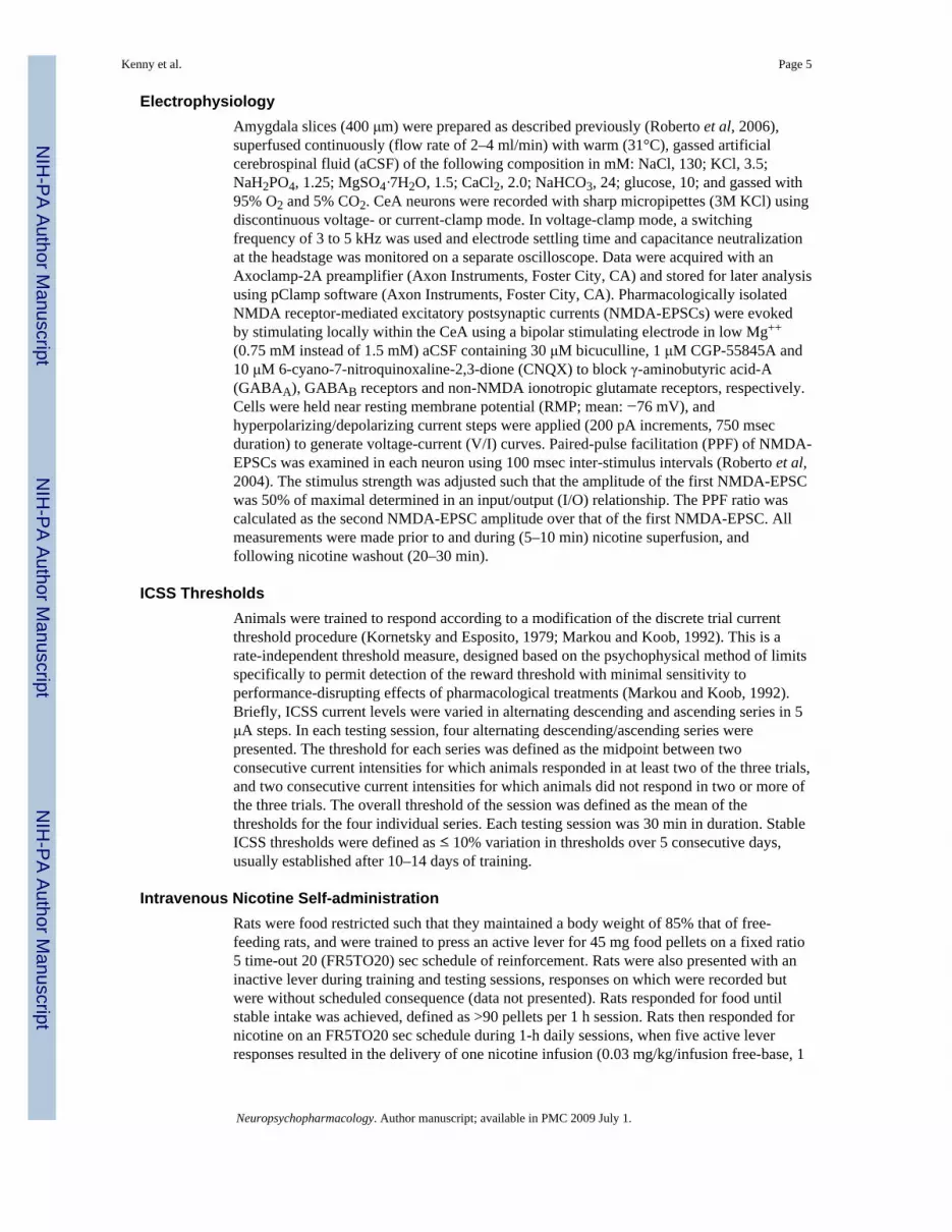
ElectrophysiologyAmygdala slices (400 μm) were prepared as described previously (Roberto et al, 2006),superfused continuously (flow rate of 2–4 ml/min) with warm (31°C), gassed artificialcerebrospinal fluid (aCSF) of the following composition in mM: NaCl, 130; KCl, 3.5;NaH2PO4, 1.25; MgSO4·7H2O, 1.5; CaCl2, 2.0; NaHCO3, 24; glucose, 10; and gassed with95% O2 and 5% CO2. CeA neurons were recorded with sharp micropipettes (3M KCl) usingdiscontinuous voltage- or current-clamp mode. In voltage-clamp mode, a switchingfrequency of 3 to 5 kHz was used and electrode settling time and capacitance neutralizationat the headstage was monitored on a separate oscilloscope. Data were acquired with anAxoclamp-2A preamplifier (Axon Instruments, Foster City, CA) and stored for later analysisusing pClamp software (Axon Instruments, Foster City, CA). Pharmacologically isolatedNMDA receptor-mediated excitatory postsynaptic currents (NMDA-EPSCs) were evokedby stimulating locally within the CeA using a bipolar stimulating electrode in low Mg++
(0.75 mM instead of 1.5 mM) aCSF containing 30 μM bicuculline, 1 μM CGP-55845A and10 μM 6-cyano-7-nitroquinoxaline-2,3-dione (CNQX) to block γ-aminobutyric acid-A(GABAA), GABAB receptors and non-NMDA ionotropic glutamate receptors, respectively.Cells were held near resting membrane potential (RMP; mean: −76 mV), andhyperpolarizing/depolarizing current steps were applied (200 pA increments, 750 msecduration) to generate voltage-current (V/I) curves. Paired-pulse facilitation (PPF) of NMDA-EPSCs was examined in each neuron using 100 msec inter-stimulus intervals (Roberto et al,2004). The stimulus strength was adjusted such that the amplitude of the first NMDA-EPSCwas 50% of maximal determined in an input/output (I/O) relationship. The PPF ratio wascalculated as the second NMDA-EPSC amplitude over that of the first NMDA-EPSC. Allmeasurements were made prior to and during (5–10 min) nicotine superfusion, andfollowing nicotine washout (20–30 min).
ICSS ThresholdsAnimals were trained to respond according to a modification of the discrete trial currentthreshold procedure (Kornetsky and Esposito, 1979; Markou and Koob, 1992). This is arate-independent threshold measure, designed based on the psychophysical method of limitsspecifically to permit detection of the reward threshold with minimal sensitivity toperformance-disrupting effects of pharmacological treatments (Markou and Koob, 1992).Briefly, ICSS current levels were varied in alternating descending and ascending series in 5μA steps. In each testing session, four alternating descending/ascending series werepresented. The threshold for each series was defined as the midpoint between twoconsecutive current intensities for which animals responded in at least two of the three trials,and two consecutive current intensities for which animals did not respond in two or more ofthe three trials. The overall threshold of the session was defined as the mean of thethresholds for the four individual series. Each testing session was 30 min in duration. StableICSS thresholds were defined as ≤ 10% variation in thresholds over 5 consecutive days,usually established after 10–14 days of training.
Intravenous Nicotine Self-administrationRats were food restricted such that they maintained a body weight of 85% that of free-feeding rats, and were trained to press an active lever for 45 mg food pellets on a fixed ratio5 time-out 20 (FR5TO20) sec schedule of reinforcement. Rats were also presented with aninactive lever during training and testing sessions, responses on which were recorded butwere without scheduled consequence (data not presented). Rats responded for food untilstable intake was achieved, defined as >90 pellets per 1 h session. Rats then responded fornicotine on an FR5TO20 sec schedule during 1-h daily sessions, when five active leverresponses resulted in the delivery of one nicotine infusion (0.03 mg/kg/infusion free-base, 1
Kenny et al. Page 5
Neuropsychopharmacology. Author manuscript; available in PMC 2009 July 1.
NIH
-PA Author Manuscript
NIH
-PA Author Manuscript
NIH
-PA Author Manuscript

sec infusion), that initiated the time-out period signaled by a light cue located above thelever, during which responding on the lever was without consequence.
Statistical AnalysesFor ICSS experiments, percentage change from baseline reward thresholds was calculatedby expressing the drug-influenced threshold scores as a percentage of the previous day’sthreshold (for experimenter-administered nicotine injections) or the mean thresholdsobtained on the three days prior to the first nicotine self-administration session. Data wereanalyzed by two-way or three-way repeated-measures analyses of variance (ANOVA) asappropriate. Post-hoc comparisons among means were conducted with the Fisher’s protectedt-tests. For nicotine self-administration and food responding experiments, percentage changefrom baseline nicotine or food reinforcers was calculated by expressing the number of drug-influenced reinforcers earned as a percentage of the baseline number of reinforcers minus100. Baseline intake was the mean number of reinforcers earned on the 3 days prior to eachinjection day. Data were subjected to two-factor repeated-measures ANOVA. IC50 valuesfor the effects of LY235959 on nicotine or food intake were calculated using GraphPadPrism software (GraphPad, San Diego, CA). For Western blots, the optical density of theprotein of interest was normalized to that of β-actin for each sample. Fold-induction datawere analyzed by three-way repeated-measures ANOVA (Brain region × Treatment ×Protein; with repeated-measures on Protein) followed by Fisher’s protected t-tests for eachbrain region. For electrophysiology experiments, all values were analyzed by between- orwithin-subjects repeated-measures ANOVA as appropriate, followed by Newman-Keulspost-hoc tests. In all cases, post-hoc comparisons were performed only after significant mainor interaction effects in an ANOVA. In this manner the null hypothesis could be rejectedand Type I errors during post-hoc test were less likely.
RESULTSExperiment 1 - NMDA receptors gate the magnitude and valence of the effects of nicotineon brain reward systems
Rats (n=9) were prepared with ICSS electrodes and trained in the ICSS threshold procedureuntil stable thresholds were established, see above. Rats were then pre-treated with saline orLY235959 (1 mg/kg) 30 min prior to testing, and received a nicotine injection (0, 0.125,0.25 or 0.5 mg/kg; within-subjects Latin-square design) 10 min prior to being tested in theICSS procedure. Next, the same rats were injected with saline or LY235959 (2.5 or 5 mg/kg;I.P.) 30 min prior to testing, and also a saline or nicotine (0.25 mg/kg; S.C.) injection 10 minprior to reward threshold assessment (Latin-square design). This dose of nicotine (0.25 mg/kg) was chosen because it induced maximal lowering of reward thresholds when testedalone or in combination with the lower dose (1 mg/kg) of LY235959 (see Fig. 1). Therewere at least 48 h between each drug injection.
Mean (± SEM) baseline ICSS threshold prior to drug treatment was 128.8 ± 13.9 μA. Two-way repeated-measures ANOVA on the data involving the 1 mg/kg LY235959 dosedemonstrated a significant main effect of Nicotine (F(3,24)=6.3, p<0.01), no effect ofLY235959 (1 mg/kg) (F(1,8)=0.18), nor a significant LY235959 × Nicotine interaction:(F (3,24)=1.1). Further analysis of the main effect of Nicotine demonstrated that thresholdswere significantly lowered in rats pre-treated with saline or LY235959 at nicotine doses0.125–0.25 mg/kg (p<0.05; Fig. 1a).
Two-way repeated-measures ANOVA on the data involving the two higher doses ofLY235959 (2.5–5 mg/kg) and the threshold-lowering effects of a single dose of nicotine(0.25 m/kg) demonstrated a main effect of LY235959 (F(1,8)=6.4, p<0.01), and a significant
Kenny et al. Page 6
Neuropsychopharmacology. Author manuscript; available in PMC 2009 July 1.
NIH
-PA Author Manuscript
NIH
-PA Author Manuscript
NIH
-PA Author Manuscript

Nicotine × LY235959 interaction (F(1,8)=6.3, p<0.01), and no statistically significant maineffect of nicotine (F(1,8)=0.02, p=0.9). Further analysis of the main effect of LY235959demonstrated that reward thresholds were significantly elevated in rats pre-treated with thehighest does of LY235959 (5 mg/kg) (p<0.01; Fig. 1b).
Experiment 2 - Reward-enhancing and reinforcing effects of self-administered nicotinerequire NMDA receptors
Rats (n=16) were prepared with ICSS electrodes and trained in the ICSS threshold procedureuntil stable reward thresholds were achieved. All rats were then prepared with intravenouscatheters. One group of rats (nicotine rats; n=9) was trained in the nicotine self-administration procedure, and reward thresholds were assessed 1-h before (pre-thresholds)and 15 min after (post-thresholds) each self-administration session. The remaining rats(control rats; n=7) were tested in the ICSS procedure at the same times each day as the self-administering rats, but were returned to their home-cages during the self-administrationsessions, and thus remained nicotine-naïve throughout the experiment.
Mean (± SEM) baseline ICSS thresholds prior to the start of nicotine self-administrationwere 92.1 ± 15.2 μA and 75.3 ± 5.2 μA in the nicotine and control rats, respectively. Mean(± SEM) baseline number of nicotine infusions earned by nicotine rats was 10.5 ± 1.0. Asexpected, pre-thresholds and post-thresholds remained stable and unaltered in the controlrats (Fig. 2a). However, in replication of our recently published data (Kenny and Markou,2006;Paterson et al, 2007), daily post-thresholds were lowered after each self-administrationsession in the nicotine rats compared with pre-thresholds during the initial 7-day access tonicotine, prior to any LY235959 treatment (Fig. 2b). In addition, a downward drift wasobserved in pre- and post-thresholds in nicotine rats (Fig. 2b) similar to that previouslyreported (Kenny and Markou, 2006), reflecting a long-lasting nicotine-induced increase inthe baseline sensitivity of brain reward systems. Accordingly, two-way repeated-measuresANOVA over this 7-day period on threshold data from control and nicotine ratsdemonstrated a significant main effect of Drug (nicotine or control group) (F(1,13)=5.7,p<0.05), a main effect of Session (pre-thresholds or post-thresholds) (F(1,13)=6.3, p<0.05),and a significant Drug × Session interaction (F(1,13)=7.9, p<0.05). Two-way ANOVA of thethreshold data obtained only from the nicotine rats over this time period revealed significantmain effects of Days (F(6,48)=2.3, p<0.05) and Session (F(1,8)=12.1, p<0.01).
After 7 consecutive days of nicotine self-administration, the effects of LY235959 on rewardthresholds were examined in nicotine and control rats. First, the effects of LY235959 on pre-thresholds, nicotine self-administration, and post-thresholds were assessed. That is, bothgroups of rats were pre-treated with LY235959 (0, 0.5 1 or 2.5 mg/kg; Latin-square design;minimum of 7 days between injections), and 15 min later pre-thresholds were assessed. Ratswere then permitted to self-administer nicotine or were returned to their home-cages (controlrats), and post-thresholds were subsequently assessed. Administration of LY235959 beforepre-threshold assessment did not alter pre-thresholds or post-thresholds in control rats (Fig.2c). However, LY235959 blocked the lowering of post-nicotine reward thresholds typicallyobserved in nicotine rats (Fig. 2d). Three-way repeated-measures ANOVA on the rewardthreshold data confirmed a significant main effect of LY235959 (F(3,42)=4.5, p<0.01), and asignificant Drug (nicotine or control group) × LY235959 interaction (F(3,42)=3.8, p<0.05).Two-way ANOVA on the reward threshold data from the nicotine rats demonstrated a maineffect of LY235959 (F(3,24)=6.5, p<0.01).
Because no effect of Session was observed in the above analysis, data from the two Sessions(pre-thresholds and post-thresholds) were combined for each dose and then compared withthe 0 mg/kg treatment condition for the nicotine rats. This analyses indicated that there wasa main effect of LY235959 (F(3,24)=10.3, p<0.001), and that thresholds tended to be elevated
Kenny et al. Page 7
Neuropsychopharmacology. Author manuscript; available in PMC 2009 July 1.
NIH
-PA Author Manuscript
NIH
-PA Author Manuscript
NIH
-PA Author Manuscript

at the 0.5 mg/kg dose (p=0.06), and were significantly elevated at the 1 mg/kg (p<0.05) and2.5 mg/kg (p<0.001) LY235959 doses compared with thresholds observed after salinetreatment (Fig. 2d).
In addition to reversing the lowering effects of self-administered nicotine on ICSSthresholds, LY235959 also decreased nicotine intake. Mean (± SEM) baseline number ofnicotine reinforcers earned by the nicotine rats prior to LY235959 treatment was 12.0 ± 1.5.LY235959 dose-dependently decreased nicotine intake in the nicotine rats (Fig. 2e),reflected in a significant effect of LY235959 (F(3,24)=41.0, p<0.001). Post-hoc analysisdemonstrated that nicotine intake was significantly decreased by the 0.5 mg/kg (p<0.05) andthe 1–2.5 mg/kg (p<0.01) LY235959 doses (Fig. 2e).
Next, we examined the effects of LY235959 (0–2.5 mg/kg; Latin-square design)administered immediately after the self-administration session on post-thresholds assessed15 min later. In this manner, we could directly assess the influence of LY235959 onthreshold lowering induced by self-administered nicotine whilst avoiding the confoundinginhibitory effects of LY235959 on nicotine self-administration. LY235959 administered inthis manner reversed the lowering of post-thresholds observed in the nicotine rats, but didnot alter thresholds in control rats (Fig. 2f). Two-way repeated-measures ANOVAdemonstrated a significant main effect of LY235959 (F(3,42)=6.5, p<0.01), and a significantDrug (nicotine or control group) × LY235959 interaction (F(3,42)=8.3, p<0.001), and noeffect of Drug (F(1,14)=1.7, p=0.2). One-way repeated-measures ANOVA of threshold datafrom nicotine rats demonstrated a significant effect of LY235959 (F(3,24)=13.5, p<0.001).Post-hoc analysis on the reward thresholds in nicotine rats demonstrated that thresholdswere significantly elevated after the 0.5 mg/kg (p<0.05), 1 mg/kg (p<0.01) and 2.5 mg/kg(p<0.001) LY235959 doses compared with thresholds after saline administration (Fig. 2f).
To determine if the inhibitory effects of LY235959 on nicotine self-administration observedin the nicotine rats above (Fig. 2e) were secondary to a disruptive effect on operantperformance, we again assessed the effects of LY235959 (0.1–5 mg/kg) on nicotine self-administration in a new cohort of rats, and compared these effects with the actions ofLY235959 on responding for food reinforcement. Nicotine self-administering rats (n=7)were injected with LY235959 (0, 0.1, 0.5, 1, 2.5 or 5 mg/kg; Latin-square design), andnicotine intake was evaluated 30 min later. The effects of LY235959 were also assessed inrats (n=8) trained to respond for food reinforcement (45 mg pellet) under a FR5TO20 secschedule, and in rats (n=7) trained under a FR5TO210 sec schedule. This adjusted scheduleincorporated a longer time-out period (210 s vs. of 20 s) that better equated rates ofresponding for nicotine and food.
Mean (± SEM) baseline number of nicotine infusions earned by this second cohort of ratsprior to treatment with LY235959 was 10.8 ± 0.7. The mean (± SEM) baseline number offood rewards earned by rats under the FR5TO20 sec and the FR5TO 210 sec schedules ofreinforcement prior to LY235959 treatment were 109.5 ± 9.6 and 16.3 ± 0.3, respectively.Two-way repeated-measures ANOVA demonstrated a significant effect of Reinforcer(F(2,19)=17.2, p<0.001), a significant effect of LY235959 (F(5,10)=19.4, p<0.001), and noReinforcer × LY235959 interaction (F(10,95)=1.4). Pre-planned comparisons demonstratedthat LY235959 decreased nicotine intake at doses ≥ 0.5 mg/kg (Fig. 2 g; confirming theresults reported above in a separate cohort of rats shown in Fig. 2e), but decreased foodintake under FR5TO20 sec or FR5TO210 sec reinforcement schedules only at the highestdoses tested (2.5–5 mg/kg; Fig. 2g). Further analyses of the above data demonstrated thatLY235959 decreased nicotine intake with an IC50 of 0.53 mg/kg, and decreased foodresponding under FR5TO20 and FR5TO210 sec schedules with IC50values of 1.88 and 2.2mg/kg, respectively.
Kenny et al. Page 8
Neuropsychopharmacology. Author manuscript; available in PMC 2009 July 1.
NIH
-PA Author Manuscript
NIH
-PA Author Manuscript
NIH
-PA Author Manuscript
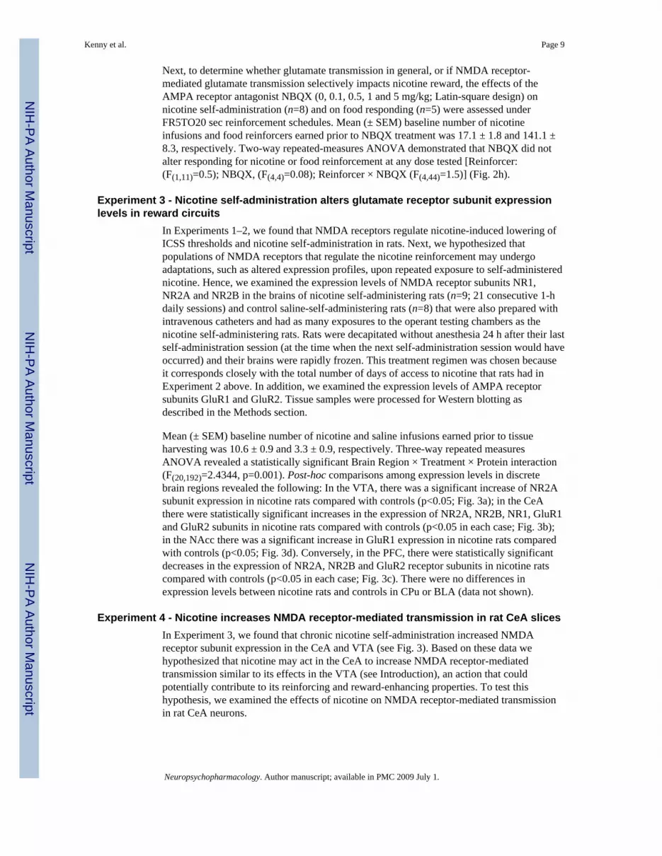
Next, to determine whether glutamate transmission in general, or if NMDA receptor-mediated glutamate transmission selectively impacts nicotine reward, the effects of theAMPA receptor antagonist NBQX (0, 0.1, 0.5, 1 and 5 mg/kg; Latin-square design) onnicotine self-administration (n=8) and on food responding (n=5) were assessed underFR5TO20 sec reinforcement schedules. Mean (± SEM) baseline number of nicotineinfusions and food reinforcers earned prior to NBQX treatment was 17.1 ± 1.8 and 141.1 ±8.3, respectively. Two-way repeated-measures ANOVA demonstrated that NBQX did notalter responding for nicotine or food reinforcement at any dose tested [Reinforcer:(F(1,11)=0.5); NBQX, (F(4,4)=0.08); Reinforcer × NBQX (F(4,44)=1.5)] (Fig. 2h).
Experiment 3 - Nicotine self-administration alters glutamate receptor subunit expressionlevels in reward circuits
In Experiments 1–2, we found that NMDA receptors regulate nicotine-induced lowering ofICSS thresholds and nicotine self-administration in rats. Next, we hypothesized thatpopulations of NMDA receptors that regulate the nicotine reinforcement may undergoadaptations, such as altered expression profiles, upon repeated exposure to self-administerednicotine. Hence, we examined the expression levels of NMDA receptor subunits NR1,NR2A and NR2B in the brains of nicotine self-administering rats (n=9; 21 consecutive 1-hdaily sessions) and control saline-self-administering rats (n=8) that were also prepared withintravenous catheters and had as many exposures to the operant testing chambers as thenicotine self-administering rats. Rats were decapitated without anesthesia 24 h after their lastself-administration session (at the time when the next self-administration session would haveoccurred) and their brains were rapidly frozen. This treatment regimen was chosen becauseit corresponds closely with the total number of days of access to nicotine that rats had inExperiment 2 above. In addition, we examined the expression levels of AMPA receptorsubunits GluR1 and GluR2. Tissue samples were processed for Western blotting asdescribed in the Methods section.
Mean (± SEM) baseline number of nicotine and saline infusions earned prior to tissueharvesting was 10.6 ± 0.9 and 3.3 ± 0.9, respectively. Three-way repeated measuresANOVA revealed a statistically significant Brain Region × Treatment × Protein interaction(F(20,192)=2.4344, p=0.001). Post-hoc comparisons among expression levels in discretebrain regions revealed the following: In the VTA, there was a significant increase of NR2Asubunit expression in nicotine rats compared with controls (p<0.05; Fig. 3a); in the CeAthere were statistically significant increases in the expression of NR2A, NR2B, NR1, GluR1and GluR2 subunits in nicotine rats compared with controls (p<0.05 in each case; Fig. 3b);in the NAcc there was a significant increase in GluR1 expression in nicotine rats comparedwith controls (p<0.05; Fig. 3d). Conversely, in the PFC, there were statistically significantdecreases in the expression of NR2A, NR2B and GluR2 receptor subunits in nicotine ratscompared with controls (p<0.05 in each case; Fig. 3c). There were no differences inexpression levels between nicotine rats and controls in CPu or BLA (data not shown).
Experiment 4 - Nicotine increases NMDA receptor-mediated transmission in rat CeA slicesIn Experiment 3, we found that chronic nicotine self-administration increased NMDAreceptor subunit expression in the CeA and VTA (see Fig. 3). Based on these data wehypothesized that nicotine may act in the CeA to increase NMDA receptor-mediatedtransmission similar to its effects in the VTA (see Introduction), an action that couldpotentially contribute to its reinforcing and reward-enhancing properties. To test thishypothesis, we examined the effects of nicotine on NMDA receptor-mediated transmissionin rat CeA neurons.
Kenny et al. Page 9
Neuropsychopharmacology. Author manuscript; available in PMC 2009 July 1.
NIH
-PA Author Manuscript
NIH
-PA Author Manuscript
NIH
-PA Author Manuscript
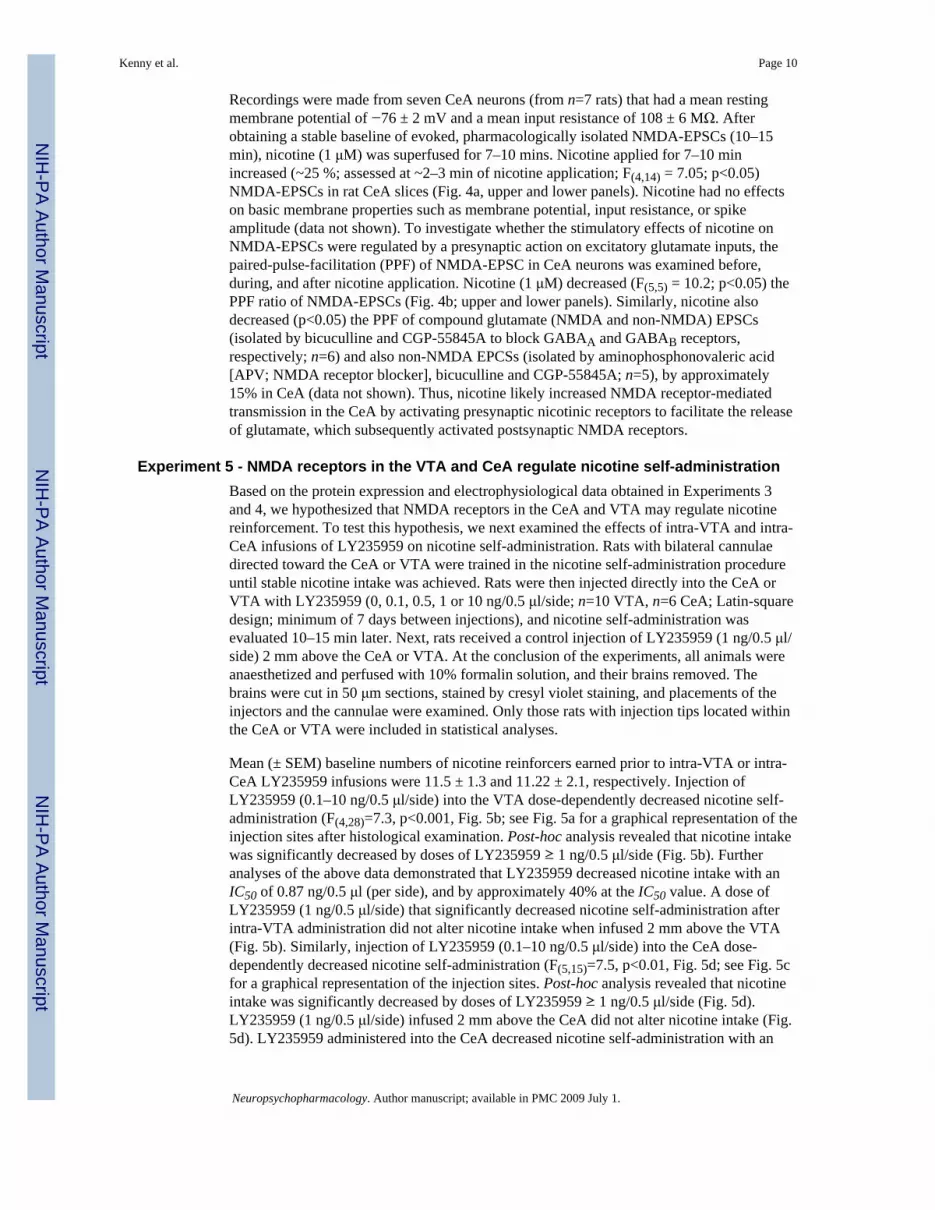
Recordings were made from seven CeA neurons (from n=7 rats) that had a mean restingmembrane potential of −76 ± 2 mV and a mean input resistance of 108 ± 6 MΩ. Afterobtaining a stable baseline of evoked, pharmacologically isolated NMDA-EPSCs (10–15min), nicotine (1 μM) was superfused for 7–10 mins. Nicotine applied for 7–10 minincreased (~25 %; assessed at ~2–3 min of nicotine application; F(4,14) = 7.05; p<0.05)NMDA-EPSCs in rat CeA slices (Fig. 4a, upper and lower panels). Nicotine had no effectson basic membrane properties such as membrane potential, input resistance, or spikeamplitude (data not shown). To investigate whether the stimulatory effects of nicotine onNMDA-EPSCs were regulated by a presynaptic action on excitatory glutamate inputs, thepaired-pulse-facilitation (PPF) of NMDA-EPSC in CeA neurons was examined before,during, and after nicotine application. Nicotine (1 μM) decreased (F(5,5) = 10.2; p<0.05) thePPF ratio of NMDA-EPSCs (Fig. 4b; upper and lower panels). Similarly, nicotine alsodecreased (p<0.05) the PPF of compound glutamate (NMDA and non-NMDA) EPSCs(isolated by bicuculline and CGP-55845A to block GABAA and GABAB receptors,respectively; n=6) and also non-NMDA EPCSs (isolated by aminophosphonovaleric acid[APV; NMDA receptor blocker], bicuculline and CGP-55845A; n=5), by approximately15% in CeA (data not shown). Thus, nicotine likely increased NMDA receptor-mediatedtransmission in the CeA by activating presynaptic nicotinic receptors to facilitate the releaseof glutamate, which subsequently activated postsynaptic NMDA receptors.
Experiment 5 - NMDA receptors in the VTA and CeA regulate nicotine self-administrationBased on the protein expression and electrophysiological data obtained in Experiments 3and 4, we hypothesized that NMDA receptors in the CeA and VTA may regulate nicotinereinforcement. To test this hypothesis, we next examined the effects of intra-VTA and intra-CeA infusions of LY235959 on nicotine self-administration. Rats with bilateral cannulaedirected toward the CeA or VTA were trained in the nicotine self-administration procedureuntil stable nicotine intake was achieved. Rats were then injected directly into the CeA orVTA with LY235959 (0, 0.1, 0.5, 1 or 10 ng/0.5 μl/side; n=10 VTA, n=6 CeA; Latin-squaredesign; minimum of 7 days between injections), and nicotine self-administration wasevaluated 10–15 min later. Next, rats received a control injection of LY235959 (1 ng/0.5 μl/side) 2 mm above the CeA or VTA. At the conclusion of the experiments, all animals wereanaesthetized and perfused with 10% formalin solution, and their brains removed. Thebrains were cut in 50 μm sections, stained by cresyl violet staining, and placements of theinjectors and the cannulae were examined. Only those rats with injection tips located withinthe CeA or VTA were included in statistical analyses.
Mean (± SEM) baseline numbers of nicotine reinforcers earned prior to intra-VTA or intra-CeA LY235959 infusions were 11.5 ± 1.3 and 11.22 ± 2.1, respectively. Injection ofLY235959 (0.1–10 ng/0.5 μl/side) into the VTA dose-dependently decreased nicotine self-administration (F(4,28)=7.3, p<0.001, Fig. 5b; see Fig. 5a for a graphical representation of theinjection sites after histological examination. Post-hoc analysis revealed that nicotine intakewas significantly decreased by doses of LY235959 ≥ 1 ng/0.5 μl/side (Fig. 5b). Furtheranalyses of the above data demonstrated that LY235959 decreased nicotine intake with anIC50 of 0.87 ng/0.5 μl (per side), and by approximately 40% at the IC50 value. A dose ofLY235959 (1 ng/0.5 μl/side) that significantly decreased nicotine self-administration afterintra-VTA administration did not alter nicotine intake when infused 2 mm above the VTA(Fig. 5b). Similarly, injection of LY235959 (0.1–10 ng/0.5 μl/side) into the CeA dose-dependently decreased nicotine self-administration (F(5,15)=7.5, p<0.01, Fig. 5d; see Fig. 5cfor a graphical representation of the injection sites. Post-hoc analysis revealed that nicotineintake was significantly decreased by doses of LY235959 ≥ 1 ng/0.5 μl/side (Fig. 5d).LY235959 (1 ng/0.5 μl/side) infused 2 mm above the CeA did not alter nicotine intake (Fig.5d). LY235959 administered into the CeA decreased nicotine self-administration with an
Kenny et al. Page 10
Neuropsychopharmacology. Author manuscript; available in PMC 2009 July 1.
NIH
-PA Author Manuscript
NIH
-PA Author Manuscript
NIH
-PA Author Manuscript

IC50 value of 0.97 ng/0.5 μl (per side), and reduced intake by approximately 20% at theIC50value.
DISCUSSIONLY235959 is the active isomer of LY274614 (Ornstein et al, 1993), and acts as a highlypotent and selective competitive antagonist at NMDA receptors in vivo and in vitro(Schoepp et al, 1991; Fischer and Dykstra, 2006). We found that LY235959 attenuated theamplification of brain reward function induced by experimenter-administered orintravenously self-administered nicotine injections, as reflected in blockade of nicotine-induced lowering of ICSS thresholds. Indeed, antagonism of NMDA receptors switched theeffects of volitional and non-volitional nicotine administration on brain reward systems fromstimulatory to inhibitory, detected by nicotine-induced elevations of ICSS thresholds.Furthermore, LY235959 decreased nicotine self-administration at doses that did not alterresponding for food reinforcement, whereas the AMPA receptor antagonist NBQX did notalter nicotine self-administration behavior. These observations suggest that NMDAreceptors, but not AMPA receptors, regulate the magnitude and valence of nicotine’s effectson brain reward systems, and may thereby regulate nicotine self-administration behavior.We also found that NMDA receptor subunit expression was up-regulated in the CeA andVTA of nicotine self-administering rats, that nicotine transiently increased EPSCs in theCeA via potentiation of glutamate release similar to its previously reported effects in theVTA, and that LY235959 infused into the CeA or VTA decreased nicotine self-administration. Taken together, these findings suggest that nicotine enhances glutamate-mediated transmission at NMDA receptors in the CeA and VTA, and that this action ofnicotine is central to its stimulatory effects on brain reward function and the motivation toconsume the drug. Further, it is likely that NMDA receptors that regulate the reinforcingactions of nicotine undergo profound adaptations after chronic exposure to volitionallyconsumed nicotine.
Reward-enhancing and reinforcing effects of nicotine require NMDA receptorsExperimenter-administered nicotine infusions lowered ICSS thresholds, suggesting thatnicotine increased the rewarding effects of ICSS; that is, nicotine enhanced the reward valueof the ICSS stimulation. We found that lower doses of LY235959 blocked, whereas a higherdose reversed the effects of experimenter-administered nicotine from reward-enhancing intoreward-inhibitory. Importantly, doses of LY235959 (2.5–5 mg/kg) that blocked or reversedthe effects of nicotine on brain reward systems had no effects on baseline reward thresholdsin saline pretreated animals. Similarly, these doses of LY235959 did not alter responselatencies, considered an accurate measure of performance in the ICSS procedure (Kornetskyand Esposito, 1979; Markou and Koob, 1992), in saline- or nicotine-treated rats (data notshown). These observations support the notion that LY235959 selectively modulates thereward-related effects of nicotine, and that the actions of LY235959 are not secondary toinhibitory effects on motor performance in the ICSS procedure.
Considerable evidence suggests that brain reward systems may respond differentially tovolitional vs. non-volitional drug consumption. Indeed, neurobiological correlates of reward(e.g., neurotransmitter release in the basal ganglia) are increased after response-contingentdrug infusions by a far greater magnitude than by response-non-contingent drug infusions(Wilson et al, 1994; Mark et al, 1999; Kimmel et al, 2005; Lecca et al, 2007). In addition,different gene expression, neurochemical, stress system and behavioral adaptations occur inresponse to volitional vs. non-volitional drug consumption (Stefanski et al, 1999; Jacobs etal, 2005). Therefore, in addition to effects on experimenter-administered nicotine injections,we also assessed the effects of LY235959 on the threshold lowering induced byintravenously self-administered nicotine infusions. We found that at lower doses LY235959
Kenny et al. Page 11
Neuropsychopharmacology. Author manuscript; available in PMC 2009 July 1.
NIH
-PA Author Manuscript
NIH
-PA Author Manuscript
NIH
-PA Author Manuscript

blocked nicotine-induced lowering of reward thresholds in self-administering rats, and athigher doses LY235959 reversed the effects of nicotine on brain reward systems fromstimulatory to inhibitory. LY235959 also reversed the long-lasting stimulatory actions ofvolitionally consumed nicotine on brain reward systems, reflected in persistently loweredreward thresholds assessed before each daily self-administration session (pre-thresholds;Fig. 2d). Importantly, doses of LY235959 that reversed the effects of nicotine on rewardthresholds in nicotine self-administering animals did not alter thresholds in control animals.Hence, together with the fact that reward thresholds detected in the rate-free procedureutilized in these studies are not affected by motoric effects of manipulations, these effects ofLY235959 are unlikely to be secondary to non-specific inhibitory actions on operantperformance. Taken together, the above data are consistent with a permissive role forNMDA receptors in regulating the stimulatory effects of nicotine on brain reward systems.Blockade of α7 nAChRs in the VTA could similarly reverse the hedonic valence of VTA-infused nicotine injections from rewarding to aversive, measured in a place conditioningprocedure (Laviolette and van der Kooy, 2003). Nicotine increases glutamate release in theVTA by a stimulatory action at α7 nAChRs, and thereby facilitates NMDA receptor-mediated transmission in this brain region (Mansvelder and McGehee, 2000; Schilstrom etal, 2000). Thus, blockade of nicotine’s stimulatory effects on NMDA receptor-mediatedtransmission, either directly or indirectly, likely switches the hedonic valence of nicotinefrom positive to negative.
In addition to reversing the reward-enhancing effects of nicotine, LY235959 also dose-dependently decreased nicotine self-administration. Furthermore, a broad range ofLY235959 doses (0.1–1 mg/kg) that decreased nicotine self-administration failed to alterresponding for food under a similar schedule of reinforcement (FR5TO20 sec), or under anadjusted schedule (FR5TO210 sec) that better equated overall rates of responding for foodand nicotine. Thus, antagonism of NMDA receptors unlikely decreased nicotine intake byinducing non-specific deficits in operant performance. Instead, these data are consistent withthe hypothesis that nicotine-induced amplification of brain reward activity provides animportant source of motivation that supports the initiation and maintenance of nicotine self-administration in rats (Kenny and Markou, 2006), and perhaps the tobacco smoking habit inhumans (Donny et al, 2003; Kenny, 2007). Based on our observation that NMDA receptorblockade reversed the valence of nicotine’s actions on reward circuitries, we may speculatethat NMDA receptor antagonism decreased nicotine intake by rendering rats less sensitive tothe appetitive effects of nicotine and more sensitive to its aversive effects, see (Laviolette etal, 2002; Laviolette and van der Kooy, 2003). Such an interpretation would account for thefact that rats did not display compensatory increases in their nicotine intake after LY235959treatment similar to those observed in cocaine or amphetamine self-administering ratstreated with doses of dopamine receptor antagonists that attenuate the rewarding effects ofthese drugs (Yokel and Wise, 1975; Koob et al, 1987).
In rats that intravenously self-administered nicotine, we observed a gradual lowering ofbaseline reward thresholds across days, an effect not observed in nicotine-naive control rats.This observation confirms recent data from our laboratory (Kenny and Markou, 2006), andsuggests that the excitatory effects of self-administered nicotine on reward systems are longlasting and at least partly cumulative(Kenny and Markou, 2006). Intriguingly, recent datafrom human patients have shown that even a single exposure to nicotine can inducepersistent increases (> 7 days) in sensitivity to non-drug rewarding environmental stimuli(Barr et al, 2007). Considering that nicotine-induced amplification of brain reward signals isconsidered an important factor in motivating nicotine consummatory behaviors (Kenny andMarkou, 2006; Kenny, 2007), the enduring stimulatory effects of nicotine on reward systemscould potentially contribute to the reinforcing effects of the drug and facilitate thedevelopment of nicotine dependence. The mechanisms by which self-administered nicotine
Kenny et al. Page 12
Neuropsychopharmacology. Author manuscript; available in PMC 2009 July 1.
NIH
-PA Author Manuscript
NIH
-PA Author Manuscript
NIH
-PA Author Manuscript

elicits persistent increases in reward sensitivity are unclear but may involve hypersensitivityof nAChRs located in brain reward systems (Dani and Heinemann, 1996; Buisson andBertrand, 2002). Indeed, the nAChR antagonist dihydro-β-erythroidine (DHβE) blockedboth the acute lowering of reward thresholds and also the persistently lowered baselinereward thresholds induced by self-administered nicotine in rats (Kenny and Markou, 2006).Similarly, in the present study LY235959 also reversed acutely lowered and persistentlylowered baseline reward thresholds in nicotine self-administering rats. Thus, is possible thatpersistently activated NMDA receptors in brain reward circuitries, either directly throughaltered number and/or function of these receptors (see below) or perhaps indirectly throughnicotine-induced hyperactivity of nAChRs, may contribute to the enduringly increasedreward sensitivity in nicotine self-administering rats.
Non-competitive antagonists of the NMDA receptor such as phencyclidine (PCP), ketamineand dizocilpine, have reward-facilitating actions and can lower ICSS thresholds in rats(Herberg and Rose, 1989; Carlezon and Wise, 1993). This stimulatory action of NMDAreceptor channel blockers on brain reward systems likely contributes to the abuse liability ofthese ligands. In the present study, we found that LY235959 had no intrinsic reward-facilitating (i.e., ICSS threshold-lowering) effects at any dose tested. Thus, it is likely thatLY235959 and mechanistically related NMDA receptor antagonists are likely hedonicallyinert. The reason for the differential sensitivity of brain reward systems to non-competitiveversus competitive NMDA receptor antagonists is presently unclear. However, one possibleexplanation is the fact that NMDA receptor channel blockers are “use dependent”antagonists that voltage-dependently occlude NMDA receptors, whereas competitiveantagonists act at the glutamate binding site to competitively antagonize NMDA receptors.Thus, it is possible that NMDA receptor channel blockers but not competitive antagonistsmay selectively target populations of NMDA receptors, perhaps in a voltage-dependentmanner, that have a tonic inhibitory influence on brain reward systems. Whatever theexplanation may be, the present data suggest that the competitive NMDA receptorantagonists are hedonically inert and therefore devoid of intrinsic abuse liability, anobservation that supports their potential clinical utility for the treatment of substance abusedisorders.
Plasticity of glutamate receptors in brain reward circuits induced by self-administerednicotine
Far lower doses of LY235959 (0.5–1 mg/kg) were required to block the ICSS threshold-lowering effects of self-administered nicotine compared with those doses ≥ 2.5 mg/kg)(required to block the effects of bolus experimenter-administered nicotine injections (Figs. 1and 2). The magnitude by which reward thresholds were lowered was equivalent followingexperimenter-administered or self-administration nicotine injections. Furthermore, ratsobtained similar amounts of acute nicotine under these two treatment conditions. Thus, theincreased sensitivity to LY235959 in nicotine self-administering rats was unlikely secondaryto differences in the magnitude by which thresholds were lowered, or to different amounts ofnicotine acting on reward systems. Instead, these observations suggest that populations ofNMDA receptors that regulate the actions of nicotine may undergo functional adaptationafter nicotine self-administration, thereby increasing their sensitivity to antagonist blockade.Based on this behavioral observation, we next assessed NMDA receptor subunit expressionlevels in the brains of nicotine self-administering rats compared with controls. Theunderlying hypothesis was that populations of NMDA receptors that regulate the reinforcingeffects of nicotine would undergo compensatory adaptations, including altered expression ofthe protein subunits that compose the receptor, upon repeated activation by self-administered nicotine infusions.
Kenny et al. Page 13
Neuropsychopharmacology. Author manuscript; available in PMC 2009 July 1.
NIH
-PA Author Manuscript
NIH
-PA Author Manuscript
NIH
-PA Author Manuscript

In general, NMDA receptors are heteromeric complexes composed of multiple NR1subunits in combination with at least one of the four types of NR2 subunits (Cull-Candy etal, 2001). Both NR1 and NR2 subunits confer distinct properties to the NMDA receptor, andchanges in expression levels of the subunits would be expected to alter NMDA receptorsubunit stoichiometry and function. We found that the expression of NMDA receptorsubunits (NR2A, NR2B and/or NR1) was increased in the VTA and CeA, decreased in thePFC and unaltered in the NAcc, BLA or CPu, of nicotine self-administering rats comparedwith controls. Importantly, similar increases in NMDA receptor subunit expression in theVTA and CeA and decreases in the PFC have been observed in rats self-administeringcocaine, opiates or alcohol and in post-mortem brain tissues from human cocaine users(Ortiz et al, 1995; Ghasemzadeh et al, 1999; Lu et al, 2003; Tang et al, 2003; Turchan et al,2003; Lu et al, 2005; Roberto et al, 2006; Ben-Shahar et al, 2007). The effects of self-administered nicotine on NMDA receptor subunit expression were far more pronounced inthe CeA compared with the VTA. Indeed, marked increases were observed in the expressionof all NMDA receptor subunits assessed in the CeA, whereas only the NR2A subunit wasincreased in the VTA. The underlying mechanism or functional significance of thisdifferential pattern of NMDA receptor subunit upregulation is unclear but may indicate thatthe CeA is particularly sensitive to the effects of nicotine and readily undergoes functionaladaptations upon consumption of the drug. A caveat to these expression data is that NMDAreceptor subunit levels were detected in tissue homogenates, and not from purifiedmembrane homogenates in which mature NMDA receptors are located. Thus, the alteredNMDA receptor subunit expression levels that were observed may reflect, in part, alteredlevels of intracellular subunits not yet incorporated into mature receptors in the membrane.In contrast to the present data, a recent study reported that nicotine intake did not alterNMDA receptor subunit expression in the VTA, and increased expression in the PFC (Wanget al, 2007). The effects of nicotine self-administration on NMDA receptor subunitexpression in the amygdala were not assessed in this previous study (Wang et al, 2007).There are a number of methodological differences that may account for these apparentdiscrepancies. First, in the present investigations Wistar rats were used, whereas Wang andcolleagues used Sprague-Dawley. Further, in our studies rats were permitted 1-h dailyaccess to nicotine self-administration, whereas the previous study employed a 23-h dailyaccess schedule. Finally, in our study brain tissues were harvested approximately 23-h afterthe final nicotine self-administration session, whereas Wang and colleagues collected brainsapproximately 30 mins after the last nicotine infusion (Wang et al, 2007). Thus, it ispossible that expression levels of NMDA receptor subunits may be temporally regulated in ahighly dynamic manner, such that the time since last nicotine exposure may play animportant role in determining the direction in which NMDA receptor subunit levels arealtered. Our data suggest that NMDA receptors in the VTA, CeA and PFC undergoprofound plasticity upon repeated exposure to rewarding doses of self-administered nicotine.Recent data have shown that other glutamate receptors, including group II metabotropicglutamate (mGlu2/3) receptors, similarly undergo profound adaptations upon repeatedexposure to self-administered nicotine and when assessed 24 hr after the last nicotine self-administration session, similar to the present study (Liechti et al, 2007). In future studies, itwill be interesting to determine if similar adaptations in NMDA receptors are observed in“yoked” animals that receive intravenous nicotine infusions non-volitionally in a mannertime-locked to a partner rat that responds for nicotine volitionally. Inasmuch as alteredNMDA receptor subunit expression levels can be correlated with complex behavioral states,we speculate that these data may explain the differential sensitivity to NMDA receptorblockade that we observed in these rats relative to non-self-administering rats. Importantly,NMDA receptor plasticity is considered a fundamental process in the regulation of synapticefficacy and remodeling in the brain (Lau and Zukin, 2007). Thus, the nicotine-inducedplasticity in NMDA receptors located in brain regions heavily implicated in drugdependence processes may play an important role in the persistence of nicotine self-
Kenny et al. Page 14
Neuropsychopharmacology. Author manuscript; available in PMC 2009 July 1.
NIH
-PA Author Manuscript
NIH
-PA Author Manuscript
NIH
-PA Author Manuscript

administration behavior in rats, and perhaps the tobacco habit in human smokers (Kenny andMarkou, 2004; Jones and Bonci, 2005; Kalivas and Volkow, 2005); see also (Kenny et al,2003).
Role of NMDA receptors in the VTA and CeA in nicotine self-administrationThe VTA is considered a key anatomical region that regulates nicotine dependenceprocesses (Corrigall et al, 1994; Kenny et al, 2003; Maskos et al, 2005; David et al, 2006;Ikemoto et al, 2006). Based on our observation that NMDA receptor subunit expression wasup-regulated in the VTA and CeA, we hypothesized that nicotine may act in these two areasto increase NMDA receptor-mediated transmission, and thereby elicit its reinforcing effects.Nicotine increases NMDA receptor-mediated transmission in the VTA of rats (Fu et al,2000; Grillner and Svensson, 2000; Mansvelder and McGehee, 2000; Schilstrom et al,2000). Considerably less is known regarding the effects of nicotine in the CeA, although arecent study suggested that nicotine may enhance glutamate-mediated transmission in themouse amygdala (Barazangi and Role, 2001). Therefore, we examined the effects ofnicotine on NMDA receptor-mediated transmission in rat CeA slices. As predicted, nicotinetransiently increased NMDA receptor-mediated transmission in the CeA, likely bystimulating nAChRs located on presynaptic glutamate terminals to facilitate glutamaterelease, subsequently activating postsynaptic NMDA receptors. A drawback to the presentstudies is that the effects of nicotine on NMDA receptor-mediated currents were assessedonly in drug-naive rats, and not in rats with prior experience of nicotine self-administration.Such an assessment may have revealed alterations in the stimulatory actions of nicotine onexcitatory glutamate release, or modifications in the response of NMDA receptors tonicotine-enhanced glutamate transmission, that represent important adaptations to nicotineself-administration that contribute to the persistence of this behavior in rats. Nevertheless,consistent with an important role for NMDA receptors the VTA and CeA in nicotinereinforcement, we found that infusion of LY235959 directly into these brain regions, but not2 mm above these sites (demonstrating anatomical specificity), dose-dependently decreasednicotine self-administration behavior. Importantly, the effects of only a single NMDAreceptor antagonist (LY235959), administered systemically or directly into discrete brainregions, were assessed on nicotine-induced lowering of ICSS thresholds and nicotine self-administration. Future studies utilizing different classes of competitive NMDA receptorantagonists will be necessary to confidently attribute the actions of LY235959 reported heresolely to an action at NMDA receptors. Further, a caveat to the central administrationstudies described above is that the effects of infusing LY235959 into the VTA or CeA onresponding for a non-drug reinforcer were not assessed. These additional control groupswould have increased confidence that NMDA receptors in the VTA or CeA regulate nicotinereinforcement. Finally, it is also important to highlight the possibility, not tested here, that inaddition to blocking the rewarding and reward-enhancing effects of nicotine, systemically orcentrally administered LY235959 may also diminish the effects of secondary reinforcerssuch as the cue light paired with each nicotine infusion in the self-administration procedureand thereby decrease nicotine-seeking behaviors (Mead and Stephens, 2003). Takentogether, the above observations suggest that cessation of nicotine intake is associated withdecreased glutamate-mediated transmission in the CeA and VTA, resulting in compensatoryincreases in the expression of postsynaptic NMDA receptor subunits, and decreasedexpression of inhibitory presynaptic mGluII receptors (Liechti et al, 2007). The present datamay be interpreted within the framework of nicotine stimulating nAChRs located onglutamate terminals in the VTA and CeA to increase NMDA receptor-mediatedtransmission, actions of nicotine that contribute to its constitutive rewarding and reward-enhancing properties that drive nicotine consumption.
Kenny et al. Page 15
Neuropsychopharmacology. Author manuscript; available in PMC 2009 July 1.
NIH
-PA Author Manuscript
NIH
-PA Author Manuscript
NIH
-PA Author Manuscript

As noted above, we observed marked decreases in NMDA receptor subunit expression in thePFC of nicotine self-administering rats, effects opposite in direction to those observed in theCeA and VTA. This observation is consistent with previous reports demonstrating thatmolecular and neurochemical adaptations in response to nicotine are often oppositeindirection in the prefrontal cortex compared with limbic or basal ganglia sites (Carboni etal, 2000; Konu et al, 2001; Hwang and Li, 2006; Sun et al, 2007). Although not assessedhere, it is an interesting possibility that the PFC may regulate nicotine reinforcement similarto the CeA and VTA, as has been shown for other drugs of abuse (Goeders and Smith, 1984;McBride et al, 1999). Consistent with this possibility, another cortical brain region, theinsula, has been shown to play a central role in motivating tobacco consumption and damageto this area can result in cessation of the tobacco smoking habit in human patients (Naqvi etal, 2007).
Role of AMPA receptors in nicotine reinforcementAMPA receptors are a subgroup of ionotropic glutamate receptors found at most excitatorysynapses, and are considered the primary postsynaptic mediators of glutamate-mediatedtransmission in the NAcc and other reward-relevant brain regions (Dingledine et al, 1999).We found that the AMPA receptor antagonist NBQX did not alter nicotine self-administration behavior. These data suggest that AMPA receptors are not involved innicotine reward, an observation consistent with the previous observations that systemic,intra-VTA or intra-NAcc administration of the closely related AMPA receptor antagonistCNQX did not alter nicotine-induced increases in mesoaccumbens dopamine transmission(Schilstrom et al, 1998; Fu et al, 2000; Sziraki et al, 2002). However, a recent studydemonstrated that ZK200775, an AMPA antagonist structurally related to NBQX andCNQX, attenuated nicotine-induced increases in NAcc dopamine release (Kosowski et al,2004), suggesting a potential role for AMPA receptors in regulating nicotine reinforcement.One explanation for this apparent discrepancy is that ZK200775 may block populations ofAMPA receptors distinct from those antagonized by NBQX (Kosowski et al, 2004).Alternatively, ZK200775 is less selective than NBQX for AMPA receptors compared withNMDA receptors (Randle et al, 1992; Kosowski et al, 2004), raising the possibility that theinhibitory actions of ZK200775 on nicotine-evoked NAcc dopamine release may reflect anindirect action at NMDA receptors. It is important to note that the doses of NBQX tested onnicotine self-administration behavior do not induce non-specific decreases in operantperformance (Jackson et al, 1996), but have been shown to precipitate withdrawal innicotine-dependent rats (Kenny et al, 2003) and decrease responding for ethanolreinforcement (Stephens and Brown, 1999). Thus, these doses of NBQX were likelysufficient to antagonize AMPA receptors in reward circuitries.
Although administration of an AMPA receptor antagonist had no effects on nicotine selfadministration, we found that AMPA receptor subunit expression was increased in the CeAand NAcc, and decreased in the PFC of nicotine self-administering rats compared withcontrols. Similar effects on AMPA subunit expression have been observed in post-mortembrain tissues of human cocaine users (Hemby et al, 2005). Interestingly, Carlezon andcolleagues have postulated that AMPA receptor subunit expression in the NAcc mayregulate reward sensitivity. Specifically, it was found that virally mediated over-expressionof GluR1 subunits in the NAcc elevated ICSS thresholds in rats (Todtenkopf et al, 2006),and that expression of this receptor subunit may be increased during periods of drugwithdrawal (Chartoff et al, 2006). Thus, although AMPA receptors are likely not involved inregulating nicotine self-administration based on the present data, it is an interestingpossibility that increased NAcc GluR1 expression may represent a compensatory(homeostatic) response in brain reward circuits to repeated exposure to the reward-enhancing effects of nicotine. In addition, considering the important roles of the PFC and
Kenny et al. Page 16
Neuropsychopharmacology. Author manuscript; available in PMC 2009 July 1.
NIH
-PA Author Manuscript
NIH
-PA Author Manuscript
NIH
-PA Author Manuscript

CeA in relapse processes (Kalivas and Volkow, 2005; Lu et al, 2007), nicotine-inducedadaptations in AMPA receptor subunit expression in these brain regions may also contributeto the persistence of the smoking habit in humans. Future studies in which moleculartechniques are utilized to induce regionally-specific alterations in NMDA or AMPA receptorsubunit expression may enable a more detailed analysis of how each change affectssensitivity to nicotine.
ConclusionsThe data presented here suggest that nicotine enhances NMDA receptor-mediatedneurotransmission in the VTA and CeA, and that this action of nicotine plays an essential“permissive” role in the reward-enhancing effects of nicotine that may motivate itsconsumption. Furthermore, analysis of protein expression levels demonstrate that a historyof nicotine self-administration induces dramatic plasticity in glutamate-mediatedtransmission throughout reward-relevant brain regions such as the VTA and CeA, an actionthat may contribute to the persistence of the drug taking habit. In addition to highlighting theimportant role for NMDA receptors located in the VTA in nicotine reinforcement, these datareveal a previously unrecognized role for the CeA in this process. In conclusion, NMDAreceptors are central to nicotine reinforcement processes and may therefore prove to beuseful therapeutic targets for the treatment of tobacco addiction.
AcknowledgmentsSupported by the National Institute on Drug Abuse (DA20686 to P.J.K.; DA12736 to W.C.; DA11946 to A.M.),and the James and Esther King Biomedical Research Program, Florida Department of Health (07KN-06 to P.J.K.).The authors thank Ms. Gina Stouffer and Ms. Nurith Amitai for technical assistance and Mr. Mike Arends foreditorial assistance. This is publication 16208 from The Scripps Research Institute.
ReferencesBarazangi N, Role LW. Nicotine-induced enhancement of glutamatergic and gabaergic synaptic
transmission in the mouse amygdala. J Neurophysiol 2001;86:463–474. [PubMed: 11431525]Barr RS, Pizzagalli DA, Culhane MA, Goff DC, Evins AE. A single dose of nicotine enhances reward
responsiveness in nonsmokers: Implications for development of dependence. Biol Psychiatry. 2007Epub ahead of print.
Bauco P, Wise RA. Potentiation of lateral hypothalamic and midline mesencephalic brain stimulationreinforcement by nicotine: Examinationof repeated treatment. J Pharmacol Exp Ther 1994;271:294–301. [PubMed: 7965727]
Ben-Shahar O, et al. Changes in levels of d1, d2, or nmda receptors during withdrawal from brief orextended daily access to iv cocaine. Brain Res 2007;1131:220–228. [PubMed: 17161392]
Brunzell DH, Russell DS, Picciotto MR. In vivo nicotine treatment regulates mesocorticolimbic creband erk signaling in c57bl/6j mice. J Neurochem 2003;84:1431–1441. [PubMed: 12614343]
Buisson B, Bertrand D. Nicotine addiction: The possible role of functional upregulation. TrendsPharmacol Sci 2002;23:130–136. [PubMed: 11879680]
Carboni E, Bortone L, Giua C, Di Chiara G. Dissociation of physical abstinence signs from changes inextracellular dopamine in the nucleus accumbens and in the prefrontal cortex of nicotine dependentrats. Drug Alcohol Depend 2000;58:93–102. [PubMed: 10669059]
Carlezon WA Jr, Wise RA. Phencyclidine-induced potentiation of brain stimulation reward: Acuteeffects are not altered by repeated administration. Psychopharmacology (Berl) 1993;111:402–408.[PubMed: 7870980]
Chartoff EH, Mague SD, Barhight MF, Smith AM, Carlezon WA Jr. Behavioral and molecular effectsof dopamine d1 receptor stimulation during naloxone-precipitated morphine withdrawal. J Neurosci2006;26:6450–6457. [PubMed: 16775132]
Corrigall WA, Coen KM, Adamson KL. Self-administered nicotine activates the mesolimbic dopaminesystem through the ventral tegmental area. Brain Res 1994;653:278–284. [PubMed: 7982062]
Kenny et al. Page 17
Neuropsychopharmacology. Author manuscript; available in PMC 2009 July 1.
NIH
-PA Author Manuscript
NIH
-PA Author Manuscript
NIH
-PA Author Manuscript

Corrigall WA, Franklin KB, Coen KM, Clarke PB. The mesolimbic dopaminergic system is implicatedin the reinforcing effects of nicotine. Psychopharmacology (Berl) 1992;107:285–289. [PubMed:1615127]
Cull-Candy S, Brickley S, Farrant M. Nmda receptor subunits: Diversity, development and disease.Curr Opin Neurobiol 2001;11:327–335. [PubMed: 11399431]
Dani JA, Heinemann S. Molecular and cellular aspects of nicotine abuse. Neuron 1996;16:905–908.[PubMed: 8630247]
David V, Besson M, Changeux JP, Granon S, Cazala P. Reinforcing effects of nicotine microinjectionsinto the ventral tegmental area of mice: Dependence on cholinergic nicotinic and dopaminergic d1receptors. Neuropharmacology 2006;50:1030–1040. [PubMed: 16580026]
Dingledine R, Borges K, Bowie D, Traynelis SF. The glutamate receptor ion channels. Pharmacol Rev1999;51:7–61. [PubMed: 10049997]
Donny EC, et al. Operant responding for a visual reinforcer in rats is enhanced by noncontingentnicotine: Implications for nicotine self-administration and reinforcement. Psychopharmacology(Berl) 2003;169:68–76. [PubMed: 12774186]
Fischer BD, Dykstra LA. Interactions between an n-methyl-d-aspartate antagonist and low-efficacyopioid receptor agonists in assays of schedule-controlled responding and thermal nociception. JPharmacol Exp Ther 2006;318:1300–1306. [PubMed: 16772537]
Fu Y, Matta SG, Gao W, Brower VG, Sharp BM. Systemic nicotine stimulates dopamine release innucleus accumbens: Re-evaluation of the role of n-methyl-d-aspartate receptors in the ventraltegmental area. J Pharmacol Exp Ther 2000;294:458–465. [PubMed: 10900219]
Ghasemzadeh MB, Nelson LC, Lu XY, Kalivas PW. Neuroadaptations in ionotropic and metabotropicglutamate receptor mrna produced by cocaine treatment. J Neurochem 1999;72:157–165.[PubMed: 9886066]
Goeders NE, Smith JE. Parameters of intracranial self-administration of cocaine into the medialprefrontal cortex. NIDA Res Monogr 1984;55:132–137. [PubMed: 6100503]
Grillner P, Svensson TH. Nicotine-induced excitation of midbrain dopamine neurons in vitro involvesionotropic glutamate receptoractivation. Synapse 2000;38:1–9. [PubMed: 10941135]
Harrison AA, Gasparini F, Markou A. Nicotine potentiation of brain stimulation reward reversed bydhβe and sch 23390, but not by eticlopride, ly 314582 or mpep in rats. Psychopharmacology (Berl)2002;160:56–66. [PubMed: 11862374]
Hatton GI, Yang QZ. Synaptic potentials mediated by alpha 7 nicotinic acetylcholine receptors insupraoptic nucleus. J Neurosci 2002;22:29–37. [PubMed: 11756485]
Hemby SE, et al. Cocaine -induced alterations in nucleus accumbens ionotropic glutamate receptorsubunits in human and non-human primates. J Neurochem 2005;95:1785–1793. [PubMed:16363995]
Herberg LJ, Rose IC. The effect of mk-801 and other antagonists of nmda-type glutamate receptors onbrain-stimulation reward. Psychopharmacology (Berl) 1989;99:87–90. [PubMed: 2550989]
Huston-Lyons D, Kornetsky C. Effects of nicotine on the threshold for rewarding brain stimulation inrats. Pharmacol Biochem Behav 1992;41:755–759. [PubMed: 1594644]
Hwang YY, Li MD. Proteins differentially expressed in response to nicotine in five rat brain regions:Identification using a 2-de/ms-based proteomics approach. Proteomics 2006;6:3138–3153.[PubMed: 16622831]
Ikemoto S, Qin M, Liu ZH. Primary reinforcing effects of nicotine are triggered from multiple regionsboth inside and outside the ventral tegmental area. J Neurosci 2006;26:723–730. [PubMed:16421292]
Jackson A, Brown G, Stephens DN. N-methyl-d-aspartate (nmda) and alpha-amino-3-hydroxy-5-methyl-4-isoxazoleproprionate (ampa) glutamate -receptor antagonists have different interactionswith the discriminative stimuli of abused drugs. Psychopharmacology (Berl) 1996;128:320–327.[PubMed: 8972552]
Jacobs EH, Smit AB, de Vries TJ, Schoffelmeer AN. Long-term gene expression in the nucleusaccumbens following heroin administration is subregion-specific and depends on the nature ofdrug administration. Addict Biol 2005;10:91–100. [PubMed: 15849023]
Kenny et al. Page 18
Neuropsychopharmacology. Author manuscript; available in PMC 2009 July 1.
NIH
-PA Author Manuscript
NIH
-PA Author Manuscript
NIH
-PA Author Manuscript

Jones S, Bonci A. Synaptic plasticity and drug addiction. Curr Opin Pharmacol 2005;5:20–25.[PubMed: 15661621]
Kalivas PW, Volkow ND. The neural basis of addiction: A pathology of motivation and choice. Am JPsychiatry 2005;162:1403–1413. [PubMed: 16055761]
Kenny PJ. Brain reward systems and compulsive drug use. Trends Pharmacol Sci 2007;28:135–141.[PubMed: 17276521]
Kenny PJ, Markou A. The ups and downs of addiction: Role of metabotropic glutamate receptors.Trends Pharmacol Sci 2004;25:265–272. [PubMed: 15120493]
Kenny PJ, Markou A. Nicotine self-administration acutely activates brain reward systems and inducesa long-lasting increase in reward sensitivity. Neuropsychopharmacology 2006;31:1203–1211.[PubMed: 16192981]
Kenny PJ, Gasparini F, Markou A. Group ii metabotropic and alpha-amino-3-hydroxy-5-methyl-4-isoxazole propionate (ampa)/kainate glutamate receptors regulate the deficit in brain rewardfunction associated with nicotine withdrawal in rats. J Pharmacol Exp Ther 2003;306:1068–1076.[PubMed: 12805481]
Kimmel HL, Ginsburg BC, Howell LL. Changes in extracellular dopamine during cocaine self-administration in squirrel monkeys. Synapse 2005;56:129–134. [PubMed: 15765537]
Konu O, et al. Region -specific transcriptional response to chronic nicotine in rat brain. Brain Res2001;909:194–203. [PubMed: 11478936]
Koob GF, Le HT, Creese I. The d1 dopamine receptor antagonist sch 23390 increases cocaine self-administration in the rat. Neurosci Lett 1987;79:315–320. [PubMed: 2958724]
Kornetsky C, Esposito RU. Euphorigenic drugs: Effects on the reward pathways of the brain. Fed Proc1979;38:2473–2476. [PubMed: 488370]
Kosowski AR, Cebers G, Cebere A, Swanhagen AC, Liljequist S. Nicotine-induced dopamine releasein the nucleus accumbens is inhibited by the novel ampa antagonist zk200775 and the nmdaantagonist cgp39551. Psychopharmacology (Berl) 2004;175:114–123. [PubMed: 15088078]
Lambe EK, Picciotto MR, Aghajanian GK. Nicotine induces glutamate release from thalamocorticalterminals in prefrontal cortex. Neuropsychopharmacology 2003;28:216–225. [PubMed: 12589374]
Lau CG, Zukin RS. Nmda receptor trafficking in synaptic plasticity and neuropsychiatric disorders.Nat Rev Neurosci 2007;8:413–426. [PubMed: 17514195]
Laviolette SR, van der Kooy D. The motivational valence of nicotine in the rat ventral tegmental areais switched from rewarding to aversive following blockade of the alpha7-subunit-containingnicotinic acetylcholine receptor. Psychopharmacology (Berl) 2003;166:306–313. [PubMed:12569428]
Laviolette SR, Alexson TO, van der Kooy D. Lesions of the tegmental pedunculopontine nucleusblock the rewarding effects and reveal the aversive effects of nicotine in the ventral tegmentalarea. J Neurosci 2002;22:8653–8660. [PubMed: 12351739]
Lecca D, Cacciapaglia F, Valentini V, Acquas E, Di Chiara G. Differential neurochemical andbehavioral adaptation to cocaine after response contingent and noncontingent exposure in the rat.Psychopharmacology (Berl) 2007;191:653–667. [PubMed: 16932924]
Liechti ME, Lhuillier L, Kaupmann K, Markou A. Metabotropic glutamate 2/3 receptors in the ventraltegmental area and the nucleus accumbens shell are involved in behaviors relating to nicotinedependence. J Neurosci 2007;27:9077–9085. [PubMed: 17715344]
Lu L, Grimm JW, Shaham Yand Hope BT. Molecular neuroadaptations in the accumbens and ventraltegmental area during the first 90 days of forced abstinence from cocaine self-administration inrats. J Neurochem 2003;85:1604–1613. [PubMed: 12787079]
Lu L, Dempsey J, Shaham Y, Hope BT. Differential long-term neuroadaptations of glutamatereceptors in the basolateral and central amygdala after withdrawal from cocaine self-administration in rats. J Neurochem 2005;94:161–168. [PubMed: 15953359]
Lu L, Uejima JL, Gray SM, Bossert JM, Shaham Y. Systemic and central amygdala injections of themglur(2/3) agonist ly379268 attenuate the expression of incubation of cocaine craving. BiolPsychiatry 2007;61:591–598. [PubMed: 16893525]
Mansvelder HD, McGehee DS. Long-term potentiation of excitatory inputs to brain reward areas bynicotine. Neuron 2000;27:349–357. [PubMed: 10985354]
Kenny et al. Page 19
Neuropsychopharmacology. Author manuscript; available in PMC 2009 July 1.
NIH
-PA Author Manuscript
NIH
-PA Author Manuscript
NIH
-PA Author Manuscript

Mark GP, Hajnal A, Kinney AE, Keys AS. Self-administration of cocaine increases the release ofacetylcholine to a greater extent than response-independent cocaine in the nucleus accumbens ofrats. Psychopharmacology (Berl) 1999;143:47–53. [PubMed: 10227079]
Markou A, Koob GF. Construct validity of a self-stimulation threshold paradigm: Effects of rewardand performance manipulations. Physiol Behav 1992;51:111–119. [PubMed: 1741436]
Maskos U, et al. Nicotine reinforcement and cognition restored by targe ted expression of nicotinicreceptors. Nature 2005;436:103–107. [PubMed: 16001069]
McBride WJ, Murphy JM, Ikemoto S. Localization of brain reinforcement mechanisms: Intracranialself-administration and intracranial place-conditioning studies. Behav Brain Res 1999;101:129–152. [PubMed: 10372570]
Mead AN, Stephens DN. Selective disruption of stimulus-reward learning in glutamate receptor gria1knock-out mice. J Neurosci 2003;23:1041–1048. [PubMed: 12574434]
Naqvi NH, Rudrauf D, Damasio H, Bechara A. Damage to the insula disrupts addiction to cigarettesmoking. Science 2007;315:531–534. [PubMed: 17255515]
Ornstein PL, et al. Unusual stereochemical preferences of decahydroisoquinoline -3-carboxylic acidcompetitive nmda antagonists. Bioorg Med Chem Lett 1993;3:2067–2072.
Ortiz J, et al. Biochemical actions of chronic eth anol exposure in the mesolimbic dopamine system.Synapse 1995;21:289–298. [PubMed: 8869159]
Pagliusi SR, Tessari M, DeVevey S, Chiamulera C, Pich EM. The reinforcing properties of nicotineare associated with a specific patterning of c-fos expression in the rat brain. Eur J Neurosci1996;8:2247–2256. [PubMed: 8950089]
Paterson NE, Balfour DJ, Markou A. Chronic bupropion partially attenuated the reinforcement-enhancing, but not the reinforcing, effects of self-administered nicotine and enhanced themotivational properties of a nicotine-associated conditioned stimulus in rats. Nicotine Tob Res.2007 In press.
Paxinos, G.; Watson, CH. Book. London: Academic Press Limited; 1986. The rat brain atlas instereotaxic coordinates.
Pellegrino, LJ.; Pellegrino, AS.; Cushman, AJ. Book. New York: Plenum Press; 1979. A stereotaxicatlas of the rat brain.
Pich EM, et al. Common neural substrates for the addictive properties of nicotine and cocaine. Science1997;275:83–86. [PubMed: 8974398]
Prince HK, Conn PJ, Blackstone CD, Huganir RL, Levey AI. Down-regulation of ampa receptorsubunit glur2 in amygdaloid kindling. J Neurochem 1995;64:462–465. [PubMed: 7798948]
Randle JC, Guet T, Cordi A, Lepagnol JM. Competitive inhibition by nbqx of kainate/ampa receptorcurrents and excitatory synaptic potentials: Importance of 6-nitro substitution. Eur J Pharmacol1992;215:237–244. [PubMed: 1382998]
Rice ME, Cragg SJ. Nicotine amplifies reward-related dopamine signals in striatum. Nat Neurosci2004;7:583–584. [PubMed: 15146188]
Roberto M, Madamba SG, Stouffer DG, Parsons LH, Siggins GR. Increased gaba release in the centralamygdala of ethanol-dependent rats. J Neurosci 2004;24:10159–10166. [PubMed: 15537886]
Roberto M, Bajo M, Crawford E, Madamba SG, Siggins GR. Chronic ethanol exposure and protractedabstinence alter nmda receptors in central amygdala. Neuropsychopharmacology 2006;31:988–996. [PubMed: 16052244]
Schilstrom B, Nomikos GG, Nisell M, Hertel P, Svensson TH. N-methyl-d-aspartate receptorantagonism in the ventral tegmental area diminishes the systemic nicotine-induced dopaminerelease in the nucleus accumbens. Neuroscience 1998;82:781–789. [PubMed: 9483535]
Schilstrom B, et al. Putative role of presynaptic alpha7* nicotinic receptors in nicotine stimulatedincreases of extracellular levels of glutamate and aspartate in the ventral tegmental area. Synapse2000;38:375–383. [PubMed: 11044884]
Schoepp DD, Ornstein PL, Salhoff CR, Leander JD. Neuroprotectant effects of ly274614, astructurally novel systemically active competitive nmda receptor antagonist. J Neural Transm GenSect 1991;85:131–143. [PubMed: 1834088]
Kenny et al. Page 20
Neuropsychopharmacology. Author manuscript; available in PMC 2009 July 1.
NIH
-PA Author Manuscript
NIH
-PA Author Manuscript
NIH
-PA Author Manuscript

Stefanski R, Ladenheim B, Lee SH, Cadet JL, Goldberg SR. Neuroadaptations in the dopaminergicsystem after active self-administration but not after passive administration of methamphetamine.Eur J Pharmacol 1999;371:123–135. [PubMed: 10357249]
Stephens DN, Brown G. Disruption of operant oral self-administration of ethanol, sucrose, andsaccharin by the ampa/kainate antagonist, nbqx, but not the ampa antagonist, gyki 52466. AlcoholClin Exp Res 1999;23:1914–1920. [PubMed: 10630610]
Sun D, et al. Regulation by nicotine of gpr51 and ntrk2 expres sion in various rat brain regions.Neuropsychopharmacology 2007;32:110–116. [PubMed: 16794563]
Sziraki I, Sershen H, Hashim A, Lajtha A. Receptors in the ventral tegmental area mediating nicotine-induced dopamine release in the nucleus accumbens. Neurochem Res 2002;27:253–261. [PubMed:11958525]
Tang WX, Fasulo WH, Mash DC, Hemby SE. Molecular profiling of midbrain dopamine regions incocaine overdose victims. J Neurochem 2003;85:911–924. [PubMed: 12716423]
Todtenkopf MS, et al. Brain reward regulated by ampa receptor subunits in nucleus accumbens shell. JNeurosci 2006;26:11665–11669. [PubMed: 17093088]
Turchan J, Maj M, Przewlocka B. The effect of drugs of abuse on nmdar1 receptor expression in therat limbic system. Drug Alcohol Depend 2003;72:193–196. [PubMed: 14636974]
Valjent E, Pages C, Herve D, Girault JA, Caboche J. Addictive and non-addictive drugs induce distinctand specific patterns of erk activation in mouse brain. Eur J Neurosci 2004;19:1826–1836.[PubMed: 15078556]
Wang F, Chen H, Steketee JD, Sharp BM. Upregulation of ionotropic glutamate receptor subunitswithin specific mesocorticolimbic regions during chronic nicotine self-administration.Neuropsychopharmacology 2007;32:103–109. [PubMed: 16452988]
Wilson JM, et al. Amygdala dopamine levels are markedly elevated after self -but not passive-administration of cocaine. Brain Res 1994;668:39–45. [PubMed: 7704616]
Yokel RAand Wise RA. Increased lever pressing for amphetamine after pimozide in rats: Implicationsfor a dopamine theory of reward. Science 1975;187:547–549. [PubMed: 1114313]
Kenny et al. Page 21
Neuropsychopharmacology. Author manuscript; available in PMC 2009 July 1.
NIH
-PA Author Manuscript
NIH
-PA Author Manuscript
NIH
-PA Author Manuscript

Fig. 1.NMDA receptor antagonism switched the effects of experimenter-administered nicotine onbrain reward systems from stimulatory to inhibitory. (a) Rats (n=9) were pretreated withsaline or LY235959 (1 mg/kg), and subsequently received saline or nicotine (0.125–0.5 mg/kg) injection. Data are expressed as mean (± SEM) percentage change from baselinethresholds. *P<0.05, effects of nicotine in saline and LY235959 pre-treated rats comparedwith saline effects in saline and LY235959 pre-treated rats, follow-up tests of the significantmain effect of Nicotine in ANOVA. (b) Rats were then pre-treated with saline or LY235959(2.5–5 mg/kg), and subsequently received saline or nicotine (0.25mg/kg) injection.**P<0.01, saline-nicotine treatment compared with saline-saline treatment; ##p<0.01,
Kenny et al. Page 22
Neuropsychopharmacology. Author manuscript; available in PMC 2009 July 1.
NIH
-PA Author Manuscript
NIH
-PA Author Manuscript
NIH
-PA Author Manuscript

LY235959-nicotine treatment compared with saline-nicotine treatment, post-hoc tests aftersignificant interaction effect in two-way ANOVA.
Kenny et al. Page 23
Neuropsychopharmacology. Author manuscript; available in PMC 2009 July 1.
NIH
-PA Author Manuscript
NIH
-PA Author Manuscript
NIH
-PA Author Manuscript

Fig. 2.NMDA receptor antagonism switched the effects of self-administered nicotine on brainreward systems from stimulatory to inhibitory and thereby decreased nicotine self-administration in rats. (a) Pre-thresholds and post-thresholds for control rats (n=7),expressed as mean (± SEM) percentage change from baseline thresholds on the 7 days priorto LY235959 treatment. (b) Pre-thresholds and post-thresholds for nicotine self-administering rats (n=9) on the 7 days prior to LY235959 treatment. **P<0.01, main effectof nicotine on reward thresholds, two-way ANOVA. (c) Effects of LY235959 on pre-thresholds and post-thresholds in control rats. (d) Effects of LY235959 on reward thresholdsin nicotine rats. Three-way ANOVA demonstrated no effect of Session (pre- or post-
Kenny et al. Page 24
Neuropsychopharmacology. Author manuscript; available in PMC 2009 July 1.
NIH
-PA Author Manuscript
NIH
-PA Author Manuscript
NIH
-PA Author Manuscript

thresholds) in nicotine rats, so pre- and post-thresholds were collapsed and analyzed by one-way ANOVA. Post-hoc analyses demonstrated that LY235959 significantly elevatedthresholds at the 1 mg/kg (*p<0.05) and 2.5 mg/kg (***p<0.001) doses. (e) Effects ofLY235959 on nicotine self-administration. Data are expressed as mean (± SEM) percentagechange from baseline nicotine infusions. *P<0.05, ***p<0.001 compared with vehicletreatment; post-hoc test after significant one-way ANOVA. (f) Effects of LY235959,administered after nicotine self-administration but prior to post-thresholds assessment, onpost-thresholds in control and nicotine rats. LY235959 was administered in this manner toexamine its effects on nicotine-induced lowering of reward thresholds without theconfounding inhibitory influence of LY235959 on nicotine self-administration. *P<0.05,**p<0.01, ***p<0.001, compared with post-thresholds in nicotine rats after saline injection;post-hoc test after significant one-way ANOVA. (g) The effects of LY235959 on nicotineself-administration compared with food intake. Data are expressed as mean (± SEM)percentage change from baseline nicotine infusions (n=7), or change from baseline foodreinforcers under a FR5TO20 sec reinforcement schedule (n=8), or under a FR5TO210 secschedule (n=7) that equated rates of responding for nicotine and food. **P<0.01 comparedwith nicotine infusions earned after vehicle injection; pairwise test after significantANOVA. #P<0.05 compared with corresponding food reinforcers under a FR5TO210 secschedule; ^^P<0.01 compared with corresponding food reinforcers earned a FR5TO20 secschedule; pairwise test after significant ANOVA. (h) Effects of NBQX on nicotine self-administration and food responding under a FR5TO20 sec schedule. Data are expressed asmean (± SEM) percentage change from baseline nicotine infusions or food reward earned.
Kenny et al. Page 25
Neuropsychopharmacology. Author manuscript; available in PMC 2009 July 1.
NIH
-PA Author Manuscript
NIH
-PA Author Manuscript
NIH
-PA Author Manuscript
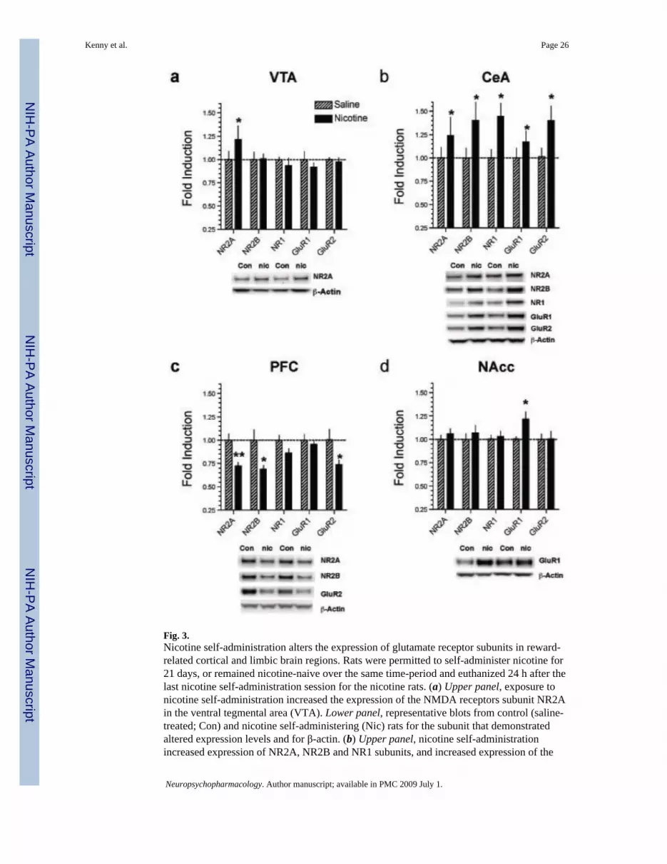
Fig. 3.Nicotine self-administration alters the expression of glutamate receptor subunits in reward-related cortical and limbic brain regions. Rats were permitted to self-administer nicotine for21 days, or remained nicotine-naive over the same time-period and euthanized 24 h after thelast nicotine self-administration session for the nicotine rats. (a) Upper panel, exposure tonicotine self-administration increased the expression of the NMDA receptors subunit NR2Ain the ventral tegmental area (VTA). Lower panel, representative blots from control (saline-treated; Con) and nicotine self-administering (Nic) rats for the subunit that demonstratedaltered expression levels and for β-actin. (b) Upper panel, nicotine self-administrationincreased expression of NR2A, NR2B and NR1 subunits, and increased expression of the
Kenny et al. Page 26
Neuropsychopharmacology. Author manuscript; available in PMC 2009 July 1.
NIH
-PA Author Manuscript
NIH
-PA Author Manuscript
NIH
-PA Author Manuscript

AMPA receptor subunits GluR1 and GluR2 compared with control rats in the centralnucleus of the amygdala (CeA). Lower panel, representative blots for the subunits thatdemonstrated altered expression levels and for β-actin. (c) Upper panel, nicotine self-administration decreased expression of NR2A, NR2B and GluR2 subunits compared withcontrol rats in the prefrontal cortex (PFC). Lower panel, representative blots for the subunitsthat demonstrated altered expression levels and for β-actin. (d) Upper panel, nicotine self-administration increased expression of GluR1 subunits compared with control rats in thenucleus accumbens (NAcc). Lower panel, representative blots for the subunit thatdemonstrated altered expression levels and for β-actin. *P<0.05, **p<0.01 compared withnicotine-naive controls; post-hoctest after significant three -way ANOVA.
Kenny et al. Page 27
Neuropsychopharmacology. Author manuscript; available in PMC 2009 July 1.
NIH
-PA Author Manuscript
NIH
-PA Author Manuscript
NIH
-PA Author Manuscript

Fig. 4.Nicotine increases NMDA receptor-mediated transmission in rat CeA slices. (a) Nicotine (1μM) increased the amplitude of evoked, pharmacologically isolated NMDA receptor EPSCsin CeA neurons. Upper panel, current records of evoked NMDA-EPSCs in a CeA neuronbefore, during and after nicotine superfusion. Lower panel, in CeA neurons, nicotine (2–3mins of application) induced a transient increase in the mean amplitudes of five evokedNMDA-EPSCs by approximately 25% compared with pre-treatment levels. *P<0.05, post-hoc test after significant ANOVA. (b) Application of nicotine (1 μM) increased glutamaterelease in CeA neurons. Upper panel, current records of NMDA-EPSCs in response to twostimuli (paired-pulses) spaced 100 ms apart in a CeA neuron. Lower panel, pooled data ofpaired-pulse facilitation (PPF) ratios expressed as the second NMDA-EPSC amplitude overthe first. Nicotine decreased the mean PPF ratio of NMDA-EPSCs, suggesting a presynapticstimulatory effect of nicotine on glutamate release in CeA tissues. *P<0.05, post-hoc testafter significant ANOVA
Kenny et al. Page 28
Neuropsychopharmacology. Author manuscript; available in PMC 2009 July 1.
NIH
-PA Author Manuscript
NIH
-PA Author Manuscript
NIH
-PA Author Manuscript

Fig. 5.NMDA receptors in the VTA and CeA regulate nicotine self-administration (a)Diagrammatic representation of coronal sections from the rat brain showing histologicalreconstruction of the injection sites in the VTA. Black lines indicate locations of cannulaeand injector tips (n=7) in the VTA and included in the statistical analysis. (b) LY235959(0.1–10 ng/0.5 μl/side) administered into the VTA, but not 2 mm above (1 ng/0.5 μl/side),decreased nicotine self-administration. *P<0.05, **p<0.01, difference from nicotineinfusions earned after vehicle injection, post-hoc test after significant one-way ANOVA. (c)Histological reconstruction of the injection sites in the CeA. Black lines indicate locations ofcannulae and injector tips (n=6) in the CeA and included in statistical analysis. (d)LY235959 (0.1–10 ng/0.5 μl/side) administered into the CeA, but not 2 mm above (1 ng/0.5μl/side), decreased nicotine self-administration. *P<0.05, **p<0.01, difference from nicotineinfusions earned after vehicle injection, post-hoctest after significant one -way ANOVA.
Kenny et al. Page 29
Neuropsychopharmacology. Author manuscript; available in PMC 2009 July 1.
NIH
-PA Author Manuscript
NIH
-PA Author Manuscript
NIH
-PA Author Manuscript
![Reduced inhibitory action of a GABAB receptor agonist on [3H]-dopamine release from rat ventral tegmental area in vitro after chronic nicotine administration](https://img.pdfslide.net/doc/110x75/635653b176ab4e2f19076989/reduced-inhibitory-action-of-a-gabab-receptor-agonist-on-3h-dopamine-release-from.jpg)


![(S)-(-)-[11C]Nicotine Binding Assessed by PET](https://img.pdfslide.net/doc/110x75/635feb4eac0cd8fcb10e64d4/s-11cnicotine-binding-assessed-by-pet.jpg)


