Embed Size (px)
Citation preview

Hyposensitivity to Gamma-Aminobutyric Acid in the VentralTegmental Area During Alcohol Withdrawal: Reversal byHistone Deacetylase Inhibitors
Devinder S Arora1, Sudarat Nimitvilai1, Tara L Teppen2,3, Maureen A McElvain1, Amul J Sakharkar2,3,Chang You1, Subhash C Pandey2,3 and Mark S Brodie*,1
1Department of Physiology and Biophysics, University of Illinois, Chicago, IL, USA; 2Department of Psychiatry, University of Illinois, Chicago, IL, USA;3Jesse Brown VA Medical Center, University of Illinois, Chicago, IL, USA
Putative dopaminergic (pDAergic) ventral tegmental area (VTA) neurons have an important role in alcohol addiction. Acute ethanol
increases the activity of pDAergic neurons, and withdrawal from repeated ethanol administration produces a decreased sensitivity of
pDAergic VTA neurons to GABA. Recent studies show that behavioral changes induced by chronic alcohol are reversed by inhibitors of
histone deacetylases (HDACs). Whether HDAC-induced histone modifications regulate changes in GABA sensitivity of VTA pDAergic
neurons during withdrawal is unknown. Here, we investigated modulation of withdrawal-induced changes in GABA sensitivity of
pDAergic VTA neurons by HDAC inhibitors (HDACi), and also measured the levels of HDAC2, histone (H3-K9) acetylation, and
GABA-Aa1 receptor (GABA (A-a1) R) subunit in VTA during ethanol withdrawal. Mice were injected intraperitoneally (ip) with either
ethanol (3.5 g/kg) or saline twice daily for 3 weeks. In recordings from pDAergic VTA neurons in brain slices from ethanol-withdrawn
mice, sensitivity to GABA (50–500 mM) was reduced. In brain slices from ethanol-withdrawn mice incubated with the HDACi SAHA
(vorinostat) or trichostatin A (TSA) for 2 h, the hyposensitivity of pDAergic VTA neurons to GABA was significantly attenuated. There
was no effect of TSA or SAHA on GABA sensitivity of pDAergic VTA neurons from saline-treated mice. In addition, ethanol withdrawal
was associated with an increase in levels of HDAC2 and a decrease in histone (H3-K9) acetylation and levels of GABA (A-a1) R subunits
in the VTA. Therefore, blockade of upregulation of HDAC2 by HDACi normalizes GABA hyposensitivity of pDAergic neurons
developed during withdrawal after chronic ethanol treatment, which suggests the possibility that inhibition of HDACs can reverse
ethanol-induced neuroadaptational changes in reward circuitry.
Neuropsychopharmacology (2013) 38, 1674–1684; doi:10.1038/npp.2013.65; published online 3 April 2013
Keywords: VTA; GABA; dopamine neurons; HDAC inhibitors; histone acetylation; alcohol withdrawal
������������������������������������������������������
INTRODUCTION
The mesolimbic dopamine (DA) system projects from theventral tegmental area (VTA) to the nucleus accumbens(NAc), prefrontal cortex, basolateral amygdala, and to othercorticolimbic structures (Albanese and Minciacchi, 1983;Oades and Halliday, 1987). Although drugs of abuse havedistinct molecular targets, they all share the ability toincrease DA levels in NAc (Luscher and Ungless, 2006;Arora et al, 2010). Acute administration of ethanol increasesthe firing rate of DA neurons, both in vitro and in vivo(Gessa et al, 1985; Brodie et al, 1990).
Chronic use of alcohol or drugs of abuse producesprofound changes in the brain. Some of these changes may
be directly related to the process of addiction (Koob, 2003).We have shown previously that repeated ethanol treatmentof C57Bl/6J mice results in an increase in sensitivity to acuteethanol and a decrease in sensitivity to GABA-mediatedinhibition (Brodie, 2002). Changes in the spontaneousfrequency of firing of VTA neurons of rats and micefollowing a variety of ethanol treatment regimens have beendemonstrated, but it is unknown whether GABA sensitivityhas a role in this change in neuronal activity (Diana et al,1992b; Shen and Chiodo, 1993; Bailey et al, 1998). Themolecular basis of changes in GABA responses in the VTAinduced by chronic alcohol exposure is not known.
In the recent years, the concept of epigenetics and alcoholabuse has provided new directions in the mechanisticapproach to alcohol research (Pandey et al, 2008; Starkmanet al, 2012). Epigenetic mechanisms include DNA methyla-tion, histone modifications, and microRNA (Renthal andNestler, 2008; Kalsi et al, 2009; Moonat et al, 2010). There isa growing interest in the possible functional consequencesof covalent modification of the chromatin in the appearanceand maintenance of behavioral changes, including the
*Correspondence: Dr MS Brodie, Department of Physiology andBiophysics, University of Illinois at Chicago, 835 S Wolcott, RoomE-202, M/C 901, Chicago, IL 60612-7342, USA, Tel: +1 312 996 2373,Fax: +1 312 996 1414, E-mail: [email protected] 31 October 2012; revised 1 February 2013; accepted 21February 2013; accepted article preview online 8 March 2013
Neuropsychopharmacology (2013) 38, 1674–1684
& 2013 American College of Neuropsychopharmacology. All rights reserved 0893-133X/13
www.neuropsychopharmacology.org

development and manifestation of addictive behavior(Sweatt, 2009; Renthal and Nestler, 2009a; Starkman et al,2012). Modifications in chromatin architecture couldunderlie the long-lasting changes in neuronal gene expres-sion and synaptic plasticity, and these changes ultimatelycould explain the processes involved in addictive behaviors(Renthal and Nestler, 2009a; Moonat et al, 2010). Recentstudies have shown that when cocaine is administered atdoses necessary to produce locomotor sensitization andconditioned place preference, histone hyperacetylation atspecific brain areas relevant to the development of addictivebehavior was observed (Kumar et al, 2005; Levine et al,2005; Renthal and Nestler, 2008). Furthermore, the func-tional role of histone acetyltransferase (Levine et al, 2005)and histone deacetylase (HDAC) (Renthal et al, 2007)activities in the mechanism of action of psychostimulantshas also been established, and is supported by experimentaluse of HDAC inhibitors (HDACi) (Kumar et al, 2005;Renthal et al, 2007; Kalda et al, 2007).
HDAC-induced chromatin remodeling in the amygdalahas been implicated in alcohol dependence (Pandey et al,2008). Anxiety-like behaviors that develop during with-drawal after chronic ethanol exposure in rats can bereversed by treatment with the HDACi trichostatin A(TSA). Rapid tolerance to anxiolytic effects of alcohol inrats can be reversed by TSA treatment, suggesting a role foramygdaloid HDAC-induced histone modifications in alco-hol tolerance (Sakharkar et al, 2012). There are severalHDAC isoforms, which are classified as Class I, II, III, andIV. The HDAC2 isoform, which belongs to Class I, has beenshown to be involved in synaptic plasticity (Guan et al,2009). The histone H3 is acetylated at the lysine nineposition (H3-K9) by HDAC2, and it is this histonemodification that has been associated with gene expressionrelated to drug abuse and neural plasticity (Renthaland Nestler, 2008; Shibasaki et al, 2011). More recently,higher HDAC2 expression-induced deficits in histoneH3-K9 acetylation and associated synaptic plasticity in theamygdala has been shown to be involved in alcohol-drinking behaviors (Moonat et al, 2013). However, the roleof HDAC-induced histone modification in the reductionof GABA sensitivity in the VTA during alcohol withdrawalhas not been examined. In the present study, we used extra-cellular recording from putative dopaminergic (pDAergic)VTA neurons in brain slices, and explored the effects ofHDACi on the reduction in GABA responsiveness in theVTA induced by chronic ethanol administration in C57Bl/6Jmice. In addition, we also investigated the effects of alcoholtreatment and withdrawal on the levels of HDAC2, histoneacetylation, and GABA-A a1 receptor (GABA (A-a1) R)subunit in the VTA of mice.
MATERIALS AND METHODS
Animals
C57BL/6J mice (4–6 weeks old) used in these studies wereobtained from Jackson Labs (Bar Harbor, ME). Allexperimental procedures in mice were in strict accordancewith the NIH Guidelines for the Care and Use of LaboratoryAnimals, and approved by the Animal Care Committee ofthe University of Illinois at Chicago.
Chronic Ethanol Treatment
All mice were treated for 3 weeks (21 days), with twice daily(morning (07:00–08:00 hour) and evening (15:00–17:00hour)) intraperitoneal (ip) injections of either normal salineor 3.5 g/kg ethanol (20% w/v in normal saline). Thisinjection paradigm has been used in other laboratories toproduce tolerance to the behavioral effects of ethanol(Crabbe et al, 1982). To reduce the stress of twice dailyip injection, each mouse (control and ethanol-treated) wasanesthetized lightly with halothane, immediately beforeeach injection.
Preparation of Brain Slices
For the electrophysiological studies, mice were sacrificed atleast 14 h after the last injection of either saline or ethanol.Brain slices containing the VTA were prepared from thesubject animals, as previously described (Brodie et al,1999a). Briefly, following rapid removal of the brain, thetissue was blocked coronally to contain the VTA andsubstantia nigra; the cerebral cortices and a portion of thedorsal mesencephalon were removed. The tissue block wasmounted in the vibratome and submerged in chilled cuttingsolution. Coronal sections (400-mm thick) were cut andplaced in 50 ml polypropylene centrifuge tubes in aCSF withthe addition of either 0.1% DMSO, 12.5 mM TSA, or 3mMSAHA; both TSA and SAHA were dissolved in DMSO andwere added to the aCSF in the centrifuge tubes such that thefinal DMSO concentration was 0.1%. After 2 h incubation inone of these solutions, each slice then was placed onto amesh platform in the recording chamber. Each slice wastotally submerged in drug-free aCSF maintained at a flowrate of 2 ml/min; the temperature in the recording chamberwas kept at 35 1C. The composition of the aCSF in theseexperiments was (in mM): NaCl 126, KCl 2.5, NaH2PO4 1.24,CaCl2 2.4, MgSO4 1.3, NaHCO3 26, glucose 11. The compo-sition of the cutting solution was (in mM): KCl 2.5,CaCl2 2.4, MgSO4 1.3, NaHCO3 26, glucose 11, and sucrose220. Both solutions were saturated with 95% O2/5% CO2
(pH¼ 7.4). Recordings were made immediately, and wereobtained up to 6 h after the slices were placed in therecording chamber.
Cell Identification
The VTA was clearly visible in the fresh tissue as a grey areamedial to the darker substantia nigra, and separated fromthe nigra by white matter. Recording electrodes were placedin the VTA under visual control. pDAergic neurons havebeen shown to have distinctive electrophysiological charac-teristics (Grace and Bunney, 1984; Lacey et al, 1989). Onlythose neurons that were anatomically located within theVTA and that conformed to the criteria for pDAergicneurons established in the literature and in this laboratory(Lacey et al, 1989; Mueller and Brodie, 1989) were studied.These criteria include broad action potentials (X2.5 ms,measured as the width of the bi- or tri-phasic waveform atthe baseline), slow spontaneous firing rate (0.5–5 Hz), and aregular interspike interval.
HDAC and GABA sensitivity in alcoholismDS Arora et al
1675
Neuropsychopharmacology

Drug Administration
Drugs were added to the aCSF by means of a calibratedinfusion pump from stock solutions 100–1000 times thedesired final concentrations. The addition of drug solutionsto the aCSF was performed in such a way as to permit thedrug solution to mix completely with aCSF before thismixture reached the recording chamber. Final concentra-tions were calculated from the aCSF flow rate, pumpinfusion rate, and concentration of drug stock solution.The small volume chamber (about 300 ml) used in thesestudies permitted the rapid application and washoutof drug solutions. Typically, drugs reach equilibrium inthe tissue after 2–3 min of application. GABA, ethanol,and most of the salts used to prepare the extracellularmedia were purchased from Sigma Aldrich Corporation(St Louis, MO). Suberoylanilide hydroxamic acid (SAHA)and TSA were obtained from Selleck Chemicals(Houston, TX).
Extracellular Recording
Extracellular recording electrodes were made from 1.5-mmdiameter glass tubing with filament and were filled with0.9% NaCl. Tip resistance of the microelectrodes rangedfrom 2 to 5 MO. A Fintronics amplifier was used in conjunc-tion with an IBM-PC-based data acquisition system(ADInstruments, Colorado Springs, CO, USA). Offlineanalysis was used to calculate, display, and store thefrequency of firing 1-min intervals. Firing rate wasdetermined before and during drug application. Firing ratewas calculated over 1-min intervals before administration ofdrugs and during the drug effect; peak drug-inducedchanges in firing rate were expressed as the percentagechange from the control firing rate according to the formula((FRD—FRC)/FRC)� 100, where FRD is the firing rateduring the peak drug effect and FRC is the control firingrate. The change in firing rate thus is expressed as apercentage of the initial firing rate, which controls for smallchanges in firing rate that may occur over time. Thisformula was used to calculate both excitatory and inhibitorydrug effects. Peak excitation was defined as the peakincrease in firing rate produced by the drug greater thanthe pre-drug baseline. Inhibition was defined as the lowestfiring rate below the pre-drug baseline.
Gold Immunolabeling Histochemical Procedure
Mice were subjected to repeated treatment with eitherethanol or saline injections, as described above. Threegroups of mice were used: saline-treated mice that receivedtheir last injection at least 14 h earlier (control), ethanol-treated mice that received their last ethanol injection at least14 h earlier (withdrawal), and ethanol-treated mice thatreceived an ethanol injection 1 h before sacrifice (ethanol).Blood was taken to assess ethanol concentration using anAnalox Alcohol Analyzer (Analox Instruments, Lunenberg,MA). Each mouse was anesthetized and then perfused intra-cardially with ice-cold normal saline (30–50 ml), followed by4% paraformaldehyde (30–50 ml; PFA) prepared in 0.1 Mphosphate buffer (pH 7.4). Brains were removed and placedin PFA for 20 h at 4 1C. Then, brains were soaked
sequentially in 10, 20, and 30% sucrose solutions (dissolvedin phosphate buffer, pH 7.4). Brains were then frozen andstored at � 80 1C until used for the gold immunolabelingwith antibodies to HDAC2 (MBL, Woburn, MA), acetylatedhistone H3-K9 (Millipore, Billerica, MA), and GABA (A-a1)R (Abcam, Cambridge, MA), as described previously(Pandey et al, 2008; Sakharkar et al, 2012). Briefly, 20 mmcoronal cryostat sections were cut and placed in 0.01 Mphosphate-buffered saline (PBS) for washing, followed bysequential incubations in RPMI (Invitrogen, Grand Island,NY), 10% normal goat serum (Vector Labs, Burlingame,CA), and 1% bovine serum albumin in PBST for 30 mineach. Sections were then incubated for 16–18 h at roomtemperature in antibodies against HDAC2 (1 : 200), acetyl-ated histone H3-K9 (1 : 500), or GABA (A-a1) R (1 : 200).Sections were further washed in PBS and incubated in goldparticle-conjugated anti-rabbit secondary antibody (1 : 200)for 1 h (Nanoprobes, Yaphank, NY), and were developedusing silver enhancement solution (Ted Pella, Redding, CA).Gold-immunolabeled HDAC2, acetylated H3-K9, and GABA(A-a1) R were quantified using the image analysis programat 100� magnification, as described previously (Pandeyet al, 2008; Sakharkar et al, 2012). Immunogold particlesin the VTA region in each of three adjacent brain sections(9–11 total object fields) were quantified; values wereaveraged for each animal and results were represented asmean±SEM of the number of immunogold particles per100 mm2 area.
Statistical Analysis
Averaged numerical values were expressed as the mean±SEM. Data were analyzed by using one- or two-way analysisof variance (ANOVA) followed by Tukey’s post hoccomparisons of means. A value of po0.05 was consideredto be significant.
RESULTS
Electrophysiology: VTA Neuron Characteristics
A total of 112 pDAergic VTA neurons from 25 micewere used in the electrophysiological portion of thisstudy. The firing rate of these neurons ranged from 0.57to 4.2 Hz, with an overall mean firing rate of 2.06±0.11 Hz.All of the neurons tested had regular firing rates andwere inhibited by GABA. Cells that did not return towithin 10% of the pre-drug firing rate during washout werenot used.
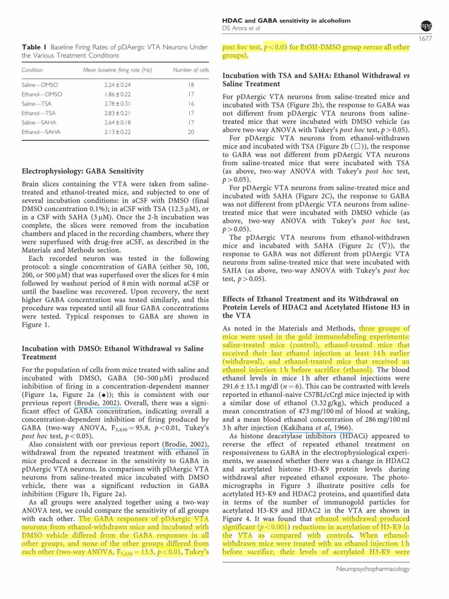
For slices treated with DMSO or HDACi, recordingswere made in up to eight neurons per slice, with an averagenumber of cells per slice of 4.9±0.42. There was a signi-ficant difference in the baseline frequency of firing rate ofpDAergic VTA neurons among these groups (one-wayANOVA, F5,99¼ 2.83; po0.02), with Tukey’s mean compa-rison indicating a significant difference between only theethanol injected, DMSO-treated (E-DMSO) cells and theethanol-injected, TSA-treated (E-TSA) cells (Tukey’s posthoc test, po0.05 for E-DMSO and E-TSA groups, p40.05for comparisons of all other groups) (Table 1).
HDAC and GABA sensitivity in alcoholismDS Arora et al
1676
Neuropsychopharmacology
Electrophysiology: GABA Sensitivity
Brain slices containing the VTA were taken from saline-treated and ethanol-treated mice, and subjected to one ofseveral incubation conditions: in aCSF with DMSO (finalDMSO concentration 0.1%); in aCSF with TSA (12.5 mM), orin a CSF with SAHA (3 mM). Once the 2-h incubation wascomplete, the slices were removed from the incubationchambers and placed in the recording chambers, where theywere superfused with drug-free aCSF, as described in theMaterials and Methods section.
Each recorded neuron was tested in the followingprotocol: a single concentration of GABA (either 50, 100,200, or 500 mM) that was superfused over the slices for 4 minfollowed by washout period of 8 min with normal aCSF oruntil the baseline was recovered. Upon recovery, the nexthigher GABA concentration was tested similarly, and thisprocedure was repeated until all four GABA concentrationswere tested. Typical responses to GABA are shown inFigure 1.
Incubation with DMSO: Ethanol Withdrawal vs SalineTreatment
For the population of cells from mice treated with saline andincubated with DMSO, GABA (50–500 mM) producedinhibition of firing in a concentration-dependent manner(Figure 1a, Figure 2a (K)); this is consistent with ourprevious report (Brodie, 2002). Overall, there was a signi-ficant effect of GABA concentration, indicating overall aconcentration-dependent inhibition of firing produced byGABA (two-way ANOVA, F3,430¼ 95.8, po0.01, Tukey’spost hoc test, po0.05).
Also consistent with our previous report (Brodie, 2002),withdrawal from the repeated treatment with ethanol inmice produced a decrease in the sensitivity to GABA inpDAergic VTA neurons. In comparison with pDAergic VTAneurons from saline-treated mice incubated with DMSOvehicle, there was a significant reduction in GABAinhibition (Figure 1b, Figure 2a).
As all groups were analyzed together using a two-wayANOVA test, we could compare the sensitivity of all groupswith each other. The GABA responses of pDAergic VTAneurons from ethanol-withdrawn mice and incubated withDMSO vehicle differed from the GABA responses in allother groups, and none of the other groups differed fromeach other (two-way ANOVA, F5,430¼ 13.5, po0.01, Tukey’s
post hoc test, po0.05 for EtOH-DMSO group versus all othergroups).
Incubation with TSA and SAHA: Ethanol Withdrawal vsSaline Treatment
For pDAergic VTA neurons from saline-treated mice andincubated with TSA (Figure 2b), the response to GABA wasnot different from pDAergic VTA neurons from saline-treated mice that were incubated with DMSO vehicle (asabove two-way ANOVA with Tukey’s post hoc test, p40.05).
For pDAergic VTA neurons from ethanol-withdrawnmice and incubated with TSA (Figure 2b (&)), the responseto GABA was not different from pDAergic VTA neuronsfrom saline-treated mice that were incubated with TSA(as above, two-way ANOVA with Tukey’s post hoc test,p40.05).
For pDAergic VTA neurons from saline-treated mice andincubated with SAHA (Figure 2C), the response to GABAwas not different from pDAergic VTA neurons from saline-treated mice that were incubated with DMSO vehicle (asabove, two-way ANOVA with Tukey’s post hoc test,p40.05).
The pDAergic VTA neurons from ethanol-withdrawnmice and incubated with SAHA (Figure 2c (r)), theresponse to GABA was not different from pDAergic VTAneurons from saline-treated mice that were incubated withSAHA (as above, two-way ANOVA with Tukey’s post hoctest, p40.05).
Effects of Ethanol Treatment and its Withdrawal onProtein Levels of HDAC2 and Acetylated Histone H3 inthe VTA
As noted in the Materials and Methods, three groups ofmice were used in the gold immunolabeling experiments:saline-treated mice (control), ethanol-treated mice thatreceived their last ethanol injection at least 14 h earlier(withdrawal), and ethanol-treated mice that received anethanol injection 1 h before sacrifice (ethanol). The bloodethanol levels in mice 1 h after ethanol injections were291.6±15.1 mg/dl (n¼ 6). This can be contrasted with levelsreported in ethanol-naive C57BL/cCrgl mice injected ip witha similar dose of ethanol (3.32 g/kg), which produced amean concentration of 473 mg/100 ml of blood at waking,and a mean blood ethanol concentration of 286 mg/100 ml3 h after injection (Kakihana et al, 1966).
As histone deacetylase inhibitors (HDACi) appeared toreverse the effect of repeated ethanol treatment onresponsiveness to GABA in the electrophysiological experi-ments, we assessed whether there was a change in HDAC2and acetylated histone H3-K9 protein levels duringwithdrawal after repeated ethanol exposure. The photo-micrographs in Figure 3 illustrate positive cells foracetylated H3-K9 and HDAC2 proteins, and quantified datain terms of the number of immunogold particles foracetylated H3-K9 and HDAC2 in the VTA are shown inFigure 4. It was found that ethanol withdrawal producedsignificant (po0.001) reductions in acetylation of H3-K9 inthe VTA as compared with controls. When ethanol-withdrawn mice were treated with an ethanol injection 1 hbefore sacrifice, their levels of acetylated H3-K9 were
Table 1 Baseline Firing Rates of pDAergic VTA Neurons Underthe Various Treatment Conditions
Condition Mean baseline firing rate (Hz) Number of cells
Saline—DMSO 2.24±0.24 18
Ethanol—DMSO 1.86±0.22 17
Saline—TSA 2.78±0.31 16
Ethanol—TSA 2.83±0.21 17
Saline—SAHA 2.64±0.18 17
Ethanol—SAHA 2.13±0.22 20
HDAC and GABA sensitivity in alcoholismDS Arora et al
1677
Neuropsychopharmacology

Figure 1 Single neurons: effects of 200 mM GABA. Each figure represents the firing rate of a single neuron of the VTA over time. Vertical bars areproportional to the firing rate over a 5-s interval, horizontal bars represent the duration of application of GABA (200 mM). Neurons were recorded in brainslices from mice treated with saline (a, c, e) or ethanol (b, d, f). Slices were incubated with DMSO (a, b), TSA (c, d), or SAHA (e, f). Addition of 200 mMGABA to the extracellular medium resulted in maximum reductions of firing in these neurons of (a) � 99.6%, (b) � 22.7%, (c) � 76.9%, (d) � 70.0%,(e) � 99.2%, (f) � 98.5%.
HDAC and GABA sensitivity in alcoholismDS Arora et al
1678
Neuropsychopharmacology

similar to control levels (Figures 3 and 4). Conversely,HDAC2 protein expression was upregulated in the VTAof the ethanol-withdrawn animals as compared with thatof saline-treated and ethanol (ethanol injection 1 hbefore sacrifice) groups (bottom panels of Figures 3and 4). Immunolabeling in the VTA for the withdrawalgroup differed from the other two groups for both HDAC2(one-way ANOVA; F2,15¼ 32.77, po0.001; n¼ 6) andH3-K9 acetylation (one-way ANOVA, F2,15¼ 25.12,po0.001; n¼ 6).
Effects of Ethanol Treatment and its Withdrawal onProtein Levels of the a1 Subunit of GABA-A Receptors
The hyposensitivity to GABA during ethanol withdrawalcould be due to reduction in GABA receptors. Figure 5shows that the inhibition of DA VTA neurons by theconcentrations of GABA used in the present study weremediated by the activation of GABA-A receptors. GABA(50–500mM) was tested on pDAergic VTA neurons fromnaive C57 mice in the absence and presence of the GABA-Breceptor antagonist CGP35348 (1 mM). There was nosignificant difference in the response to GABA whetherCGP35348 was present or not (two-way ANOVA, F1,51¼1.4E-5, p40.99), indicating that GABA-induced inhibitionover the concentration range tested is primarily because ofthe action at GABA-A receptors.
Next, we examined the changes in one GABA-A receptorsubunit that has been shown to be reduced by chronicethanol treatment: the a 1 subunit (Kumar et al, 2003). Thephotomicrograph in Figure 6a illustrates GABA (A-a1)R-positive cells in the VTA of various group of C57 micetreated according to the repeated injection protocol.The Figure 6b shows the mean number of immunogoldparticles under the same conditions as in Figures 3 and 4.The protein levels of GABA (A-a1) R were downregulated inthe VTA during withdrawal, and significantly (po0.001)differed from the groups treated with saline or chronicethanol, with an ethanol injection 1 h before sacrifice(one-way ANOVA, F 2,15¼ 35.25, po0.001; n¼ 6).
DISCUSSION
The novel findings of present study demonstrate thatdecreased GABA responses in DA VTA neurons inducedby 3 weeks of repeated ethanol administration can bereversed by a relatively brief (2 h) incubation with theHDACi, TSA, or SAHA. Complementary and relatedimmunohistochemical examination of acetylated histoneH3-K9 and HDAC2 indicates that withdrawal from repeatedethanol increases HDAC2 protein levels and decreaseshistone acetylation (acetylated H3-K9) in the VTA. Thereversal by SAHA or TSA of the effect of repeated ethanolon sensitivity to GABA suggests a new paradigm fortreatment of alcohol-induced brain changes; this goesbeyond pharmacological amelioration of symptoms andapproaches a class of therapy that normalizes neurotrans-mission in the alcoholic brain. This is further supported byrecent findings that TSA treatment during withdrawal afterchronic ethanol exposure corrected the deficits in H3-K9acetylation by blocking the upregulation of HDAC activityin the amygdala and prevented the development of anxiety-like behaviors in rats (Pandey et al, 2008). Furthermore, ithas been shown that knockdown of HDAC2 in the centralnucleus of amygdala attenuated alcohol intake in alcohol-preferring rats (Moonat et al, 2013). The current report isthe first of which we are aware demonstrating that in vitroadministration of an HDACi can reverse the effects ofin vivo ethanol treatment. Some epigenetic changes in theVTA during withdrawal from chronic ethanol may berelated to an increased expression of HDAC2, and clearlythere is sufficient transcription and turnover in thebrain slice preparation over a 2-h period to reverse
Figure 2 Pooled responses: effects of GABA. Pooled concentration–response curves for pDAergic VTA neurons from brain slices from micetreated with repeated injections of either saline (filled symbols) or ethanol(open symbols). Slices were incubated for 2 h with 0.1% DMSO (a),12.5 mM TSA (b), or 3 mM SAHA (c). (a) Incubation in DMSO (vehiclecontrols): slices from saline-treated (K) or ethanol-treated mice (J); (b)incubation in TSA: slices from saline-treated (’) or ethanol-treated mice(&); (c) incubation in SAHA: slices from saline-treated (.) or ethanol-treated mice (r).
HDAC and GABA sensitivity in alcoholismDS Arora et al
1679
Neuropsychopharmacology

withdrawal-induced GABA hyposensitivity, once HDAC isinhibited by TSA or SAHA.
Acute ethanol exposure excites VTA DA neurons directly(Brodie et al, 1999b); however, other factors may enhanceethanol-induced excitation of DA VTA neurons. Forexample, ethanol can hyperpolarize the GABA-containingneurons of the VTA (Gallegos et al, 1999; Xiao and Ye, 2008)and can influence NMDA receptor-mediated excitation ofVTA DA neurons (Stobbs et al, 2004). Chronic ethanol islikely to affect various ethanol-sensitive properties differ-ently, resulting in apparent increases or decreases in theeffects of ethanol. This idea is supported by evidence fromour laboratory (Brodie, 2002) that DA VTA neurons frommice subjected to repeated ethanol treatment were moresensitive to the excitatory action of ethanol and lesssensitive to inhibition by GABA, but responsiveness toNMDA was unaffected. Chronic ethanol and subsequentwithdrawal may have more profound effects on GABAneurons of the VTA; chronic exposure to ethanol enhancesthe baseline activity of GABA neurons and inducestolerance to ethanol inhibition of the firing rate of theseneurons (Gallegos et al, 1999). In contrast, tolerance has notbeen observed to ethanol-induced excitation of DA VTAneurons after chronic ethanol treatment (Diana et al, 1992a;Brodie, 2002). As we report here and have reportedpreviously (Brodie, 2002), there is a decrease in theresponse of pDAergic VTA neurons in vitro to exogenouslyapplied GABA during ethanol withdrawal. Furthermore,consistent with the previous studies (Brodie, 2002), therewas no significant difference in the basal spontaneous firingbetween the ethanol exposed and control groups under eachcondition, although there was a difference between two ofthe groups.
The phenomenon of reduced sensitivity to GABA couldbe attributed to the postsynaptic adaptive changes in theGABA receptor subunit composition during withdrawalfrom chronic ethanol. Although activation of GABA-Breceptors can cause inhibition of pDAergic VTA neurons(Lacey et al, 1988; Mueller and Brodie, 1989), data aboveindicate that the concentrations of GABA used in thepresent study caused inhibition by GABA-A receptoractivation. It has been shown that there is a shift in thea-subunit composition in the mesolimbic system of ratfollowing chronic ethanol administration (Papadeas et al,2001; Liang et al, 2007). Other proposed mechanisms forchronic ethanol-induced GABA-A receptor adaptationsinclude alterations in gene expression, post-translationalmodifications, synaptic localization, and intracellular sig-naling (Kumar et al, 2004). Increased internalization ofGABA (A-a1) R subunit has been demonstrated in cerebralcortex after chronic ethanol consumption (Kumar et al,2003). No change in the GABA (A-a1) R subunit wasobserved in the VTA in rats chronically drinking alcohol,but levels during withdrawal were not examined (Papadeaset al, 2001). Here we demonstrated that withdrawal, notethanol exposure per se, produced downregulation of GABA(A-a1) R protein levels in the VTA of mice. Certaincombinations of GABA-A receptor subunits form GABA-Areceptors that respond to GABA with lower efficacy. Forexample, a4b1g2 GABA-A receptors respond to GABA withlower efficacy than a1b1g2 GABA-A receptors (Whittemoreet al, 1996). Although another group has shown no changein a4 subunit following 14-day ethanol consumption(Papadeas et al, 2001), it is known that different treatmentregimens can produce different effects on GABA subunits(Matthews et al, 1998), and there may be differences in
Figure 3 Representative photomicrographs (scale bar¼ 40mm) showing acetylated histone H3-K9 (top panel) and HDAC2 (bottom panel) goldimmunolabeling in the VTA of repeated saline treatment (Control), repeated ethanol treatment with last ethanol injection 1 h before sacrifice (Ethanol), andrepeated ethanol treatment with last ethanol injection 414 h before sacrifice (Withdrawal).
HDAC and GABA sensitivity in alcoholismDS Arora et al
1680
Neuropsychopharmacology

GABA receptor subunits during withdrawal that are notseen during ethanol treatment. The b3 subunit may beparticularly important for GABA-A receptor trafficking andcontrol of responsiveness to GABA (Parker et al, 2011).Furthermore, in humans, the genes encoding the GABA-Areceptor subunits are clustered in several chromosomalregions, and the chromosome 4 clusters of genes are likelyto be important in addiction and anxiety and may bevulnerable to epigenetic effects in early development (Enochet al, 2009).
The focus of the electrophysiological portion ofthe present study was to assess the control of ethanolwithdrawal-induced GABA hyposensitivity by HDAC byincubating the slices with HDACi. We observed thatinhibition of HDACs abolished the ethanol withdrawal-induced changes in GABA sensitivity of pDAergic VTAneurons in the VTA. It seems plausible that decreases in
histone acetylation in VTA induced by upregulation ofHDAC2 produces the adaptive changes in the GABA-Areceptors, accounting for the GABA hyposensitivityof pDAergic neurons that we observed during ethanolwithdrawal. Note that no HDACi was present during theassessment of GABA inhibition; a 2-h incubation periodpreceded the placement of the brain slices in the recordingchambers, and no HDACi was present in the superfusionmedium. As the recordings were obtained over a 5–7-hperiod after the slices were removed from the HDACi-containing medium, it is unlikely that any significantamount of HDACi was present during the recordings. Thissuggests that the 2-h incubation produced a sustainedchange in GABA responsiveness due to a persistentbiochemical change, rather than a direct interaction of theHDACi with GABA receptors during the GABA adminis-tration in the electrophysiology experiments. Furthermore,treatment with TSA or SAHA did not alter the overall GABAsensitivity of VTA neurons from saline-treated mice. Theseresults suggest a possibility for HDAC-induced histonedeacetylation as a mechanism for the changes in GABAsensitivity in the VTA induced by repeated ethanoladministration.
The ability of HDACi to reverse changes in GABAsensitivity observed in VTA neurons from ethanol-treatedmice in withdrawal suggests a change in HDAC activity thatis induced by repeated ethanol treatment. We observed thatHDAC2 levels were significantly elevated in the ethanol-treated mice at a time equivalent to the time period duringwhich recordings would have been made. We do not knowwhether other HDAC isoforms were similarly increasedduring withdrawal after repeated ethanol treatment. Corre-lated with the HDAC2 increase was a complementarydecrease in histone H3-K9 acetylation and expression of
Figure 4 Bar diagrams showing the differences in the acetylated H3 (H3-K9) and HDAC2 protein levels in the VTA of repeated saline treatment(Control, n¼ 6), repeated ethanol treatment with last ethanol injection 1 hbefore sacrifice (Ethanol, n¼ 6), and repeated ethanol treatment with lastethanol injection 414 h before sacrifice (Withdrawal, n¼ 6). There was asignificant decrease in acetylated H3 (*po0.001) and a reciprocal increasein the levels of HDAC2 (*po0.001) in the VTA of mice in withdrawal fromethanol (Withdrawal) compared with that of saline-treated controls(Control) or to ethanol-treated mice that received an ethanol injection1 h before sacrifice (Ethanol).
Figure 5 The effects of GABA-B antagonist CGP35348 on GABA-induced inhibition of pDAergic VTA neurons. Pooled concentration–response curves for responses to GABA of pDAergic VTA neurons frombrain slices from mice in the absence (K) or presence (’) of CGP35348(1 mM). GABA (50–500 mM) was applied to each neuron, then CGP35348was added to the medium and the same concentrations of GABA weretested. There was no significant difference in response to GABA betweenthe groups (two-way ANOVA, F1,51¼ 1.4E-5, p40.99).
HDAC and GABA sensitivity in alcoholismDS Arora et al
1681
Neuropsychopharmacology

GABA (A-a1) R subunit in the VTA; although there mayhave been changes in other HDAC isoforms, the overalleffect of withdrawal after repeated ethanol was to produce asignificant decrease in histone H3 acetylation. Interestingly,when ethanol was present, the levels of HDAC2 and histoneacetylation as well as GABA (A-a1) R were not differentfrom controls. This suggests that these changes in VTA maybe related to the withdrawal state, and are rapidly reversedby ethanol exposure. As the VTA has been linked to reward/reinforcement and craving, the change in HDAC activityduring withdrawal observed in the present study might belinked to withdrawal-induced craving (Lopez and Becker,2005; Enoch, 2008). Future studies will be needed todetermine whether in vivo HDACi modulate increases inHDAC2 and concomitant decreases in histone acetylationspecific to various GABA-A receptor subunits, and whetherthis treatment could reverse changes in GABA responsive-ness in the VTA during withdrawal after chronic ethanolexposure.
Neuroadaptational changes in the histone acetylation andHDAC activity in the amygdala induced during withdrawalafter chronic ethanol have been reported to be reversed orprevented by TSA. The few studies performed with respect
to HDAC-induced histone deacetylation in response toalcohol administration have focused on the forebrain(Pandey et al, 2008; Moonat et al, 2013; Sakharkar et al,2012). In particular, changes in histone acetylation asso-ciated with drug abuse have centered on the amygdala(Pandey et al, 2008; Sakharkar et al, 2012) and the NAc(Renthal and Nestler, 2009b). One group has observedincreases in acetylated histone H3 protein in midbrainduring a single withdrawal from a 9-day course of ethanolvapor inhalation in male ddY mice (Shibasaki et al, 2011);the differences between that and the present study may berelated to brain region specificity and differences induration and treatment paradigm of ethanol. Nonetheless,the present study is the first to examine the actions ofHDACi on ethanol-induced changes in GABA electro-physiological responses in the VTA. The observation thatHDACi appears to restore GABA sensitivity in the VTAsuggests that ethanol-induced changes in histone acetyla-tion may be the mechanism for abnormal reward circuitryoccurred during ethanol exposure. It is known thatexperience with alcohol increases alcohol self-administrationand alcohol seeking (Lopez and Becker, 2005; Griffin et al,2009). As the VTA is associated with the rewarding/reinforcing
Figure 6 (a) Representative low-magnification photomicrographs (scale bar¼ 40mm) of gold immunolabeling of GABA (A-a1) R subunit in the VTA ofrepeated saline treatment (Control), repeated ethanol treatment with last ethanol injection 1 h before sacrifice (Ethanol), and repeated ethanol treatmentwith last ethanol injection 414 h before sacrifice (Withdrawal). (b) Bar diagrams showing the differences in the protein levels of GABA (A-a1) R subunit inthe VTA of repeated saline treatment (Control, n¼ 6), repeated ethanol treatment with last ethanol injection 1 h before sacrifice (Ethanol, n¼ 6), andrepeated ethanol treatment with last ethanol injection 414 h before sacrifice (Withdrawal, n¼ 6). There was a significant decrease in GABA (A-a1) Rsubunit (*po0.001) in the VTA of mice in withdrawal from ethanol (Withdrawal) compared with that of saline-treated controls (Control) or to ethanol-treated mice.
HDAC and GABA sensitivity in alcoholismDS Arora et al
1682
Neuropsychopharmacology

effects of alcohol and other drugs of abuse, it is provocativeto speculate on the possibility that inhibition of HDAC mayproduce a decrease in drug-seeking behavior related toreward and reinforcement.
ACKNOWLEDGEMENTS
This work was supported by the National Institute onAlcohol Abuse and Alcoholism Grants AA-016690, AA-019971(NADIA project), and by the Department of VeteransAffairs (Merit Review Grant-l01BX000143; Research CareerScientist award) to SCP, and by the National Institute onAlcohol Abuse and Alcoholism Grants AA-05846 andAA09125 to MSB.
DISCLOSURE
Dr Pandey reports that a US patent application entitled‘Histone acetyl transferase activators and histone deacety-lase inhibitors in the treatment of alcoholism’ (serialnumber 60/848237 filed on 29 September, 2006), which isdirected to a proposed method for reducing symptomsrelated to alcohol withdrawal in humans, is currentlypending. The remaining authors declare no conflicts ofinterest.
REFERENCES
Albanese A, Minciacchi D (1983). Organization of the ascendingprojections from the ventral tegmental area: a multiplefluorescent retrograde tracer study in the rat. J Comp Neurol216: 406–420.
Arora D, Haluk DM, Kourrich S, Pravetoni M, Fernandez-Alacid L,Nicolau JC et al (2010). Altered neurotransmission in themesolimbic reward system of Girk mice. J Neurochem 114:1487–1497.
Bailey CP, Manley SJ, Watson WP, Wonnacott S, Molleman A,Little HJ (1998). Chronic ethanol administration alters activity inventral tegmental area neurons after cessation of withdrawalhyperexcitability. Brain Res 803: 144–152.
Brodie MS (2002). Increased ethanol excitation of dopaminergicneurons of the ventral tegmental area after chronic ethanoltreatment. Alcohol Clin Exp Res 26: 1024–1030.
Brodie MS, McElvain MA, Bunney EB, Appel SB (1999a).Pharmacological reduction of small conductance calcium-activated potassium current (SK) potentiates the excitatoryeffect of ethanol on ventral tegmental area dopamine neurons.J Pharmacol Exp Ther 290: 325–333.
Brodie MS, Pesold C, Appel SB (1999b). Ethanol directly excitesdopaminergic ventral tegmental area reward neurons. AlcoholClin Exper Res 23: 1848–1852.
Brodie MS, Shefner SA, Dunwiddie TV (1990). Ethanol increasesthe firing rate of dopamine neurons of the rat ventral tegmentalarea in vitro. Brain Res 508: 65–69.
Crabbe JC, Johnson NA, Gray DK, Kosobud A, Young ER (1982).Biphasic effects of ethanol on open-field activity: sensitivity andtolerance in C57BL/6N and DBA/2N mice. J Comp PhysiolPsychol 96: 440–451.
Diana M, Gessa GL, Rossetti ZL (1992a). Lack of tolerance toethanol-induced stimulation of mesolimbic dopamine system.Alcohol Alcohol 27: 329–333.
Diana M, Pistis M, Muntoni A, Rossetti ZL, Gessa G (1992b).Marked decrease of A10 dopamine neuronal firing during
ethanol withdrawal syndrome in rats. Eur J Pharmacol 221:403–404.
Enoch MA (2008). The role of GABA(A) receptors in the develop-ment of alcoholism. Pharmacol Biochem Behav 90: 95–104.
Enoch MA, Hodgkinson CA, Yuan Q, Albaugh B, Virkkunen M,Goldman D (2009). GABRG1 and GABRA2 as independentpredictors for alcoholism in two populations. Neuropsychophar-macology 34: 1245–1254.
Gallegos RA, Lee RS, Criado JR, Henriksen SJ, Steffensen SC(1999). Adaptive responses of gamma-aminobutyric acidneurons in the ventral tegmental area to chronic ethanol.J Pharmacol Exp Ther 291: 1045–1053.
Gessa GL, Muntoni F, Collu M, Vargiu L, Mereu G (1985). Lowdoses of ethanol activate dopaminergic neurons in the ventraltegmental area. Brain Res 348: 201–203.
Grace AA, Bunney BS (1984). The control of firing pattern in nigraldopamine neurons: single spike firing. J Neurosci 4: 2866–2876.
Griffin WC III, Lopez MF, Yanke AB, Middaugh LD, Becker HC(2009). Repeated cycles of chronic intermittent ethanol exposurein mice increases voluntary ethanol drinking and ethanolconcentrations in the nucleus accumbens. Psychopharmacology201: 569–580.
Guan JS, Haggarty SJ, Giacometti E, Dannenberg JH, Joseph N,Gao J et al (2009). HDAC2 negatively regulates memoryformation and synaptic plasticity. Nature 459: 55–60.
Kakihana R, Brown DR, McClearn GE, Tabershaw IR (1966).Brain sensitivity to alcohol in inbred mouse strains. Science 154:1574–1575.
Kalda A, Heidmets LT, Shen HY, Zharkovsky A, Chen JF (2007).Histone deacetylase inhibitors modulates the induction andexpression of amphetamine-induced behavioral sensitizationpartially through an associated learning of the environment inmice. Behav Brain Res 181: 76–84.
Kalsi G, Prescott CA, Kendler KS, Riley BP (2009). Unraveling themolecular mechanisms of alcohol dependence. Trends Genet 25:49–55.
Koob GF (2003). Alcoholism: allostasis and beyond. Alcohol ClinExp Res 27: 232–243.
Kumar A, Choi KH, Renthal W, Tsankova NM, Theobald DE,Truong HT et al (2005). Chromatin remodeling is a keymechanism underlying cocaine-induced plasticity in striatum.Neuron 48: 303–314.
Kumar S, Fleming RL, Morrow AL (2004). Ethanol regulation ofgamma-aminobutyric acid A receptors: genomic and non-genomic mechanisms. Pharmacol Ther 101: 211–226.
Kumar S, Kralic JE, O’Buckley TK, Grobin AC, Morrow AL (2003).Chronic ethanol consumption enhances internalization of alpha1subunit-containing GABAA receptors in cerebral cortex.J Neurochem 86: 700–708.
Lacey MG, Mercuri NB, North RA (1988). On the potassium con-ductance increase activated by GABAB and dopamine D2 recep-tors in rat substantia nigra neurones. J Physiol 401: 437–453.
Lacey MG, Mercuri NB, North RA (1989). Two cell types in ratsubstantia nigra zona compacta distinguished by membraneproperties and the actions of dopamine and opioids. J Neurosci9: 1233–1241.
Levine AA, Guan Z, Barco A, Xu S, Kandel ER, Schwartz JH (2005).CREB-binding protein controls response to cocaine by acetylat-ing histones at the fosB promoter in the mouse striatum. ProcNatl Acad Sci USA 102: 19186–19191.
Liang J, Suryanarayanan A, Abriam A, Snyder B, Olsen RW,Spigelman I (2007). Mechanisms of reversible GABAA receptorplasticity after ethanol intoxication. J Neurosci 27: 12367–12377.
Lopez MF, Becker HC (2005). Effect of pattern and number ofchronic ethanol exposures on subsequent voluntary ethanolintake in C57BL/6J mice. Psychopharmacology 181: 688–696.
Luscher C, Ungless MA (2006). The mechanistic classification ofaddictive drugs. PLoS Med 3: e437.
HDAC and GABA sensitivity in alcoholismDS Arora et al
1683
Neuropsychopharmacology

Matthews DB, Devaud LL, Fritschy JM, Sieghart W, Morrow AL(1998). Differential regulation of GABA(A) receptor geneexpression by ethanol in the rat hippocampus versus cerebralcortex. J Neurochem 70: 1160–1166.
Moonat S, Sakharkar AJ, Zhang H, Tang L, Pandey SC (2013).Aberrant HDAC2-mediated histone modifications and synapticplastcity in the amygdala predisposes to anxiety and alcoholism.Biol Psychiatry 3223: 00052–00058.
Moonat S, Starkman BG, Sakharkar A, Pandey SC (2010).Neuroscience of alcoholism: molecular and cellular mechanisms.Cell Mol Life Sci 67: 73–88.
Mueller AL, Brodie MS (1989). Intracellular recording fromputative dopamine-containing neurons in the ventral tegmentalarea of Tsai in a brain slice preparation. J Neurosci Methods 28:15–22.
Oades RD, Halliday GM (1987). Ventral tegmental (A10) system:neurobiology. 1. Anatomy and connectivity. Brain Res 434:117–165.
Pandey SC, Ugale R, Zhang H, Tang L, Prakash A (2008). Brainchromatin remodeling: a novel mechanism of alcoholism.J Neurosci 28: 3729–3737.
Papadeas S, Grobin AC, Morrow AL (2001). Chronic ethanolconsumption differentially alters GABA(A) receptor alpha1 andalpha4 subunit peptide expression and GABA(A) receptor-mediated 36 Cl(-) uptake in mesocorticolimbic regions of ratbrain. Alcohol Clin Exp Res 25: 1270–1275.
Parker JG, Wanat MJ, Soden ME, Ahmad K, Zweifel LS, BamfordNS et al (2011). Attenuating GABAA receptor signaling indopamine neurons selectively enhances reward learning andalters risk preference in mice. J Neurosci 31: 17103–17112.
Renthal W, Maze I, Krishnan V, Covington HE III, Xiao G,Kumar A et al (2007). Histone deacetylase 5 epigeneticallycontrols behavioral adaptations to chronic emotional stimuli.Neuron 56: 517–529.
Renthal W, Nestler EJ (2008). Epigenetic mechanisms in drugaddiction. Trends Mol Med 14: 341–350.
Renthal W, Nestler EJ (2009a). Chromatin regulation in drugaddiction and depression. Dialogues Clin Neurosci 11: 257–268.
Renthal W, Nestler EJ (2009b). Histone acetylation in drugaddiction. Semin Cell Dev Biol 20: 387–394.
Sakharkar AJ, Zhang H, Tang L, Shi G, Pandey SC (2012). Histonedeacetylases (HDAC)-induced histone modifications in theamygdala: a role in rapid tolerance to the anxiolytic effects ofethanol. Alcohol Clin Exp Res 36: 61–71.
Shen R-Y, Chiodo LA (1993). Acute withdrawal after repeatedethanol treatment reduces the number of spontaneously activedopaminergic neurons in the ventral tegmental area. Brain Res622: 289–293.
Shibasaki M, Mizuno K, Kurokawa K, Ohkuma S (2011).Enhancement of histone acetylation in midbrain of mice withethanol physical dependence and its withdrawal. Synapse 65:1244–1250.
Starkman BG, Sakharkar AJ, Pandey SC (2012). Epigenetics-beyond the genome in alcoholism. Alcohol Res 34: 293–305.
Stobbs SH, Ohran AJ, Lassen MB, Allison DW, Brown JE,Steffensen SC (2004). Ethanol suppression of ventral tegmentalarea GABA neuron electrical transmission involves N-methyl-D-aspartate receptors. J Pharmacol Exp Ther 311: 282–289.
Sweatt JD (2009). Experience-dependent epigenetic modificationsin the central nervous system. Biol Psychiatry 65: 191–197.
Whittemore ER, Yang W, Drewe JA, Woodward RM (1996).Pharmacology of the human gamma-aminobutyric acidAreceptor alpha 4 subunit expressed in Xenopus laevis oocytes.Mol Pharmacol 50: 1364–1375.
Xiao C, Ye JH (2008). Ethanol dually modulates GABAergicsynaptic transmission onto dopaminergic neurons in ventraltegmental area: role of mu-opioid receptors. Neuroscience 153:240–248.
HDAC and GABA sensitivity in alcoholismDS Arora et al
1684
Neuropsychopharmacology


![Molecular characterization of four pharmacologically distinct gamma-aminobutyric acid transporters in mouse brain [corrected]](https://img.pdfslide.net/doc/110x75/635e4d80095e4caf22062878/molecular-characterization-of-four-pharmacologically-distinct-gamma-aminobutyric.jpg)


