Embed Size (px)
Citation preview

HAL Id: tel-03510164https://tel.archives-ouvertes.fr/tel-03510164
Submitted on 4 Jan 2022
HAL is a multi-disciplinary open accessarchive for the deposit and dissemination of sci-entific research documents, whether they are pub-lished or not. The documents may come fromteaching and research institutions in France orabroad, or from public or private research centers.
L’archive ouverte pluridisciplinaire HAL, estdestinée au dépôt et à la diffusion de documentsscientifiques de niveau recherche, publiés ou non,émanant des établissements d’enseignement et derecherche français ou étrangers, des laboratoirespublics ou privés.
Non-coding RNA and protein effectors of ciliarybiology : identification of let-7b as a modulator of
ciliogenesis and putative ciliary roles for Ataxin-7 inspinocerebellar ataxia 7
Lorraine Fievet
To cite this version:Lorraine Fievet. Non-coding RNA and protein effectors of ciliary biology : identification of let-7bas a modulator of ciliogenesis and putative ciliary roles for Ataxin-7 in spinocerebellar ataxia 7.Neurobiology. Université de Strasbourg, 2019. English. �NNT : 2019STRAJ050�. �tel-03510164�

UNIVERSITÉ DE STRASBOURG
ÉCOLE DOCTORALE DES SCIENCES DE LA VIE ET DE LA SANTÉ
IGBMC, CNRS UMR7104, Inserm U1258
THÈSE présentée par :
Lorraine FIEVET
soutenue le : 19 Septembre 2019
pour obtenir le grade de : Docteur de l’université de Strasbourg
Discipline/ Spécialité : Aspects moléculaires et cellulaires de la biologie
ARN non-codants et protéines effecteurs de la biologie du cil
Identification du microARN let-7b comme modulateur de la ciliogenèse et rôle ciliaire putatif de l'Ataxine-7 dans l'ataxie
spinocérébelleuse 7
THÈSE dirigée par : Dr TROTTIER Yvon Directeur de recherche, université de Strasbourg Dr DAVIS Erica Professeure des universités, université de Duke
RAPPORTEURS : Dr BACHMANN-GAGESCU Ruxandra Professeures des universités, université de Zurich Dr ROEPMAN Ronald Professeur des universités, université de Nimègue
AUTRES MEMBRES DU JURY :
Dr DOLLFUS Hélène Professeure des universités, université de Strasbourg

Acknowledgements
Acknowledgements
I am grateful for the continuous guidance I received from my thesis co-supervisors Dr. Yvon
Trottier and Dr. Erica Davis. Thank you for sharing your immense knowledge with me. Thank
you Yvon for the freedom to experience and for your encouragements. Thank you Erica for
your advices and enthusiasm.
I would like to thank my thesis committee: Dr. Ruxandra Bachmann-Gagescu, Dr.
Ronald Roepman and Dr. Jamel Chelly for accepting to evaluate my work. And thank you to
my mid-thesis committee: Dr. Christelle Golzio and Dr. Edor Kabashi who helped me
transitioning from one project to another.
A special thanks to three wonderful scientists and friends who I have met during my
studies. A thousand thanks to Samantha for teaching me all about zebrafish and for being an
enthusiastic, generous and wonderful friend. Thank you Maria for your advices and support,
both professional and personal. And special thanks to Ashot for committing to the success of
my least favorite experiments and for the support during my frustrations.
I could not forget the Company with Francesca and Dami O. We were very involved
into brainstorming to find ideas to become business partners and make a better world. I
cannot remember laughing more than during those lunches.
I am grateful to the great minds and helpful colleagues with who I have shared
stimulating scientific conversations: the polyQ and CHDM teams, and especially Fabrice,
Julien, Wes, Julie, Dami A., Gary, and Marie. Thank you Chantal for the tips (solution 1, 2, 3!),
support and friendship. I also acknowledge the zebrafish caretakers and especially Sandrine,
Zak and Ed.
Also, to the Student and Post-doc Board of IGBMC and the Duke Zebrafish
Community: it was great working with you and organizing events. Special thanks to Dr. John
Rawls for coordinating the Zebrafish symposiums and to Jamie for her friendship.
And last but not least, I would like to thank my wonderful parents Bruno and Laure, my
brilliant brother and sister Frédéric and Tiphaine for their continuous support.

Table of Contents
Table of Contents
ACKNOWLEDGEMENTS .................................................................................................................................... I
TABLE OF CONTENTS ....................................................................................................................................... II
LIST OF TABLES – INTRODUCTION ...................................................................................................................VI
LIST OF FIGURES - INTRODUCTION ..................................................................................................................VI
LIST OF TABLES .............................................................................................................................................. VII
LIST OF FIGURES ............................................................................................................................................ VII
ABBREVIATIONS AND SYMBOLS ..................................................................................................................... IX
INTRODUCTION............................................................................................................................................... 2
I. CILIA STRUCTURE ............................................................................................................................................... 4
II. CILIOGENESIS AND MAINTENANCE ......................................................................................................................... 8
III. CILIA FUNCTION............................................................................................................................................... 14
1. Primary cilia facilitate transduction of signaling pathways ................................................................... 14
2. Cell-type specific specialization of primary or motile cilia ...................................................................... 20
3. Primary cilia in cancer ............................................................................................................................ 24
IV. CILIOPATHIES .................................................................................................................................................. 25
1. Clinical synopses and diagnosis .............................................................................................................. 25
2. Genetic architecture of ciliopathies ........................................................................................................ 29
3. Spinocerebellar ataxia 7, a candidate ciliopathy? .................................................................................. 35
V. CHALLENGES AND OPPORTUNITIES ...................................................................................................................... 44
1. Non-coding RNAs and cilia ..................................................................................................................... 44
THESIS AIM 1 ................................................................................................................................................................ 48
2. New ciliary role for an already characterized protein ............................................................................ 48
THESIS AIM 2 ................................................................................................................................................................ 49
3. Exploring other aspects of ciliopathies and SCA7 ................................................................................... 49
THESIS AIM 3 ................................................................................................................................................................ 49

Table of Contents
MATERIAL AND METHODS ............................................................................................................................ 51
Zebrafish maintenance ............................................................................................................................................ 51
Transient suppression and overexpression of let-7b in zebrafish ........................................................................... 51
Brightfield examination of zebrafish embryos......................................................................................................... 52
spaw whole mount in situ hybridization ................................................................................................................. 53
Cilia measurement in the Kupffer’s vesicle of zebrafish embryos ........................................................................... 53
Evaluation of convergent-extension ........................................................................................................................ 54
Targeted sequencing of a ciliopathy cohort and analysis ........................................................................................ 54
Cloning and construct preparation .......................................................................................................................... 55
Mature mRNA synthesis and microinjection into zebrafish embryos ..................................................................... 56
Cerebellum immunostaining ................................................................................................................................... 56
SoFa1 retina imaging ............................................................................................................................................... 56
Cryo-sections ........................................................................................................................................................... 57
Comparisons of eGFP kinetics ................................................................................................................................. 57
Immunostaining of the proximal convoluted tubule ............................................................................................... 57
RESULTS ........................................................................................................................................................ 60
I. THE MICRORNA LET-7B CAN MODULATE CILIOGENESIS ........................................................................................... 60
1. Use of zebrafish for in vivo validation of the effect of let-7b on ciliogenesis ......................................... 60
Suppression of let-7b results in established ciliary phenotypes in zebrafish........................................................... 60
Suppression of let-7b leads to shorter cilia in the zebrafish Kupffer’s vesicle ......................................................... 61
Cilia defects were recapitulated in let-7b morphants and were partially rescued when the morpholino was co-
injected with let-7b-5p mimic .................................................................................................................................. 62
Suppression of let-7b in zebrafish also leads to phenotypes related to primary cilia defects ................................. 63
2. Identification of possible let-7b targets which might act on ciliogenesis ............................................... 64
Target gene predicting tool and database: TargetScan ........................................................................................... 64
Insights from transcriptomics data from cells treated with let-7b inhibitor or mimic ............................................ 65
Identifying direct targets of let-7b ........................................................................................................................... 66
Verifying LIMK2 candidacy in zebrafish ................................................................................................................... 67
3. Explore the hypothesis that the let-7 family of miRNAs is a modifier gene of ciliopathies .................... 68
Identification of the target loci for sequencing ....................................................................................................... 68
Analysis of variants .................................................................................................................................................. 69

Table of Contents
4. Summary and concluding remarks ......................................................................................................... 69
II. DEVELOPING A ZEBRAFISH MODEL FOR SPINOCEREBELLAR ATAXIA 7 TO EXPLORE THE LINK BETWEEN ATAXIN-7 AND CILIA ..... 98
1. A SCA7 zebrafish model with phenotypes characteristic of ciliary defects ............................................ 98
Preliminary results show cilia-related phenotypes in a zebrafish model of SCA7 ................................................... 98
Evaluation of organs affected in SCA7 ................................................................................................................... 100
Shorter peptides with predicted augmentation of proteotoxicity ........................................................................ 101
2. Spatio-temporal expression pattern of ATXN7 mRNAs ........................................................................ 101
Fluorescent fusion protein to monitor expression ................................................................................................ 102
Insights from the cellular level .............................................................................................................................. 102
Kinetic studies to define expression timeline ........................................................................................................ 103
3. Early cilia defects and protein context ................................................................................................. 104
Convergent-extension assay enables detection of ciliary defects at an early stage .............................................. 104
Co-injection of human WT and mutant mRNA ...................................................................................................... 105
Injection of zebrafish mRNA to favor endogenous interactions ............................................................................ 105
4. Summary and concluding remarks ....................................................................................................... 106
III. OTHER OPPORTUNITIES TO EXPLORE THE ADVANTAGES OF THE ZEBRAFISH TO MODEL HUMAN GENETIC DISEASES, ............. 120
1. Studying organs in zebrafish embryos which are typically affected in ciliopathies .............................. 120
1.1 Mutations in the Kinesin-2 motor KIF3B cause an autosomal dominant ciliopathy ............................................. 120
1.2 Association study identifies GREB1L mutations in congenital kidney malformations .......................................... 122
2. The role of Atxn7 in the development of the zebrafish brain and eye .................................................. 122
DISCUSSION AND PERSPECTIVES ................................................................................................................. 125
The zebrafish model in comparison to other model organisms to explore ciliary biology .................................... 125
on let-7b miRNA ......................................................................................................................................................... 127
Strengths and limits of the zebrafish depleted in let-7b or overexpressing let-7b-5p ........................................... 127
Understanding the biological processes targeted by let-7b which affect ciliogenesis .......................................... 129
Challenges in identifying let-7b direct targets ....................................................................................................... 130
Further validation of best candidates .................................................................................................................... 132
Lessons from the targeted sequencing.................................................................................................................. 133
on ATXN7 and cilia ..................................................................................................................................................... 134
High concentration of mRNA but low level of accumulated protein ..................................................................... 134
Zebrafish Atxn7 and cilia ....................................................................................................................................... 136

Table of Contents
eno2:gal4 x 4xnrUAS-Ataxin-P2A-mCherry transgenic zebrafish........................................................................... 137
Putative ciliary role for ATXN7 ............................................................................................................................... 138
ANNEX ........................................................................................................................................................ 142
Supplementary methods ....................................................................................................................................... 143
BIBLIOGRAPHY ............................................................................................................................................ 147
RESUME DE LA THESE .................................................................................................................................. 171

List of Tables – Introduction
List of Tables – Introduction
Table 1: Genetic overlap and genes contributing to total mutational load in 10 ciliopathies. .. 31
Table 2: Overview of polyglutamine disorders ........................................................................ 38
List of Figures - Introduction
Figure 1: Structure of primary and motile cilia. ......................................................................... 3
Figure 2: Ciliogenesis and cilia maintenance. ......................................................................... 11
Figure 3: Signaling through the primary cilium. ....................................................................... 17
Figure 4: Specialized cilia in vertebrates. ............................................................................... 21
Figure 5: Spectrum and overlap of clinical manifestations in ciliopathies. .............................. 26
Figure 6: ATXN7 is mutated in spinocerebellar ataxia 7. ........................................................ 36
Figure 7: Pathomechanisms of SCA7. .................................................................................... 40
Figure 8: ATXN7 localizes at the centrosome and is present at the base of primary cilia. ..... 42
Figure 9: Summary of canonical miRNA biogenesis pathway. ............................................... 45
Figure 10: let-7b is a positive regulator of ciliogenesis. .......................................................... 47

List of Tables
List of Tables
let-7b chapter
Table 1: Intra- and inter-species conservation of let-7b miRNAs ............................................ 72
Table 2: let-7 targets and negative regulators of ciliogenesis ................................................. 73
Table 3: Genes with altered expression in the cells treated with let-7b inhibitor or mimic, and
which are also negative regulators of ciliogenesis ................................................................... 74
Table 4: Final list of let-7b candidate targets ........................................................................... 75
Table 5: Genes with let-7b target sites selected for targeted sequencing in the ciliopathy
cohort ....................................................................................................................................... 84
Table 6: Burden analysis between Meckel-Grüber and Bardet-Biedl syndrome patients ....... 85
Table 7: Depletion or enrichment analysis of candidate variants ............................................ 86
List of Figures
let-7b chapter
Figure 1: Suppression of let-7b leads to cilia defects in zebrafish .......................................... 87
Figure 2: Ciliary phenotypes are recapitulated in let-7b morphants but overexpression of let-
7b does not lead to opposite cilia defects ................................................................................ 88
Figure 3: Filtering strategy to identify let-7 candidate targets with a role of negative regulator
of ciliogenesis .......................................................................................................................... 90
Figure 4: Testing the candidacy of LIMK2 in zebrafish ........................................................... 91
Figure 5: Representation of variants relative to the let-7 seed target ..................................... 92
Supplementary Figure 1: Validation of let-7b knockdown and overexpression ..................... 93

List of Figures
Supplementary Figure 2: Quantification of developmental defects at 4 days-post-fertilization.
................................................................................................................................................. 94
Supplementary Figure 3: Kupffer’s vesicles tend to be smaller in injected embryos. ........... 95
Supplementary Figure 4: Suppression but not overexpression of let-7b leads to convergent-
extension defects ..................................................................................................................... 96
ATXN7 chapter
Figure 1: Discrepancy in SCA7 models from mRNA injections. ........................................... 107
Figure 2: Characterization of the cerebellum and retina of SCA7 larvae. ............................. 109
Figure 3: Injection of ATXN7∆ mRNA does not trigger more phenotypes in a dose-dependent
manner. .................................................................................................................................. 111
Figure 4: Injection of mRNAs coding for fusion fluorescent ATXN7 proteins to control for
expression. ............................................................................................................................. 112
Figure 5: eGFP kinetic comparison studies. ......................................................................... 114
Figure 6: Convergent-extension assay. ................................................................................ 115
Supplementary Figure 1: Embryos injected with H2B-GFP mRNA are fluorescent. ........... 116
Supplementary Figure 2: Embryos injected with plasmids coding for ATXN7 proteins are
severely affected. ................................................................................................................... 117
Supplementary Figure 3: Embryos injected with mRNA coding for zebrafish Atxn7 do not
present the expected ciliary-phenotypes. .............................................................................. 119
Abbreviations and Symbols
Abbreviations and Symbols
AcTub Acetylated tubulin
ADAMTS9 A Disintegrin And Metalloproteinase with Thrombospondin type 1 motif 9
AGO Argonaute
AHI1 Abelson Helper Integration site 1
AMPK Adenosine MonoPhosphate-activated protein Kinase
APC Adenomatous Polyposis Coli Protein
ARL13B ADP Ribosylation Factor Like GTPase 13B
ARPE-19 Adult Retinal Pigment Epithelial cell line-19
ATAT1 Alpha Tubulin Acetyltransferase 1
ATP Adenosine TriPhosphate
ATXN7 Human Ataxin-7; gene is italicized
AURKA Aurora Kinase A
B9D1 B9 Domain containing 1
B9D2 B9 Domain containing 2
BB Basal body
BBS Bardet-Biedl Syndrome
BMPR Bone Morphogenic Protein Receptor
CaMKII Calcium/Calmodulin dependent protein Kinase II
cAMP Cyclic Adenosine MonoPhosphate
CaN Calcinurin
CAV1 Caveolin 1
CC2D2A Coiled-Coil And C2 Domain Containing 2A
CED Sensenbrenner Syndrome
CEP Centrosomal Protein
cGMP Cyclic Guanosine MonoPhosphate
CHDM Center for Human Disease Modeling, Duke University Medical Center
CK1 Casein Kinase 1
CP110 Centrosomal protein
CRX Cone-Rod homeobox
CSL Also called RBPJ for Recombination signal Binding Protein for immunoglobulin kappa J region
CTEF Corrected Total Embryo Fluorescence
DAAM1 Dishevelled Associated Activator of Morphogenesis 1
DAG1 Dystroglycan 1
DAV Distal Appendage Vesicle
DGCR8 DiGeorge syndrome Critical Region 8
DIDO3 Death Inducer-Obliterator 3
DVL Dishevelled
EDH1 EH Domain-containing protein 1
EvC Ellis-Van Creveld syndrome protein

Abbreviations and Symbols
EZR Ezrin
FBF1 Fas Binding Factor 1
FOXJ1 Forkhead Box J1
FZD Frizzled
GLI Glioma family zinc-finger
GO Gene Ontology
GPCR G-Protein Coupled Receptor
GPR161 G-Protein coupled Receptor 161
GRB2 Growth factor Receptor Bound protein 2
GSK Glycogen Synthase Kinase
GTPase Guanosine TriPhosphate hydrolase
H2B-GFP Histone 2B-Green Fluorescent Protein
H9c2 Rat embryonic cardiomyocyte cell line
HDAC6 Histone Deacetylase 6
HEF1 also called NEDD9 for Neural precursor cell Expressed, Developmentally Down-Regulated 9
her12 Hairy related 12
HH Hedgehog
Hpf/dpf Hours- or days-post-fertilization
HTT Huntingtin
IFT Intraflagellar transport
IFT-A Intraflagellar transport complex A
IFT-B Intraflagellar transport complex B
IMCD3 Inner Medullary Collecting Duct 3 (mouse cell line)
INPP5E Inositol Polyphosphate-5-Phosphatase E
ITGA or ITGB Integrin Alpha or Integrin Beta
JATD Jeune Asphyxiating Thoracic Dystrophy
JBTS Joubert Syndrome
JNK c-Jun N-terminal kinase
KAP Kinesin associated protein
KIAA0586 also called Talpid3
KIF Kinesin family
LATS Large Tumor Suppressor
LCA Leber congenital amaurosis
LEF Lymphoid Enhancer Binding Factor
LIN28 Lineage 28
LKB1 also called STK11 for Serine/Threonine Kinase 11
LRP Low density lipoprotein Receptor-related Protein
MAF Minor Allele Frequency
MAPK/ERK Mitogen-Activated Protein Kinase / Extracellular signal-Regulated Kinase
MARK4 Microtubule Affinity Regulating Kinase 4
mATXN7 mutant Ataxin-7
miRNA micro-RNA

Abbreviations and Symbols
MKS Meckel-Grüber Syndrome
mRNA messenger RNA
MSN Moesin
MST1/2 Myeloproliferative Syndrome, Transient
MTOC Microtubule Organization Center
mTOR mechanistic Target Of Rapamycin
mTORC1 mechanistic Target Of Rapamycin Complex 1
mTORC2 mechanistic Target Of Rapamycin Complex 2
NCID Notch intracellular domain
NDE1 NudE Neurodevelopment Protein 1
NEK2 (Never In Mitosis Gene A)-Related Kinase 2
NES Nuclear Exportation Signal
NF-κB Nuclear Factor Kappa B
NFAT Nuclear Factor Of Activated T Cells
NLS Nuclear Localization Signal
NPHP Nephronophthisis
NRK Nik Related Kinase
OFD Orofaciodigital Syndrome
OMIM Online Mendelian Inheritance in Man
P85 Phosphoinositide-3-kinase regulatory subunit 1
PC1 Polycycstin-1
PC2 Polycycstin-2
PCP Planar Cell Polarity
PDGF Platelet-Derived Growth Factor
PDGFR Platelet-Derived Growth Factor receptor
PI Phosphoinositide
PI3K-AKT Phosphatidylinositol-4,5-Bisphosphate 3-Kinase – protein kinase
PIFO Primary Cilia Formation
PKA Protein Kinase A
PKC Protein Kinase C
PKD Polycystic Kidney Disease
PLCγ Phospholipase C Gamma
pLI probability of being Loss-of-function Intolerant
PP1 Protein Phosphastase 1
PP2A Protein Phosphatase 2 phosphastase Activator
polyQ polyglutamine
PR Photoreceptor
pre-miRNa Precursor micro-RNA
pri-miRNA Primary micro-RNA
PTCH Patched
PVRL2 Also called NECTIN2, nectin cell adhesion molecule 2
RAB Ras-Associated Protein

Abbreviations and Symbols
RAB3IP RAB3A Interacting Protein
RAC Rac GTPase
RDX Radixin
RFX Regulatory Factor X
RHEB Ras Homolog Enriched In Brain
RHO Ras Homolog
RISC RNA-induced silencing complex
RNA-seq RNA-sequencing
RPGR Retinitis Pigmentosa GTPase Regulator
RPGRIP1L Retinitis Pigmentosa GTPase Regulator Interacting Protein 1 Like
SAGA Spt-Ada-Gcn5 Acetyltransferase
SAV1 Salvador Family WW Domain Containing Protein 1
SAV1 Salvador Family WW Domain Containing Protein 1
SCA7 Spinocerebellar ataxia 7
SCLT1 Sodium Channel And Clathrin Linker 1
siRNA silencing RNA
SKP S-phase Kinase associated Protein
SLC9A1 Solute Carrier family 9 member A1
SLS Senior-Loken Syndrome
SMAD Mother Against Decapentaplegic
SMO Smoothened
SRP Short rib polydactyly
ss Somite-stage
SUFU Suppressor of Fused
TCF Transcription Factor
TCHP Trichoplein
TCTN Tectonic
TEAD1-4 TEA Domain transcription factor
TGF Transforming Growth Factor
TMEM Transmembrane protein
TPM1 Tropomyosin 1
TRAM-1a Translocating chain Associated Membrane protein 1a
TRBP Trans-activation responsive RNA-Binding Protein
TRPC1 Transient Receptor Potential Cation Channel Subfamily C Member 1
TRPV4 Transient Receptor Potential Cation Channel Subfamily V Member 4
TSC Tuberin
TTBK2 Tau Tubulin Kinase 2
TULP3 Tubby Like Protein 3
TUT4 Terminal Uridylyl Transferase 4
TZ Transition zone
UI Uninjected control embryo
UTR Untranslated Region

Abbreviations and Symbols
VANGL2 Van Gogh-Like protein 2
VCL Vinculin
WDR35 or 44 WD Repeat Domain 35 or 44
WNT Wingless-integrated
XPO5 Exportin 5
YAP/TAZ Yes Associated Protein / Tafazzin
ZnF Zinc-Finger
β-TrCP beta-Transducin repeat Containing Protein

NON-CODING RNA AND PROTEIN
EFFECTORS OF CILIARY BIOLOGY
Identification of let-7b as a modulator of ciliogenesis and putative ciliary roles for Ataxin-7 in
spinocerebellar ataxia 7

Abbreviations and Symbols
INTRODUCTION

Introduction
Introduction
Cilia are conserved microtubule-based appendages residing at the apical surface of almost all
terminally-differentiated cell types. Based predominantly on the past two decades of research,
we now know that cilia function both during embryogenesis and also in differentiated tissues
to regulate a multitude of cellular processes [1-3]. From a historical perspective, cilia are
among the oldest observed organelles; they were discovered in the 17th century by van
Leeuwenhoek and noted for their remarkable motile properties [4]. The appreciation for motile
cilia intensified with the realization that multiciliated cells are present in diverse vertebrate
tissue types and include cilia lining the respiratory tract, lungs, inner ear, and brain ventricles
[5-7]. In 1998, a landmark study uncovered a link between a distinct form of motile cilia in the
node and mammalian determination of left–right asymmetry during development [8]. While
initial functional studies were focused on motile cilia because of their obvious functions in fluid
or cell propulsion, the primary cilium was largely neglected and considered a vestigial
structure, despite documentation in the 19th century [9]. Several key findings brought
functional relevance to the primary cilium; these include but are not limited to: (1) the causal
relationship between primary ciliary dysfunction and the cystic renal pathology in the Oak
Ridge Polycystic Kidney mouse (Tg737OPRK) [10,11]; (2) the discovery of a role for primary
cilia in Hedgehog (HH) signal transduction in mice [12]; and (3) the observation that
polycystin-1 and polycystin-2, both associated with renal disease, mediate calcium
mechanosensation in a primary cilia-dependent manner [13]. Subsequent in vitro and in vivo
work associated the primary cilium to additional morphogenetic pathways including Notch,
Wnt, Hippo, mTOR, and PDGFR signaling, redefining the cilium as a cellular antenna critical
for development, homeostasis and regenerative processes [14,15]. (Paragraph from [16])
This opening paragraph, extracted from a recent review on cilia written by CHDM
investigators, summarizes how our appreciation of cilia grew over the years and how cilia
progressively gained importance in the understanding of physiology and human disease.

Introduction
Due to the extraordinary inter- and intra-species diversity of cilia (Figures 1 and 4),
which range from the flagella of Chlamydomonas reinhardtii to the stereocilia in the human
organ of sound perception cochlea, it is challenging to describe a typical ciliary organelle
which could represent all. Therefore, I will focus on the vertebrate cilium which is more
relevant to this work focusing on human pathology. However, it is important to note that work
performed in C. reinhardtii, Caenorhabditis elegans, Drosophila melanogaster and other
invertebrates have greatly contributed to the knowledge of cilia and often pioneered our
understanding of cilia structures and functions which explain why some citations will refer to
studies in those organisms.
Figure 1: Structure of primary and motile cilia.
A. Primary cilia (green) from cultured inner medullary collecting duct (IMCD3) cells extend
from the basal body (magenta). Cell–cell junctions are shown in red. B, C. Scanning electron
micrographs of mouse nodal cilia (B) and mouse tracheal motile cilia (C). D. Simplified
schematic of ciliary ultrastructure. E, E’. Cross-section diagrams of a non-motile primary
cilium (E) and transmission electron microscopy of its structure (E’). F, F’. Cross-section
diagrams of a motile cilium (F) and transmission electron microscopy of its structure. Scale
bars = 50 nm. Figures A-C, E and F are from [17], D is from [18] and E’ and F’ are from [19].

Introduction
I. Cilia structure
Cilia can be divided in 5 functional sub-domains which are: the basal body (BB), the transition
zone (TZ), the ciliary membrane, the axoneme and the tip (Figure 1).
Whether they are present as a single primary cilium on a neuron or multiple motile cilia
on an epithelial cell, each cilium is anchored to a BB (Figure 1D). The BB is derived from the
mother centriole which docks to the cell membrane after exit from the cell cycle. This barrel-
shaped structure is composed of a characteristic scaffold of 9 peripheral microtubule triplets,
where a triplet is composed of 1 complete tubule A and 2 partial tubules B and C, and where a
tubule is made of γ-tubulin dimers [20,21]. The BB is attached to the cell cortex under the
plasma membrane via transition fibers and associated proteins CEP164, CEP83, CEP89,
SCLT1 and FBF1 [22]. It also possesses basal feet which have been shown to play a role in
stability and maintenance of the submerged portion of the cilium [23]. In the case of motile
cilia, there is a unique basal foot per cilium which is aligned with the direction of the beating to
allow coordinated movements [24]. The role of the BB is to serve as an anchor for the
axoneme of the cilium and as a hub to import proteins into the ciliary compartment. Numerous
proteins have been found to localize to the BB such as CEP170, OFD1, RPGRIP1L, MKS1
and the 8 components of the BBSome (a complex made of proteins which are mutated in
Bardet-Biedl syndrome (BBS)), and most of them are involved in the formation of the BB or
protein trafficking [20]. The end of the BB sub-domain is defined by the end of the C tubules
(only tubules A and B elongate in the axoneme) and the transition fibers.
The TZ is composed of microtubule-to-membrane connectors called y-links and the
ciliary necklace which together form a physical barrier to prevent cytoplasmic proteins to freely
enter the ciliary compartment (Figure 1D). The ciliary necklace consists in defined wave-like
rows of membrane particles that encircle the base of the cilium [25,26]. More than 20 soluble
and membrane associated proteins are associated with the TZ and enable selective
trafficking. There are three conserved modules of TZ proteins that have been described: the
NPHP module (NPHP1 and NPHP4), the MKS module (MKS1, B9D1, B9D2, TCTN1, TCTN2,

Introduction
TCTN3, CC2D2A, TMEM17, TMEM67, TMEM107, TMEM216, TMEM231, TMEM237) and the
CEP290 module (CEP290 and NPHP5). These modules interact with each other and with
additional proteins such as RPGRIP1L and TMEM138. Since some interactions between
components of each module have been observed in both ciliated and non-ciliated cells, parts
of the modules are thought to be assembled before reaching the TZ during ciliogenesis [27].
Mutations in any of the TZ proteins can lead to severe developmental defects as observed in
Meckel-Grüber syndrome [28]. The TZ is often called a ciliary gate for its role in the selective
trafficking of ciliary components in collaboration with the BBSome, intraflagellar transport (IFT)
complexes and small-GTPases [29]. For instance, CEP290 prevents TRAM-1a from entering
the ciliary compartment and ARL13B from leaking out [30]. The TZ also acts as a lipid gate to
regulate the composition of the ciliary membrane and modulate signal pathways [31,32].
The invagination of the plasma membrane at the level of the transition zone is called
the ciliary pocket (Figure 1D). It is shaped by an actin skeleton and is enriched in clathrin-
coated pits to allow endocytosis [33]. Although it is continuous with the plasma membrane, the
ciliary membrane has a distinct protein and lipid composition. The protein ARL13B is a small
GTPase (can bind to GTP but is not able to hydrolyze it), which localizes exclusively to the
ciliary membrane with cooperation from TULP3, and plays an important role in the assembly
and stabilization of the axoneme [34,35]. The cilium is often referred as an antenna because
of its role in sensing the extracellular environment and detecting signaling molecules and this
is possible through the presence of receptors and channels concentrated in the ciliary
membrane [36]. For instance, the mammalian HH signaling pathway is cilia-dependent and
necessitates Smoothened (SMO) receptors to be targeted at the ciliary membrane by ARL13B
to allow signal transduction [34,37]. Additionally, the distribution of proteins in the ciliary
membrane is regulated by lipid composition. While both the plasma membrane and the ciliary
membrane contain phosphoinositides (PI) PI(4)P and PI(4,5)P2, the ratio is different: PI(4)P is
more abundant in the ciliary membrane while PI(4,5)P2 is rare [32]. This is due to the
presence of the phosphatase INPP5E which is enriched in the ciliary membrane via targeting
by ARL13B [38]. It has been shown that unbalanced lipid composition affects the Sonic HH

Introduction
signaling pathway [31,32]. Although observed predominantly in C. reinhardtii and C. elegans,
the ciliary membrane of vertebrates is also able to form extracellular vesicles as exemplified
by the shedding of the photoreceptor outer segment [39], or as suggested by the presence of
polycyctin-1 and -2 (PC1 and PC2) in extracellular vesicles found in urine [40,41]. More recent
studies showed that cilia in cultured glioblastoma cells were capable of releasing vesicles to
communicate with surrounding cells [42], and that cultured IMCD3 cells were producing
exosomes to regulate the accumulation of receptors at the tips of their cilia [43].
The axoneme is anchored in the BB from which the microtubule skeleton elongates
(Figure 1D). The microtubule skeleton is composed of two types of tubules which differ by
their shape: microtubules A are complete and present a tubular shape, while microtubules B
are incomplete with a half-tubular shape and are only found adjacent to microtubules A.
(Figure 1E, F) [17]. Ciliary axonemes are typically composed of either the 9+0 conformation
with nine peripheral microtubule doublets (Figure 1E, E’), or the 9+2 conformation which has
2 additional central microtubule singlets (Figure 1F, F’). Motile cilia usually have the 9+2
conformation while primary cilia have the 9+0 conformation. Known exceptions include nodal
cilia which are motile but have a 9+0 conformation; and the olfactory nonmotile cilia with either
9+0 and 9+2 conformations depending on the distance from the basal body [44,45]. In
addition to these examples of discordance between function (motile or nonmotile) and
structure (9+2 or 9+0), studies have shown the presence of non-traditional conformations
such as the 9+4 microtubule conformation in the rabbit node [19]. In the axoneme,
microtubules are heterodimers of α- and β-tubulin and often carry post-translational marks
such as the acetylation on lysine 40 of α-tubulin by ATAT1 which is thought to increase the
microtubule flexibility and stability [46,47], or glutamylation by CEP41 required for proper
axoneme formation [48]. To allow beating of motile cilia, additional structures articulate the
axoneme. Peripheral microtubule doublets are connected to the central pair and depend upon
the radial spokes, which are themselves connected to central pair projections. Additionally,
peripheral doublets are linked to their neighbors via nexin. Outer and inner dynein arms are
periodically distributed at 24 nm and 96 nm respectively along the microtubule A. [49,50]

Introduction
(Figure 1F). Towards the distal part of the axoneme, the doublets usually become singlets
(loss of B tubules) and decrease in number while preserving their radial organization [51]. The
axoneme serves as a track for the members of the IFT complexes, associated with their
molecular motor kinesin or dynein for anterograde or retrograde transport respectively, to
transport the cargos essential for ciliary function and maintenance.
The tip is the most distal part of the cilium (Figure 1D), it is a dynamic turnover zone
where microtubules polymerize or depolymerize during the elongation and maintenance of the
axoneme. Several structures can be found at the tip of motile cilia including the central cap
and the ciliary crown. The central cap links the central pair of microtubules of the axoneme to
the ciliary membrane through distal filaments and has been observed at the tips of cilia on
tracheal cells [52]. A possible role could be the strengthening of the ciliary skeleton to resist to
forces generated by the beating [53]. Ciliary crowns are trans-membranous filaments
anchored in the central cap that protrude from the ciliary tip. They have been observed in a
number of mammalian epithelial cilia such as the murine oviduct epithelium [54] and the
guinea pig tracheal epithelium [52]. The precise role(s) of the ciliary crown remains unclear
but studies suggest that its negative charge might play a role in the adhesive property of cilia
[55]. The axoneme of primary cilia usually ends with A tubules of unequal length, topped by
an electron-dense area bound to the ciliary membrane which remains to be characterized
[56]. In the kidney, the A tubules of the primary cilia bend progressively toward the center at
the tip of the cilium to create a narrow end [57]. Finally, a fascinating example of a ciliary tip is
exemplified by the outer segment of photoreceptors present in the retina. This highly
specialized structure corresponds to an elongated ciliary tip shaped as a rod or a cone and
packed with membrane discs containing Opsin. This sensory cilium can detect light and
convert it to an electric signal transmitted by the retinal neurons to the brain [58].

Introduction
II. Ciliogenesis and maintenance
Ciliogenesis is an intricate process which requires the proteasomal degradation of a pool of
inhibitory proteins [59,60] and the coordination of vesicle and protein transport to the site of
microtubule nucleation [61] (Figure 2).
Cilia are formed when cells are quiescent (G0) or during the interphase (in G1
particularly) for proliferating cells [62,63]. NDE1, a regulator of the mitotic spindle, is highly
expressed during mitosis and is a known negative regulator of ciliogenesis. Its degradation
through the proteasome is one of the first ciliogenesis-permissive steps [59]. Next,
ciliogenesis requires the maturation of the mother centriole into a BB. Multiciliated cells have
one BB per cilium and thus additional BB are generated through deuterosomes which first
appear adjacent to the daughter centriole and then disperse [64]. The abundance of BB in this
cell type is regulated by the availability of the cell surface area [65]. The mother centriole is
located at the center of the cell and its structure is similar to that of the BB with an array of 9
peripheral microtubule triplets. It possesses a distal appendage which will give rise to the
transition fibers, and a sub-distal appendage which will gives rise to the basal foot/feet. The
mother centriole is connected to the daughter centriole via engagement fibers to form the
centrosome, also called the Microtubule Organizing Center (MTOC), which is responsible for
the mitotic spindle formation and chromosome segregation during the cell cycle [66].
CEP164 protein mediates the accumulation of RAB11-positive recycling endosomes at
the distal appendage, called distal appendage vesicles (DAVs), when the mother centriole
progresses slowly towards the apical cell plasma membrane [67,68] (Figure 2A). The
mechanisms by which the mother centriole recognizes the apical pole of a cell remain unclear.
The transition fibers anchor and stabilize the BB to the cell cortex [22]. DAVs contain EDH1
proteins which remove the capping protein CP110 from the mother centriole to allow
maturation into the BB and elongation of the axoneme. ODF2, MARK4 and TTBK2 have also
been associated with the removal of the CP110-CEP97 inhibitory complex [69,70].
Proteasomal degradation of the ciliogenesis inhibitor TCHP is also necessary for axoneme

Introduction
growth [71]. EDH1 allows the DAVs to fuse into the ciliary vesicle which grows with the
accumulation of RAB3IP and RAB8 (Figure 2A). Additional vesicles, derived from the Golgi
apparatus and containing IFT20 and TZ proteins, are delivered to the ciliary vesicle [72]. The
axoneme grows under the ciliary vesicle which differentiates into the ciliary membrane and the
TZ is formed. BBSome proteins associated with RAB8 have been shown to participate in the
extension of the ciliary membrane [73]. In sum, the initial steps of ciliogenesis require the
degradation of the inhibitory proteins NDE1, CP110-CEP97, TCHP; and the growth of a ciliary
vesicle which is formed through the fusion vesicles containing ciliary components.
Once the BB is correctly localized, the axoneme elongates and ciliary proteins need to
be distributed along the ciliary membrane. Elongation of the microtubule skeleton through
tubulin polymerization occurs at the distal part of the axoneme, at the plus (+) end of tubules
A and B [74], but there is no protein synthesis in the ciliary compartment. Therefore, a
constant importation of axonemal components is operated by the members of the intraflagellar
transport complex B (IFT-B) and Kinesin-2, which are capable of anterograde transport
(Figure 2B).
The IFT-B complex is composed of a core subcomplex with IFT22, IFT25/HSPB11,
IFT27, IFT46, IFT52, IFT56/TTC56, IFT70, IFT74, IFT81 and IFT88, and a peripheral
subcomplex with IFT20, IFT38/CLUAP1, IFT54/TRAF3IP1, IFT57, IFT80 and IFT172 [75].
Kinesin-2 motors are composed of KIF3A and KIF3B subunits with an accessory subunit KAP
[76]. Tomographic electron microcopy has enabled the visualization of “long trains” of IFT-
B/Kinesin-2 complexes bound to the B tubules of the axoneme [77]. Those “long trains” not
only carry tubulin but also contain IFT complex A members (IFT-A) and Dynein-2 which are
used for retrograde transport.Dynein-2 is loaded on anterograde trains away from the
microtubules and in an autoinhibitory form [78]. Knockdown or knockout of IFT-B proteins or
Kinesin-2 usually results in short cilia or complete absence of cilia [10,79-81]. BBSome
proteins have also been observed travelling up and down the cilium to import proteins [73]. As

Introduction
the microtubules grow, there is an accumulation of tubulin marks such as glycylation which
participate in the increase of cilia length [82,83].
At the tip of the cilium, tubulin is continuously incorporated into the axoneme, whether
it is growing or after the optimal length is reached. Thus, microtubule growth and cilia length
are regulated by KIF7 [84], and tubulin can also be removed by the ciliary tip protein KIF19A
[85].

Introduction
Figure 2: Ciliogenesis and cilia maintenance.
Figure 2: Ciliogenesis and cilia maintenance.
A. Model of early ciliogenesis. Rab11-positive recycling endosomes trafficked toward the
mother centriole-associated distal appendages (DA). When the vesicles dock into the DA,
they become distal appendage vesicles (DAVs). EHD1 included in the DAVs triggers SNARE-
dependent membrane fusion to form the ciliary vesicle (CV). EHD1 also triggers transition of
the mother centriole to the basal body (BB) by removing CP110 from the mother centriole.
After the formation of CV and BB, small GTPase Rab8 is accumulated in the CV and is
activated by Rabin8 from Rab11, facilitating elongation of the CV and axoneme. In addition,
IFT20 and TZ proteins are delivered to the CV by Golgi-derived vesicles. During elongation of
the CV and axoneme, the TZ is formed and the CV membranes are differentiated into the
ciliary membrane (orange) and the ciliary sheath (green). Finally, the cilium is formed by the
fusion of the ciliary sheath with the apical plasma membrane. The ciliary sheath becomes the
periciliary membrane (green). B. Intraflagellar transport machinery. The canonical
anterograde intraflagellar transport (IFT) motor, heterotrimeric Kinesin-2, transports IFT
complexes A and B, axonemal proteins and cytoplasmic dynein 2 (previously known as
cytoplasmic dynein 1b) to the tip of cilium. During this anterograde motion, Kinesin-2 is active
and the retrograde motor, cytoplasmic dynein 2, is kept inactive to allow smooth processive
anterograde movement. At the tip of cilium, anterograde IFT trains release axonemal proteins
and rearrange their conformation for retrograde IFT. Cytoplasmic dynein 2 is activated and
transports retrograde IFT trains to the cell body. Subsets of IFT trains are involved in
transporting membrane proteins and the BBSome. Figure A and legend from [61], Figure B
and legend from [17].
The switch from anterograde to retrograde transport also occurs at the tip of the cilium.
IFT proteins, molecular motors and cargo dissociate and undergo a reorganization process to
switch from anterograde to retrograde transport, now operated by IFT-A [86,87] (Figure 2B).
IFT-A is composed of IFT43, IFT121, IFT122, IFT139, IFT140 and IFT144 and its associated
molecular motor Dynein-2 is composed of DYNC2H1, WDR34, DYNC2L1 and DYNLL1.
Together, they enable the repatriation of IFT-B particles to the BB for turnover by forming
“short trains” associated to A tubules [77]. Retrograde transport is not required for ciliogenesis

Introduction
but mutations in Dynein-2 components lead to stubby cilia in mice caused by protein
accumulation [88]. Thus, cargo transport along the axoneme occurs at a steady state to
enable the growth and maintenance of the axoneme [89]. Ciliary proteins first need to be
brought to the ciliary compartment before they can circulate inside the cilium. Proteins
involved in the importation of components from the trans-Golgi network to the TZ include the
BBSome, IFT20 and small Rab GTPases [18]. The first transport mechanism is mediated by
RAB11 and RAB8, similar to the formation of the ciliary vesicle. Vesicles coming from the
trans-Golgi network and containing ciliary proteins, dock to the periciliary membrane with the
cooperation of the exocyst complex, and deliver proteins to the TZ [90,91]. An important
negative regulator of vesicle trafficking to the ciliary base is actin remodeling triggered by the
Hippo pathway [92]. The second mechanism relies on IFT20 which is the only known IFT
particle to localize to the Golgi apparatus [93]. IFT20 has been suggested to facilitate
transport of ciliary integral membrane proteins from the trans-Golgi network to the BB [94].
Finally, the BBSome can recognize cilia-targeting sequences on transmembrane proteins and
mediate vesicle transport to the BB. Bbs2 and Bbs4 knockout mice were shown to present
structural and functional cilia defects. Bbs2 knockout mice have fewer connecting cilia in their
photoreceptors which results in the mis-localization of rhodopsin and cell death [95].
Additionally, Bbs4 knockout mice have shorter and fewer cilia on olfactory sensory neurons
[96], but longer cilia on renal tubule epithelial cells [97]. These examples illustrate a key role
for the BBSome in the importation of proteins to the ciliary compartment. Proteins are then
selectively allowed to cross the TZ. TULP3 has been shown to target ARL13B to the ciliary
membrane but also works with IFT-A to assist the trafficking of G-protein-coupled receptors
(GPCR) to the ciliary membrane [98]. The Hippo pathway complex MST1/2-SAV1 associates
with the NPHP module and promotes ciliary localization of cargo during ciliogenesis [99]. The
NPHP module also recruits KIF13B, of which localization in the ciliary compartment is
necessary to maintain the restricted localization of CAV1 at the base of primary cilia
membrane to promote Sonic HH signaling [100]. The TZ also regulates exportation of proteins

Introduction
outside the ciliary compartment. Indeed, BBSome and IFT-B proteins facilitate the export of
SMO and Patched (PTCH) receptors to allow transduction of the HH signaling pathway [101].
A feedback loop between autophagy, the proteasome, cilia and the mTOR pathway
have been linked to ciliogenesis and cilia length regulation. Autophagy processes are required
to provide amino acids for the building of cilia, and inhibition of autophagy typically impedes
ciliogenesis [102]. Additionally, ciliogenesis prevents mTOR from inhibiting autophagy to allow
ciliary elongation. On the other hand, short cilia activate the mTOR pathway which inhibits
autophagy through HH signaling and subsequently increases the proteasome activity and thus
decreases cilia length [103]. In sum, elongation of the axoneme and maintenance of the cilium
is performed through the coordinated transport of proteins to the TZ mainly by the BBSome
and the vesicular transport from the trans-Golgi network. Once, inside the ciliary
compartment, transport along the axoneme is facilitated by IFT-B and IFT-A complexes.
Upon reentry in mitosis, a cell needs to disassemble and resorb its cilium to free the
mother centriole, which is required for the formation of the MTOC. Several parallel signals can
trigger this process. When activated, HDAC6 can destabilize microtubules by removing the
acetylation marks on tubulin. This protein is usually spread in the cytoplasm but can be
recruited to the ciliary compartment by the actin cytoskeleton and tumor-suppressor protein
DIDO3 [104]. The cell reentry in mitosis corresponds to the inactivation of the Hippo pathway,
and thus the activation of the downstream YAP/TAZ pathway and Aurora kinase A, which is
encoded by AURKA. AURKA is a regulator of cell cycle progression and can associate with
HEF1 and PIFO to phosphorylate and activate HDAC6 [92,105]. Depolymerization of
microtubules is achieved by KIF24 when activated by the NEK2 kinase, which is highly
expressed in S/G2 phase [106].

Introduction
III. Cilia function
1. Primary cilia facilitate transduction of signaling pathways
Non-motile primary cilia function as antennae which expose receptors to detect external cues
and trigger an appropriate intracellular response (Figure 3 and Figure 4E). Primary cilia can
also modulate a number of signaling pathways not because of their shape but through the
intervention of ciliary proteins. The well-studied signaling pathways associated with primary
cilia are usually critical for organogenesis, cell homeostasis, and regeneration, including the
HH pathway and the WNT pathway. Consequently, mutations in genes coding for ciliary
components often result in defective signal transduction and thus severe developmental
defects or cancer [107]. Here, I briefly describe some of the pathways known to signal through
primary cilia in vertebrates to illustrate the complex role of what used to be called a vestigial
organelle [9].
Hedgehog signaling. HH signaling plays a major role during embryonic development,
where gradients of HH expression dictate patterns of cell fate for proper organogenesis. It was
discovered in 1980 in Drosophila mutants with segmentation defects that were covered with
denticles, hence the hedgehog analogy [108]. HH signaling is cilia-independent in Drosophila.
In 2003, Huangfu et al. discovered the first clue associating the HH pathway and cilia in
vertebrates when they identified two IFT-B mouse mutants (Ift172 and Ift88) with phenotypes
characteristics of Sonic HH signaling defects. They showed that IFT proteins regulate HH
transduction and intervene downstream of the trans-membrane receptor PTCH and upstream
of HH targets [12]. A few years later, different independent groups further characterized the
link between HH signaling and cilia in vertebrates and showed that IFT proteins are
responsible for trafficking HH effectors in and out of the cilia [37,88,109,110]. There are a
variety of HH ligands produced across species, for instance mammals can produce three HH
ligands: Sonic HH, Indian HH and Desert HH, and the zebrafish (Danio rerio) can produce six
HH ligands: Sonic HHa, Tiggy-Winkle HH (or Sonic HHb), Indian HHa, Echidna HH (or Indian

Introduction
HHb), Qiqihar HH and Desert HH. Despite being paralogs, they are all processed the same
way and only differ by their expression patterns [111]. In the absence of HH ligand, PTCH
receptors are localized to the ciliary membrane to prevent SMO from entering the ciliary
compartment and GLI transcription factors are brought by IFT-B and sequestered at the tips of
primary cilia by Suppressor of Fused (SUFU) (Figure 3A). Additionally, G-Protein coupled
receptor 161 (GPR161) is targeted to the cilium by TULP3 and activates Protein kinase A
(PKA), which in turn allows GLI3 repressor (GLI3R) to localize to the nucleus and prevent
expression of target genes. Binding of HH ligand to PTCH receptors triggers PTCH and
GPR161 to exit the ciliary compartment. PTCH exit from the ciliary compartment allows SMO
to enter in the cilium where it is activated by EvC and can then inhibit SUFU to release GLI.
Activated GLI2 then travels down the cilium via IFT-A and translocates to the nucleus where it
can activate the transcription of target genes. GPR161 exit interrupts the production of GLI3R
and activated GLI3 can trigger expression of target genes instead [112,113] (Figure 3A).
Wingless-integrated or WNT pathway. Components of the WNT pathway were first
described in the late 1970’s-early 1980’s. wingless was discovered in Drosophila and
described as a key regulator of fly development [114], and its ortholog Integrated1 was
identified in mouse mammary tumor cells and described as an oncogenic factor [115]. The
WNT pathway has three mechanisms of action which are β-catenin dependent (canonical
WNT) or independent (Planar Cell Polarity or PCP, and calcium pathway). β-catenin is an
integral E-cadherin involved in cell-cell adhesion and it also functions as a transcriptional co-
regulator. The role of primary cilia in canonical WNT remains unclear [116]. Some evidence
shows that canonical WNT is functional in the absence of cilia in mouse embryo [117] and
developing zebrafish [118], but also that cilia can modulate the pathway, notably by restricting
the activation of Dishevelled (DVL) [119] and sequestering AHI1 [120], which are two effectors
of the WNT pathway.
Briefly, in unstimulated cells, the “destruction complex” (formed by Axin, APC and
GSK-β) and CK1 phosphorylate β-catenin to trigger its ubiquitination by β-TrCP/SKP and thus

Introduction
proteasomal degradation. In stimulated cells, WNT ligands bind to Frizzled (FZD) receptors
which recruit the co-receptor LRP5/6 and activate DVL through phosphorylation. DVL
destabilizes the “destruction complex” by removing GSK-β which then allows β-catenin to
translocate to the nucleus where it associates with the transcription factors TCF and LEF to
activate expression of target genes [121].
In contrast, the link between primary cilia and PCP has been well established because
of the polarizing role of cilia on the epithelium. PCP corresponds to the homogenous
orientation of cells within the plane of an epithelium and requires coordinated remodeling of
the cytoskeleton and positioning of the centrosome/BB. To accomplish collective cell
movements, cells secrete WNT ligands that will bind to FZD or VANGL2 trans-membrane
receptors present on their neighboring cells, which in turn recruit the protein DVL. DVL can
form a complex with intracellular DAAM1 to activate downstream effectors including the small
GTPases RHO and RAC to initiate the c-Jun N-terminal kinase (JNK) pathway and trigger
actin cytoskeleton remodeling [122] (Figure 3B). Actin cytoskeleton remodeling, as well as
DVL and other BB proteins are then required for the proper migration of the BB to the apical
pole of the cells and to allow PCP [123-125]. Alternatively, the binding of WNT-activated FZD
to DVL can induce a calcium influx. Elevated intracellular calcium activates CaMKII, CaN and
PKC which in turn activate the transcription factors NFAT and NF-κB to trigger the expression
of target genes [122]. Not only do primary cilia enable non-canonical WNT signaling, but it has
also been shown that WNT signaling can trigger cilia disassembly. CK1 can phosphorylate
DVL2 to allow its interaction with the kinase PLK1, mostly known for its role at the
kinetochores during the M phase of the cell cycle. The DVL2/PLK1 complex then stabilizes
HEF1/AURKA complex to trigger cilia disassembly [126].

Introduction
Figure 3: Signaling through the primary cilium.

Introduction
Figure 3: Signaling through the primary cilium.
A. Unstimulated state: PTCH (here Ptc1) excludes SMO from the cilium. GLI are sequestered
and inhibited by SUFU at the ciliary tip. Hedgehog signaling in stimulated state: HH binds to
PTCH and relieves the repression of SMO. SMO represses SUFU and releases activated GLI
(GLIA) which translocates to the nucleus and activates expression of downstream target
genes. B. Non-canonical WNT ligand binds to FZD (here Fzd3) which can recruit DVL.
Subsequent activation cascades result in changes in intracellular calcium and actin
cytoskeleton remodeling. In PCP, the migration of the BB is regulated by DVL, TMEM67,
TMEM216, INVS and MKS1. C. After contact between a Notch receptor and a membrane-
bound Delta or Jagged ligand on an adjacent cell, Presenilin cleaves the NICD which can
enter the nucleus and associate with RBPj to activate expression of target genes. D.
Unstimulated state: a phosphorylation cascade inhibits YAP/TAZ. Phosphorylated YAP/TAZ is
sequestered in the cytoplasm by 14-3-3. Hippo pathway activation: inhibitors of YAP/TAZ
localize at the basal body through interactions with the NPHP complex, thus YAP/TAZ can
enter the nucleus and complex with TEAD1-4 to activate transcription of target genes. E.
GPCR signaling in olfactory cilia relies on the cAMP‐dependent opening of ion channels,
leading to an influx of Na+ and Ca2+ ions into the ciliary compartment, which in turn activates
chloride channels, causing efflux of Cl−, which results in a further depolarization of the cell.
Olf: ligands for olfactory receptors. Figures A-D from [15] with adapted legends, E and its
legend are from [127].
Notch signaling. Notch signaling is involved in cell fate and tissue homeostasis, and
relies on the contact between the Notch receptor in the ciliary membrane and the
transmembrane Delta or Jagged proteins from a neighboring cell. The γ-secretase Presenilin
is located at the BB and cleaves the Notch intracellular domain (NICD) after the trans
interaction occurred. The released NICD translocates to the nucleus and associates with the
CSL transcription factor complex (composed of CBF1, Suppressor of Hairless, and Lag-1) to
activate the expression of target genes [128] (Figure 3C). In zebrafish, Notch signaling leads
to transcription of her12 in the embryonic node which modulates the ratio of motile and
immotile cilia required for proper establishment of the left-right axis [129].
Hippo pathway. This pathway obtained its name from the organ overgrowth
phenotypes observed in Drosophila mutants for members of this pathway [130,131]. The
Hippo pathway regulates cell fate during development and regeneration and is inhibited when
cells are cycling, and thus do not possess primary cilia. In unstimulated cells, the transcription

Introduction
co-activators and effectors of Hippo signaling YAP/TAZ are either degraded or retained in the
cytoplasm by 14-3-3 because of their phosphorylated state (Figure 3D). In a stimulated cell
starting to differentiate and grow a primary cilium, the Hippo pathway kinases MST1/2 and
LATS1/2 localize at the BB through binding to the NPHP complex. MST1/2 forms a complex
with SAV1 to phosphorylate and activate LATS1/2, which in turn phosphorylates YAP/TAZ
[132]. MST1/2 can phosphorylate AURKA to prevent cilia disassembly and regulate cargo
transport into the ciliary compartment to allow ciliogenesis [99], while non-phosphorylated
YAP/TAZ can enter the nucleus and associate with TEAD1-4 to trigger transcription of target
genes [132] (Figure 3D).
Platelet-Derived Growth Factor (PDGF) signaling. PDGF signaling is involved in
cell migration and tissue homeostasis. PDGF receptors (PDGFR) are tyrosine-kinase
receptors present in the ciliary membrane (PDGFRα) or cytoplasmic membrane (PDGFRβ),
which need to dimerize upon ligand binding to initiate a response cascade. PDGF ligand
binding to PDGFRα causes the dimer to auto-phosphorylate and thus be recognized by either
P85 to initiate the PI3K-AKT cascade, or GRB2 to initiate the MEK-ERK cascade. Those
signals function in parallel to allow activation and proper homogenous localization of the
SLC9A1 Na+/H+ antiporter channels at the leading edge of migrating cells, as well as
cytoskeleton reorganization [133]. Additionally, PDGFRβ initiates ciliary resorption by
activating the PLCγ pathway and AURKA [134].
Transforming Growth Factor β (TGF-β) and Bone Morphogenic Protein (BMP)
signaling. Receptors of TGF-β and BMP ligands, TGF-βR and BMPR respectively, are
located at the tip of primary cilia and form homodimers upon activation. TGF-βR then travel to
the ciliary pocket where they undergo clathrin-mediated endocytosis but can also activate
ERK1/2 while still in the ciliary compartment [135]. The transcription factors SMAD2/3 are
anchored in early endosomes at the base of the cilia and are activated by TGF-βR. Activated
SMAD2/3 then associates with SMAD4 to translocate to the nucleus and initiate expression of
target genes [136]. On the other hand, activated BMPR travels down the cilium and activates

Introduction
SMAD1/5/8 directly, and also requires SMAD4 for entry into the nucleus. Proteins involved in
the recycling of TGF-βR [137,138] and feedback inhibitors of TGF-β/BMP [135,139] signaling
are localized at the base of the cilia to allow rapid turnover.
mechanistic Target Of Rapamycin (mTOR) signaling. mTOR signaling can be
activated downstream of WNT signaling, pathways triggered by growth factors (such as
PDGF) and hormones (such as insulin), and many others. mTOR is an atypical
serine/threonine kinase which can assemble in two complexes: rapamycin-sensitive mTORC1
which modulates protein and lipid synthesis, autophagy and energy metabolism; and
rapamycin-insensitive mTORC2 which regulates cell survival and metabolism as well as
cytoskeletal organization [140]. In the context of primary cilia, mTOR has been studied
predominantly in kidneys. Urine flow in renal tubules activates LKB1 located in cilia which then
travels down to the BB to phosphorylate AMPK, which in turn phosphorylates Tuberin (TSC).
TSC can increase GTP hydrolysis on the small GTPase RHEB to inactivate it. As a
consequence, RHEB can no longer phosphorylate and activate mTORC1 [141]. Additionally,
PC1, a protein mutated in polycystic kidney disease (PKD), also inhibits mTORC1 to prevent
cell hypertrophy and proliferation. Rapamycin can thus be used as a treatment to reverse
PKD caused by mutations in PC1 [142]. In zebrafish, stimulation of mTORC1 leads to longer
cilia while rapamycin treatment leads to shorter cilia through a possible regulation of
autophagy and thus protein availability for cilia growth [143].
2. Cell-type specific specialization of primary or motile cilia
Besides facilitating the transduction of critical signaling pathways involved in development and
homeostasis, a subset of primary cilia is also specialized in the perception of the environment.
One example is the previously mentioned connecting cilium in the photoreceptors, which uses
a different signaling cascade involving the GPCRs to communicate with other retinal neurons.
Furthermore, motile cilia present on epithelial cells, allow fluid flow in tubular organs.

Introduction
Figure 4: Specialized cilia in vertebrates.
The numbers of cilia drawn indicate how many cilia are present per cell (one or many),
whereas the average lengths of the cilia found in humans, mice or rats is given in the bottom
right of each panel. The insets depict the ultrastructure of a transverse section of the cilium
(position of sections indicated in green). A. The sperm flagellum moves with a whip-like
motion. B. Motile nodal cilia move in a vertical manner. C. Human airway cilia with bitter taste
receptors. D. Biciliated ependymal cells. E. HH signaling pathway signals through the solitary
signaling cilia. F. Mechanosensory proteins PKD1/2 localize to renal cilia to sense urine flow.
G. Cilia on olfactory sensory neurons. H. The kinocilium serves to polarize the stereocilia
(gray) during development of auditory hair cells. CDH23, cadherin 23; PCDH15, protocadherin
15. I. Retinal cells have a specialized connecting cilium that gives way to the outer segment.
Figure and shortened legend from [144].

Introduction
GPCRs. GPCRs can be found on the primary cilia of a variety of cells including
neurons, cholangiocytes and renal epithelial cells. In the inactivated state, GPCRs are bound
to an inactive heterotrimeric complex of α, β and γ G-proteins. Upon ligand binding to the
GPCR, the G-protein α subunit exchanges its GDP for a GTP which triggers its dissociation
from the two other subunits β and γ. G-protein α subunit and the complex formed by the β and
γ subunits can then regulate their own effectors. In the meantime, the activated GPCR is
phosphorylated by a G-protein-coupled-receptor kinase (GRK) which can then attract β-
arrestins. β-arrestins promote the internalization of activated GPCR to regulate signaling
[145]. GPCRs on neurons can be activated by neurotransmitters such as dopamine [146],
photons [147] and odorant molecules [148]. The photoreceptors in the retina are specialized
neurons capable of sensing light through their outer segment (Figure 4I). Opsins are GPCR
chromophores with 11-cis-retinal which undergo a conformation change upon absorption of a
photon. Downstream effectors cause a decrease in intracellular cGMP levels, the closing of
cGMP-gated channels and thus a hyperpolarization of the photoreceptors. The electrical
stimulus is then transmitted to the brain via the retinal neurons [145,147]. Olfactory sensory
neurons are multiciliated cells and each ciliary tip is covered with odorant GPCRs. Their
activation causes the following ordered response: an increase in the intra-cilium level of
cAMP, an increase in calcium level, the activation of calcium-gated chloride channels and the
depolarization of the olfactory sensory neuron [145,148] (Figure 3E). GPCRs on
cholangiocytes are activated by bile acids and can trigger ERK1/2 signaling to regulate cell
proliferation. Defective cilia on cholangiocytes can affect fluid secretion and lead to hepatic
fibrosis [149,150]. The primary cilia on kidney epithelial cells have been associated with at
least two types of GPCRs: vasopressin receptors [151] (Figure 3E) and the dopaminergic
receptors. Vasopressin hormone plays a key role in maintaining the hydroelectrolytic
homeostasis in mammals and dopamine has been associated with ciliary maintenance in the
context of the kidney [152].
Mechanosensation. Specialized cilia can be stimulated physically by fluid flow or
vibrations. In renal epithelial cells, PC1 and PC2 localize to primary cilia membranes and are

Introduction
capable of sensing urine flow in the kidney tubules [13] (Figure 4F). The bending of cilia by
fluid is detected by the large extracellular domain of PC1. PC1 then activates the non-
selective cation channel PC2 which causes a calcium influx in the primary cilium which is
propagated into the cytoplasm via activation of the ryanodine receptor [153,154]. PC2 is also
able to complex with TRPC1 or TRPV4 in response to fluid flow to increase intracellular
calcium and regulate MEK/ERK signaling [155,156]. Another example of mechanosensory
cilia, stereocilia in the cochlea are arranged in small arrowhead-like groups composed of three
rows of cilia sorted according to their size (Figure 4H). Sound waves trigger vibrations in the
tectorial membrane in the organ of Corti which causes the stereocilia to bend. This change
activates the mechanoelectrical transduction channels which allow calcium entry into the cells
and subsequent electrical signal transmission to the brain [157]. Although most of the
literature currently links mechanosensation to calcium signaling, recent findings questioned
this hypothesis by showing that no calcium flux was detected in cultured stimulated kidney
epithelial cells [158].
Cilia beating generates fluid flow and allows motility. Multiciliated epithelial cells
(up to 300 motile cilia/cell) can be found lining the surface of the ependymal canal and brain
ventricles to allow circulation of the cerebrospinal fluid and signaling molecules [159] (Figure
4D); the respiratory tracts to move mucus [160] (Figure 4C); and in the Fallopian tubes to
transport eggs to the uterus [161]. Motile monocilia include nodal cilia and sperm flagella
(Figure 4A, B). Early in development, nodal cilia generate a leftward fluid flow [8,129] which is
detected by surrounding non-motile primary cilia bearing PC2 channels. The subsequent
signaling cascade involving calcium influx and the TGF-β Nodal allow transcription of target
genes to establish the left-right patterning [162]. However, recent findings argue that fluid
sensing is not sufficient to trigger symmetry breakdown and that the fluid flow might also
enable the transport of signaling molecules [163]. Sperm are propelled by flagella to travel to
the oocyte for fertilization [164]. The length of the cilia and the beating speed vary across
tissues and species: ependymal cilia have been reported to be longer (8.2 µm) and to beat
faster (40.7 Hertz) than tracheal cilia (5.5 µm, 20.9 Hertz) in rats [165], and human sperm

Introduction
flagella length can vary between 36.18 to 49.75 µm [166]. Beating of motile cilia is facilitated
by the molecular motor dynein, which uses ATP to provoke sliding of the peripheral
microtubule doublets and the bending of the axoneme [50]. The radial spokes and central
microtubule doublet coordinate the sliding. Studies in Chlamydomonas showed that kinases
(PKA and CK1) and phosphatases (PP1 and PP2A) localize to the radial spokes in flagella,
and are involved in regulating cilia motility by controlling dynein activity [167-169].
3. Primary cilia in cancer
Primary cilia and cancer are inevitably linked because of the dual identity of the
centrosome being the MTOC during mitosis or the BB in differentiated cells. Loss of primary
cilia have been observed in several types of cancer including pancreatic, breast and ovarian
cancers [170-172]. The presence of a cilium on a cell inhibits its proliferation until it is
resorbed. As a result, any defect in ciliogenesis or cilia disassembly will have consequences
on the cycling capacity of a cell. The ciliary disassembly proteins HEF1, NEK2 and KIF24
have been found to be overexpressed in breast cancer and to contribute to metastasis [173].
Additionally, the signaling pathways described above (HH, Hippo, etc) regulate cell fate, cell
proliferation and cell homeostasis which can lead to cancer if mis-regulated [174]. Knockdown
of IFT20 in mouse fibroblasts has been showed to inhibit ciliogenesis and to prevent the
localization of PDGFRα at the ciliary membrane. Spread in the plasma membrane, PDGFRα
were overactivated and triggered uncontrolled cell proliferation [175].
To conclude, cilia are involved in virtually every step of life: from embryonic
development to later homeostasis and allow organisms to perceive and respond to their
environment.

Introduction
IV. Ciliopathies
Consistent with the near-ubiquitous presence of cilia across tissues, and their critical role in
organogenesis and maintenance, it is not surprising that perturbation in cilia structure or
function causes a host of human genetic disorders. Primary ciliary dyskinesia, characterized
by situs inversus, hydrocephalus and chronic airway infections [176], garnered initial attention
as a clinical entity caused by impaired motile ciliary beating capacity. In the early 2000s,
defects in the primary cilium were implicated as the molecular cause of additional rare human
genetic disorders, including isolated renal cystic disease (nephronophthisis [177,178]); and
BBS, a genetically heterogeneous disorder characterized by retinitis pigmentosa, polydactyly,
obesity, learning difficulties, and renal anomalies [179]. These studies established a role for
cilia in human disease; defined the ciliopathies as a clinical collection of organellar disorders;
and led to the identification of a multitude of additional phenotypically-overlapping pathologies
[180]. Ciliopathies manifest in a spectrum of hallmark phenotypes with variable penetrance
and expressivity [3,180,181]. Although individually rare, there are ∼100 suspected or
established cilia-related clinical synopses reported in the Online Mendelian Inheritance in Man
database (https://omim.org/) with a collective incidence of ∼1:1000, which is comparable to
Down syndrome [182]. (Paragraph from [16]),
1. Clinical synopses and diagnosis
Hallmark ciliopathy phenotypes include: retinopathy, cysts (in kidneys, liver, pancreas),
skeletal abnormalities (thoracic dystrophy, craniofacial anomalies and polydactyly),
hydrocephaly, encephalocele, infertility, obesity, respiratory infections, cerebellar hypoplasia,
intellectual disability and situs inversus [180,182] (Figure 5). Leber congenital amaurosis, a
non-syndromic ciliopathy characterized primarily by retinitis pigmentosa, is incapacitating but
not life-threatening and represents an example of the mild end of the ciliopathy severity
spectrum [15,183].

Introduction
Figure 5: Spectrum and overlap of clinical manifestations in ciliopathies.
Non-exhaustive chart showing the different symptoms predominant in ciliopathies and how
they overlap across syndromes. There is an intuitive correlation between the number of
affected organs and the severity of a ciliopathy. Ciliopathies at the beginning of the spectrum
(left) usually correspond to non-syndromic manifestations that do not impair reproductive
fitness while syndromes at the end of the spectrum (right) can be lethal. Figure 5 produced
with data from [184] and [182].
One approach for defining ciliopathy disease severity has been proposed based on the
number of organ systems that are affected: Usher syndrome and Senior-Loken syndrome are
both characterized by a retinopathy plus an additional affected organ (deafness and
nephronophthisis, respectively), and Joubert syndrome is clinically characterized by a
retinopathy, kidney defects, cerebellar hypoplasia and intellectual disability. Among the most
severe ciliopathies, Meckel-Grüber syndrome is characterized by defects in all assessed
organs, and the non-closure of the neural tube is embryonic lethal [185] (Figure 5).

Introduction
Proposed classification of ciliopathies. Recently, Reiter & Leroux have proposed a
classification for ciliopathies [3]. First, they proposed a distinction between first-order
ciliopathies caused by mutations in genes coding for proteins directly involved in cilia biology,
and second-order ciliopathies caused by mutations in genes coding for proteins which do not
localize to cilia but indirectly regulate ciliogenesis and cilia maintenance. For instance,
mutations in the IFT-A complex member IFT139/TTC21B, involved in retrograde transport in
cilia, have been associated with the first-order ciliopathies nephronophthisis and short-rib
thoracic dysplasia (also known as Jeune syndrome) (Table 1) [186]. Alternatively, mutations
in the dynein assembly factors which localize to the cytoplasm are responsible for the second-
order ciliopathy primary ciliary dyskinesia [187]. The RFX family of transcription factors and
the FOXJ1 transcription factor have been associated with the expression of many genes
coding for ciliary proteins [144]. Although no ciliopathies have been reported to be caused by
mutations in either gene yet, they are predicted to result in second-order ciliopathies. Second,
an intuitive separation between motile and sensory ciliopathies has been proposed [3]. Thus,
primary ciliary dyskinesia can be considered a second-order motile ciliopathy and
nephronophthisis is a first-order sensory ciliopathy. Finally, Reiter & Leroux suggested a third
group of ciliopathies called extraciliary disorders to account for diseases caused by mutations
in proteins having ciliary and non-ciliary functions. To date, no human disease has been firmly
associated with this category. However, the craniofacial phenotype caused by impaired
collagen secretion in mice with Ift20 specifically knocked-out in neural crest cells could fall in
this category [3,188].
From mutation to clinical manifestation. Due to the distinct functions of motile
versus primary cilia, it is possible to predict, albeit with some exceptions, the symptoms that
can result from defects in each ciliary subtype. Defects in proteins involved in ciliary motility
likely cause chronic airway infections due to improper mucus clearance; infertility due to poor
sperm motility or oocyte transport; and headaches or hydrocephalus due to dysregulated flow
of the cerebrospinal fluid [176,189]. Establishing relationships between protein defects and
symptoms in non-motile ciliopathies is more challenging because of the multiple roles of

Introduction
primary cilia. Most skeletal anomalies have been attributed to ciliary defects affecting HH
signaling [190,191] and retinopathies have been linked to degeneration of the photoreceptors
caused by defects in the connecting cilia [192,193], however the tissue-specific manifestation
of defective ubiquitous ciliary proteins remains to be elucidated. Symptoms can also be
categorized in developmental or degenerative defects. If a signaling pathway essential for
organogenesis or the establishment of the neuronal network connectivity is affected,
symptoms will likely be detected prenatally or during early childhood. For instance, 61% of
individuals with Bardet–Biedl syndrome have learning difficulties [194], likely due to impaired
brain development [195]. However, the degeneration of photoreceptors, hair cells in the ear,
or the formation of cysts, is progressive and symptoms might appear later in life [196]. Finally,
the variable phenotypic severity observed within affected members of a family highlight the
variable expressivity of ciliopathies. For example, in a case report describing a family with a
deleterious mutation in WDR35, patients’ symptoms range from fulfilling clinical diagnostic
criteria for Sensenbrenner syndrome to an unclassified short rib-polydactyly condition [197].
Thus, improved mechanistic knowledge about ciliary proteins intersected with mutational data
and longitudinal phenotype data from ciliopathy cases will improve our predictive capacity and
clinical management of patients.
Treatments. To date, available treatments for ciliopathies are few and seek to
alleviate symptoms. Even so, research in gene therapy, stem cell transplantation, and
discovery of pharmacological molecules is still ongoing [198,199].The intranasal injection of a
IFT88-GFP adenovirus in Tg737OPRK mice successfully rescued ciliary defects and restored
olfactory functions [200]. More recently, two novel approaches were developed to improve
visual acuity of individuals with Leber congenital amaurosis caused by the intronic mutation
c.2991+1655A>G, which leads to the aberrant inclusion of intronic sequence and a premature
STOP codon in CEP290. The intravitreal injection of an antisense oligonucleotide was shown
to efficiently restore the correct splicing of CEP290 transcripts in patients [201], and Maeder et
al. developed the EDIT-101 genome-editing therapeutic approach which uses the
CRISPR/Cas9 technology to crop out a genomic region around the mutation in patients’

Introduction
fibroblasts [202]. Human stem cell therapy also offers a therapeutic opportunity to treat
retinopathies. However, only RPE cells have been successfully transplanted to date and
further trials are required to allow photoreceptor transplants [203]. Read-though therapy can
be used in the case of disorders caused by a premature STOP codon. It consists in
transforming a premature STOP codon into a missense mutation by administrating drugs
which destabilize proofreading mechanisms [204]. It has been proposed in the treatment of
Usher syndrome through the PTC124 drug [205], retinitis pigmentosa via the G418 and
PTC124 drugs [206], and primary ciliary dyskinesia through aminoglycosides [207]. In other
cases, molecules are used to modulate specific biological pathways. The administration of
valproic acid, guanabenz or a Caspase12 inhibitor can protect photoreceptors from cell death
in Bbs12 mutant mice [208] and cAMP signaling has been proposed as a target in the
treatment of PKD [209]. More recently, the flavonoid compound eupatilin has been shown to
restore the localization of Nphp5 at the ciliary base in Cep290 mutant mice and to improve
their retinopathy [210]. Finally, the pharmaceutical company Rhythm has an ongoing clinical
trial to treat obesity in BBS patients. It consists in daily subcutaneous injections of
setmelanotide, an agonist of the melanocortin-4 receptor, to reduce hunger in affected
individuals [211]. Overall, the genetic heterogeneity of the ciliopathies has imposed major
challenges for therapeutic development, but the greatest potential for drug advancement is
currently in the readily accessible cell types of the eye.
2. Genetic architecture of ciliopathies
Ciliopathies are inherited in an autosomal recessive manner with few exceptions: autosomal
dominant polycystic kidney disease [212], Von Hippel-Lindau syndrome (autosomal dominant)
[213] and X-linked forms of primary ciliary dyskinesia [214] and retinitis pigmentosa [215].
Overlap across ciliopathies are seen not only at the clinical manifestation level but also at the
genetic level (Table 1), suggesting that the genetics underlying ciliopathies is more complex
than a simple monogenic paradigm.

Introduction
Linear relationships between allele and symptom. In some cases, there is a robust
correlation between a mutation and its clinical manifestation. This can be specific to a
particular gene, the allele strength (e.g. loss of function versus hypomorph), and the protein
domain in which the mutation resides. For instance, BBS8 is enriched in mammalian
photoreceptors and a case report of a consanguineous pedigree showed that a splice-site
mutation in BBS8 was sufficient to cause non-syndromic retinitis pigmentosa [216]. This type
of relationship between tissue specificity and symptoms was also observed at the level of
specific isoforms of BBS8 [217] and RPGR [218]. Some genes do not show such a strong
organ specificity but have a tendency to associate with defects in specific tissues. For
instance, mutations in IFT140 are associated with severe kidney defects [219], but an
explanation for this organ sensitivity remains to be elucidated. In some instances, the severity
of a syndrome could be linked to the disrupting level of an allele: in the same gene MKS1, a
frameshift mutation and a substitution in a splice donor site result in Meckel-Grüber syndrome
[220], while substitutions causing amino acid missense lead to the milder BBS [221]. Similarly,
truncating mutations in MKS3 cause Meckel-Grüber syndrome but missense mutations cause
nephronophthisis [222]. Furthermore, it is hypothesized that the outcome of missense
mutations in IFT172 depends on which macromolecular complex association is disrupted by
the mutation (IFT or BBSome) [3,223]. This illustrates how interdisciplinary studies merging
genetics (nature of alleles), cell biology (role of proteins) and biochemistry (protein
interactions) has contributed to refined explanations for a portion of the variability observed in
ciliopathies. Finally, a global study on the morbid genome of ciliopathies based on the
sequencing of 371 ciliopathy patients from 265 mostly consanguineous families concluded on
a fully penetrant model for the 225 causal mutations identified (85% of the families), further
supporting a monogenic model for ciliopathies [224].

Introduction
Table 1: Genetic overlap and genes contributing to total mutational load in 10 ciliopathies.
Ciliopathies →
Gene ↓ LCA NPHP SLS JBTS BBS MKS OFD CED SRP JATD
AIPL1
CRB1
CRX
GUCY2D
IMPDH1
RDH12
RPE65
RPGRIP1
LCA5
CEP290
NPHP1
INVS
NPHP3
NPHP4
NPHP5
GLIS2
NEK8
ATXN10
AHI1
TMEM67
RPGRIP1L
ARL13B
OFD1
INPP5E
TMEM216
TMEM138
TMEM237
TCTN1
TCTN2
KIF7
CEP41
BBS1
BBS2
BBS3
BBS4
BBS5
BBS6
BBS7
BBS8
BBS9
BBS10
BBS11
BBS12
SDCCAG8
WDPCP
MGC1203
MKS1
CC2D2A
B9D1
B9D2
WDR35
IFT43
IFT144
DYNC2H1
NEK1
IFT80
TTC21B
LCA: Leber congenital amaurosis, NPHP: Nephronophthisis, SLS: Senior-Loken Syndrome,
JBTS: Joubert Syndrome, BBS: Bardet-Biedl Syndrome, MKS: Meckel-Grüber Syndrome,
OFD: Orofaciodigital Syndrome, CED: Sensenbrenner Syndrome, SRP: Short rib polydactyly,
JATD: Jeune Asphyxiating Thoracic Dystrophy. Primary locus, modifier locus, both. Adapted
from [182].

Introduction
Table 1 gives an overview of the genetic complexity of ciliopathies: while hypomorphic
mutations in ARL13B are associated strictly with Joubert syndrome, the implication of null
alleles of CEP290 in five different ciliopathies with a wide range of severity, from Leber
congenital amaurosis to Meckel-Grüber syndrome, strongly argues against a model of
monogenic disease [185]. Hypotheses to explain the variation in the clinical manifestation are
the presence of modifier genes and increased mutational burden in ciliary protein encoding
genes.
Modifier genes. Several modifiers of ciliary phenotypes have been reported (Table 1).
For instance, a common allele in RPGRIP1L has been proposed to be a modifier of the
retinopathy observed in ciliopathies with support from genetics, biochemical and in vivo
modeling data [225]. Additional examples in the literature include: heterozygous deleterious
variants in TTC21B/IFT139 which might act as modifiers of polycystic kidney disease [226],
and some IFT140 alleles which might act as modifiers of short-rib thoracic dysplasia [219].
The identification of modifier genes is not only key to better understand the genetics behind
ciliopathies and improve prognosis but it also has the potential to offer additional therapeutic
targets which might be more accessible than the primary pathogenic cause. However,
identification of phenotype modifiers using human genetic data remains challenging because
it requires large cohort numbers (intractable in the individually rare ciliopathies), and the
modifier locus must be common enough to be detected (with sufficient statistical power). To
bypass these limitations and to identify candidate modifiers which may not be naturally
occurring, researchers have deployed new techniques such as suppressor screens and the
use of model organisms. Suppressor screens aim to identify mutations and genes which can
modulate the phenotype of a disease causing mutation and thus uncover potential modifiers.
A suppressor screen performed on mouse models of Rett syndrome, a severe form of autism,
identified cholesterol homeostasis as a potential treatment target for patients [227], and we
postulate that the same powerful approach could prove to be informative in a ciliopathy
context. The zebrafish has been proven to not only be a robust model to test human allele
pathogenicity but also to investigate genetic interactions [228]. This approach allows

Introduction
exploration of genetic interactions by knocking down, knocking out or over-expressing several
genes in parallel. One example is the identification of vangl2 as a modifier of connecting cilia
length in the photoreceptors of arl13b zebrafish mutants, thus proposing that PCP genes
should be considered for modifiers of Joubert syndrome and other ciliopathies [229].
Oligogenic phenomena and mutational burden. BBS was among the first
documented disorders wherein some pedigrees required three alleles (the recessive locus
plus an additional heterozygous pathogenic variant) to manifest the syndrome [230]. The
study showed that two siblings carried compound heterozygous nonsense mutations in BBS2
but that only the child bearing a third nonsense mutation in BBS6 was affected. Although this
phenomenon was observed in thirteen BBS families [231] and was also suspected for
nephronophthisis [232], the rarity of this inheritance model has made it subject to controversy
[224,233]. Nonetheless, these initial reports of oligogenic alleles paved the way to a new
revised hypothesis to explain phenotype variability within and among affected individuals;
mutational burden at other ciliary loci can influence ciliopathy phenotype. This paradigm was
already observed in two non-ciliopathic disorders includingCharcot-Marie-Tooth disease [234]
and hypogonadotropic hypogonadism [235]. By revisiting this hypothesis directed at BBS, it
was found that copy-number variations could also contribute to the mutational burden as
secondary sites in addition to a recessive BBS locus [236]. Moreover, recent work suggests
that additive and multiplicative effects of recessive variants with deleterious heterozygous
variants inside a same biochemical module (BBSome) is enriched in BBS [237]. Replication of
this study by evaluating the IFT, NPHP or other recently characterized modules [238] could
refine further our understanding of ciliopathies as well as other Mendelian disorders
hallmarked by phenotypic variation and genetic heterogeneity.
Genetics of ciliary modules. Prior to genetic studies that sought to quantify
mutational burden, ciliary modules and interactome maps had gained interest through the
concept of systems-biology. The characterization of the different ciliary modules and their
components enabled the identification of candidate ciliopathy genes and brought insights into

Introduction
the pathomechanisms of ciliopathies. The first characterization of the BBSome showed that
seven of the twelve BBS genes known at the time were coding for members of the same
complex which illustrates the relationship between functional module and phenotype [73]. The
study of the proteins mutated in Meckel-Grüber syndrome and nephronophthisis which
present overlapping symptoms, allowed the identification of the MKS and NPHP modules. The
characterization of those modules enabled us to understand the role of the TZ as a selective
gate operated by the MKS and NPHP modules [239]. By using those modules as baits in
proteomics analysis, in combination with linkage and sequencing analysis of patients affected
by nephronophthisis and Joubert syndrome, Sang et al. identified ATXN10 and TCTN2 as
novel ciliopathy genes [240]. A study on the members of the IFT-B complex identified IFT172
as a novel gene for short-rib thoracic dysplasia, and suggested a relationship between
members of the IFT-B complex and skeletal ciliopathies, similar to what was already observed
for the members of the IFT-A complex [241]. A yeast two-hybrid assay enabled to understand
the function of FAM161A which is lost in recessive forms of retinitis pigmentosa [242]. Indeed,
only the localization of FAM161A at the connecting cilium of photoreceptors was known, but
the identification of its interacting partners enabled to elucidate its function in the Golgi-
centrosomal network [242]. Those different studies and work from other groups allowed Reiter
& Leroux to dissect the structural network of motile versus non-motile cilia, the ciliogenesis
temporal network, and the ciliary trafficking network to build maps of proteins and ciliopathies
[3]. This visualization can inform on symptoms being linked to defect in a specific
compartment such as hearing loss being associated to early defects in basal body maturation,
or organ sensitivity such as the kidney being affected exclusively in case of ciliary transport
defects.

Introduction
3. Spinocerebellar ataxia 7, a candidate ciliopathy?
By evaluating the function of a protein or the clinical manifestations of a syndrome, several
reviews have proposed inclusion of some human genetic disorders in the spectrum of
ciliopathies [180,243]. For instance, short-rib thoracic dysplasia syndrome was proposed to be
a ciliopathy based on its clinical manifestation before the ciliary defect was identified as the
cause of the disease [243]. Spinocerebellar ataxia 7 (SCA7) is an autosomal dominant
neurodegenerative disorder caused by a mutation in Ataxin-7 (ATXN7) and characterized by
progressive cerebellar ataxia and retinal degeneration [244,245] (Figure 6). These hallmark
cerebellar and retinal features resemble characteristics listed among the top nine relevant
features to predict ciliopathies [243]. Further, ATXN7 has been shown to interact with the
centrosomal/BB proteins CEP70 and CEP72 [246]. These observations have led us to
hypothesize that SCA7 could be part of the ciliopathy spectrum.
The well-characterized genetics of SCA7. The expansion of the polyglutamine
(polyQ) tract [244] and the gene [245] causing SCA7 were identified in 1995 and 1998
respectively. Based on the mutational mechanism, SCA7 has been classified as one of nine
polyQ expansion disorders which also include spinocerebellar ataxias 1, 2, 3, 6 and 17,
dentatorubralpallidoluysian atrophy, spinal-bulbal muscular atrophy and Huntington’s disease.
They are autosomal dominant diseases (with the exception of X-linked spinal-bulbal muscular
atrophy) caused by an expansion of cytosine-adenine-guanine (CAG) repeats in their
corresponding genes coding for polyQ in unrelated proteins [247]. Common features and
differences between polyQ disorders are summarized in Table 2. Healthy individuals typically
carry 4 to 35 CAG repeats in ATXN7 while expansion above this threshold is pathogenic.
There is a strong positive correlation between the length of the repeat and the severity of the
disease. Indeed, alleles with 38-70 repeats lead to the classic progressive adult form of SCA7
and larger expansions lead to accelerated juvenile forms (70-100), and infantile forms (>100)
which are lethal soon after birth [248]. The largest number of repeats reported in a SCA7
patient to date is 460 repeats [249], highlighting the fact that CAG repeats in SCA7 are the
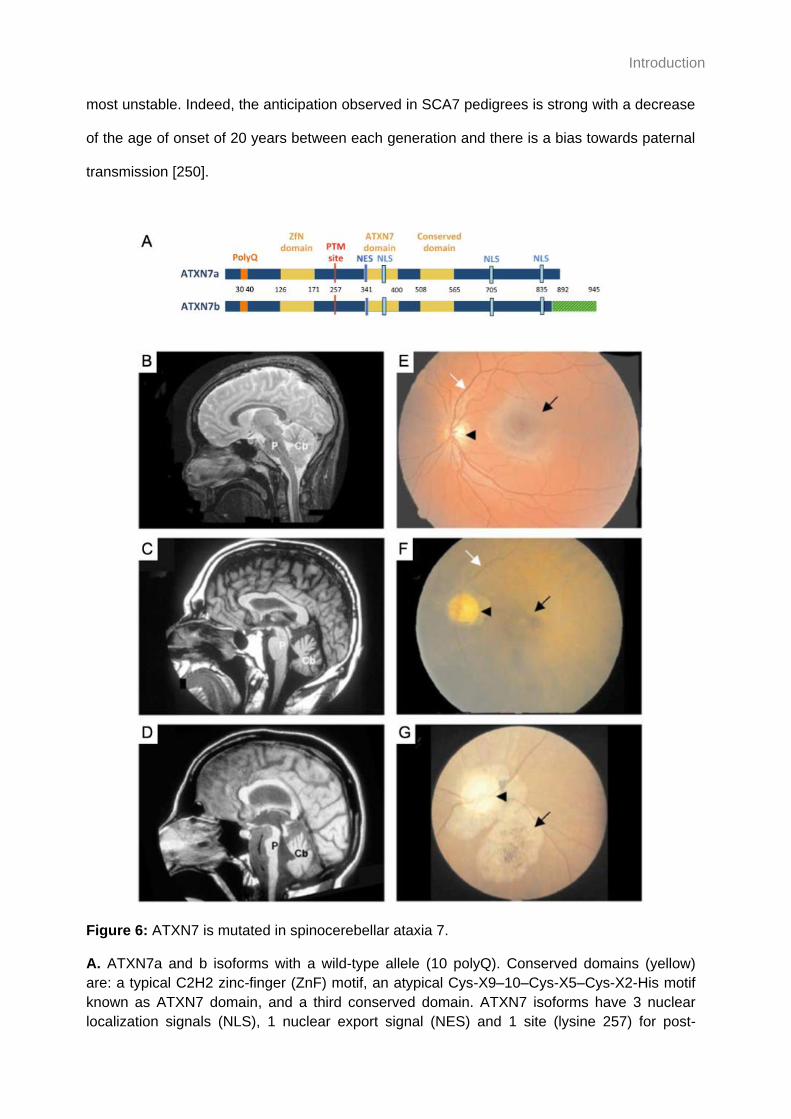
Introduction
most unstable. Indeed, the anticipation observed in SCA7 pedigrees is strong with a decrease
of the age of onset of 20 years between each generation and there is a bias towards paternal
transmission [250].
Figure 6: ATXN7 is mutated in spinocerebellar ataxia 7.
A. ATXN7a and b isoforms with a wild-type allele (10 polyQ). Conserved domains (yellow)
are: a typical C2H2 zinc-finger (ZnF) motif, an atypical Cys-X9–10–Cys-X5–Cys-X2-His motif
known as ATXN7 domain, and a third conserved domain. ATXN7 isoforms have 3 nuclear
localization signals (NLS), 1 nuclear export signal (NES) and 1 site (lysine 257) for post-

Introduction
translational modifications including acetylation and SUMOylation, but differ at their carboxy-
terminal end. B-D. Magnetic resonance imaging of a control individual (B) and two SCA7
patients (C, D). The patient in (C) (38 polyQ, examined at 60 years and showed mild
unsteadiness of gait) displays severe atrophy of the cerebellum (Cb), but almost complete
sparing of the pons (P). In contrast, the patient in (D) (52 polyQ, examined at 40 years),
shows severe cerebellar atrophy, as well as marked atrophy of the pons (P). E-G Fundoscopy
images of a control individual (E) and two SCA7 patients (F, G). Normal appearance of the
macula (black arrow) and the optic disk (arrowhead), and the well-developed vasculature
(white arrow) in the control individual (E). On the contrary, the retina of patient (F) (52 polyQ,
examined at 40 years) displays an atrophic macula (black arrow), very pale optic disk
(arrowhead), and poor vasculature (white arrow). Moreover, the retina of patient (G) (unknown
CAG repeats, examined at 20 years and showed gait ataxia, dysarthria, and decreased
eyesight). Figures A from [251], B adapted from [248] with legend shortened.
Pathomechanisms of SCA7. Although not fully understood, we know the baseline
pathomechanism of SCA7 and polyQ disorders (Figure 7). The polyQ expansion has a gain-
of-function effect where the mutant protein assembles in toxic aggregates not only with itself
but also to trap additional proteins [252]. ATXN7 is part of the chromatin remodeling and
transcription coactivator of RNA polymerase II SAGA [253], thus its localization is mostly
nuclear. Several studies showed the absence of correlation between the abundance of
ATXN7 transcripts and protein suggesting that a rapid protein turnover is necessary for proper
regulation of gene expression [254-256]. In the context of SCA7, mutant ATXN7 (mATXN7) is
cleaved by caspase-7 which releases a smaller peptide containing the polyQ and a zinc-finger
domain but deprived of the putative nuclear exportation signal and SCA7 domain [257]. The
polyQ seems to stabilize the protein which results in altered transcriptional programs and
neurodegeneration [258-261]. Additionally, a nuclear clearance system through clastosomes
is quickly overwhelmed by the accumulation of the toxic fragments in nuclear inclusions.
Those nuclear inclusions, hallmarks of the disease, are found ubiquitously in post-mortem
brains [262] but seem to accumulate faster in the SCA7 hallmark tissues of SCA7 mouse
models [256].

Introduction
Table 2: Overview of polyglutamine disorders T
ab
le 2
: O
verv
iew
of p
oly
glu
tam
ine d
isord
ers
Pa
tho
ge
nic
alle
le3
45
-91
35
-50
0
61
-87
20
-33
36
-46
0
49
-66
>4
0
49
-93
38
-62
Info
rmation c
om
pile
d in t
his
table
is f
rom
the f
ollo
win
g o
nlin
e d
ata
bases: 1
Pro
babili
ty o
f bein
g loss-o
f-fu
nctio
n into
lera
nt
fro
m g
no
mA
D (
the c
loser
the
pLI
is t
o 1
, th
e
more
into
lera
nt
it is),
2 U
nip
rot, a
nd 3
OM
IM.
Wil
d-t
yp
e
alle
le3
6-3
9
14
-31
11
-44
4-1
8
4-3
5
25
-42
9-3
5
6-3
5
9-3
4
Pro
tein
fu
ncti
on
2
Ata
xin
-1:
chro
matin-b
ind
ing f
acto
r, r
epre
ssor
of
Notc
h s
ign
alin
g,
ma
y b
e in
volv
ed in
RN
A m
eta
bolis
m a
nd b
rain
de
velo
pm
ent
Ata
xin
-2:
acts
as a
neg
ative r
egu
lato
r of
end
ocytic E
GF
R inte
rna
liza
tio
n a
t th
e p
lasm
a
mem
bra
ne
Ata
xin
-3:
deub
iquitin
atin
g e
nzym
e invo
lved in p
rote
in h
om
eosta
sis
ma
inte
nan
ce
,
transcription,
cyto
skele
ton r
eg
ula
tion,
myoge
nesis
and d
egra
da
tio
n o
f m
isfo
lde
d
chap
ero
ne s
ub
str
ate
s
Voltage-d
epe
ndent
P/Q
-type c
alc
ium
chan
nel sub
unit α
-1A
: m
edia
te t
he e
ntr
y C
a2
+
into
excita
ble
cells
and is involv
ed in a
va
riety
of
calc
ium
-dep
end
ent
pro
ce
sse
s
Ata
xin
-7:
com
pone
nt
of
the S
AG
A t
ranscription c
oactivato
r-H
AT
co
mp
lex,
me
dia
tes t
he
inte
raction o
f S
AG
A c
om
ple
x w
ith C
RX
, necessary
for
mic
rotu
bu
le c
yto
ske
leto
n
sta
bili
zation
TA
TA
box b
ind
ing p
rote
in:
ge
nera
l tr
anscription f
acto
r, f
unctions a
t th
e c
ore
of
the D
NA
-
bin
din
g m
ultip
rote
in f
acto
r T
FII
D t
o a
ctivate
RN
A p
oly
mera
se
II
tra
nscriptio
n o
f
eukary
otic g
en
es
Huntingtin:
ma
y p
lay a
role
in m
icro
tub
ule
-media
ted t
ransp
ort
or
ve
sic
le f
un
ction
Atr
ophin
-1:
transcriptiona
l core
pre
ssor
Andro
ge
n r
ece
pto
r: s
tero
id h
orm
one
-activate
d t
ranscription f
acto
r th
at
regu
late
s
eukary
otic g
en
e e
xpre
ssio
n a
nd a
ffects
cellu
lar
pro
lifera
tion a
nd
diffe
ren
tia
tion
in t
arg
et
tissues
pL
I1
0.9
7
0.8
5
0.9
7
1.0
0
0.9
6
0.0
2
1.0
0
1.0
0
0.9
9
Ge
ne
AT
XN
1
AT
XN
2
AT
XN
3
CA
CN
A1A
AT
XN
7
TB
P
HT
T
AT
N1
AR
Dis
eas
e
Sp
ino
ce
reb
ella
r
ata
xia
1
Sp
ino
ce
reb
ella
r
ata
xia
2
Sp
ino
ce
reb
ella
r
ata
xia
3
Sp
ino
ce
reb
ella
r
ata
xia
6
Sp
ino
ce
reb
ella
r
ata
xia
7
Sp
ino
ce
reb
ella
r
ata
xia
17
Hu
nting
ton
’s
dis
ea
se
De
nta
toru
bra
lpa
lli
do
luysia
n a
tro
phy
Sp
ina
lbu
lba
l
mu
scu
lar
atr
op
hy

Introduction
Neurodegeneration in SCA7. In the classic adult form, SCA7 first manifests as a
cerebellar pathology. Patients progressively lose coordination of balance, gait and speech and
present with pyramidal signs due to neuronal degeneration in the cerebellum, in the Purkinje
cell layer and in the dentate nuclei especially, but also in other brain structures [248,263].
Cerebellar atrophy can be visualized through magnetic resonance imaging (Figure 6). SCA7
retinopathy usually appears second and manifests as dyschromatopsia followed by decreased
central visual acuity (loss of cone photoreceptors). The degeneration progresses into a cone-
rod dystrophy until the loss of the entire visual field and complete blindness (Figure 6). SCA7
disease progresses over several decades and most patients die from pneumonia caused by
dysphagia [264]. In the most severe form of SCA7, the retinopathy manifests prior to
spinocerebellar ataxia.
Neurodevelopmental component. In the past two decades, the definition of late
onset diseases evolved due to accumulating evidence of developmental components to
degenerative manifestations. A well-known example is the observation that post-mortem
tissues from schizophrenia patients revealed brain developmental anomalies which were not
related to neurodegeneration [265,266]. More closely related to SCA7, subtle brain
differences were observed in individuals who were at risk for Huntington’s disease [267,268].
A possible explanation for those early manifestations could be a partial loss-of-function of the
protein. Indeed, overexpression of wild-type HTT and ATXN3 ameliorated some phenotypes
of a mouse model of Huntington’s disease [269,270], and the neurodegeneration of a
Drosophila model of SCA3 [271] respectively. Further lines of support include the high
probability of being Loss-of-function Intolerant (pLI) of the genes mutated in polyQ disorders
(Table 2) and the non-viability of HTT knockout mice. More recently, we showed that atxn7
plays a role in the development of the eyes and brain of the zebrafish ([272] with results
description in section Results III.2).
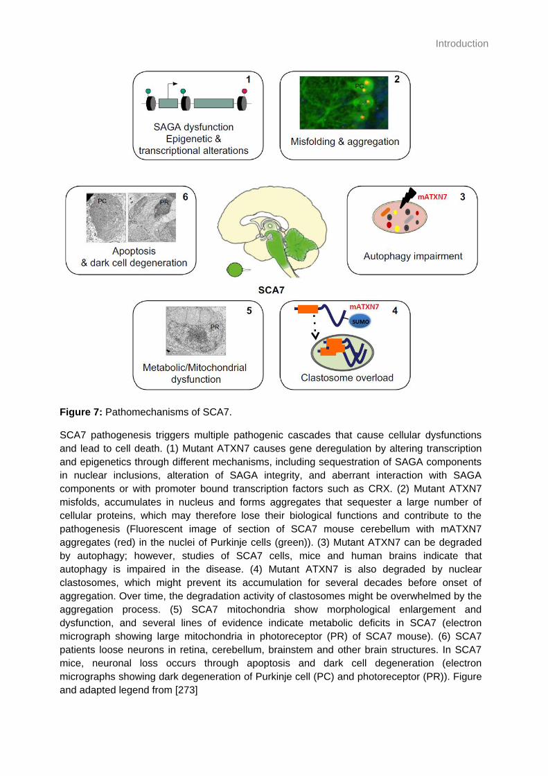
Introduction
Figure 7: Pathomechanisms of SCA7.
SCA7 pathogenesis triggers multiple pathogenic cascades that cause cellular dysfunctions
and lead to cell death. (1) Mutant ATXN7 causes gene deregulation by altering transcription
and epigenetics through different mechanisms, including sequestration of SAGA components
in nuclear inclusions, alteration of SAGA integrity, and aberrant interaction with SAGA
components or with promoter bound transcription factors such as CRX. (2) Mutant ATXN7
misfolds, accumulates in nucleus and forms aggregates that sequester a large number of
cellular proteins, which may therefore lose their biological functions and contribute to the
pathogenesis (Fluorescent image of section of SCA7 mouse cerebellum with mATXN7
aggregates (red) in the nuclei of Purkinje cells (green)). (3) Mutant ATXN7 can be degraded
by autophagy; however, studies of SCA7 cells, mice and human brains indicate that
autophagy is impaired in the disease. (4) Mutant ATXN7 is also degraded by nuclear
clastosomes, which might prevent its accumulation for several decades before onset of
aggregation. Over time, the degradation activity of clastosomes might be overwhelmed by the
aggregation process. (5) SCA7 mitochondria show morphological enlargement and
dysfunction, and several lines of evidence indicate metabolic deficits in SCA7 (electron
micrograph showing large mitochondria in photoreceptor (PR) of SCA7 mouse). (6) SCA7
patients loose neurons in retina, cerebellum, brainstem and other brain structures. In SCA7
mice, neuronal loss occurs through apoptosis and dark cell degeneration (electron
micrographs showing dark degeneration of Purkinje cell (PC) and photoreceptor (PR)). Figure
and adapted legend from [273]
Introduction
ATXN7 and cilia. One of the possible implications of the wild-type ATXN7 being lost is
a putative impact in ciliary function. ATXN7 is found in both the nucleus and the cytoplasm in
many human tissues [274]. Consistently, a study aiming to build an ataxia interactome map
used ATXN7 as a bait in a yeast two-hybrid screen and identified multiple interactions
between ATXN7 and nuclear or cytoplasmic proteins [246]. Of interest was the interaction
between ATXN7 and the two centrosomal/BB proteins CEP70 and CEP72. Furthermore,
ATXN7 was shown to associate with and stabilize the microtubule cytoskeleton in vitro [275].
A number of experiments performed in our lab showed that 1) ATXN7 and CEP72 indeed
colocalize to the centrosome in human telomerase-immortalized retinal pigmented epithelial
(htRPE-1) cells; 2) ATXN7 can be observed at the base of the primary cilia in mouse primary
cortical and hippocampal neurons, as well as in IMCD3 cells and embryonic rat
cardiomyocytes (H9c2 cells); and 3) ATXN7 colocalizes to the BB of connecting cilia of
photoreceptors in the retina of wild-type mice (Figure 8, [276]). Together, these data show
that ATXN7 can associate with two ciliary components: the centrosome/BB and tubulin, and
could suggest a role for ATXN7 in ciliary biology. However, further functional studies are
required to characterize this putative and unexpected role of ATXN7.

Introduction
Figure 8: ATXN7 localizes at the centrosome and is present at the base of primary cilia.
A. Co-immunolabeling of htRPE-1 cells showing the colocalization of endogenous ATXN7
with its centrosomal CEP72 interactor (arrowhead). B. Co-immunostaining of ATXN7 (1262
polyclonal antibody against N-terminus fragment of the protein) and cilia markers
(polyglutamylation modification, GT335; acetylated α-tubulin, AcTub; or tubulin,1D5). ATXN7
is present at the base of primary cilia of cultured primary neurons of mouse hippocampus and
cortex. ATXN7 is detected in the shaft of motile cilia of ependymal and tracheal epithelium of
14-day old mice. ATXN7 is detected at the base of primary cilia of IMCD3 and H9c2 cells. C.
Retinal sections of 3-week old mice were labeled with the 1262 antibody. Cilia were detected
with anti-acetylated α-tubulin (AcTub) which labels the connecting cilum (CC) (C’). C’’ is a
schematic map of ATXN7 localization in photoreceptors. Scale bars 5 μm. Figure and adapted
legend from [276]
Introduction
Scientific progress and next challenges. Our understanding of ciliopathies has
greatly increased in the past two decades. Even if some aspects of these disorders are now
well documented, such as the cause and progression of retinal degeneration through the
impairment of connecting cilia [277], there are a number of symptoms which are not fully
understood. For instance, the mechanisms leading to the kidney pathology in autosomal
dominant polycystic kidney disease remain unsolved despite the different hypotheses to
explain the formation of cysts [278]. Perhaps the non-ciliary roles of ciliary proteins and the
ciliary roles of hitherto unknown ciliary proteins could explain knowledge gaps. Establishment
of a comprehensive “ciliary landscape” integrating data from gene location and regulation to
protein maturation, localization, interactions and functions would considerably improve the
diagnosis and prognosis of ciliopathies [279,280]. Thus, an emergent trend is a systems-level
approach integrating proteomics and patients’ data aiming to capture ciliary protein networks.
One example includes the identification of KIAA0586 as the causative gene for 5% of
unexplained Joubert syndrome cases through combinatorial approaches of siRNA screening,
proteomics data integration and whole-exome sequencing [281]. Another recent report has
documented the simultaneous identification of ADAMTS9 as the causative gene for a form of
nephronophthisis [282] and the role of ADAMTS9 in ciliary growth [283].

Introduction
V. Challenges and opportunities
1. Non-coding RNAs and cilia
The first ciliary proteome database contained more than 1,200 proteins selected through
multiple approaches, including data on gene expression and proteins from different
organisms [284]. The most recent databases of ciliary and centrosomal genes and proteins
(including data from multiple species) include: the original SYSCILIA Gold Standard (1,319
proteins, [238,285]), Cildb3.1 [286,287] and CiliaCarta which integrates data from the
SYSCILIA Gold Standard (836 genes, [288]). However, those resources focused primarily on
proteins because of technical choice (yeast-two hybrid screens, whole exome sequencing,
mass spectrometry, etc). Despite playing a substantial role on regulating cell biology, non-
coding RNAs have not been integrated in the ciliary databases. This mostly unexplored
molecular repertoire offers us the opportunity to contribute to the building of the “ciliary
landscape”.
The candidacy of microRNAs. microRNAs (miRNA) are short (about 20 nucleotides
long) non-coding RNAs which act as post-transcriptional regulators of messenger RNA
(mRNA) expression that bind to complementary regions in their 3’UTR [289]. They are
involved in almost every biological process and are highly conserved across species. miRNAs
are scattered throughout the genome in both intergenic regions and introns and are often
present in paralogous copies coding for miRNAs of a same family. For instance, let-7 was the
second miRNA to be discovered, is highly conserved, and has been established as a key
promoter of differentiation in C. elegans, Drosophila, mouse and human cell types [290].
There are twelve let-7 loci in the human genome: three give rise to let-7a, two to let-7f and the
others code for let-7b, c, d, e, g, i, and miR-28 [291]. They exist in three forms: the immature
pri-miRNA, the pre-miRNA hairpin and the mature miRNA. Pri-miRNAs are transcribed in the
nucleus by the RNA polymerase II and bear a 5’ cap and a polyA tail, similar to mRNAs [292].
In cases where miRNAs are located in introns, pri-miRNAs are released as part of the splicing

Introduction
process allowing maturation of both mRNAs and miRNAs. The Microprocessor complex
formed by the nuclear RNase III Drosha and its cofactor DGCR8 can crop pri-miRNAs and
release pre-miRNA hairpins which can then be exported to the cytoplasm by Exportin5/Ran-
GTP complexes located at the nuclear envelope (Figure 9) [293,294]. The cytoplasmic
RNase III Dicer and TRBP further crop the hairpin to release a small double strand fragment
[295]. One hairpin can hold 2 mature miRNAs: one on its 5’ strand and the other on its 3’
strand. However, only one mature miRNA can then be incorporated to the RISC (RNA-
induced silencing complex) at a time, alongside with AGO, to target mRNAs for degradation or
repress their translation [289] (Figure 9). Regulation of miRNAs can occur at several levels:
through regulation of the processing proteins necessary for miRNA maturation or through
post-transcriptional modifications such as miRNA tailing and methylation [296]. LIN28 is a
well-studied regulator of the let-7 family: it can bind to the pri-miRNAs to prevent the first
maturation step by the Microprocessor, or recruit TUT4 to add uridylyl to the 3’ end of pre-let-7
to prevent processing by Dicer and target them for degradation. Because LIN28 transcripts
harbor let-7 targets in their 3’UTR, LIN28 and let-7 are mutual repressors [297].
Figure 9: Summary of canonical miRNA biogenesis pathway.

Introduction
Figure 9: Summary of canonical miRNA biogenesis pathway.
miRNA genes are transcribed from intergenic or intragenic regions of noncoding RNA
transcripts mediated by RNA polymerase II, called pri-miRNA. These long pri-miRNAs are
processed by the DROSHA–DGCR8 complex to form pre-miRNAs (~60 nucleotides long).
XPO5 mediates the export of these pre-miRNAs to the cytoplasm for further processing by
DICER. DICER cleaves pre-miRNAs to form mature miRNAs of ~20 nucleotides long. One of
the strands of mature miRNA (guide strand) gets incorporated into RISC involving DICER and
AGO2 enzymes to target mRNAs to cause degradation or translational suppression of the
target gene. Figure and legend adapted from [298].
Paucity of studies on microRNAs and cilia. Despite the regulatory role of miRNAs
and their potential effect on the expression of ciliary genes, there have been few studies
connecting miRNAs and cilia. In particular, the miR-34/miR-449 family has been implicated in
several mechanisms linked to reduced ciliogenesis such as the direct repression of the
Delta/Notch signaling pathway and the relocalization of actin binding protein Filamin-A in
human airway epithelium and embryonic Xenopus epidermis [299,300]; the repression of
CP110 in mouse and Xenopus models [301]; and the inhibition of cell cycle genes in a miR-
34/miR-449 knockout mouse model [302]. Another study has reported that miR-129-3p can
also control ciliogenesis through the direct repression of CP110, as well as by repressing F-
actin formation [303]. These different studies showed a role for miR-34/miR-449 and miR-129-
3p in the initiation of ciliogenesis and suggest that other miRNAs could contribute to ciliary
biology.
let-7b identified as a modulator of ciliogenesis. In 2010, our group (CHDM)
performed a medium throughput screen, testing approximately 800 miRNA inhibitors, to
identify miRNAs that could significantly induce or suppress ciliogenesis. The top candidate
predicted to enhance ciliogenesis was the let-7 family with let-7b showing the most substantial
effect. It was further shown that let-7b could promote ciliogenesis and increase cilia length
independent of its role on the cell cycle (Figure 9). However, these preliminary in vitro data
required validation in an in vivo developmental context.

Introduction
Figure 10: let-7b is a positive regulator of ciliogenesis.
A. Immunofluorescence of htRPE-1 cells stably expressing Smo-EGFP (green) and stained
for acetylated α-tubulin (red). Cells transfected with let-7b miRIDIAN inhibitor (middle panel)
show a marked reduction in the number of cilia in comparison to a control inhibitor (top panel).
Elevation of let-7 levels by transfecting a let-7b mimic (bottom panel) results in a greater
fraction of ciliated cells and significantly longer cilia. B. Quantification of the fraction of ciliated
cells and C. ciliary length. Statistics two-tailed t-test, ****p<0.0001, ***p<0.001; *p<0.05. Scale
bars=10µm, 2µm in insets. (Jon Robinson’s thesis; [304])

Introduction
THESIS AIM 1
The first objective of this work is to validate the potential role of let-7b on ciliogenesis using
three approaches: 1) the suppression and overexpression of let-7b in the zebrafish model
organism to monitor ciliary defect in vivo; 2) identification of let-7b target genes which are
negative regulators of ciliogenesis using transcriptomic data; and 3) exploration of the
hypothesis that human ciliopathy cohorts might harbor an enrichment of variation in the
3’UTRs of let-7b target genes. Outcomes of this work could contribute to the “ciliary
landscape” and help to explain the phenotypic variability observed in patients, but could also
improve diagnostics and prognostics of ciliopathies.
2. New ciliary role for an already characterized protein
The observation that there are ciliary proteins with non-ciliary roles and non-ciliary proteins
with ciliary roles has challenged the mapping of the “ciliary landscape”. ATXN7 has been
characterized previously to participate to the mediation of transcription through the SAGA
complex [305]. However, protein interaction and localization experiments suggest a novel role
linked to cilia. Thus, ATXN7 is suspected to be a non-ciliary protein with a ciliary role.
Primary cilia in brain development. Primary cilia play an important role in brain
development not only through the transduction of signaling pathways but also through
regulated autophagy [306]. In particular, they are involved in the morphogenesis of the
cerebellum, which is severely affected in SCA7, through HH signaling [307,308]. Sonic HH is
secreted by the Purkinje cells in the cerebellum and allows the proliferation of cerebellar
neuronal precursors including in the granule cell layer [309,310]. Defects in primary cilia have
been observed in olfactory neuronal precursors obtained from patients affected with late onset
disorders, including schizophrenia and bipolar disorder [311,312].
SCA7, a second-order ciliopathy? Considering: 1) the probable developmental
component of SCA7 with a role for ATXN7 in brain and eye development [272]; 2) the putative

Introduction
ciliary role of ATXN7; and 3) the role of primary cilia in cerebellum development, it is possible
that SCA7 first manifests through subtle brain anomalies caused by a partial loss of ATXN7’s
putative ciliary role.
THESIS AIM 2
The second objective of this thesis is to explore the potential link between ATXN7 and cilia in
the development of SCA7 by overexpressing wild-type and mutant ATXN7 in the developing
zebrafish. The zebrafish offers an opportunity to evaluate the brain of a transparent vertebrate
with ex vivo development and is also a model of choice to observe potential ciliary defects
[313]. A zebrafish model of SCA7 would be a relevant tool not only to understand the disease
by complementing mouse models, which are more expensive and take 5 weeks to 11 months
to develop relevant phenotypes [251], but would also have the potential to be used in
therapeutic screens to test compounds such as anti-aggregation molecules identified in vitro
by our group (IGBMC) [314].
3. Exploring other aspects of ciliopathies and SCA7
The zebrafish model organism presented an opportunity to explore phenotypic aspects of
ciliopathies and to study the role of ATXN7 in development. Indeed, the zebrafish produces a
large amount of offspring, develops externally and rapidly, is transparent during development
[315] and 70% of its genome is conserved with the one of humans [316].
THESIS AIM 3
The third objective is to broaden my range of technical skills by participating in other projects
lead in my host laboratories. Notably, by exploiting the zebrafish model organism to study
organs which are typically affected in ciliopathies (kidney [317] and retina (manuscript
submitted)) and to contribute to the study on the consequences of atxn7 inactivation on
development [272].

Introduction
MATERIAL AND METHODS

Material and Methods
Material and Methods
Material and methods for the two attached publications (chapter III of the results) are included
in the publications and are not mentioned here.
Zebrafish maintenance
Zebrafish adults from the Duke University facility or IGBMC facility were maintained at 28°C.
Embryos were maintained at 28.5°C in egg water (0.55mM CaSO4, 0.45mM CHNaO3,
5.13mM NaCl). Wild-type embryos were from the ZDR strain (let-7b chapter) or AB strain
(ATXN7 chapter), and transgenic SoFa1 strain was kindly provided by Dr. William A. Harris
[318]. Embryos were staged in somite-stage (ss), hours-post-fertilization (hpf) or days-post-
fertilization (dpf) as described [315].
CRISPR/Cas9 genome editing of let-7b in zebrafish
We used ChopChop to design a guide RNA (gRNA) targeting the Drosha processing site
required for let-7b pri-miRNA maturation into pre-miRNA (miRBase ID: MI0001865; 5′-
AGTGGTGTTCGGACAGGGTGAGG-3′). We synthesized the gRNA using the Gene Art
precision gRNA synthesis Kit (Invitrogen) according to manufacturer’s instructions. To test our
gRNA efficiency, we co-injected 100pg of gRNA with 200pg of Cas9 protein (PNA Bio) in the
cell of 1-cell stage zebrafish embryos (1nL/embryo). We extracted the genomic DNA at 1dpf,
amplified the region targeted by the gRNA and loaded the product on a 15% polyacrylamide
gel to visualize heteroduplexes after a denaturation-reannealing treatment. PCR products
from different embryos were cloned and Sanger sequenced. Percent mosaicism was
calculated by counting the percentage of mutant clones per total clones and averaging across
all embryos tested.
Transient suppression and overexpression of let-7b in zebrafish
The mature sequence of human let-7-b-5p (miRbase ID: MI0000063) is 100% conserved in
zebrafish. We designed a morpholino (MO) complementary to the mature sequence of let-7b

Material and Methods
to inhibit its activity (5’- AAACCACACAACCTACTACCTCACC -3’; GeneTools), and obtained
a dsRNA let-7b-5p mimic from Dharmacon (5’- UGAGGUAGUAGGUUGUGUGGUU -3’). We
first titrated our MO by injecting increasing concentrations of MO (2, 4 and 6ng/nL) and mimic
(0.2 and 0.8μM) in the cell of 1-cell stage zebrafish embryos (1nL/embryo) then used 6ng/nL
of MO and 0.08μM of let-7b-5p mimic to perform the rescue experiments.
RT-qPCR on zebrafish embryos
Total RNA was extracted from 30hpf embryos with TRIzol (Invitrogen) according to
manufacturer’s instructions. Total RNA was reverse transcribed with either the miRNA 1st-
Strand cDNA Synthesis Kit (Agilent) or the QuantiTect Reverse Transcription Kit (Qiagen),
from 800ng or 500ng of total RNA, respectively, according to manufacturer’s instructions.
qPCR was performed with Power SYBR Green PCR Master Mix (Applied Biosystems)
according to manufacturer’s instructions. let-7b levels were normalized to miR-206 and limk2
levels were normalized to gapdh. qPCR data were analyzed with the 2-∆∆CT method [319] and
statistical analyses were performed with a two-tailed t-tests (GraphPad PRISM software;
version 8.0). The following primers were used: let-7b: 5’-TGAGGTAGTAGGTTGTGTGGTT-3’;
miR-206: 5’-TGGAATGTAAGGAAGTGTGTGG-3’; universal reverse primer from the Agilent
kit; limk2-F: 5’-CCATGGTGGCAGGAGACTAT-3’ and limk2-R: 5’-GGACCACCTGCTTGT
AGCAT-3’; gapdh-F: 5’- TTGTAAGCAATGCCTCCTGC-3’ and gapdh-R:5’-CTGTGTTGCTGT
GATGGCAT-3’.
Brightfield examination of zebrafish embryos
let-7b chapter: Embryos at the desired developmental stage were anesthetized with 0.02%
Tricaine (A5040, SigmaAldrich) and positioned in a 2% agarose mold. Heart looping
assessment was performed when the embryos were still in their chorion to facilitate
manipulation. Statistical comparison was performed with a two-tailed Fisher’s exact test
(GraphPad online QuickCalcs tool). Images were acquired using a Nikon SMZ745
stereoscope equipped with a Digital Sight camera and with NIS-Elements software. The
background of the images was normalized with Gimp software.

Material and Methods
ATXN7 chapter: Embryos older than or at the 24hpf stage were anesthetized with 0.02%
Tricaine (A5040, SigmaAldrich) and imaged in egg water or in a 1.5% agarose mold. Images
were captured with a M420 Macroscope (Leica) with COOLSNAP coupled camera and
analyzed using FiJi software (NIH).
spaw whole mount in situ hybridization
18ss embryos were fixed in 4% paraformaldehyde (PFA) at 4°C overnight. in situ hybridization
on whole-mount embryos was performed as described [320]. Statistical comparison was
performed with a two-tailed Fisher’s exact test (GraphPad online QuickCalcs tool). Images
were acquired using a Nikon SMZ745 stereoscope equipped with a Digital Sight camera and
with NIS-Elements software. The background of the pictures was normalized with Gimp
software.
Cilia measurement in the Kupffer’s vesicle of zebrafish embryos
Embryos were reared at 23°C and fixed at the 10ss in Dent’s solution (80% methanol, 20%
dimethylsulfoxide) overnight at 4°C. Embryos were rehydrated with a decreasing series of
methanol and phosphate-buffered saline with 0.1% Tween-20 (PBST); washed with PBST
and dechorionated. Cilia were stained with acetylated tubulin primary antibody (T745, Sigma
Aldrich, 1:1000 dilution) and goat anti-mouse IgG secondary antibody (488 Alexa Fluor,
Invitrogen; 1:500 dilution). The tail buds were dissected and mounted in 50% glycerol on a
slide prior imaging with a 90i fluorescent microscope (Nikon) at 60x magnification, a digital
black and white camera (Nikon), and the NIS Elements software (Nikon). Z-stacks were
combined using the Maximum Intensity projection in FiJi (NIH) and cilia were measured with
the Segmented Line tool. Statistical comparisons were performed with a non-parametric one-
way ANOVA followed by Tukey’s multiple comparison (GraphPad PRISM software; version
8.0).

Material and Methods
Evaluation of convergent-extension
let-7b chapter: Convergent-extension (CE) was assessed at the 10ss. Images were acquired
with an Axio Zoom V16 (Zeiss) at 80x magnification, and the ZEN Blue software (Zeiss).
ATXN7 chapter: Fixed (4% PFA overnight at 4°C) embryos at the 10-11ss were imaged in a
1.5% agarose mold with a M420 Macroscope (Leica) coupled to a COOLSNAP camera.
Images were analyzed in ImageJ software (NIH) where the body-gap angle was measured
using the zebrafish head (before the polster) and tail tip as landmarks. Statistical comparison
was performed with a non-parametric one-way ANOVA followed by Tukey’s multiple
comparison (GraphPad PRISM software; version 8.0).
Targeted sequencing of a ciliopathy cohort and analysis
Human gDNA was quantified using fluorometric quantification with the Quant-iT PicoGreen
dsDNA kit according to manufacturer’s instruction on a FLUOStar Omega plate reader (BMG
Labtech). For each sample, gDNA was subsequently diluted to 2.5ng/μL to a final volume of
10μL, and transferred to the Duke sequencing core. Raw sequencing results were analyzed
by the Duke bioinformatics Core which produced an Excel file the filtered data. Detailed
methods of the targeted sequencing and bioinformatics analysis can be found in the annex.
Each variant was manually verified in the Integrative Genomics Viewer version 2.4.10 (Broad
Institute) and variants were called at a cutoff value of 20%. Ethnicity, sex, and primary causal
mutation were reattributed to each patient as well as the variants detected in the targeted
sequencing. Variants were then manually counted and we used control data from the Genome
Aggregation Database (gnomAD; https://gnomad.broadinstitute.org/) to perform the
enrichment analysis. Statistical comparisons were performed with a Chi-square test with
Yates’ correction (GraphPad online QuickCalcs tool) and Bonferroni correction was applied to
the p-values.

Material and Methods
Cloning and construct preparation
Truncated ATXN7 constructs were obtained as follows: 4xnrUAS-Ataxin (wild-type or mutant)-
P2A-mCherry vectors obtained from the AMAGEN platform (Gif-sur-Yvette) were digested
with NheI and XmaI to extract the coding sequences of truncated ATXN7 which was then
purified by gel electrophoresis and ligated back into pCS2+_ATXN7a vector after digestion by
the same enzyme to remove the sequence coding for the full-length wild-type protein. The
addition of eGFP downstream of truncated wild-type and mutant ATXN7 sequence was
performed as follows: the sequence coding for eGFP was amplified from a pEGFP-N2
plasmid using the following primers 5’-GAATTCTGCAGTCGACGGTA-3’ and 5’-
TATGTTCTAGAG TCGCGGCC-3’ because the XbaI site was methylated and could not be
used for digestion, the PCR product was then cloned in a p-GMT vector and recovered by
digestion with XmaI and XbaI, finally the eGFP fragment was cloned into the pCS2+_wild-
type/mutant-ATXN7∆ vectors which were first digested with XmaI and XbaI. The addition of
eGFP downstream of full-length wild-type and mutant ATXN7 sequence was performed using
a three-fragment strategy to increase specificity as follows: pCS2+_wild-type/mutant-
ATXN7∆-eGFP vectors were digested with NheI, XmaI and DraII and the XmaI-DraIII and
DraIII-NheI fragments were gel purified, in parallel, the pCS2+_ATXN7 (wild-type, with 60
CAG repeats and with 104 CAG repeats) vectors were digested with NdeI, NheI and XmaI
and only the NheI-XmaI fragments were gel purified, finally the three purified fragments were
ligated all together. Removal of the STOP codon which was kept between ATXN7 and eGFP
sequences was achieved by amplifying the 3’ region of ATXN7 sequence with a forward
primer upstream of the XhoI digestion site 5’-CCTGTCAACTCCCACGGCAG
TTTTTCCCACTCAC-3’ and a reverse primer overlapping the XmaI digestion site but
excluding the STOP codon 3’-CCGGTGGATCCCGGGACGTGCCTTTGGC TGATGA-5’, the
amplicon was then digested with XhoI and XmaI and used to replace the faulty fragment
containing the STOP codon.

Material and Methods
Mature mRNA synthesis and microinjection into zebrafish embryos
pCS2+ vectors containing a SP6 promoter upstream of the sequence of interest were
linearized with KpnI then capped mRNAs were prepared according to manufacturer’s
intructions using the mMessage mMachine SP6 Transcription kit (AM1340, ThermoFisher).
mRNAs were aliquoted to prevent freeze-thaw cycles and stored at -80°C. Injection cocktails
were prepared with Phenol red and nuclease-free water. Injection cocktails were injected in
the cell of one-cell stage zebrafish embryo using a Picospritzer III microinjector (Parker
Hannifin) in Duke University facilities or in the yolk of one- to two-cell stage zebrafish embryo
using a Nanoject II micro-injector (Drummond Scientific) in IGBMC facilities.
Cerebellum immunostaining
Embryos were anesthetized and fixed in Dent’s solution (80% methanol, 20%
dimethylsulfoxide) at 3 dpf. Whole-mount immunostaining was performed as described [321].
Briefly, embryos were rehydrated with a decreasing series of methanol and phosphate-
buffered saline with 0.1% TWEEN-20 (PBST), washed with PBST, bleached (with 3%
H2O2/0.5% KOH) and permeabilized with Proteinase K at 10μg/mL (25530-049,
ThermoFisher). Embryos were then incubated with primary antibody anti-acetylated α-tubulin
(T7451, mouse, Sigma-Aldrich, dilution 1:1000) and then secondary antibody Alexa Fluor 488
goat anti-mouse IgG (ThermoFisher, 1:500). Dorsal images were acquired with an AZ100
fluorescent microscope (Nikon), digital sight black and white camera (Nikon), and NIS
Elements software (Nikon) at 7X magnification. The area of the cerebellum was measured
with ImageJ (NIH) and statistical differences between conditions were determined with a non-
parametric one-way ANOVA followed by Tukey’s multiple comparison (GraphPad PRISM
software; version 8.0).
SoFa1 retina imaging
Embryos were kept in egg water with 0.003% 1-phenyl-2-thiourea (PTU, P7629-10G, Sigma-
Aldrich) starting at 24hpf to prevent pigment formation. Embryos were manually
dechorionated if younger than 50hpf, anesthetized with 0.02% Tricaine (A5040, SigmaAldrich)
Material and Methods
and mounted into 3%-methylcellulose (M0387, SigmaAldrich) on depression slides. Pictures
were captured with a DM 400B microscope (Leica) with COOLSNAP coupled camera.
Cryo-sections
Embryos were fixed at 24hpf in 4% paraformaldehyde (PFA) for 10 min at room-temperature
to preserve the eGFP, washed in PBST, incubated in 30% sucrose overnight then mounted in
optimal cutting temperature compound (OCT; AGR1180; Agar scientific) and stored at -80°C
until ready for sectioning. 7μm-thick sections were cut using a CM3050 S cryostat (Leica).
Comparisons of eGFP kinetics
Live embryos were mounted in 3%-methylcellulose (M0387, SigmaAldrich) after being
anesthetized with 0.02% Tricaine (A5040, SigmaAldrich). If older than 24hpf, images were
captured with a DM 400B microscope (Leica) with COOLSNAP coupled camera. Images were
then analyzed in ImageJ (NIH). To calculate the Corrected Total Embryo Fluorescence
(CTEF), I traced the contour of each embryo using the brightfield picture and measured the
area and integrated density using the fluorescent image. Then I used the following formula
adapted from [322]: CTEF = Integrated density – (Area of embryo X Mean fluorescence of
background reading) where the mean fluorescence of uninjected controls was used as the
mean fluorescence of background reading. For the first experiment (II. Figure 5A), a defined
exposure time was set too high which is why saturation was observed in the eGFP injected
embryos. For the second experiment (II. Figure 5B), the exposure time was not set but
recorded and used to adjust the integrated density. Statistical differences between the CTEF
of embryos injected with ATXN7 constructs were determined with two-way ANOVA followed
by Tukey’s multiple comparison.
Immunostaining of the proximal convoluted tubule
Embryos were anesthetized and fixed in Dent’s solution (80% methanol, 20%
dimethylsulfoxide) at 4 dpf. Whole-mount immunostaining was performed as follows: embryos
were rehydrated with a decreasing series of methanol and phosphate-buffered saline with

Material and Methods
0.1% TWEEN-20 (PBST), washed with PBST, bleached (with 3% H2O2/0.5% KOH) and
permeabilized with 1% Triton X-100 (85112, ThermoFisher). Embryos were then incubated
with primary antibody anti-Na+/K+/ATPase alpha-1 subunit (a6F, mouse, Developmental
Studies Hybridoma B, 1:20 dilution) and then secondary antibody Alexa Fluor 488 goat anti-
mouse IgG (ThermoFisher, 1:500). Lateral images were acquired with an AZ100 fluorescent
microscope (Nikon), digital sight black and white camera (Nikon), and NIS Elements software
(Nikon) at 4X magnification. The area of the proximal convoluted tubule was measured with
ImageJ (NIH) and statistical differences between conditions were determined with a non-
parametric one-way ANOVA followed by Tukey’s multiple comparison (GraphPad PRISM
software; version 8.0).

Material and Methods
RESULTS

Results
Results
I. The microRNA let-7b can modulate ciliogenesis
A medium throughput screen conducted on htRPE-1 cells in our group (CHDM)
enabled the identification of the let-7 family of miRNAs to significantly induce or suppress
ciliogenesis. To validate further the role of let-7 on ciliary biology, we chose to test let-7 in
vivo.
1. Use of zebrafish for in vivo validation of the effect of let-7b on ciliogenesis
We used zebrafish as a model organism to test whether let-7 has similar effects on cilia in
vivo. let-7 miRNAs are conserved in zebrafish and expressed at low levels in early
embryogenesis [291,323]. Based on conservation, number of copies, and ranking from our in
vitro screen, the most experimentally tractable let-7 member is let-7b (100% identity, single
copy and top hit) (Table1).
Suppression of let-7b results in established ciliary phenotypes in zebrafish
We designed a guide RNA (gRNA) with 100% efficiency targeting the Drosha processing site
required for let-7b pri-miRNA maturation into pre-miRNA (Supp. Figure 1A,D, E). We co-
injected 100pg of gRNA with 200pg of Cas9 enzyme into zebrafish embryos to generate let-7b
mosaic F0, and we measured a 36% reduction of mature let-7b via RT-qPCR (p=1.62E-4;
Supp. Figure 1B). Next, we examined the mosaic let-7b F0 at different developmental
stages. Left-right defects are a hallmark of ciliopathies and can be assessed in the zebrafish
by the directionality of the heart looping at 2 days post-fertilization (dpf) [324,325]. We

Results
observed minimal abnormal cardiac looping among our injected embryos with a greater extent
in embryos injected with gRNA+Cas9 (5.3%) compared to their siblings injected with gRNA
alone (2.4%; not significant) or uninjected (0%; p<0.0001; Figure 1A, B). Left-right patterning
during embryogenesis is governed by spatially restricted expression of nodal genes, such as
southpaw (spaw) [326], so we performed RNA in situ hybridization to assess the expression
pattern of this gene. 59.3% of mutants presented either bilateral, contralateral or absent
expression of spaw compared to 8.3% in embryos injected with gRNA alone (p=0.0001) and
3.3% in uninjected embryos (p<0.0001; Figure 1C, D). The increased number of affected
embryos can be explained by the fact that bilateral or absent expression of spaw could give
rise to normal looping by chance. We raised the zebrafish embryos up to 4dpf and noticed
that 18% of mosaic let-7b F0 were smaller, lacked a swim bladder and had cardiac edema.
Less than 3% of such embryos were found in the control conditions (Supp. Figure 2).
Suppression of let-7b leads to shorter cilia in the zebrafish Kupffer’s vesicle
The asymmetrical expression of nodal genes is created by the detection of a flow generated
by the motile cilia in the Kupffer’s vesicle (KV) which forms at approximately the 4-5ss somite-
stage and persists until 18ss. Thus, we hypothesized that the left-right defects observed in the
let-7b mosaic F0 were caused by ciliary defects in the KV. We stained 10ss embryos using an
antibody against acetylated α-tubulin and examined the KV. The size of KV is naturally
variable from one embryo to another [327] and we observed a trend toward a smaller KV in
injected embryos (Supp. Figure 3A). To account for this variability, we measured the number
of cilia per µm2 of the KV lumen for each condition. We did not observe a change in the cilia
density (Figure 1F), but cilia were significantly shorter in the KV of embryos injected with
gRNA+Cas9 (4.3µm) compared to their siblings injected with gRNA alone (4.9µm; p<0.0001)
or uninjected controls (5.1µm; p<0.0001; Figure 1G). These results suggest that suppression
of let-7b in zebrafish embryos does not affect the number of ciliated cells but leads to shorter

Results
cilia in the KV. This defect might prevent the generation of a proper flow to trigger restricted
asymmetrical expression of laterality genes and could explain the heterotaxia phenotypes.
Cilia defects were recapitulated in let-7b morphants and were partially rescued when
the morpholino was co-injected with let-7b-5p mimic
To validate further the effect of let-7b on cilia length, we injected a morpholino (MO) against
let-7b or a let-7b-5p mimic (Supp. Figure 1A, Figure 2). Injection of increasing
concentrations of let-7b MO from 2ng/µL to 6ng/µL led to a significant reduction of mature let-
7b (2ng: 47% reduction, p=7.08E-5; 4ng: 48% reduction, p=9.31E-5; 6ng: 53% reduction,
p=6.63E-6), while injection of the mimic at 0.2µM and 0.8µM led to a significant increase
(0.2µM: 287% increase, p=1.57E-8; 0.8µM: 902% increase, p=2.64E-7) (Supp. Figure 1C).
The injection of the MO recapitulated the heart looping anomalies (5.4-9.6%, p≤0.0001;
Figure 2A), the spaw expression defects (36.8-65.3%, p-values<0.0001; Figure 2B), and cilia
length defects (3.5-4.5µm, p<0.0001; Figure 2F) in a dose dependent manner. The KV
tended to be smaller but the cilia density in the KV lumen was not affected (p>0.5, except for
2ngMO, p=0.02; Supp. Figure 3B, Figure 2G). Longer cilia have also been shown to affect
left-right development in zebrafish [328] so we expected to see similar phenotypes in the
embryos injected with the let-7-5p mimic. The injection of the let-7b-5p mimic led to dose-
dependent heart looping anomalies (3.3% at 0.2µM, p=0.0001; and 25.3% at 0.8µM,
p<0.0001) and spaw expression defects (30.6% at 0.2µM, p=0.0004; and 54.3% at 0.8µM,
p<0.0001) (Figure 2A, B). However, we found no effect on the cilia density or length in the KV
(Figure 2F, G). There was a trend for a smaller KV lumen in the embryos injected with the
mimic but differences were not statistically significant (Supp. Figure 3B). The absence of cilia
phenotypes in fixed samples, despite the presence of ciliary phenotypes, could suggest a
mechanical or a signaling defect in embryos injected with the mimic. It is also possible that the
left-right anomalies are caused by imbalance in cell differentiation due to excessive levels of
let-7b-5p at these developmental stages.

Results
Next, we co-injected let-7b MO with let-7b-5p mimic to see whether we could rescue
the ciliary phenotypes observed in the morphants. All observed phenotypes were partially
rescued when the MO was co-injected with 0.08μM of let-7b-5p mimic suggesting specificity:
the heart looping anomalies were improved by 7.3% (from 9% in the morphants to 1.7% in the
rescued embryos, p<0.0001; Figure 2C), the spaw expression defects were improved by
41.9% (from 64.9% to 23%, p =5.8E-3; Figure 2D), and the cilia length by 0.6 µm (from 3.7µm
to 4.3µm, p<0.0001; Figure 2H). The cilia density calculated based on the size of the KV
lumen (Supp. Figure 3C) and the number of cilia showed no difference (Figure 2I).
Suppression of let-7b in zebrafish also leads to phenotypes related to primary cilia
defects
Convergent-extension (CE) is a process where cells migrate collectively and intercalate to
allow body axis elongation in the developing zebrafish. The main pathway regulating CE is the
cilia-dependent Wnt/PCP pathway and CE defects have been observed in several zebrafish
ciliopathy models [313,329]. Thus, we examined 10ss let-7b mosaic F0, morphants and
embryos expressing let-7b-5p mimic. We observed a significant increase of the angle
between the anterior- and posterior-most ends of let-7b mosaic F0 (64.5°, p<0.0001), let-7b
morphants (57,6° at 2ng/µL, 60.3° at 4ng/µL, and 77° at 6ng/µL, all p<0.0001) compared to
controls (45° in embryos injected with gRNA alone, and 39.2 - 46,6° in uninjected siblings)
suggesting a defect in primary cilia (Supp. Figure 4). Embryos injected with let-7b-5p mimic
did not show a significant increase of their body-gap angle compared to uninjected siblings
(51.8° at 0.2µM, p=0.21; and 52.2° at 0.8µM, p=0.14; Supp. Figure 4). This suggests that the
overexpression of let-7b-5p in zebrafish embryos does not affect ciliary signaling.

Results
2. Identification of possible let-7b targets which might act on ciliogenesis
Both in vitro and in vivo results support the candidacy of let-7b as a positive regulator of
ciliogenesis but its mechanism of action remains unclear. To identify candidate genes that are
regulated by let-7b and could influence ciliogenesis, we used the online prediction tool
TargetScan and RNA-seq data performed on htRPE-1 cells treated with let-7b inhibitor or
mimic.
Target gene predicting tool and database: TargetScan
The fewer and shorter cilia observed upon knockdown of let-7b in vitro and in vivo suggest
that let-7b targets transcripts which negatively regulate ciliogenesis and cilia maintenance
(Figure 3A), thus we looked in the literature for either validated or predicted negative
regulators of ciliogenesis and identified 213 genes (Supp. Table 1). Next, we looked at the
TargetScan database [330] which predicted 1207 target genes for the let-7 family based on
the presence of conserved sites matching the seed region and experimental evidence (Supp.
Table 2). We found an overlap of 21 genes between the TargetScan predicted targets and the
list of confirmed or predicted negative regulators: MDM4, DUSP16, PBX1, PLA2G3, ARL4D,
EIF4G2, ELOVL4, RTCA, ABL2, KDM3A, DUSP9, CCND1, SLC6A15, LIMK2, APC2,
RASGRP1, EIF2S2, UTP15, B3GALT1, SCARA3, and MTDH (Figure 3B, Table 2). Of note,
ARL4D codes for an ADP Ribosylation Factor Like GTPase protein, and ARL6/BBS3 codes
for a protein of the same family and has been associated with BBS [331]. Since let-7b was the
let-7 family member which had the most marked effect in vitro, we filtered the predicted target
genes by preferred let-7 member and obtained a list of 60 genes which were let-7b preferred
targets (Supp. Table 2). None of them were known or predicted negative regulators of
ciliogenesis.

Results
Insights from transcriptomics data from cells treated with let-7b inhibitor or mimic
Next, we switched from an in silico approach to an in vitro approach to account for the gene
expression in the context of the htRPE-1 cells which allowed the identification of let-7 as a
candidate regulator of ciliogenesis. We hypothesized that the negative regulators of
ciliogenesis targeted by the let-7 family, and let-7b in particular, would be up-regulated in the
context of let-7b inhibitor and down-regulated in the context of let-7b mimic. RNA-seq on
htRPE-1 cells that were treated with let-7b inhibitor or let-7b-5p mimic was previously
performed at CHDM. Transcriptomic data analysis led to the identification of 1,221 genes
(1,152 were protein coding) of which expression was significantly increased when the cells
were treated with the inhibitor and significantly decreased in the context of the mimic
(FDR<0.05) (see methods in the annex). There was an overlap of 18 genes with the list of
validated or predicted negative regulators of ciliogenesis, and 5 were also predicted with
TargetScan (Figure 3C, Table 3). Functional annotation using the DAVID bioinformatics
resource 6.8 [332] showed an enrichment in KEGG pathways related to proteoglycans in
cancer and cell adhesion, the first term being expected considering the characterized function
of let-7 (Figure 3D). The Gene Ontology term analysis of biological processes replicated the
enrichment in cell adhesion pathways and also suggested an enrichment for terms associated
to the vesicle trafficking between the endoplasmic reticulum and the Golgi apparatus, and the
extracellular matrix organization (Figure 3). However, it has been shown that endocardial-
derived cells of conditional Ift88 knock-out mice, which lack cilia in the heart, produce
significantly more collagen and versican than their control littermates [333]. This indicated that
the enrichment analyses might reveal consequences of the loss of cilia (increased protein
synthesis and trafficking to the extracellular matrix) rather than let-7b direct upstream effect.
Therefore, we further filtered our list of candidate targets.

Results
Identifying direct targets of let-7b
It is likely that some gene expression alteration is an indirect effect of the modulation of let-7b
levels. Therefore, we combined both in silico and in vitro approaches by using the TargetScan
search tool on the transcriptomic data. We identified a total of 3,882 let-7 seed targets on
1,979 transcripts and only retained the 1,526 transcripts coding for proteins. After
consolidating the multiple isoforms, we obtained a list of 733 genes of which expression was
perturbed in cells treated with let-7b inhibitor and mimic, and bearing at least one let-7 target
(interactive Table 4 mentioned in the annex). When we restricted the search to let-7b-5p
targets to be consistent with the let-7b-5p mimic treatment, we narrowed down the list of
candidate genes to 499 (Table 4). Ten genes were overlapping with the list of negative
regulators of ciliogenesis: ARL4D, EIF4G2, ELOVL4, RTCA, LIMK2, CXXC4, RIOK2, PARVA,
NCK2, and TEAD1, of which 5 were also predicted targets by TargetScan (Figure 3E, Table
4). To guide our filtering, we also noted whether the gene was associated with cilia based on
different studies including the SYSCILIA lists [285], our own data obtained from the ciliary
proteome [284] and ciliopathy patients sequencing [334], the ciliary landscape study [238],
and OMIM database (Table 4, Supp. Table 3). The small number of genes prevented us from
reaching statistical significance in an enrichment analysis, however, manual review of the
genes enabled the identification of three different patterns which could link the transcript
regulation by let-7b and ciliogenesis: cell cycle regulation, actin cytoskeleton remodeling and
the PI3K/Akt pathway.
We identified a number of candidates, including CDC25A, CXXC4 and RIOK2, which
are involved in the regulation of the cell cycle which ultimately affects ciliogenesis. However,
those genes do not fit the model where let-7 regulates ciliogenesis in a cell cycle-independent
manner (Jon Robinson’s thesis, [304]). Thus, those genes were not further explored.
Among the validated negative regulators, we found the actin regulator PARVA which
has been identified in a RNAi screen where authors further demonstrated the inhibitory effect
of the actin cytoskeleton on ciliogenesis [335]. Additionally, LIMK2 was shown to

Results
phosphorylate CFL to inactivate its actin-depolarization activity which blocks vesicle trafficking
to the BB, but also allows nuclear translocation of YAP/TAZ, complexing with TEAD4, and
transcription of AURKA and PLK1 [92]. Since TEAD1 is closely related to TEAD4 [336], it is
possible that it plays a role in this cascade as well. Among the candidate genes not
established or predicted negative regulators of ciliogenesis, we found NRK, which is predicted
to play a similar role as LIMK2 [337], ACTR2 and a number of genes coding for cytoplasmic
membrane proteins which serves as anchor for the actin cytoskeleton. These genes include
VCL, RDX, EZR, MSN, TPM1, DAG1, PVRL2 and three genes coding for integrins: ITGA11,
ITGAV, ITGB3.
We found 15 genes coding for proteins stimulating the PI3K/AKT pathway: PDGFB,
GNB4, CDKN1A, NGF, TLR4, THBS1, ITGA11, ITGAV, OSMR, YWHAZ, IRS1, GNG5,
CREB3L2, NRAS, and ITGB3. It has recently been shown that the PI3K/AKT pathway is
activated by lysophosphatidic acid present in the serum delivered to htRPE-1 cells in culture,
and that it leads to the phosphorylation of WDR44. WDR44 then binds to RAB11 to prevent it
from initiating the preciliary trafficking [338].
Verifying LIMK2 candidacy in zebrafish
LIMK2 was the top candidate as it was selected by TargetScan, by our transcriptomics
analysis with low FDRs (FDRInhibitor=6.53E-46, FDRMimic=1.65E-5) and high fold changes (Log2
(FoldChange)Inhibitor=1.50 and Log2 (FoldChange)Mimic=-0.47), had three let-7b-5p target sites,
and is a validated negative regulator of ciliogenesis (Table 4, Figure 4A). Thus, we asked
whether we could detect an increase in the level of limk2 in let-7b F0 mosaic zebrafish and
morphants, and a decrease of limk2 in the embryos expressing let-7b-5p mimic. We did not
detect significant changes in the expression of limk2 in our injected embryos at 30hpf (Figure
4B,C). However, this does not exclude the possibility that let-7b modulates ciliogenesis
through LIMK2 and the regulation of the actin cytoskeleton in another context. It is possible
that LIMK2 played a role in the htRPE-1 cells in vitro but not in the developing zebrafish

Results
embryos at the observed stage. It would be relevant to knock down limk2 in let-7b F0 mosaic
and morphants to see if it rescues the cilia length phenotype. Phenocopy could also be tested
by overexpressing limk2 by mRNA injection in the zebrafish embryo.
3. Explore the hypothesis that the let-7 family of miRNAs is a modifier gene of
ciliopathies
To explore the hypothesis that the let-7 family of miRNAs acts as a modifier of ciliopathies, we
proposed that ciliopathy patients might harbor an enrichment of variants in the 3’UTR of let-7
target genes. We selected a cohort of 185 ciliopathy patients affected with a severe or a
milder form of ciliopathy (68 with Meckel-Grüber syndrome, and 117 with BBS). Patients were
from different ethnicities and split into two groups in accordance with the gnomAD for the
downstream analysis: North European (Non-Finnish) or other (Latino, African, East Asian, and
other). First, we Sanger sequenced the let-7b locus and confirmed that there was no mutation
in any sample from our cohort.
Identification of the target loci for sequencing
We built a list of let-7b targets to sequence based on the filtered data obtained from RNA-seq
data and the TargetScan tool. Here, we decided to focus on genes associated with
ciliopathies or established ciliary genes. Thus, we filtered the list of 733 genes (dysregulated
in treated htRPE-1 cells and containing at least one let-7b-5p or let-7b-3p binding site) with a
list of genes which have an established ciliary role obtained from the SYSCILIA list [285], and
genes associated with ciliopathies (our own data obtained from patients sequencing [334],
and OMIM database, Supp. Table 3). We identified 42 genes with 113 let-7b target loci (let-
7b-5p or let-7b-3p, Table 5) and performed targeted sequencing on our cohort of 185
ciliopathy patients. The mean target coverage was 1,369 reads across all samples.

Results
Analysis of variants
We looked for rare homozygous variants in let7b target sites which might contribute to
recessive ciliopathies but we found no such variant. Next, we ask whether there were more
variants in the 3’UTR of Meckel-Grüber patients compared to BBS patients to investigate a
correlation between mutational burden and disease severity. We observed no enrichment at a
5% or 1% minor allele frequency (MAF) suggesting no mutational burden at the sequenced
loci in these patients (Table 6). Then, we used the controls from the gnomAD to ask whether
there was an enrichment or depletion of the variants we identified through targeted
sequencing. We identified 2 variants which were significantly depleted or enriched in our
ciliopathy cohort. rs6679100 (1-198288709-A-G, MAF=0.70%) in the 3’UTR of NEK7 was
significantly depleted in our ciliopathy cohort (p=0.02; Table 7). NEK7 codes for NIMA related
kinase 7 which accelerates microtubule shrinkage speed [339] and is related to NEK2, a
protein mutated in Retinitis pigmentosa [340]. On the other hand, rs182129505 (10-
113912476-C-T; MAF=8.22E-5) in the 3’UTR of GPAM was significantly enriched in our
cohort (p <0.01; Table 7). GPAM codes for a glycerol-3-phosphate acyltransferase located in
mitochondria and has been associated with primary cilia but its function in relation to cilia
biology remains unclear [341]. Those 2 variants were confirmed by Sanger sequencing.
Figure 5 shows where the variants are located relative to let-7 seed target sequence. It is
possible that the depletion of rs6679100 observed in our ciliopathy cohort is due to an
ethnicity bias. Indeed, 37% of our ethnicity group “Other” is composed of patients of Pakistani
origin and gnomAD lacks data on individuals from South Asia at this particular region in the
genome. This potential population bias and the fact that the variants were not directly
disrupting the seed target sequence dissuaded us from pursuing validation in vitro.
4. Summary and concluding remarks
Following a medium-throughput screen in vitro suggesting that let-7 is a positive regulator of
ciliogenesis and cilia maintenance, we further validated the function of let-7b in vivo.

Results
Consistent with the shorter cilia observed in the htRPE-1 cells treated with let-7b inhibitor, we
showed that knocking down let-7b in the zebrafish embryos using two different methods,
namely the CRISPR/Cas9 genome editing technology and the injection of MO, leads to
shorter cilia in the KV and associated ciliary phenotypes. We did not observe a decrease in
the fraction of ciliated cells measured by cilia density in the KV lumen, which might be due to
differences between in vitro and in vivo models. The overexpression of let-7b-5p mimic in vivo
did not recapitulate the increased cilia length and fraction of ciliated cells observed in vitro.
However, the presence of left-right patterning defects at the concentrations of 0.2µM and
0.8µM suggests that cilia might be affected in a different way, perhaps in their beating
capacity.
By combining in silico and in vitro data with a thorough literature search, we obtained a
list of 499 candidate targets which could link the transcript regulation activity of let-7 and the
downstream effect on ciliogenesis and cilia maintenance. We identified two networks of genes
which could contribute to the cilia phenotypes observed in vitro and in vivo: the actin
cytoskeleton remodeling and the PI3K/Akt pathway. When focusing on single genes, we
identified ten strong candidates which have already been associated with roles of negative
regulators of ciliogenesis and five of them were also present in the TargetScan database
which lists predicted and validated targets of let-7. LIMK2 was our top candidate but could not
be validated in vivo. However, further experiments are needed to conclude with certainty on
the relationship between let-7, LIMK2, and ciliogenesis.
Finally, to explore the hypothesis that let-7 is a modifier gene of ciliopathies, we
performed targeted sequencing on let-7 targets in the 3’UTR of fourty ciliary genes of which
expression was showed to be disturbed through transcriptomic analysis of htRPE-1 cells. We
did not find variants in the seed targets that we sequenced in our ciliopathy cohort at an allele
balance cut-off of 20%, nor strong candidate variants for enrichment analysis. A possible
explanation is the small size of our cohort once split according to ethnicity groups. However,
this study as a whole revealed the potential of non-coding RNA in affecting ciliogenesis and

Results
cilia maintenance, and suggests a need to include the UTRs in whole-exome sequencing
(WES) analysis performed on patients. Once more UTRs are available, it would be worthwhile
to reevaluate the let-7 targets across all genes.

Results
Table 1: Intra- and inter-species conservation of let-7b miRNAs
miRNA Human loci Human mature sequence Zebrafish loci Zebrafish mature sequence
let-7a* let-7a-1,2,3 UGAGGUAGUAGGUUGUAUAGUU let-7a-1,2,3,4,5,6 UGAGGUAGUAGGUUGUAUAGUU
let-7b* let-7b UGAGGUAGUAGGUUGUGUGGUU let-7b UGAGGUAGUAGGUUGUGUGGUU
let-7c* let-7c UGAGGUAGUAGGUUGUAUGGUU let-7c-1,2 UGAGGUAGUAGGUUGUAUGGUU
let-7d let-7d AGAGGUAGUAGGUUGCAUAGUU let-7d-1,2 UGAGGUAGUUGGUUGUAUGGUU
let-7e let-7e UGAGGUAGGAGGUUGUAUAGUU let-7e UGAGGUAGUAGAUUGAAUAGUU
let-7f* let-7f-1,2 UGAGGUAGUAGAUUGUAUAGUU let-7f UGAGGUAGUAGAUUGUAUAGUU
let-7g let-7g UGAGGUAGUAGUUUGUACAGUU let-7g1,2 UGAGGUAGUAGUUUGUAUAGUU
let-7h let-7h UGAGGUAGUAAGUUGUGUUGUU
let-7i* let-7i UGAGGUAGUAGUUUGUGCUGUU let-7i UGAGGUAGUAGUUUGUGCUGUU
let-7j let-7j UGAGGUAGUUGUUUGUACAGUU
miR-98 miR-98 UGAGGUAGUAAGUUGUAUUGUU
miRNA mature sequences which are 100% conserved between human and zebrafish are indicated with *. miRNA seed is
underlined. Nucleotides which differ from one let-7 member to another are indicated in bold.

Results
Table 2: let-7 targets and negative regulators of ciliogenesis
Ensembl ID Gene Name Gene Description TCS Negative Regulator
Reference (PMID)
ENSG00000198625 MDM4 Mdm4 p53 binding protein homolog (mouse) -0.84 26026149
ENSG00000185630 PBX1 pre-B-cell leukemia homeobox 1 -0.7 26026149
ENSG00000111266 DUSP16 dual specificity phosphatase 16 -0.68 26026149
ENSG00000182541 LIMK2 LIM domain kinase 2 -0.48 26026149
ENSG00000100078 PLA2G3 phospholipase A2, group III -0.45 20393563
ENSG00000175906 ARL4D ADP-ribosylation factor-like 4D -0.42 26026149
ENSG00000172575 RASGRP1
RAS guanyl releasing protein 1 (calcium and DAG-
regulated) -0.42 26026149
ENSG00000125977 EIF2S2
eukaryotic translation initiation factor 2, subunit 2
beta, 38kDa -0.41 26026149
ENSG00000110321 EIF4G2 eukaryotic translation initiation factor 4 gamma, 2 -0.4 26026149
ENSG00000110092 CCND1 cyclin D1 -0.4 26026149
ENSG00000118402 ELOVL4 ELOVL fatty acid elongase 4 -0.39 26167768
ENSG00000147649 MTDH metadherin -0.39 26026149
ENSG00000137996 RTCA RNA 3'-terminal phosphate cyclase -0.37 26026149
ENSG00000164338 UTP15
UTP15, U3 small nucleolar ribonucleoprotein,
homolog (S. cerevisiae) -0.37 26026149
ENSG00000143322 ABL2 c-abl oncogene 2, non-receptor tyrosine kinase -0.36 26026149
ENSG00000115548 KDM3A lysine (K)-specific demethylase 3A -0.33 28246120
ENSG00000130829 DUSP9 dual specificity phosphatase 9 -0.31 26026149
ENSG00000072041 SLC6A15
solute carrier family 6 (neutral amino acid
transporter), member 15 -0.27 26026149
ENSG00000172318 B3GALT1
UDP-Gal:betaGlcNAc beta 1,3-
galactosyltransferase, polypeptide 1 -0.26 26026149
ENSG00000115266 APC2 adenomatosis polyposis coli 2 -0.13 26026149
ENSG00000168077 SCARA3 scavenger receptor class A, member 3 -0.05 26026149
TCS: Total Context++ Score is based on several parameters such as the type of the binding site and the length of the 3’UTR [342]. It measures the strength of a miRNA inhibition on a particular transcript, the lower the score the stronger the predicted inhibition.

Results
Table 3: Genes with altered expression in the cells treated with let-7b inhibitor or mimic, and which are also negative regulators of ciliogenesis
Ensembl ID Gene Name Gene Description Log(FC)inhibitor FDRinhibitor Log(FC)mimic FDRmimic Negative Regulator
Reference (PMID)
ENSG00000182541 LIMK2 LIM domain kinase 2 1.5 6.53E-46 -0.47 1.65E-05 25849865
ENSG00000120708 TGFBI transforming growth factor beta induced 1.05 6.26E-21 -0.75 1.86E-11 26026149
ENSG00000141524 TMC6 transmembrane channel like 6 1.15 5.64E-18 -1.01 3.22E-10 26026149
ENSG00000197702 PARVA parvin alpha 0.61 8.95E-15 -0.36 5.89E-06 20393563
ENSG00000110321 EIF4G2 eukaryotic translation initiation factor 4 gamma 2 0.42 7.59E-09 -0.67 3.58E-22 26026149
ENSG00000188488 SERPINA5 serpin family A member 5 1.23 1.26E-06 -4 9.64E-26 26026149
ENSG00000168772 CXXC4 CXXC finger protein 4 0.73 1.58E-05 -0.55 2.03E-03 26026149
ENSG00000115091 ACTR3 actin related protein 3 0.35 3.18E-05 -0.49 4.39E-10 20393563
ENSG00000187079 TEAD1 TEA domain transcription factor 1 0.29 6.16E-05 -0.23 1.01E-03 26026149
ENSG00000168615 ADAM9 ADAM metallopeptidase domain 9 0.3 3.84E-04 -0.37 4.88E-06 26026149
ENSG00000118402 ELOVL4 ELOVL fatty acid elongase 4 0.28 7.19E-04 -0.81 1.19E-24 26167768
ENSG00000027847 B4GALT7 beta-1,4-galactosyltransferase 7 0.28 8.45E-04 -0.19 1.79E-02 26026149
ENSG00000162302 RPS6KA4 ribosomal protein S6 kinase A4 0.3 1.33E-03 -0.29 1.21E-03 26026149
ENSG00000058729 RIOK2 RIO kinase 2 0.25 3.72E-03 -0.25 2.13E-03 26026149
ENSG00000175906 ARL4D ADP ribosylation factor like GTPase 4D 0.31 1.03E-02 -1.15 5.68E-24 26026149
ENSG00000119326 CTNNAL1 catenin alpha like 1 0.28 1.22E-02 -0.91 7.13E-20 20393563
ENSG00000071051 NCK2 NCK adaptor protein 2 0.26 2.66E-02 -0.45 2.41E-05 26026149
ENSG00000137996 RTCA RNA 3'-terminal phosphate cyclase 0.18 3.32E-02 -1.07 5.95E-46 26026149
FC: fold change; FDR: false discovery rate

Results
Table 4: Final list of let-7b candidate targets
Ensembl ID Gene Name
Number of -3p Sites
Number of -5p Sites
TargetScan TCS
Preferred let-7 L2FC (IC)
FDR (IC)
L2FC (MC)
FDR (MC)
Negative Regulator Reference (PMID)
Ciliary Gene
ENSG00000197702 PARVA 0 1 N/A N/A 0.61 8.95E-15 -0.36 5.89E-06 20393563 FALSE ENSG00000182541 LIMK2 0 3 -0.48 hsa-miR-98-5p 1.50 6.53E-46 -0.47 1.65E-05 25849865 FALSE ENSG00000110321 EIF4G2 0 1 -0.4 hsa-let-7d-5p 0.42 7.59E-09 -0.67 3.58E-22 26026149 FALSE ENSG00000168772 CXXC4 6 2 N/A N/A 0.73 1.58E-05 -0.55 2.03E-03 26026149 FALSE ENSG00000187079 TEAD1 6 1 N/A N/A 0.29 6.16E-05 -0.23 1.01E-03 26026149 FALSE ENSG00000058729 RIOK2 2 2 N/A N/A 0.25 3.72E-03 -0.25 2.13E-03 26026149 FALSE ENSG00000175906 ARL4D 0 1 -0.42 hsa-let-7a-5p 0.31 1.03E-02 -1.15 5.68E-24 26026149 FALSE ENSG00000071051 NCK2 0 1 N/A N/A 0.26 2.66E-02 -0.45 2.41E-05 26026149 FALSE ENSG00000137996 RTCA 0 1 -0.37 hsa-let-7d-5p 0.18 3.32E-02 -1.07 5.95E-46 26026149 FALSE ENSG00000118402 ELOVL4 1 1 -0.39 hsa-let-7f-5p 0.28 7.19E-04 -0.81 1.19E-24 26167768 FALSE ENSG00000159217 IGF2BP1 0 6 -1.51 hsa-let-7d-5p 2.42 2.39E-148 -0.42 2.38E-05 N/A FALSE ENSG00000078401 EDN1 0 1 -0.54 hsa-let-7d-5p 4.51 4.46E-98 -1.16 1.64E-05 N/A FALSE ENSG00000011201 KAL1 4 2 N/A N/A 2.07 3.45E-97 -1.87 4.33E-56 N/A FALSE ENSG00000001617 SEMA3F 0 1 N/A N/A 1.81 1.50E-93 -1.59 4.97E-70 N/A FALSE ENSG00000100311 PDGFB 1 1 -0.32 hsa-let-7f-5p 3.71 4.19E-85 -1.08 1.79E-05 N/A FALSE ENSG00000173065 FAM222B 0 4 -1.03 hsa-let-7d-5p 1.33 3.16E-80 -0.67 1.15E-19 N/A FALSE ENSG00000100697 DICER1 3 3 -0.96 hsa-let-7d-5p 1.56 1.53E-76 -0.29 1.15E-03 N/A FALSE ENSG00000144057 ST6GAL2 4 2 N/A N/A 2.84 2.06E-76 -2.14 2.98E-34 N/A FALSE ENSG00000134198 TSPAN2 5 1 -0.28 hsa-let-7g-5p 3.04 1.14E-66 -2.18 1.83E-20 N/A FALSE ENSG00000099337 KCNK6 0 1 -0.47 hsa-let-7d-5p 1.78 1.43E-59 -1.40 1.44E-32 N/A FALSE ENSG00000143641 GALNT2 1 1 -0.29 hsa-let-7g-5p 1.10 4.81E-59 -1.06 8.22E-54 N/A FALSE ENSG00000137801 THBS1 1 2 -0.3 hsa-let-7c-5p 2.14 7.26E-56 -1.14 9.76E-18 N/A FALSE ENSG00000099953 MMP11 1 1 -0.36 hsa-let-7d-5p 2.28 9.75E-53 -1.59 4.84E-24 N/A FALSE ENSG00000004399 PLXND1 0 1 -0.32 hsa-let-7d-5p 1.22 3.27E-50 -0.80 2.49E-21 N/A FALSE ENSG00000122367 LDB3 1 1 N/A N/A 2.78 3.25E-49 -1.31 5.82E-05 N/A FALSE ENSG00000158258 CLSTN2 4 3 N/A N/A 1.80 9.58E-48 -1.35 6.00E-28 N/A FALSE ENSG00000136603 SKIL 2 3 -0.77 hsa-let-7d-5p 0.99 6.95E-46 -1.00 2.12E-44 N/A FALSE ENSG00000128510 CPA4 0 2 -0.57 hsa-let-7d-5p 3.25 1.07E-44 -0.74 1.02E-03 N/A FALSE ENSG00000149948 HMGA2 5 10 -2.69 hsa-let-7d-5p 2.26 3.06E-43 -0.65 8.01E-05 N/A FALSE ENSG00000164292 RHOBTB3 5 2 N/A N/A 1.12 4.98E-41 -0.65 9.64E-15 N/A FALSE ENSG00000205978 NYNRIN 0 3 -0.4 hsa-let-7d-5p 2.14 1.28E-40 -0.85 2.83E-05 N/A FALSE ENSG00000060718 COL11A1 3 1 -0.26 hsa-let-7d-5p 2.56 9.81E-40 -1.48 7.45E-14 N/A FALSE ENSG00000204516 MICB 0 1 N/A N/A 0.95 8.04E-39 -1.19 2.00E-51 N/A FALSE ENSG00000151233 GXYLT1 3 2 -0.63 hsa-let-7d-5p 1.14 1.78E-38 -0.39 2.74E-05 N/A FALSE ENSG00000129657 SEC14L1 1 1 -0.26 hsa-let-7d-5p 0.99 4.70E-38 -0.48 1.12E-09 N/A FALSE ENSG00000241852 C8orf58 0 1 -0.65 hsa-let-7d-5p 1.13 6.20E-37 -0.65 4.17E-11 N/A FALSE ENSG00000173706 HEG1 1 1 N/A N/A 1.35 6.54E-37 -0.54 5.85E-07 N/A FALSE ENSG00000135912 TTLL4 0 2 -0.87 hsa-let-7d-5p 0.81 1.25E-36 -0.34 4.95E-07 N/A FALSE ENSG00000035403 VCL 3 1 N/A N/A 1.02 9.97E-36 -1.37 6.84E-64 N/A Ciliary-landscape_2016 ENSG00000119927 GPAM 2 2 N/A N/A 1.09 1.39E-35 -0.19 4.92E-02 N/A Katsanis_2006 ENSG00000185909 KLHDC8B 0 1 -0.57 hsa-let-7d-5p 1.11 2.17E-35 -1.04 5.30E-27 N/A FALSE ENSG00000090097 PCBP4 0 1 N/A N/A 0.87 3.15E-35 -1.19 1.74E-60 N/A FALSE ENSG00000156466 GDF6 1 1 -0.51 hsa-let-7d-5p 2.34 1.47E-34 -0.48 1.74E-02 N/A FALSE ENSG00000197930 ERO1L 3 3 -0.63 hsa-let-7a-5p 0.88 2.21E-34 -0.92 5.80E-37 N/A FALSE ENSG00000130958 SLC35D2 0 1 -0.92 hsa-let-7d-5p 0.77 1.06E-33 -0.79 1.27E-32 N/A FALSE ENSG00000182054 IDH2 0 1 -0.36 hsa-let-7d-5p 0.96 5.69E-33 -1.96 8.72E-115 N/A FALSE ENSG00000166897 ELFN2 0 1 N/A N/A 1.05 1.26E-32 -0.74 1.09E-14 N/A FALSE ENSG00000141429 GALNT1 1 2 -0.69 hsa-let-7d-5p 1.13 2.14E-32 -0.33 9.08E-04 N/A Katsanis_2006 ENSG00000182752 PAPPA 3 3 -0.81 hsa-miR-98-5p 1.96 6.00E-32 -1.20 2.75E-13 N/A FALSE ENSG00000073792 IGF2BP2 1 2 -0.54 hsa-let-7d-5p 0.98 2.24E-31 -1.14 5.25E-42 N/A FALSE ENSG00000196428 TSC22D2 6 1 -0.31 hsa-let-7d-5p 0.92 2.85E-30 -0.48 5.96E-09 N/A FALSE ENSG00000144810 COL8A1 2 1 N/A N/A 0.94 4.07E-30 -0.70 2.03E-17 N/A FALSE ENSG00000139719 VPS33A 1 1 N/A N/A 1.12 1.80E-29 -0.51 2.86E-06 N/A FALSE ENSG00000075420 FNDC3B 6 3 -0.36 hsa-let-7d-5p 0.85 2.71E-29 -0.27 7.01E-04 N/A Katsanis_2006

Results
Ensembl ID Gene Name
Number of -3p Sites
Number of -5p Sites
TargetScan TCS
Preferred let-7 L2FC (IC)
FDR (IC)
L2FC (MC)
FDR (MC)
Negative Regulator Reference (PMID)
Ciliary Gene
ENSG00000135622 SEMA4F 1 1 -0.38 hsa-let-7d-5p 0.84 4.43E-29 -1.04 4.28E-41 N/A FALSE ENSG00000106080 FKBP14 4 1 N/A N/A 0.76 6.62E-29 -0.39 3.39E-08 N/A FALSE ENSG00000015532 XYLT2 0 1 N/A N/A 0.83 1.31E-28 -0.57 5.96E-14 N/A Ciliary-landscape_2016 ENSG00000129128 SPCS3 2 2 N/A N/A 0.95 7.15E-26 -0.99 4.54E-28 N/A FALSE ENSG00000164574 GALNT10 1 1 N/A N/A 0.84 2.61E-25 -1.36 1.59E-64 N/A FALSE ENSG00000126003 PLAGL2 1 1 -0.41 hsa-let-7b-5p 0.84 2.84E-25 -1.03 3.04E-32 N/A FALSE ENSG00000168256 NKIRAS2 0 1 -0.3 hsa-let-7d-5p 0.65 3.75E-25 -0.47 3.98E-13 N/A FALSE ENSG00000114302 PRKAR2A 2 1 -0.44 hsa-let-7d-5p 0.84 5.84E-25 -1.01 3.04E-34 N/A Katsanis_2006 ENSG00000130508 PXDN 1 3 -0.54 hsa-let-7d-5p 1.12 8.61E-24 -1.27 4.91E-31 N/A FALSE ENSG00000259207 ITGB3 1 4 -0.46 hsa-let-7d-5p 1.57 1.08E-23 -3.77 9.92E-38 N/A FALSE ENSG00000179361 ARID3B 0 5 -1.68 hsa-let-7d-5p 1.72 3.33E-23 -0.80 2.95E-05 N/A FALSE ENSG00000158156 XKR8 0 3 -0.77 hsa-let-7d-5p 0.73 2.44E-22 -1.05 2.24E-39 N/A FALSE ENSG00000136295 TTYH3 0 2 N/A N/A 1.12 3.07E-22 -0.54 2.78E-06 N/A FALSE ENSG00000174695 TMEM167A 4 2 -0.62 hsa-let-7d-5p 0.78 3.13E-22 -0.49 2.46E-09 N/A FALSE ENSG00000114554 PLXNA1 1 1 N/A N/A 0.66 6.82E-22 -0.84 4.00E-35 N/A FALSE ENSG00000183023 SLC8A1 8 3 N/A N/A 0.92 4.55E-21 -0.78 1.08E-14 N/A FALSE ENSG00000114115 RBP1 0 1 N/A N/A 0.88 4.55E-21 -1.50 1.64E-45 N/A FALSE ENSG00000060982 BCAT1 1 1 -0.42 hsa-let-7d-5p 1.06 9.53E-21 -0.90 1.03E-15 N/A FALSE ENSG00000112763 BTN2A1 0 1 N/A N/A 0.64 1.67E-20 -0.61 5.45E-18 N/A FALSE ENSG00000085491 SLC25A24 1 1 -0.4 hsa-let-7a-5p 0.81 2.41E-20 -1.10 1.10E-35 N/A FALSE ENSG00000080493 SLC4A4 7 1 -0.25 hsa-let-7d-5p 1.19 2.41E-20 -1.12 1.60E-13 N/A FALSE ENSG00000104660 LEPROTL1 4 2 -0.59 hsa-let-7d-5p 0.73 3.15E-20 -0.81 3.35E-23 N/A FALSE ENSG00000177058 SLC38A9 1 1 -0.31 hsa-let-7d-5p 0.77 4.05E-20 -0.29 1.21E-03 N/A FALSE ENSG00000065308 TRAM2 0 1 N/A N/A 0.83 5.60E-20 -0.70 6.23E-15 N/A FALSE ENSG00000180758 GPR157 0 2 -0.36 hsa-let-7f-5p 0.85 5.78E-20 -0.33 1.27E-03 N/A FALSE ENSG00000119681 LTBP2 1 1 N/A N/A 0.95 5.94E-20 -0.78 3.94E-14 N/A FALSE ENSG00000134871 COL4A2 0 1 -0.32 hsa-miR-98-5p 1.06 1.76E-19 -0.70 1.79E-09 N/A FALSE ENSG00000196923 PDLIM7 0 1 N/A N/A 0.71 2.45E-19 -0.91 4.74E-31 N/A Ciliary-landscape_2016 ENSG00000105419 MEIS3 0 3 N/A N/A 0.96 2.52E-19 -1.28 4.56E-28 N/A FALSE ENSG00000078804 TP53INP2 1 1 N/A N/A 0.71 2.67E-19 -0.78 2.69E-22 N/A FALSE ENSG00000114268 PFKFB4 0 1 N/A N/A 0.83 3.88E-19 -0.78 2.79E-15 N/A FALSE ENSG00000107443 CCNJ 1 2 -0.63 hsa-let-7d-5p 0.70 4.43E-19 -0.74 4.24E-19 N/A FALSE ENSG00000134762 DSC3 4 1 N/A N/A 0.63 4.45E-19 -0.74 1.28E-25 N/A FALSE ENSG00000173473 SMARCC1 0 2 -0.31 hsa-let-7g-5p 0.70 7.58E-19 -0.92 4.92E-32 N/A FALSE ENSG00000154678 PDE1C 4 1 N/A N/A 0.87 1.12E-18 -1.12 1.47E-29 N/A Katsanis_2006 ENSG00000159263 SIM2 0 1 N/A N/A 0.70 1.42E-18 -0.26 1.45E-03 N/A FALSE ENSG00000058799 YIPF1 0 1 N/A N/A 0.67 5.20E-18 -0.88 7.05E-27 N/A FALSE ENSG00000173641 HSPB7 3 1 N/A N/A 1.29 6.22E-18 -1.47 5.63E-15 N/A FALSE ENSG00000130021 HDHD1 2 2 -0.46 hsa-let-7d-5p 1.20 6.58E-18 -1.61 2.16E-26 N/A FALSE ENSG00000163362 C1orf106 1 1 N/A N/A 2.09 6.58E-18 -0.95 1.26E-02 N/A FALSE ENSG00000126067 PSMB2 0 1 N/A N/A 0.60 2.92E-17 -0.57 4.25E-16 N/A FALSE ENSG00000164741 DLC1 2 2 -0.36 hsa-let-7d-5p 0.93 3.89E-17 -1.45 1.27E-39 N/A FALSE ENSG00000164294 GPX8 1 2 N/A N/A 0.62 4.28E-17 -1.38 3.88E-79 N/A FALSE ENSG00000165072 MAMDC2 2 1 N/A N/A 1.04 4.80E-17 -0.40 1.76E-03 N/A FALSE ENSG00000166086 JAM3 0 1 N/A N/A 0.66 5.31E-17 -1.63 5.16E-91 N/A FALSE ENSG00000172548 NIPAL4 0 1 -0.3 hsa-let-7d-5p 1.24 1.07E-16 -2.34 2.07E-32 N/A FALSE ENSG00000088854 C20orf194 1 2 -0.48 hsa-let-7d-5p 0.66 1.29E-16 -0.44 9.54E-08 N/A FALSE ENSG00000121058 COIL 1 1 -0.63 hsa-let-7f-5p 0.65 4.27E-16 -0.20 2.34E-02 N/A FALSE ENSG00000160094 ZNF362 1 2 -0.26 hsa-let-7b-5p 0.69 6.51E-16 -0.67 9.78E-14 N/A FALSE ENSG00000154380 ENAH 2 3 N/A N/A 0.60 1.04E-15 -0.29 1.63E-04 N/A FALSE ENSG00000107551 RASSF4 0 2 N/A N/A 0.94 1.10E-15 -1.16 8.89E-21 N/A FALSE ENSG00000054983 GALC 1 1 -0.43 hsa-let-7f-5p 1.10 1.47E-15 -1.61 3.93E-25 N/A FALSE ENSG00000106799 TGFBR1 4 3 -0.76 hsa-let-7d-5p 0.66 1.62E-15 -1.15 2.23E-43 N/A FALSE ENSG00000146109 ABT1 0 1 N/A N/A 0.70 1.64E-15 -0.50 5.07E-08 N/A FALSE ENSG00000166860 ZBTB39 3 2 -0.28 hsa-let-7d-5p 0.72 1.72E-15 -0.35 3.42E-04 N/A FALSE ENSG00000196233 LCOR 9 3 -0.51 hsa-let-7d-5p 0.62 2.49E-15 -0.49 6.57E-10 N/A FALSE

Results
Ensembl ID Gene Name
Number of -3p Sites
Number of -5p Sites
TargetScan TCS
Preferred let-7 L2FC (IC)
FDR (IC)
L2FC (MC)
FDR (MC)
Negative Regulator Reference (PMID)
Ciliary Gene
ENSG00000213281 NRAS 2 2 -0.7 hsa-let-7f-5p 0.57 2.72E-15 -0.63 2.06E-18 N/A FALSE ENSG00000156140 ADAMTS3 1 1 N/A N/A 0.87 3.09E-15 -1.16 1.02E-20 N/A FALSE ENSG00000169612 FAM103A1 0 2 -0.74 hsa-let-7d-5p 1.19 3.14E-15 -0.50 3.99E-03 N/A FALSE ENSG00000184117 NIPSNAP1 0 1 N/A N/A 0.55 5.69E-15 -1.42 1.68E-90 N/A Ciliary-landscape_2016 ENSG00000125170 DOK4 0 1 N/A N/A 0.56 6.59E-15 -0.66 3.14E-18 N/A FALSE ENSG00000154813 DPH3 1 1 -0.77 hsa-let-7d-5p 0.71 8.52E-15 -0.87 3.17E-21 N/A FALSE ENSG00000170185 USP38 1 2 -0.49 hsa-let-7g-5p 0.49 1.61E-14 -0.79 1.76E-34 N/A FALSE ENSG00000204304 PBX2 1 2 -0.54 hsa-let-7d-5p 0.63 1.86E-14 -0.68 2.22E-16 N/A FALSE ENSG00000113657 DPYSL3 1 1 -0.25 hsa-let-7d-5p 0.82 2.70E-14 -0.50 4.10E-06 N/A FALSE ENSG00000248905 FMN1 4 3 N/A N/A 1.64 3.88E-14 -1.13 4.21E-07 N/A FALSE ENSG00000175727 MLXIP 0 4 -0.17 hsa-let-7f-5p 0.49 4.70E-14 -0.45 4.83E-12 N/A FALSE ENSG00000092531 SNAP23 0 2 -0.38 hsa-let-7d-5p 0.50 5.02E-14 -0.71 1.13E-26 N/A FALSE ENSG00000166349 RAG1 0 1 -0.25 hsa-let-7a-5p 1.39 5.58E-14 -1.18 1.06E-06 N/A FALSE ENSG00000128487 SPECC1 1 2 N/A N/A 0.67 5.60E-14 -0.83 5.52E-21 N/A FALSE ENSG00000040487 PQLC2 0 2 -0.68 hsa-let-7d-5p 0.62 8.89E-14 -0.35 5.82E-05 N/A FALSE ENSG00000196663 TECPR2 1 3 -0.37 hsa-let-7d-5p 0.55 1.26E-13 -1.34 1.57E-68 N/A FALSE ENSG00000134986 NREP 3 1 N/A N/A 1.22 1.33E-13 -0.69 4.01E-05 N/A FALSE ENSG00000168672 FAM84B 1 2 -0.23 hsa-let-7g-5p 1.40 1.63E-13 -0.95 5.57E-07 N/A FALSE ENSG00000163900 TMEM41A 0 1 -0.41 hsa-let-7d-5p 0.71 2.19E-13 -0.77 4.47E-15 N/A FALSE ENSG00000137075 RNF38 3 1 -0.26 hsa-let-7g-5p 0.51 2.27E-13 -0.19 1.08E-02 N/A FALSE ENSG00000176697 BDNF 2 1 N/A N/A 1.02 3.49E-13 -0.99 2.04E-12 N/A FALSE ENSG00000174600 CMKLR1 1 1 N/A N/A 1.82 3.53E-13 -2.34 3.18E-18 N/A FALSE ENSG00000083454 P2RX5 0 1 N/A N/A 1.25 4.71E-13 -1.19 1.01E-08 N/A FALSE ENSG00000095539 SEMA4G 0 1 -0.22 hsa-let-7d-5p 0.89 6.33E-13 -2.06 1.18E-33 N/A FALSE ENSG00000126768 TIMM17B 0 1 -0.36 hsa-let-7f-5p 0.56 7.17E-13 -1.19 1.22E-48 N/A Ciliary-landscape_2016 ENSG00000072682 P4HA2 1 2 -0.49 hsa-let-7d-5p 0.63 1.02E-12 -0.87 9.87E-24 N/A FALSE ENSG00000138131 LOXL4 0 1 -0.29 hsa-let-7a-5p 3.40 1.19E-12 -2.14 4.59E-02 N/A FALSE ENSG00000169857 AVEN 0 1 N/A N/A 0.59 3.53E-12 -1.49 2.91E-59 N/A FALSE ENSG00000111642 CHD4 0 1 -0.28 hsa-let-7a-5p 0.54 3.84E-12 -0.92 1.85E-34 N/A SYSCILIA_potential ENSG00000120868 APAF1 0 1 N/A N/A 0.63 4.01E-12 -0.43 4.17E-06 N/A FALSE ENSG00000185127 C6orf120 3 1 N/A N/A 0.58 5.04E-12 -0.32 1.57E-04 N/A FALSE ENSG00000120925 RNF170 1 2 -0.52 hsa-let-7d-5p 0.68 5.88E-12 -0.39 1.06E-04 N/A FALSE ENSG00000132000 PODNL1 0 1 N/A N/A 1.28 6.22E-12 -3.55 1.03E-57 N/A FALSE ENSG00000142623 PADI1 0 1 N/A N/A 1.16 7.32E-12 -1.05 1.09E-08 N/A FALSE ENSG00000047230 CTPS2 1 1 N/A N/A 0.54 7.86E-12 -0.71 1.75E-18 N/A FALSE ENSG00000170153 RNF150 5 5 N/A N/A 0.92 8.30E-12 -0.57 8.04E-05 N/A FALSE ENSG00000148154 UGCG 2 2 -0.48 hsa-let-7d-5p 0.99 1.24E-11 -1.07 1.28E-13 N/A FALSE ENSG00000150593 PDCD4 0 2 N/A N/A 0.52 1.26E-11 -0.25 1.82E-03 N/A FALSE ENSG00000171302 CANT1 0 1 -0.41 hsa-let-7b-5p 0.46 2.05E-11 -1.32 5.52E-82 N/A FALSE ENSG00000196182 STK40 1 2 -0.37 hsa-let-7d-5p 0.48 2.10E-11 -0.41 2.18E-08 N/A FALSE ENSG00000073969 NSF 1 1 N/A N/A 0.54 4.05E-11 -0.22 8.68E-03 N/A Ciliary-landscape_2016 ENSG00000171877 FRMD5 2 1 -0.33 hsa-let-7d-5p 0.50 4.41E-11 -0.52 5.39E-12 N/A FALSE ENSG00000134259 NGF 0 1 -0.79 hsa-let-7d-5p 0.66 5.13E-11 -0.79 7.39E-15 N/A FALSE ENSG00000087095 NLK 1 2 -0.29 hsa-miR-98-5p 0.45 5.14E-11 -0.40 8.16E-09 N/A FALSE ENSG00000170442 KRT86 1 1 N/A N/A 1.14 6.75E-11 -1.61 1.27E-11 N/A FALSE ENSG00000026103 FAS 0 2 -0.58 hsa-let-7d-5p 0.69 8.04E-11 -0.75 1.39E-12 N/A FALSE ENSG00000168872 DDX19A 0 1 -0.77 hsa-let-7g-5p 0.46 8.61E-11 -0.22 2.87E-03 N/A FALSE ENSG00000117500 TMED5 1 1 -0.39 hsa-let-7f-5p 0.45 8.89E-11 -0.37 7.48E-08 N/A FALSE ENSG00000117335 CD46 5 1 N/A N/A 0.49 9.51E-11 -0.21 6.27E-03 N/A FALSE ENSG00000164976 KIAA1161 0 2 -0.24 hsa-let-7f-5p 1.33 1.19E-10 -1.65 1.67E-14 N/A FALSE ENSG00000127452 FBXL12 0 1 N/A N/A 0.61 1.21E-10 -0.41 2.59E-05 N/A FALSE ENSG00000213190 MLLT11 0 1 N/A N/A 0.77 1.33E-10 -0.34 5.89E-03 N/A FALSE ENSG00000224531 SMIM13 2 1 -0.26 hsa-let-7d-5p 0.52 1.93E-10 -0.62 6.33E-14 N/A FALSE ENSG00000153317 ASAP1 2 1 -0.34 hsa-let-7d-5p 0.54 2.73E-10 -0.72 5.78E-18 N/A SYSCILIA_2019 ENSG00000181026 AEN 0 2 -0.59 hsa-let-7g-5p 0.57 2.95E-10 -0.24 1.17E-02 N/A FALSE ENSG00000123572 NRK 2 2 -0.39 hsa-let-7d-5p 1.24 3.12E-10 -1.44 5.04E-12 N/A FALSE

Results
Ensembl ID Gene Name
Number of -3p Sites
Number of -5p Sites
TargetScan TCS
Preferred let-7 L2FC (IC)
FDR (IC)
L2FC (MC)
FDR (MC)
Negative Regulator Reference (PMID)
Ciliary Gene
ENSG00000221866 PLXNA4 3 3 -0.32 hsa-let-7d-5p 0.58 3.81E-10 -1.82 1.23E-85 N/A FALSE ENSG00000092841 MYL6 1 2 N/A N/A 0.55 5.45E-10 -0.24 7.89E-03 N/A Ciliary-landscape_2016 ENSG00000144655 CSRNP1 0 1 N/A N/A 0.87 5.63E-10 -0.49 4.86E-04 N/A FALSE ENSG00000107719 PALD1 0 1 -0.42 hsa-let-7d-5p 0.94 5.89E-10 -0.80 3.03E-06 N/A Ciliary-landscape_2016 ENSG00000163904 SENP2 0 2 -0.65 hsa-let-7d-5p 0.45 5.94E-10 -0.84 1.90E-31 N/A FALSE ENSG00000156017 C9orf41 0 1 -0.32 hsa-let-7f-5p 0.62 6.13E-10 -0.43 2.65E-05 N/A FALSE ENSG00000198959 TGM2 1 1 N/A N/A 0.69 7.36E-10 -1.03 3.69E-21 N/A FALSE ENSG00000151414 NEK7 2 1 N/A N/A 0.43 9.95E-10 -0.67 2.19E-22 N/A Ciliary-landscape_2016 ENSG00000222047 C10orf55 1 1 N/A N/A 1.13 1.19E-09 -0.72 6.34E-04 N/A FALSE ENSG00000114529 C3orf52 1 1 -0.33 hsa-let-7c-5p 0.60 1.63E-09 -0.29 5.00E-03 N/A FALSE ENSG00000135535 CD164 1 2 N/A N/A 0.47 2.15E-09 -0.84 2.95E-28 N/A FALSE ENSG00000076685 NT5C2 5 1 N/A N/A 0.51 2.69E-09 -0.40 1.58E-06 N/A SYSCILIA_potential ENSG00000108679 LGALS3BP 0 1 N/A N/A 0.55 3.05E-09 -0.24 1.24E-02 N/A FALSE ENSG00000174437 ATP2A2 1 1 -0.34 hsa-let-7d-5p 0.42 3.28E-09 -0.34 5.82E-07 N/A FALSE ENSG00000171992 SYNPO 3 2 N/A N/A 0.88 3.46E-09 -0.69 3.29E-06 N/A FALSE ENSG00000131236 CAP1 0 1 N/A N/A 0.46 3.53E-09 -1.29 5.86E-66 N/A FALSE ENSG00000103066 PLA2G15 0 1 -0.26 hsa-let-7b-5p 0.64 6.15E-09 -1.63 1.46E-49 N/A FALSE ENSG00000174021 GNG5 0 1 -0.63 hsa-let-7a-5p 0.42 7.40E-09 -1.55 3.18E-104 N/A FALSE ENSG00000133997 MED6 2 1 -0.35 hsa-let-7d-5p 0.63 8.62E-09 -0.54 2.84E-06 N/A FALSE ENSG00000184988 TMEM106A 0 1 N/A N/A 0.64 1.04E-08 -1.50 1.27E-32 N/A FALSE ENSG00000181027 FKRP 1 1 -0.27 hsa-let-7d-5p 0.58 1.13E-08 -0.92 9.39E-20 N/A FALSE ENSG00000087152 ATXN7L3 0 1 -0.23 hsa-let-7d-5p 0.36 1.21E-08 -0.14 3.24E-02 N/A FALSE ENSG00000110881 ASIC1 2 1 N/A N/A 0.46 1.25E-08 -1.01 4.69E-38 N/A FALSE ENSG00000221988 PPT2 0 1 N/A N/A 0.50 1.34E-08 -0.74 1.75E-15 N/A FALSE ENSG00000152683 SLC30A6 0 1 -0.4 hsa-let-7d-5p 0.46 1.61E-08 -0.35 1.98E-05 N/A FALSE ENSG00000168758 SEMA4C 0 1 -0.35 hsa-let-7d-5p 0.45 1.71E-08 -0.25 1.64E-03 N/A FALSE ENSG00000101367 MAPRE1 0 1 N/A N/A 0.36 1.87E-08 -0.64 8.81E-25 N/A SYSCILIA_2019 ENSG00000123684 LPGAT1 0 4 -0.88 hsa-let-7g-5p 0.38 2.27E-08 -0.41 4.38E-10 N/A FALSE ENSG00000156990 RPUSD3 0 2 N/A N/A 0.53 2.50E-08 -0.77 4.76E-16 N/A FALSE ENSG00000254986 DPP3 0 1 -0.48 hsa-let-7d-5p 0.61 3.43E-08 -0.75 4.45E-12 N/A FALSE ENSG00000153310 FAM49B 1 1 N/A N/A 0.42 4.00E-08 -0.41 5.81E-08 N/A FALSE ENSG00000175697 GPR156 1 1 -0.3 hsa-let-7g-5p 1.08 5.94E-08 -0.60 1.72E-02 N/A FALSE ENSG00000135083 CCNJL 0 1 -0.31 hsa-let-7d-5p 0.57 7.15E-08 -0.30 6.17E-03 N/A FALSE ENSG00000130202 PVRL2 0 1 N/A N/A 0.39 7.15E-08 -0.70 2.20E-23 N/A FALSE ENSG00000188112 C6orf132 3 1 N/A N/A 0.48 7.17E-08 -0.52 5.85E-09 N/A FALSE ENSG00000049239 H6PD 0 2 N/A N/A 0.45 7.22E-08 -0.31 1.79E-04 N/A FALSE ENSG00000164100 NDST3 2 2 N/A N/A 0.81 7.45E-08 -1.31 7.44E-16 N/A FALSE ENSG00000128606 LRRC17 1 1 N/A N/A 0.66 7.84E-08 -1.44 4.28E-34 N/A FALSE ENSG00000112186 CAP2 0 1 N/A N/A 0.53 9.17E-08 -0.99 2.31E-25 N/A FALSE ENSG00000105556 MIER2 0 1 N/A N/A 0.42 9.59E-08 -1.18 5.13E-50 N/A FALSE ENSG00000160208 RRP1B 0 1 -0.23 hsa-let-7d-5p 0.35 1.01E-07 -0.46 5.48E-13 N/A FALSE ENSG00000196839 ADA 0 1 N/A N/A 0.83 1.15E-07 -1.22 3.97E-15 N/A FALSE ENSG00000100364 KIAA0930 0 2 -0.36 hsa-let-7d-5p 0.34 1.22E-07 -0.42 3.49E-11 N/A Ciliary-landscape_2016 ENSG00000142252 GEMIN7 0 1 -0.71 hsa-let-7d-5p 0.64 1.34E-07 -0.57 5.06E-06 N/A FALSE ENSG00000100599 RIN3 0 1 N/A N/A 0.53 1.53E-07 -0.32 2.90E-03 N/A FALSE ENSG00000140950 TLDC1 2 2 N/A N/A 0.51 1.57E-07 -0.80 3.54E-17 N/A FALSE ENSG00000099804 CDC34 0 2 -0.92 hsa-let-7d-5p 0.43 1.79E-07 -1.34 2.21E-61 N/A FALSE ENSG00000119231 SENP5 0 1 -0.28 hsa-let-7d-5p 0.39 1.90E-07 -0.45 5.92E-10 N/A FALSE ENSG00000148843 PDCD11 0 1 N/A N/A 0.37 1.98E-07 -0.45 1.07E-10 N/A FALSE ENSG00000169047 IRS1 10 2 N/A N/A 0.40 2.03E-07 -0.30 5.53E-05 N/A FALSE ENSG00000164054 SHISA5 1 1 N/A N/A 0.38 2.08E-07 -1.21 7.35E-68 N/A FALSE ENSG00000174840 PDE12 5 2 -0.65 hsa-let-7a-5p 0.40 2.18E-07 -0.30 1.16E-04 N/A FALSE ENSG00000197183 C20orf112 1 1 -0.34 hsa-let-7d-5p 0.47 3.21E-07 -1.64 9.28E-71 N/A FALSE ENSG00000102158 MAGT1 4 1 N/A N/A 0.34 3.33E-07 -1.08 2.54E-62 N/A Ciliary-landscape_2016 ENSG00000046604 DSG2 3 1 N/A N/A 0.46 3.52E-07 -0.90 3.78E-26 N/A FALSE ENSG00000105245 NUMBL 0 1 -0.22 hsa-let-7d-5p 0.36 4.37E-07 -0.59 1.53E-17 N/A Ciliary-landscape_2016

Results
Ensembl ID Gene Name
Number of -3p Sites
Number of -5p Sites
TargetScan TCS
Preferred let-7 L2FC (IC)
FDR (IC)
L2FC (MC)
FDR (MC)
Negative Regulator Reference (PMID)
Ciliary Gene
ENSG00000072501 SMC1A 4 4 -0.7 hsa-let-7a-5p 0.51 5.11E-07 -0.76 8.98E-15 N/A Ciliary-landscape_2016 ENSG00000102781 KATNAL1 7 2 -0.3 hsa-let-7d-5p 0.36 5.56E-07 -0.97 4.88E-44 N/A FALSE ENSG00000086544 ITPKC 0 2 N/A N/A 0.38 6.75E-07 -1.05 1.27E-42 N/A FALSE ENSG00000114026 OGG1 0 1 N/A N/A 0.42 7.04E-07 -0.55 1.55E-10 N/A FALSE ENSG00000160325 CACFD1 1 1 -0.3 hsa-let-7a-5p 0.55 8.13E-07 -0.84 2.62E-13 N/A FALSE ENSG00000164023 SGMS2 0 2 N/A N/A 0.56 9.23E-07 -0.35 1.64E-03 N/A FALSE ENSG00000155034 FBXL18 1 1 N/A N/A 0.36 1.26E-06 -0.26 5.37E-04 N/A FALSE ENSG00000108433 GOSR2 0 1 N/A N/A 0.44 1.36E-06 -0.33 2.73E-04 N/A SYSCILIA_potential ENSG00000122482 ZNF644 1 2 -0.48 hsa-let-7d-5p 0.35 1.82E-06 -0.17 2.07E-02 N/A FALSE ENSG00000131446 MGAT1 0 1 N/A N/A 0.34 2.30E-06 -0.70 4.81E-25 N/A FALSE ENSG00000097033 SH3GLB1 4 2 N/A N/A 0.33 2.34E-06 -0.26 8.21E-05 N/A Ciliary-landscape_2016 ENSG00000132017 DCAF15 0 1 -0.37 hsa-let-7b-5p 0.40 2.93E-06 -1.01 4.67E-33 N/A FALSE ENSG00000115486 GGCX 0 3 N/A N/A 0.34 3.14E-06 -0.32 5.73E-06 N/A FALSE ENSG00000155016 CYP2U1 3 1 N/A N/A 0.37 3.45E-06 -0.68 9.62E-19 N/A FALSE ENSG00000115318 LOXL3 0 2 N/A N/A 0.66 3.64E-06 -1.27 6.20E-17 N/A FALSE ENSG00000087303 NID2 0 2 -0.36 hsa-let-7d-5p 0.66 3.90E-06 -1.30 1.73E-21 N/A FALSE ENSG00000167595 C19orf55 0 1 N/A N/A 0.54 4.05E-06 -0.38 1.39E-03 N/A FALSE ENSG00000163214 DHX57 2 1 -0.37 hsa-let-7i-5p 0.36 4.47E-06 -0.47 1.24E-09 N/A FALSE ENSG00000123191 ATP7B 0 1 N/A N/A 0.52 4.68E-06 -1.10 3.23E-21 N/A FALSE ENSG00000146828 SLC12A9 0 1 -0.37 hsa-let-7d-5p 0.41 5.18E-06 -0.41 2.16E-06 N/A FALSE ENSG00000244242 IFITM10 0 1 N/A N/A 0.54 5.58E-06 -1.61 1.64E-41 N/A FALSE ENSG00000120913 PDLIM2 0 1 N/A N/A 0.47 6.58E-06 -0.61 1.30E-09 N/A FALSE ENSG00000180914 OXTR 1 1 N/A N/A 1.12 7.82E-06 -2.00 2.78E-16 N/A FALSE ENSG00000087266 SH3BP2 2 3 N/A N/A 0.34 8.26E-06 -0.83 1.75E-29 N/A FALSE ENSG00000187867 PALM3 0 1 -0.33 hsa-let-7b-5p 1.67 8.31E-06 -1.46 8.45E-03 N/A FALSE ENSG00000106636 YKT6 0 1 N/A N/A 0.38 8.46E-06 -0.39 1.89E-06 N/A FALSE ENSG00000127946 HIP1 1 1 -0.28 hsa-let-7d-5p 0.48 8.51E-06 -1.20 2.48E-31 N/A FALSE ENSG00000158769 F11R 0 1 N/A N/A 0.45 8.83E-06 -0.23 2.66E-02 N/A FALSE ENSG00000177374 HIC1 1 1 -0.24 hsa-let-7i-5p 0.56 1.03E-05 -0.86 3.37E-11 N/A FALSE ENSG00000068383 INPP5A 1 1 -0.28 hsa-let-7d-5p 0.63 1.05E-05 -0.49 5.40E-04 N/A Katsanis_2006 ENSG00000167889 MGAT5B 0 1 N/A N/A 0.60 1.09E-05 -1.18 3.82E-19 N/A FALSE ENSG00000171365 CLCN5 1 8 -0.87 hsa-let-7d-5p 0.54 1.09E-05 -0.73 2.34E-09 N/A FALSE ENSG00000101856 PGRMC1 0 2 -0.73 hsa-let-7d-5p 0.36 1.19E-05 -1.90 5.03E-120 N/A FALSE ENSG00000142687 KIAA0319L 0 1 N/A N/A 0.31 1.30E-05 -0.42 2.48E-10 N/A FALSE ENSG00000164045 CDC25A 0 3 -0.59 hsa-let-7d-5p 0.77 1.33E-05 -1.09 1.07E-09 N/A FALSE ENSG00000143816 WNT9A 2 1 -0.45 hsa-let-7d-5p 0.74 1.39E-05 -1.15 2.03E-11 N/A FALSE ENSG00000175376 EIF1AD 4 1 N/A N/A 0.37 1.41E-05 -0.68 9.24E-17 N/A FALSE ENSG00000108039 XPNPEP1 0 1 N/A N/A 0.36 1.51E-05 -0.71 7.18E-20 N/A FALSE ENSG00000108582 CPD 4 1 -0.29 hsa-let-7i-5p 0.43 1.54E-05 -0.38 5.65E-05 N/A SYSCILIA_potential ENSG00000102034 ELF4 0 1 -0.33 hsa-let-7a-5p 0.46 1.68E-05 -1.85 5.59E-72 N/A FALSE ENSG00000137710 RDX 3 1 -0.53 hsa-let-7d-5p 0.32 1.76E-05 -0.94 4.83E-40 N/A FALSE ENSG00000163359 COL6A3 0 1 N/A N/A 0.82 1.79E-05 -1.78 1.18E-18 N/A FALSE ENSG00000111711 GOLT1B 1 2 -0.59 hsa-let-7d-5p 0.41 1.92E-05 -1.08 1.25E-32 N/A FALSE ENSG00000153404 PLEKHG4B 0 2 N/A N/A 0.94 2.13E-05 -1.48 3.90E-07 N/A FALSE ENSG00000128791 TWSG1 0 1 N/A N/A 0.34 2.15E-05 -0.90 1.17E-32 N/A FALSE ENSG00000108829 LRRC59 0 1 -0.22 hsa-let-7d-5p 0.44 2.32E-05 -0.87 5.35E-19 N/A SYSCILIA_potential ENSG00000106460 TMEM106B 1 1 N/A N/A 0.32 2.53E-05 -0.22 2.96E-03 N/A FALSE ENSG00000164086 DUSP7 1 1 -0.23 hsa-let-7d-5p 0.36 2.86E-05 -0.93 1.58E-29 N/A FALSE ENSG00000175130 MARCKSL1 0 1 N/A N/A 0.42 2.94E-05 -0.49 2.64E-07 N/A FALSE ENSG00000104131 EIF3J 1 2 N/A N/A 0.33 3.05E-05 -0.26 8.24E-04 N/A FALSE ENSG00000122863 CHST3 1 3 -0.27 hsa-let-7d-5p 0.68 3.08E-05 -1.05 1.17E-11 N/A Ciliary-landscape_2016 ENSG00000131791 PRKAB2 2 2 -0.25 hsa-let-7a-5p 0.33 3.08E-05 -0.55 9.82E-13 N/A FALSE ENSG00000106829 TLE4 3 1 N/A N/A 0.28 3.10E-05 -0.29 1.02E-05 N/A FALSE ENSG00000167977 KCTD5 1 1 N/A N/A 0.31 3.13E-05 -1.00 8.91E-43 N/A Ciliary-landscape_2016 ENSG00000138193 PLCE1 3 1 N/A N/A 0.31 3.21E-05 -1.32 7.78E-77 N/A SYSCILIA_potential ENSG00000164933 SLC25A32 0 1 -0.33 hsa-miR-98-5p 0.36 3.23E-05 -0.73 1.12E-18 N/A FALSE

Results
Ensembl ID Gene Name
Number of -3p Sites
Number of -5p Sites
TargetScan TCS
Preferred let-7 L2FC (IC)
FDR (IC)
L2FC (MC)
FDR (MC)
Negative Regulator Reference (PMID)
Ciliary Gene
ENSG00000090971 NAT14 0 2 N/A N/A 0.38 3.25E-05 -0.90 5.05E-25 N/A Ciliary-landscape_2016 ENSG00000166171 DPCD 0 1 N/A N/A 0.39 3.33E-05 -0.74 8.14E-16 N/A SYSCILIA_2019 ENSG00000147533 GOLGA7 2 1 -0.34 hsa-let-7d-5p 0.32 3.35E-05 -0.35 2.33E-06 N/A FALSE ENSG00000149313 AASDHPPT 2 2 N/A N/A 0.33 3.57E-05 -0.96 3.03E-36 N/A FALSE ENSG00000164924 YWHAZ 2 1 N/A N/A 0.31 3.92E-05 -0.82 9.54E-33 N/A FALSE ENSG00000010270 STARD3NL 0 1 -0.35 hsa-let-7d-5p 0.32 4.28E-05 -1.09 6.61E-50 N/A FALSE ENSG00000115540 MOB4 7 1 -0.33 hsa-let-7a-5p 0.34 4.43E-05 -0.40 9.86E-07 N/A FALSE ENSG00000198108 CHSY3 0 1 -0.27 hsa-let-7d-5p 0.78 4.49E-05 -1.45 3.29E-13 N/A FALSE ENSG00000165915 SLC39A13 1 2 N/A N/A 0.29 4.50E-05 -0.16 2.81E-02 N/A FALSE ENSG00000136869 TLR4 3 2 N/A N/A 0.57 4.69E-05 -0.52 1.23E-04 N/A FALSE ENSG00000205213 LGR4 3 1 -0.49 hsa-let-7f-5p 0.28 4.75E-05 -0.48 2.37E-13 N/A FALSE ENSG00000006695 COX10 0 2 N/A N/A 0.47 4.87E-05 -0.53 3.48E-06 N/A FALSE ENSG00000095139 ARCN1 1 1 N/A N/A 0.36 4.92E-05 -1.18 5.21E-45 N/A FALSE ENSG00000198862 LTN1 1 1 -0.27 hsa-let-7d-5p 0.32 5.40E-05 -0.63 4.08E-17 N/A FALSE ENSG00000091527 CDV3 0 1 -0.46 hsa-let-7d-5p 0.31 5.80E-05 -0.59 4.45E-16 N/A FALSE ENSG00000172113 NME6 0 1 -0.45 hsa-let-7f-5p 0.40 6.38E-05 -0.59 3.34E-09 N/A FALSE ENSG00000177119 ANO6 0 3 N/A N/A 0.40 6.54E-05 -1.32 1.73E-44 N/A FALSE ENSG00000141699 FAM134C 0 1 N/A N/A 0.29 6.95E-05 -0.40 5.27E-09 N/A FALSE ENSG00000204682 CASC10 0 2 N/A N/A 0.64 7.19E-05 -0.67 1.20E-04 N/A FALSE ENSG00000137809 ITGA11 0 1 N/A N/A 0.94 7.39E-05 -1.21 9.76E-08 N/A FALSE ENSG00000105229 PIAS4 1 2 -0.24 hsa-let-7a-5p 0.32 7.77E-05 -0.31 6.47E-05 N/A FALSE ENSG00000138316 ADAMTS14 0 2 -0.25 hsa-let-7d-5p 1.09 7.82E-05 -1.35 3.47E-07 N/A FALSE ENSG00000173166 RAPH1 6 2 N/A N/A 0.47 8.20E-05 -0.54 1.71E-06 N/A FALSE ENSG00000128590 DNAJB9 1 1 -0.3 hsa-let-7d-5p 0.35 8.46E-05 -0.40 3.57E-06 N/A FALSE ENSG00000162104 ADCY9 1 3 -0.18 hsa-let-7d-5p 0.31 8.53E-05 -0.62 1.98E-16 N/A FALSE ENSG00000140406 MESDC1 0 1 -0.32 hsa-let-7d-5p 0.39 8.64E-05 -1.09 1.03E-29 N/A FALSE ENSG00000133056 PIK3C2B 0 1 N/A N/A 0.67 8.95E-05 -0.51 3.14E-03 N/A FALSE ENSG00000111319 SCNN1A 0 1 N/A N/A 0.77 9.32E-05 -1.24 1.08E-07 N/A FALSE ENSG00000087502 ERGIC2 0 1 -0.36 hsa-let-7g-5p 0.40 9.66E-05 -1.27 2.37E-40 N/A FALSE ENSG00000242265 PEG10 2 3 -0.36 hsa-let-7d-5p 0.78 9.68E-05 -1.28 1.33E-11 N/A FALSE ENSG00000172687 ZNF738 4 3 N/A N/A 0.59 1.00E-04 -0.65 6.46E-05 N/A FALSE ENSG00000162980 ARL5A 7 2 -0.32 hsa-let-7d-5p 0.29 1.01E-04 -0.91 2.54E-38 N/A FALSE ENSG00000128595 CALU 1 1 -0.31 hsa-let-7d-5p 0.41 1.03E-04 -0.85 1.99E-18 N/A FALSE ENSG00000133740 E2F5 1 1 -0.47 hsa-let-7a-5p 0.49 1.06E-04 -1.28 2.77E-26 N/A FALSE ENSG00000124762 CDKN1A 0 1 -0.46 hsa-let-7d-5p 0.41 1.07E-04 -0.50 5.69E-07 N/A FALSE ENSG00000143324 XPR1 3 2 N/A N/A 0.29 1.08E-04 -0.68 3.98E-22 N/A FALSE ENSG00000153006 SREK1IP1 3 3 -0.78 hsa-let-7d-5p 0.36 1.14E-04 -0.74 1.48E-16 N/A FALSE ENSG00000006831 ADIPOR2 1 2 -0.32 hsa-let-7d-5p 0.29 1.17E-04 -0.42 5.85E-09 N/A FALSE ENSG00000103202 NME4 0 1 -0.45 hsa-let-7d-5p 0.28 1.17E-04 -1.01 7.70E-49 N/A FALSE ENSG00000163171 CDC42EP3 2 1 N/A N/A 0.44 1.20E-04 -0.49 6.09E-06 N/A FALSE ENSG00000181619 GPR135 1 2 N/A N/A 0.56 1.20E-04 -0.70 2.58E-06 N/A FALSE ENSG00000126785 RHOJ 0 1 N/A N/A 0.42 1.24E-04 -1.33 2.33E-38 N/A FALSE ENSG00000141179 PCTP 1 3 -0.55 hsa-let-7d-5p 0.52 1.24E-04 -0.37 7.08E-03 N/A FALSE ENSG00000134243 SORT1 2 2 N/A N/A 0.37 1.26E-04 -0.64 2.11E-12 N/A FALSE ENSG00000123159 GIPC1 0 2 -0.49 hsa-let-7d-5p 0.29 1.27E-04 -1.03 3.62E-47 N/A FALSE ENSG00000152767 FARP1 1 2 -0.28 hsa-let-7d-5p 0.30 1.30E-04 -0.60 1.62E-16 N/A FALSE ENSG00000166471 TMEM41B 1 1 N/A N/A 0.35 1.39E-04 -0.36 4.57E-05 N/A Ciliary-landscape_2016 ENSG00000136014 USP44 3 5 -1.3 hsa-let-7d-5p 0.70 1.60E-04 -0.46 2.28E-02 N/A FALSE ENSG00000102100 SLC35A2 0 1 N/A N/A 0.42 1.74E-04 -1.29 2.37E-33 N/A FALSE ENSG00000183762 KREMEN1 1 2 -0.37 hsa-let-7i-5p 0.70 1.97E-04 -0.83 2.83E-05 N/A FALSE ENSG00000196814 MVB12B 3 2 -0.19 hsa-let-7d-5p 0.39 2.00E-04 -0.33 1.76E-03 N/A FALSE ENSG00000128829 EIF2AK4 0 1 N/A N/A 0.28 2.00E-04 -0.52 3.31E-13 N/A FALSE ENSG00000002834 LASP1 1 2 N/A N/A 0.29 2.13E-04 -0.46 1.50E-10 N/A FALSE ENSG00000131069 ACSS2 0 1 N/A N/A 0.28 2.22E-04 -0.43 9.27E-10 N/A FALSE ENSG00000106948 AKNA 3 3 N/A N/A 0.39 2.30E-04 -0.75 1.60E-13 N/A Katsanis_2006 ENSG00000107021 TBC1D13 0 1 N/A N/A 0.31 2.31E-04 -0.26 1.52E-03 N/A FALSE

Results
Ensembl ID Gene Name
Number of -3p Sites
Number of -5p Sites
TargetScan TCS
Preferred let-7 L2FC (IC)
FDR (IC)
L2FC (MC)
FDR (MC)
Negative Regulator Reference (PMID)
Ciliary Gene
ENSG00000167695 FAM57A 0 1 N/A N/A 0.34 2.75E-04 -0.96 3.66E-26 N/A FALSE ENSG00000077585 GPR137B 2 1 N/A N/A 0.37 2.77E-04 -0.57 3.11E-09 N/A FALSE ENSG00000146476 C6orf211 0 1 -0.3 hsa-let-7a-5p 0.32 3.16E-04 -0.60 3.56E-13 N/A FALSE ENSG00000176986 SEC24C 1 1 N/A N/A 0.28 3.37E-04 -0.35 1.59E-06 N/A FALSE ENSG00000125970 RALY 0 1 N/A N/A 0.25 3.47E-04 -0.84 3.16E-40 N/A FALSE ENSG00000205726 ITSN1 6 2 -0.27 hsa-let-7d-5p 0.28 3.61E-04 -0.19 1.30E-02 N/A FALSE ENSG00000176771 NCKAP5 1 1 N/A N/A 0.69 3.92E-04 -1.55 4.69E-16 N/A FALSE ENSG00000146555 SDK1 1 2 -0.21 hsa-let-7f-5p 1.07 3.95E-04 -1.50 3.04E-04 N/A FALSE ENSG00000156709 AIFM1 0 2 -0.77 hsa-let-7d-5p 0.27 4.23E-04 -0.25 8.74E-04 N/A FALSE ENSG00000115419 GLS 5 2 N/A N/A 0.38 4.24E-04 -0.23 2.66E-02 N/A FALSE ENSG00000104081 BMF 1 2 N/A N/A 0.68 4.32E-04 -0.45 1.39E-02 N/A FALSE ENSG00000160688 FLAD1 0 1 N/A N/A 0.33 4.66E-04 -0.46 2.77E-07 N/A FALSE ENSG00000103591 AAGAB 0 1 N/A N/A 0.28 4.71E-04 -0.38 4.64E-07 N/A FALSE ENSG00000138448 ITGAV 6 2 N/A N/A 0.30 5.14E-04 -0.71 1.62E-19 N/A FALSE ENSG00000102898 NUTF2 0 1 N/A N/A 0.26 5.18E-04 -0.66 3.43E-21 N/A SYSCILIA_potential ENSG00000166145 SPINT1 0 2 N/A N/A 1.86 5.36E-04 -2.64 2.17E-03 N/A FALSE ENSG00000141542 RAB40B 1 1 N/A N/A 0.40 5.49E-04 -0.68 2.54E-09 N/A FALSE ENSG00000109066 TMEM104 0 1 N/A N/A 0.38 5.71E-04 -0.55 1.36E-07 N/A FALSE ENSG00000172731 LRRC20 0 3 -0.27 hsa-let-7a-5p 0.33 5.82E-04 -0.90 2.15E-23 N/A SYSCILIA_potential ENSG00000225830 ERCC6 0 1 -0.38 hsa-let-7d-5p 0.47 6.59E-04 -0.42 1.37E-03 N/A FALSE ENSG00000185551 NR2F2 4 1 N/A N/A 0.52 6.98E-04 -1.25 3.99E-19 N/A FALSE ENSG00000172936 MYD88 0 2 N/A N/A 0.46 7.75E-04 -0.62 4.58E-06 N/A FALSE ENSG00000063601 MTMR1 1 1 N/A N/A 0.25 8.28E-04 -0.52 8.49E-14 N/A FALSE ENSG00000196230 TUBB 1 1 N/A N/A 0.30 8.39E-04 -1.38 3.56E-64 N/A Katsanis_2006 ENSG00000137073 UBAP2 0 1 N/A N/A 0.25 8.87E-04 -0.63 1.69E-19 N/A FALSE ENSG00000170266 GLB1 0 1 N/A N/A 0.44 8.88E-04 -1.80 5.55E-48 N/A Ciliary-landscape_2016 ENSG00000139977 NAA30 4 5 -0.77 hsa-let-7d-5p 0.23 9.09E-04 -0.34 2.06E-07 N/A FALSE ENSG00000049449 RCN1 1 1 -0.34 hsa-let-7d-5p 0.28 9.41E-04 -0.36 5.89E-06 N/A FALSE ENSG00000106692 FKTN 1 4 N/A N/A 0.32 9.84E-04 -0.68 5.08E-14 N/A FALSE ENSG00000143845 ETNK2 0 1 -0.27 hsa-let-7d-5p 0.57 1.16E-03 -0.86 1.66E-05 N/A FALSE ENSG00000138768 USO1 0 2 N/A N/A 0.26 1.17E-03 -1.12 1.23E-53 N/A FALSE ENSG00000044574 HSPA5 0 1 N/A N/A 0.40 1.26E-03 -0.74 5.10E-11 N/A SYSCILIA_potential ENSG00000180182 MED14 5 1 N/A N/A 0.26 1.26E-03 -0.72 2.83E-22 N/A FALSE ENSG00000115084 SLC35F5 2 2 N/A N/A 0.32 1.33E-03 -0.66 3.95E-13 N/A FALSE ENSG00000148358 GPR107 0 2 N/A N/A 0.27 1.34E-03 -0.28 4.20E-04 N/A FALSE ENSG00000159479 MED8 0 1 -0.65 hsa-let-7d-5p 0.33 1.47E-03 -0.92 1.94E-21 N/A FALSE ENSG00000130311 DDA1 0 3 N/A N/A 0.26 1.48E-03 -0.58 5.21E-14 N/A FALSE ENSG00000144231 POLR2D 0 1 -0.5 hsa-let-7b-5p 0.27 1.59E-03 -0.70 2.32E-18 N/A FALSE ENSG00000182389 CACNB4 2 1 -0.27 hsa-let-7a-5p 0.35 1.59E-03 -0.42 9.39E-05 N/A FALSE ENSG00000104067 TJP1 3 1 N/A N/A 0.25 1.63E-03 -0.32 1.18E-05 N/A FALSE ENSG00000147065 MSN 0 1 -0.23 hsa-let-7b-5p 0.27 1.64E-03 -0.62 3.10E-15 N/A FALSE ENSG00000131941 RHPN2 1 1 N/A N/A 0.34 1.69E-03 -0.79 1.73E-13 N/A FALSE ENSG00000182197 EXT1 3 2 N/A N/A 0.32 1.83E-03 -0.51 8.99E-08 N/A Katsanis_2006 ENSG00000196591 HDAC2 7 1 N/A N/A 0.23 1.86E-03 -0.59 4.18E-19 N/A Ciliary-landscape_2016 ENSG00000168005 C11orf84 1 3 -0.3 hsa-let-7g-5p 0.26 1.97E-03 -1.61 6.44E-89 N/A FALSE ENSG00000121578 B4GALT4 1 2 N/A N/A 0.27 2.00E-03 -0.68 3.15E-17 N/A FALSE ENSG00000059804 SLC2A3 0 1 N/A N/A 0.37 2.03E-03 -0.72 9.31E-11 N/A FALSE ENSG00000164970 FAM219A 3 4 N/A N/A 0.23 2.10E-03 -0.64 7.07E-21 N/A FALSE ENSG00000005238 FAM214B 0 1 -0.45 hsa-let-7d-5p 0.26 2.18E-03 -0.86 3.69E-28 N/A FALSE ENSG00000137076 TLN1 0 1 N/A N/A 0.24 2.27E-03 -0.70 1.34E-22 N/A FALSE ENSG00000198933 TBKBP1 0 2 -0.44 hsa-let-7d-5p 0.30 2.34E-03 -0.26 6.70E-03 N/A Ciliary-landscape_2016 ENSG00000156966 B3GNT7 0 2 -0.32 hsa-let-7d-5p 0.62 2.42E-03 -0.73 5.01E-04 N/A FALSE ENSG00000161558 TMEM143 0 1 -0.35 hsa-let-7a-5p 0.51 2.48E-03 -0.85 1.14E-07 N/A FALSE ENSG00000146267 FAXC 11 3 -0.26 hsa-let-7d-5p 0.49 2.50E-03 -0.62 1.14E-04 N/A FALSE ENSG00000074047 GLI2 2 1 N/A N/A 0.39 2.52E-03 -0.76 8.73E-10 N/A SYSCILIA_2019 ENSG00000082805 ERC1 0 3 N/A N/A 0.27 2.55E-03 -0.48 4.96E-09 N/A Ciliary-landscape_2016

Results
Ensembl ID Gene Name
Number of -3p Sites
Number of -5p Sites
TargetScan TCS
Preferred let-7 L2FC (IC)
FDR (IC)
L2FC (MC)
FDR (MC)
Negative Regulator Reference (PMID)
Ciliary Gene
ENSG00000112679 DUSP22 0 2 -0.44 hsa-let-7d-5p 0.37 2.89E-03 -0.45 1.00E-04 N/A FALSE ENSG00000145194 ECE2 0 1 N/A N/A 0.51 3.00E-03 -0.55 2.32E-03 N/A FALSE ENSG00000137166 FOXP4 0 1 N/A N/A 0.23 3.10E-03 -1.15 4.48E-60 N/A FALSE ENSG00000100379 KCTD17 0 1 -0.55 hsa-let-7d-5p 0.26 3.24E-03 -0.91 1.48E-28 N/A FALSE ENSG00000221914 PPP2R2A 1 1 -0.34 hsa-let-7a-5p 0.20 3.39E-03 -0.27 1.89E-05 N/A FALSE ENSG00000127463 EMC1 2 2 N/A N/A 0.23 3.43E-03 -0.59 4.49E-16 N/A FALSE ENSG00000149541 B3GAT3 1 1 -0.32 hsa-let-7a-5p 0.28 3.76E-03 -0.70 1.85E-15 N/A FALSE ENSG00000123178 SPRYD7 0 2 N/A N/A 0.31 4.05E-03 -0.84 2.23E-16 N/A FALSE ENSG00000171793 CTPS1 0 1 -0.28 hsa-let-7a-5p 0.33 4.31E-03 -0.28 1.03E-02 N/A FALSE ENSG00000136888 ATP6V1G1 1 1 N/A N/A 0.28 4.47E-03 -0.49 4.66E-08 N/A FALSE ENSG00000145996 CDKAL1 1 1 N/A N/A 0.29 4.53E-03 -0.98 3.12E-24 N/A FALSE ENSG00000138071 ACTR2 1 1 N/A N/A 0.22 4.63E-03 -0.45 2.64E-10 N/A FALSE ENSG00000168924 LETM1 0 1 N/A N/A 0.23 4.66E-03 -0.35 3.83E-06 N/A FALSE ENSG00000198925 ATG9A 0 1 N/A N/A 0.20 4.69E-03 -0.31 1.58E-06 N/A FALSE ENSG00000132953 XPO4 2 1 N/A N/A 0.31 4.70E-03 -0.75 1.11E-13 N/A FALSE ENSG00000204291 COL15A1 1 1 -0.3 hsa-let-7d-5p 1.44 4.88E-03 -1.41 3.76E-02 N/A FALSE ENSG00000116396 KCNC4 0 1 -0.07 hsa-let-7b-5p 0.61 4.99E-03 -2.18 1.09E-18 N/A FALSE ENSG00000185009 AP3M1 0 1 N/A N/A 0.20 5.16E-03 -0.65 1.80E-23 N/A FALSE ENSG00000160404 TOR2A 0 1 N/A N/A 0.41 5.23E-03 -0.45 1.49E-03 N/A FALSE ENSG00000058335 RASGRF1 1 1 N/A N/A 0.62 5.49E-03 -1.02 1.07E-04 N/A FALSE ENSG00000196588 MKL1 1 1 N/A N/A 0.23 5.94E-03 -0.32 2.75E-05 N/A FALSE ENSG00000113569 NUP155 1 1 -0.49 hsa-let-7g-5p 0.26 5.97E-03 -0.57 2.37E-11 N/A FALSE ENSG00000166479 TMX3 0 2 N/A N/A 0.19 6.61E-03 -0.46 4.91E-13 N/A FALSE ENSG00000129925 TMEM8A 0 1 N/A N/A 0.34 6.73E-03 -1.92 5.16E-64 N/A FALSE ENSG00000171303 KCNK3 0 2 N/A N/A 0.61 7.13E-03 -2.52 1.02E-27 N/A FALSE ENSG00000028203 VEZT 0 1 -0.54 hsa-let-7g-5p 0.18 7.18E-03 -0.85 1.60E-46 N/A FALSE ENSG00000239672 NME1 0 1 N/A N/A 0.35 7.24E-03 -0.26 3.49E-02 N/A Katsanis_2006 ENSG00000134874 DZIP1 1 1 -0.3 hsa-let-7d-5p 0.23 7.95E-03 -0.46 4.99E-09 N/A FALSE ENSG00000104886 PLEKHJ1 0 1 N/A N/A 0.24 8.45E-03 -0.29 7.81E-04 N/A FALSE ENSG00000142178 SIK1 1 1 N/A N/A 0.34 8.54E-03 -0.71 1.12E-09 N/A FALSE ENSG00000165195 PIGA 1 1 -0.42 hsa-let-7a-5p 0.29 8.56E-03 -0.24 2.56E-02 N/A FALSE ENSG00000104611 SH2D4A 0 1 N/A N/A 0.26 8.56E-03 -0.68 2.58E-14 N/A FALSE ENSG00000113070 HBEGF 1 1 -0.26 hsa-miR-98-5p 0.30 8.95E-03 -1.13 1.05E-27 N/A FALSE ENSG00000177425 PAWR 4 3 -0.43 hsa-let-7g-5p 0.26 9.06E-03 -0.28 2.07E-03 N/A FALSE ENSG00000094916 CBX5 4 3 -0.96 hsa-let-7d-5p 0.33 9.12E-03 -0.77 8.55E-12 N/A FALSE ENSG00000164073 MFSD8 2 3 N/A N/A 0.33 9.26E-03 -0.44 1.86E-04 N/A FALSE ENSG00000181192 DHTKD1 0 1 N/A N/A 0.25 9.42E-03 -0.76 9.79E-18 N/A FALSE ENSG00000148248 SURF4 0 1 -0.34 hsa-let-7d-5p 0.22 9.45E-03 -0.91 7.32E-35 N/A FALSE ENSG00000102317 RBM3 0 1 N/A N/A 0.25 1.00E-02 -0.85 6.25E-23 N/A FALSE ENSG00000134717 BTF3L4 2 2 -0.41 hsa-let-7g-5p 0.19 1.05E-02 -0.17 1.67E-02 N/A Katsanis_2006 ENSG00000170085 SIMC1 0 1 N/A N/A 0.29 1.07E-02 -1.23 4.96E-30 N/A FALSE ENSG00000196313 POM121 1 1 N/A N/A 0.23 1.10E-02 -1.29 4.16E-55 N/A FALSE ENSG00000133872 TMEM66 1 1 N/A N/A 0.21 1.15E-02 -0.74 3.01E-24 N/A FALSE ENSG00000115107 STEAP3 1 1 -0.31 hsa-let-7d-5p 0.29 1.15E-02 -0.36 4.95E-04 N/A Katsanis_2006 ENSG00000136052 SLC41A2 0 3 N/A N/A 0.29 1.16E-02 -0.40 2.55E-04 N/A FALSE ENSG00000008838 MED24 1 1 N/A N/A 0.20 1.17E-02 -0.48 5.76E-12 N/A FALSE ENSG00000123728 RAP2C 2 1 N/A N/A 0.18 1.18E-02 -0.41 1.97E-10 N/A Katsanis_2006 ENSG00000106367 AP1S1 0 2 -0.67 hsa-let-7a-5p 0.20 1.24E-02 -1.19 4.72E-64 N/A FALSE ENSG00000094880 CDC23 2 2 N/A N/A 0.19 1.27E-02 -0.53 2.64E-14 N/A FALSE ENSG00000113621 TXNDC15 1 1 N/A N/A 0.17 1.29E-02 -0.23 3.39E-04 N/A FALSE ENSG00000167065 DUSP18 1 2 N/A N/A 0.35 1.29E-02 -0.46 4.33E-04 N/A FALSE ENSG00000161791 FMNL3 3 2 N/A N/A 0.22 1.33E-02 -0.44 5.59E-08 N/A FALSE ENSG00000151693 ASAP2 4 1 N/A N/A 0.22 1.41E-02 -0.43 4.21E-08 N/A Ciliary-landscape_2016 ENSG00000072849 DERL2 3 2 N/A N/A 0.18 1.59E-02 -0.20 3.84E-03 N/A FALSE ENSG00000156853 ZNF689 0 2 -0.43 hsa-let-7d-5p 0.24 1.59E-02 -0.65 1.21E-12 N/A FALSE ENSG00000260027 HOXB7 0 1 N/A N/A 0.49 1.64E-02 -1.27 7.66E-09 N/A FALSE

Results
Ensembl ID Gene Name
Number of -3p Sites
Number of -5p Sites
TargetScan TCS
Preferred let-7 L2FC (IC)
FDR (IC)
L2FC (MC)
FDR (MC)
Negative Regulator Reference (PMID)
Ciliary Gene
ENSG00000188501 LCTL 2 1 N/A N/A 0.48 1.76E-02 -0.80 2.36E-05 N/A FALSE ENSG00000173406 DAB1 1 2 -0.28 hsa-let-7d-5p 0.69 1.85E-02 -1.95 1.46E-10 N/A FALSE ENSG00000198467 TPM2 0 1 N/A N/A 0.26 1.86E-02 -0.55 8.11E-09 N/A FALSE ENSG00000173226 IQCB1 0 1 -0.46 hsa-let-7f-5p 0.28 1.86E-02 -0.74 5.04E-12 N/A SYSCILIA_2019 ENSG00000145623 OSMR 0 1 -0.36 hsa-let-7d-5p 0.24 1.89E-02 -0.46 4.65E-07 N/A FALSE ENSG00000130309 COLGALT1 0 1 N/A N/A 0.19 1.93E-02 -0.41 2.34E-08 N/A FALSE ENSG00000151553 FAM160B1 5 1 N/A N/A 0.22 1.98E-02 -0.61 1.31E-13 N/A FALSE ENSG00000132256 TRIM5 2 1 N/A N/A 0.20 1.99E-02 -0.63 6.90E-16 N/A FALSE ENSG00000149809 TM7SF2 0 1 N/A N/A 0.27 2.00E-02 -0.26 1.86E-02 N/A FALSE ENSG00000186073 C15orf41 1 1 -0.41 hsa-let-7b-5p 0.24 2.04E-02 -0.83 5.23E-18 N/A FALSE ENSG00000166908 PIP4K2C 0 1 N/A N/A 0.20 2.06E-02 -0.19 1.35E-02 N/A FALSE ENSG00000068438 FTSJ1 0 1 N/A N/A 0.17 2.10E-02 -0.42 3.19E-10 N/A FALSE ENSG00000160218 TRAPPC10 3 1 N/A N/A 0.17 2.13E-02 -0.29 1.15E-05 N/A SYSCILIA_2019 ENSG00000129515 SNX6 0 1 -0.38 hsa-let-7c-5p 0.21 2.50E-02 -0.45 3.01E-08 N/A FALSE ENSG00000182158 CREB3L2 3 2 N/A N/A 0.26 2.65E-02 -0.93 2.64E-19 N/A FALSE ENSG00000072736 NFATC3 1 1 N/A N/A 0.19 2.65E-02 -0.44 4.54E-09 N/A FALSE ENSG00000114450 GNB4 2 1 N/A N/A 0.20 2.67E-02 -0.26 1.68E-03 N/A FALSE ENSG00000122176 FMOD 1 2 N/A N/A 0.41 2.77E-02 -3.22 1.08E-49 N/A FALSE ENSG00000143815 LBR 1 1 -0.4 hsa-let-7d-5p 0.28 2.79E-02 -0.35 3.15E-03 N/A Katsanis_2006 ENSG00000187244 BCAM 0 2 N/A N/A 0.24 2.88E-02 -0.58 8.94E-10 N/A FALSE ENSG00000101654 RNMT 3 3 N/A N/A 0.19 2.93E-02 -0.30 1.02E-04 N/A FALSE ENSG00000090924 PLEKHG2 0 1 N/A N/A 0.29 3.02E-02 -0.48 7.66E-05 N/A FALSE ENSG00000163602 RYBP 2 3 N/A N/A 0.21 3.07E-02 -0.45 2.70E-07 N/A FALSE ENSG00000163697 APBB2 2 2 N/A N/A 0.24 3.07E-02 -0.34 8.23E-04 N/A FALSE ENSG00000065060 UHRF1BP1 1 1 N/A N/A 0.21 3.15E-02 -0.21 2.18E-02 N/A Ciliary-landscape_2016 ENSG00000107949 BCCIP 1 2 N/A N/A 0.17 3.19E-02 -0.17 2.41E-02 N/A FALSE ENSG00000010704 HFE 3 1 N/A N/A 0.22 3.22E-02 -0.57 9.65E-10 N/A FALSE ENSG00000204138 PHACTR4 4 3 N/A N/A 0.17 3.23E-02 -0.36 4.87E-07 N/A FALSE ENSG00000124783 SSR1 2 1 N/A N/A 0.18 3.36E-02 -0.84 6.90E-31 N/A FALSE ENSG00000144043 TEX261 0 2 -0.35 hsa-let-7d-5p 0.18 3.37E-02 -0.44 4.57E-09 N/A FALSE ENSG00000145545 SRD5A1 0 1 N/A N/A 0.22 3.41E-02 -1.12 1.70E-31 N/A FALSE ENSG00000135503 ACVR1B 1 4 -0.29 hsa-let-7c-5p 0.22 3.43E-02 -1.21 3.29E-40 N/A FALSE ENSG00000089723 OTUB2 0 1 -0.21 hsa-let-7d-5p 0.36 3.46E-02 -0.53 9.03E-04 N/A FALSE ENSG00000128989 ARPP19 3 2 -0.56 hsa-miR-98-5p 0.16 3.57E-02 -0.51 3.89E-15 N/A FALSE ENSG00000167118 URM1 0 1 N/A N/A 0.16 3.58E-02 -0.39 1.83E-08 N/A FALSE ENSG00000063587 ZNF275 0 2 -0.52 hsa-let-7b-5p 0.20 3.60E-02 -0.74 3.95E-19 N/A FALSE ENSG00000125871 MGME1 0 1 N/A N/A 0.23 3.66E-02 -0.43 1.29E-05 N/A FALSE ENSG00000110917 MLEC 2 2 N/A N/A 0.20 3.73E-02 -1.03 4.18E-35 N/A SYSCILIA_potential ENSG00000137941 TTLL7 3 2 N/A N/A 0.21 4.01E-02 -0.25 5.96E-03 N/A Katsanis_2006 ENSG00000168297 PXK 1 1 N/A N/A 0.17 4.12E-02 -0.77 1.04E-27 N/A FALSE ENSG00000158863 FAM160B2 0 1 -0.2 hsa-let-7d-5p 0.19 4.18E-02 -0.54 1.67E-11 N/A FALSE ENSG00000136492 BRIP1 1 1 N/A N/A 0.34 4.21E-02 -0.98 6.10E-11 N/A FALSE ENSG00000129493 HEATR5A 1 1 N/A N/A 0.19 4.22E-02 -0.34 6.17E-05 N/A FALSE ENSG00000049245 VAMP3 0 1 N/A N/A 0.16 4.25E-02 -1.17 1.82E-64 N/A FALSE ENSG00000168016 TRANK1 1 1 -0.4 hsa-let-7b-5p 0.52 4.31E-02 -0.55 4.14E-02 N/A SYSCILIA_potential ENSG00000166986 MARS 0 1 N/A N/A 0.16 4.50E-02 -0.68 8.86E-24 N/A FALSE ENSG00000156162 DPY19L4 2 2 N/A N/A 0.17 4.52E-02 -0.41 4.94E-08 N/A FALSE ENSG00000204308 RNF5 0 1 N/A N/A 0.18 4.53E-02 -0.55 2.89E-12 N/A FALSE ENSG00000124571 XPO5 0 1 N/A N/A 0.16 4.75E-02 -0.65 1.07E-19 N/A FALSE ENSG00000136731 UGGT1 0 3 -0.29 hsa-let-7d-5p 0.21 4.76E-02 -0.76 4.96E-17 N/A FALSE ENSG00000134490 TMEM241 1 1 N/A N/A 0.25 4.92E-02 -0.77 1.93E-11 N/A FALSE ENSG00000125447 GGA3 0 1 N/A N/A 0.16 4.99E-02 -0.22 4.02E-03 N/A FALSE ENSG00000197702 PARVA 0 1 N/A N/A 0.61 8.95E-15 -0.36 5.89E-06 20393563 FALSE
TCS: Total Context++ Score; L2FC: Log2(FoldChange); IC: inhibitor versus control condition; MC: mimic versus control condition; FDR: False Discovery Rate; N/A: not applicable; Negative regulator of ciliogenesis are highlighted in light gray or dark grey when they also have a TCS score; let-7b-5p preferred targets are indicated in bold

Results
Table 5: Genes with let-7b target sites selected for targeted sequencing in the ciliopathy
cohort
Ensembl ID Gene Name L2FC(IC) FDR(IC) L2FC(MC) FDR(MC) Number of -3p sites
Number of -5p sites
ENSG00000106948 AKNA 0.39 2.30E-04 -0.75 1.60E-13 3 3
ENSG00000153317 ASAP1 0.54 2.73E-10 -0.72 5.78E-18 2 1
ENSG00000134717 BTF3L4 0.19 1.05E-02 -0.17 1.67E-02 2 2
ENSG00000165376 CLDN2 2.07 1.92E-25 -1.67 4.78E-09 1 0
ENSG00000166171 DPCD 0.39 3.33E-05 -0.74 8.14E-16 0 1
ENSG00000182197 EXT1 0.32 1.83E-03 -0.51 8.99E-08 3 2
ENSG00000092820 EZR 0.20 3.62E-02 -0.67 1.15E-16 2 0
ENSG00000088832 FKBP1A 0.49 1.26E-13 -0.18 7.10E-03 1 0
ENSG00000075420 FNDC3B 0.85 2.71E-29 -0.27 7.01E-04 6 3
ENSG00000141429 GALNT1 1.13 2.14E-32 -0.33 9.08E-04 1 2
ENSG00000090863 GLG1 0.20 3.00E-02 -0.49 1.52E-09 2 0
ENSG00000111087 GLI1 0.41 1.01E-02 -0.41 6.46E-03 2 0
ENSG00000074047 GLI2 0.39 2.52E-03 -0.76 8.73E-10 2 1
ENSG00000119927 GPAM 1.09 1.39E-35 -0.19 4.92E-02 2 2
ENSG00000107937 GTPBP4 0.33 3.94E-05 -0.29 2.10E-04 1 0
ENSG00000061918 GUCY1B3 0.36 1.67E-03 -1.33 1.36E-33 5 0
ENSG00000155304 HSPA13 0.18 3.87E-02 -0.60 1.77E-16 4 0
ENSG00000044574 HSPA5 0.40 1.26E-03 -0.74 5.10E-11 0 1
ENSG00000068383 INPP5A 0.63 1.05E-05 -0.49 5.40E-04 1 1
ENSG00000132321 IQCA1 0.60 5.06E-04 -0.44 1.42E-02 2 0
ENSG00000173226 IQCB1 0.28 1.86E-02 -0.74 5.04E-12 0 1
ENSG00000143815 LBR 0.28 2.79E-02 -0.35 3.15E-03 1 1
ENSG00000108829 LRRC59 0.44 2.32E-05 -0.87 5.35E-19 0 1
ENSG00000101367 MAPRE1 0.36 1.87E-08 -0.64 8.81E-25 0 1
ENSG00000110917 MLEC 0.20 3.73E-02 -1.03 4.18E-35 2 2
ENSG00000100345 MYH9 0.64 1.09E-15 -0.76 3.38E-22 1 0
ENSG00000151414 NEK7 0.43 9.95E-10 -0.67 2.19E-22 2 1
ENSG00000239672 NME1 0.35 7.24E-03 -0.26 3.49E-02 0 1
ENSG00000154678 PDE1C 0.87 1.12E-18 -1.12 1.47E-29 4 1
ENSG00000114302 PRKAR2A 0.84 5.84E-25 -1.01 3.04E-34 2 1
ENSG00000104388 RAB2A 0.24 4.70E-03 -0.37 2.24E-06 5 0
ENSG00000108774 RAB5C 0.24 2.48E-03 -0.17 3.05E-02 1 0
ENSG00000123728 RAP2C 0.18 1.18E-02 -0.41 1.97E-10 2 1
ENSG00000168385 SEPT2 0.33 2.60E-05 -0.19 1.10E-02 1 0
ENSG00000099956 SMARCB1 0.40 9.09E-06 -0.61 7.86E-13 1 0
ENSG00000115107 STEAP3 0.29 1.15E-02 -0.36 4.95E-04 1 1
ENSG00000119977 TCTN3 0.17 4.94E-02 -0.17 3.44E-02 1 0
ENSG00000041982 TNC 2.38 6.25E-48 -0.37 2.55E-02 1 0
ENSG00000160218 TRAPPC10 0.17 2.13E-02 -0.29 1.15E-05 3 1
ENSG00000137941 TTLL7 0.21 4.01E-02 -0.25 5.96E-03 3 2
ENSG00000196230 TUBB 0.30 8.39E-04 -1.38 3.56E-64 1 1
ENSG00000152763 WDR78 0.24 3.81E-02 -0.44 4.16E-05 4 0
L2FC: Log2(FoldChange); IC: inhibitor versus control condition; MC: mimic versus control condition; FDR: False Discovery Rate

Results
Table 6: Burden analysis between Meckel-Grüber and Bardet-Biedl syndrome patients
<5% MAF Number of patients Number of alleles Number of variants P-Value
(Bonferroni correction)
European (Non-Finnish)
BBS 102 204 14
MKS 28 56 11 2.61
Other
BBS 15 30 1
MKS 40 80 16 7.88
<1% MAF Number of patients Number of alleles Number of variants P-Value
(Bonferroni correction)
European (Non-Finnish)
BBS 102 204 11
MKS 28 56 10 1.49
Other
BBS 15 30 1
MKS 40 80 15 13.12
MAF: Minor Allele Frequency; BBS: Bardet-Biedl Syndrome; MKS: Meckel-Grüber Syndrome

Results
Table 7: Depletion or enrichment analysis of candidate variants
Other (Other, Latino, African and East Asian) rs6679100 (1-198288709-A-G) Clinical
diagnosis N Genotype Counts Genotype Frequency Allele Count
Allele Frequency
P-value (Bonferroni)
AA AG GG AA AG GG A G A G
Case vs Controls
BBS vs MKS
Controls 6068 604 2383 3081 0.10 0.39 0.51 3591 8545 0.30 0.70
BBS 15 2 9 4 0.13 0.60 0.27 13 17 0.43 0.57 16.7353
MKS 40 12 14 14 0.30 0.35 0.35 38 42 0.48 0.53 0.0791
Ciliopathy 55 14 23 18 0.25 0.42 0.33 51 59 0.46 0.54 0.0226 97.2478
rs182129505 (10-113912476-C-T)
Clinical diagnosis
N Genotype Counts Genotype Frequency Allele Count Allele
Frequency P-value (Bonferroni)
CC CT TT CC CT TT C T C T
Case vs Controls
BBS vs MKS
Controls 6082 6081 1 0 1.00 0.00 0.00 12163 1.00 1.00 0.00
BBS 15 15 0 0 1.00 0.00 0.00 30 0.00 1.00 0.00 108.5252
MKS 40 39 1 0 0.98 0.03 0.00 79 1.00 0.99 0.01 <0.01
Ciliopathy 55 54 1 0 0.98 0.02 0.00 109 1.00 0.99 0.01 0.339 61.9692
BBS: Bardet-Biedl Syndrome; MKS: Meckel-Grüber Syndrome; significant P-values are indicated in bold

Results
Figure 1: Suppression of let-7b leads to cilia defects in zebrafish
A. Representative images of 2 days-post-fertilization (dpf) embryos with normal leftward heart
looping, no looping and situs inversus. White and black arrowheads indicate the ventricle and
atrium respectively. B. Percentage of embryos with each type of heart looping at 2dpf. n=84-
112 embryos per condition, N=3 biological replicates. C. Dorsal view of whole-mount in situ
hybridization showing normal left-side restricted expression of spaw, and abnormal bilateral,
contralateral or absent expression at the 18 somites-stage (ss). D. Percentage of embryos
with each pattern of spaw expression at 18ss. n=17-32 embryos per condition, N=2
biologiccal replicates. E. Kupffer’s vesicle (KV) of 10ss embryos stained with anti-acetylated
α-tubulin to label cilia. Density (F) and length (G) of cilia in the KV at 10ss. n=20-27 KV, 416-
865 cilia/condition, N=3 biological replicates. Error bars indicate standard deviation.
****p≤0.0001; ns: not significant; UI: uninjected control. Black scale bars=100µm, white scale
bar=20µm.

Results
Figure 2: Ciliary phenotypes are recapitulated in let-7b morphants but overexpression of let-
7b does not lead to opposite cilia defects

Results
Figure 2: Ciliary phenotypes are recapitulated in let-7b morphants but overexpression of let-
7b does not lead to opposite cilia defects
A. Percentage of embryos with each type of heart looping at 2 days-post-fertilization (dpf).
n=90-110 embryos per condition, N=3. B. Percentage of embryos with each pattern of spaw
expression at the 18 somites-stage (ss). n=35-72 embryos per condition, N=2 biological
replicates. Co-injection of 6ng MO + 0.08µM mimic partially rescues the heart looping
phenotype (C) and spaw expression defects (D). n=35-72 embryos per condition, N=2
biological replicates. E. KV of 10ss embryos stained with anti-acetylated α-tubulin to label
cilia. Measurements (F) and density (G) of cilia in the KV at 10ss. n=11-16 KV, 233-629
cilia/condition, N=3 biological replicates. Co-injection of 6ng MO + 0.08µM mimic partially
rescues the cilia length anomaly (H) and does not affect the cilia density (I). n=21-31
embryos per condition, N=3 biological replicates. Error bars indicate standard deviation.
*p≤0.05; **p≤0.01; ****p≤0.0001; ns: not significant; UI: uninjected controls; MO: morpholino.
Scale bar=20µm.

Results
Figure 3: Filtering strategy to identify let-7 candidate targets with a role of negative regulator
of ciliogenesis
A. Expected mechanism of action of let-7b on ciliogenesis and cilia maintenance. B. Venn
diagram representing the overlap between different lists of genes. C. Venn diagram showing
the restricted overlap between the transcriptomic data, the TargetScan predicted let-7 targets,
and the list of negative regulators of ciliogenesis. D. Functional enrichment analysis for KEGG
pathways and biological processes (Gene Ontology). Terms with an FDR<0.5 are displayed
(values indicated on histograms). E. Venn diagram showing the overlap between the different
filters applied to identify let-7b-5p target genes.

Results
Figure 4: Testing the candidacy of LIMK2 in zebrafish
A. Putative mechanism of action of let-7 and LIMK2 on ciliogenesis. Dashed lines indicate
weak effect in the presence of let-7, P indicate phosphorylation events: ROCK phosphorylates
LIMK2 to activate the protein but LIMK2 phosphorylates CFL to inactivate it. B. Expression of
limk2 measured in let-7b mosaic F0 and controls at 30hpf. C. Expression of limk2 measured
in let-7b morphants and embryos overexpressing let-7b-5p at 30hpf. Error bars indicated
standard deviation. ns: not significant; UI: uninjected control; MO: morpholino; ROCK: Rho
associated coiled-coil Containing protein Kinase; CFL: cofilin.

Results
Figure 5: Representation of variants relative to the let-7 seed target
Schematic of let-7b mature miRNA and map of the variants identified in our ciliopathy cohort.
let-7 seed is indicated in red and bold. Variants are indicated in blue, asterisks indicate the
position of the variant and the protruding letter indicates the alternative allele.

Results
Supplementary Figure 1: Validation of let-7b knockdown and overexpression
A. Schematics of human and zebrafish let-7b immature stem-loops. let-7b-5p is indicated in
green and let-7b-3p is indicated in purple. The Protospacer Adjacent Motif (PAM) targeted for
cleavage by the CRISPR system is indicated in light blue, and the Drosha processing site
predicted to be altered after DNA repair is highlighted in red. The target sequence of the
morpholino (MO) is indicated in orange. B. and C. indicate the level of mature let-7b
measured in a pool of 50 embryos for each condition at 30 hours-post-fertilization. D.
Heteroduplex analysis of 2 uninjected controls and 10 embryos injected with gRNA + Cas9
protein and harvested for genomic DNA extraction at 2 days-post-fertilization. The 15%
polyacrylamide gel was loaded with denatured-reannealed PCR products flanking the gRNA
target. E. Representative chromatograms of PCR products that were cloned and sequenced
from uninjected controls or let-7b mosaic F0 (individuals marked with a black dot in (D)). let-7b
mature sequence is indicated in green and the PAM in light blue.
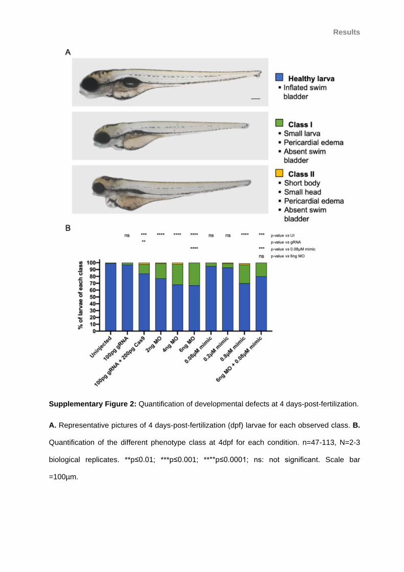
Results
Supplementary Figure 2: Quantification of developmental defects at 4 days-post-fertilization.
A. Representative pictures of 4 days-post-fertilization (dpf) larvae for each observed class. B.
Quantification of the different phenotype class at 4dpf for each condition. n=47-113, N=2-3
biological replicates. **p≤0.01; ***p≤0.001; ****p≤0.0001; ns: not significant. Scale bar
=100µm.

Results
Supplementary Figure 3: Kupffer’s vesicles tend to be smaller in injected embryos.
The area of the Kupffer’s vesicle (KV) lumen at 10 somites-stage is variable in let-7b mosaic
F0 (A), embryos injected with let-7b morpholino (MO) or mimic (B), and embryos co-injected
with let-7b MO and mimic (C). n=11-27, N=3 biological replicates. ns: not significant.

Results
Supplementary Figure 4: Suppression but not overexpression of let-7b leads to convergent-
extension defects

Results
Supplementary Figure 4: Suppression but not overexpression of let-7b leads to convergent-
extension defects
A. Lateral view of 10 somites-stage embryos. Angle between the anterior- and posterior-most
ends of the embryo (white dashed lines) was used for the convergent-extension assay. White
arrowhead indicates first somite. B. Plot of the angles measured for each embryo. n=73-92
embryos per condition, pool of 3 independent experiments. C. Plot of the angles measured for
each morphant and embryos overexpressing let-7b-5p compared to control embryos. n=83-91
embryos per condition, pool of 3 independent experiments. *p≤0.05; ****p≤0.0001; ns: not
significant; UI: uninjected controls. Scale bar=100µm.

Results
II. Developing a zebrafish model for spinocerebellar ataxia 7
to explore the link between Ataxin-7 and cilia
To investigate the interplay between ATXN7 and cilia in the context of SCA7, we first
attempted to develop a zebrafish model of the disease. Our approach consisted of
overexpressing human ATXN7 in zebrafish embryos by injecting mRNA.
Human ATXN7 has a single ortholog in the zebrafish genome encoded by atxn7. The
zebrafish Atxn7 protein shares 46% identity and 59% similarity with human ATXN7. All
domains are conserved, including the polyQ stretch which is shorter with only 5Q [272].
1. A SCA7 zebrafish model with phenotypes characteristic of ciliary defects
Preliminary results show cilia-related phenotypes in a zebrafish model of SCA7
pCS2+ vectors containing cDNA encoding wild-type (WT) ATXN7 with 10Q (N10; normal 10)
and for two forms of mutant (m) ATXN7 with 60Q (E60; expanded 60) and 104 or 106Q (E104
or E106; expanded 104 or 106) were used to generated mature mRNA coding for the human
proteins. Preliminary injections performed in our group produced embryos with several cilia-
related phenotypes such as body curvature, otolith defects, cardiac edema and kidney cysts
[343,344], which seemed polyQ dependent when the embryos were injected with E60 and
E106 mRNAs at concentrations ranging from 250 to 400ng/µL (Figure 1A-D). At 2dpf,
approximately 10% of embryos injected with E60 mRNA presented with otolith defects and
cardiac edemas and almost 30% had a curved body. Embryos injected with mRNA coding for
E106 mRNA presented with the same phenotype at higher frequencies: 35% with otolith
defects, almost 40% with cardiac edema and nearly 80% with body curvature. Less than 5%
of uninjected siblings showed those defects (Figure 1D). Although not counted, few cyclops
were observed among embryos injected with mRNA coding for E106 (Figure 1B).

Results
Discrepancy between preliminary and new experiments
Contrary to our expectations, the injections of the same concentrations of the original mRNAs,
or freshly prepared mRNAs from the same vectors, did not recapitulate those phenotypes. We
first compared the embryos injected with 250ng/µL of fresh mRNA with the images obtained
from preliminary data at 4dpf but 95% of injected embryos looked like uninjected controls
(Figure 1B, E). Next, we repeated the injections but included a control mRNA and monitored
the development of the embryos at earlier time points in case affected embryos might have
died before 4dpf. The control mRNA was injected at 10ng/µL and coded for the fusion protein
Histone 2B-GFP (H2B-GFP) and enabled to control for the injection and mRNA expression.
Every injection generated more than 98% fluorescent embryos (Supp. Figure 1). At 2dpf with
250ng/µL of ATXN7 mRNA, less than 6% of injected embryos presented with no phenotype
while control embryos, including H2B-GFP mRNA injected and uninjected siblings, had a
phenotype (Figure 1G). The injection of mRNA at 400ng/µL triggered more phenotypes but
the proportions of affected embryos were still minimal compared to preliminary experiments
(Figure 1D, H). For instance, body curvature was found in 16% and 8% of embryos injected
with E60 and E104 respectively (Figure 1H). At 3dpf, the mortality in embryos injected with
250ng/µL of mRNA did not exceed 6% but it augmented up to 38% in embryos injected with
400ng/µL of E104 mRNA (Figure 1F). The latest is exceptionally high and might explain the
discrepancy between the preliminary and new injections, however a mortality rate of 35% as
early as at 1dpf prevents any characterization of the phenotypes of interest. Next, we
compared the injection of mature mRNA with the injection of plasmids which is a technique
preferentially used by colleagues [345]. We used plasmids containing either eGFP alone as a
control, or eGFP fused to WT ATXN7 or mATXN7 (pEGFP, pEGFP-N10 and pEGFP-E104
respectively). The injection of 30 or 100ng/µL of pEGFP-N10 and pEGFP-E104 lead to 23-
53% mortality at 2dpf, which was surprising as the fluorescence evidenced low expression
(Supp. Figure 2). Besides, pEGFP-N10 and pEGFP-E104 were equally toxic. Surviving
embryos looked delayed and had gnarled tails which are two signs of non-specific toxicity
[346] (Supp. Figure 2). pEGFP, and second control pCS2+ H2B-GFP, were correctly
Results
expressed as evidenced by fluorescence in about half of the injected embryos. The mortality
was also high in embryos injected with control plasmids. Therefore, this approach raised more
questions than brought solutions and we chose to pursue with the mRNA injection strategy.
The lower mortality and increased percentage of fluorescent embryos observed in
experiments conducted with the injection of mRNA suggest that this tool is more appropriate
for our experiments.
Evaluation of organs affected in SCA7
We did not detect significant phenotypes at the level of the whole body in the embryos
injected with mRNA coding for mATXN7, however, the hallmarks of SCA7 are
neurodegeneration in the cerebellum and in the retina [248], and Atxn7 is known to play a role
in the development of the brain and eye of the zebrafish [272]. Thus, we asked whether the
injected embryos had more subtle phenotypes in each respective cell type. We used an anti-
acetylated α-tubulin antibody to label the cerebellum (Figure 2A) and the transgenic zebrafish
SoFa1 line which highlights the retinal neurons [318] (Figure 2D). We did not detect any
significant hypoplasia of the cerebellum in the embryos injected with E104 compared to the
embryos injected with N10 or uninjected siblings at 3dpf (Figure 2A-C). Observation of retinas
at 48 and 72hpf did not show any anomalies: retinas of embryos injected with mRNA coding
for WT or mATXN7 were indistinguishable from uninjected controls with defined retinal neuron
layers (Figure 2E). Retinas from embryos injected with H2B-GFP mRNA were saturated with
GFP and were not imaged. We concluded that the injection of mRNA coding for ATXN7 at
concentrations ranging from 100ng/µL to 400ng/µL in our zebrafish lines was not sufficient to
generate a robust zebrafish SCA7 model. Knowing that embryos used for preliminary
experiments were obtained from adult fish of almost 2-years of age, we hypothesized that
they might have been more sensitive to proteotoxicity.

Results
Shorter peptides with predicted augmentation of proteotoxicity
To address the possibility of variable proteotoxic sensitivity, we injected mRNAs coding for a
fragment of ATXN7 (ATXN7∆) predicted to be more toxic than the full-length proteins [257].
Previous studies showed that expanded-polyQ-eGFP was sufficient to cause protein
aggregation, nuclear inclusion and death in zebrafish [347,348]. Therefore, we cloned WT and
mutant ATXN7∆ (WT ATXN7∆ coding for 10Q and mATXN7∆ coding for 104Q respectively)
constructs into pCS2+ vectors to produce mRNA. The injection of mRNA coding for WT
ATXN7∆ and mATXN7∆ at concentrations ranging from 10ng/µL up to 400ng/µL did not lead
to significant phenotypes. Less than 10% of embryos presented with anomalies in every
condition. For instance, no more than 6% embryos injected with 400ng/µL of mATXN7∆
mRNA had a cardiac edema at 2dpf (Figure 3A), and mortality was at 10% at 3dpf (Figure
3B). Like in the preliminary injections, we noted few cyclops but we did not count them
because of their rarity. At the concentration of 1000ng/µL, which was used in [348], we
observed up to 35% of cardiac edemas in embryos injected with WT ATXN7∆ and high
mortality rates (27-58%) (Figure 3A, B). The phenotype frequencies being higher in the
embryos injected with WT ATXN7∆ compared to mATXN7∆, and the high mortality rate
dissuaded us from pursuing the characterization of these embryos. Because embryos injected
with mATXN7∆ mRNA showed less phenotypes compared to embryos injected with WT
ATXN7∆ mRNA despite the predicted proteotoxicity of the mATXN7∆, we decided to control
the expression of the injected mRNAs.
2. Spatio-temporal expression pattern of ATXN7 mRNAs
Despite our positive control H2B-GFP suggesting a proper expression of the injected mRNA,
we wanted to verify the expression of the mRNA coding for ATXN7. We generated fluorescent
fusion proteins by adding an eGFP in the C-terminus of the ATXN7∆ peptides. To do so, we
amplified the eGFP coding sequence from a pEGFP-N2 vector and cloned it downstream of
the ATXN7∆ sequences and upstream of the STOP codon.

Results
Fluorescent fusion protein to monitor expression
We followed the evolution of the fluorescent signal of WT ATXN7∆eGFP, mATXN7∆eGFP
and of an eGFP control in live embryos injected with 400ng/µL from 4hpf to 7dpf.
Fluorescence was detectable as early as 4hpf in all injected embryos suggesting that all
mRNAs were correctly expressed (Figure 4A). However, we observed a strong decrease of
fluorescence after 24hpf in the embryos injected with WT ATXN7∆eGFP and mATXN7∆eGFP
mRNAs while the fluorescence in control embryos injected with eGFP mRNA lasted until the
experimental endpoint at 7dpf (Figure 4A and data not shown). This suggested that either the
ATXN7∆eGFP were degraded too quickly or that the produced proteins were very unstable.
Less than 10% of injected embryos had a body curvature, cardiac edema or otolith defect and
we noted a low but constant appearance of cyclops (<5%) at 2dpf (Figure 4D). The mortality
was less than 10% at 3dpf (Figure 4E).
Insights from the cellular level
To investigate if the injection of ATXN7∆eGFP mRNAs at 400ng/µL triggered protein
aggregation, we fixed embryos at 24hpf, the latest stage with visible fluorescence, and
prepared cryo-sections of the heads to look for nuclear inclusions. We observed fluorescence
in both the nucleus and the cytoplasm despite all nuclear localization and exportation signals
being ablated in ATXN7∆ constructs, with a preference for the nucleus in the cells from
embryos injected with WT ATXN7∆eGFP and mATXN7∆eGFP mRNAs (Figure 4C). Cells
from the control embryos injected with eGFP mRNA had a strong cytoplasmic signal (Figure
4C). The absence of puncta at 24hpf characteristic of protein aggregation indicated that the
amount of available protein was not enough to cause proteotoxicity at this stage.

Results
Kinetic studies to define expression timeline
To better understand the expression timeline of the injected mRNAs and to determine the
developmental stage with the peak of mRNA expression and potentially protein aggregation,
we performed comparative analyses of eGFP kinetics. We first cloned eGFP in C-terminus of
the full-length ATXN7 proteins to compare them with the ATXN7∆eGFP. Then we injected
equimolar concentrations of mRNA (0.7mM) to facilitate the comparison of signal intensity,
and imaged six live embryos for each condition from the cleavage period at 1hpf to protruding
mouth at 72hpf (Figure 5A). The fluorescence intensity for each embryo was measured and
corrected in Image J software and called Corrected Total Embryo Fluorescence (CTEF). We
first compared WT ATXN7 full-length and WT ATXN7∆ with the eGFP control and observed
that the highest CTEF was measured in eGFP controls at 18hpf (77x106 a.u.) (Figure 5A).
However, images of embryos injected with eGFP mRNA were saturated between 6hpf and
18hpf suggesting that the real fluorescence peak was probably reached at one of these time
points (Figure 5A). CTEF from embryos injected with WT ATXN7∆eGFP mRNA were much
lower with a peak at 10hpf of 50x106 a.u., and CTEF from embryos injected with WT
ATXN7eGFP mRNA was even lower with a peak at 10hpf of 5x10⁶ a.u. (Figure 5A). This
experiment suggests that mRNA coding for ATXN7∆ is more highly expressed or that the
produced peptide is more stable, and thus that the overexpression of ATXN7∆ is a better
approach to develop the SCA7 model rather than the overexpression of the full-length
proteins. Therefore, we repeated this kinetic study and compared WT ATXN7∆eGFP with
mATXN7∆eGFP kinetics (Figure 5B). We did not detect significant difference between the
CTEF of embryos injected with ATXN7∆eGFP or mATXN7∆eGFP but we confirmed that both
mRNAs were correctly expressed. This time the highest CTEF was detected at 24hpf which
might be due to the more accurate method used for these measurements (see methods).
To conclude, the spatio-temporal tracking of ATXN7 mRNA expression showed that: 1) small
levels of protein were present at the observed stages (50% less than eGFP protein according

Results
to the CTEF) suggesting a problem in the translation of the mRNAs coding for ATXN7, or the
instability of the produced proteins; and that 2) even the polyQ peptide predicted to be the
most toxic did not aggregate in cells. The low levels, the short time window of expression
(increasing CTEF until 24hpf then drop of fluorescence) and the absence of aggregates can
nevertheless explain the absence of significant phenotype in all conditions analyzed.
3. Early cilia defects and protein context
The kinetic analyses indicated that the peak of protein accumulation occurred between 6hpf
and 24hpf and that the amount of protein quickly dropped after that stage. Therefore, we
established a new time point to phenotype zebrafish embryos.
Convergent-extension assay enables detection of ciliary defects at an early stage
The convergent-extension (CE) assay is performed at the 8-12ss (corresponds to ~14hpf at
28.5°C or ~24hpf at 23°C) and enables the assessment of collective cell migration
movements orchestrated by PCP which is a ciliary dependent pathway. It involves the
assessment of the body-gap angle during zebrafish embryo segmentation and has been used
to measure cilia defects in zebrafish in several studies [313,329]. We first performed this
assay on embryos injected with 100 and 200ng/µL of mRNA coding for the full-length ATXN7
N10 or E104 to verify if those constructs could trigger an early ciliary phenotype despite
producing small amounts of proteins. We observed a significant increase in the body-gap
angle of embryos injected with 100 and 200ng/µL of N10 or E104 mRNA compared to H2B-
GFP injected controls and uninjected siblings (Figure 6A, B). There was a significant increase
in the body-gap angle of E104 injected embryos compared to N10 injected embryos at
200ng/µL but not at 100ng/µL (Figure 6B) suggesting that the overexpression of E104 is
more pathogenic than N10 at 200ng/µL and that it might interfere with PCP signaling at this
concentration. We monitored the development of the siblings of the embryos evaluated for CE

Results
until 3dpf and confirmed the absence of significant phenotypes in accordance with our first
experiments.
Co-injection of human WT and mutant mRNA
The analysis of nuclear inclusions found in mammalian cells overexpressing mATXN7
revealed that they contained not only truncated polyQ expanded ATXN7 but also additional
proteins including WT ATXN7 [252] and USP22, another component of SAGA [349].
Therefore, we hypothesized that the absence of aggregate and phenotype in the injected
embryos might be due to unfavorable context such as the lack of human WT ATXN7 or the
48% of difference in the protein sequence of human ATXN7 and its zebrafish ortholog which
might be necessary for interactions. Thus, we compared the injection of 100 or 200ng/µL of
mRNA coding for N10 or E104 alone with the co-injection of 100 or 200ng/µL of each mRNA
in zebrafish embryos (Figure 6A). We performed the CE assay and followed the development
of the injected siblings until 3dpf. We measured a significant difference in the body-gap angle
of embryos co-injected with 100 or 200ng/µL of N10 and E104 (average of 51.6° and 58.9° at
100 or 200ng/µL respectively) compared to H2B-GFP injected (45°) and uninjected controls
(43.7°) (Figure 6B). However, the co-injection of N10 and E104 mRNAs were not significantly
different from the injection of N10 mRNA alone. We concluded that the co-injection of mRNA
coding for WT and mutant ATXN7 did not increase proteotoxicity. At later stages, we
observed few cyclops (1-6%) but no significant phenotypes.
Injection of zebrafish mRNA to favor endogenous interactions
To favor interactions with endogenous proteins, we switched to ATXN7 of zebrafish origin.
Therefore, we prepared mRNA coding for WT Atxn7 mRNA (bearing 5 CAG repeats) and
performed injections and phenotyping. The CE assay showed a significant increase in the
body-gap angle of embryos injected with Atxn7 mRNA (83.7°) compared to embryos injected
with H2B-GFP mRNA (41.4°) and uninjected siblings (41.3°) (Figure 6C). Observations at

Results
later stages showed low frequencies of eye phenotypes at 24hpf: 0% and 6% of cyclopia, and
4% and 9% of unilateral ano- or microphthalmia at the concentrations of 100 and 200ng/uL of
mRNA respectively (Supp. Figure 3A). We prepared mutant atxn7 construct (bearing 61 CAG
repeats; mAtxn7; Eurofins) and the preliminary injections of 100 and 200ng/uL of mRNA did
not lead to any significant phenotype, no more than 1% of the injected embryos presented
with defects in the assessed organs (Supp. Figure 3A). Mortality was low in every condition
(≤1%) (Supp. Figure 3B). Because embryos injected with mAtxn7 mRNA were
indistinguishable from uninjected siblings at 2dpf, we decided not to perform the CE assay.
Considering the strong effect of WT Atxn7 mRNA on the body-gap angle, we would have first
needed to titrate down the mRNA to find a concentration where WT Atxn7 mRNA would not
trigger a phenotype to allow interpretation.
4. Summary and concluding remarks
The injection of dose curves of human WT and mutant ATXN7 mRNA, which were shown to
be expressed properly via the visualization of fluorescence, were not sufficient to trigger
protein aggregation or significant ciliary phenotypes that could be characterized further in our
zebrafish models. The co-injection of both WT and mutant ATXN7 mRNA did not exacerbate
proteotoxicity as assessed by the CE experiment and the general aspect of the embryos until
3dpf. We suspect that no aggregates were formed and this was verified in cryo-sections from
early stage injected embryos (<24hpf). The injection of zebrafish mRNA generated
encouraging results and further work could inform on the relationship between Atxn7 and cilia.
However, the absence of striking phenotypes at 2dpf diminishes the potential of a zebrafish
developmental model to study SCA7. Now that the SCA7 knock-in mouse model SCA7104Q/5Q
is available in our group, investigation of cilia anomalies can be performed in a mammalian
model which is more relevant to SCA7 disease and therapeutic opportunities. Therefore,
efforts were concentrated towards the other projects.

Results
Figure 1: Discrepancy in SCA7 models from mRNA injections.
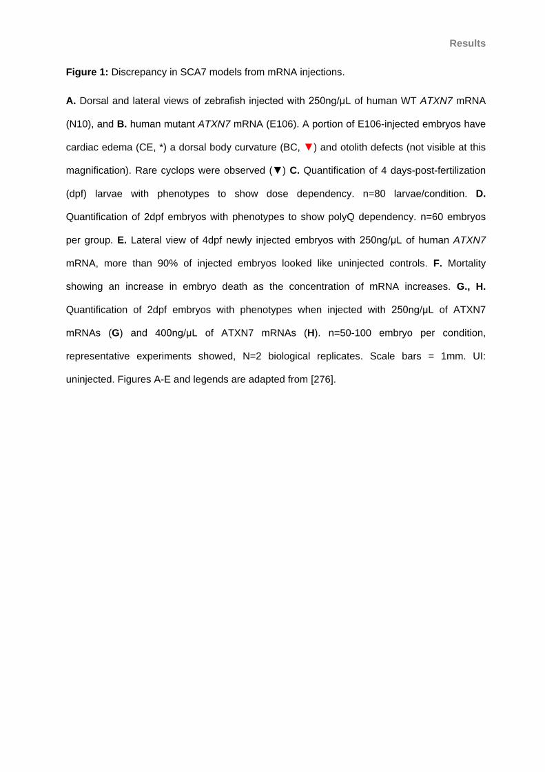
Results
Figure 1: Discrepancy in SCA7 models from mRNA injections.
A. Dorsal and lateral views of zebrafish injected with 250ng/μL of human WT ATXN7 mRNA
(N10), and B. human mutant ATXN7 mRNA (E106). A portion of E106-injected embryos have
cardiac edema (CE, *) a dorsal body curvature (BC, ▼) and otolith defects (not visible at this
magnification). Rare cyclops were observed (▼) C. Quantification of 4 days-post-fertilization
(dpf) larvae with phenotypes to show dose dependency. n=80 larvae/condition. D.
Quantification of 2dpf embryos with phenotypes to show polyQ dependency. n=60 embryos
per group. E. Lateral view of 4dpf newly injected embryos with 250ng/μL of human ATXN7
mRNA, more than 90% of injected embryos looked like uninjected controls. F. Mortality
showing an increase in embryo death as the concentration of mRNA increases. G., H.
Quantification of 2dpf embryos with phenotypes when injected with 250ng/μL of ATXN7
mRNAs (G) and 400ng/μL of ATXN7 mRNAs (H). n=50-100 embryo per condition,
representative experiments showed, N=2 biological replicates. Scale bars = 1mm. UI:
uninjected. Figures A-E and legends are adapted from [276].

Results
Figure 2: Characterization of the cerebellum and retina of SCA7 larvae.

Results
Figure 2: Characterization of the cerebellum and retina of SCA7 larvae.
A. Dorsal view of an uninjected control stained with anti-acetylated α-tubulin to highlight the
brain structures of the larva: OT: optic tectum, IC: intertectal connections and C: cerebellum.
B. Dorsal views of stained larva injected with ATXN7 mRNAs. C. Quantification of the
cerebellum area (dashed area in A) for all conditions. n=39-48 embryos per condition,
repeated. Error bars indicate standard error of the mean. *p<0.05. D. Schematic of the retina
of a SoFa1 embryo showing the different fluorescent layers of retinal neurons and when they
first differentiate. E. Lateral views of the eyes of 3dpf zebrafish embryos injected with
400ng/μL of human ATXN7 mRNA. UI: uninjected, GC: ganglion cell layer, AC: amacrine
cells, HC: horizontal cells, BP: bipolar cells, PR: photoreceptors. White scale bars = 100μm,
black scale bars = 50μm.

Results
Figure 3: Injection of ATXN7∆ mRNA does not trigger more phenotypes in a dose-
dependent manner.
A. Quantification of 2dpf embryos with phenotypes when injected with increasing
concentrations of mRNA. B. Cumulative mortality at 3dpf shows that 1000ng/μL of ATXN7∆
mRNA is highly toxic. n=60-100 embryos per condition, representative experiments are
shown, N=2 biological replicates.

Results
Figure 4: Injection of mRNAs coding for fusion fluorescent ATXN7 proteins to control for
expression.

Results
Figure 4: Injection of mRNAs coding for fusion fluorescent ATXN7 proteins to control for
expression.
A. Images showing that eGFP is visible at 4 hours-post-fertilization (hpf) in embryos injected
with 400ng/μL of mRNA coding for control eGFP, wild-type and mutant ATXN7∆eGFP
(mATXN7∆eGFP). B. The fluorescent signal remains at 24hpf, embryos injected with mRNA
coding for eGFP are the brightest. C. Cryo-sections of 24hpf embryos showing fluorescence
in cerebellar cells. D. Quantification of 2 days-post-fertilization (dpf) embryos with phenotypes.
WT ATXN7∆eGFP and mATXN7∆eGFP mRNAs injected at 400ng/μL. E. Quantification of the
mortality at 3dpf. n=60-150 embryos per condition, N=2 biological replicates, representative
experiments are shown. Black scale bars = 200μm, white scale bars = 10μm. UI: uninjected.

Results
Figure 5: eGFP kinetic comparison studies.
A. Plot of the average CTEF of 6 live embryos per condition followed during development.
Saturation was observed in images of eGFP injected embryos between 4hpf and 24hpf
suggesting that the true CTEF is higher than what was calculated (dashed line). B.
Comparison between the CTEF of ATXN7∆eGFP and mATXN7∆eGFP injected embryos
revealed no significant differences.

Results
Figure 6: Convergent-extension assay.
A. Images of fixed (controls and human mRNA) and live (zebrafish mRNA) 10 somites-stage
embryos. Concentration of mRNA in ng/μL injected indicated in brackets. The body-gap angle
measured for this assay is indicated in white. Scale bar = 200μm. B. Quantification of the
body-gap angle of control embryos and embryos injected with human ATXN7 mRNA. n=98-
100 embryos per condition, repeated. C. Quantification of the body-gap angle of control
embryos and embryos injected with zebrafish atxn7 mRNA. n=25-60 embryos per condition,
representative experiments are showen, N=2 biological replicates. Error bars indicate
standard error of the mean. *p<0.05, **p<0.01, ****p<0.0001, ns: not significant. UI:
uninjected.

Results
Supplementary Figure 1: Embryos injected with H2B-GFP mRNA are fluorescent.
Representative image of a 2 days-post-fertilization embryo injected with 10ng/μL of mRNA
coding for H2B-GFP. No apparent phenotype is visible and fluorescence can be detected
which confirms the expression of the injected mRNA. Scale bar = 500μm.

Results
Supplementary Figure 2: Embryos injected with plasmids coding for ATXN7 proteins are
severely affected.

Results
Supplementary Figure 2: Embryos injected with plasmids coding for ATXN7 proteins are
severely affected.
A. Representative images of a 2 days-post-fertilization uninjected (UI) zebrafish embryo and
siblings injected with 30ng/μL of expression plasmids coding for WT ATXN7 (pEGFP-N10)
and mATXN7 (pEGFP-E104) fused to eGFP. Surviving embryos have small heads and eyes,
are developmentally delayed and have gnarled tails which are signs of non-specific toxicity.
Scale bar = 500μm. B. Quantification of fluorescent and dead embryos at 2dpf.
Concentrations of injected plasmids in ng/μL are indicated below the histogram. n=60-95
embryos per condition.

Results
Supplementary Figure 3: Embryos injected with mRNA coding for zebrafish Atxn7 do not
present the expected ciliary-phenotypes.
A. Quantification of embryos with a phenotype observed at 2 days-post-fertilization. B.
Mortality at 3dpf, concentrations of injected mRNA in ng/μL are indicated below the histogram.
n=60-135 embryos per condition, N=2 biological replicates, representative experiments are
shown.

Results
III. Other opportunities to explore the advantages of the
zebrafish to model human genetic diseases,
1. Studying organs in zebrafish embryos which are typically affected in ciliopathies
An overarching mission of the CHDM is to understand the genetic architecture of rare
human genetic disorders. Thus, I had the opportunity to work on two additional projects: the
first one focused on an autosomal dominant retinal ciliopathy, and the second one focused on
congenital kidney malformations which are commonly found in ciliopathies. My contribution to
both studies consisted of characterization of the affected organs in zebrafish models.
1.1 Mutations in the Kinesin-2 motor KIF3B cause an autosomal dominant ciliopathy
Our collaborators identified two families with typical ciliopathy symptoms including
retinal degeneration and polydactyly. WES enabled to identification of candidate variants in
the Kinesin-2 motor KIF3B which were inherited in an autosomal dominant manner in one
pedigree, and appeared de novo in the proband of the second pedigree. Analysis of patients’
fibroblasts from a single proband revealed longer primary cilia. To functionally confirm the
variants in vivo, we injected wild-type and mutated human KIF3B mRNA into zebrafish
embryos and monitored their development. We were especially interested in characterizing
their eye phenotypes. I participated to this project by 1) performing cryo-sections of 5dpf
embryos; 2) staining retina sections with antibodies labelling cilia to measure the connecting
cilia in photoreceptors; 3) performing TUNEL on retina sections to measure cell death; and 4)
editing the manuscript (results are shown in Figure 4 D,E, Figure S3 and Figure S4 of the
following manuscript). This work lead to the preparation of a manuscript which is in revision at
the American Journal of Human Genetics.

Results
In this study, we show that the variants identified in patients lead to longer cilia in vitro
and in vivo; and that rhodopsin, which is transported via the connecting cilium from the inner
to the outer segments of photoreceptors, is mis-localized in rod inner segments of zebrafish
embryos injected with human mutated KIF3B mRNA. Considering that most ciliopathies are
inherited in an autosomal recessive pattern, this study represents a rare example of an
autosomal dominant ciliopathy. Our work also shows that perturbation of KIF3B can result in
either dominant toxic effects as shown here for humans, or it can result in recessive disease
as reported previously for multiple mutant model organisms including mouse and zebrafish
[8,350].

Results
1.2 Association study identifies GREB1L mutations in congenital kidney malformations
Kidney defects are one of the hallmarks of ciliopathies and can be found through the
whole spectrum of ciliopathies: from some severe forms of Senior-Loken syndrome to
perinatal lethal Meckel-Grüber cases [351]. Although the cellular defect did not involve ciliary
dysfunction, this project was an opportunity to learn how to stain, measure and interpret data
regarding the zebrafish pronephros. Because this study is less closely related to the topic of
this thesis manuscript, the publication will not be included.
Renal agenesis and hypodysplasia are severe conditions which affect about 0.5% of
the population, but only 15% of the cases have known genetic causes. Our collaborators
identified a candidate gene which could contribute to kidney anomalies in cohorts of patients
with renal agenesis and hypodysplasia. To further validate the candidacy of GREB1L, we
characterized renal phenotypes in zebrafish mutants and morphants and performed functional
testing of the missense variants identified in patient cohorts. I contributed to this project by 1)
providing support to Kamal Khan to characterize the loss of function models and to test the
different variants; 2) preparing Figure 2, Figure 3, Figure S3, Figure S4 and Table S7; and
3) editing the manuscript. I performed whole-mount immunostaining of 4dpf larvae and
imaged them (results are showed in Figure 3, S3, S4 and Table S7 of the publication: [317]).
This work enabled the identification of GREB1L as a new gene contributing to renal
agenesis and hypodysplasia which improves our understanding of the complex genetics
behind these conditions, and will inform future diagnosis of kidney malformations.
2. The role of Atxn7 in the development of the zebrafish brain and eye
The primary research interest of Dr. Yvon Trottier’s laboratory is to understand the
pathomechanisms underlying two polyglutamine expansion diseases: Huntington’s disease
and SCA7. Therefore, the thesis project led by Dr. Samantha Carrillo-Rosas was to develop
and characterize a model of ATXN7 loss of function with a focus on the photoreceptor.

Results
Because the Atxn7 knockout mouse was not available, the zebrafish offered a quick and
relevant alternative to generate this model. Indeed, the use of morpholinos and CRISPR/Cas9
tools enabled the partial and complete loss-of-function of atxn7 and the rapid development of
the embryos enabled monitoring of the development of the retina, a major affected structure in
SCA7, over the course of a week. Humans and mice have an overall similar cone-rod ratio in
their retina (mouse: 97:3, human:95:5). However, humans are diurnal and their fovea is
enriched in cones. As the fovea corresponds to the site where neurodegeneration initiates in
SCA7, diurnal zebrafish with a cone-rod ratio of 65:35 are relevant to model SCA7 retinopathy
[352]. I contributed to this project by 1) performing preliminary ISH; 2) titrating the splice-
blocking morpholino as well as the human wild-type ATXN7 mRNA to perform the rescue
experiments; and 3) editing the manuscript. This work led to a publication which has been
cited in this manuscript: [272].
In this paper, we show that Atxn7 plays an important role in the development of the
zebrafish eye and brain. In particular, we show that Atxn7 is required for the proximo-distal
patterning of the optic stalk and vesicle, as well as for the closure of the choroid fissure.
Deeper characterization of the loss of function models (morphants and CRISPR mutants)
suggests that Atxn7 is an indirect negative regulator of the members of the Hedgehog family
and that it is required for morphogenesis of the photoreceptor outer segment through the
control of crx expression. The latest being directly correlated with SCA7 retinopathy and
suggesting that partial loss of function of normal ATXN7 in SCA7 patients could account for
the retinal degeneration.

Results
DISCUSSION AND PERSPECTIVES

Discussion and Perspectives
Discussion and Perspectives
In this study, we explored functional links between cilia, a non-coding RNA, and a
protein mainly characterized for its role in transcriptional regulation. We used a broad range of
techniques which was complemented by the participation in additional projects. From genetic
manipulation in vivo to immunostaining and targeted sequencing, we investigated ciliary
biology from different angles and dissected contributing mechanisms to ciliogenesis and
pathogenic consequences of ciliary defects. We validated the role of let-7b as a positive
regulator of ciliogenesis in vivo after previous identification of this miRNA through a medium-
throughput in vitro screen, and we identified 499 potential let-7b targets which could explain
its mechanism of action. Despite the non-validation of our hypothesis of let-7 as a modifier of
ciliopathies in our targeted sequencing experiment, we will pursue this work in expanded
cohorts and/or candidate target sites. Publication of the results obtained on let-7 will likely
motivate the community to include UTRs in their WES analyses. The ATXN7 study confronted
biological obstacles that we could not overcome despite our diverse approaches. However,
the hypothesis of a putative role for ATXN7 was neither confirmed nor refuted and further
work in SCA7 mouse models is ongoing in our group (IGBMC).
The zebrafish model in comparison to other model organisms to explore ciliary biology
Aside from cilia, the common theme among the different studies described was the use of the
zebrafish model organism to explore questions around the cilium. From the characterization of
immotile flagella in Chlamydomonas mutants which highlighted the role of the central pair of
tubules cilia motility [353], to the mouse model of primary ciliary dyskinesia [11], a collection
of model organisms contributed to the expansion of our understanding of ciliary biology and
ciliopathies.

Discussion and Perspectives
Chlamydomonas is a unicellular algae which can explore its environment by propelling
itself with its two flagella [354]. It presents several advantages to study motile cilia: flagella are
long (12µm) and can be easily isolated for proteomics analyses [355], and it is inexpensive to
maintain. Although mostly used in fundamental research, there are several examples where
studies in Chlamydomonas enabled to explore ciliopathy mechanisms. For instance,
Miertzschke et al. used the crystal structure of arl13b to predict the functional effect of the
mutation p.R79Q identified in ARL13B in patients affected with Joubert syndrome. They
concluded that the arginine residue was playing an important role in the protein conformation
and that the mutation was probably leading to a loss of the protein function [356]. Perhaps the
most famous contribution of Drosophila to the field of cilia research is the study of
morphogenes which function through the primary cilium. Close to five decades after the first
observation of PCP defects [114], we are still learning from Drosophila about Wnt
mechanisms. Indeed, recent work showed that the polarization of centrioles in Drosophila
wings depended on two different PCP protein complexes: the Frizzled-PCP and the Fat-PCP
[357].
As more genetic manipulations and imaging technologies became available, more
complex organisms including zebrafish [313], mice [95,96], rabbits [19] and even hamsters
[55] were used to study cilia. Mice remain the favored model to study underlying mechanisms
of ciliopathy due to their closest relationship to humans (99% of their genes possess a human
homolog [358]). However, zebrafish are less expensive to maintain and are transparent, with
ciliated organs which are easily accessible. The KV mentioned in this study is an examplar.
Additionally, the ex vivo development of zebrafish and shorter generation time allow easy
gene manipulations. Zebrafish also present the opportunity to screen therapeutic compounds
[359].
In this work, zebrafish appeared as a powerful tool by successfully replicating in vitro
observations such as the cilia shortening in the let-7b experiments or the cilia lengthening in
the KIF3B experiments. It was also shown to be relevant in modeling human diseases such as
the eye anomalies caused by dominant mutations in KIF3B, and the kidney developmental

Discussion and Perspectives
defects caused by loss of function of GREB1L. However, the zebrafish also showed
limitations in the modeling of SCA7 where low protein expression could not trigger relevant
phenotypes. Nonetheless, zebrafish remains a relevant organism to model neurological
disorders [360,361].
on let-7b miRNA
Strengths and limits of the zebrafish depleted in let-7b or overexpressing let-7b-5p
To validate further the effect of let-7b on ciliogenesis and cilia maintenance, we used the
zebrafish to test let-7b in vivo. We recapitulated the decrease in cilia length: we measured a
decrease of 0.6μm in the let-7b mosaic F0, and 1.49μm in the let-7b morphants. Those
reductions are comparable to the decrease of 1.23μm observed in vitro which highlights the
strength of our model.
The mild cilia length decrease observed in the let-7b mosaic F0 correlates with the
mild knockdown measured by RT-qPCR despite the guide efficiency calculated at 100%. It is
common to observe milder phenotypes in mosaic F0 compared to morphants due to three
factors. First, we cannot certify the disruptive nature of the mutations generated by
CRISPR/CAS9 technology in our organ of interest, and some mutations might not lead to a
complete loss of function of a gene. Second, it has been shown in zebrafish how
compensatory mechanisms can arise after a gene ablation but not in the case of a gene
knockdown [362]. It is thus possible that cells which completely lost let-7b and contributed to
form the KV, triggered a compensatory mechanism. Third, the genome manipulation is
permanent but does not affect maternal transcripts which are actively expressed until 4hpf.
Whereas morpholinos regulate expression of all transcripts. In the case of the let-7b mosaic
F0, the hypothesis of the maternal transcript to explain mild phenotypes is unlikely since it is

Discussion and Perspectives
crucial for the zebrafish embryonic cells to maintain their proliferative capacity and
pluripotency until at least 6hpf [363], and that let-7 is known to contribute to cell differentiation
[364]. We targeted only the let-7b locus, and within that strategy, our gRNA targeted the
Drosha processing site and not the mature sequence directly due to technical constraints. It is
thus likely that the mild effect observed in let-7b mosaic F0 is caused by the two other
possible explanations. There might be compensation from the 17 other let-7 loci coding for the
nine other members of the let-7 family, and/or the mutations affecting the Drosha processing
site might not all be disruptive and some mature let-7b might still be produced in some cells.
Future work on the let-7b F3 knockout will ensure stability of the mutation and absence of any
potential maternal transcripts. The F1 generation has just reached maturity and should be
propagated for eventual further experiments, including the phenotyping of other ciliated
organs such as the retina and the kidney.
Other approaches to obtain stable mutants depleted of let-7 consist of either knocking
down all let-7 members, or a subset of them since all let-7 members were shown to affect
ciliogenesis in vitro. A previous study demonstrated that it is possible to knockdown the 10
members of the let-7 family in zebrafish, however the embryos were only used for genotyping
at 1dpf or sensor assays at 2dpf and were not characterized [365]. Since the loss of let-7 is
lethal in C. elegans, it is likely that the F0 would not survive to adulthood for propagation and
that studies might be restricted to mosaic F0. Alternatively, targeting a cluster of let-7
members might have allowed us to observe stronger phenotypes such as shorter cilia and
associated developmental defects including retinal degeneration and kidney cysts [344]. This
approach was used by a group who observed an increased activation of B cells in a let-7adf
knockout mouse model [366]. In their study, authors targeted one let-7 cluster and knocked
out three let-7 genes altogether.
A limitation of our in vivo study of the role of let-7b is the normal length and normal
cilia density observed in the embryos injected with the let-7b-5p mimic despite the observed
left-right anomalies. After the striking increase in ciliation and cilia length observed in vitro
upon transfection of let-7b-5p mimic, it was reasonable to expect a similar pattern in vivo. The

Discussion and Perspectives
presence of left-right defects could suggest an anomaly at the level of the beating mechanism.
To test this hypothesis, future studies should monitor ciliary beating in the KV of live embryos
injected with let-7b-5p mimic by either using transgenic zebrafish with a cilia reporter
Tg(βact::Arl13b–GFP), or fluorescent beads directly injected in the KV lumen [367]. Another
possible explanation is that the RISC was overwhelmed with the quantity of let-7b-5p which
led to a global dysregulation gene expression and the observed phenotypes. However, this is
unlikely as the over-expression of miR-145 mimic, miR-203a mimic, and miR-210 mimic did
not trigger toxicity in zebrafish embryos [368-370]. miR-145, miR-203a, and miR-210 are
involved in gut and heart development, myelopoiesis, and muscle development respectively,
and are expressed at similar levels with that of let-7b in the developing zebrafish [323]. miR-
203a mimic was injected at a concentration as high as 20μM and generated a 46-fold
expression increase which is far superior to the 9-fold increase obtained with the injection of
0.8μM of let-7b-5p mimic. These observations, and the partial rescue of the left-right
asymmetry patterning defect when the mimic was co-injected with let-7b morpholino, argue
against a toxicity effect via RISC.
Understanding the biological processes targeted by let-7b which affect ciliogenesis
To validate further let-7b role on ciliogenesis and cilia maintenance, and to understand the
level at which let-7b is operating to regulate cilia biology, we could perform rescue
experiments in different models harboring longer cilia. Depending on the mechanisms leading
to longer cilia, such rescue experiments will help identify the steps in cilia biology where let-7b
intervenes. The zebrafish model developed in the KIF3B study represents an opportunity to
test whether let-7b operates at the level the axoneme elongation. Indeed, KIF3B is involved in
anterograde transport of tubulin to the tip of the cilium [371]. To refine the chronology of let-7b
intervention in ciliogenesis, we could compare this experiment with another model where an
earlier step in ciliogenesis is affected. Ciliary membrane elongation occurs prior axoneme
elongation and ARL13B is one actor of this initial step. Its over-expression in zebrafish and

Discussion and Perspectives
mammalian cells leads to longer cilia [372]. Thus, we could test whether let-7b intervenes
early in ciliogenesis by co-injecting let-7b MO with arl13b mRNA in zebrafish. We could also
focus on ciliary modules such as the BBSome. The over-expression of mutant BBS3 (p.T31R
or p.Q73L) leads to a higher proportion of ciliated htRPE-1 cells in culture [373]. There is a
collection of additional models harboring longer cilia such as cultured renal epithelial cells
from both Bbs4 [97] and Nek8 knockout mice [374], or Rpgrip1l knockout mice [375].
However, the longer cilia are generated by the loss of a protein in those cases and it would be
challenging to track multiple inhibitory relationships.
Challenges in identifying let-7b direct targets
The in vitro cilia length data in the context of let-7 modulation and the recapitulation of the cilia
length phenotype in vivo strongly support the candidacy of let-7, especially let-7b, as a
positive regulator of ciliogenesis and cilia maintenance. The subsequent identification of direct
targets of let-7b remains challenging. Indeed, it is possible that the transcripts targeted by let-
7 that function to inhibit ciliogenesis are individually weakly modulated. Only the cumulative
knockdown would regulate ciliogenesis rather than the strong negative regulation of a single
transcript [376]. Moreover, the study is limited by our current knowledge on both miRNAs and
cilia. Those different aspects are discussed below. However, we identified 499 candidates and
possible regulatory networks providing a partial answer to the complex role of let-7.
Gene network. Since miRNAs have many different target transcripts, it is likely that
let-7 regulates not only the expression of one negative regulator of ciliogenesis, but a network
of genes which act together. The functional enrichment analysis we performed on the
transcriptomic data suggested a role for let-7b on cell adhesion and organization of the
extracellular matrix. However, extracellular matrix changes, including excessive secretion of
collagen, were observed in the heart of conditional Ift88 knockout mice lacking cilia in this
organ [333]. Additionally, collagen is known to interact with integrins which represents a
significant proportion of cell adhesion molecules detected in our study [377]. Thus, we

Discussion and Perspectives
hypothesized that the enrichment analysis showed pathways stimulated as a consequence of
cilia loss, and not pathways directly controlled by let-7b. Review of our list of 499 candidates
obtained after further filtering enabled confirmation of the enrichment in cell adhesion proteins.
However, we also found actin-interacting proteins which shape the actin cytoskeleton for cell
motility; these are associated with tumor invasiveness (ENAH, AFAP1, FSCN1). Thus, we
focused on the actin anchoring property of the cell adhesion molecules rather than their extra
cellular matrix interaction properties. It has been shown at different levels how the actin
cytoskeleton can interfere with ciliogenesis [92,303,335], therefore, we propose that let-7 is a
modulator of the actin cytoskeleton at the expression level. To validate this hypothesis, we
could first use phalloidin dye to stain actin in the htRPE-1 cells and compare the cytoskeleton
between control cells and cells treated with either let-7b inhibitor and let-7b-5p mimic. Of note,
the cytoskeleton dynamics are crucial to cell motility and a role for let-7 on the regulation of
actin could bring insights in the mechanism behind metastasis observed in cancers where let-
7 is mis-regulated [378,379].
let-7b specificity. The knockdown of each let-7 member in htRPE-1 cells led to fewer
ciliated cells and shorter cilia, and loss of let-7b had the most marked effect. This suggests
that let-7b has a specific affinity for a gene, or a network of genes, regulating ciliogenesis and
cilia maintenance. When we used this affinity criterion which was available in the TargetScan
database, we did not find an overlap with our list of predicted and validated negative
regulators of ciliogenesis. There are two possible explanations for this result. First, the list of
predicted and validated negative regulators is non-exhaustive and is likely to grow in the
future as our knowledge of cilia extends. Second, the measurement of the affinity of the let-7
members is based on the sequences on the 5’ arm of their coding stem-loop. Indeed, the
scientific community has gathered much more data on the miRNAs coded on the guide strand
which are considered to be the primary active miRNA compared to the other strand of the
stem-loop [380]. Thus, we are lacking information on the repression capacity of let-7b-3p
which could contribute to the regulation of gene expression through let-7b. However, the

Discussion and Perspectives
strong effect obtained in vitro from overexpressing only let-7b-5p supports a major role for let-
7b-5p and a minor role for let-7b-3p.
Atypical regulation by miRNAs. The mechanism by which a miRNA regulates gene
expression at the level of the transcripts depends partly on the type of binding between the
seed sequence and the 3’UTR of a mRNA. Perfect base-pairing usually leads to mRNA
degradation while imperfect base-pairing leads to translational repression [381]. This has
been confirmed for let-7 in human cultured HeLa cells where constructs bearing perfect base-
pairing were degraded, while constructs with imperfect base-pairing were repressed from
translation and stored in P bodies (membrane-less organelles with a concentration of RNA-
protein complexes for RNA decay) [382]. This is why we performed RNA-seq on htRPE-1
cells and prioritized transcripts which were 1) up-regulated in the context of let-7b inhibitor; 2)
down-regulated in the context of let-7b-5p mimic; and 3) bearing at least one seed
complementary sequence. However, it is possible that let-7 acts through translational
repression in htRPE-1 cells, or in zebrafish, to regulate ciliogenesis. It this case, the RNA-seq
dataset might not reflect the regulation by let-7b. Despite not being translated, mRNAs would
still be present in the cytoplasm. In this instance, proteomic analysis would be more relevant
to identify miRNA targets. Finally, although this mechanism has not been reported for the let-7
family of miRNA, some miRNAs can positively regulate gene expression by binding to the
3’UTR of target transcripts and competing with TTP to prevent AU-rich element mediated
mRNA decay [383,384]. Notably, we obtained a list of 910 genes which were down-regulated
in the context of let-7b inhibitor and up-regulated in the context of let-7b-5p mimic. It could be
relevant to analyze this list of genes and identify possible networks stimulating ciliogenesis.
Further validation of best candidates
Our final list of 499 candidate let-7b targets can be refined once we intersect it with the list
generated by TargetSan and the list of predicted and validated negative regulators of
ciliogenesis. We have tools available to validate and further study the 5 top candidates LIMK2,

Discussion and Perspectives
EIF4G2, ARL4D, RTCA and ELOVL4. Indeed, we could perform rescue experiments by
knocking down those genes in htRPE-1 cells, zebrafish stable mutants or let-7b morphants
with siRNAs or MO depending on the model. We could also perform phenocopy experiments
where we would overexpress those genes and see if it recapitulates the cilia defects.
Lessons from the targeted sequencing
To test whether let-7 could be a modifier gene of ciliopathies, we sequenced let-7 targets in
the 3’UTR of 40 cilia-associated genes, which were selected after analysis of the RNA-seq
analysis and contained one or multiple let-7b-5p or let-7b-3p targets sequences. A mutational
burden analysis requires individual data and not aggregated data like the one available in the
gnomAD. Indeed, we need to be able to count and compare the number of rare variants
(MAF<5% or 1%) in let-7 target sequences for each individual. It would have thus required the
sequencing of a control cohort. However, the paucity of rare variants detected in our patients
dissuaded us from performing this financially burdensome experiment. This observation
suggests a high conservation of those sites which actually strengthen our hypothesis that
miRNA regulation could play an important role in determining the expressivity of a ciliopathy.
As the cost of WES decreases every year, it would be relevant to include UTRs to
complement WES without necessarily doing whole-genome sequencing.
In our study, we were confronted to a potential population substructure bias which we
were unable to overcome. Indeed, a significant portion of our cohort was of Pakistani origin
and the gnomAD lacks data on controls from South Asia, at least at the site of the variants
investigated. An alternative database is the Great Middle East (GME) variome
(http://igm.ucsd.edu/gme/) which is composed of exomes of controls from North Africa, Middle
East and South-West Asia which includes Pakistan. However, this database contains
exclusively exome data and no UTR.
In the scenario wherein we would have identified candidate variants in let-7 targets, we
would have first validated the effect of the mutation on let-7 binding in vitro through a GFP

Discussion and Perspectives
sensor assay [385]. We would have transfected cells with let-7b-5p mimic alongside with
mRNA coding for eGFP associated to the 3’UTR containing the mutation in let-7 target. We
would have measured the resulting fluorescence and compared it to cells transfected with the
same reagents but with a corrected let-7 target. Next, we would have validated the effect of
the overexpression of the gene with the mutated 3’UTR by over-expressing it in the htRPE-1
cells and quantifying the fraction of ciliated cells as well as the cilia length.
on ATXN7 and cilia
The injection of our different human constructs did not generate SCA7-related
phenotypes (protein aggregation) nor did it reproduce the cilia-related phenotypes obtained in
preliminary experiments (otolith defects, cardiac edema, kidney cysts and body curvature).
This can be due to an anomaly relative to the injected mRNA or resulting protein, but it could
also be due to the model itself. Indeed, some model organisms can be more or less
appropriate to the in vivo analysis of human diseases. Here, we will discuss both of these
aspects, starting with the reagents used to generate the SCA7 zebrafish model.
High concentration of mRNA but low level of accumulated protein
By controlling the expression of the injected mRNA with a fluorescent tag, we understood that
little protein was accumulated in the embryos and that it could explain the absence of
phenotypic manifestations. However, the reasons for low protein accumulation remain
unclear. Our two control mRNAs H2B-GFP and eGFP were expressed normally as indicated
by fluorescence that was visible up to 7dpf, suggesting that the anomaly was specific to
ATXN7 mRNAs. We propose two hypotheses: 1) there is a problem in the translation or

Discussion and Perspectives
stability of the mRNAs; and 2) the produced protein is unstable and quickly degraded.
Inefficient mRNA translation can arise from errors in the 5’UTR while mRNA instability can be
due to missing 5’cap and missing or short polyA tail [386,387]. Since all mRNAs, including
controls, were generated from the same pCS2+ backbone and SP6 polymerase kit, we can
speculate that the difference in expression is not caused by anomalies of the cap, UTR, or
polyA tail. There is an inter- and intra-species codon bias affecting translation efficiency [388-
390] and zebrafish are no exception [391]. To investigate if a codon bias could explain
translation inefficacy of the ATXN7 mRNAs, we uploaded the human and zebrafish ATXN7
coding sequences to the Integrated DNA Technologies (IDT) online tool for codon
optimization (https://www.idtdna.com/CodonOpt) and simulated a codon optimization for
expression in the zebrafish. About 28% of each sequence (human: 27.7%, zebrafish: 28.2%)
required codon optimization according to the Integrated DNA Technologies online tool, which
suggests that codons from the human and zebrafish ATXN7 coding sequences are equally
efficient. Thus, the hypothesis of the protein instability remains. To test the protein stability of
human ATXN7 and H2B, we could use the technique of fluorescence decay after
photoconversion [392] on injected embryos younger than 24hpf (before the drop of
fluorescence) to calculate the half-life of each protein.
The observation that ATXN7∆eGFP mRNAs enable more protein accumulation than
the mRNAs coding for ATXN7 full-length was an encouraging step in our development of a
zebrafish SCA7 model. Even though we were not able to observe the phenotypes noted
during the preliminary experiments, we were confident that the mRNAs were expressed
correctly. As mentioned in the introduction on SCA7 disease, ATXN7 has a rapid turnover
which seems to be necessary for its proper function. According to the fluorescent tag, WT
ATXN7∆eGFP has a longer half-life than N10eGFP, but WT and mutant ATXN7∆eGFP
behave similarly in terms of protein accumulation. Since WT ATXN7∆ lacks the functional
domains of ATXN7 which are required for its function within the SAGA complex, it is not
surprising that its turnover is different than the full-length protein. However, in the context of
SCA7, the polyQ in mATXN7 stabilizes the protein so we could have expected stronger
Discussion and Perspectives
fluorescence from embryos injected with mATXN7∆eGFP mRNA compared to WT
ATXN7∆eGFP mRNA.
Zebrafish Atxn7 and cilia
The injection of WT and mutant zebrafish Atxn7 at 100 and 200ng/μL did not trigger the
expected cilia-related phenotypes at 2dpf including otolith defects, cardiac edema and body
curvature. A small fraction of embryos (less than 10%) presented an anomaly in the formation
of the eyes: either cyclopia or unilateral ano- or microphthalmia, which is consistent with the
presence of cyclops among embryos injected with human mRNA. However, the injection of
zebrafish mRNA generated a CE phenotype while the human WT ATXN7 mRNA did not
trigger defects at the same concentration of 100ng/μL. As discussed above, differences in the
manifestation of a mRNA over-expression can be caused by anomalies at the level of the
translation, or at the level of the protein. Because the same tools were used to prepare the
different mRNAs, we concluded that the differences might arise from the level of the
synthesized protein instead. Atxn7 is more likely to localize properly and interact with its
appropriate partners in the zebrafish embryo compared to the human ATXN7. Indeed, there is
41% of sequence difference between the human and zebrafish ATXN7.
Nonetheless, the CE assay suggests that WT Atxn7 can interfere with PCP, and thus
cilia, when overexpressed at early stages of development. To determine with whether Atxn7
interacts with cilia, one should first confirm its expression through quantification or
visualization. The preferred technique for protein quantification is the Western blot, however,
this technique is challenging because of the lack of an efficient Atxn7 antibody. Therefore, a
protein tag could be used to monitor the protein level. We previously chose a fluorescent tag
eGFP to follow the course of the expression in real-time. An alternative would be a smaller
tag, such as a Flag, Myc or PA tag, which are predicted to be less disruptive than a
fluorophore [393]. A small tag presents additional advantages such as the flexibility to choose
the detection method: Western blot, whole-mount immunostaining and immunostaining on

Discussion and Perspectives
cryo-sections with the choice of fluorophore. Furthermore, a Flag tag in N- and C-termini
would allow us to account for protein cleavage and investigate the fate of each peptide. Once
the expression is confirmed, we could investigate whether Atxn7 participates in ciliary biology.
As seen in the let-7b morphants, it is possible that cilia defects do not lead to body curvature
or otolith defects. Consequently, it is possible that Atxn7 interacts with cilia in zebrafish and
further experiments are required to resolve this possibility. We could analyze the KV of the
injected animals and label the cilia, the basal body and Atxn7 to look for cilia anomalies, and
see if Atxn7 colocalizes with the BB as seen in vitro. Then, to characterize the role of Atxn7 in
the context of SCA7, we could perform parallel injections of WT and mutant Atxn7 and assess
cilium morphology in the retina and cerebellum. A concentration where mAtxn7 triggers cilia
anomalies but not WT Atxn7 would be ideal to generate a SCA7-ciliopathy model.
eno2:gal4 x 4xnrUAS-Ataxin-P2A-mCherry transgenic zebrafish
We hypothesized that the low accumulation of the ATXN7 was the reason for the absence of
SCA7 phenotype in our injected embryos. Regardless of the protein stability, the stock of
injected mRNA is degraded over time which also limits protein aggregation. Thus, an
alternative would be the use of a transgenic zebrafish line to allow a continuous production
and gradual accumulation of protein. This would also give us the opportunity to analyze more
timepoints and organs. Such transgenics would not only have the potential to study the
relationship between ATNX7 and cilia but could also be used for the discovery of small
compounds which could ameliorate SCA7 phenotypes. To identify drugs capable of improving
symptoms of amyotrophic lateral sclerosis, McGown et al. have developed a drug screen on
6dpf zebrafish model [394] which corresponds to a stage where we could expect protein
aggregation. At the onset of this work, we had ordered a transgenic zebrafish line with a UAS
responder module: 4xnrUAS-Ataxin-P2A-mCherry (AMAGEN). When crossed with the GAL4
driver line eno2:gal4 [395], those fish stably overexpress human ATXN7∆ specifically in
mature neurons. ATXN7∆ is fused to mCherry through a P2A peptide, which enables the

Discussion and Perspectives
release of mCherry after synthesis, to monitor expression but to limit interference with
ATXN7∆ conformation. The purpose of these fish is to study the interplay of ATXN7 and cilia
specifically in the neurons which are more sensitive to mATXN7 proteotoxicity. They are now
available but have not been characterized yet. Considering the role of Atxn7 in the
differentiation of the photoreceptors [272], it would be appropriate to start with retinal sections
and evaluate the presence of anomalies of the connecting cilium.
The choice of the model organism
Rather than a technical issue, it is possible that the observed levels of protein are
representative of the rapid turnover of ATXN7, which could be accelerated and polyQ-
independent in the context of the zebrafish. Indeed, some model organisms can be more or
less appropriate to the modeling of human diseases and there are several instances where
humans and model organisms react differently to a pathogenic mutation. For example, there
are at least 20 pathogenic variants reported in patients which are fixed in mice [396], which
highlights the importance of the context of a variant (whole protein sequence, interactors).
More relevant is the collection of Huntington’s disease models which are necessary to study
the different aspects of the disease [397]. Thus, it is possible that the zebrafish might not
develop SCA7 phenotypes with a human protein but remains relevant to study the interplay
between Atxn7 and cilia.
Putative ciliary role for ATXN7
Despite the lack of in vivo data on the interaction between ATXN7 and cilia at the onset of this
work, the strong in vitro data and the accumulated observations in SCA7 mouse models
support this relationship. As mentioned in the introduction, labelling ATXN7, cilia axonemes
and BB in different cell types cultured in vitro showed that ATXN7 colocalizes with the BB and
is also present in the ciliary compartment. Recently, our group showed that knocking down
ATXN7 via siRNA in cultured ARPE-19 cells leads to longer cilia while ATXN7 overexpression

Discussion and Perspectives
leads to shorter cilia suggesting that ATXN7 could act as a negative regulator of ciliogenesis
(Figure Annex; Yvon Trottier, unpublished data). Furthermore, we have observed kidney
cysts in the SCA7104Q/5Q knock-in mice (Figure Annex; Yvon Trottier, unpublished data) which
can also be found in a mouse model of Bardet-Biedl syndrome (Bbs4 knock-out mice, [398]).
Cilia were found on podocytes in adult SCA7104Q/5Q knock-in mice while those cells should not
be ciliated at this stage (Yvon Trottier, unpublished data) [399].
Interestingly, a function for ATXN7 at the primary cilium could provide a mechanistic
explanation for the Shh disturbance observed in the atxn7 zebrafish morphants. Indeed, we
showed in [272] that the loss of function of endogenous Atxn7 leads to increased Shh levels
and coloboma, and suggested that Atxn7 indirectly and negatively regulates Shh levels.
Since, HH pathway functions through the cilia in zebrafish [118], it is reasonable to
hypothesize that Atxn7 might regulate Shh through cilia. This would be consistent with the
rare but consistent presence of cyclops in embryos overexpressing ATXN7 as cyclopia is an
expected outcome of Shh downregulation [400]. Thus, the level of Atxn7 negatively correlates
with the level of Shh. To validate the specificity of this rare but constant phenotype, we would
need to show that ATXN7 expression is higher in the cyclops than in their siblings. This could
be achieved by Western blot by using an antibody against the human protein or by introducing
a tag in our constructs. To further validate our hypothesis, we could perform an ISH or a RT-
qPCR on cyclops and siblings to measure the level of Shh in those animals. Alternatively, it is
possible that ATXN7 influences the level of Shh through its role in SAGA and transcription
regulation.
The microtubule-binding and -stabilizing properties of ATXN7 [275] represent a
possible avenue of investigation for the functional link between this protein and cilia. In their
study, Nakamura et al., prepared different constructs to identify the ATXN7 residues involved
in the binding to microtubules. Although they concluded that the full protein was most likely
necessary for an optimal binding, they identified the amino acid residues 120 to 400 to play a
major role in the binding to α-tubulin. It would be relevant to 1) investigate which of these
constructs maintain their BB colocalization in different cell types; and 2) test these constructs

Discussion and Perspectives
in the ARPE-19 cells treated with ATXN7 siRNA to see which constructs would rescue the
cilia length phenotype. These experiments will help to determine whether ATXN7 affects
ciliogenesis through its microtubule binding property.

Discussion and Perspectives
ANNEX

Annex
Annex
An Excel version of Table 4 and the four supplementary tables attached to the results part on
the miRNA can be read as Excel files:
Table 4: Final list of let-7b candidate targets sorted by evidence for being a negative
regulator, then by FDR(IC).
Supp Table 1: Validated and predicted negative regulators of ciliogenesis
Supp Table 2: let-7 targets according to TargetScan database (last download on 06/10/2019)
Supp Table 3: List of cilia-associated genes
Supp Table 4: List of genes which are up-regulated in htRPE-1 cells treated with let-7b
inhibitor and down-regulated in htRPE-1 cells treated with let-7b-5p mimic
Supp Table 5: Loci targeted for sequencing in our ciliopathy cohort
Supplementary information include methods on RNA-seq and targeted sequencing performed
in collaboration with Duke sequenging and bioinformatics core, and a figure to illustrate the
discussion on the putative ciliary role of ATXN7.

Annex
Supplementary methods
RNA-sequencing on htRPE-1 cells
Total RNA was isolated from htRPE-1 cells with TRIzol (Invitrogen) following manufacturer’s
instructions. RNA-seq data was processed using the TrimGalore toolkit (1), which employs
Cutadapt (2) to trim low quality bases and Illumina sequencing adapters from the 3’ end of the
reads. Only reads that were 20nt or longer were kept for further analysis. Reads were
mapped to the GrCh37v75 version of the human genome and transcriptome (3) using the
STAR RNA-seq alignment tool (4). Reads were kept for subsequent analysis if they mapped
to a single genomic location. Gene counts were compiled using the HTSeq tool (5). Only
genes that had at least 10 reads in any given library were used in subsequent analysis.
Normalization and differential expression was carried out using the EdgeR (6) Bioconductor
(7) package with the R statistical programming environment (8). Because a batch effect was
observed in the data, we included batch as a cofactor in the linear model when calculating the
significance of the differential expression.
Targeted sequencing
Human gDNA was quantified using fluorometric quantitation on the Qubit 2.0 (ThermoFisher
Scientific). Diluted gDNA was then hybridized with a mix of oligos designed to amplify regions
of interests across the genome. This amplicon-based assay is designed to capture multiple
targets of interest simultaneously and sequence up to 1,536 amplicons in a single pool, using
a single reaction. The custom set of oligos used for this study was designed using Illumina
Design Studio (9). The list of targeted regions can be found in Supp. Table 5. The Illumina
TruSeq Custom Amplicon Low Input Library Prep Kit was used to generate the libraries and
the manufacturer protocol was followed. After unbound oligos are removed, bound oligos are
extended and ligated. A PCR reaction then amplifies the regions of interest while also adding
indexes to each library. Amplified and indexed libraries are cleaned and quantified using
Qubit. Before pooling and sequencing, fragment length distribution is first assessed on a

Annex
2100 Bioanalyzer using the High Sensitivity DNA Kit (Agilent Technologies). Each pool
contained a total of ninety-six libraries and were sequenced on a Illumina MiSeq instrument
using a 150bp PE protocol. Sequence data was demultiplexed and Fastq files generated
using Bcl2Fastq conversion software provided by Illumina.
1. http://www.bioinformatics.babraham.ac.uk/projects/trim_galore 2. Martin, M. Cutadapt removes adapter sequences from high-throughput sequencing reads.
EMBnet.journal 17, 10–12 (2011). 3. Kersey, P. J. et al. Ensembl Genomes: an integrative resource for genome-scale data
from non-vertebrate species. Nucleic Acids Res. 40, D91–D97 (2011). 4. Dobin, A. et al. STAR: ultrafast universal RNA-seq aligner. Bioinformatics 29, 15–21
(2012). 5. Anders S, Pyl PT, Huber W. HTSeq-a Python framework to work with high-throughput
sequencing data. Bioinformatics. 2015 Jan 15;31(2):166-9. 6. Robinson, M. D., McCarthy, D. J. & Smyth, G. K. edgeR: a Bioconductor package for
differential expression analysis of digital gene expression data. Bioinformatics 26, 139–140 (2009).
7. Gentleman, R. C. et al. Bioconductor: open software development for computational biology and bioinformatics. Genome Biol. 5, R80 (2004).
8. www.r-project.org 9. https://www.illumina.com/informatics/sample-experiment-management/custom-assay-
design.html

Annex
Figure Annex: Recently acquired data supporting a ciliary role for ATXN7.
A. Primary cilia formation in ARPE19 cells treated with ATXN7 siRNA, control (ctrl) siRNA or
not treated (NT). 72h-starved cells were immunostained with GT335 antibody to label primary
cilia. Percentage of ciliated cells were determined from three independent experiments
performed in triplicates (n>900 cells counted per condition; * p<0.05). B. Quantification of
primary cilium length performed in three independent experiments (n>850 cilia per condition; *
p<0.05). C. Primary cilia formation in ARPE19 cells transfected with ATXN7-10Q or ATXN7-
60Q expression vectors or empty vector (mock) as control. Percentage of ciliated cells were
determined from 2 independent experiments performed in triplicates (n>425 cells per
condition). Error bars are standard deviation. D. A lower magnification showing the density of
cysts. Boxes in (D) show higher magnifications of glomerular cysts (E) and proximal
(asterisks) and distal tubular cysts (F). Scale bars: D: 500 µm; E, F: 50 µm. (Yvon Trottier,
unpublished data)

Annex
BIBLIOGRAPHY

Bibliography
Bibliography
1. Goetz SC, Anderson KV: The primary cilium: a signalling centre during vertebrate development. Nat Rev Genet 2010, 11:331-344.
2. Gerdes JM, Davis EE, Katsanis N: The vertebrate primary cilium in development, homeostasis, and disease. Cell 2009, 137:32-45.
3. Reiter JF, Leroux MR: Genes and molecular pathways underpinning ciliopathies. Nat Rev Mol Cell Biol 2017, 18:533-547.
4. Satir P: Landmarks in cilia research from Leeuwenhoek to us. Cell Motil Cytoskeleton 1995, 32:90-94.
5. Worthington WC, Jr., Cathcart RS, 3rd: Ependymal cilia: distribution and activity in the adult human brain. Science 1963, 139:221-222.
6. Tilley AE, Walters MS, Shaykhiev R, Crystal RG: Cilia dysfunction in lung disease. Annu Rev Physiol 2015, 77:379-406.
7. Whitfield TT: Development of the inner ear. Curr Opin Genet Dev 2015, 32:112-118.
8. Nonaka S, Tanaka Y, Okada Y, Takeda S, Harada A, Kanai Y, Kido M, Hirokawa N: Randomization of left-right asymmetry due to loss of nodal cilia generating leftward flow of extraembryonic fluid in mice lacking KIF3B motor protein. Cell 1998, 95:829-837.
9. Bloodgood RA: From central to rudimentary to primary: the history of an underappreciated organelle whose time has come. The primary cilium. Methods Cell Biol 2009, 94:3-52.
10. Pazour GJ, Dickert BL, Vucica Y, Seeley ES, Rosenbaum JL, Witman GB, Cole DG: Chlamydomonas IFT88 and its mouse homologue, polycystic kidney disease gene tg737, are required for assembly of cilia and flagella. J Cell Biol 2000, 151:709-718.
11. Schrick JJ, Onuchic LF, Reeders ST, Korenberg J, Chen XN, Moyer JH, Wilkinson JE, Woychik RP: Characterization of the human homologue of the mouse Tg737 candidate polycystic kidney disease gene. Hum Mol Genet 1995, 4:559-567.
12. Huangfu D, Liu A, Rakeman AS, Murcia NS, Niswander L, Anderson KV: Hedgehog signalling in the mouse requires intraflagellar transport proteins. Nature 2003, 426:83-87.
13. Nauli SM, Alenghat FJ, Luo Y, Williams E, Vassilev P, Li X, Elia AE, Lu W, Brown EM, Quinn SJ, et al.: Polycystins 1 and 2 mediate mechanosensation in the primary cilium of kidney cells. Nat Genet 2003, 33:129-137.
14. Singla V, Reiter JF: The primary cilium as the cell's antenna: signaling at a sensory organelle. Science 2006, 313:629-633.
15. Wheway G, Nazlamova L, Hancock JT: Signaling through the Primary Cilium. Front Cell Dev Biol 2018, 6:8.
16. Heydeck W, Fievet L, Davis EE, Katsanis N: The complexity of the cilium: spatiotemporal diversity of an ancient organelle. Curr Opin Cell Biol 2018, 55:139-149.
17. Ishikawa H, Marshall WF: Ciliogenesis: building the cell's antenna. Nat Rev Mol Cell Biol 2011, 12:222-234.

Bibliography
18. Malicki JJ, Johnson CA: The Cilium: Cellular Antenna and Central Processing Unit. Trends Cell Biol 2017, 27:126-140.
19. Feistel K, Blum M: Three types of cilia including a novel 9+4 axoneme on the notochordal plate of the rabbit embryo. Dev Dyn 2006, 235:3348-3358.
20. Marshall WF: Basal bodies platforms for building cilia. Curr Top Dev Biol 2008, 85:1-22.
21. Janke C: The tubulin code: molecular components, readout mechanisms, and functions. J Cell Biol 2014, 206:461-472.
22. Tanos BE, Yang HJ, Soni R, Wang WJ, Macaluso FP, Asara JM, Tsou MF: Centriole distal appendages promote membrane docking, leading to cilia initiation. Genes Dev 2013, 27:163-168.
23. Mazo G, Soplop N, Wang WJ, Uryu K, Tsou MF: Spatial Control of Primary Ciliogenesis by Subdistal Appendages Alters Sensation-Associated Properties of Cilia. Dev Cell 2016, 39:424-437.
24. Clare DK, Magescas J, Piolot T, Dumoux M, Vesque C, Pichard E, Dang T, Duvauchelle B, Poirier F, Delacour D: Basal foot MTOC organizes pillar MTs required for coordination of beating cilia. Nat Commun 2014, 5:4888.
25. Gilula NB, Satir P: The ciliary necklace. A ciliary membrane specialization. J Cell Biol 1972, 53:494-509.
26. Szymanska K, Johnson CA: The transition zone: an essential functional compartment of cilia. Cilia 2012, 1:10.
27. Gupta GD, Coyaud E, Goncalves J, Mojarad BA, Liu Y, Wu Q, Gheiratmand L, Comartin D, Tkach JM, Cheung SW, et al.: A Dynamic Protein Interaction Landscape of the Human Centrosome-Cilium Interface. Cell 2015, 163:1484-1499.
28. Hartill V, Szymanska K, Sharif SM, Wheway G, Johnson CA: Meckel-Gruber Syndrome: An Update on Diagnosis, Clinical Management, and Research Advances. Front Pediatr 2017, 5:244.
29. Goetz SC, Bangs F, Barrington CL, Katsanis N, Anderson KV: The Meckel syndrome- associated protein MKS1 functionally interacts with components of the BBSome and IFT complexes to mediate ciliary trafficking and hedgehog signaling. PLoS One 2017, 12:e0173399.
30. Li C, Jensen VL, Park K, Kennedy J, Garcia-Gonzalo FR, Romani M, De Mori R, Bruel AL, Gaillard D, Doray B, et al.: MKS5 and CEP290 Dependent Assembly Pathway of the Ciliary Transition Zone. PLoS Biol 2016, 14:e1002416.
31. Chavez M, Ena S, Van Sande J, de Kerchove d'Exaerde A, Schurmans S, Schiffmann SN: Modulation of Ciliary Phosphoinositide Content Regulates Trafficking and Sonic Hedgehog Signaling Output. Dev Cell 2015, 34:338-350.
32. Garcia-Gonzalo FR, Phua SC, Roberson EC, Garcia G, 3rd, Abedin M, Schurmans S, Inoue T, Reiter JF: Phosphoinositides Regulate Ciliary Protein Trafficking to Modulate Hedgehog Signaling. Dev Cell 2015, 34:400-409.
33. Molla-Herman A, Ghossoub R, Blisnick T, Meunier A, Serres C, Silbermann F, Emmerson C, Romeo K, Bourdoncle P, Schmitt A, et al.: The ciliary pocket: an endocytic membrane domain at the base of primary and motile cilia. J Cell Sci 2010, 123:1785-1795.
34. Caspary T, Larkins CE, Anderson KV: The graded response to Sonic Hedgehog depends on cilia architecture. Dev Cell 2007, 12:767-778.

Bibliography
35. Han S, Miyoshi K, Shikada S, Amano G, Wang Y, Yoshimura T, Katayama T: TULP3 is required for localization of membrane-associated proteins ARL13B and INPP5E to primary cilia. Biochem Biophys Res Commun 2019, 509:227-234.
36. Garcia G, 3rd, Raleigh DR, Reiter JF: How the Ciliary Membrane Is Organized Inside-Out to Communicate Outside-In. Curr Biol 2018, 28:R421-R434.
37. Corbit KC, Aanstad P, Singla V, Norman AR, Stainier DY, Reiter JF: Vertebrate Smoothened functions at the primary cilium. Nature 2005, 437:1018-1021.
38. Humbert MC, Weihbrecht K, Searby CC, Li Y, Pope RM, Sheffield VC, Seo S: ARL13B, PDE6D, and CEP164 form a functional network for INPP5E ciliary targeting. Proc Natl Acad Sci U S A 2012, 109:19691-19696.
39. Young RW, Bok D: Participation of the retinal pigment epithelium in the rod outer segment renewal process. J Cell Biol 1969, 42:392-403.
40. Hogan MC, Manganelli L, Woollard JR, Masyuk AI, Masyuk TV, Tammachote R, Huang BQ, Leontovich AA, Beito TG, Madden BJ, et al.: Characterization of PKD protein-positive exosome-like vesicles. J Am Soc Nephrol 2009, 20:278-288.
41. Hogan MC, Bakeberg JL, Gainullin VG, Irazabal MV, Harmon AJ, Lieske JC, Charlesworth MC, Johnson KL, Madden BJ, Zenka RM, et al.: Identification of Biomarkers for PKD1 Using Urinary Exosomes. J Am Soc Nephrol 2015, 26:1661-1670.
42. Hoang-Minh LB, Dutra-Clarke M, Breunig JJ, Sarkisian MR: Glioma cell proliferation is enhanced in the presence of tumor-derived cilia vesicles. Cilia 2018, 7:6.
43. Nager AR, Goldstein JS, Herranz-Perez V, Portran D, Ye F, Garcia-Verdugo JM, Nachury MV: An Actin Network Dispatches Ciliary GPCRs into Extracellular Vesicles to Modulate Signaling. Cell 2017, 168:252-263 e214.
44. Satir P, Christensen ST: Overview of structure and function of mammalian cilia. Annu Rev Physiol 2007, 69:377-400.
45. Falk N, Losl M, Schroder N, Giessl A: Specialized Cilia in Mammalian Sensory Systems. Cells 2015, 4:500-519.
46. Xu Z, Schaedel L, Portran D, Aguilar A, Gaillard J, Marinkovich MP, Thery M, Nachury MV: Microtubules acquire resistance from mechanical breakage through intralumenal acetylation. Science 2017, 356:328-332.
47. Eshun-Wilson L, Zhang R, Portran D, Nachury MV, Toso DB, Lohr T, Vendruscolo M, Bonomi M, Fraser JS, Nogales E: Effects of alpha-tubulin acetylation on microtubule structure and stability. Proc Natl Acad Sci U S A 2019, 116:10366-10371.
48. Lee JE, Silhavy JL, Zaki MS, Schroth J, Bielas SL, Marsh SE, Olvera J, Brancati F, Iannicelli M, Ikegami K, et al.: CEP41 is mutated in Joubert syndrome and is required for tubulin glutamylation at the cilium. Nat Genet 2012, 44:193-199.
49. Satir P, Heuser T, Sale WS: A Structural Basis for How Motile Cilia Beat. Bioscience 2014, 64:1073-1083.
50. Ibanez-Tallon I, Heintz N, Omran H: To beat or not to beat: roles of cilia in development and disease. Hum Mol Genet 2003, 12 Spec No 1:R27-35.
51. Soares H, Carmona B, Nolasco S, Viseu Melo L, Goncalves J: Cilia Distal Domain: Diversity in Evolutionarily Conserved Structures. Cells 2019, 8.
52. Dalen H: An ultrastructural study of the tracheal epithelium of the guinea-pig with special reference to the ciliary structure. J Anat 1983, 136:47-67.
53. Fisch C, Dupuis-Williams P: Ultrastructure of cilia and flagella - back to the future! Biol Cell 2011, 103:249-270.
Bibliography
54. Dirksen ER, Satir P: Ciliary activity in the mouse oviduct as studied by transmission and scanning electron microscopy. Tissue Cell 1972, 4:389-403.
55. Lam X, Gieseke C, Knoll M, Talbot P: Assay and importance of adhesive interaction between hamster (Mesocricetus auratus) oocyte-cumulus complexes and the oviductal epithelium. Biol Reprod 2000, 62:579-588.
56. Wilsman NJ, Farnum CE, Reed-Aksamit DK: Incidence and morphology of equine and murine chondrocytic cilia. Anat Rec 1980, 197:355-361.
57. Gluenz E, Hoog JL, Smith AE, Dawe HR, Shaw MK, Gull K: Beyond 9+0: noncanonical axoneme structures characterize sensory cilia from protists to humans. FASEB J 2010, 24:3117-3121.
58. Nir I, Cohen D, Papermaster DS: Immunocytochemical localization of opsin in the cell membrane of developing rat retinal photoreceptors. J Cell Biol 1984, 98:1788-1795.
59. Maskey D, Marlin MC, Kim S, Kim S, Ong EC, Li G, Tsiokas L: Cell cycle-dependent ubiquitylation and destruction of NDE1 by CDK5-FBW7 regulates ciliary length. EMBO J 2015, 34:2424-2440.
60. Inoko A, Matsuyama M, Goto H, Ohmuro-Matsuyama Y, Hayashi Y, Enomoto M, Ibi M, Urano T, Yonemura S, Kiyono T, et al.: Trichoplein and Aurora A block aberrant primary cilia assembly in proliferating cells. J Cell Biol 2012, 197:391-405.
61. Lee J, Chung YD: Ciliary subcompartments: how are they established and what are their functions? BMB Rep 2015, 48:380-387.
62. Fonte VG, Searls RL, Hilfer SR: The relationship of cilia with cell division and differentiation. J Cell Biol 1971, 49:226-229.
63. Tucker RW, Pardee AB, Fujiwara K: Centriole ciliation is related to quiescence and DNA synthesis in 3T3 cells. Cell 1979, 17:527-535.
64. Shahid U, Singh P: Emerging Picture of Deuterosome-Dependent Centriole Amplification in MCCs. Cells 2018, 7.
65. Nanjundappa R, Kong D, Shim K, Stearns T, Brody SL, Loncarek J, Mahjoub MR: Regulation of cilia abundance in multiciliated cells. Elife 2019, 8.
66. Doxsey S, Zimmerman W, Mikule K: Centrosome control of the cell cycle. Trends Cell Biol 2005, 15:303-311.
67. Schmidt KN, Kuhns S, Neuner A, Hub B, Zentgraf H, Pereira G: Cep164 mediates vesicular docking to the mother centriole during early steps of ciliogenesis. J Cell Biol 2012, 199:1083-1101.
68. Hehnly H, Chen CT, Powers CM, Liu HL, Doxsey S: The centrosome regulates the Rab11- dependent recycling endosome pathway at appendages of the mother centriole. Curr Biol 2012, 22:1944-1950.
69. Kuhns S, Schmidt KN, Reymann J, Gilbert DF, Neuner A, Hub B, Carvalho R, Wiedemann P, Zentgraf H, Erfle H, et al.: The microtubule affinity regulating kinase MARK4 promotes axoneme extension during early ciliogenesis. J Cell Biol 2013, 200:505-522.
70. Cajanek L, Nigg EA: Cep164 triggers ciliogenesis by recruiting Tau tubulin kinase 2 to the mother centriole. Proc Natl Acad Sci U S A 2014, 111:E2841-2850.
71. Kasahara K, Kawakami Y, Kiyono T, Yonemura S, Kawamura Y, Era S, Matsuzaki F, Goshima N, Inagaki M: Ubiquitin-proteasome system controls ciliogenesis at the initial step of axoneme extension. Nat Commun 2014, 5:5081.

Bibliography
72. Lu Q, Insinna C, Ott C, Stauffer J, Pintado PA, Rahajeng J, Baxa U, Walia V, Cuenca A, Hwang YS, et al.: Early steps in primary cilium assembly require EHD1/EHD3-dependent ciliary vesicle formation. Nat Cell Biol 2015, 17:228-240.
73. Nachury MV, Loktev AV, Zhang Q, Westlake CJ, Peranen J, Merdes A, Slusarski DC, Scheller RH, Bazan JF, Sheffield VC, et al.: A core complex of BBS proteins cooperates with the GTPase Rab8 to promote ciliary membrane biogenesis. Cell 2007, 129:1201-1213.
74. Rosenbaum JL, Child FM: Flagellar regeneration in protozoan flagellates. J Cell Biol 1967, 34:345-364.
75. Katoh Y, Terada M, Nishijima Y, Takei R, Nozaki S, Hamada H, Nakayama K: Overall Architecture of the Intraflagellar Transport (IFT)-B Complex Containing Cluap1/IFT38 as an Essential Component of the IFT-B Peripheral Subcomplex. J Biol Chem 2016, 291:10962-10975.
76. Cole DG, Chinn SW, Wedaman KP, Hall K, Vuong T, Scholey JM: Novel heterotrimeric kinesin-related protein purified from sea urchin eggs. Nature 1993, 366:268-270.
77. Stepanek L, Pigino G: Microtubule doublets are double-track railways for intraflagellar transport trains. Science 2016, 352:721-724.
78. Jordan MA, Diener DR, Stepanek L, Pigino G: The cryo-EM structure of intraflagellar transport trains reveals how dynein is inactivated to ensure unidirectional anterograde movement in cilia. Nat Cell Biol 2018, 20:1250-1255.
79. Haycraft CJ, Schafer JC, Zhang Q, Taulman PD, Yoder BK: Identification of CHE-13, a novel intraflagellar transport protein required for cilia formation. Exp Cell Res 2003, 284:251-263.
80. Morris RL, Scholey JM: Heterotrimeric kinesin-II is required for the assembly of motile 9+2 ciliary axonemes on sea urchin embryos. J Cell Biol 1997, 138:1009-1022.
81. Mueller J, Perrone CA, Bower R, Cole DG, Porter ME: The FLA3 KAP subunit is required for localization of kinesin-2 to the site of flagellar assembly and processive anterograde intraflagellar transport. Mol Biol Cell 2005, 16:1341-1354.
82. Bre MH, Redeker V, Vinh J, Rossier J, Levilliers N: Tubulin polyglycylation: differential posttranslational modification of dynamic cytoplasmic and stable axonemal microtubules in paramecium. Mol Biol Cell 1998, 9:2655-2665.
83. Gadadhar S, Dadi H, Bodakuntla S, Schnitzler A, Bieche I, Rusconi F, Janke C: Tubulin glycylation controls primary cilia length. J Cell Biol 2017, 216:2701-2713.
84. He M, Subramanian R, Bangs F, Omelchenko T, Liem KF, Jr., Kapoor TM, Anderson KV: The kinesin-4 protein Kif7 regulates mammalian Hedgehog signalling by organizing the cilium tip compartment. Nat Cell Biol 2014, 16:663-672.
85. Niwa S, Nakajima K, Miki H, Minato Y, Wang D, Hirokawa N: KIF19A is a microtubule-depolymerizing kinesin for ciliary length control. Dev Cell 2012, 23:1167-1175.
86. Chien A, Shih SM, Bower R, Tritschler D, Porter ME, Yildiz A: Dynamics of the IFT machinery at the ciliary tip. Elife 2017, 6.
87. Pedersen LB, Miller MS, Geimer S, Leitch JM, Rosenbaum JL, Cole DG: Chlamydomonas IFT172 is encoded by FLA11, interacts with CrEB1, and regulates IFT at the flagellar tip. Curr Biol 2005, 15:262-266.
88. May SR, Ashique AM, Karlen M, Wang B, Shen Y, Zarbalis K, Reiter J, Ericson J, Peterson AS: Loss of the retrograde motor for IFT disrupts localization of Smo to cilia and prevents the expression of both activator and repressor functions of Gli. Dev Biol 2005, 287:378-389.

Bibliography
89. Lechtreck KF, Van De Weghe JC, Harris JA, Liu P: Protein transport in growing and steady-state cilia. Traffic 2017, 18:277-286.
90. Yoshimura S, Egerer J, Fuchs E, Haas AK, Barr FA: Functional dissection of Rab GTPases involved in primary cilium formation. J Cell Biol 2007, 178:363-369.
91. Zuo X, Guo W, Lipschutz JH: The exocyst protein Sec10 is necessary for primary ciliogenesis and cystogenesis in vitro. Mol Biol Cell 2009, 20:2522-2529.
92. Kim J, Jo H, Hong H, Kim MH, Kim JM, Lee JK, Heo WD, Kim J: Actin remodelling factors control ciliogenesis by regulating YAP/TAZ activity and vesicle trafficking. Nat Commun 2015, 6:6781.
93. Follit JA, Tuft RA, Fogarty KE, Pazour GJ: The intraflagellar transport protein IFT20 is associated with the Golgi complex and is required for cilia assembly. Mol Biol Cell 2006, 17:3781-3792.
94. Fogelgren B, Lin SY, Zuo X, Jaffe KM, Park KM, Reichert RJ, Bell PD, Burdine RD, Lipschutz JH: The exocyst protein Sec10 interacts with Polycystin-2 and knockdown causes PKD-phenotypes. PLoS Genet 2011, 7:e1001361.
95. Nishimura DY, Fath M, Mullins RF, Searby C, Andrews M, Davis R, Andorf JL, Mykytyn K, Swiderski RE, Yang B, et al.: Bbs2-null mice have neurosensory deficits, a defect in social dominance, and retinopathy associated with mislocalization of rhodopsin. Proc Natl Acad Sci U S A 2004, 101:16588-16593.
96. Uytingco CR, Williams CL, Xie C, Shively DT, Green WW, Ukhanov K, Zhang L, Nishimura DY, Sheffield VC, Martens JR: BBS4 is required for intraflagellar transport coordination and basal body number in mammalian olfactory cilia. J Cell Sci 2019, 132.
97. Mokrzan EM, Lewis JS, Mykytyn K: Differences in renal tubule primary cilia length in a mouse model of Bardet-Biedl syndrome. Nephron Exp Nephrol 2007, 106:e88-96.
98. Mukhopadhyay S, Wen X, Chih B, Nelson CD, Lane WS, Scales SJ, Jackson PK: TULP3 bridges the IFT-A complex and membrane phosphoinositides to promote trafficking of G protein-coupled receptors into primary cilia. Genes Dev 2010, 24:2180-2193.
99. Kim M, Kim M, Lee MS, Kim CH, Lim DS: The MST1/2-SAV1 complex of the Hippo pathway promotes ciliogenesis. Nat Commun 2014, 5:5370.
100. Schou KB, Mogensen JB, Morthorst SK, Nielsen BS, Aleliunaite A, Serra-Marques A, Furstenberg N, Saunier S, Bizet AA, Veland IR, et al.: KIF13B establishes a CAV1-enriched microdomain at the ciliary transition zone to promote Sonic hedgehog signalling. Nat Commun 2017, 8:14177.
101. Eguether T, San Agustin JT, Keady BT, Jonassen JA, Liang Y, Francis R, Tobita K, Johnson CA, Abdelhamed ZA, Lo CW, et al.: IFT27 links the BBSome to IFT for maintenance of the ciliary signaling compartment. Dev Cell 2014, 31:279-290.
102. Tang Z, Lin MG, Stowe TR, Chen S, Zhu M, Stearns T, Franco B, Zhong Q: Autophagy promotes primary ciliogenesis by removing OFD1 from centriolar satellites. Nature 2013, 502:254-257.
103. Wang S, Livingston MJ, Su Y, Dong Z: Reciprocal regulation of cilia and autophagy via the MTOR and proteasome pathways. Autophagy 2015, 11:607-616.
104. Sanchez de Diego A, Alonso Guerrero A, Martinez AC, van Wely KH: Dido3-dependent HDAC6 targeting controls cilium size. Nat Commun 2014, 5:3500.
105. Pugacheva EN, Jablonski SA, Hartman TR, Henske EP, Golemis EA: HEF1-dependent Aurora A activation induces disassembly of the primary cilium. Cell 2007, 129:1351-1363.

Bibliography
106. Kim S, Lee K, Choi JH, Ringstad N, Dynlacht BD: Nek2 activation of Kif24 ensures cilium disassembly during the cell cycle. Nat Commun 2015, 6:8087.
107. Anvarian Z, Mykytyn K, Mukhopadhyay S, Pedersen LB, Christensen ST: Cellular signalling by primary cilia in development, organ function and disease. Nat Rev Nephrol 2019, 15:199-219.
108. Nusslein-Volhard C, Wieschaus E: Mutations affecting segment number and polarity in Drosophila. Nature 1980, 287:795-801.
109. Liu A, Wang B, Niswander LA: Mouse intraflagellar transport proteins regulate both the activator and repressor functions of Gli transcription factors. Development 2005, 132:3103-3111.
110. Haycraft CJ, Banizs B, Aydin-Son Y, Zhang Q, Michaud EJ, Yoder BK: Gli2 and Gli3 localize to cilia and require the intraflagellar transport protein polaris for processing and function. PLoS Genet 2005, 1:e53.
111. Petrov K, Wierbowski BM, Salic A: Sending and Receiving Hedgehog Signals. Annu Rev Cell Dev Biol 2017, 33:145-168.
112. Bangs F, Anderson KV: Primary Cilia and Mammalian Hedgehog Signaling. Cold Spring Harb Perspect Biol 2017, 9.
113. Kong JH, Siebold C, Rohatgi R: Biochemical mechanisms of vertebrate hedgehog signaling. Development 2019, 146.
114. Sharma RP, Chopra VL: Effect of the Wingless (wg1) mutation on wing and haltere development in Drosophila melanogaster. Dev Biol 1976, 48:461-465.
115. Nusse R, Varmus HE: Many tumors induced by the mouse mammary tumor virus contain a provirus integrated in the same region of the host genome. Cell 1982, 31:99-109.
116. Oh EC, Katsanis N: Context-dependent regulation of Wnt signaling through the primary cilium. J Am Soc Nephrol 2013, 24:10-18.
117. Ocbina PJ, Tuson M, Anderson KV: Primary cilia are not required for normal canonical Wnt signaling in the mouse embryo. PLoS One 2009, 4:e6839.
118. Huang P, Schier AF: Dampened Hedgehog signaling but normal Wnt signaling in zebrafish without cilia. Development 2009, 136:3089-3098.
119. Corbit KC, Shyer AE, Dowdle WE, Gaulden J, Singla V, Chen MH, Chuang PT, Reiter JF: Kif3a constrains beta-catenin-dependent Wnt signalling through dual ciliary and non-ciliary mechanisms. Nat Cell Biol 2008, 10:70-76.
120. Lancaster MA, Louie CM, Silhavy JL, Sintasath L, Decambre M, Nigam SK, Willert K, Gleeson JG: Impaired Wnt-beta-catenin signaling disrupts adult renal homeostasis and leads to cystic kidney ciliopathy. Nat Med 2009, 15:1046-1054.
121. Nusse R, Clevers H: Wnt/beta-Catenin Signaling, Disease, and Emerging Therapeutic Modalities. Cell 2017, 169:985-999.
122. Wang Y, Zhou CJ, Liu Y: Wnt Signaling in Kidney Development and Disease. Prog Mol Biol Transl Sci 2018, 153:181-207.
123. Park TJ, Mitchell BJ, Abitua PB, Kintner C, Wallingford JB: Dishevelled controls apical docking and planar polarization of basal bodies in ciliated epithelial cells. Nat Genet 2008, 40:871-879.
124. Simons M, Gloy J, Ganner A, Bullerkotte A, Bashkurov M, Kronig C, Schermer B, Benzing T, Cabello OA, Jenny A, et al.: Inversin, the gene product mutated in nephronophthisis type II, functions as a molecular switch between Wnt signaling pathways. Nat Genet 2005, 37:537-543.

Bibliography
125. Dawe HR, Adams M, Wheway G, Szymanska K, Logan CV, Noegel AA, Gull K, Johnson CA: Nesprin-2 interacts with meckelin and mediates ciliogenesis via remodelling of the actin cytoskeleton. J Cell Sci 2009, 122:2716-2726.
126. Lee KH, Johmura Y, Yu LR, Park JE, Gao Y, Bang JK, Zhou M, Veenstra TD, Yeon Kim B, Lee KS: Identification of a novel Wnt5a-CK1varepsilon-Dvl2-Plk1-mediated primary cilia disassembly pathway. EMBO J 2012, 31:3104-3117.
127. Schou KB, Pedersen LB, Christensen ST: Ins and outs of GPCR signaling in primary cilia. EMBO Rep 2015, 16:1099-1113.
128. Ezratty EJ, Stokes N, Chai S, Shah AS, Williams SE, Fuchs E: A role for the primary cilium in Notch signaling and epidermal differentiation during skin development. Cell 2011, 145:1129-1141.
129. Tavares B, Jacinto R, Sampaio P, Pestana S, Pinto A, Vaz A, Roxo-Rosa M, Gardner R, Lopes T, Schilling B, et al.: Notch/Her12 signalling modulates, motile/immotile cilia ratio downstream of Foxj1a in zebrafish left-right organizer. Elife 2017, 6.
130. Wu S, Huang J, Dong J, Pan D: hippo encodes a Ste-20 family protein kinase that restricts cell proliferation and promotes apoptosis in conjunction with salvador and warts. Cell 2003, 114:445-456.
131. Harvey KF, Pfleger CM, Hariharan IK: The Drosophila Mst ortholog, hippo, restricts growth and cell proliferation and promotes apoptosis. Cell 2003, 114:457-467.
132. Misra JR, Irvine KD: The Hippo Signaling Network and Its Biological Functions. Annu Rev Genet 2018, 52:65-87.
133. Clement DL, Mally S, Stock C, Lethan M, Satir P, Schwab A, Pedersen SF, Christensen ST: PDGFRalpha signaling in the primary cilium regulates NHE1-dependent fibroblast migration via coordinated differential activity of MEK1/2-ERK1/2-p90RSK and AKT signaling pathways. J Cell Sci 2013, 126:953-965.
134. Nielsen BS, Malinda RR, Schmid FM, Pedersen SF, Christensen ST, Pedersen LB: PDGFRbeta and oncogenic mutant PDGFRalpha D842V promote disassembly of primary cilia through a PLCgamma- and AURKA-dependent mechanism. J Cell Sci 2015, 128:3543-3549.
135. Clement CA, Ajbro KD, Koefoed K, Vestergaard ML, Veland IR, Henriques de Jesus MP, Pedersen LB, Benmerah A, Andersen CY, Larsen LA, et al.: TGF-beta signaling is associated with endocytosis at the pocket region of the primary cilium. Cell Rep 2013, 3:1806-1814.
136. Derynck R, Gelbart WM, Harland RM, Heldin CH, Kern SE, Massague J, Melton DA, Mlodzik M, Padgett RW, Roberts AB, et al.: Nomenclature: vertebrate mediators of TGFbeta family signals. Cell 1996, 87:173.
137. Mitchell H, Choudhury A, Pagano RE, Leof EB: Ligand-dependent and -independent transforming growth factor-beta receptor recycling regulated by clathrin-mediated endocytosis and Rab11. Mol Biol Cell 2004, 15:4166-4178.
138. Monnich M, Borgeskov L, Breslin L, Jakobsen L, Rogowski M, Doganli C, Schroder JM, Mogensen JB, Blinkenkjaer L, Harder LM, et al.: CEP128 Localizes to the Subdistal Appendages of the Mother Centriole and Regulates TGF-beta/BMP Signaling at the Primary Cilium. Cell Rep 2018, 22:2584-2592.
139. Koefoed K, Skat-Rordam J, Andersen P, Warzecha CB, Pye M, Andersen TA, Ajbro KD, Bendsen E, Narimatsu M, Vilhardt F, et al.: The E3 ubiquitin ligase SMURF1 regulates cell-fate specification and outflow tract septation during mammalian heart development. Sci Rep 2018, 8:9542.
140. Laplante M, Sabatini DM: mTOR signaling in growth control and disease. Cell 2012, 149:274-293.

Bibliography
141. Li Y, Corradetti MN, Inoki K, Guan KL: TSC2: filling the GAP in the mTOR signaling pathway. Trends Biochem Sci 2004, 29:32-38.
142. Shillingford JM, Murcia NS, Larson CH, Low SH, Hedgepeth R, Brown N, Flask CA, Novick AC, Goldfarb DA, Kramer-Zucker A, et al.: The mTOR pathway is regulated by polycystin-1, and its inhibition reverses renal cystogenesis in polycystic kidney disease. Proc Natl Acad Sci U S A 2006, 103:5466-5471.
143. Yuan S, Li J, Diener DR, Choma MA, Rosenbaum JL, Sun Z: Target-of-rapamycin complex 1 (Torc1) signaling modulates cilia size and function through protein synthesis regulation. Proc Natl Acad Sci U S A 2012, 109:2021-2026.
144. Choksi SP, Lauter G, Swoboda P, Roy S: Switching on cilia: transcriptional networks regulating ciliogenesis. Development 2014, 141:1427-1441.
145. Mykytyn K, Askwith C: G-Protein-Coupled Receptor Signaling in Cilia. Cold Spring Harb Perspect Biol 2017, 9.
146. Marley A, von Zastrow M: DISC1 regulates primary cilia that display specific dopamine receptors. PLoS One 2010, 5:e10902.
147. Mannu GS: Retinal phototransduction. Neurosciences (Riyadh) 2014, 19:275-280.
148. Firestein S: How the olfactory system makes sense of scents. Nature 2001, 413:211-218.
149. Masyuk AI, Huang BQ, Radtke BN, Gajdos GB, Splinter PL, Masyuk TV, Gradilone SA, LaRusso NF: Ciliary subcellular localization of TGR5 determines the cholangiocyte functional response to bile acid signaling. Am J Physiol Gastrointest Liver Physiol 2013, 304:G1013-1024.
150. Brancati F, Iannicelli M, Travaglini L, Mazzotta A, Bertini E, Boltshauser E, D'Arrigo S, Emma F, Fazzi E, Gallizzi R, et al.: MKS3/TMEM67 mutations are a major cause of COACH Syndrome, a Joubert Syndrome related disorder with liver involvement. Hum Mutat 2009, 30:E432-442.
151. Raychowdhury MK, Ramos AJ, Zhang P, McLaughin M, Dai XQ, Chen XZ, Montalbetti N, Del Rocio Cantero M, Ausiello DA, Cantiello HF: Vasopressin receptor-mediated functional signaling pathway in primary cilia of renal epithelial cells. Am J Physiol Renal Physiol 2009, 296:F87-97.
152. Upadhyay VS, Muntean BS, Kathem SH, Hwang JJ, Aboualaiwi WA, Nauli SM: Roles of dopamine receptor on chemosensory and mechanosensory primary cilia in renal epithelial cells. Front Physiol 2014, 5:72.
153. Praetorius HA, Spring KR: Bending the MDCK cell primary cilium increases intracellular calcium. J Membr Biol 2001, 184:71-79.
154. Jin X, Mohieldin AM, Muntean BS, Green JA, Shah JV, Mykytyn K, Nauli SM: Cilioplasm is a cellular compartment for calcium signaling in response to mechanical and chemical stimuli. Cell Mol Life Sci 2014, 71:2165-2178.
155. Bai CX, Giamarchi A, Rodat-Despoix L, Padilla F, Downs T, Tsiokas L, Delmas P: Formation of a new receptor-operated channel by heteromeric assembly of TRPP2 and TRPC1 subunits. EMBO Rep 2008, 9:472-479.
156. Kottgen M, Buchholz B, Garcia-Gonzalez MA, Kotsis F, Fu X, Doerken M, Boehlke C, Steffl D, Tauber R, Wegierski T, et al.: TRPP2 and TRPV4 form a polymodal sensory channel complex. J Cell Biol 2008, 182:437-447.
157. Fettiplace R, Hackney CM: The sensory and motor roles of auditory hair cells. Nat Rev Neurosci 2006, 7:19-29.
158. Delling M, Indzhykulian AA, Liu X, Li Y, Xie T, Corey DP, Clapham DE: Primary cilia are not calcium-responsive mechanosensors. Nature 2016, 531:656-660.

Bibliography
159. Ibanez-Tallon I, Pagenstecher A, Fliegauf M, Olbrich H, Kispert A, Ketelsen UP, North A, Heintz N, Omran H: Dysfunction of axonemal dynein heavy chain Mdnah5 inhibits ependymal flow and reveals a novel mechanism for hydrocephalus formation. Hum Mol Genet 2004, 13:2133-2141.
160. Mall MA: Role of cilia, mucus, and airway surface liquid in mucociliary dysfunction: lessons from mouse models. J Aerosol Med Pulm Drug Deliv 2008, 21:13-24.
161. Lyons RA, Saridogan E, Djahanbakhch O: The reproductive significance of human Fallopian tube cilia. Hum Reprod Update 2006, 12:363-372.
162. Yoshiba S, Shiratori H, Kuo IY, Kawasumi A, Shinohara K, Nonaka S, Asai Y, Sasaki G, Belo JA, Sasaki H, et al.: Cilia at the node of mouse embryos sense fluid flow for left-right determination via Pkd2. Science 2012, 338:226-231.
163. Ferreira RR, Vilfan A, Julicher F, Supatto W, Vermot J: Physical limits of flow sensing in the left-right organizer. Elife 2017, 6.
164. Inaba K: Molecular architecture of the sperm flagella: molecules for motility and signaling. Zoolog Sci 2003, 20:1043-1056.
165. O'Callaghan C, Sikand K, Rutman A: Respiratory and brain ependymal ciliary function. Pediatr Res 1999, 46:704-707.
166. Mossman JA, Pearson JT, Moore HD, Pacey AA: Variation in mean human sperm length is linked with semen characteristics. Hum Reprod 2013, 28:22-32.
167. Yang P, Fox L, Colbran RJ, Sale WS: Protein phosphatases PP1 and PP2A are located in distinct positions in the Chlamydomonas flagellar axoneme. J Cell Sci 2000, 113 ( Pt 1):91-102.
168. Gaillard AR, Fox LA, Rhea JM, Craige B, Sale WS: Disruption of the A-kinase anchoring domain in flagellar radial spoke protein 3 results in unregulated axonemal cAMP-dependent protein kinase activity and abnormal flagellar motility. Mol Biol Cell 2006, 17:2626-2635.
169. Gokhale A, Wirschell M, Sale WS: Regulation of dynein-driven microtubule sliding by the axonemal protein kinase CK1 in Chlamydomonas flagella. J Cell Biol 2009, 186:817-824.
170. Seeley ES, Carriere C, Goetze T, Longnecker DS, Korc M: Pancreatic cancer and precursor pancreatic intraepithelial neoplasia lesions are devoid of primary cilia. Cancer Res 2009, 69:422-430.
171. Menzl I, Lebeau L, Pandey R, Hassounah NB, Li FW, Nagle R, Weihs K, McDermott KM: Loss of primary cilia occurs early in breast cancer development. Cilia 2014, 3:7.
172. Egeberg DL, Lethan M, Manguso R, Schneider L, Awan A, Jorgensen TS, Byskov AG, Pedersen LB, Christensen ST: Primary cilia and aberrant cell signaling in epithelial ovarian cancer. Cilia 2012, 1:15.
173. Fabbri L, Bost F, Mazure NM: Primary Cilium in Cancer Hallmarks. Int J Mol Sci 2019, 20.
174. Hassounah NB, Nunez M, Fordyce C, Roe D, Nagle R, Bunch T, McDermott KM: Inhibition of Ciliogenesis Promotes Hedgehog Signaling, Tumorigenesis, and Metastasis in Breast Cancer. Mol Cancer Res 2017, 15:1421-1430.
175. Schmid FM, Schou KB, Vilhelm MJ, Holm MS, Breslin L, Farinelli P, Larsen LA, Andersen JS, Pedersen LB, Christensen ST: IFT20 modulates ciliary PDGFRalpha signaling by regulating the stability of Cbl E3 ubiquitin ligases. J Cell Biol 2018, 217:151-161.
176. Afzelius BA: A human syndrome caused by immotile cilia. Science 1976, 193:317-319.
Bibliography
177. Olbrich H, Fliegauf M, Hoefele J, Kispert A, Otto E, Volz A, Wolf MT, Sasmaz G, Trauer U, Reinhardt R, et al.: Mutations in a novel gene, NPHP3, cause adolescent nephronophthisis, tapeto-retinal degeneration and hepatic fibrosis. Nat Genet 2003, 34:455-459.
178. Otto EA, Schermer B, Obara T, O'Toole JF, Hiller KS, Mueller AM, Ruf RG, Hoefele J, Beekmann F, Landau D, et al.: Mutations in INVS encoding inversin cause nephronophthisis type 2, linking renal cystic disease to the function of primary cilia and left-right axis determination. Nat Genet 2003, 34:413-420.
179. Ansley SJ, Badano JL, Blacque OE, Hill J, Hoskins BE, Leitch CC, Kim JC, Ross AJ, Eichers ER, Teslovich TM, et al.: Basal body dysfunction is a likely cause of pleiotropic Bardet-Biedl syndrome. Nature 2003, 425:628-633.
180. Badano JL, Mitsuma N, Beales PL, Katsanis N: The ciliopathies: an emerging class of human genetic disorders. Annu Rev Genomics Hum Genet 2006, 7:125-148.
181. Tobin JL, Beales PL: The nonmotile ciliopathies. Genet Med 2009, 11:386-402.
182. Davis EE, Katsanis N: The ciliopathies: a transitional model into systems biology of human genetic disease. Curr Opin Genet Dev 2012, 22:290-303.
183. Gillespie FD: Congenital amaurosis of Leber. Am J Ophthalmol 1966, 61:874-880.
184. Wheway G, Mitchison HM: Opportunities and Challenges for Molecular Understanding of Ciliopathies-The 100,000 Genomes Project. Front Genet 2019, 10:127.
185. Novarino G, Akizu N, Gleeson JG: Modeling human disease in humans: the ciliopathies. Cell 2011, 147:70-79.
186. Davis EE, Zhang Q, Liu Q, Diplas BH, Davey LM, Hartley J, Stoetzel C, Szymanska K, Ramaswami G, Logan CV, et al.: TTC21B contributes both causal and modifying alleles across the ciliopathy spectrum. Nat Genet 2011, 43:189-196.
187. Mitchison HM, Schmidts M, Loges NT, Freshour J, Dritsoula A, Hirst RA, O'Callaghan C, Blau H, Al Dabbagh M, Olbrich H, et al.: Mutations in axonemal dynein assembly factor DNAAF3 cause primary ciliary dyskinesia. Nat Genet 2012, 44:381-389, S381-382.
188. Noda K, Kitami M, Kitami K, Kaku M, Komatsu Y: Canonical and noncanonical intraflagellar transport regulates craniofacial skeletal development. Proc Natl Acad Sci U S A 2016, 113:E2589-2597.
189. Jabourian Z, Lublin FD, Adler A, Gonzales C, Northrup B, Zwillenberg D: Hydrocephalus in Kartagener's syndrome. Ear Nose Throat J 1986, 65:468-472.
190. Ruiz-Perez VL, Blair HJ, Rodriguez-Andres ME, Blanco MJ, Wilson A, Liu YN, Miles C, Peters H, Goodship JA: Evc is a positive mediator of Ihh-regulated bone growth that localises at the base of chondrocyte cilia. Development 2007, 134:2903-2912.
191. Schock EN, Brugmann SA: Discovery, Diagnosis, and Etiology of Craniofacial Ciliopathies. Cold Spring Harb Perspect Biol 2017, 9.
192. Tsujikawa M, Malicki J: Intraflagellar transport genes are essential for differentiation and survival of vertebrate sensory neurons. Neuron 2004, 42:703-716.
193. Dharmat R, Eblimit A, Robichaux MA, Zhang Z, Nguyen TT, Jung SY, He F, Jain A, Li Y, Qin J, et al.: SPATA7 maintains a novel photoreceptor-specific zone in the distal connecting cilium. J Cell Biol 2018, 217:2851-2865.
194. Forsythe E, Beales PL: Bardet-Biedl syndrome. Eur J Hum Genet 2013, 21:8-13.
195. Youn YH, Han YG: Primary Cilia in Brain Development and Diseases. Am J Pathol 2018, 188:11-22.

Bibliography
196. Beales PL, Elcioglu N, Woolf AS, Parker D, Flinter FA: New criteria for improved diagnosis of Bardet-Biedl syndrome: results of a population survey. J Med Genet 1999, 36:437-446.
197. Bacino CA, Dhar SU, Brunetti-Pierri N, Lee B, Bonnen PE: WDR35 mutation in siblings with Sensenbrenner syndrome: a ciliopathy with variable phenotype. Am J Med Genet A 2012, 158A:2917-2924.
198. McIntyre JC, Williams CL, Martens JR: Smelling the roses and seeing the light: gene therapy for ciliopathies. Trends Biotechnol 2013, 31:355-363.
199. Mirra V, Werner C, Santamaria F: Primary Ciliary Dyskinesia: An Update on Clinical Aspects, Genetics, Diagnosis, and Future Treatment Strategies. Front Pediatr 2017, 5:135.
200. McIntyre JC, Davis EE, Joiner A, Williams CL, Tsai IC, Jenkins PM, McEwen DP, Zhang L, Escobado J, Thomas S, et al.: Gene therapy rescues cilia defects and restores olfactory function in a mammalian ciliopathy model. Nat Med 2012, 18:1423-1428.
201. Cideciyan AV, Jacobson SG, Drack AV, Ho AC, Charng J, Garafalo AV, Roman AJ, Sumaroka A, Han IC, Hochstedler MD, et al.: Effect of an intravitreal antisense oligonucleotide on vision in Leber congenital amaurosis due to a photoreceptor cilium defect. Nat Med 2019, 25:225-228.
202. Maeder ML, Stefanidakis M, Wilson CJ, Baral R, Barrera LA, Bounoutas GS, Bumcrot D, Chao H, Ciulla DM, DaSilva JA, et al.: Development of a gene-editing approach to restore vision loss in Leber congenital amaurosis type 10. Nat Med 2019, 25:229-233.
203. Mandai M, Watanabe A, Kurimoto Y, Hirami Y, Morinaga C, Daimon T, Fujihara M, Akimaru H, Sakai N, Shibata Y, et al.: Autologous Induced Stem-Cell-Derived Retinal Cells for Macular Degeneration. N Engl J Med 2017, 376:1038-1046.
204. Keeling KM, Xue X, Gunn G, Bedwell DM: Therapeutics based on stop codon readthrough. Annu Rev Genomics Hum Genet 2014, 15:371-394.
205. Goldmann T, Overlack N, Wolfrum U, Nagel-Wolfrum K: PTC124-mediated translational readthrough of a nonsense mutation causing Usher syndrome type 1C. Hum Gene Ther 2011, 22:537-547.
206. Schwarz N, Carr AJ, Lane A, Moeller F, Chen LL, Aguila M, Nommiste B, Muthiah MN, Kanuga N, Wolfrum U, et al.: Translational read-through of the RP2 Arg120stop mutation in patient iPSC-derived retinal pigment epithelium cells. Hum Mol Genet 2015, 24:972-986.
207. Bukowy-Bieryllo Z, Dabrowski M, Witt M, Zietkiewicz E: Aminoglycoside-stimulated readthrough of premature termination codons in selected genes involved in primary ciliary dyskinesia. RNA Biol 2016, 13:1041-1050.
208. Mockel A, Obringer C, Hakvoort TB, Seeliger M, Lamers WH, Stoetzel C, Dollfus H, Marion V: Pharmacological modulation of the retinal unfolded protein response in Bardet-Biedl syndrome reduces apoptosis and preserves light detection ability. J Biol Chem 2012, 287:37483-37494.
209. Torres VE, Harris PC: Strategies targeting cAMP signaling in the treatment of polycystic kidney disease. J Am Soc Nephrol 2014, 25:18-32.
210. Kim YJ, Kim S, Jung Y, Jung E, Kwon HJ, Kim J: Eupatilin rescues ciliary transition zone defects to ameliorate ciliopathy-related phenotypes. J Clin Invest 2018, 128:3642-3648.
211. Haws RM, Fletty KL, McIntee TJ, Green C, Pomeroy J, Hylan M, Folster C, Davis EK, Brady SM, Fiedorek FT, et al.: Effect of the Melanocortin-4 Receptor Agonist, Setmelanotide, on Obesity and Hyperphagia in Individuals Affected by Bardet-

Bibliography
Biedl Syndrome. In European Society for Paediatric Endocrinology. Edited by. Athens, Greece; 2018.
212. Kaye C, Lewy PR: Congenital appearance of adult-type (autosomal dominant) polycystic kidney disease. J Pediatr 1974, 85:807-810.
213. Schermer B, Ghenoiu C, Bartram M, Muller RU, Kotsis F, Hohne M, Kuhn W, Rapka M, Nitschke R, Zentgraf H, et al.: The von Hippel-Lindau tumor suppressor protein controls ciliogenesis by orienting microtubule growth. J Cell Biol 2006, 175:547-554.
214. Narayan D, Krishnan SN, Upender M, Ravikumar TS, Mahoney MJ, Dolan TF, Jr., Teebi AS, Haddad GG: Unusual inheritance of primary ciliary dyskinesia (Kartagener's syndrome). J Med Genet 1994, 31:493-496.
215. Klein D, Franceschetti A, Hussels I, Race RR, Sanger R: X-linked retinitis pigmentosa and linkage studies with the Xg blood-groups. Lancet 1967, 1:974-975.
216. Riazuddin SA, Iqbal M, Wang Y, Masuda T, Chen Y, Bowne S, Sullivan LS, Waseem NH, Bhattacharya S, Daiger SP, et al.: A splice-site mutation in a retina-specific exon of BBS8 causes nonsyndromic retinitis pigmentosa. Am J Hum Genet 2010, 86:805-812.
217. Murphy D, Singh R, Kolandaivelu S, Ramamurthy V, Stoilov P: Alternative Splicing Shapes the Phenotype of a Mutation in BBS8 To Cause Nonsyndromic Retinitis Pigmentosa. Mol Cell Biol 2015, 35:1860-1870.
218. He S, Parapuram SK, Hurd TW, Behnam B, Margolis B, Swaroop A, Khanna H: Retinitis Pigmentosa GTPase Regulator (RPGR) protein isoforms in mammalian retina: insights into X-linked Retinitis Pigmentosa and associated ciliopathies. Vision Res 2008, 48:366-376.
219. Schmidts M, Frank V, Eisenberger T, Al Turki S, Bizet AA, Antony D, Rix S, Decker C, Bachmann N, Bald M, et al.: Combined NGS approaches identify mutations in the intraflagellar transport gene IFT140 in skeletal ciliopathies with early progressive kidney Disease. Hum Mutat 2013, 34:714-724.
220. Kyttala M, Tallila J, Salonen R, Kopra O, Kohlschmidt N, Paavola-Sakki P, Peltonen L, Kestila M: MKS1, encoding a component of the flagellar apparatus basal body proteome, is mutated in Meckel syndrome. Nat Genet 2006, 38:155-157.
221. Leitch CC, Zaghloul NA, Davis EE, Stoetzel C, Diaz-Font A, Rix S, Alfadhel M, Lewis RA, Eyaid W, Banin E, et al.: Hypomorphic mutations in syndromic encephalocele genes are associated with Bardet-Biedl syndrome. Nat Genet 2008, 40:443-448.
222. Otto EA, Tory K, Attanasio M, Zhou W, Chaki M, Paruchuri Y, Wise EL, Wolf MT, Utsch B, Becker C, et al.: Hypomorphic mutations in meckelin (MKS3/TMEM67) cause nephronophthisis with liver fibrosis (NPHP11). J Med Genet 2009, 46:663-670.
223. Bujakowska KM, Zhang Q, Siemiatkowska AM, Liu Q, Place E, Falk MJ, Consugar M, Lancelot ME, Antonio A, Lonjou C, et al.: Mutations in IFT172 cause isolated retinal degeneration and Bardet-Biedl syndrome. Hum Mol Genet 2015, 24:230-242.
224. Shaheen R, Szymanska K, Basu B, Patel N, Ewida N, Faqeih E, Al Hashem A, Derar N, Alsharif H, Aldahmesh MA, et al.: Characterizing the morbid genome of ciliopathies. Genome Biol 2016, 17:242.
225. Khanna H, Davis EE, Murga-Zamalloa CA, Estrada-Cuzcano A, Lopez I, den Hollander AI, Zonneveld MN, Othman MI, Waseem N, Chakarova CF, et al.: A common allele in RPGRIP1L is a modifier of retinal degeneration in ciliopathies. Nat Genet 2009, 41:739-745.

Bibliography
226. Bullich G, Vargas I, Trujillano D, Mendizabal S, Pinero-Fernandez JA, Fraga G, Garcia-Solano J, Ballarin J, Estivill X, Torra R, et al.: Contribution of the TTC21B gene to glomerular and cystic kidney diseases. Nephrol Dial Transplant 2017, 32:151-156.
227. Buchovecky CM, Turley SD, Brown HM, Kyle SM, McDonald JG, Liu B, Pieper AA, Huang W, Katz DM, Russell DW, et al.: A suppressor screen in Mecp2 mutant mice implicates cholesterol metabolism in Rett syndrome. Nat Genet 2013, 45:1013-1020.
228. Niederriter AR, Davis EE, Golzio C, Oh EC, Tsai IC, Katsanis N: In vivo modeling of the morbid human genome using Danio rerio. J Vis Exp 2013:e50338.
229. Song P, Dudinsky L, Fogerty J, Gaivin R, Perkins BD: Arl13b Interacts With Vangl2 to Regulate Cilia and Photoreceptor Outer Segment Length in Zebrafish. Invest Ophthalmol Vis Sci 2016, 57:4517-4526.
230. Katsanis N, Ansley SJ, Badano JL, Eichers ER, Lewis RA, Hoskins BE, Scambler PJ, Davidson WS, Beales PL, Lupski JR: Triallelic inheritance in Bardet-Biedl syndrome, a Mendelian recessive disorder. Science 2001, 293:2256-2259.
231. Eichers ER, Lewis RA, Katsanis N, Lupski JR: Triallelic inheritance: a bridge between Mendelian and multifactorial traits. Ann Med 2004, 36:262-272.
232. Hoefele J, Wolf MT, O'Toole JF, Otto EA, Schultheiss U, Deschenes G, Attanasio M, Utsch B, Antignac C, Hildebrandt F: Evidence of oligogenic inheritance in nephronophthisis. J Am Soc Nephrol 2007, 18:2789-2795.
233. Mykytyn K, Nishimura DY, Searby CC, Beck G, Bugge K, Haines HL, Cornier AS, Cox GF, Fulton AB, Carmi R, et al.: Evaluation of complex inheritance involving the most common Bardet-Biedl syndrome locus (BBS1). Am J Hum Genet 2003, 72:429-437.
234. Gonzaga-Jauregui C, Harel T, Gambin T, Kousi M, Griffin LB, Francescatto L, Ozes B, Karaca E, Jhangiani SN, Bainbridge MN, et al.: Exome Sequence Analysis Suggests that Genetic Burden Contributes to Phenotypic Variability and Complex Neuropathy. Cell Rep 2015, 12:1169-1183.
235. Pitteloud N, Quinton R, Pearce S, Raivio T, Acierno J, Dwyer A, Plummer L, Hughes V, Seminara S, Cheng YZ, et al.: Digenic mutations account for variable phenotypes in idiopathic hypogonadotropic hypogonadism. J Clin Invest 2007, 117:457-463.
236. Lindstrand A, Frangakis S, Carvalho CM, Richardson EB, McFadden KA, Willer JR, Pehlivan D, Liu P, Pediaditakis IL, Sabo A, et al.: Copy-Number Variation Contributes to the Mutational Load of Bardet-Biedl Syndrome. Am J Hum Genet 2016, 99:318-336.
237. Kousi M, Söylemez O, Ozanturk A, Akle S, Jungreis I, Muller J, Cassa CA, Brand H, Mokry JA, Wolf MY, et al.: Evidence for secondary-variant genetic burden and non-random distribution across biological modules in a recessive ciliopathy. bioRXiv 2018.
238. Boldt K, van Reeuwijk J, Lu Q, Koutroumpas K, Nguyen TM, Texier Y, van Beersum SE, Horn N, Willer JR, Mans DA, et al.: An organelle-specific protein landscape identifies novel diseases and molecular mechanisms. Nat Commun 2016, 7:11491.
239. Williams CL, Li C, Kida K, Inglis PN, Mohan S, Semenec L, Bialas NJ, Stupay RM, Chen N, Blacque OE, et al.: MKS and NPHP modules cooperate to establish basal body/transition zone membrane associations and ciliary gate function during ciliogenesis. J Cell Biol 2011, 192:1023-1041.

Bibliography
240. Sang L, Miller JJ, Corbit KC, Giles RH, Brauer MJ, Otto EA, Baye LM, Wen X, Scales SJ, Kwong M, et al.: Mapping the NPHP-JBTS-MKS protein network reveals ciliopathy disease genes and pathways. Cell 2011, 145:513-528.
241. Halbritter J, Bizet AA, Schmidts M, Porath JD, Braun DA, Gee HY, McInerney-Leo AM, Krug P, Filhol E, Davis EE, et al.: Defects in the IFT-B component IFT172 cause Jeune and Mainzer-Saldino syndromes in humans. Am J Hum Genet 2013, 93:915-925.
242. Di Gioia SA, Farinelli P, Letteboer SJ, Arsenijevic Y, Sharon D, Roepman R, Rivolta C: Interactome analysis reveals that FAM161A, deficient in recessive retinitis pigmentosa, is a component of the Golgi-centrosomal network. Hum Mol Genet 2015, 24:3359-3371.
243. Baker K, Beales PL: Making sense of cilia in disease: the human ciliopathies. Am J Med Genet C Semin Med Genet 2009, 151C:281-295.
244. Trottier Y, Lutz Y, Stevanin G, Imbert G, Devys D, Cancel G, Saudou F, Weber C, David G, Tora L, et al.: Polyglutamine expansion as a pathological epitope in Huntington's disease and four dominant cerebellar ataxias. Nature 1995, 378:403-406.
245. David G, Abbas N, Stevanin G, Durr A, Yvert G, Cancel G, Weber C, Imbert G, Saudou F, Antoniou E, et al.: Cloning of the SCA7 gene reveals a highly unstable CAG repeat expansion. Nat Genet 1997, 17:65-70.
246. Kahle JJ, Gulbahce N, Shaw CA, Lim J, Hill DE, Barabasi AL, Zoghbi HY: Comparison of an expanded ataxia interactome with patient medical records reveals a relationship between macular degeneration and ataxia. Hum Mol Genet 2011, 20:510-527.
247. Zoghbi HY, Orr HT: Glutamine repeats and neurodegeneration. Annu Rev Neurosci 2000, 23:217-247.
248. Michalik A, Martin JJ, Van Broeckhoven C: Spinocerebellar ataxia type 7 associated with pigmentary retinal dystrophy. Eur J Hum Genet 2004, 12:2-15.
249. van de Warrenburg BP, Frenken CW, Ausems MG, Kleefstra T, Sinke RJ, Knoers NV, Kremer HP: Striking anticipation in spinocerebellar ataxia type 7: the infantile phenotype. J Neurol 2001, 248:911-914.
250. Gouw LG, Castaneda MA, McKenna CK, Digre KB, Pulst SM, Perlman S, Lee MS, Gomez C, Fischbeck K, Gagnon D, et al.: Analysis of the dynamic mutation in the SCA7 gene shows marked parental effects on CAG repeat transmission. Hum Mol Genet 1998, 7:525-532.
251. Karam A, Trottier Y: Molecular Mechanisms and Therapeutic Strategies in Spinocerebellar Ataxia Type 7. Adv Exp Med Biol 2018, 1049:197-218.
252. Zander C, Takahashi J, El Hachimi KH, Fujigasaki H, Albanese V, Lebre AS, Stevanin G, Duyckaerts C, Brice A: Similarities between spinocerebellar ataxia type 7 (SCA7) cell models and human brain: proteins recruited in inclusions and activation of caspase-3. Hum Mol Genet 2001, 10:2569-2579.
253. Bonnet J, Wang CY, Baptista T, Vincent SD, Hsiao WC, Stierle M, Kao CF, Tora L, Devys D: The SAGA coactivator complex acts on the whole transcribed genome and is required for RNA polymerase II transcription. Genes Dev 2014, 28:1999-2012.
254. Mauger C, Del-Favero J, Ceuterick C, Lubke U, van Broeckhoven C, Martin J: Identification and localization of ataxin-7 in brain and retina of a patient with cerebellar ataxia type II using anti-peptide antibody. Brain Res Mol Brain Res 1999, 74:35-43.
Bibliography
255. Yvert G, Lindenberg KS, Devys D, Helmlinger D, Landwehrmeyer GB, Mandel JL: SCA7 mouse models show selective stabilization of mutant ataxin-7 and similar cellular responses in different neuronal cell types. Hum Mol Genet 2001, 10:1679-1692.
256. Yoo SY, Pennesi ME, Weeber EJ, Xu B, Atkinson R, Chen S, Armstrong DL, Wu SM, Sweatt JD, Zoghbi HY: SCA7 knockin mice model human SCA7 and reveal gradual accumulation of mutant ataxin-7 in neurons and abnormalities in short-term plasticity. Neuron 2003, 37:383-401.
257. Young JE, Gouw L, Propp S, Sopher BL, Taylor J, Lin A, Hermel E, Logvinova A, Chen SF, Chen S, et al.: Proteolytic cleavage of ataxin-7 by caspase-7 modulates cellular toxicity and transcriptional dysregulation. J Biol Chem 2007, 282:30150-30160.
258. La Spada AR, Fu YH, Sopher BL, Libby RT, Wang X, Li LY, Einum DD, Huang J, Possin DE, Smith AC, et al.: Polyglutamine-expanded ataxin-7 antagonizes CRX function and induces cone-rod dystrophy in a mouse model of SCA7. Neuron 2001, 31:913-927.
259. Abou-Sleymane G, Chalmel F, Helmlinger D, Lardenois A, Thibault C, Weber C, Merienne K, Mandel JL, Poch O, Devys D, et al.: Polyglutamine expansion causes neurodegeneration by altering the neuronal differentiation program. Hum Mol Genet 2006, 15:691-703.
260. Helmlinger D, Hardy S, Abou-Sleymane G, Eberlin A, Bowman AB, Gansmuller A, Picaud S, Zoghbi HY, Trottier Y, Tora L, et al.: Glutamine-expanded ataxin-7 alters TFTC/STAGA recruitment and chromatin structure leading to photoreceptor dysfunction. PLoS Biol 2006, 4:e67.
261. Chou AH, Chen CY, Chen SY, Chen WJ, Chen YL, Weng YS, Wang HL: Polyglutamine-expanded ataxin-7 causes cerebellar dysfunction by inducing transcriptional dysregulation. Neurochem Int 2010, 56:329-339.
262. Rub U, Brunt ER, Gierga K, Seidel K, Schultz C, Schols L, Auburger G, Heinsen H, Ippel PF, Glimmerveen WF, et al.: Spinocerebellar ataxia type 7 (SCA7): first report of a systematic neuropathological study of the brain of a patient with a very short expanded CAG-repeat. Brain Pathol 2005, 15:287-295.
263. Horton LC, Frosch MP, Vangel MG, Weigel-DiFranco C, Berson EL, Schmahmann JD: Spinocerebellar ataxia type 7: clinical course, phenotype-genotype correlations, and neuropathology. Cerebellum 2013, 12:176-193.
264. Rub U, Brunt ER, Petrasch-Parwez E, Schols L, Theegarten D, Auburger G, Seidel K, Schultz C, Gierga K, Paulson H, et al.: Degeneration of ingestion-related brainstem nuclei in spinocerebellar ataxia type 2, 3, 6 and 7. Neuropathol Appl Neurobiol 2006, 32:635-649.
265. Harrison PJ: The neuropathology of schizophrenia. A critical review of the data and their interpretation. Brain 1999, 122 ( Pt 4):593-624.
266. Schmidt MJ, Mirnics K: Neurodevelopment, GABA system dysfunction, and schizophrenia. Neuropsychopharmacology 2015, 40:190-206.
267. Lee JK, Mathews K, Schlaggar B, Perlmutter J, Paulsen JS, Epping E, Burmeister L, Nopoulos P: Measures of growth in children at risk for Huntington disease. Neurology 2012, 79:668-674.
268. Nopoulos PC, Aylward EH, Ross CA, Mills JA, Langbehn DR, Johnson HJ, Magnotta VA, Pierson RK, Beglinger LJ, Nance MA, et al.: Smaller intracranial volume in prodromal Huntington's disease: evidence for abnormal neurodevelopment. Brain 2011, 134:137-142.

Bibliography
269. Leavitt BR, Guttman JA, Hodgson JG, Kimel GH, Singaraja R, Vogl AW, Hayden MR: Wild-type huntingtin reduces the cellular toxicity of mutant huntingtin in vivo. Am J Hum Genet 2001, 68:313-324.
270. Van Raamsdonk JM, Pearson J, Murphy Z, Hayden MR, Leavitt BR: Wild-type huntingtin ameliorates striatal neuronal atrophy but does not prevent other abnormalities in the YAC128 mouse model of Huntington disease. BMC Neurosci 2006, 7:80.
271. Warrick JM, Morabito LM, Bilen J, Gordesky-Gold B, Faust LZ, Paulson HL, Bonini NM: Ataxin-3 suppresses polyglutamine neurodegeneration in Drosophila by a ubiquitin-associated mechanism. Mol Cell 2005, 18:37-48.
272. Carrillo-Rosas S, Weber C, Fievet L, Messaddeq N, Karam A, Trottier Y: Loss of zebrafish Ataxin-7, a SAGA subunit responsible for SCA7 retinopathy, causes ocular coloboma and malformation of photoreceptors. Hum Mol Genet 2018.
273. Trottier Y, Niewiadomska-Cimicka A: Molecular Targets and Therapeutic Strategies in Spinocerebellar Ataxia Type 7. Neurotherapeutics 2019.
274. Jonasson J, Strom AL, Hart P, Brannstrom T, Forsgren L, Holmberg M: Expression of ataxin-7 in CNS and non-CNS tissue of normal and SCA7 individuals. Acta Neuropathol 2002, 104:29-37.
275. Nakamura Y, Tagawa K, Oka T, Sasabe T, Ito H, Shiwaku H, La Spada AR, Okazawa H: Ataxin-7 associates with microtubules and stabilizes the cytoskeletal network. Hum Mol Genet 2012, 21:1099-1110.
276. Karam A: Retinal ciliopathies in Huntington's and SCA7 disorders. In Genomics. Edited by. Université de Strasbourg: Université de Strasbourg; 2013. [Trottier Y (Series Editor):
277. Pazour GJ, Baker SA, Deane JA, Cole DG, Dickert BL, Rosenbaum JL, Witman GB, Besharse JC: The intraflagellar transport protein, IFT88, is essential for vertebrate photoreceptor assembly and maintenance. J Cell Biol 2002, 157:103-113.
278. Ghata J, Cowley BD, Jr.: Polycystic Kidney Disease. Compr Physiol 2017, 7:945-975.
279. Mitchison HM, Valente EM: Motile and non-motile cilia in human pathology: from function to phenotypes. J Pathol 2017, 241:294-309.
280. Lee JE, Gleeson JG: A systems-biology approach to understanding the ciliopathy disorders. Genome Med 2011, 3:59.
281. Roosing S, Hofree M, Kim S, Scott E, Copeland B, Romani M, Silhavy JL, Rosti RO, Schroth J, Mazza T, et al.: Functional genome-wide siRNA screen identifies KIAA0586 as mutated in Joubert syndrome. Elife 2015, 4:e06602.
282. Choi YJ, Halbritter J, Braun DA, Schueler M, Schapiro D, Rim JH, Nandadasa S, Choi WI, Widmeier E, Shril S, et al.: Mutations of ADAMTS9 Cause Nephronophthisis-Related Ciliopathy. Am J Hum Genet 2019, 104:45-54.
283. Nandadasa S, Kraft CM, Wang LW, O'Donnell A, Patel R, Gee HY, Grobe K, Cox TC, Hildebrandt F, Apte SS: Secreted metalloproteases ADAMTS9 and ADAMTS20 have a non-canonical role in ciliary vesicle growth during ciliogenesis. Nat Commun 2019, 10:953.
284. Gherman A, Davis EE, Katsanis N: The ciliary proteome database: an integrated community resource for the genetic and functional dissection of cilia. Nat Genet 2006, 38:961-962.

Bibliography
285. van Dam TJ, Wheway G, Slaats GG, Group SS, Huynen MA, Giles RH: The SYSCILIA gold standard (SCGSv1) of known ciliary components and its applications within a systems biology consortium. Cilia 2013, 2:7.
286. Arnaiz O, Malinowska A, Klotz C, Sperling L, Dadlez M, Koll F, Cohen J: Cildb: a knowledgebase for centrosomes and cilia. Database (Oxford) 2009, 2009:bap022.
287. Arnaiz O, Cohen J, Tassin AM, Koll F: Remodeling Cildb, a popular database for cilia and links for ciliopathies. Cilia 2014, 3:9.
288. van Dam TJP, Kennedy J, van der Lee R, de Vrieze E, Wunderlich KA, Rix S, Dougherty GW, Lambacher NJ, Li C, Jensen VL, et al.: CiliaCarta: An integrated and validated compendium of ciliary genes. PLoS One 2019, 14:e0216705.
289. O'Brien J, Hayder H, Zayed Y, Peng C: Overview of MicroRNA Biogenesis, Mechanisms of Actions, and Circulation. Front Endocrinol (Lausanne) 2018, 9:402.
290. Bussing I, Slack FJ, Grosshans H: let-7 microRNAs in development, stem cells and cancer. Trends Mol Med 2008, 14:400-409.
291. Lee H, Han S, Kwon CS, Lee D: Biogenesis and regulation of the let-7 miRNAs and their functional implications. Protein Cell 2016, 7:100-113.
292. Lee Y, Kim M, Han J, Yeom KH, Lee S, Baek SH, Kim VN: MicroRNA genes are transcribed by RNA polymerase II. EMBO J 2004, 23:4051-4060.
293. Gregory RI, Yan KP, Amuthan G, Chendrimada T, Doratotaj B, Cooch N, Shiekhattar R: The Microprocessor complex mediates the genesis of microRNAs. Nature 2004, 432:235-240.
294. Yi R, Qin Y, Macara IG, Cullen BR: Exportin-5 mediates the nuclear export of pre-microRNAs and short hairpin RNAs. Genes Dev 2003, 17:3011-3016.
295. Chendrimada TP, Gregory RI, Kumaraswamy E, Norman J, Cooch N, Nishikura K, Shiekhattar R: TRBP recruits the Dicer complex to Ago2 for microRNA processing and gene silencing. Nature 2005, 436:740-744.
296. Ha M, Kim VN: Regulation of microRNA biogenesis. Nat Rev Mol Cell Biol 2014, 15:509-524.
297. Tsialikas J, Romer-Seibert J: LIN28: roles and regulation in development and beyond. Development 2015, 142:2397-2404.
298. Rupaimoole R, Calin GA, Lopez-Berestein G, Sood AK: miRNA Deregulation in Cancer Cells and the Tumor Microenvironment. Cancer Discov 2016, 6:235-246.
299. Marcet B, Chevalier B, Luxardi G, Coraux C, Zaragosi LE, Cibois M, Robbe-Sermesant K, Jolly T, Cardinaud B, Moreilhon C, et al.: Control of vertebrate multiciliogenesis by miR-449 through direct repression of the Delta/Notch pathway. Nat Cell Biol 2011, 13:693-699.
300. Chevalier B, Adamiok A, Mercey O, Revinski DR, Zaragosi LE, Pasini A, Kodjabachian L, Barbry P, Marcet B: miR-34/449 control apical actin network formation during multiciliogenesis through small GTPase pathways. Nat Commun 2015, 6:8386.
301. Song R, Walentek P, Sponer N, Klimke A, Lee JS, Dixon G, Harland R, Wan Y, Lishko P, Lize M, et al.: miR-34/449 miRNAs are required for motile ciliogenesis by repressing cp110. Nature 2014, 510:115-120.
302. Otto T, Candido SV, Pilarz MS, Sicinska E, Bronson RT, Bowden M, Lachowicz IA, Mulry K, Fassl A, Han RC, et al.: Cell cycle-targeting microRNAs promote differentiation by enforcing cell-cycle exit. Proc Natl Acad Sci U S A 2017, 114:10660-10665.

Bibliography
303. Cao J, Shen Y, Zhu L, Xu Y, Zhou Y, Wu Z, Li Y, Yan X, Zhu X: miR-129-3p controls cilia assembly by regulating CP110 and actin dynamics. Nat Cell Biol 2012, 14:697-706.
304. Robinson JF: Genetic and functional dissection of the mechanisms of ciliogenesis Edited by: Johns Hopkins University; 2012. [Reed R (Series Editor):
305. Helmlinger D, Hardy S, Sasorith S, Klein F, Robert F, Weber C, Miguet L, Potier N, Van-Dorsselaer A, Wurtz JM, et al.: Ataxin-7 is a subunit of GCN5 histone acetyltransferase-containing complexes. Hum Mol Genet 2004, 13:1257-1265.
306. Park SM, Jang HJ, Lee JH: Roles of Primary Cilia in the Developing Brain. Front Cell Neurosci 2019, 13:218.
307. Chizhikov VV, Davenport J, Zhang Q, Shih EK, Cabello OA, Fuchs JL, Yoder BK, Millen KJ: Cilia proteins control cerebellar morphogenesis by promoting expansion of the granule progenitor pool. J Neurosci 2007, 27:9780-9789.
308. Spassky N, Han YG, Aguilar A, Strehl L, Besse L, Laclef C, Ros MR, Garcia-Verdugo JM, Alvarez-Buylla A: Primary cilia are required for cerebellar development and Shh-dependent expansion of progenitor pool. Dev Biol 2008, 317:246-259.
309. Haldipur P, Bharti U, Govindan S, Sarkar C, Iyengar S, Gressens P, Mani S: Expression of Sonic hedgehog during cell proliferation in the human cerebellum. Stem Cells Dev 2012, 21:1059-1068.
310. Lewis PM, Gritli-Linde A, Smeyne R, Kottmann A, McMahon AP: Sonic hedgehog signaling is required for expansion of granule neuron precursors and patterning of the mouse cerebellum. Dev Biol 2004, 270:393-410.
311. Munoz-Estrada J, Lora-Castellanos A, Meza I, Alarcon Elizalde S, Benitez-King G: Primary cilia formation is diminished in schizophrenia and bipolar disorder: A possible marker for these psychiatric diseases. Schizophr Res 2018, 195:412-420.
312. Pruski M, Lang B: Primary Cilia-An Underexplored Topic in Major Mental Illness. Front Psychiatry 2019, 10:104.
313. Zaghloul NA, Katsanis N: Zebrafish assays of ciliopathies. Methods Cell Biol 2011, 105:257-272.
314. Aviolat H, Nomine Y, Gioria S, Bonhoure A, Hoffmann D, Ruhlmann C, Nierengarten H, Ruffenach F, Villa P, Trottier Y, et al.: SynAggreg: A Multifunctional High-Throughput Technology for Precision Study of Amyloid Aggregation and Systematic Discovery of Synergistic Inhibitor Compounds. J Mol Biol 2018, 430:5257-5279.
315. Kimmel CB, Ballard WW, Kimmel SR, Ullmann B, Schilling TF: Stages of embryonic development of the zebrafish. Dev Dyn 1995, 203:253-310.
316. Howe K, Clark MD, Torroja CF, Torrance J, Berthelot C, Muffato M, Collins JE, Humphray S, McLaren K, Matthews L, et al.: The zebrafish reference genome sequence and its relationship to the human genome. Nature 2013, 496:498-503.
317. Sanna-Cherchi S, Khan K, Westland R, Krithivasan P, Fievet L, Rasouly HM, Ionita-Laza I, Capone VP, Fasel DA, Kiryluk K, et al.: Exome-wide Association Study Identifies GREB1L Mutations in Congenital Kidney Malformations. Am J Hum Genet 2017, 101:789-802.
318. Almeida AD, Boije H, Chow RW, He J, Tham J, Suzuki SC, Harris WA: Spectrum of Fates: a new approach to the study of the developing zebrafish retina. Development 2014, 141:1971-1980.
319. Livak KJ, Schmittgen TD: Analysis of relative gene expression data using real-time quantitative PCR and the 2(-Delta Delta C(T)) Method. Methods 2001, 25:402-408.

Bibliography
320. Thisse C, Thisse B: High-resolution in situ hybridization to whole-mount zebrafish embryos. Nat Protoc 2008, 3:59-69.
321. Spataro R, Kousi M, Farhan SMK, Willer JR, Ross JP, Dion PA, Rouleau GA, Daly MJ, Neale BM, La Bella V, et al.: Mutations in ATP13A2 (PARK9) are associated with an amyotrophic lateral sclerosis-like phenotype, implicating this locus in further phenotypic expansion. Hum Genomics 2019, 13:19.
322. Fitzpatrick M: Measuring cell fluorescence using ImageJ. Edited by; 2014. vol 2017.]
323. Chen PY, Manninga H, Slanchev K, Chien M, Russo JJ, Ju J, Sheridan R, John B, Marks DS, Gaidatzis D, et al.: The developmental miRNA profiles of zebrafish as determined by small RNA cloning. Genes Dev 2005, 19:1288-1293.
324. Olson AJ, Krentz AD, Finta KM, Okorie UC, Haws RM: Thoraco-Abdominal Abnormalities in Bardet-Biedl Syndrome: Situs Inversus and Heterotaxy. J Pediatr 2019, 204:31-37.
325. French VM, van de Laar IM, Wessels MW, Rohe C, Roos-Hesselink JW, Wang G, Frohn-Mulder IM, Severijnen LA, de Graaf BM, Schot R, et al.: NPHP4 variants are associated with pleiotropic heart malformations. Circ Res 2012, 110:1564-1574.
326. Long S, Ahmad N, Rebagliati M: The zebrafish nodal-related gene southpaw is required for visceral and diencephalic left-right asymmetry. Development 2003, 130:2303-2316.
327. Gokey JJ, Ji Y, Tay HG, Litts B, Amack JD: Kupffer's vesicle size threshold for robust left-right patterning of the zebrafish embryo. Dev Dyn 2016, 245:22-33.
328. Pintado P, Sampaio P, Tavares B, Montenegro-Johnson TD, Smith DJ, Lopes SS: Dynamics of cilia length in left-right development. R Soc Open Sci 2017, 4:161102.
329. Gerdes JM, Liu Y, Zaghloul NA, Leitch CC, Lawson SS, Kato M, Beachy PA, Beales PL, DeMartino GN, Fisher S, et al.: Disruption of the basal body compromises proteasomal function and perturbs intracellular Wnt response. Nat Genet 2007, 39:1350-1360.
330. Lewis BP, Burge CB, Bartel DP: Conserved seed pairing, often flanked by adenosines, indicates that thousands of human genes are microRNA targets. Cell 2005, 120:15-20.
331. Fan Y, Esmail MA, Ansley SJ, Blacque OE, Boroevich K, Ross AJ, Moore SJ, Badano JL, May-Simera H, Compton DS, et al.: Mutations in a member of the Ras superfamily of small GTP-binding proteins causes Bardet-Biedl syndrome. Nat Genet 2004, 36:989-993.
332. Huang da W, Sherman BT, Lempicki RA: Systematic and integrative analysis of large gene lists using DAVID bioinformatics resources. Nat Protoc 2009, 4:44-57.
333. Toomer KA, Fulmer D, Guo L, Drohan A, Peterson N, Swanson P, Brooks B, Mukherjee R, Body S, Lipschutz JH, et al.: A role for primary cilia in aortic valve development and disease. Dev Dyn 2017, 246:625-634.
334. Hjeij R, Lindstrand A, Francis R, Zariwala MA, Liu X, Li Y, Damerla R, Dougherty GW, Abouhamed M, Olbrich H, et al.: ARMC4 mutations cause primary ciliary dyskinesia with randomization of left/right body asymmetry. Am J Hum Genet 2013, 93:357-367.
335. Kim J, Lee JE, Heynen-Genel S, Suyama E, Ono K, Lee K, Ideker T, Aza-Blanc P, Gleeson JG: Functional genomic screen for modulators of ciliogenesis and cilium length. Nature 2010, 464:1048-1051.
Bibliography
336. Holden JK, Cunningham CN: Targeting the Hippo Pathway and Cancer through the TEAD Family of Transcription Factors. Cancers (Basel) 2018, 10.
337. Nakano K, Kanai-Azuma M, Kanai Y, Moriyama K, Yazaki K, Hayashi Y, Kitamura N: Cofilin phosphorylation and actin polymerization by NRK/NESK, a member of the germinal center kinase family. Exp Cell Res 2003, 287:219-227.
338. Walia V, Cuenca A, Vetter M, Insinna C, Perera S, Lu Q, Ritt DA, Semler E, Specht S, Stauffer J, et al.: Akt Regulates a Rab11-Effector Switch Required for Ciliogenesis. Dev Cell 2019, 50:229-246 e227.
339. Cohen S, Aizer A, Shav-Tal Y, Yanai A, Motro B: Nek7 kinase accelerates microtubule dynamic instability. Biochim Biophys Acta 2013, 1833:1104-1113.
340. Nishiguchi KM, Tearle RG, Liu YP, Oh EC, Miyake N, Benaglio P, Harper S, Koskiniemi-Kuendig H, Venturini G, Sharon D, et al.: Whole genome sequencing in patients with retinitis pigmentosa reveals pathogenic DNA structural changes and NEK2 as a new disease gene. Proc Natl Acad Sci U S A 2013, 110:16139-16144.
341. Narita K, Kozuka-Hata H, Nonami Y, Ao-Kondo H, Suzuki T, Nakamura H, Yamakawa K, Oyama M, Inoue T, Takeda S: Proteomic analysis of multiple primary cilia reveals a novel mode of ciliary development in mammals. Biol Open 2012, 1:815-825.
342. Agarwal V, Bell GW, Nam JW, Bartel DP: Predicting effective microRNA target sites in mammalian mRNAs. Elife 2015, 4.
343. Choksi SP, Babu D, Lau D, Yu X, Roy S: Systematic discovery of novel ciliary genes through functional genomics in the zebrafish. Development 2014, 141:3410-3419.
344. Shi Y, Su Y, Lipschutz JH, Lobo GP: Zebrafish as models to study ciliopathies of the eye and kidney. Clin Nephrol Res 2017, 1:6-9.
345. Sellier C, Campanari ML, Julie Corbier C, Gaucherot A, Kolb-Cheynel I, Oulad-Abdelghani M, Ruffenach F, Page A, Ciura S, Kabashi E, et al.: Loss of C9ORF72 impairs autophagy and synergizes with polyQ Ataxin-2 to induce motor neuron dysfunction and cell death. EMBO J 2016, 35:1276-1297.
346. Bedell VM, Westcot SE, Ekker SC: Lessons from morpholino-based screening in zebrafish. Brief Funct Genomics 2011, 10:181-188.
347. Miller VM, Nelson RF, Gouvion CM, Williams A, Rodriguez-Lebron E, Harper SQ, Davidson BL, Rebagliati MR, Paulson HL: CHIP suppresses polyglutamine aggregation and toxicity in vitro and in vivo. J Neurosci 2005, 25:9152-9161.
348. Schiffer NW, Broadley SA, Hirschberger T, Tavan P, Kretzschmar HA, Giese A, Haass C, Hartl FU, Schmid B: Identification of anti-prion compounds as efficient inhibitors of polyglutamine protein aggregation in a zebrafish model. J Biol Chem 2007, 282:9195-9203.
349. Yang H, Liu S, He WT, Zhao J, Jiang LL, Hu HY: Aggregation of Polyglutamine-expanded Ataxin 7 Protein Specifically Sequesters Ubiquitin-specific Protease 22 and Deteriorates Its Deubiquitinating Function in the Spt-Ada-Gcn5-Acetyltransferase (SAGA) Complex. J Biol Chem 2015, 290:21996-22004.
350. Zhao C, Omori Y, Brodowska K, Kovach P, Malicki J: Kinesin-2 family in vertebrate ciliogenesis. Proc Natl Acad Sci U S A 2012, 109:2388-2393.
351. Arts HH, Knoers NV: Current insights into renal ciliopathies: what can genetics teach us? Pediatr Nephrol 2013, 28:863-874.
352. Hollbach N, Tappeiner C, Jazwinska A, Enzmann V, Tschopp M: Photopic and scotopic spatiotemporal tuning of adult zebrafish vision. Front Syst Neurosci 2015, 9:20.

Bibliography
353. Randall J, Warr JR, Hopkins JM, McVittie A: A Single-Gene Mutation of Chlamydomonas Reinhardii Affecting Motility: A Genetic and Electron Microscope Study. Nature 1964, 203:912-914.
354. Harris EH: Chlamydomonas as a Model Organism. Annu Rev Plant Physiol Plant Mol Biol 2001, 52:363-406.
355. Craige B, Brown JM, Witman GB: Isolation of Chlamydomonas flagella. Curr Protoc Cell Biol 2013, Chapter 3:Unit 3 41 41-49.
356. Miertzschke M, Koerner C, Spoerner M, Wittinghofer A: Structural insights into the small G-protein Arl13B and implications for Joubert syndrome. Biochem J 2014, 457:301-311.
357. Garrido-Jimenez S, Roman AC, Carvajal-Gonzalez JM: Diminished Expression of Fat and Dachsous PCP Proteins Impaired Centriole Planar Polarization in Drosophila. Front Genet 2019, 10:328.
358. Mouse Genome Sequencing C, Waterston RH, Lindblad-Toh K, Birney E, Rogers J, Abril JF, Agarwal P, Agarwala R, Ainscough R, Alexandersson M, et al.: Initial sequencing and comparative analysis of the mouse genome. Nature 2002, 420:520-562.
359. Gehrig J, Pandey G, Westhoff JH: Zebrafish as a Model for Drug Screening in Genetic Kidney Diseases. Front Pediatr 2018, 6:183.
360. Fontana BD, Mezzomo NJ, Kalueff AV, Rosemberg DB: The developing utility of zebrafish models of neurological and neuropsychiatric disorders: A critical review. Exp Neurol 2018, 299:157-171.
361. Bhattarai P, Thomas AK, Cosacak MI, Papadimitriou C, Mashkaryan V, Zhang Y, Kizil C: Modeling Amyloid-beta42 Toxicity and Neurodegeneration in Adult Zebrafish Brain. J Vis Exp 2017.
362. Rossi A, Kontarakis Z, Gerri C, Nolte H, Holper S, Kruger M, Stainier DY: Genetic compensation induced by deleterious mutations but not gene knockdowns. Nature 2015, 524:230-233.
363. Wagner DE, Weinreb C, Collins ZM, Briggs JA, Megason SG, Klein AM: Single-cell mapping of gene expression landscapes and lineage in the zebrafish embryo. Science 2018, 360:981-987.
364. Su JL, Chen PS, Johansson G, Kuo ML: Function and regulation of let-7 family microRNAs. Microrna 2012, 1:34-39.
365. Narayanan A, Hill-Teran G, Moro A, Ristori E, Kasper DM, C AR, Lu J, Nicoli S: In vivo mutagenesis of miRNA gene families using a scalable multiplexed CRISPR/Cas9 nuclease system. Sci Rep 2016, 6:32386.
366. Jiang S, Yan W, Wang SE, Baltimore D: Let-7 Suppresses B Cell Activation through Restricting the Availability of Necessary Nutrients. Cell Metab 2018, 27:393-403 e394.
367. Wang G, Yost HJ, Amack JD: Analysis of gene function and visualization of cilia-generated fluid flow in Kupffer's vesicle. J Vis Exp 2013.
368. Zeng L, Carter AD, Childs SJ: miR-145 directs intestinal maturation in zebrafish. Proc Natl Acad Sci U S A 2009, 106:17793-17798.
369. Lu C, Wu J, Xiong S, Zhang X, Zhang J, Mei J: MicroRNA-203a regulates fast muscle differentiation by targeting dmrt2a in zebrafish embryos. Gene 2017, 625:49-54.
370. Jia W, Liang D, Li N, Liu M, Dong Z, Li J, Dong X, Yue Y, Hu P, Yao J, et al.: Zebrafish microRNA miR-210-5p inhibits primitive myelopoiesis by silencing foxj1b and slc3a2a mRNAs downstream of gata4/5/6 transcription factor genes. J Biol Chem 2019, 294:2732-2743.

Bibliography
371. Cole DG, Diener DR, Himelblau AL, Beech PL, Fuster JC, Rosenbaum JL: Chlamydomonas kinesin-II-dependent intraflagellar transport (IFT): IFT particles contain proteins required for ciliary assembly in Caenorhabditis elegans sensory neurons. J Cell Biol 1998, 141:993-1008.
372. Lu H, Toh MT, Narasimhan V, Thamilselvam SK, Choksi SP, Roy S: A function for the Joubert syndrome protein Arl13b in ciliary membrane extension and ciliary length regulation. Dev Biol 2015, 397:225-236.
373. Wiens CJ, Tong Y, Esmail MA, Oh E, Gerdes JM, Wang J, Tempel W, Rattner JB, Katsanis N, Park HW, et al.: Bardet-Biedl syndrome-associated small GTPase ARL6 (BBS3) functions at or near the ciliary gate and modulates Wnt signaling. J Biol Chem 2010, 285:16218-16230.
374. Smith LA, Bukanov NO, Husson H, Russo RJ, Barry TC, Taylor AL, Beier DR, Ibraghimov-Beskrovnaya O: Development of polycystic kidney disease in juvenile cystic kidney mice: insights into pathogenesis, ciliary abnormalities, and common features with human disease. J Am Soc Nephrol 2006, 17:2821-2831.
375. Gerhardt C, Lier JM, Burmuhl S, Struchtrup A, Deutschmann K, Vetter M, Leu T, Reeg S, Grune T, Ruther U: The transition zone protein Rpgrip1l regulates proteasomal activity at the primary cilium. J Cell Biol 2015, 210:115-133.
376. Kehl T, Backes C, Kern F, Fehlmann T, Ludwig N, Meese E, Lenhof HP, Keller A: About miRNAs, miRNA seeds, target genes and target pathways. Oncotarget 2017, 8:107167-107175.
377. Jokinen J, Dadu E, Nykvist P, Kapyla J, White DJ, Ivaska J, Vehvilainen P, Reunanen H, Larjava H, Hakkinen L, et al.: Integrin-mediated cell adhesion to type I collagen fibrils. J Biol Chem 2004, 279:31956-31963.
378. Hur K, Toiyama Y, Schetter AJ, Okugawa Y, Harris CC, Boland CR, Goel A: Identification of a metastasis-specific MicroRNA signature in human colorectal cancer. J Natl Cancer Inst 2015, 107.
379. Thammaiah CK, Jayaram S: Role of let-7 family microRNA in breast cancer. Noncoding RNA Res 2016, 1:77-82.
380. Griffiths-Jones S, Hui JH, Marco A, Ronshaugen M: MicroRNA evolution by arm switching. EMBO Rep 2011, 12:172-177.
381. Bartel DP: MicroRNAs: genomics, biogenesis, mechanism, and function. Cell 2004, 116:281-297.
382. Pillai RS, Bhattacharyya SN, Artus CG, Zoller T, Cougot N, Basyuk E, Bertrand E, Filipowicz W: Inhibition of translational initiation by Let-7 MicroRNA in human cells. Science 2005, 309:1573-1576.
383. Ma F, Liu X, Li D, Wang P, Li N, Lu L, Cao X: MicroRNA-466l upregulates IL-10 expression in TLR-triggered macrophages by antagonizing RNA-binding protein tristetraprolin-mediated IL-10 mRNA degradation. J Immunol 2010, 184:6053-6059.
384. Murphy AJ, Guyre PM, Pioli PA: Estradiol suppresses NF-kappa B activation through coordinated regulation of let-7a and miR-125b in primary human macrophages. J Immunol 2010, 184:5029-5037.
385. Nicoli S, Standley C, Walker P, Hurlstone A, Fogarty KE, Lawson ND: MicroRNA-mediated integration of haemodynamics and Vegf signalling during angiogenesis. Nature 2010, 464:1196-1200.
386. van der Velden AW, Thomas AA: The role of the 5' untranslated region of an mRNA in translation regulation during development. Int J Biochem Cell Biol 1999, 31:87-106.

Bibliography
387. Gallie DR: The cap and poly(A) tail function synergistically to regulate mRNA translational efficiency. Genes Dev 1991, 5:2108-2116.
388. Murray EE, Lotzer J, Eberle M: Codon usage in plant genes. Nucleic Acids Res 1989, 17:477-498.
389. Camiolo S, Toome-Heller M, Aime MC, Haridas S, Grigoriev IV, Porceddu A, Mannazzu I: An analysis of codon bias in six red yeast species. Yeast 2019, 36:53-64.
390. Bornelov S, Selmi T, Flad S, Dietmann S, Frye M: Codon usage optimization in pluripotent embryonic stem cells. Genome Biol 2019, 20:119.
391. Mishima Y, Tomari Y: Codon Usage and 3' UTR Length Determine Maternal mRNA Stability in Zebrafish. Mol Cell 2016, 61:874-885.
392. Rogers KW, Blassle A, Schier AF, Muller P: Measuring protein stability in living zebrafish embryos using fluorescence decay after photoconversion (FDAP). J Vis Exp 2015:52266.
393. Vandemoortele G, Eyckerman S, Gevaert K: Pick a Tag and Explore the Functions of Your Pet Protein. Trends Biotechnol 2019.
394. McGown A, Shaw DP, Ramesh T: ZNStress: a high-throughput drug screening protocol for identification of compounds modulating neuronal stress in the transgenic mutant sod1G93R zebrafish model of amyotrophic lateral sclerosis. Mol Neurodegener 2016, 11:56.
395. Bai Q, Garver JA, Hukriede NA, Burton EA: Generation of a transgenic zebrafish model of Tauopathy using a novel promoter element derived from the zebrafish eno2 gene. Nucleic Acids Res 2007, 35:6501-6516.
396. Gao L, Zhang J: Why are some human disease-associated mutations fixed in mice? Trends Genet 2003, 19:678-681.
397. Pouladi MA, Morton AJ, Hayden MR: Choosing an animal model for the study of Huntington's disease. Nat Rev Neurosci 2013, 14:708-721.
398. Guo DF, Beyer AM, Yang B, Nishimura DY, Sheffield VC, Rahmouni K: Inactivation of Bardet-Biedl syndrome genes causes kidney defects. Am J Physiol Renal Physiol 2011, 300:F574-580.
399. Ichimura K, Kurihara H, Sakai T: Primary cilia disappear in rat podocytes during glomerular development. Cell Tissue Res 2010, 341:197-209.
400. Chen JK, Taipale J, Cooper MK, Beachy PA: Inhibition of Hedgehog signaling by direct binding of cyclopamine to Smoothened. Genes Dev 2002, 16:2743-2748.
Résumé de la thèse
Résumé de la thèse
Introduction
Le cil est une organelle conservée au fil de l’évolution qui se projette de presque toutes les
cellules de vertébrés. Les cils sont recouverts par la membrane cytoplasmique et sont composés
d’un squelette de microtubules ancré dans la cellule via le corps basal. Ils accomplissent diverses
fonctions : ils sont requis pour la transduction de signaux extracellulaires pendant le
développement, ils jouent un rôle dans la perception de l’environnement tels que les cils
connecteurs dans les photorécepteurs de la rétine, ou peuvent avoir une fonction de motilité pour
permettre le flux de liquide tel que dans le canal de l’épendyme. Des mutations dans les gènes
codant pour des protéines impliquées dans la structure ou la fonction des cils peuvent causer
différentes anomalies développementales ou dégénératives, qui sont collectivement appelées
ciliopathies. Ce groupe de syndromes, soulignés par un défaut de la même organelle, donne lieu à
un ensemble de caractéristiques cliniques distinctives touchant multiples organes.
Les individus atteints de ciliopathies présentent une importante variabilité inter- et intra-
familliale, suggérant la présence de gènes modificateurs de la pénétrance et de l’expression de la
maladie. De récentes études génétiques et fonctionnelles indiquent que la charge mutationelle à
divers loci associés au cil peut influencer le phénotype d’une ciliopathie. Cependant, la recherche
des molécules qui régulent les gènes du cil est sous-explorée.
Les microARNs (miARN) sont de petits ARNs non codant, d’environ 22 nucléotides de long, qui
inhibent la traduction d’ARN messagers (ARNm) cibles en se fixant sur des séquences
complémentaires dans leur région non traduites en 3’ (3’UTR). Leur rôle dans la régulation de
l’expression des gènes en fait des candidats plausibles pour être des gènes modificateurs des
ciliopathies. Afin d’explorer cette hypothèse, mon laboratoire américain a réalisé un criblage de
moyenne échelle utilisant 800 inhibiteurs de miARNs dans des cellules épithéliales pigmentées de
rétine (RPE) possédant un rapporteur fluorescent du cil (Smo:eGFP). Cette étude a montré que la
famille des miARNs let-7 pouvait promouvoir la ciliogenèse et augmenter la longueur des cils de
façon indépendante de son rôle sur le cycle cellulaire. La famille let-7 fut la deuxième famille de
miARNs découverte chez Caenorhabditis elegans et a depuis été identifiée dans la plupart des
espèces avec un rôle dans la prolifération et différentiation cellulaire. Étant donné que la majorité
des cibles connues de let-7 sont des oncogènes, let-7 a été principalement étudié dans le
contexte du cancer.
De nombreuses études fonctionnelles ont permis d’identifier prêt de 1000 protéines
impliquées dans la structure ou fonction du cil et cette liste ne cesse de s’agrandir. Ces protéines
peuvent avoir une seule fonction caractérisée liée au cil ou plusieurs fonctions connues
dépendantes ou indépendantes de la biologie du cil. Des résultats préliminaires dans mon
laboratoire d’accueil français ont montré que la protéine Ataxine-7 (ATXN7), une des protéines du
complexe de coactivation de la transcription SAGA, pouvait se localiser à la base du cil
connecteur des photorécepteurs de souris sauvages, et que cette localisation était perdue lorsque
la protéine était mutée par une expansion de la répétition d’acides aminés glutamine (polyQ)
située en N-terminus de la protéine. Cette mutation en particulier cause une maladie autosomale
dominante nommée Ataxie SpinoCérébelleuse 7 (SCA7) qui fait partie du groupe de maladies par
expansion de polyQ. Cette maladie neurodégénérative à manifestation tardive, est caractérisée
par une dégénération progressive des neurones du cervelet et de la rétine conduisant
respectivement à l’ataxie et à la cécité. Étant donné les études de plus en plus nombreuses
suggérant la contribution d’anomalies développementales à des maladies à manifestation tardives
Résumé de la thèse
telles que les maladies d’Huntington (également une maladie à expansion de polyQ), d’Alzheimer,
de Parkinson et la schizophrénie ; ainsi que notre récente publication qui suggère un rôle pour
l’Atxn7 dans le développement du cerveau et de l’œil du poisson-zèbre, il est raisonnable de
postuler que l’ATXN7 ait un rôle dans le développement de ces organes qui soit lié au cil et qui
contribue au développement de la maladie de SCA7 lorsque l’ATXN7 est mutée.
Objectifs
I. Le premier objectif de ma thèse est d’étudier le rôle potentiel de let-7b comme
modulateur de la ciliogenèse, au travers de deux approches : d’une part en inactivant l’expression
de let-7b dans le modèle poisson-zèbre, et d’autre part en explorant l’hypothèse que les cohortes
de patients atteints de ciliopathies ont un enrichissement de mutations dans les 3’UTR des cibles
de let-7b. Notre étude devrait enrichir notre compréhension non seulement de la biologie du cil,
mais également des facteurs pouvant expliquer la variabilité phénotypique observée chez les
patients atteints de ciliopathies. Une meilleure appréhension de la génétique des ciliopathies a le
potentiel d’améliorer les diagnostiques, les pronostics et donc le bien-être des patients.
II. Le second objectif de ma thèse est d’explorer le lien potentiel entre l’ATXN7 et le cil
dans le développement de la maladie SCA7 en utilisant le modèle poisson-zèbre. Un modèle
poisson-zèbre de SCA7 permettrait une meilleure compréhension de cette maladie encore
incurable et aurait aussi le potentiel d’être utilisé pour des criblages de composés thérapeutiques
tels que des molécules à propriété anti-agrégeante identifiées dans mon laboratoire français.
III. Le troisième objectif est mineur et consiste à élargir mon éventail technique en
participant à d’autre projets menés dans mes laboratoires hôtes américain et français.
Méthodes
I. Afin de valider le rôle de let-7b sur la ciliogenèse, j’ai réalisé deux modèles poisson-
zèbre inactivés pour let-7b : le premier, généré avec la technologie d’édition de génome
CRISPR/Cas9 permet une inactivation permanente ; et le second, généré via l’injection
d’oligonucléotide anti-sens morpholino (MO) bloque de façon transitoire la maturation du transcrit
de let-7b. J’ai caractérisé ces deux modèles en déficience de let-7b mature à différent stades du
développement en utilisant des techniques de microscopie conventionnelle, d’hybridation in situ,
d’immunomarquage fluorescent et de RT-qPCR. Pour identifier des transcrits candidats cibles de
let-7b, j’ai utilisé des données d’ARN-seq de RPE traitées avec un inhibiteur ou imitateur
synthétique de let-7b (obtenues précédemment dans mon laboratoire américain). J’ai filtré la liste
d’ARNs surexprimés en présence de l’inhibiteur de let-7b et sous-exprimés en présence de
l’imitateur avec 2 listes : 1) une liste de cibles prédites de let-7b par l’outil en ligne TargetScan qui
scrute les 3’UTR de transcrits, et 2) une liste de gènes codant pour des protéines inhibant la
ciliogenèse générée par recherche bibliographique. Les données d’ARN-seq filtrées avec une liste
de gènes liés au cil a permis d’établir la liste des loci à séquencer dans une cohorte de patients
atteints de ciliopathies tels le syndrome de Meckel-Grüber (MKS) et le syndrome de Bardet-Biedl
(BBS). Le séquençage et la pré-analyse des données ont été réalisés par les plateformes de
séquençage et bio-informatique de l’Université de Duke. J’ai vérifié l’authenticité de chaque
mutation détectée dans le logiciel IGV, réattribué les informations de chaque patient à chaque
mutation (sexe, ethnicité, locus pathogène primaire), et ai compté les allèles pour détecter un
éventuel enrichissement ou appauvrissement chez les patients.
II. Pour étudier si la mutation de l’ATXN7 pouvait avoir un impact sur les fonctions du cil,
j’ai utilisé différentes stratégies afin d’exprimer l’ATXN7 mutante dans le poisson-zèbre au stade

Résumé de la thèse
embryonnaire ou adulte. D’abord, j’ai préparé plusieurs vecteurs portant les séquences codantes
de l’ATXN7 (sauvage ou mutée, pleine longueur (PL) ou tronquée (Δ), avec ou sans le marqueur
fluorescent « green fluorescent protein ») à partir desquels j’ai synthétisé de l’ARNm. J’ai ensuite
injecté ces ARNms dans des embryons de poisson-zèbre et utilisé des techniques de microscopie
conventionnelle et de cryosections pour caractériser l’expression des ARNms au niveau spatio-
temporel, avant une caractérisation phénotypique plus approfondies. Par ailleurs, un modèle
poisson-zèbre adulte de SCA7 a été généré par la plateforme AMAGEN qui a introduit la
séquence sauvage ou mutée de l’ATXN7 tronquée en aval de la séquence du promoteur UAS.
Ces poissons adultes doivent être croisés avec une lignée exprimant GAL4 sous le contrôle du
promoteur ENO2 afin de générer une progéniture qui surexprime l’ATXN7 exclusivement dans les
neurones matures.
III. Dans mon laboratoire américain, j’ai utilisé les techniques d’immunomarquage sur
embryons entiers ou cryosections et de marquage TUNEL dans le cadre de projets indépendants
mais impliquant l’étude d’organes typiquement affectés dans le cadre des ciliopathies : le rein et la
rétine. J’ai également utilisé divers instruments robotisés tels que le Vertebrate Automated
Screening Technology et le DanioVison pour capturer des images/vidéos d’embryons en temps
réel. Dans mon laboratoire français, j’ai apporté une aide technique notamment pour des
expériences de sauvetage du phénotype à une étudiante en thèse qui s’intéressait aux
conséquences de l’inactivation de l’atxn7 sur le développement du poisson-zèbre.
Résultats
I. Étude du rôle potentiel de let-7b comme modulateur de la ciliogenèse.
1. Let-7b, un modulateur de la ciliogenèse dans le poisson-zèbre.
Afin d’inactiver let-7b par la technique Crispr/Cas9 dans le poisson-zèbre, j’ai conçu deux ARN
guides (ARNg) ciblant un site de maturation de let-7b. L’efficacité de ces ARNg a été déterminée à
94 et 100%, c’est donc le second qui a été utilisé pour toutes les expériences suivantes.
L’inactivation de let-7b dans le poisson-zèbre dans la génération F0 a donné lieu à plusieurs
phénotypes embryonnaires caractéristiques des modèles poissons de ciliopathies, tels que
l’hétérotaxie à 2 jours post-fécondation (jpf) évaluée via l’orientation de la boucle cardiaque, et des
défauts de migration cellulaire par convergence-extension au stade de 10 somites. Pour expliquer
le phénotype d’hétérotaxie, j’ai réalisé une hybridation in situ pour le gène nodale southpaw,
connu pour être impliqué dans la formation de l’asymétrie gauche-droite de l’embryon de poisson-
zèbre et ai observé une expression bilatérale ou contra-latérale de southpaw dans plus de 50%
des embryons. Parce que l’expression unilatérale des gènes nodaux est causée par le flux généré
par les cils motiles présents dans la vésicule de Kupffer (VK), je me suis intéressée à cet organe
et ai compté et mesuré le nombre de cils. J’ai observé des cils plus courts dans les embryons
injectés avec l’ARNg et l’enzyme Cas9, comparé aux contrôles non-injectés ou injectés avec
l’ARNg seul. J’ai maintenu cette lignée de poisson-zèbre mutante jusqu’à la génération F3 mais
une trop grande présence d’œdèmes cardiaques dans les trois génotypes (sauvage, hétérozygote
ou homozygote), possiblement due à une mutation dans le fond génétique des poissons, a
interféré avec l’interprétation des résultats.
Les phénotypes observés dans la génération de mutants F0 ont été répliqués avec une
plus grande pénétrance dans des embryons de poisson-zèbre injectés avec le MO contre let-7b
(morphants). Un sauvetage phénotypique partiel de chaque phénotype a été réalisé en co-
injectant le MO avec l’imitateur de let-7b. Le chevauchement des phénotypes observés dans les

Résumé de la thèse
poissons F0 et les morphants ainsi que le sauvetage phénotypique partiel sont indicateurs de la
spécificité des phénotypes observés.
2. Les potentiels gènes cibles de let-7b et acteurs de la ciliogenèse.
L’analyse des transcriptomes (ARN-seq) de cellules RPE traitées avec un inhibiteur ou avec un
imitateur de let-7b a permis d’identifier une liste de 1221 transcrits potentiellement cibles de let-7b.
La recherche in silico de séquences complémentaires à let-7b dans les 3’UTR des 1221 ARNm
ainsi que l’intersection avec une liste de gènes impliqués dans l’inhibition de la ciliogenèse a
permis de réduire la liste à 499 candidats. 5 d’entre eux ont été identifiés via les 3 méthodes
employées à savoir l’ARN-seq, la recherche in silico de cibles de let-7b et la recherche
bibliographique sur les inhibiteurs de la ciliogénèse.
3. let-7b comme gène modificateur des ciliopathies
Nous avons choisi une cohorte de patients atteints de forme sévère (68 patients atteints du MKS)
et de forme intermédiaire (117 patients atteints du BBS) de ciliopathie dans le but de pouvoir
comparer non seulement des patients versus contrôles mais aussi une forme sévère versus
intermédiaire. Une analyse bioinformatique nous a indiqué que les 40 gènes candidats obtenus
précédemment portaient un total de 113 séquences cibles de let-7b (en effet, un gène peut coder
pour plusieurs ARNm possédant eux-mêmes plusieurs cibles de let-7b dans leur 3’UTR). Les 113
loci ont été séquencé chez les patients sélectionnés. Aucun allèle rare n’a été identifié. J’ai
comparé le nombre et la nature des polymorphismes mononucléotidiques (PMN) détectés chez
les patients avec ceux présents chez les contrôles de la base de données en ligne gnomAD.
Après correction par la méthode de Bonferroni, 2 mutations se sont avérées enrichie ou appauvrie
chez les patients. Cependant, nous suspectons que cela soit dû à la composition ethnique de
notre cohorte. J’ai également évalué la charge mutationelle entre les patients MKS et BBS en
comparant le nombre de PMN par patients. Elle ne s’est pas révélée significativement différente. Il
est important de noter qu’à ce stade notre cohorte était de petite taille et stratifiée par plusieurs
ethnicités ce qui a réduit le pouvoir statistique de l’étude.
II. Fonction et dysfonction de l’Ataxine-7 dans l’Ataxie Spinocérébelleuse 7
1. L’Ataxine-7 et le cil
Des données préliminaires du laboratoire, dont la perte de la localisation de l’ATXN7 à la base du
cil chez les souris SCA7, et l’observation de phénotypes de ciliopathies dans des embryons de
poisson-zèbre injectés avec de l’ARNm d’ATXN7 mutée, permettent d’anticiper un rôle de l’ATXN7
mutée dans le développement de SCA7 qui serait lié au cil. Toutefois, dans mes expériences, des
injections d’ARNm d’ATXN7 sauvage ou mutée à des concentrations croissantes n’ont pas réussi
à reproduire les résultats préliminaires observés dans l’embryon de poisson-zèbre pour des
raisons qui restent encore inconnues. De plus, l’injection d’ARNm codant pour l’ATXN7-Δ mutée,
prédite pour être plus toxique, n’a pas non plus donné lieu à des phénotypes. Afin de comprendre
cette absence de phénotype, j’ai exploré les hypothèses que les ARNm injectés étaient
incorrectement exprimés ou que les protéines produites aient été instables ou dégradées avant
d’avoir des effets toxiques. Pour ce faire, des ARNm codant pour des ATXN7 (PL ou Δ)
fusionnées à la GFP ont été injectés chez l’embryon et leur expression a été suivie au cours du
temps. Bien qu’il s’avère que l’ATXN7-Δ soit plus exprimée que l’ATXN7-PL, les deux formes
montrent un niveau d’expression très faible lorsque comparé aux embryons injectés avec de
l’ARNm codant pour la GFP seule. Des expériences de suivi spatial de la GFP dans les tissus ont

Résumé de la thèse
révélé un signal principalement nucléaire de la fluorescence avec une faible intensité
cytoplasmique. Cependant je n’ai pas observé d’agrégat d’ATXN7, suggérant encore que le
niveau d’expression était sous optimal pour générer un phénotype.
2. Le rôle de l’Ataxin-7 dans la dégénérescence rétinienne
Les poissons transgéniques générés par la plateforme AMAGEN devaient nous permettre
d’étudier la dégénérescence rétinienne de SCA7 de l’embryon au stade adulte dans le poisson-
zèbre qui, contrairement au modèle murin, est diurne et possède donc une composition rétinienne
plus proche de celle de l’homme. Cette partie du projet a été abandonnée suite à mon
déménagement aux Etats-Unis pour la réalisation de l’autre partie du projet de thèse.
Conclusion
I. En utilisant le poisson-zèbre, j’ai pu valider le rôle du miARN let-7b sur la ciliogenèse et
mon analyse des données d’ARN-seq ont permis d’identifier de solides candidats, acteurs de la
ciliogenèse. Cette découverte permet d’impliquer ce miARN largement étudié dans le monde du
cancer dans le contexte différent de la biologie du cil. Bien que nous n’ayons pas pu démontrer
notre hypothèse que let-7b puisse être un gène modificateur des ciliopathies dans notre cohorte
actuelle, il est possible que les effets aient été masqués par la taille et stratification. De plus, il
serait pertinent d’étendre l’étude à l’ensemble de la famille let-7.
II. Grâce à des suivis spatio-temporels de protéines de fusion ATXN7-GFP, j’ai pu
expliquer les difficultés à générer mon modèle poisson-zèbre SCA7. Des expériences
supplémentaires sont donc nécessaires pour générer le modèle et pour pouvoir répondre à
l’hypothèse de la relation ATXN7-cil.

Lorraine FIEVET
ARN non-codants et protéines effecteurs de la biologie du cil
Résumé
Le cil est une organelle conservée au fil de l’évolution qui se projette de presque toutes les cellules
de vertébrés. Des mutations dans les gènes codant pour des protéines impliquées dans la structure
ou la fonction des cils peuvent causer différentes anomalies développementales ou dégénératives,
qui sont collectivement appelés ciliopathies. Les individus atteints de ciliopathies présentent une
importante variabilité inter- et intra-familliale, suggérant la présence de gènes modificateurs de la
pénétrance et de l’expression de la maladie. L’utilisation du poisson-zèbre comme organisme
modèle permet de 1) valider le rôle du microARN let-7 comme modulateur de la ciliogenèse et ainsi
de le considérer comme potentiel gène modificateurs dans les ciliopathies; et 2) d’explorer le rôle
putatif de l’Ataxine-7 dans la biologie du cil et une potentielle contribution dévelopmentale à la
maladie d’ataxie spinocérebelleuse 7 qui est une maladie dégénérative. Une meilleure
compréhension de la biologie du cil et des ciliopathies permettrait de mieux diagnostiquer et prendre
en charge les patients.
Mots-clés : cil, microARN, ataxie spinocérebelleuse 7, poisson-zèbre
Résumé en anglais
Cilia are conserved organelles projecting from almost every vertebrate cell. Mutations in genes
coding for proteins involved in cilia structure or function can cause different developmental and
degenerative anomalies, which are collectively termed ciliopathies. Ciliopathy patients present an
important inter- and intra-familial phenotypic variability, suggesting the presence of modifier genes of
the penetrance and expressivity of the disease. Using the zebrafish model organism enables to 1)
validate the role of the microRNA let-7 as a modulator of ciliogenesis, and thus consider it as a
potential modifier gene of ciliopathies; and 2) explore the putative ciliary role of Ataxin-7 and a
potential developmental contribution to spinocerebellar ataxia 7, which is a degenerative disease. A
better understanding of cilia biology and ciliopathies would allow better diagnostics and care of
patients.
Keywords: cilia, microRNA, spinocerebellar ataxia 7, zebrafish


