Embed Size (px)
Citation preview
Proc. Natl. Acad. Sci. USAVol. 94, pp. 6965–6970, June 1997Medical Sciences
Homo- and heterodimeric interactions between the gene productsof PKD1 and PKD2
(polycystic kidney diseaseyyeast two-hybrid systemyprotein–protein interactions)
LEONIDAS TSIOKAS*†, EMILY KIM†‡, THIERRY ARNOULD*, VIKAS P. SUKHATME*, AND GERD WALZ*§
*Renal Division, Department of Medicine, Beth Israel Deaconess Medical Center, Harvard Medical School, Boston, MA 02215; and ‡Laboratory of Molecularand Developmental Neuroscience, Massachusetts General Hospital, Harvard Medical School, Boston, MA 02114
Communicated by Irving M. London, Massachusetts Institute of Technology, Cambridge, MA, May 1, 1997 (received for review January 3, 1997)
ABSTRACT PKD1 and PKD2 are two recently identifiedgenes that are responsible for the vast majority of autosomalpolycystic kidney disease, a common inherited disease thatcauses progressive renal failure. PKD1 encodes polycystin, alarge glycoprotein that contains several extracellular motifsindicative of a role in cell–cell or cell–matrix interactions, andthe PKD2 encodes a protein with homology to a voltage-activated calcium channel and to PKD1. It is currently un-known how mutations of either protein functionally causeautosomal polycystic kidney disease. We show that PKD1 andPKD2 interact through their C-terminal cytoplasmic tails.This interaction resulted in an up-regulation of PKD1 but notPKD2. Furthermore, the cytoplasmic tail of PKD2 but notPKD1 formed homodimers through a coiled–coil domaindistinct from the region required for interaction with PKD1.These interactions suggest that PKD1 and PKD2 may functionthrough a common signaling pathway that is necessary fornormal tubulogenesis and that PKD1 may require the pres-ence of PKD2 for stable expression.
Autosomal dominant polycystic kidney disease (ADPKD) is acommon hereditary disease that accounts for 8–10% of end-stage renal disease. ADPKD is genetically heterogeneous withloci mapped to chromosome 16p13.3 (PKD1) (1) and tochromosome 4q21–23 (PKD2) (2–4), with the likelihood of athird unmapped locus. PKD1 (5, 6) and PKD2 (4) have recentlybeen cloned and found to be broadly expressed (4, 7). Thepredicted PKD1 protein is a glycoprotein with multiple trans-membrane domains and a C-terminal cytoplasmic tail of 225amino acids. The N-terminal extracellular region of '2,557amino acids contains multiple domains that implicate PKD1 incell–cell or cell–matrix interactions. These include leucine-richrepeats, a C-type lectin domain, 16 immunoglobulin-like re-peats, and 4 type III fibronectin-related domains. PKD2 en-codes an integral membrane protein of 968 amino acidscontaining six transmembrane domains flanked by cytoplasmicN and C termini. Homology of PKD2 to the a1E-1 subunit ofa voltage-activated calcium channel (VACCa1E-1) (4) is evi-dent throughout most of the transmembrane domains and thecytoplasmic C-terminal tail, including a potential E-F handmotif. Similarities between PKD1 and PKD2 are restricted tothe transmembrane domains I through IV of PKD2. Thepredicted structures of PKD1 and PKD2, and their similardisease profiles, are highly suggestive of their involvement ina common signaling pathway that links extracellular adhesiveevents to alterations in ion transport (4).
Although various functional abnormalities have been de-tected in cultured human epithelial cells isolated from cysticlesions of patients with ADPKD, these observations have not
clarified the nature of the aberrant gene products caused bymutations of PKD1 and PKD2. Renal cysts are thought to arisethrough a process of persistent epithelial proliferation relatedto the lack of terminal differentiation. Both abnormal growthfactor responsiveness (8–12) and the elevated expression ofcertain oncogenes appear to support this hypothesis (13–16).Recently, loss of heterozygosity was discovered within a subsetof cysts for two closely linked polymorphic markers locatedwithin the PKD1 gene, indicating that cyst formation inADPKD1 requires the absence of functional PKD1 protein(17).
On the basis of the overlapping expression patterns of PKD1and PKD2, the similar disease presentations of the two mu-tated genes, and predicted protein structures that support arole in signal transduction, we hypothesized that PKD1 andPKD2 are closely associated in a signal transduction pathway.In the present study, we have demonstrated that PKD1 andPKD2 interact through their C-terminal cytoplasmic tails. Thisinteraction resulted in an up-regulation of PKD1 but notPKD2. Furthermore, the cytoplasmic tail of PKD2 but notPKD1 formed homodimers through a region distinct from thedomain required for interaction with PKD1. These results areconsistent with a mechanism whereby mutations in PKD2could impede the function of PKD1 and thereby result in adisease presentation similar to that of PKD1 through distinctmolecular lesions of a common signaling pathway operationalin normal tubulogenesis.
MATERIALS AND METHODS
Constructs and Materials. The C-terminal cytoplasmicdomains of wild-type PKD1 and PKD2 were amplified by PCRfrom KG8 (M. C. Schneider, Brigham and Women’s Hospital,Boston, MA) and yj63h09, an expressed sequence tag cloneobtained from a human breast library (Genome Systems),respectively. DNA fragments for PKD1 and PKD2 deletionconstructs were obtained by PCR-directed introduction ofin-frame stop codons at the positions of the amino acids asindicated in Fig. 1. For the yeast two-hybrid analysis, thefragments were cloned by PCR-introduced MluI–NotI sitesinto pLEX, a derivative of plex202 (18), to generate a LexAfusion protein (bait) or into pCGA, a derivative of pcgatrp2containing a bacterial transcriptional activator (prey) underthe control of the galactose-inducible promoter GAL1 (E.K.and B. Seed, unpublished data). Membrane-bound fusions of
The publication costs of this article were defrayed in part by page chargepayment. This article must therefore be hereby marked ‘‘advertisement’’ inaccordance with 18 U.S.C. §1734 solely to indicate this fact.
© 1997 by The National Academy of Sciences 0027-8424y97y946965-6$2.00y0
Abbreviations: ADPKD, autosomal dominant polycystic kidney dis-ease; GFP, green fluorescent protein; F, FLAG-tag without leadersequence; sF, FLAG-tag with the leader sequence of preprotrypsin;sIg.7, leader sequence of CD5 followed by the CH2 and CH3 domainof human IgG1 and the transmembrane domain of CD7; CD16.7,extracellular domain of CD16 followed by the transmembrane domainof CD7; BAP2, bacterial alkaline phosphatase; REJ, sea urchin spermreceptor for egg jelly; MBP, maltose-binding protein.†L.T. and E.K. contributed equally to this work.§To whom reprint requests should be addressed.
6965
PKD1 and PKD2 domains were expressed in 293T cells byusing a derivative of pCDM8 containing the leader sequenceof CD5 fused to the CH2 and CH3 domain of human IgG1followed by the transmembrane region of CD7 (provided by B.Seed, Massachusetts General Hospital, Boston). The C-terminal cytoplasmic domains of PKD1 and PKD2 were fusedto the 39 end of the CD7 transmembrane region. In someexperiments, the CD5-CH2-CH3 sequences were replaced bythe extracellular domain of human CD16 (19–21). The PKD1construct sF.3TM–PKD1 containing the last three putativetransmembrane domains plus the cytoplasmic tail of PKD1 wastagged at its N terminus with the leader sequence of prepro-trypsin followed by a FLAG epitope (Kodak). FLAG tagswithout a leader sequence were used to express cytoplasmicversions of PKD1 and PKD2. A plasmid directing the expres-sion of green fluorescent protein (GFP) (22) was used tomonitor transfection efficiency. Sequence identity of the PCRconstructs was confirmed by restriction analysis and DNAsequencing. The following antibodies were used for Westernblot analysis: the anti-FLAG mouse monoclonal antibody M2(Kodak), an anti-GFP rabbit polyclonal antiserum (CLON-TECH), the anti-a-catenin monoclonal antibody (Transduc-tion Laboratories, Lexington, KY), and the anti-CD16 mono-clonal antibody BMA209 (Behring).
Immunofluorescence. To demonstrate surface expression ofthe constructs CD16.7–PKD1.2 and sIg.7–PKD2, transientlytransfected 293T cells were incubated with either phyco-erythrin-conjugated anti-CD16 monoclonal antibody (Immu-notech, Westbrook, ME) or f luorescein isothiocyanate-conjugated goat anti-human IgG polyclonal antiserum (Cap-pel) prior to fixation. Cells were washed twice in PBS, fixed in
3% formaldehydeyPBS, and visualized by fluorescence mi-croscopy.
Two-Hybrid Interaction Analysis. Yeast transformation andinteraction analysis were performed as described (18). Forassessing the interactions of PKD1 and PKD2, pLEX express-ing the appropiate fragments of PKD1 and PKD2 were trans-formed into the yeast strain EGY48ypRB1840 (23) bearing thelacZ reporter plasmid JK103 under the control of LexAbinding sites. These bait stains were subsequently transformedwith pCGA-PKD2 deletion plasmids. Interaction betweenbait- and prey-encoding fusion proteins was determined byb-galactosidase production and leucine prototrophy of yeastgrown in the presence of galactose.
Coimmunoprecipitations. To analyze protein–protein inter-actions in vivo, 293T cells were transiently transfected with 10mg of plasmid DNA by the calcium phosphate method. Afterincubation for 24 hr, cells were washed twice with PBS andthen lysed in 1 ml of 1% Triton X-100 buffer containing 150mM NaCl, 10 mM TriszHCl (pH 7.5), 1 mM EDTA, 1 mMEGTA, 0.2 mM sodium vanadate, 0.2 mM phenylmethylsul-fonyl f luoride, 0.5% Nonidet P-40, aprotinin (1 mgyml), andpepstatin (1 mgyml). Cell lysates containing equal amounts oftotal protein were incubated for 1 hr at 4°C with 50 ml of 30%protein A-Sepharose beads (Pharmacia). The beads werewashed extensively with lysis buffer and bound proteins werefractionated by SDSyPAGE on 12% gels and transferred to apoly(vinylidene difluoride) membrane (Millipore) with a semi-dry transfer system (Bio-Rad). Western blot analysis wasperformed with anti-FLAG M2 (10 mgyml) or anti-CD16BMA209 (1:5,000 dilution) followed by incubation with horse-radish peroxidase-coupled sheep anti-mouse immunoglobulin(Amersham). Immunoglobulin-fusion proteins were directlylabeled by horseradish peroxidase-coupled donkey anti-humanIgG immunoglobulin (Amersham). Immobilized antibodieswere detected by chemiluminescence (Pierce). For analysis ofmetabolically labeled proteins, 750 mCi (1 Ci 5 37 GBq) of[35S]methioniney[35S]cysteine per 1.5 ml of methionineycysteine-free medium was added to the cells '24 hr aftertransfection. Cells were harvested after 6 hr, and coimmuno-precipitations were performed as above. Labeled proteinswere detected by autoradiography after separation on a 12%SDSyPAGE gel. The intensity of the protein bands wasrecorded by using a Molecular Dynamics PhosphorImager andconverted into molar ratios based on the number of methi-onines and cysteines incorporated into each molecule. For invitro coimmunoprecipitations, [35S]methionine labeledF–PKD2 was generated with the Promega TNT system. Afusion between PKD1.2 and the maltose-binding protein(MBP) was generated, by using a modification of the pmal-c2vector (New England Biolabs). Ten microliters of the reactionmixture was incubated with 2 mg of MBP–PKD1.2 or with 2 mgof MBP–FIT, an unrelated control protein of equal length(E.K. and B. Seed, unpublished data), immobilized on amyloseresin in the presence of 450 ml of reaction buffer [50 mMpotassium phosphate, pH 7.5y150 mM KCly1 mM MgCl2y10%glyceroly1% Triton X-100yEscherichia coli lysate (10 mgyml)yprotease inhibitors). The reaction mixture was incubated for 2hr, washed three times in reaction buffer (0.5% Triton X-100),separated on a SDSy10% polyacrylamide gel, and visualized byautoradiography.
RESULTS
PKD1 Interacts with PKD2 in Yeast. A physical interactionbetween the C-terminal cytoplasmic domains of PKD1 andPKD2 was observed by using the yeast two-hybrid system (Fig.1A). In this experiment, the C-terminal cytoplasmic domain ofPKD1 served as a bait, and the C-terminal cytoplasmic domainof PKD2 was used as a prey. After cotransfection of bait andprey plasmids into yeast cells, interaction was assessed by the
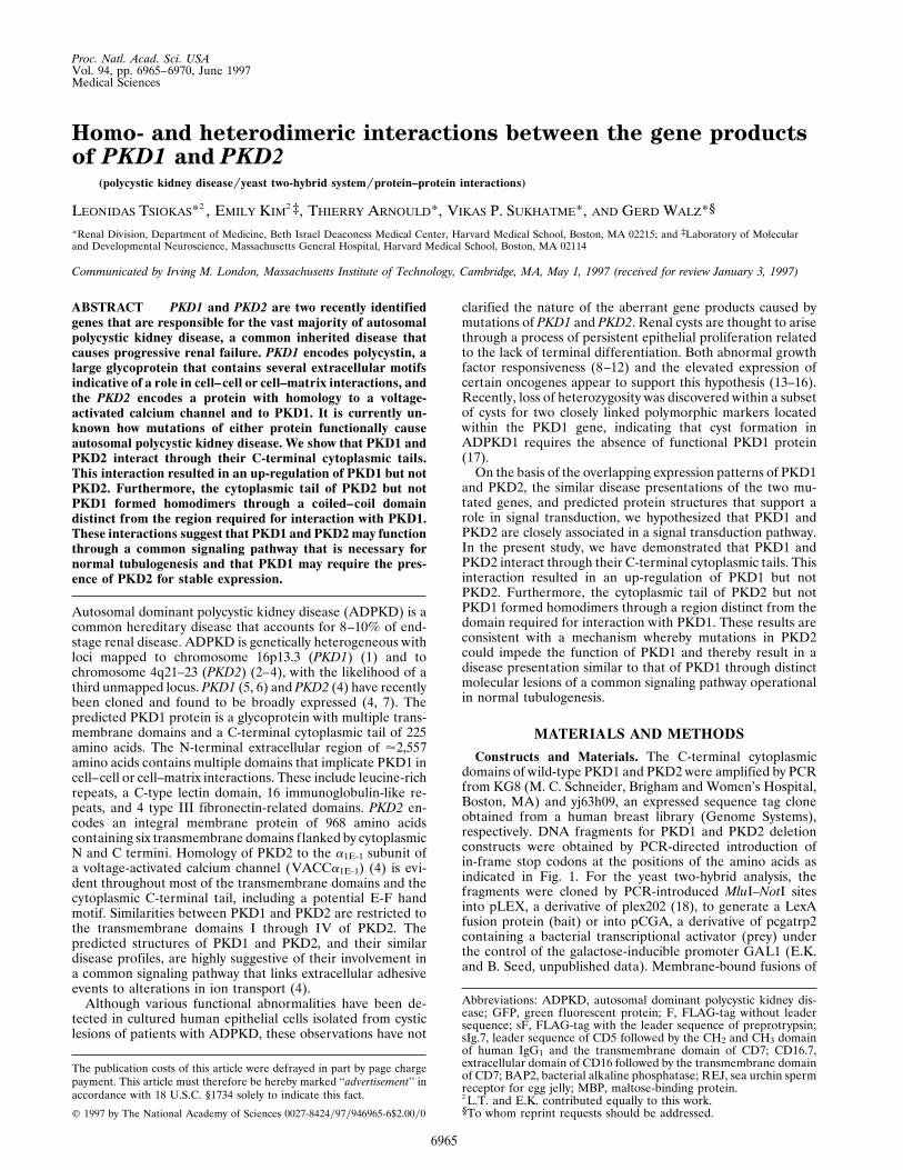
FIG. 1. Mapping the heterodimerization domains of PKD1 andPKD2 (A) and the homodimerization domains of PKD2 (B) in theyeast two-hybrid system. (A) The C-terminal 226 amino acids of PKD1and progressive C- and N-terminal deletions of this domain wereinserted into pLEX. For PKD2 binding assays, the pLex bait constructsand PKD2 prey constructs were sequentially transfected into the yeaststrain EGY48 bearing a lacZ reporter. Interaction (1) was indicatedby b-galactosidase production and leucine prototrophy in yeast; theminimal interacting domain, PKD1.4, caused leucine prototrophywithout b-galactosidase production (p). A putative coiled–coil struc-ture is depicted as a shaded box, a potential PEST sequence is depictedas a solid box. (B) The C-terminal 289 amino acids of PKD2 andprogressive C- and N-terminal deletions of this domain were insertedinto pLEX to map the interaction with PKD1 and PKD2. PKD2 bindsPKD1 through the C-terminal 97 amino acids. The homodimerizationof PKD2 is mediated by a region spanning amino acids 63–192, whichcontains a putative coiled–coil structure (shaded box).
6966 Medical Sciences: Tsiokas et al. Proc. Natl. Acad. Sci. USA 94 (1997)
survival of yeast on leucine-deficient medium and the produc-tion of b-galactosidase. PKD1 specifically interacted withPKD2 but not with a number of heterologous baits or withitself (data not shown). Further two-hybrid analysis usingdeletions of PKD1 and PKD2 localized the interacting domainof PKD1 to a 40-amino acids region contained within the last76 amino acids of PKD1, a region that contains a probablecoiled–coil structure (24) (Fig. 1). The domain of PKD2responsible for binding of PKD1 was found to reside in a regionspanning the last 97 amino acids of PKD2 (Fig. 1A).
PKD2 Forms Homodimers Through a C-Terminal Cyto-plasmic Coiled–Coil Domain in Yeast. The detection of aprobable coiled–coil domain in PKD2 outside of the PKD1-binding domain suggested to us a possible mechanism forself-dimerization. The yeast two-hybrid system was used toconfirm the homodimerization of PKD2 and to map thedomain mediating this interaction (Fig. 1B). The formation ofPKD2 homodimers was found to be mediated by a domainwithin amino acids 63–192 of the C terminus. These resultsindicate that PKD2 interacts with PKD1 through a domain thatis distinct from the region of PKD2 required for its ho-modimerization.
Expression of Recombinant PKD2 Up-Regulates the Ex-pression of PKD1 in Mammalian Cells. Because both PKD1and PKD2 appear to represent integral membrane proteinsthat have not yet been expressed in full-length in vivo (ref. 25and L.T., unpublished results), we designed an expressionsystem that targeted the cytoplasmic domains of PKD1 andPKD2 to the plasma membrane by using a single heterologoustransmembrane domain. The transmembrane region of humanCD7 has been used to anchor cytoplasmic peptides to theplasma membrane while retaining their functional activity(19–21). The C-terminal cytoplasmic tails of PKD1 and PKD2were each fused to the 39 end of the CD7 transmembranedomain, which was in turn preceded by the extracellulardomains of either CD16 or the leader sequence of CD5followed by the CH2 and CH3 domain of human IgG1 (Fig.2A). Plasmids directing the expression of CD16-CD7 (CD16.7)or CH2-CH3-CD7 (sIg.7) fusion proteins were transfected into293T cells and the surface expression of the individual con-structs was demonstrated by immunofluorescence (Fig. 2B). Inaddition, a PKD1 construct bearing three putative C-terminaltransmembrane domains and the cytoplasmic tail of PKD1 wastagged at its N terminus with the leader sequence of prepro-trypsin followed by the FLAG epitope (sF.3TM–PKD1; Fig.2A). Cytoplasmic versions of both the C-terminal domains ofPKD1 and PKD2 were expressed as FLAG-tagged proteinswithout a proceeding leader sequence (F). These constructspermitted us to analyze the interaction between PKD1 andPKD2 with either or both partners expressed as membrane-bound receptors. We noticed very low levels of expression forF–PKD1 in the lysates of cells cotransfected with the controlplasmid sIg.7 (Fig. 3, lane 5). The presence of a potential PESTsequence spanning amino acids 93–109 (26, 27) may mediatethis effect since deletion of the first 113 amino acids of thecytoplasmic domain resulted in increased protein levels ofF–PKD1.2 (Fig. 3, lanes 3 and 4).
An up-regulation of both membrane-bound and cytoplasmicPKD1 protein levels was detected when plasmids SF.3TM–PKD1, F–PKD1, and F–PKD1.2 were coexpressed with sIg.7–PKD2. This was not apparent for cytoplasmic PKD2; in fact,F–PKD2 protein levels were slightly reduced when cotrans-fected with sIg.7–PKD2 (see Fig. 5 A, lane 2, and D, lane 3).To allow a more quantitative assessment of the PKD1 up-regulation in the presence of PKD2, an expression vectorencoding GFP was included in the transfection mixture at aconstant ratio. Lysates of transfected 293T cells were probedsequentially for FLAG-tagged fusions, GFP, and an endoge-nous structural protein, a-catenin. The expression of GFPprovided a measure for transfection efficiency, and the ex-
pression of a-catenin served to normalize for the amount oftotal protein loaded in each lane. Cotransfection of sIg.7–PKD2 but not sIg.7 resulted in a significant increase of all threePKD1 fusion proteins (Fig. 3, lanes 4, 6, and 8). These resultsprovide indirect evidence that one role of membrane-boundPKD2 may be to interact with and stabilize PKD1.
Heterodimerization Between PKD1 And PKD2. To confirmthe results obtained in the yeast two-hybrid assay, 293T cellswere cotransfected with combinations of membrane-boundand cytoplasmic versions of PKD1 and PKD2. A specificinteraction was detected between F–PKD2 and sIg.7–PKD1.2(Fig. 4A, lane 2), sIg.7–PKD2 and F–PKD1.2 (Fig. 4A, lane 4),and sIg.7–PKD2 and F–PKD1 (Fig. 4A, lane 6), demonstratingthat the PKD2 binding region of PKD1 maps to the mostC-terminal 113 amino acids. No interaction was detectable
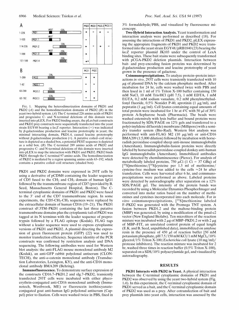
FIG. 2. Construction (A) and surface expression (B) of membrane-bound PKD1 and PKD2 fusion proteins. (A) The C-terminal 113amino acids of PKD1 (PKD1.2) and the extracellular domain of CD16were fused to the transmembrane region of CD7 to yield the chimericintegral membrane protein CD16.7–PKD1.2. The construct sF.3TM–PKD1, containing the last three putative transmembrane domains plusthe cytoplasmic tail of PKD1, is tagged at its N terminus with theleader sequence of preprotrypsin followed by a FLAG epitope. Themembrane-bound fusion of PKD2, sIg.7–PKD2, was generated byfusing the C-terminal 289 amino acids of PKD2 to a cell surfaceexpressed immunoglobulin consisting of the leader sequence of CD5,the CH2 and CH3 domain of human IgG1, and the transmembraneregion of CD7. (B) Surface expression of CD16.7–PKD1.2 andsIg.7–PKD2 fusion proteins. 293T cells were transfected with vector(a), CD16.7 (b), CD16.7–PKD1.2 (c), vector (d), sIg.7 (e), sIg.7–PKD2(f), and labeled with anti-CD16-fluorescein isothiocyanate (a–c) oranti-human IgG-phycoerythrin (d–f). Photographs were taken at 3400magnification under fluorescence microscopy 24 hr after transfection.(Insets) Lower magnification (3100) are located in the right lower ofb, c, e, and f. Expression of CD16.7–PKD1.2 (c) and sIg.7–PKD2 (f)was clearly detected on the surface of unfixed cells.
Medical Sciences: Tsiokas et al. Proc. Natl. Acad. Sci. USA 94 (1997) 6967

between the FLAG-tagged PKD1 and PKD2 fusion proteinswith the control protein sIg.7 (Fig. 4A, lanes 1, 3, and 5) orsIg.7–CD40 (data not shown). Thus, membrane-bound PKD1or PKD2 has the ability to interact with soluble forms of eachother. Furthermore, this interaction occurs when both PKD1and PKD2 are expressed as integral membrane proteins. Thefusion protein sF.3TM–PKD1 was used to cotransfect 293Tcells in combination with sIg.7–PKD2. Immunoprecipitationwith protein A and subsequent Western blot analysis withanti-FLAG antibody showed that sF.3TM–PKD1 was copre-cipitated with sIg.7–PKD2 (Fig. 4B, lane 1) and not with thecontrol protein sIg.7–CD40 (Fig. 4B, lane 2). To rule out anonspecific interaction between the CH2-CH3 domains ofsIg.7–PKD2 and the FLAG epitope, we cotransfected sIg.7–PKD2 and a FLAG-tagged version of bacterial alkaline phos-phatase (BAP2) and showed that sIg.7–PKD2 and F–BAP2did not coimmunoprecipitate (Fig. 4B, lane 3). Having dem-onstrated that sF.3TM–PKD1, the three-membrane spanningversion of PKD1, specifically associates with membrane-anchored PKD2, we confirmed the association of the cyto-plasmic tails of PKD1 and PKD2 by coexpressing two differentmembrane-bound forms of PKD1.2, CD16.7–PKD1.2 andsIg.7–PKD2, in 293T cells. Cell lysates were immunoprecipi-tated with protein A and coprecipitating sIg.7–PKD2 wasdetected by immunoblotting with anti-CD16 (Fig. 4B, lane 6).No association was seen between sIg.7–PKD2 and CD16.7(Fig. 4B, lane 4) or sIg.7–CD40 and CD16.7–PKD1.2 (Fig. 4B,lane 5). These results indicate that the C-terminal tails ofPKD1 and PKD2 associate in mammalian cells. To determinewhether other cellular proteins participate in the interactionbetween PKD1 and PKD2, coimmunoprecipitation experi-ments were performed with 35S-labeled 293T cells. Immuno-
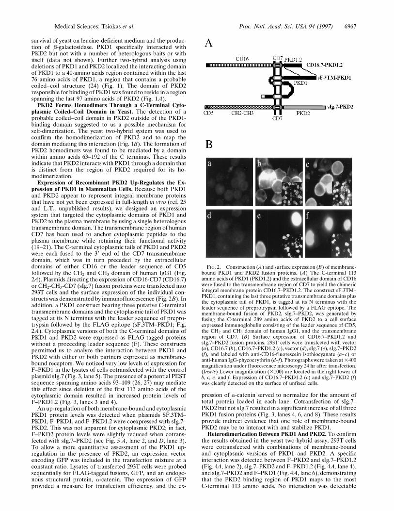
precipitation of F–PKD1.2 with anti-FLAG M2 resulted in thecoimmunoprecipitation of sIg.7–PKD2 (Fig. 5A, lane 1), con-firming the results obtained by Western blot analysis. Inaddition, a faint '50-kDa protein appeared in the precipitatescontaining F–PKD1.2 and sIg.7–PKD2 (Fig. 5A, lane 1),
FIG. 3. Up-regulation of PKD1 fusion proteins by membrane-bound PKD2 (sIg.7–PKD2). 293T cells were cotransfected with 4 mgof expression vector for FLAG-tagged PKD1 or PKD2 truncations and4 mg of expression vector for sIg.7 (control) or sIg.7–PKD2. Alltransfection mixes contained 2 mg of GFP. Cell lysates were immu-noprecipitated with protein A, followed by sequential Western blotanalysis with anti-FLAG, anti-GFP, and anti-a-catenin. Increasedlevels of PKD1 proteins, but not PKD2 protein, were observed (lanes4, 6, and 8). Control for transfection efficiency was provided by GFPlevels that were similar or lower than in control transfections contain-ing sIg.7, and total amount of protein loaded in each lane wasmonitored by a-catenin expression.
FIG. 4. Heterodimerization of PKD1 and PKD2 in vivo. (A)Coimmunoprecipitation of membrane-bound chimeric PKD1 andPKD2 proteins with cytoplasmic PKD2 and PKD1 fusion proteins,respectively. 293T cells were transfected with the indicated combina-tions of expression vectors for PKD1 and PKD2, and cell lysates wereimmunoprecipitated with protein A, followed by Western blot analysiswith anti-FLAG. sIg.7–PKD1.2 interacted with F–PKD2 (lane 2) butnot with the sIg.7 control protein (lane 1). The interaction betweensIg.7–PKD2 and F–PKD1.2 is demonstrated in lane 4; the interactionbetween sIg.7–PKD2 and F–PKD1 is shown in lane 6. NeitherF–PKD1.2 nor F–PKD1 interacted with the sIg.7 control protein. (B)Coimmunoprecipitation of membrane-bound fusion proteins of PKD2and PKD1. 293T cells were transfected with the expression vectorsindicated above. Immunoprecipitations were performed with proteinA, followed by Western blot analysis with anti-FLAG (lanes 1–3) oranti-CD16 (lanes 4–6). Lane 1 demonstrates the interaction withsIg.7–PKD2 and sF.3TM–PKD1. No interaction was detectable be-tween sF.3TM–PKD1 and sIg.7–CD40 or between sIg.7–PKD2 andF–BAP2. Lane 6 shows the interaction between the two membrane-bound fusion proteins CD16.7–PKD1.2 and sIg.7–PKD2. No interac-tion was detectable with the control proteins CD16.7 (lane 4), orsIg.7–CD40 (lane 5).
6968 Medical Sciences: Tsiokas et al. Proc. Natl. Acad. Sci. USA 94 (1997)
potentially in precipitates of F–PKD2 and sIg.7–PKD2 (Fig.5A, lane 2), and in F–PKD2 alone (Fig. 5A, lane 4), indicatingthat other cellular protein(s) may coimmunoprecipitate withPKD2 or the PKD1–PKD2 complex. To exclude the possibilitythat a third cellular protein is required for the interactionbetween PKD1 and PKD2, we performed coimmunoprecipi-tations in vitro with a recombinant MBP–PKD1.2 fusionprotein. Fig. 5B demonstrates that in vitro transcribed andtranslated [35S]methionine-labeled F–PKD2 bound to PKD1.2(Fig. 5B, lane 3), but not to an unrelated control protein ofsimilar length (Fig. 5B, lane 2). Collectively, these resultsindicate that the interaction between PKD1 and PKD2 doesnot require additional cellular proteins.
Homodimerization of PKD2 in Vivo. Whereas no ho-modimerization between PKD1 was detectable in either theyeast two-hybrid system or by coimmunoprecipitation in 293Tcells, PKD2 was found to homodimerize in both systems. 293T
cells were cotransfected with sIg.7–PKD1.2 or sIg.7 andF–PKD1.2, lysates were immunoprecipitated with protein A,and bound proteins were immunoblotted with anti-FLAG. Nointeraction was detectable between the cytoplasmic and mem-brane-anchored versions of PKD1 (data not shown). However,in a similar experiment, cotransfection of sIg.7–PKD2 andF–PKD2 in 293T cells resulted in coimmunoprecipitation ofF–PKD2 (Fig. 5C, lane 3). This interaction was specific and didnot occur when F–PKD2 was coexpressed with sIg.7 or sIg.7–CD40 (Fig. 5C, lanes 1 and 2). Thus, the C terminus of PKD2forms homodimers and heterodimers with the C terminus ofPKD1. This is in agreement with results from the two-hybridanalysis, which showed that PKD1 and PKD2 bind to non-overlapping sites within the C terminus of PKD2.
DISCUSSION
Two genes responsible for ADPKD have recently been iden-tified (4, 5) and sequence analysis of PKD1 and PKD2 hasfueled speculation over the mechanism by which mutations ofthese two genes cause a similar presentation of polycystickidney disease associated with liver cysts and cerebral aneu-rysm. Several structural elements found in the extracellulardomain of PKD1 point to a role in cell–cell andyor cell–matrixinteraction: a leucine-rich region, the C-type lectin domain, therepetitive Ig-like domains, and putative fibronectin type IIIrepeats found in other cell adhesion molecules (6). In addition,the extracellular region of PKD1 contains a region (aminoacids 2,146–2,882) with significant homology to the sea urchinsperm receptor for egg jelly (REJ) (28), an integral membraneprotein whose function is coupled to calcium channels: bindingof monoclonal antibodies to REJ initiates the acrosome reac-tion in sperm by increasing the level of intracellular calcium,a prerequisite for subsequent Na1yH1 exchange (28). Inter-estingly, the combination of Ig-like domains and fibronectintype III repeats is present in the neural cell adhesion moleculeand L1, a class of cellular adhesion molecules that translateextracellular events into the activation of L- and N-typeneuronal calcium channels (29). Homology between PKD2and the a1E-1 subunit of voltage-activated calcium channelsthroughout most of the transmembrane pore-forming domainsis equally suggestive of a signal transduction cascade involvingPKD1 and PKD2. On the basis of the structural elements andthe overlapping clinical presentation of ADPKD1 and AD-PKD2, we conjecture that PKD1 and PKD2 operate in acommon signaling pathway, transducing extracellular adhesiveevents into alterations in ion transport.
To begin to elucidate the molecular mechanisms underlyingADPKD, we evaluated the interaction of PKD1 and PKD2 byusing the yeast two-hybrid system and in vivo and in vitrocoimmunoprecipitation assays. The association of the twoproteins in all three systems supports a model in which the twogene products participate in a common signaling pathway. Weused deletional analysis to define sequences within the cyto-plasmic domains that are required for binding. A probablecoiled–coil structure in PKD1 was found to be essential andsufficient for interaction with PKD2, and a probable coiled–coil structure in PKD2 was located in the domain required forits homodimerization. Since PKD2 has six membrane spans,the self-associating domain could help mediate the homomul-timeric complexing necessary for function as an ion channel ina configuration similar to that of potassium channels (30).
One intriguing motif is a potential PEST sequence in the Cterminus of PKD1 that appears to target PKD1 for rapiddegradation. Deletion of this sequence, as in the constructCD16.7–PKD1.2, resulted in increased protein levels andstable cell surface expression. Several PKD1 versions werefound to be up-regulated in the presence of PKD2. A similarup-regulation of a- and b-catenin has been documented inwhich catenin–cadherin complex formation prevents b-cate-
FIG. 5. Hetero- and homodimerization of PKD1 and PKD2. (A)Coimmunoprecipitations of 35S-labeled PKD1 and PKD2 fusionsproteins. 293T cells were transfected with sIg.7–PKD2 and F–PKD1.2(lane 1), sIg.7–PKD2 and F–PKD2 (lane 2), sIg.7 and F–PKD1.2 (lane3), sIg.7 and F–PKD2 (lane 4), or remained untransfected (lane 5).Cells were harvested after a 6-hr chase with [35S]methionineycysteine.Immunoprecipitations were performed with anti-FLAG M2 affinitygel. The arrow marks a putative 50-kDa protein present in lane 1 (andpotentially in lanes 2 and 4), which appears to associate with PKD2 orthe PKD1–PKD2 complex. Similar results were obtained with recip-rocal immunoprecipitation using protein A to immunoprecipiate theIgG-tagged constructs (data not shown). (B) Coimmunoprecipitationof 35S-labeled F–PKD2 with MBP–PKD1.2 fusion protein. F–PKD2was in vitro transcribed and translated (lane 1). An aliquot of thereaction mixture was then incubated with a control MBP–protein (lane2) or MBP–PKD1.2 (lane 3). (C) Homodimerization of PKD2 in vivo.293T cells were cotransfected with sIg.7 and F–PKD2 (lane 1),sIg.7–CD40 and F–PKD2 (lane 2), and sIg.7–PKD2 and F–PKD2 (lane3). Lysates were immunoprecipitated with protein A, and boundproteins were blotted with anti-FLAG. (D) Cell lysates demonstratingcomparable expression of the F–PKD2 fusion protein in all threeconditions (lanes 1–3 as in C).
Medical Sciences: Tsiokas et al. Proc. Natl. Acad. Sci. USA 94 (1997) 6969
nin degradation (31, 32). It is possible that during tubulogen-esis, stable expression of PKD1 depends upon the presence ofPKD2. In this scenario, mutations of PKD1 and PKD2 thatcompromise protein–protein interaction would interupt thesignal transduction cascade and enhance PKD1 degradation.In support of this model is a reported ADPKD mutation thatdeletes the C-terminal 76 amino acids of PKD1 (33), a domainwe found essential for interaction with PKD2. These resultssuggest that protein stability resulting from the formation of amultimeric complex consisting of PKD1–PKD2 heteromersand PKD2–PKD2 homomultimers may be essential for normaltubulogenesis.
Collectively, these data support the hypothesis that PKD1and PKD2 participate in a common signaling pathway thatprevents cyst formation (4, 17). If PKD2 indeed functions asa voltage-gated ion channel, it is possible that extracellularevents sensed by PKD1 could regulate PKD2 channel activitythrough interactions between the C-terminal cytoplasmic do-mains of PKD1 and PKD2, and a more N-terminal coiled–coilstructure in PKD2 may mediate homomultimeric complexingPKD2 required for the formation of a functional channel.
Note Added in Proof. Qian et al. demonstrated the interaction betweenPKD1 and PKD2 in yeast and in vitro (34).
We thank Michael C. Schneider for the KG8 clone and Brian Seedfor several constructs and expression cassettes. We thank Steven A.Bossone, Herbert Cohen, Bertrand Knebelmann, and other membersof the lab for helpful comments. E.K. was supported by Public HealthService Grant MH-01147, and T.A. was supported by a ResearchFellowship of the Belgian American Educational Foundation. Thiswork was supported by National Institutes of Health Grant RO1-DK-51060 (V.P.S.) and a grant from the Polycystic Kidney ResearchFoundation (G.W.).
1. Reeders, S. T., Breuning, M. H., Davies, K. E., Nicholls, R. D.,Jarman, A. P., Higgs, D. R., Pearson, P. L. & Weatherall, D. J.(1985) Nature (London) 317, 542–544.
2. Kimberling, W. J., Kumar, S., Gabow, P. A., Kenyon, J. B.,Connolly, C. J. & Somlo, S. (1993) Genomics 18, 467–472.
3. Peters, D. J., Spruit, L., Saris, J. J., Ravine, D., Sandkuijl, L. A.,Fossdal, R., Boersma, J., van Eijk, R., Norby, S., Constantinou-Deltas, C. D., Pierides, A., Brissenden, J. E., Frants, R. R., vanOmmen, G.-J. B. & Breuning, M. H. (1993) Nat. Genet. 5,359–362.
4. Mochizuki, T., Wu, G., Hayashi, T., Xenophontos, S. L.,Veldhuisen, B., Saris, J. J., Reynolds, D. M., Cai, Y., Gavow,P. A., Pierides, A., Kimberling, W. J., Breuning, M. H., Deltas,C. C., Peters, D. J. M. & Somlo, S. (1996) Science 272, 1339–1342.
5. The International Polycystic Kidney Disease Consortium (1995)Cell 81, 289–298.
6. Hughes, J., Ward, C. J., Peral, B., Aspinwall, R., Clark, K., SanMillan, J. L., Gamble, V. & Harris, P. C. (1995) Nat. Genet. 10,151–160.
7. Ward, C. J., Turley, H., Ong, A. C. M., Comley, M., Biddolph, S.,Chetty, R., Ratcliffe, P. J., Gatter, K. & Harris, P. C. (1996) Proc.Natl. Acad. Sci. USA 93, 1524–1528.
8. Wilson, P. D. (1991) Am. J. Kidney Dis. 17, 634–637.9. Wilson, P. D., Sherwood, A. C., Palla, K., Du, J., Watson, R. &
Norman, J. T. (1991) Am. J. Physiol. 260, F420–F430.10. Wilson, P. D. & Burrow, C. R. (1992) Adv. Nephrol. Necker. Hosp.
21, 125–142.11. Wilson, P., Falkenstein, D., Gatti, L., Eng, E. & Burrow, C. (1995)
Kidney Int. 47, 724–725.12. Du, J. & Wilson, P. D. (1995) Am. J. Physiol. 269, C487–C495.13. Cowley, B. D., Jr., Chadwick, L. J., Grantham, J. J. & Calvet, J. P.
(1991) J. Am. Soc. Nephrol. 1, 1048–1053.14. Rankin, C. A., Grantham, J. J. & Calvet, J. P. (1992) J. Cell.
Physiol. 152, 578–586.15. Schaffner, D. L., Barrios, R., Massey, C., Banez, E. I., Ou, C. N.,
Rajagopalan, S., Aguilar-Cordova, E., Lebovitz, R. M., Over-beek, P. A. & Lieberman, M. W. (1993) Am. J. Pathol. 142,1051–1060.
16. Trudel, M., D’Agati, V. & Constantini, F. (1991) Kidney Int. 39,665–671.
17. Qian, F., Watnick, T. J., Onuchic, L. F. & Germino, G. G. (1996)Cell 87, 979–987.
18. Stanger, B., Leder, P., Lee, T., Kim, E. & Seed, B. (1995) Cell 81,513–523.
19. Kolanus, W., Romeo, C. & Seed, B. (1993) Cell 74, 171–183.20. Romeo, C., Kolanus, W., Amiot, M. & Seed, B. (1992) Cold
Spring Harbor Symp. Quant. Biol. 57, 117–125.21. Romeo, C., Amiot, M. & Seed, B. (1992) Cell 68, 889–897.22. Haas, J., Park, E. C. & Seed, B. (1996) Curr. Biol. 6, 315–324.23. Gyuris, J., Golemis, E., Chertkov, H. & Brent, R. (1993) Cell 75,
791–803.24. Lupas, A., van Dyke, M. & Stock, J. (1991) Science 252, 1162–
1164.25. Ibraghimov-Beskrovnaya, O., Dackowski, W., Petry, L., Burn, T.,
Connors, T., van Raay, T., Qian, F., Onuchic, L., Watnik, T.,Piontek, K., Hakim, R., Landes, G., Germino, G. & Klinger, K.(1996) J. Am. Soc. Nephrol. 7, 1599 (abstr.).
26. Rechsteiner, M. (1988) Adv. Enzyme Regul. 27, 135–51.27. Rechsteiner, M. (1990) Semin. Cell Biol. 1, 433–440.28. Moy, G. W., Mendoza, L. M., Schulz, J. R., Swanson, W. J.,
Glabe, C. G. & Vacquier, V. D. (1996) J. Cell Biol. 133, 809–817.29. Doherty, P., Ashton, S. V., Moore, S. E. & Walsh, F. S. (1991)
Cell 67, 21–33.30. Xu, J., Yu, W., Jan, Y. N., Jan, L. Y. & Li, M. (1995) J. Biol.
Chem. 270, 24761–24768.31. Hinck, L., Nathke, I. S., Papkoff, J. & Nelson, W. J. (1994) J. Cell
Biol. 125, 1327–1340.32. Nakagawa, S. & Takeichi, M. (1995) Development (Cambridge,
U.K.) 121, 1321–1332.33. Peral, B., San Millan, J. L., Ong, A. C., Gamble, V., Ward, C. J.,
Strong, C. & Harris, P. C. (1996) Am. J. Hum. Genet. 58, 86–96.34. Qian, F., Germino, F. J., Cai, Y., Zhang, X., Somlo, S. &
Germino, G. G. (1997) Nat. Genet., in press.
6970 Medical Sciences: Tsiokas et al. Proc. Natl. Acad. Sci. USA 94 (1997)


