Embed Size (px)
Citation preview
pubs.acs.org/jmc Published on Web 07/29/2010 r 2010 American Chemical Society
6100 J. Med. Chem. 2010, 53, 6100–6111
DOI: 10.1021/jm100507q
Non-Peptide Macrocyclic Histone Deacetylase Inhibitors Derived from Tricyclic Ketolide Skeleton
Sandra C. Mwakwari,† William Guerrant,† Vishal Patil,† Shabana I. Khan,§ Babu L. Tekwani,§ Zachary A. Gurard-Levin, )
Milan Mrksich, ) and Adegboyega K. Oyelere*,†,‡
†School of Chemistry and Biochemistry, and ‡Parker H. Petit Institute for Bioengineering and Bioscience, Georgia Institute of Technology,Atlanta, Georgia 30332-0400, §National Center for Natural Products Research and Department of Pharmacology, School of Pharmacy,University of Mississippi, University, Mississippi 38677-1848, and )Department of Chemistry and Howard Hughes Medical Institute,University of Chicago, Chicago, Illinois 60637-1511
Received April 26, 2010
Inhibitionofhistonedeacetylase (HDAC) function is a validated therapeutic strategy for cancer treatment.Ofthe several structurally distinct small molecule histone deacetylase inhibitors (HDACi) reported,macrocyclicdepsipeptides possess the most complex cap groups and have demonstrated excellent HDAC inhibitionpotency and isoform selectivity. Unfortunately, the development of macrocyclic depsipeptides has beenhampered in part because of development problems characteristic of large peptides and the complex reactionschemes required for their synthesis.Hereinwe report that tricyclic ketolideTE-802 is an excellentmimetic forthe peptide backbone of macrocyclic HDACi. Compounds derived from this template are particularlyselective againstHDACs1 and2with nanomolar inhibitory activity. Interrogationof the associationbetweena subset of these compounds and key HDAC isoforms, using AutoDock, enables a molecular description ofthe interaction between the HDAC enzyme’s outer rim and the inhibitors’ macrocyclic cap group that areresponsible for compound affinity and presumably isoform selectivity.
Introduction
Histone deacetylase (HDACa) and histone acetyltransfer-ase (HAT) are two functionally opposing enzymes, many ofwhich tightly regulate the chromatin structure and functionvia sustenance of equilibrium between the acetylated anddeacetylated states of histones. By catalyzing the removal ofacetyl groups, HDACs induce a condensed chromatin struc-ture resulting in transcription repression, whereas acetylatedhistones are associatedwith amoreaccessible/open chromatinstructure and activation of transcription.1-4 In addition,many non-histone proteins such as tubulin, ERR, p53, HSP90, NF-YA, and GATA-1 have been found in an acetylatedstate and may be substrates of HDACs.5-10 Eighteen humanHDAC isoforms are known, and they are subdivided into theclassical zinc dependent HDACs comprising classes I, II, andIV and NADþ dependent sirtuins, class III enzymes.9,11,12
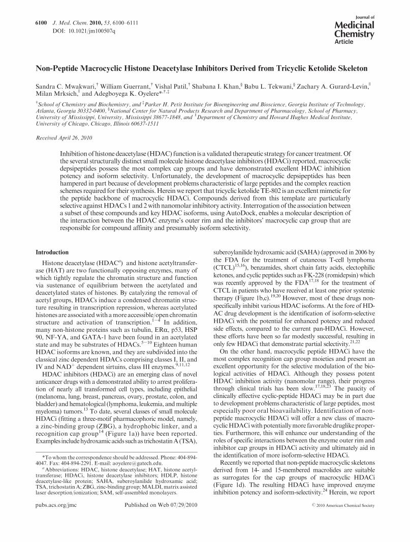
HDAC inhibitors (HDACi) are an emerging class of novelanticancer drugs with a demonstrated ability to arrest prolifera-tion of nearly all transformed cell types, including epithelial(melanoma, lung, breast, pancreas, ovary, prostate, colon, andbladder) andhematological (lymphoma, leukemia, andmultiplemyeloma) tumors.13 To date, several classes of small moleculeHDACi (fitting a three-motif pharmacophoric model, namely,a zinc-binding group (ZBG), a hydrophobic linker, and arecognition cap group14 (Figure 1a)) have been reported.Examples includehydroxamicacids suchas trichostatinA(TSA),
suberoylanilide hydroxamic acid (SAHA) (approved in 2006 bythe FDA for the treatment of cutaneous T-cell lymphoma(CTCL)15,16), benzamides, short chain fatty acids, electophilicketones, and cyclic peptides such asFK-228 (romidepsin) whichwas recently approved by the FDA17,18 for the treatment ofCTCL in patients who have received at least one prior systemictherapy (Figure 1b,c).19,20 However, most of these drugs non-specifically inhibit various HDAC isoforms. At the fore of HD-AC drug development is the identification of isoform-selectiveHDACi with the potential for enhanced potency and reducedside effects, compared to the current pan-HDACi. However,these efforts have been so far modestly successful, resulting inonly few HDACi that demonstrate partial selectivity.21,22
On the other hand, macrocyclic peptide HDACi have themost complex recognition cap group moieties and present anexcellent opportunity for the selective modulation of the bio-logical activities of HDACi. Although they possess potentHDAC inhibition activity (nanomolar range), their progressthrough clinical trials has been slow.17,18,23 The paucity ofclinically effective cyclic-peptide HDACi may be in part dueto development problems characteristic of large peptides, mostespecially poor oral bioavailability. Identification of non-peptide macrocyclic HDACi will offer a new class of macro-cyclicHDACiwithpotentiallymore favorable druglikeproper-ties. Furthermore, this will enhance our understanding of theroles of specific interactions between the enzyme outer rim andinhibitor cap groups in HDACi activity and ultimately aid inthe identification of more isoform-selective HDACi.
Recentlywe reported that non-peptidemacrocyclic skeletonsderived from 14- and 15-membered macrolides are suitableas surrogates for the cap groups of macrocyclic HDACi(Figure 1d). The resulting HDACi have improved enzymeinhibition potency and isoform-selectivity.24 Herein, we report
*Towhom the correspondence should be addressed. Phone: 404-894-4047. Fax: 404-894-2291. E-mail: [email protected].
aAbbreviations: HDAC, histone deacetylase; HAT, histone acetyl-transferase; HDACi, histone deacetylase inhibitors; HDLP, histonedeacetylase-like protein; SAHA, suberoylanilide hydroxamic acid;TSA, trichostatinA; ZBG, zinc-binding group;MALDI,matrix assistedlaser desorption/ionization; SAM, self-assembled monolayers.
Article Journal of Medicinal Chemistry, 2010, Vol. 53, No. 16 6101
that enhancement of the 14-membered macrolide ring hydro-phobicity and rigidity facilitates specific drug interactions withthe enzyme’s outer rim residues, maximizes HDAC inhibition,and improves drug cytotoxicity against human cancer cell lines.Moreover, these compoundshaveantiparasitic activitiesagainstcausative parasites of malaria and leishmaniasis in a mannerthat reveals structural attributes that confer a specific anti-parasitic response.
Results and Discussion
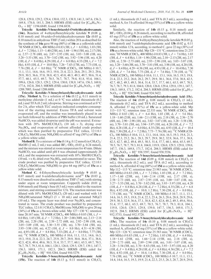
Molecular Docking Analysis. Previous molecular dock-ing studies on HDACi derived from 14- and 15-membered
macrolides clarithromycin and azithromycin, respectively,using histone deacetylase-like protein (HDLP), revealed thestructural basis for the enhanced activity of these macrocycliccompounds. Either ring system adopted docked orientationsthat displayed molecular surface complementarities betweenthe macrolide skeleton and the enzyme outer rim.24 In thesedocked structures, most of the hydrophobic components of themacrolide ring optimally interact with the hydrophobic resi-dues within HDLP hydrophobic pockets. Common to bothring systems are the vicinal diols atC11andC12andatC12andC13 for clarithromycin and azithromycin, respectively, whichhave to be accommodated within the enzyme hydrophobic
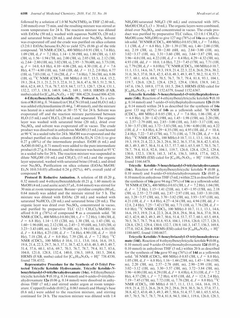
Figure 1. (a) PharmacophoricmodelofHDACiandrepresentativeexamplesof (b) acyclic, (c) cyclicpeptide,and(d)macrolide-basedHDACinhibitors.
6102 Journal of Medicinal Chemistry, 2010, Vol. 53, No. 16 Mwakwari et al.
pocket. We postulated that refinement of the ring hydropho-bicity through appropriate modification of the vicinal diolscould further modulate the HDAC inhibition of these non-peptide macrocyclic HDACi. In the previous report, we ob-served that the twomacrolide skeletons contributeddistinctly tothe overall HDAC binding. For 14- and 15-membered macro-lides with C5 and C6 linkers, the 14-membered compounds areabout 2- to 5-foldmore potentHDACi than their 15-memberedcounterparts.24 On the basis of the foregoing, we focused on thetransformation of the 14-membered macrocyclic template.
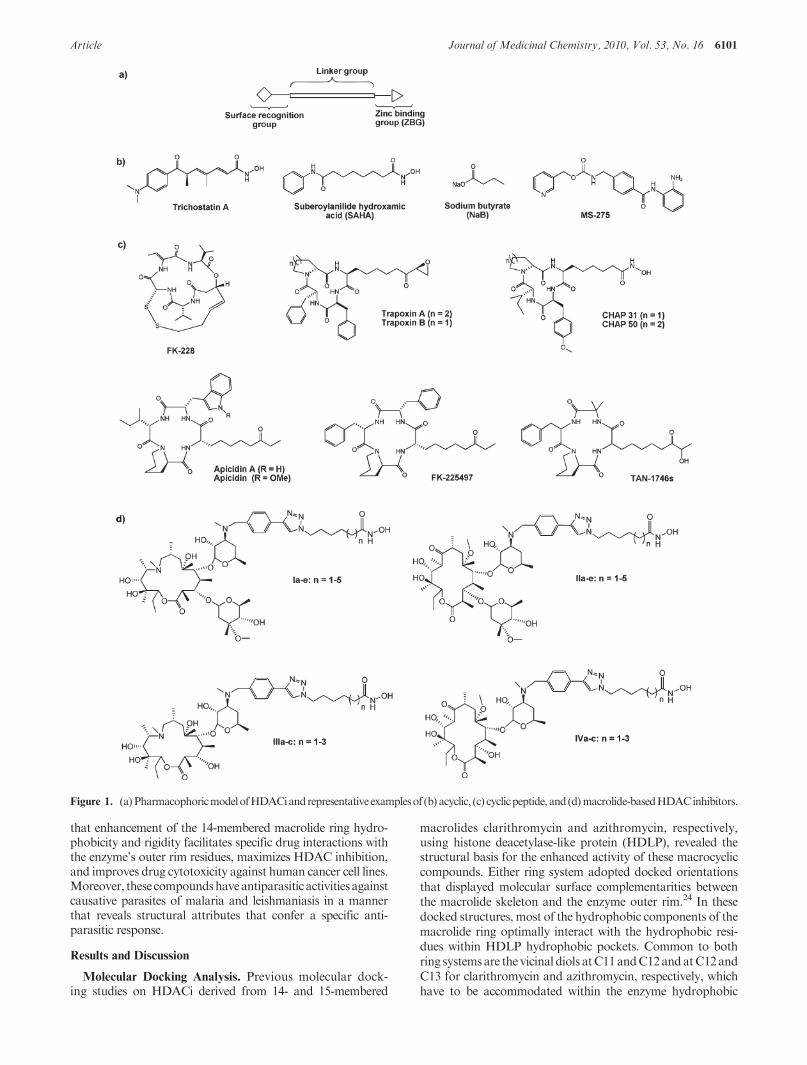
Conversion of the 14-membered ring vicinal diols to variousfive-membered carbamates is a common and synthetically trac-table approach toward their transformation to relatively morehydrophobicketolide skeletons (Figure 2).25-27The templateofketolide 10 (TE-802),25 in which the carbamate NH group hasbeen further substituted with a small hydrophobic group thatresults in a tricyclic ring system, is very attractive in this regard.The rigid template of 10minimally perturbs the ring within theregion of interest while maximizing ring hydrophobicity andshould be compatible with the enzyme outer rim hydrophobicpocket. To verify this prospect, we proceeded to interrogate thebinding interaction between HDAC1 homology model builtfrom human HDAC2 X-ray structure 3MAX coordinates28
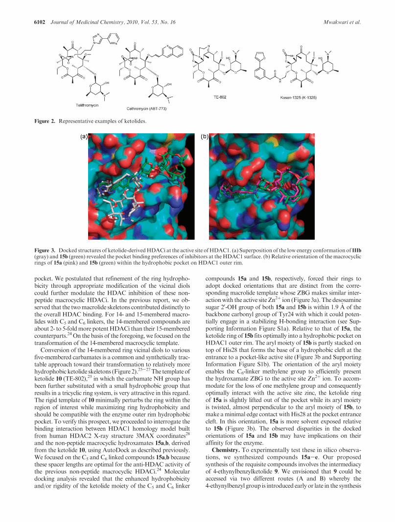
and the non-peptide macrocyclic hydroxamates 15a,b, derivedfrom the ketolide 10, using AutoDock as described previously.We focused on the C5 and C6 linked compounds 15a,b becausethese spacer lengths are optimal for the anti-HDAC activity ofthe previous non-peptide macrocyclic HDACi.24 Moleculardocking analysis revealed that the enhanced hydrophobicityand/or rigidity of the ketolide moiety of the C5 and C6 linker
compounds 15a and 15b, respectively, forced their rings toadopt docked orientations that are distinct from the corre-sponding macrolide template whose ZBG makes similar inter-actionwith the active site Zn2þ ion (Figure 3a). The desosaminesugar 20-OH group of both 15a and 15b is within 1.9 A of thebackbone carbonyl group of Tyr24 with which it could poten-tially engage in a stabilizing H-bonding interaction (see Sup-porting Information Figure S1a). Relative to that of 15a, theketolide ring of 15b fits optimally into a hydrophobic pocket onHDAC1 outer rim. The aryl moiety of 15b is partly stacked ontop of His28 that forms the base of a hydrophobic cleft at theentrance to a pocket-like active site (Figure 3b and SupportingInformation Figure S1b). The orientation of the aryl moietyenables the C6-linker methylene group to efficiently presentthe hydroxamate ZBG to the active site Zn2þ ion. To accom-modate for the loss of one methylene group and consequentlyoptimally interact with the active site zinc, the ketolide ringof 15a is slightly lifted out of the pocket while its aryl moietyis twisted, almost perpendicular to the aryl moiety of 15b, tomake aminimal edge contact withHis28 at the pocket entrancecleft. In this orientation, 15a is more solvent exposed relativeto 15b (Figure 3b). The observed disparities in the dockedorientations of 15a and 15b may have implications on theiraffinity for the enzyme.
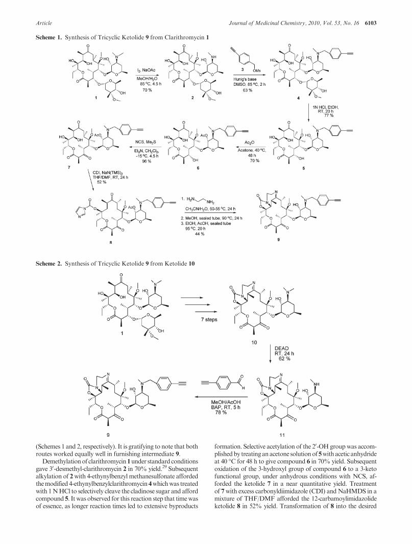
Chemistry. To experimentally test these in silico observa-tions, we synthesized compounds 15a-e. Our proposedsynthesis of the requisite compounds involves the intermediacyof 4-ethynylbenzylketolide 9. We envisioned that 9 could beaccessed via two different routes (A and B) whereby the4-ethynylbenzyl group is introduced early or late in the synthesis
Figure 2. Representative examples of ketolides.
Figure 3. Docked structures of ketolide-derivedHDACi at the active site ofHDAC1. (a) Superposition of the low energy conformation of IIIb(gray) and 15b (green) revealed the pocket binding preferences of inhibitors at the HDAC1 surface. (b) Relative orientation of the macrocyclicrings of 15a (pink) and 15b (green) within the hydrophobic pocket on HDAC1 outer rim.
Article Journal of Medicinal Chemistry, 2010, Vol. 53, No. 16 6103
(Schemes 1 and 2, respectively). It is gratifying to note that bothroutes worked equally well in furnishing intermediate 9.
Demethylationof clarithromycin1under standardconditionsgave 30-desmethyl-clarithromycin 2 in 70% yield.29 Subsequentalkylation of 2with 4-ethynylbenzyl methanesulfonate affordedthemodified 4-ethynylbenzylclarithromycin 4whichwas treatedwith 1NHCl to selectively cleave the cladinose sugar and affordcompound 5. It was observed for this reaction step that timewasof essence, as longer reaction times led to extensive byproducts
formation. Selective acetylation of the 20-OH groupwas accom-plishedby treatinganacetone solutionof5withacetic anhydrideat 40 �C for 48 h to give compound 6 in 70% yield. Subsequentoxidation of the 3-hydroxyl group of compound 6 to a 3-ketofunctional group, under anhydrous conditions with NCS, af-forded the ketolide 7 in a near quantitative yield. Treatmentof 7with excess carbonyldiimidazole (CDI) andNaHMDS in amixture of THF/DMF afforded the 12-carbamoylimidazolideketolide 8 in 52% yield. Transformation of 8 into the desired
Scheme 1. Synthesis of Tricyclic Ketolide 9 from Clarithromycin 1
Scheme 2. Synthesis of Tricyclic Ketolide 9 from Ketolide 10
6104 Journal of Medicinal Chemistry, 2010, Vol. 53, No. 16 Mwakwari et al.
tricyclic ketolide 9 was achieved in two successive cyclizationsteps adapting literature procedures.25Reactionof imidazolide8with ethylenediamine followed by intramolecularMichael addi-tion led to the formation of 11,12-cyclic carbamate. Subsequentintramolecular dehydration completed the cyclization process,affording the desired product 9 in 44% yield (Scheme 1).
Alternatively, intermediate 9 could be directly obtainedfrom 10. We synthesized 10 from clarithromycin 1 accordingto a literature procedure.25,26,30,31 Subsequent N-demethyla-tion of 10 using diethyl azodicarboxylate (DEAD) gave theexpected N-desmethyl ketolide 11 in 62% yield.31 Reductiveamination of 11 with 4-ethynylbenzaldehyde using borane-pyridine complex (BAP) afforded the requisite intermediate9 in 78% yield (Scheme 2).
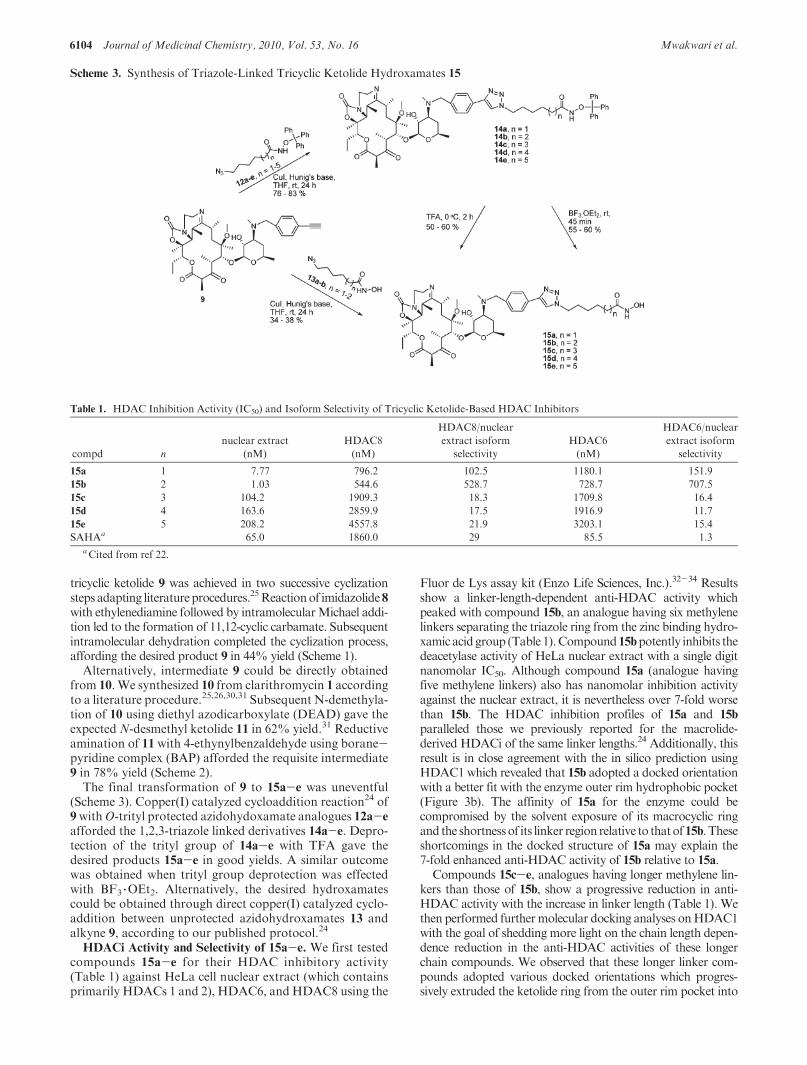
The final transformation of 9 to 15a-e was uneventful(Scheme 3). Copper(I) catalyzed cycloaddition reaction24 of9withO-trityl protected azidohydoxamate analogues 12a-e
afforded the 1,2,3-triazole linked derivatives 14a-e. Depro-tection of the trityl group of 14a-e with TFA gave thedesired products 15a-e in good yields. A similar outcomewas obtained when trityl group deprotection was effectedwith BF3 3OEt2. Alternatively, the desired hydroxamatescould be obtained through direct copper(I) catalyzed cyclo-addition between unprotected azidohydroxamates 13 andalkyne 9, according to our published protocol.24
HDACi Activity and Selectivity of 15a-e. We first testedcompounds 15a-e for their HDAC inhibitory activity(Table 1) against HeLa cell nuclear extract (which containsprimarily HDACs 1 and 2), HDAC6, and HDAC8 using the
Fluor de Lys assay kit (Enzo Life Sciences, Inc.).32-34 Resultsshow a linker-length-dependent anti-HDAC activity whichpeaked with compound 15b, an analogue having six methylenelinkers separating the triazole ring from the zinc binding hydro-xamic acidgroup (Table 1).Compound15bpotently inhibits thedeacetylase activity of HeLa nuclear extract with a single digitnanomolar IC50. Although compound 15a (analogue havingfive methylene linkers) also has nanomolar inhibition activityagainst the nuclear extract, it is nevertheless over 7-fold worsethan 15b. The HDAC inhibition profiles of 15a and 15b
paralleled those we previously reported for the macrolide-derived HDACi of the same linker lengths.24 Additionally, thisresult is in close agreement with the in silico prediction usingHDAC1 which revealed that 15b adopted a docked orientationwith a better fit with the enzyme outer rim hydrophobic pocket(Figure 3b). The affinity of 15a for the enzyme could becompromised by the solvent exposure of its macrocyclic ringand the shortnessof its linker region relative to thatof15b.Theseshortcomings in the docked structure of 15a may explain the7-fold enhanced anti-HDAC activity of 15b relative to 15a.
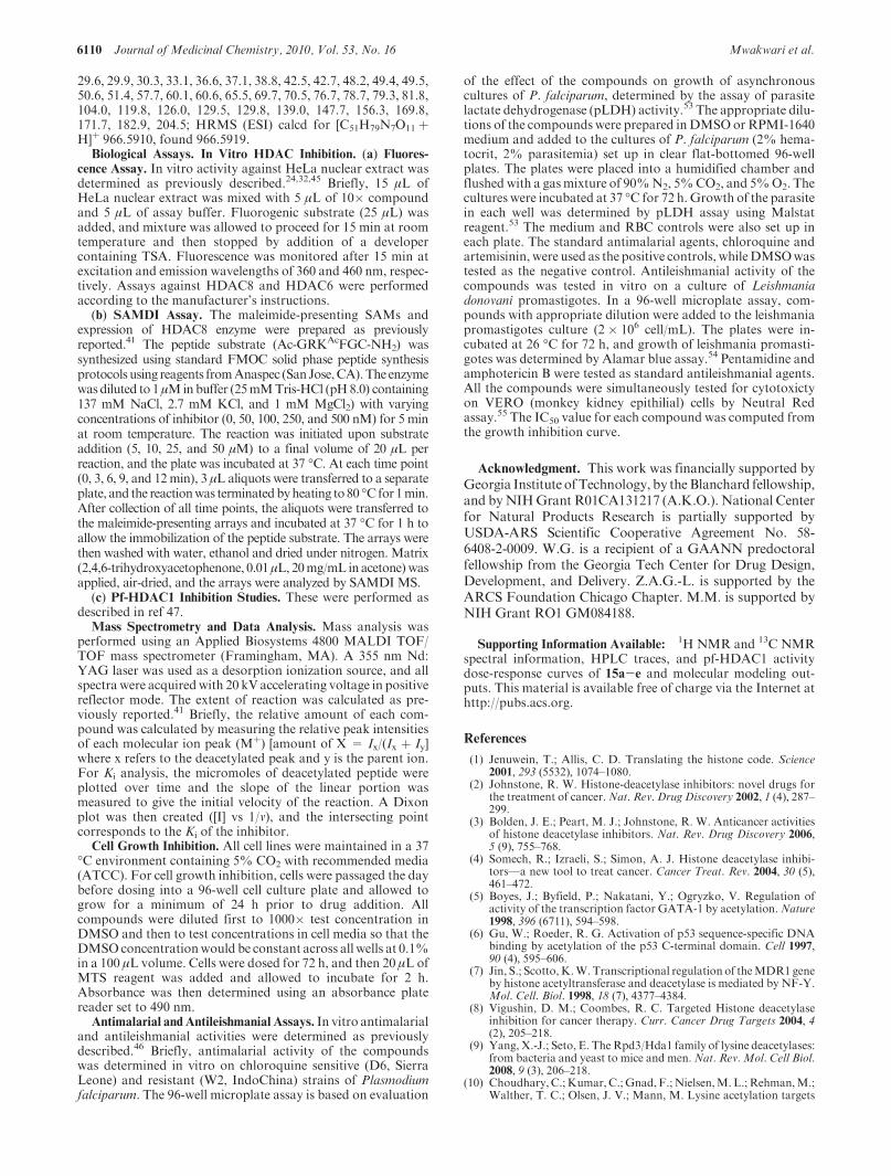
Compounds 15c-e, analogues having longer methylene lin-kers than those of 15b, show a progressive reduction in anti-HDAC activity with the increase in linker length (Table 1). Wethen performed furthermolecular docking analyses onHDAC1with the goal of shedding more light on the chain length depen-dence reduction in the anti-HDAC activities of these longerchain compounds. We observed that these longer linker com-pounds adopted various docked orientations which progres-sively extruded the ketolide ring from the outer rim pocket into
Scheme 3. Synthesis of Triazole-Linked Tricyclic Ketolide Hydroxamates 15
Table 1. HDAC Inhibition Activity (IC50) and Isoform Selectivity of Tricyclic Ketolide-Based HDAC Inhibitors
compd n
nuclear extract
(nM)
HDAC8
(nM)
HDAC8/nuclear
extract isoform
selectivity
HDAC6
(nM)
HDAC6/nuclear
extract isoform
selectivity
15a 1 7.77 796.2 102.5 1180.1 151.9
15b 2 1.03 544.6 528.7 728.7 707.5
15c 3 104.2 1909.3 18.3 1709.8 16.4
15d 4 163.6 2859.9 17.5 1916.9 11.7
15e 5 208.2 4557.8 21.9 3203.1 15.4
SAHAa 65.0 1860.0 29 85.5 1.3aCited from ref 22.
Article Journal of Medicinal Chemistry, 2010, Vol. 53, No. 16 6105
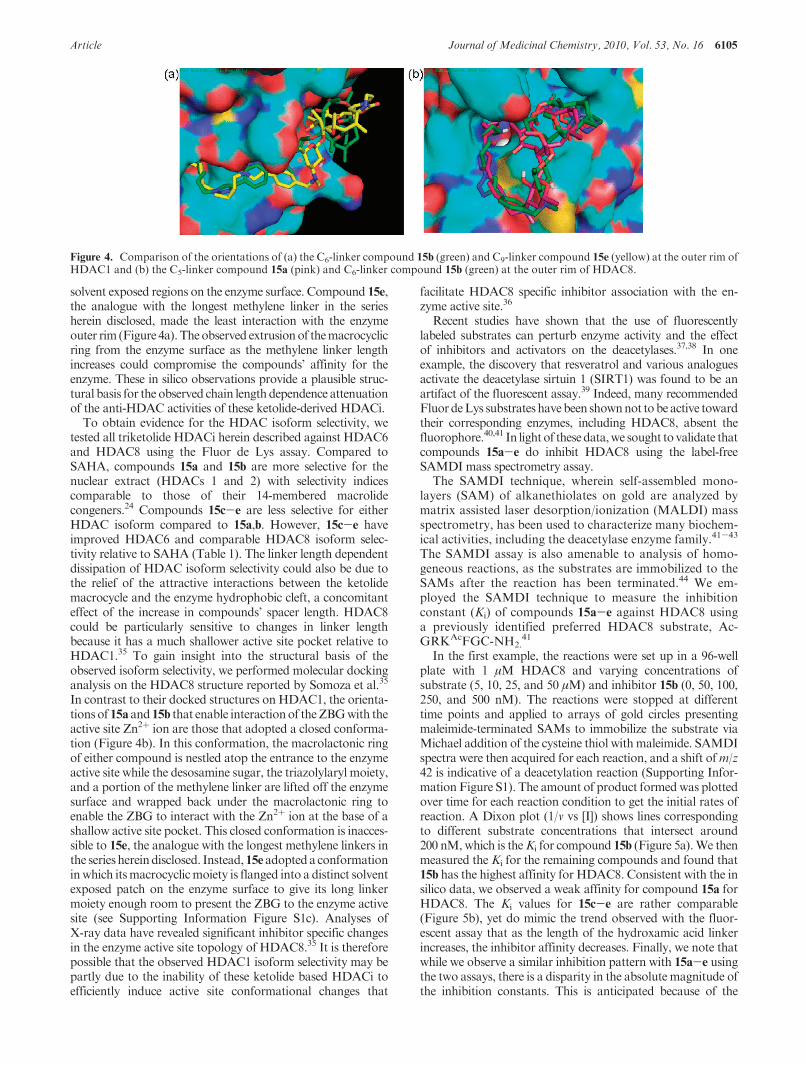
solvent exposed regions on the enzyme surface. Compound 15e,the analogue with the longest methylene linker in the seriesherein disclosed, made the least interaction with the enzymeouter rim (Figure 4a).Theobserved extrusionof themacrocyclicring from the enzyme surface as the methylene linker lengthincreases could compromise the compounds’ affinity for theenzyme. These in silico observations provide a plausible struc-tural basis for the observed chain length dependence attenuationof the anti-HDAC activities of these ketolide-derived HDACi.
To obtain evidence for the HDAC isoform selectivity, wetested all triketolide HDACi herein described against HDAC6and HDAC8 using the Fluor de Lys assay. Compared toSAHA, compounds 15a and 15b are more selective for thenuclear extract (HDACs 1 and 2) with selectivity indicescomparable to those of their 14-membered macrolidecongeners.24 Compounds 15c-e are less selective for eitherHDAC isoform compared to 15a,b. However, 15c-e haveimproved HDAC6 and comparable HDAC8 isoform selec-tivity relative to SAHA (Table 1). The linker length dependentdissipation of HDAC isoform selectivity could also be due tothe relief of the attractive interactions between the ketolidemacrocycle and the enzyme hydrophobic cleft, a concomitanteffect of the increase in compounds’ spacer length. HDAC8could be particularly sensitive to changes in linker lengthbecause it has a much shallower active site pocket relative toHDAC1.35 To gain insight into the structural basis of theobserved isoform selectivity, we performed molecular dockinganalysis on the HDAC8 structure reported by Somoza et al.35
In contrast to their docked structures on HDAC1, the orienta-tionsof 15a and15b that enable interactionof theZBGwith theactive site Zn2þ ion are those that adopted a closed conforma-tion (Figure 4b). In this conformation, the macrolactonic ringof either compound is nestled atop the entrance to the enzymeactive site while the desosamine sugar, the triazolylaryl moiety,and a portion of the methylene linker are lifted off the enzymesurface and wrapped back under the macrolactonic ring toenable the ZBG to interact with the Zn2þ ion at the base of ashallow active site pocket. This closed conformation is inacces-sible to 15e, the analogue with the longest methylene linkers inthe series hereindisclosed. Instead,15eadopteda conformationinwhich itsmacrocyclicmoiety is flanged into a distinct solventexposed patch on the enzyme surface to give its long linkermoiety enough room to present the ZBG to the enzyme activesite (see Supporting Information Figure S1c). Analyses ofX-ray data have revealed significant inhibitor specific changesin the enzyme active site topology of HDAC8.35 It is thereforepossible that the observed HDAC1 isoform selectivity may bepartly due to the inability of these ketolide based HDACi toefficiently induce active site conformational changes that
facilitate HDAC8 specific inhibitor association with the en-zyme active site.36
Recent studies have shown that the use of fluorescentlylabeled substrates can perturb enzyme activity and the effectof inhibitors and activators on the deacetylases.37,38 In oneexample, the discovery that resveratrol and various analoguesactivate the deacetylase sirtuin 1 (SIRT1) was found to be anartifact of the fluorescent assay.39 Indeed, many recommendedFluordeLys substrateshavebeen shownnot tobeactive towardtheir corresponding enzymes, including HDAC8, absent thefluorophore.40,41 In lightof thesedata,we sought tovalidate thatcompounds 15a-e do inhibit HDAC8 using the label-freeSAMDI mass spectrometry assay.
The SAMDI technique, wherein self-assembled mono-layers (SAM) of alkanethiolates on gold are analyzed bymatrix assisted laser desorption/ionization (MALDI) massspectrometry, has been used to characterize many biochem-ical activities, including the deacetylase enzyme family.41-43
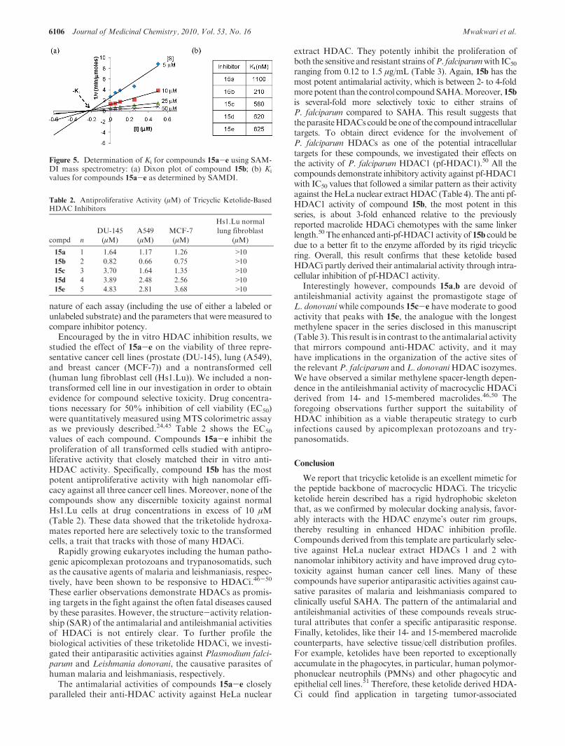
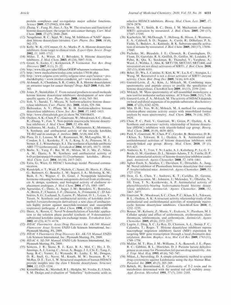
The SAMDI assay is also amenable to analysis of homo-geneous reactions, as the substrates are immobilized to theSAMs after the reaction has been terminated.44 We em-ployed the SAMDI technique to measure the inhibitionconstant (Ki) of compounds 15a-e against HDAC8 usinga previously identified preferred HDAC8 substrate, Ac-GRKAcFGC-NH2.
41
In the first example, the reactions were set up in a 96-wellplate with 1 μM HDAC8 and varying concentrations ofsubstrate (5, 10, 25, and 50 μM) and inhibitor 15b (0, 50, 100,250, and 500 nM). The reactions were stopped at differenttime points and applied to arrays of gold circles presentingmaleimide-terminated SAMs to immobilize the substrate viaMichael addition of the cysteine thiol withmaleimide. SAMDIspectra were then acquired for each reaction, and a shift ofm/z42 is indicative of a deacetylation reaction (Supporting Infor-mation Figure S1). The amount of product formedwas plottedover time for each reaction condition to get the initial rates ofreaction. A Dixon plot (1/v vs [I]) shows lines correspondingto different substrate concentrations that intersect around200 nM,which is theKi for compound 15b (Figure 5a).We thenmeasured the Ki for the remaining compounds and found that15b has the highest affinity for HDAC8. Consistent with the insilico data, we observed a weak affinity for compound 15a forHDAC8. The Ki values for 15c-e are rather comparable(Figure 5b), yet do mimic the trend observed with the fluor-escent assay that as the length of the hydroxamic acid linkerincreases, the inhibitor affinity decreases. Finally, we note thatwhile we observe a similar inhibition pattern with 15a-e usingthe two assays, there is a disparity in the absolutemagnitude ofthe inhibition constants. This is anticipated because of the
Figure 4. Comparison of the orientations of (a) the C6-linker compound 15b (green) and C9-linker compound 15e (yellow) at the outer rim ofHDAC1 and (b) the C5-linker compound 15a (pink) and C6-linker compound 15b (green) at the outer rim of HDAC8.
6106 Journal of Medicinal Chemistry, 2010, Vol. 53, No. 16 Mwakwari et al.
nature of each assay (including the use of either a labeled orunlabeled substrate) and the parameters that weremeasured tocompare inhibitor potency.
Encouraged by the in vitro HDAC inhibition results, westudied the effect of 15a-e on the viability of three repre-sentative cancer cell lines (prostate (DU-145), lung (A549),and breast cancer (MCF-7)) and a nontransformed cell(human lung fibroblast cell (Hs1.Lu)). We included a non-transformed cell line in our investigation in order to obtainevidence for compound selective toxicity. Drug concentra-tions necessary for 50% inhibition of cell viability (EC50)were quantitatively measured using MTS colorimetric assayas we previously described.24,45 Table 2 shows the EC50
values of each compound. Compounds 15a-e inhibit theproliferation of all transformed cells studied with antipro-liferative activity that closely matched their in vitro anti-HDAC activity. Specifically, compound 15b has the mostpotent antiproliferative activity with high nanomolar effi-cacy against all three cancer cell lines. Moreover, none of thecompounds show any discernible toxicity against normalHs1.Lu cells at drug concentrations in excess of 10 μM(Table 2). These data showed that the triketolide hydroxa-mates reported here are selectively toxic to the transformedcells, a trait that tracks with those of many HDACi.
Rapidly growing eukaryotes including the human patho-genic apicomplexan protozoans and trypanosomatids, suchas the causative agents of malaria and leishmaniasis, respec-tively, have been shown to be responsive to HDACi.46-50
These earlier observations demonstrate HDACs as promis-ing targets in the fight against the often fatal diseases causedby these parasites. However, the structure-activity relation-ship (SAR) of the antimalarial and antileishmanial activitiesof HDACi is not entirely clear. To further profile thebiological activities of these triketolide HDACi, we investi-gated their antiparasitic activities against Plasmodium falci-parum and Leishmania donovani, the causative parasites ofhuman malaria and leishmaniasis, respectively.
The antimalarial activities of compounds 15a-e closelyparalleled their anti-HDAC activity against HeLa nuclear
extract HDAC. They potently inhibit the proliferation ofboth the sensitive and resistant strains ofP. falciparumwith IC50
ranging from 0.12 to 1.5 μg/mL (Table 3). Again, 15b has themost potent antimalarial activity, which is between 2- to 4-foldmore potent than the control compoundSAHA.Moreover, 15bis several-fold more selectively toxic to either strains ofP. falciparum compared to SAHA. This result suggests thattheparasiteHDACscouldbeoneof the compound intracellulartargets. To obtain direct evidence for the involvement ofP. falciparum HDACs as one of the potential intracellulartargets for these compounds, we investigated their effects onthe activity of P. falciparum HDAC1 (pf-HDAC1).50 All thecompounds demonstrate inhibitory activity against pf-HDAC1with IC50 values that followed a similar pattern as their activityagainst the HeLa nuclear extract HDAC (Table 4). The anti pf-HDAC1 activity of compound 15b, the most potent in thisseries, is about 3-fold enhanced relative to the previouslyreported macrolide HDACi chemotypes with the same linkerlength.50 The enhanced anti-pf-HDAC1activity of 15b could bedue to a better fit to the enzyme afforded by its rigid tricyclicring. Overall, this result confirms that these ketolide basedHDACi partly derived their antimalarial activity through intra-cellular inhibition of pf-HDAC1 activity.
Interestingly however, compounds 15a,b are devoid ofantileishmanial activity against the promastigote stage ofL. donovaniwhile compounds 15c-e have moderate to goodactivity that peaks with 15e, the analogue with the longestmethylene spacer in the series disclosed in this manuscript(Table 3). This result is in contrast to the antimalarial activitythat mirrors compound anti-HDAC activity, and it mayhave implications in the organization of the active sites ofthe relevant P. falciparum and L. donovaniHDAC isozymes.We have observed a similar methylene spacer-length depen-dence in the antileishmanial activity of macrocyclic HDACiderived from 14- and 15-membered macrolides.46,50 Theforegoing observations further support the suitability ofHDAC inhibition as a viable therapeutic strategy to curbinfections caused by apicomplexan protozoans and try-panosomatids.
Conclusion
We report that tricyclic ketolide is an excellent mimetic forthe peptide backbone of macrocyclic HDACi. The tricyclicketolide herein described has a rigid hydrophobic skeletonthat, as we confirmed by molecular docking analysis, favor-ably interacts with the HDAC enzyme’s outer rim groups,thereby resulting in enhanced HDAC inhibition profile.Compounds derived from this template are particularly selec-tive against HeLa nuclear extract HDACs 1 and 2 withnanomolar inhibitory activity and have improved drug cyto-toxicity against human cancer cell lines. Many of thesecompounds have superior antiparasitic activities against cau-sative parasites of malaria and leishmaniasis compared toclinically useful SAHA. The pattern of the antimalarial andantileishmanial activities of these compounds reveals struc-tural attributes that confer a specific antiparasitic response.Finally, ketolides, like their 14- and 15-membered macrolidecounterparts, have selective tissue/cell distribution profiles.For example, ketolides have been reported to exceptionallyaccumulate in the phagocytes, in particular, human polymor-phonuclear neutrophils (PMNs) and other phagocytic andepithelial cell lines.51 Therefore, these ketolide derived HDA-Ci could find application in targeting tumor-associated
Figure 5. Determination of Ki for compounds 15a-e using SAM-DI mass spectrometry: (a) Dixon plot of compound 15b; (b) Ki
values for compounds 15a-e as determined by SAMDI.
Table 2. Antiproliferative Activity (μM) of Tricyclic Ketolide-BasedHDAC Inhibitors
compd n
DU-145
(μM)
A549
(μM)
MCF-7
(μM)
Hs1.Lu normal
lung fibroblast
(μM)
15a 1 1.64 1.17 1.26 >10
15b 2 0.82 0.66 0.75 >10
15c 3 3.70 1.64 1.35 >10
15d 4 3.89 2.48 2.56 >10
15e 5 4.83 2.81 3.68 >10
Article Journal of Medicinal Chemistry, 2010, Vol. 53, No. 16 6107
macrophages and further clarify the roles of HDACi in theinhibition of macrophage migration inhibitory factor (MIF)gene, a phenomenon that has been suggested to be an essentialpart of their antitumorigenic effects.52
Experimental Section
Materials and Methods. All commercially available startingmaterials were used without further purification. Clarithromy-cin 1 was purchased from Greenfield Chemical. 4-Ethynylben-zyl alcohol, ethyl 6-bromohexanoate, ethyl 7-bromoheptanoate,8-bromooctanoic acid, and methyl 10-bromodecanoate werepurchased from Sigma Aldrich. 9-Bromononanoic acid waspurchased from Karl Industries Inc. Reaction solvents wereeither high performance liquid chromatography (HPLC) gradeor American Chemical Society (ACS) grade and used withoutfurther purification. HDAC fluorimetric assay kit and recom-binant HDACs were procured from Enzo Life Sciences, BIO-MOL International, PA.Analtech silica gel plates (60 F254) wereused for analytical TLC, and Analtech preparative TLC plates(UV 254, 2000 μm) were used for purification. UV light andanisaldehyde/iodine stain were used to visualize the spots. Silicagel (200-400 mesh) was used in column chromatography.Nuclear magnetic resonance (NMR) spectra were recorded ona Varian-Gemini 400 magnetic resonance spectrometer. 1HNMR spectra were recorded in parts per million (ppm) relativeto the peak of CDCl3, (7.24 ppm). 13C spectra were recordedrelative to the central peak of the CDCl3 triplet (77.0 ppm) andwere recordedwith complete heterodecoupling.High-resolutionmass spectra were recorded at the Georgia Institute of Techno-logy mass spectrometry facility in Atlanta. Melting points wererecorded uncorrected on a MEL-TEMP II apparatus. HPLCassays were performed on a Beckman Coulter instrument usinga Phenomenex RP C-18 column (250 mm � 4.6 mm), elutingwith solvent A (0.1% formic acid/acetonitrile) and solvent B(0.1% formic acid/water) at a gradient of 5-50% over 30 min,with detection at 251 nm and a flow rate of 1 mL/min. Sampleconcentrations were 250 μM, injecting 5 μL. 30-Desmethylclar-ithromycin 2, 4-ethynylbenzylclarithromycin 4, descladinose-4-ethynylbenzylclarithromycin 5, and azidoalkylhydroxamic acid
(13a,b) were synthesized as we previously reported.24 Azido-O-trityl alkylhydroxamate derivatives (12a-e) were synthesized byadapting literature protocol.46
20-O-Acetyldescladinose-4-ethynylbenzylclarithromycin (6).To a solution of descladinose-4-ethynylbenzylclarithromycin 5(3.80 g, 5.50 mmol) in acetone (20 mL) was added Ac2O (0.62 g,6.00 mmol), and the resulting mixture was stirred at 40 �C for36 h. The reaction mixture was diluted with EtOAc (100 mL)andwashedwith aqueousNaHCO3 (70mL) and saturated brine(70 mL). Purification on silica column (6:1 CH2Cl2/acetone)afforded 2.8 g (70%) of the title compound as a yellowish solid.1H NMR (CDCl3, 400 MHz) δ 0.80 (3H, t, J = 7.2 Hz), 0.90(3H, d, J=7.2Hz), 1.08-1.47 (12H,m), 1.58 (6H, s), 1.63-2.05(7H, m), 2.08 (3H, s), 2.16 (3H, s), 2.42-2.80 (3H, m), 2.92 (3H,s), 2.94-3.00 (1H,m), 3.03-3.68 (3H,m), 3.79 (2H, s), 3.94 (1H,s), 4.08 (1H,m), 4.54 (1H, d, J=8.0Hz), 4.80 (1H,m), 5.15 (1H,dd, J=11.6, 2.4 Hz), 7.17 (2H, d, J=8.4 Hz), 7.38 (2H, d, J=8.0 Hz); 13C NMR (CDCl3, 100 MHz) δ 8.0, 10.7, 12.8, 15.5,16.4, 18.1, 19.5, 21.3, 21.5, 21.6, 31.1, 31.9, 36.0, 37.0, 37.5, 38.7,44.3, 45.7, 50.0, 58.4, 62.5, 68.9, 69.8, 71.6, 74.4, 76.9, 77.0, 77.8,78.1, 81.3, 83.9, 99.9, 120.7, 128.4, 132.2, 141.1, 170.1, 174.9,221.2; HRMS (FAB, mnba) calcd for [C40H61NO11 þ H]þ
732.4323, found 732.4311.2-Oxo-20-O-acetyldescladinose-4-ethynylbenzylclarithromy-
cin (7). Methyl sulfide (1.42 g, 22.80 mmol) was added to amixture of N-chlorosuccinimide (2.61 g, 19.50 mmol) andCH2Cl2 (10 mL) while maintaining the temperature at -15 �C.Compound 6 (10.0 g, 13.7 mmol) dissolved in CH2Cl2 (50 mL)was added to the reaction flask, followed by Et3N (1.56 g, 15.4mmol) and stirred at-15 �C for 4.5 h. The reaction mixture waspoured into EtOAc (350 mL) and 0.5 N aqueous NaOH (250mL). The organic layer was separated, washed with saturatedbrine (250mL), and dried overNa2SO4. Solvent was evaporatedoff and the crude was purified on silica column (2:3:0.1 EtOAc/hexane/Et3N) to afford 9.54 g (96%) of the title compound as anoff-white solid. 1H NMR (CDCl3, 400 MHz) δ 0.80-0.86 (6H,m), 1.09-1.57 (12H, m), 1.58 (6H, s), 1.62-2.02 (7H, m), 2.05(3H, s), 2.15 (3H, s), 2.44-2.80 (2H, m), 2.92 (3H, s), 2.95-3.00(1H, m), 3.05-3.66 (4H, m), 3.80 (1H, q, J=14.0, 6.8 Hz), 3.89(1H, s), 4.12 (1H, m), 4.38 (1H, d, J= 8.0 Hz), 4.79-4.83 (1H,m), 5.14 (1H, dd, J=11.2, 2.0Hz), 7.16 (2H, d, J=7.6Hz), 7.38(2H, d, J= 7.6 Hz); 13C NMR (CDCl3, 100 MHz) δ 10.8, 12.4,14.4, 14.5, 16.5, 17.9, 19.6, 21.3, 21.5, 31.3, 36.9, 37.6, 39.2, 45.1,46.3, 49.7, 51.1, 58.5, 62.5, 69.2, 69.6, 71.5, 74.1, 77.0, 77.1, 77.2,78.1, 83.9, 101.1, 120.7, 128.5, 132.2, 140.9, 169.6, 170.0, 205.7,221.1; HRMS (FAB, mnba) calcd for [C40H59NO11 þ H]þ
730.4166, found 730.4132.12-Carbamoylimidazole Ketolide (8). To a suspension of 7
(1.50 g, 2.06mmol) in amixture of anhydrous THF (20mL) andanhydrous DMF (7 mL) was added CDI (1.30 g, 8.20 mmol)
Table 3. In Vitro Antileishmanial (μg/mL) and Antimalarial (μg/mL) Activities of Tricyclic Ketolide-Based HDAC Inhibitor Derivativesa
antileishmanial activityb antimalarial activityc
compd n
IC50
(μg/mL) IC90(μg/mL)
Plasmodium
falciparum (D6 clone)
IC50 (μg/mL)
Plasmodium
falciparum (W2 clone)
IC50 (μg/mL)
cytotoxicity
(VERO)c
IC50(μg/mL) SI D6 (W2)
15a 1 NA NA 0.897 ( 0.198 1.789 ( 0.254 NC >5.3 (>2.68)
15b 2 NA NA 0.137 ( 0.027 0.133 ( 0.027 NC >35.0 (>36.0)
15c 3 19.7 ( 2.4 35.8 ( 4.4 1.317 ( 0.231 1.475 ( 0.244 NC >3.6 (>3.3)
15d 4 14.9 ( 1.7 32.3 ( 3.1 1.296 ( 0.129 1.376 ( 0.079 NC >3.7 (>3.5)
15e 5 4.8 ( 0.5 23.7 ( 2.4 1.195 ( 0.058 0.897 ( 0.025 NC >4.0 (>5.3)
chloroquine NT NT 0.012 ( 0.002 0.147 ( 0.018 NT NT
artemisinin NT NT 0.0066 ( 0.0007 0.0091 ( 0.0011 NT NT
pentamidine 1.17 ( 0.25 2.45 ( 0.34 NT NT NT NT
amphotericine B 0.079 ( 0.012 0.395 ( 0.054 NT NT NT NT
SAHA 21.5 ( 3.51 51.7 ( 8.1 0.279 ( 0.077 0.392 ( 0.045 1.375 ( 0.255 4.9 (3.5)aNA=not active up to highest concentration tested. NC= not cytotoxic up to the highest concentration tested. NT= not tested. SI = selectivity
index (IC50(Vero)/IC50).bMaximum tested concentration = 40 μg/mL. cMaximun tested concentration = 4.8 μg/mL.
Table 4. In Vitro Inhibition of pf-HDAC1 by Tricyclic Ketolide-BasedHDACi
compd n IC50 (nM)
15a 1 36 ( 2.9
15b 2 10 ( 0.5
15c 3 40 ( 1.8
15d 4 290 ( 16
15e 5 304 ( 17
SAHA 130 ( 9.2
6108 Journal of Medicinal Chemistry, 2010, Vol. 53, No. 16 Mwakwari et al.
followed by a solution of 1.0 M NaN(TMS)2 in THF (2.60 mL,2.60 mmol) over 75 min, and the resulting mixture was stirred atroom temperature for 24 h. The reaction mixture was dilutedwith EtOAc (50 mL), washed with aqueous NaHCO3 (20 mL)and saturated brine (20 mL), and dried over Na2SO4. Solventwas evaporated off and the crude was purified on silica column(3:2:0.1 EtOAc/hexane/Et3N) to yield 52% (0.86 g) of the titlecompound. 1H NMR (CDCl3, 400 MHz) δ 0.91 (3H, t, 7.6 Hz),1.09 (3H, d, J = 7.2 Hz), 1.14-1.50 (9H, m), 1.80 (3H, s), 1.83(3H, s), 1.56-1.94 (7H, m), 2.00 (3H, s), 2.05 (3H, s), 2.12 (3H,s), 2.64-2.80 (1H,m), 3.02 (3H, s), 2.95-3.70 (4H,m), 3.73 (1H,q, J = 14.0, 6.8 Hz), 4.10-4.06 (2H, m), 4.30 (1H, d, J = 7.6Hz), 4.77-4.80 (1H, m), 5.68 (1H, dd, J = 11.2, 2.0 Hz), 6.77(1H, s), 7.03 (1H, s), 7.16 (2H, d, J=7.6Hz), 7.36 (3H, m), 8.06(1H, s); 13C NMR (CDCl3, 100 MHz) δ 10.7, 13.5, 14.4, 15.2,19.1, 20.4, 21.1, 21.2, 21.5, 22.8, 31.2, 36.8, 47.6, 50.5, 51.2, 58.5,60.6, 62.8, 69.3, 71.6, 78.7, 83.9, 84.7, 117.3, 120.7, 128.4, 131.1,132.2, 137.3, 138.8, 140.9, 146.2, 169.1, 169.8; HRMS (FAB,mnba) calcd for [C44H59N3O11þH]þ 806.4228, found 806.4210.
4-Ethynylbenzyltricyclic Ketolide (9). Protocol A. To a solu-tion of 8 (0.60 g, 0. 74mmol) inCH3CN (10mL) andH2O (1mL)was added ethylenediamine (0.44 g, 7.40mmol), and themixturewas heated in a sealed tube at 50-55 �C for 24 h. Solvent wasevaporated off and the reaction mixture partitioned betweenH2O (15 mL) and CH2Cl2 (20 mL) and separated. The organiclayer was washed with saturated brine (20 mL), dried overNa2SO4, and solvent was evaporated off in vacuo. The crudeproduct was dissolved in anhydrousMeOH (15 mL) and heatedat 90 �C in a sealed tube for 24 h.MeOHwas evaporated and theproduct purified on silica column (EtOAc/Et3N 12:0.1) to give0.27 g (48%) of the intermediate product. EtOH (6 mL) andAcOH (0.043 g, 0.71mmol) were added to the pure intermediateproduct (0.27 g, 0.34mmol), and themixturewas heated at 95 �Cin a sealed tube for 20 h. The reaction mixture was suspended indilute NH4OH (10 mL) and CH2Cl2 (15 mL) and the organiclayer separated, washed with saturated brine (10 mL), and driedover Na2SO4. Purification on silica column (EtOAc/MeOH/Et3N 10:0.5:0.05) afforded 0.24 g (92%), 44% overall yield ofcompound 9.
Protocol B: Reductive Amination. A solution of 11 (0.20 g,0.32 mmol) and 4-ethynylbenzaldehyde (0.21 g, 1.60 mmol) inMeOH (4mL) and acetic acid (37 μL, 0.64mmol) was stirred for30 min at room temperature. Borane-pyridine complex (80 μL,0.64 mmol) was added and the mixture stirred for 5 h. Themixture was diluted with EtOAc (20 mL) and washed withsaturated NaHCO3 (20 mL) and saturated brine (20 mL). Theorganic layer was dried over Na2SO4, concentrated in vacuo,and purified by preparative TLC (12:1 CH2Cl2/MeOH) toafford 0.18 g (78%) of compound 9 as a creamish solid. 1HNMR (CDCl3, 400MHz) δ 0.80 (3H, t, J=7.2Hz), 1.00 (3H, d,J = 6.8 Hz), 1.14-1.55 (12H, m), 1.30 (3H, s), 1.43 (3H, s),1.60-1.96 (6H,m), 2.12 (3H, s), 2.45-2. 95 (6H,m), 3.03 (3H, s)3.23-3.43 (4H, m), 3.64-3.78 (4H, m), 3. 94 (1H, m), 4.16 (1H,d, J = 8.4 Hz), 4.25 (1H, d, J= 7.6 Hz), 4.90 (1H, d, J = 10.0Hz), 7.18 (2H, d, J = 8.0 Hz), 7.39 (2H, d, J = 7.2 Hz); 13CNMR (CDCl3, 100 MHz) δ 10.6, 11.1, 13.0, 14.6, 16.6, 19.3,19.8, 21.4, 22.3, 29.7, 36.5, 37.1, 38.7, 42.5, 43.0, 48.3, 49.3, 49.7,51.4, 57.6, 60.1, 65.6, 69.7, 70.5, 76.7, 78.7, 79.4, 81.7, 83.6,104.0, 121.0, 128.8, 132.4, 140.0, 156.3, 169.8, 181.5, 204.4;HRMS (FAB, mnba) calcd for [C41H59N3O9 þ H]þ 738.4330,found 738.4332.
Representative Procedure for the Synthesis of O-Trityl Pro-
tected Tricyclic Ketolide Hydroxamate. Tricyclic Ketolide-N-
benzyltriazolyl-O-tritylhexahydroxamate (14a). 4-Ethynylbenzyl-tricyclic ketolide 9 (0.10 g, 0.14 mmol) and 6-azido-O-tritylhex-ahydroxamate 12a (0.06 g, 0.14 mmol) were dissolved in anhy-drous THF (7 mL) and stirred under argon at room temper-ature. Copper(I) iodide (0.012 g, 0.063 mmol) and Hunig’s base(0.6 mL) were added to the reaction mixture, and stirringcontinued for 24 h. The reaction mixture was diluted with 1:4
NH4OH/saturated NH4Cl (50 mL) and extracted with 10%MeOH/CH2Cl2 (3� 30mL). The organic layers were combined,dried over Na2SO4, and concentrated in vacuo. The crude pro-duct was purified by preparative TLC (silica, 12:1:0.1 CH2Cl2/MeOH/conc NH4OH) to give 127 mg (78%) of 14a as a yellow-ish solid. 1HNMR(CDCl3, 400MHz)δ 0.85 (3H, t, J=7.6Hz),1.1 (3H, d, J = 6.8 Hz), 1.20-1.39 (17H, m), 1.46-2.00 (15H,m), 2.19 (3H, s), 2.58-2.80 (6H, m), 2.84-3.00 (1H, m),3.02-3.17 (1H, m), 3.31-3.60 (3H, m), 3.64-3.87 (5H, m),3.96-4.0 (1H, m), 4.22 (1H, d, J=8.4 Hz), 4.29-4.32 (3H, m),4.93 (1H, dd, J = 10.0, 1.6 Hz), 7.23-7.45 (17H, m), 7.71 (1H,s), 7.79 (2H, d, J=8.0Hz); 13CNMR(CDCl3, 100MHz)δ 10.7,11.1, 13.1, 14.6, 16.7, 19.3, 19.9, 21.4, 22.3, 22.8, 26.0, 29.6, 30.2,31.0, 36.5, 37.0, 38.8, 42.5, 43.0, 48.3, 49.3, 49.7, 50.2, 51.4, 53.7,57.7, 60.1, 65.6, 69.8, 70.5, 76.7, 78.7, 79.4, 81.8, 93.1, 104.1,119.7, 126.0, 128.2, 128.4, 129.2, 129.4, 130.0, 138.9, 141.2,147.6, 156.3, 169.8, 177.0, 181.5, 204.5; HRMS (ESI) calcd for[C66H85N7O11 þ H]þ 1152.6379, found 1152.6367.
Tricyclic Ketolide-N-benzyltriazolyl-O-tritylheptahydroxa-mate (14b).Reaction of 4-ethynylbenzyltricyclic ketolide 9 (0.10g, 0.14 mmol) and 7-azido-O-tritylheptahydroxamate 12b (0.06g, 0.14 mmol) within 24 h as described for the synthesis of 14agave 133 mg (83%) of 14b as a yellowish solid. 1H NMR(CDCl3, 400 MHz) δ 0.85 (3H, t, J = 7.6 Hz), 1.06 (3H, d, J= 6.8 Hz), 1.20-1.42 (19H, m), 1.45-1.98 (15H, m), 2.20 (3H,s), 2.57-2.79 (6H, m), 2.85-3.00 (1H, m), 3.03-3.17 (1H, m),3.30-3.57 (3H, m), 3.72-3.83 (5H, m), 3.96-4.00 (1H, m), 4.22(1H, d, J=8.8 Hz), 4.29-4.35 (3H, m), 4.95 (1H, dd, J=10.4,2.4 Hz), 7.22-7.45 (17H, m), 7.71 (1H, s), 7.78 (2H, d, J = 8.4Hz); 13C NMR (CDCl3, 100MHz) δ 10.7, 11.1, 13.1, 14.6, 16.6,19.3, 19.9, 21.4, 22.3, 26.3, 29.6, 30.2, 36.5, 37.0, 38.8, 42.5, 43.0,48.3, 49.3, 49.7, 50.4, 51.4, 53.7, 57.7, 60.1, 65.5, 69.7, 70.5, 76.7,78.7, 79.4, 81.8, 92.8, 104.1, 119.7, 126.0, 128.4, 129.2, 129.4,130.0, 132.3, 138.9, 141.3, 147.6, 156.3, 169.8, 177.6, 182.5,204.5; HRMS (ESI) calcd for [C67H87N7O11 þ H]þ 1166.6536,found 1166.6478.
Tricyclic Ketolide-N-benzyltriazolyl-O-trityloctahydroxamate
(14c). Reaction of 4-ethynylbenzyltricyclic ketolide 9 (0.08 g,0.10 mmol) and 8-azido-O-trityloctahydroxamate 12c (0.05 g,0.10mmol) in anhydrousTHF (5mL)within 22h asdescribed forthe synthesis of 14a gave 94mg (79%) of 14c as a yellowish solid.1HNMR(CDCl3, 400MHz) δ 0.83 (3H, t, J=7.2Hz), 1.04 (3H,d, J = 7.2 Hz), 1.15-1.41 (21H, m), 1.45-1.95 (15H, m), 2.18(3H, s), 2.55-2.73 (6H, m), 2.87-2.97 (1H, m), 3.03-3.10 (1H,m), 3.28-3.57 (3H, m), 3.70-3.82 (5H, m), 3.93-3.97 (1H, m),4.21 (1H, d, J = 8.4 Hz), 4.27-4.34 (3H, m), 4.94 (1H, dd, J =12.8, 2.4 Hz), 7.25-7.42 (17H, m), 7.71 (1H, s), 7.76 (2H, d, J=8.0 Hz); 13C NMR (CDCl3, 100 MHz) δ 10.7, 11.1, 13.1, 14.6,16.6, 19.3, 19.9, 21.4, 22.3, 26.4, 28.8, 29.6, 30.4, 36.6, 37.0, 38.8,42.5, 42.9, 48.3, 49.3, 49.7, 50.6, 51.4, 53.7, 57.7, 60.1, 65.5, 69.8,70.5, 76.7, 78.7, 79.4, 81.8, 93.8, 104.1, 119.6, 126.0, 127.7, 128.3,128.8, 129.2, 129.4, 130.0, 132.7, 138.9, 141.3, 147.7, 156.3, 169.8,177.0, 182.4, 204.4; HRMS (ESI) calcd for [C68H89N7O11 þH]þ
1180.6692, found 1180.6637.Tricyclic Ketolide-N-benzyltriazolyl-O-tritylnonahydroxa-
mate (14d).Reaction of 4-ethynylbenzyltricyclic ketolide 9 (0.08 g,0.10 mmol) and 9-azido-O-tritylnonahydroxamate 12d (0.05 g,0.10 mmol) in anhydrous THF (5 mL) within 20 h as describedfor the synthesis of 14a gave 93 mg (76%) of 14d as a yellowishsolid. 1H NMR (CDCl3, 400 MHz) δ 0.85 (3H, t, J = 8.0 Hz),1.05 (3H, d, J= 6.8 Hz), 1.16-1.44 (23H, m), 1.45-1.96 (15H,m), 2.20 (3H, m), 2.57-2.78 (6H, m), 2.90-2.99 (1H, m),3.02-3.12 (1H, m), 3.30-3.57 (3H, m), 3.72-3.84 (5H, m),3.96-4.00 (1H,m), 4.29 (1H, d, J=8.4Hz), 4.31 (1H, d, J=7.2Hz), 4.37 (2H, t, J = 7.2 Hz), 4.95 (1H, dd, J = 12.8, 2.4 Hz),7.19-7.45 (17H, m), 7.72 (1H, s), 7.78 (2H, d, J= 8.0 Hz); 13CNMR (CDCl3, 100 MHz) δ 10.7, 11.1, 13.1, 14.6, 16.6, 19.3,19.9, 21.4, 22.3, 26.6, 28.9, 29.2, 29.6, 29.9, 30.5, 36.5, 37.0, 37.1,38.8, 42.5, 43.0, 48.3, 49.3, 49.7, 50.6, 51.4, 57.7, 60.1, 65.5, 65.6,69.7, 70.5, 76.7, 78.7, 79.4, 81.8, 94.3, 104.1, 119.6, 126.0, 128.3,
Article Journal of Medicinal Chemistry, 2010, Vol. 53, No. 16 6109
128.8, 129.0, 129.2, 129.4, 130.0, 132.3, 138.9, 141.3, 147.6, 156.3,169.8, 178.0, 181.5, 204.5; HRMS (ESI) calcd for [C69H91N7-O11 þ H]þ 1194.6849, found 1194.6838.
Tricyclic Ketolide-N-benzyltriazolyl-O-trityldecahydroxamate
(14e). Reaction of 4-ethynylbenzyltricyclic ketolide 9 (0.08 g,0.10 mmol) and 10-azido-O-trityldecahydroxamate 12e (0.05 g,0.10mmol) in anhydrous THF (5mL) within 20 h as described forthe synthesis of 14a gave 98 mg (80%) of 14e as a yellowish solid.1HNMR (CDCl3, 400MHz) δ 0.83 (3H, t, J=6.8Hz), 1.03 (3H,d, J=7.2Hz), 1.15-1.40 (25H,m), 1.44-1.96 (15H,m), 2.17 (3H,s), 2.57-2.78 (6H, m), 2.87-2.95 (1H, m), 3.03-3.08 (1H, m),3.28-3.53 (3H, m), 3.70-3.82 (5H, m), 3.92-3.96 (1H, m), 4.20(1H, d, J=8.4 Hz), 4.29 (1H, d, J=6.8 Hz), 4.33 (2H, t, J=7.2Hz), 4.93 (1H, d, J=10.0 Hz), 7.26-7.43 (17H, m), 7.73 (1H, s),7.76 (2H, d, J = 8.0 Hz); 13C NMR (CDCl3, 100 MHz) δ 10.7,11.1, 13.1, 14.6, 16.6, 19.3, 19.8, 21.4, 22.3, 26.6, 29.1, 29.3, 29.6,29.9, 30.5, 36.6, 37.0, 38.8, 42.5, 43.0, 48.3, 49.3, 49.7, 50.6, 51.4,57.7, 60.1, 65.5, 69.7, 70.5, 76.7, 78.7, 79.4, 81.8, 93.4, 104.1,199.6, 126.0, 128.3, 129.2, 129.4, 130.0, 138,9, 147.6, 156.3, 169.8,177.6, 182.0, 204.5; HRMS (ESI) calcd for [C70H93N7O11 þ H]þ
1208.7005, found 1208.6888.Tricyclic Ketolide-N-benzyltriazolylhexahydroxamic Acid
(15a). Method A. To a solution of 14a (0.12 g, 0.11 mmol) inmethylene chloride (1 mL) at 0 �C was added thioanisole (0.2mL) and TFA (0.2mL) dropwise. Stirring was continued at 0 �Cfor 2 h, after which TLC analysis indicated complete consump-tion of the starting material. Excess TFA and solvent wereevaporated off, and the residue was immediately placed in anice bath followed by addition of PBS buffer (10 mL). SaturatedNaHCO3 was added dropwise until the pH was neutral. Extrac-tion with 20% MeOH/CH2Cl2 (10 mL � 3), drying overNa2SO4, and evaporation of solvent afforded crude productwhich was then purified by preparative TLC (silica, 12:1:0.1CH2Cl2/MeOH/conc NH4OH) to afford 47 mg (50%) of 15a asa yellow-white solid.
MethodB.To amixture of 14a (0.14 g, 0.12mmol) in CH2Cl2/MeOH (2 mL/2 mL) was added BF3 3OEt2 (0.03 g, 0.24 mmol),and themixturewas stirred at room temperature for 45min.DiluteNaHCO3 was added until pH 8 was obtained (approximately 20mL), and the suspension was extracted with 10%MeOH/CH2Cl2(10mL�x 4), dried over Na2SO4, and concentrated in vacuo. Thecrude product was purified by preparative TLC (silica, 12:1:0.1CH2Cl2/MeOH/conc NH4OH) to give 64 mg (60%) of 15a as ayellow-white solid.
Method C. 4-Ethynylbenzyltricyclic ketolide 9 (0.05 g,0.07 mmol) and 6-azidohexahydroxamic acid24 13a (0.03 g,0.15mmol) were dissolved in anhydrous THF (7mL) with stirringunder argon at room temperature. Copper(I) iodide (0.01 g,0.04 mmol) and Hunig’s base (0.5 mL) were added to the reactionmixture, and stirring continued for 12 h. The reactionmixture wasdiluted with 10% MeOH/CH2Cl2 (20 mL) and washed with 1:4NH4OH/saturated NH4Cl (3 � 10 mL) and saturated NH4Cl(10 mL). The organic layer was dried over Na2SO4 and concen-trated in vacuo. The crude product was purified by preparativeTLC (silica, 12:1:0.1 CH2Cl2/MeOH/concNH4OH) to give 20mg(34%) of 15a as a brown-white solid. Mp 127-130 �C; retentiontime 20.167 min; 1H NMR (CDCl3, 400 MHz) δ 0.85 (3H, t, J =8.0 Hz), 1.05 (3H, d, J= 7.2 Hz), 1.20-2.00 (30H, m), 2.13-2.18(2H, m), 2.20 (3H, s), 2.58-2.72 (6H, m), 2.92-2.98 (1H, m),3.05-3.09 (1H, m), 3.30-3.55 (3H, m), 3.70-3.84 (5H, m),3.95-3.98 (1H, m), 4.22 (1H, d, J = 8.0 Hz), 4.31-4.38 (3H,m), 4.95 (1H, d, J = 8.8 Hz), 7.33 (2H, d, J = 8.0 Hz), 7.77 (3H,m); 13C NMR (CDCl3, 100 MHz) δ 10.6, 11.1, 13.1, 14.6, 16.5,19.3, 19.8, 21.4, 22.3, 24.7, 25.9, 29.7, 29.9, 30.0, 36.6, 37.1, 38.8,42.5, 42.8, 49.4, 49.6, 50.3, 51.4, 53.7, 57.7, 60.1, 65.5, 69.7, 70.5,76.7, 78.7, 79.3, 81.8, 104.1, 120.1, 126.0, 129.5, 129.7, 139.1, 147.7,156.3, 169.8, 171.1, 182.3, 204.5; HRMS (ESI) calcd for[C47H71N7O11 þ H]þ 910.5284, found 910.5279.
Tricyclic Ketolide-N-benzyltriazolylheptahydroxamic Acid
(15b). The reaction of 14b (0.13 g, 0.11 mmol) in CH2Cl2
(1 mL), thioanisole (0.3 mL), and TFA (0.3 mL), according tomethodA, for 3 h afforded 56mg (55%) of 15b as a yellow-whitesolid.
Similarly, the reaction of 14b (0.15 g, 0.18 mmol) andBF3 3OEt2 (0.04 g, 0.26mmol), according tomethod B, afforded65 mg (55%) of 15b as a yellow-white solid.
Also, the reaction of 4-ethynylbenzyltricyclic ketolide 9 (0.05 g,0.06 mmol) and 7-azidoheptahydroxamic acid 13b (0.02 g, 0.11mmol) within 12 h, according, to method C, gave 22 mg (38%) of15b as a brown-white solid.Mp 128-131 �C; retention time 21.233min; 1HNMR (CDCl3, 400MHz) δ 0.83 (3H, t, J= 7.6 Hz), 1.03(3H, d, J= 6.4Hz), 1.18-1.90 (32H,m), 2.10-2.13 (2H, m), 2.17(3H, s), 2.58-2.73 (6H, m), 2.93-2.98 (1H, m), 3.03-3.07 (1H,m), 3.28-3.60 (3H,m), 3.70-3.81 (5H,m), 3.98 (1H,m), 4.20 (1H,d, J= 8.4Hz), 4.29-4.38 (3H,m), 4.93 (1H, d, J= 10.4Hz), 7.30(2H, d, J = 7.6 Hz), 7.75 (2H, d, J = 7.6 Hz), 7.78 (1H, s); 13CNMR (CDCl3, 100 MHz) δ 10.6, 11.1, 13.1, 14.6, 16.5, 19.3, 19.8,21.4, 22.3, 25.2, 26.0, 28.2, 29.7, 29.9, 30.1, 36.6, 37.0, 38.8, 42.5,42.7, 48.2, 49.3, 49.5, 50.4, 51.4, 53.7, 57.7, 60.1, 65.5, 69.7, 70.5,76.7, 78.7, 79.3, 81.8, 104.0, 120.0, 126.0, 129.4, 129.8, 139.1, 147.7,156.3, 169.8, 171.2, 182.4, 204.5; HRMS (ESI) calcd for [C48H73-N7O11 þ H]þ 924.5440, found 924.5422.
Tricyclic Ketolide-N-benzyltriazolyloctahydroxamic Acid (15c).The reaction of 14c (0.09 g, 0.08 mmol) in CH2Cl2 (1 mL),thioanisole (0.2 mL), and TFA (0.2 mL), according to methodA, afforded 37 mg (51%) of 15c as a yellow-white solid. Mp113-115 �C; retention time 22.533 min; 1H NMR (CDCl3, 400MHz) δ 0.83 (3H, t, J = 6.4 Hz), 1.03 (3H, d, J = 5.6 Hz),1.18-1.60 (21H, m), 1.66-2.1 (15H, m), 2.18 (3H, s), 2.56-2.70(6H, m), 2.90-2.98 (1H, m), 3.02-3.07 (1H, m), 3.28-3.58 (3H,m), 3.70-3.81 (5H, m), 3.94-3.97 (1H, m), 4.20 (1H, d, J = 7.2Hz), 4.28 (1H, d, J= 6.0 Hz), 4.35 (2H, m), 4.93 (1H, d, J= 10.4Hz), 7.30 (2H, d, J= 7.2Hz), 7.73-7.76 (3H,m); 13CNMR (CD-Cl3, 100 MHz) δ 10.6, 11.1, 13.1, 14.4, 14.6, 16.5, 19.3, 19.8, 21.3,21.4, 22.3, 25.2, 26.1, 28.3, 29.7, 29.9, 30.2, 32.9, 36.6, 37.1, 38.8,42.5, 42.7, 48.2, 49.4, 49.5, 50.5, 51.4, 57.7, 60.1, 60.6, 65.5, 69.7,70.5, 76.7, 78.7, 79.3, 81.8, 104.0, 119.9, 126.0, 129.5, 129.8, 139.0,147.7, 156.3, 169.8, 171.7, 182.4, 204.5; HRMS (ESI) calcd for[C49H75N7O11 þ H]þ 938.5597, found 938.5556.
Tricyclic Ketolide-N-benzyltriazolylnonahydroxamic Acid
(15d). The reaction of 14d (0.09 g, 0.08 mmol) in CH2Cl2 (1mL), thioanisole (0.2 mL), and TFA (0.2 mL), according tomethod A, afforded 43 mg (60%) of 15d as a yellow-white solid.Mp 112-115 �C; retention time 23.983 min; 1H NMR (CDCl3,400 MHz) δ 0.83 (3H, t, J = 7.2 Hz), 1.03 (3H, d, J = 7.2 Hz),1.17-1.60 (23H, m), 1.66-2.14 (15H, m), 2.17 (3H, s),2.58-2.73 (6H, m), 2.87-2.95 (1H, m), 3.00-3.07 (1H, m),3.27-3.55 (3H, m), 3.70-3.82 (5H, m), 3.93-3.97 (1H, m), 4.20(1H, d, J= 8.8Hz), 4.28 (1H, d, J= 7.2Hz), 4.35 (2H, t, J= 6.8Hz), 4.92 (1H, dd, J = 10.0, 1.2 Hz), 7.30 (2H, d, J = 8.0 Hz),7.75 (3H, m); 13C NMR (CDCl3, 100 MHz) δ 10.7, 11.1, 13.1,14.6, 16.5, 19.3, 19.8, 21.4, 22.3, 25.4, 26.3, 28.6, 28.8, 28.9, 29.6,29.9, 30.3, 32.9, 36.6, 37.1, 38.8, 42.5, 42.8, 48.2, 49.3, 49.6, 50.6,51.4, 57.7, 60.1, 65.5, 69.7, 70.5, 76.7, 78.7, 79.3, 81.8, 104.1,119.8, 126.0, 129.5, 129.8, 139.0, 147.7, 156.3, 169.8, 171.3,182.9, 204.5; HRMS (ESI) calcd for [C50H77N7O11 þ H]þ
952.5753, found 952.5728.Tricyclic Ketolide-N-benzyltriazolyldecahydroxamic Acid
(15e). The reaction of 14e (0.10 g, 0.08 mmol) in CH2Cl2(1 mL), thioanisole (0.2 mL), and TFA (0.2 mL), according tomethod A, afforded 42 mg (55%) of 15e as a yellow-white solid.Mp 123-126 �C; retention time 25.583 min; 1H NMR (CDCl3,400 MHz) δ 0.83 (3H, t, J = 7.2 Hz), 1.03 (3H, d, J = 6.4 Hz),1.18-1.61 (25H, m), 1.66-2.14 (15H, m), 2.18 (3H, s),2.59-2.73 (6H, m), 2.89-2.94 (1H, m), 3.01-3.07 (1H, m),3.28-3.54 (3H, m), 3.70-8.83 (5H, m), 3.93-3.97 (1H, m), 4.20(1H, d, J= 8.4Hz), 4.28 (1H, d, J= 6.8Hz), 4.36 (2H, t, J= 7.2Hz), 4.92 (1H, dd, J = 10.4, 1.6 Hz), 7.30 (2H, d, J = 8.0 Hz),7.76 (3H, m); 13C NMR (CDCl3, 100 MHz) δ 10.7, 11.1, 13.1,14.4, 14.6, 16.5, 19.3, 19.9, 21.4, 22.3, 25.5, 26.3, 28.7, 28.9, 29.0,
6110 Journal of Medicinal Chemistry, 2010, Vol. 53, No. 16 Mwakwari et al.
29.6, 29.9, 30.3, 33.1, 36.6, 37.1, 38.8, 42.5, 42.7, 48.2, 49.4, 49.5,50.6, 51.4, 57.7, 60.1, 60.6, 65.5, 69.7, 70.5, 76.7, 78.7, 79.3, 81.8,104.0, 119.8, 126.0, 129.5, 129.8, 139.0, 147.7, 156.3, 169.8,171.7, 182.9, 204.5; HRMS (ESI) calcd for [C51H79N7O11 þH]þ 966.5910, found 966.5919.
Biological Assays. In Vitro HDAC Inhibition. (a) Fluores-
cence Assay. In vitro activity against HeLa nuclear extract wasdetermined as previously described.24,32,45 Briefly, 15 μL ofHeLa nuclear extract was mixed with 5 μL of 10� compoundand 5 μL of assay buffer. Fluorogenic substrate (25 μL) wasadded, and mixture was allowed to proceed for 15 min at roomtemperature and then stopped by addition of a developercontaining TSA. Fluorescence was monitored after 15 min atexcitation and emission wavelengths of 360 and 460 nm, respec-tively. Assays against HDAC8 and HDAC6 were performedaccording to the manufacturer’s instructions.
(b) SAMDI Assay. The maleimide-presenting SAMs andexpression of HDAC8 enzyme were prepared as previouslyreported.41 The peptide substrate (Ac-GRKAcFGC-NH2) wassynthesized using standard FMOC solid phase peptide synthesisprotocols using reagents fromAnaspec (San Jose,CA).The enzymewasdiluted to 1μMinbuffer (25mMTris-HCl (pH8.0) containing137 mM NaCl, 2.7 mM KCl, and 1 mM MgCl2) with varyingconcentrations of inhibitor (0, 50, 100, 250, and 500 nM) for 5 minat room temperature. The reaction was initiated upon substrateaddition (5, 10, 25, and 50 μM) to a final volume of 20 μL perreaction, and the plate was incubated at 37 �C. At each time point(0, 3, 6, 9, and 12min), 3 μL aliquots were transferred to a separateplate, and the reactionwas terminatedbyheating to80 �Cfor1min.After collection of all time points, the aliquots were transferred tothe maleimide-presenting arrays and incubated at 37 �C for 1 h toallow the immobilization of the peptide substrate. The arrays werethen washed with water, ethanol and dried under nitrogen. Matrix(2,4,6-trihydroxyacetophenone, 0.01μL, 20mg/mL inacetone) wasapplied, air-dried, and the arrays were analyzed by SAMDIMS.
(c) Pf-HDAC1 Inhibition Studies. These were performed asdescribed in ref 47.
Mass Spectrometry and Data Analysis. Mass analysis wasperformed using an Applied Biosystems 4800 MALDI TOF/TOF mass spectrometer (Framingham, MA). A 355 nm Nd:YAG laser was used as a desorption ionization source, and allspectra were acquiredwith 20 kV accelerating voltage in positivereflector mode. The extent of reaction was calculated as pre-viously reported.41 Briefly, the relative amount of each com-pound was calculated by measuring the relative peak intensitiesof each molecular ion peak (Mþ) [amount of X = Ix/(Ix þ Iy]where x refers to the deacetylated peak and y is the parent ion.For Ki analysis, the micromoles of deacetylated peptide wereplotted over time and the slope of the linear portion wasmeasured to give the initial velocity of the reaction. A Dixonplot was then created ([I] vs 1/v), and the intersecting pointcorresponds to the Ki of the inhibitor.
Cell Growth Inhibition. All cell lines were maintained in a 37�C environment containing 5% CO2 with recommended media(ATCC). For cell growth inhibition, cells were passaged the daybefore dosing into a 96-well cell culture plate and allowed togrow for a minimum of 24 h prior to drug addition. Allcompounds were diluted first to 1000� test concentration inDMSO and then to test concentrations in cell media so that theDMSOconcentrationwould be constant across all wells at 0.1%in a 100 μL volume. Cells were dosed for 72 h, and then 20 μL ofMTS reagent was added and allowed to incubate for 2 h.Absorbance was then determined using an absorbance platereader set to 490 nm.
Antimalarial andAntileishmanial Assays. In vitro antimalarialand antileishmanial activities were determined as previouslydescribed.46 Briefly, antimalarial activity of the compoundswas determined in vitro on chloroquine sensitive (D6, SierraLeone) and resistant (W2, IndoChina) strains of Plasmodiumfalciparum. The 96-well microplate assay is based on evaluation
of the effect of the compounds on growth of asynchronouscultures of P. falciparum, determined by the assay of parasitelactate dehydrogenase (pLDH) activity.53 The appropriate dilu-tions of the compounds were prepared inDMSOorRPMI-1640medium and added to the cultures of P. falciparum (2% hema-tocrit, 2% parasitemia) set up in clear flat-bottomed 96-wellplates. The plates were placed into a humidified chamber andflushed with a gasmixture of 90%N2, 5%CO2, and 5%O2. Thecultures were incubated at 37 �C for 72 h. Growth of the parasitein each well was determined by pLDH assay using Malstatreagent.53 The medium and RBC controls were also set up ineach plate. The standard antimalarial agents, chloroquine andartemisinin, were used as the positive controls, whileDMSOwastested as the negative control. Antileishmanial activity of thecompounds was tested in vitro on a culture of Leishmaniadonovani promastigotes. In a 96-well microplate assay, com-pounds with appropriate dilution were added to the leishmaniapromastigotes culture (2� 106 cell/mL). The plates were in-cubated at 26 �C for 72 h, and growth of leishmania promasti-gotes was determined by Alamar blue assay.54 Pentamidine andamphotericin B were tested as standard antileishmanial agents.All the compounds were simultaneously tested for cytotoxictyon VERO (monkey kidney epithilial) cells by Neutral Redassay.55 The IC50 value for each compound was computed fromthe growth inhibition curve.
Acknowledgment. This work was financially supported byGeorgia Institute ofTechnology, by theBlanchard fellowship,and byNIHGrant R01CA131217 (A.K.O.). National Centerfor Natural Products Research is partially supported byUSDA-ARS Scientific Cooperative Agreement No. 58-6408-2-0009. W.G. is a recipient of a GAANN predoctoralfellowship from the Georgia Tech Center for Drug Design,Development, and Delivery. Z.A.G.-L. is supported by theARCS Foundation Chicago Chapter. M.M. is supported byNIH Grant RO1 GM084188.
Supporting Information Available: 1H NMR and 13C NMRspectral information, HPLC traces, and pf-HDAC1 activitydose-response curves of 15a-e and molecular modeling out-puts. This material is available free of charge via the Internet athttp://pubs.acs.org.
References
(1) Jenuwein, T.; Allis, C. D. Translating the histone code. Science2001, 293 (5532), 1074–1080.
(2) Johnstone, R. W. Histone-deacetylase inhibitors: novel drugs forthe treatment of cancer.Nat. Rev. Drug Discovery 2002, 1 (4), 287–299.
(3) Bolden, J. E.; Peart, M. J.; Johnstone, R. W. Anticancer activitiesof histone deacetylase inhibitors. Nat. Rev. Drug Discovery 2006,5 (9), 755–768.
(4) Somech, R.; Izraeli, S.; Simon, A. J. Histone deacetylase inhibi-tors—a new tool to treat cancer. Cancer Treat. Rev. 2004, 30 (5),461–472.
(5) Boyes, J.; Byfield, P.; Nakatani, Y.; Ogryzko, V. Regulation ofactivity of the transcription factor GATA-1 by acetylation.Nature1998, 396 (6711), 594–598.
(6) Gu, W.; Roeder, R. G. Activation of p53 sequence-specific DNAbinding by acetylation of the p53 C-terminal domain. Cell 1997,90 (4), 595–606.
(7) Jin, S.; Scotto,K.W.Transcriptional regulation of theMDR1geneby histone acetyltransferase and deacetylase is mediated by NF-Y.Mol. Cell. Biol. 1998, 18 (7), 4377–4384.
(8) Vigushin, D. M.; Coombes, R. C. Targeted Histone deacetylaseinhibition for cancer therapy. Curr. Cancer Drug Targets 2004, 4(2), 205–218.
(9) Yang, X.-J.; Seto, E. TheRpd3/Hda1 family of lysine deacetylases:from bacteria and yeast to mice and men.Nat. Rev. Mol. Cell Biol.2008, 9 (3), 206–218.
(10) Choudhary,C.;Kumar,C.;Gnad,F.;Nielsen,M.L.;Rehman,M.;Walther, T. C.; Olsen, J. V.; Mann, M. Lysine acetylation targets
Article Journal of Medicinal Chemistry, 2010, Vol. 53, No. 16 6111
protein complexes and co-regulates major cellular functions.Science 2009, 325 (5942), 834–840.
(11) Zhang, Y.; Fang, H.; Jiao, J.; Xu,W. The structure and function ofhistone deacetylases: the target for anti-cancer therapy.Curr.Med.Chem. 2008, 15 (27), 2840–2849.
(12) Neugebauer, R. C.; Sippl, W.; Jung, M. Inhibitors of NADþ depen-dent histone deacetylases (sirtuins). Curr. Pharm. Des. 2008, 14 (6),562–573.
(13) Kelly, W. K.; O’Connor, O. A.; Marks, P. A. Histone deacetylaseinhibitors: from target to clinical trials. Expert Opin. Invest. Drugs2002, 11, 1695–1713.
(14) Miller, T. A.; Witter, D. J.; Belvedere, S. Histone deacetylaseinhibitors. J. Med. Chem. 2003, 46 (24), 5097–5116.
(15) Grant, S.; Easley, C.; Kirkpatrick, P. Vorinostat. Nat. Rev. DrugDiscovery 2007, 6 (1), 21–22.
(16) http://www.fda.gov/cder/Offices/OODP/whatsnew/vorinostat.htm.(17) http://www.medicalnewstoday.com/articles/170146.php.(18) http://www.celgene.com/utility/celgene-inter.aspx?source=pro-
duct&display=www.istodax.com&exit_url=www.istodax.com.(19) Al-Janadi, A.; Chandana, S. R.; Conley, B. A. Histone deacetylation:
an attractive target for cancer therapy? Drugs R&D 2008, 9 (6), 369–383.
(20) Jones, P.; Steink€uhler, C. From natural products to small moleculeketone histone deacetylase inhibitors: development of new classspecific agents. Curr. Pharm. Des. 2008, 14 (6), 545–561.
(21) Itoh, Y.; Suzuki, T.; Miyata, N. Isoform-selective histone deace-tylase inhibitors. Curr. Pharm. Des. 2008, 14 (6), 529–544.
(22) Bieliauskas, A. V.; Pflum, M. K. H. Isoform-selective histonedeacetylase inhibitors. Chem. Soc. Rev. 2008, 37, 1402–1413.
(23) http://www.clinicaltrials.gov/ct/show/NCT00106431.(24) Oyelere,A.K.;Chen, P.C.;Guerrant,W.;Mwakwari, S.C.;Hood,
R.; Zhang, Y.; Fan, Y. Non-peptide macrocyclic histone deacety-lase inhibitors. J. Med. Chem. 2009, 52, 456–468.
(25) Kashimura,M.;Asaka,T.;Misawa,Y.;Matsumoto,K.;Morimoto,S. Synthesis and antibacterial activity of the tricyclic ketolidesTE-802 and its analogs. J. Antibiot. 2001, 54 (8), 664–678.
(26) Plata, D. J.; Leanna, M. R.; Rasmussen, M.; McLaughlin, M. A.;Condon, S. L.; Kerdesky, F. A. J.; King, S. A.; Peterson, M. J.;Stoner, E. J.;Wittenberger, S. J. The synthesis of ketolide antibioticABT-773 (cethromycin). Tetrahedron 2004, 60 (45), 10171–10180.
(27) Beebe, X.; Yang, F.; Bui, M. H.; Mitten, M. J.; Ma, Z.; Nilius,A. M.; Djuric, S. W. Synthesis and antibacterial activity of6-O-arylpropargyl-9-oxime-11,12-carbamate ketolides. Bioorg.Med. Chem. Lett. 2004, 14 (10), 2417–2421.
(28) Estiu, G.; Wiest, O. HDAC1 homology model. Personal commu-nication.
(29) Randolph, o. T.; Waid, P.; Nichols, C.; Sauer, D.; Haviv, F.; Diaz,G.; Bammert, G.; Besecke, L. M.; Segreti, J. A.; Mohning, K. M.;Bush, E. N.; Wegner, C. D.; Greer, J. Nonpeptide luteinizinghormone-releasing hormone antagonists derived from erythromy-cin A: design, synthesis, and biological activity of cladinose re-placement analogues. J. Med. Chem. 2004, 47 (5), 1085–1097.
(30) Agouridas, C.; Denis, A.; Auger, J.-M.; Benedetti, Y.; Bonnefoy,A.; Bretin, F.; Chantot, J.-F.; Dussarat, A.; Fromentin, C.; D’Am-bri�eres, S. G.; Lachaud, S.; Laurin, P.; Martret, O. L.; Loyau, V.;Tessot, N. Synthesis and antibacterial activity of ketolides (6-O-methyl-3-oxoerythromycin derivatives): a new class of antibacter-ials highly potent against macrolide-resistant and -susceptiblerespiratory pathogens. J. Med. Chem. 1998, 41 (21), 4080–4100.
(31) Denis, A.; Renou, C. Novel N-demethylation of ketolide: applica-tion to the solution phase parallel synthesis of N-desosaminyl-substituted ketolides using ion exchange resins. Tetrahedron Lett.2002, 43 (23), 4171–4174.
(32) HDAC Fluorimetric Assay/Drug Discovery Kit. AK-500 Manual.Fluorescent Assay System; ENZO Life Sciences International, Inc.:Plymouth Meeting, PA, 2009.
(33) HDAC 8 Fluorimetric Drug Discovery Kit. AK-518 Manual; ENZOLife Sciences International, Inc.: Plymouth Meeting, PA, 2009.
(34) HDAC 6. SE-508 Manual; ENZO Life Sciences International, Inc.:Plymouth Meeting, PA, 2009.
(35) Somoza, J. R.; Skene, R. J.; Katz, B. A.; Mol, C.; Ho, J. D.;Jennings, A. J.; Luong, C.; Arvai, A.; Buggy, J. J.; Chi, E.; Tang, J.;Sang, B.-C.; Verner, E.; Wynands, R.; Leahy, E. M.; Dougan,D. R.; Snell, G.; Navre, M.; Knuth, M. W.; Swanson, R. V.;McRee, D. E.; Tari, L.W. Structural snapshots of humanHDAC8provide insights into the class I histone deacetylases. Structure2004, 12, 1325–1334.
(36) KrennHrubec,K.;Marshall, B. L.;Hedglin,M.;Verdin, E.;Ulrich,S. M. Design and evaluation of “linkerless” hydroxamic acids as
selective HDAC8 inhibitors. Bioorg. Med. Chem. Lett. 2007, 17,2874–2878.
(37) Borra, M. T.; Smith, B. C.; Denu, J. M. Mechanism of humanSIRT1 activation by resveratrol. J. Biol. Chem. 2005, 280 (17),17187–17195.
(38) Kaeberlein, M.; McDonagh, T.; Heltweg, B.; Hixon, J.; Westman,E. A.; Caldwell, S. D.; Napper, A.; Curtis, R.; DiStefano, P. S.;Fields, S.; Bedalov, A.; Kennedy, B. K. Substrate-specific activa-tion of sirtuins by resveratrol. J. Biol. Chem. 2005, 280 (17), 17038–17045.
(39) Pacholec, M.; Bleasdale, J. E.; Chrunyk, B.; Cunningham, D.;Flynn, D.; Garofalo, R. S.; Griffith, D.; Griffor, M.; Loulakis, P.;Pabst, B.; Qiu, X.; Stockman, B.; Thanabal, V.; Varghese, A.;Ward, J.; Withka, J.; Ahn, K. SRT1720, SRT2183, SRT1460, andresveratrol are not direct activators of SIRT1. J. Biol. Chem. 2010,285 (11), 8340–8351.
(40) Beher, D.;Wu, J.; Cumine, S.; Kim, K.W.; Lu, S.-C.; Atangan, L.;Wang, M. Resveratrol is not a direct activator of SIRT1 enzymeactivity. Chem. Biol. Drug Des. 2009, 74 (6), 619–624.
(41) Gurard-Levin, Z. A.; Kim, J.; Mrksich, M. Combining massspectrometry and peptide arrays to profile the specificities ofhistone deacetylases. ChemBioChem 2009, 10 (13), 2159–2161.
(42) Mrksich, M. Mass spectrometry of self-assembled monolayers: anew tool formolecular surface science.ACSNano 2008, 2 (1), 7–18.
(43) Gurard-Levin, Z.A.;Mrksich,M.The activity ofHDAC8dependson local and distal sequences of its peptide substrates.Biochemistry2008, 47 (23), 6242–6250.
(44) Min, D.-H.; Yeo, W.-S.; Mrksich, M. A method for connectingsolution-phase enzyme activity assays with immobilized formatanalysis by mass spectrometry. Anal. Chem. 2004, 76 (14), 3923–3929.
(45) Chen, P. C.; Patil, V.; Guerrant, W.; Green, P.; Oyelere, A. K.Synthesis and structure-activity relationship of histone deacety-lase (HDAC) inhibitors with triazole-linked cap group. Bioorg.Med. Chem. 2008, 16 (9), 4839–4853.
(46) Patil, V.; Guerrant, W.; Chen, P. C.; Gryder, B.; Benicewicz, D. B.;I.Khan, S.; Tekwani, B. L.; Oyelere, A. K. Antimalarial andantileishmanial activities of histone deacetylase inhibitors withtriazole-linked cap group. Bioorg. Med. Chem. 2010, 18 (1),415–425.
(47) Andrews, K. T.; Tran, T. N.; Lucke, A. J.; Kahnberg, P.; Le, G. T.;Boyle, G.M.; Gardiner, D. L.; Skinner-Adams, T. S.; Fairlie, D. P.Potent antimalarial activity of histone deacetylase inhibitor analo-gues. Antimicrob. Agents Chemother. 2008, 52, 1454–1461.
(48) Agbor-Enoh, S.; Seudieu, C.; Davidson, E.; Dritschilo, A.; Jung,M. Novel inhibitor of Plasmodium histone deacetylase that curesP. berghei-infected mice. Antimicrob. Agents Chemother. 2009, 53,1727–1734.
(49) Dow, G. S.; Chen, Y.; Andrews, K. T.; Caridha, D.; Gerena,L.; Gettayacamin, M.; Johnson, J.; Melendez, V.; Obaldia, N.,III; Tran, T. N.; Kozikowski, A. P. Antimalarial activity ofphenylthiazolyly-bearing hydroxamate-based histone deace-tylase inhibitors. Antimicrob. Agents Chemother. 2008, 52,3467–3477.
(50) Guerrant,W.;Mwakwari, S. C.; Chen, P. C.; Khan, S. I.; Tekwani,B. L.; Oyelere, A. K. A structure activity relationship study of theantimalarial and antileishmanial activities of nonpeptide macro-cyclic histone deacetylase inhibitors. ChemMedChem 2010, 5,1232–1235.
(51) Bosnar, M.; Kelneric, Z.; Munic, V.; Erakovic, V.; Parnham,M. J.Cellular uptake and efflux of azithromycin, erythromycin, clari-thromycin, telithromycin, and cethromycin. Antimicrob. AgentsChemother. 2005, 49 (6), 2372–2377.
(52) Lugrin, J.; Ding, X. C.; Le Roy, D.; Chanson, A.-L.; Sweep, F. C.;Calandra, T.; Roger, T. Histone deacetylase inhibitors repressmacrophage migration inhibitory factor (MIF) expression bytargeting MIF gene transcription through a local chromatin dea-cetylation. Biochim. Biophys. Acta, Mol. Cell Res. 2009, 1793 (11),1749–1758.
(53) Makler, M. T.; Ries, J. M.; Williams, J. A.; Bancroft, J. E.; Piper,R. C.; Gibbins, B. L.; Hinriches, D. J. Parasite lactate dehydro-genase as an assay forPlasmodium falciparum drug sensitivity.Am.J. Trop. Med. Hyg. 1993, 48 (6), 742–747.
(54) Mikus, J.; Steverding, D. A simple colorimetric method to screendrug cytotoxicity against Leishmania using the dye Alamar Blue.Parasitol. Int. 2009, 48 (3), 265–269.
(55) Babich, H.; Borenfreund, E. Cytotoxicity of T-2 toxin and itsmetabolites determined with the neutral red cell viability assay.Appl. Environ. Microbiol. 1991, 57 (7), 2101–2103.


