Embed Size (px)
Citation preview
REVIEW
Nonmuscle myosin-2: mix and match
Sarah M. Heissler • Dietmar J. Manstein
Received: 26 February 2012 / Revised: 16 April 2012 / Accepted: 17 April 2012
� The Author(s) 2012. This article is published with open access at Springerlink.com
Abstract Members of the nonmuscle myosin-2 (NM-2)
family of actin-based molecular motors catalyze the con-
version of chemical energy into directed movement and force
thereby acting as central regulatory components of the
eukaryotic cytoskeleton. By cyclically interacting with
adenosine triphosphate and F-actin, NM-2 isoforms promote
cytoskeletal force generation in established cellular processes
like cell migration, shape changes, adhesion dynamics, endo-
and exo-cytosis, and cytokinesis. Novel functions of the
NM-2 family members in autophagy and viral infection are
emerging, making NM-2 isoforms regulators of nearly all
cellular processes that require the spatiotemporal organiza-
tion of cytoskeletal scaffolding. Here, we assess current
views about the role of NM-2 isoforms in these activities
including the tight regulation of NM-2 assembly and acti-
vation through phosphorylation and how NM-2-mediated
changes in cytoskeletal dynamics and mechanics affect cell
physiological functions in health and disease.
Keywords Nonmuscle myosin-2 � Regulation �Cytoskeleton � Review
Introduction
Nonmuscle myosins constitute one of the most abundant
and versatile group of molecular motors in eukaryotic cells.
Their name is a misnomer as NM-2 isoforms are also
present in cardiac, skeletal, and smooth muscle cells,
though in much smaller quantities than the sarcomeric
myosins. Both during embryonic development and in
mature multicellular organisms, NM-2 isoforms act as
important regulators of the highly flexible and adaptable
actin cytoskeleton [1, 2]. In response to extra- and intra-
cellular cues, the motor activity of NM-2 isoforms
contributes to the spatiotemporal organization of the local
actomyosin network resulting in contractility and pattern-
ing. NM-2 isoforms contribute thus in a critical way to the
cell’s ability to respond to changing requirements in order
to carry out physiological functions [3].
NM-2 isoforms are conventional members of the myosin
superfamily of actin-based molecular motors, one of the
largest and most diverse protein families in eukaryotes. The
members of the myosin family have been assigned to 35
classes, 12 of them are produced in humans [4]. Conven-
tional or class-2 myosins comprising the so-called skeletal,
smooth, cardiac, and nonmuscle isoforms form the largest
subfamily [4].
NM-2 is a collective term defining three distinct iso-
forms in vertebrates; nonmuscle myosin-2A (NM-2A), -2B
(NM-2B), and -2C (NM-2C). The corresponding heavy
chains (NMHC) are encoded by different genes (MYH9,
MYH10, MYH14), which are located on three different
chromosomes [5–8]. NM-2 isoforms exhibit 60–80 %
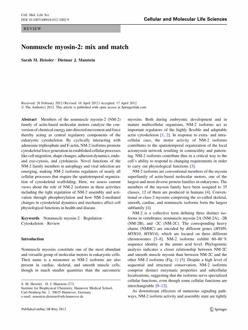
sequence identity at the amino acid level. Phylogenetic
analysis indicates a closer relationship between NM-2C
and smooth muscle myosin than between NM-2C and the
other NM-2 isoforms (Fig. 1) [5]. Despite a high level of
sequential and structural conservation, NM-2 isoforms
comprise distinct enzymatic properties and subcellular
localizations, suggesting that the isoforms serve specialized
cellular functions, even though some cellular functions are
interchangeable [9–12].
As downstream effectors of numerous signaling path-
ways, NM-2 isoform activity and assembly state are tightly
S. M. Heissler � D. J. Manstein (&)
Institute for Biophysical Chemistry, Hannover Medical School,
Carl-Neuberg-Str. 1, 30625 Hannover, Germany
e-mail: [email protected]
Cell. Mol. Life Sci.
DOI 10.1007/s00018-012-1002-9 Cellular and Molecular Life Sciences
123
regulated. Aberrant regulation and functional impairment
of NM-2 isoforms has been associated with the onset and
progression of malignancies, including cancer and altered
immune response. The prominent role of NM-2 aberrations
in disease processes emphasizes the protein’s role in
maintaining mammalian homeostasis [13].
Current knowledge about the function of conventional
myosins in nonmuscle cells is in part derived from model
organisms such as Dictyostelium discoideum, Caenorhab-
ditis elegans, Drosophila melanogaster, and Xenopus
laevis. Dictyostelium and Drosophila express a single
NM-2 gene, Dd mhcA and Dm zipper, making them well
suited for genetic analysis and biochemical studies on gene
expression, function, and regulation [14, 15]. However, the
production of three NM-2 isoforms in vertebrates creates
the need for studying NM-2 function and regulation in
more complex systems such as mouse models. As outlined
below, ablation of NM-2 isoforms in murine models pro-
vides the opportunity to study NM-2 isoforms in a tissue-
specific and developmentally dependent context and serves
as a model system for NM-2-related diseases.
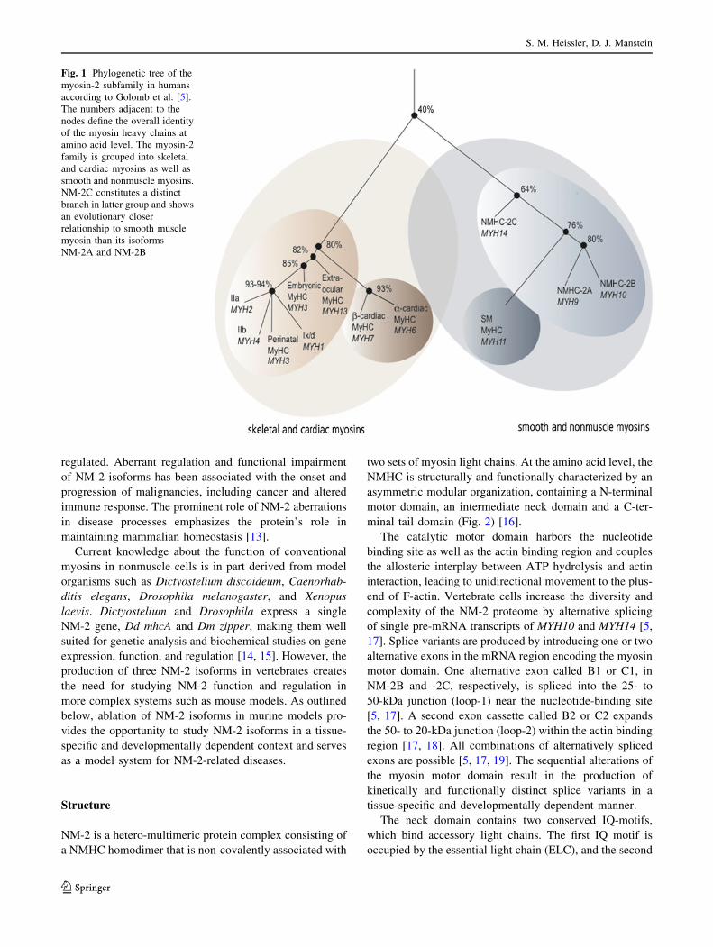
Structure
NM-2 is a hetero-multimeric protein complex consisting of
a NMHC homodimer that is non-covalently associated with
two sets of myosin light chains. At the amino acid level, the
NMHC is structurally and functionally characterized by an
asymmetric modular organization, containing a N-terminal
motor domain, an intermediate neck domain and a C-ter-
minal tail domain (Fig. 2) [16].
The catalytic motor domain harbors the nucleotide
binding site as well as the actin binding region and couples
the allosteric interplay between ATP hydrolysis and actin
interaction, leading to unidirectional movement to the plus-
end of F-actin. Vertebrate cells increase the diversity and
complexity of the NM-2 proteome by alternative splicing
of single pre-mRNA transcripts of MYH10 and MYH14 [5,
17]. Splice variants are produced by introducing one or two
alternative exons in the mRNA region encoding the myosin
motor domain. One alternative exon called B1 or C1, in
NM-2B and -2C, respectively, is spliced into the 25- to
50-kDa junction (loop-1) near the nucleotide-binding site
[5, 17]. A second exon cassette called B2 or C2 expands
the 50- to 20-kDa junction (loop-2) within the actin binding
region [17, 18]. All combinations of alternatively spliced
exons are possible [5, 17, 19]. The sequential alterations of
the myosin motor domain result in the production of
kinetically and functionally distinct splice variants in a
tissue-specific and developmentally dependent manner.
The neck domain contains two conserved IQ-motifs,
which bind accessory light chains. The first IQ motif is
occupied by the essential light chain (ELC), and the second
Fig. 1 Phylogenetic tree of the
myosin-2 subfamily in humans
according to Golomb et al. [5].
The numbers adjacent to the
nodes define the overall identity
of the myosin heavy chains at
amino acid level. The myosin-2
family is grouped into skeletal
and cardiac myosins as well as
smooth and nonmuscle myosins.
NM-2C constitutes a distinct
branch in latter group and shows
an evolutionary closer
relationship to smooth muscle
myosin than its isoforms
NM-2A and NM-2B
S. M. Heissler, D. J. Manstein
123
by the regulatory light chain (RLC). The ELC stabilizes the
NMHC, whereas the RLC has stabilizing and modulating
functions. Alternatively spliced ELCs and RLCs were
identified, but it is currently not known if there is any
isoform-specificity to a given NMHC. Myosin light chains,
especially the RLC, constitute an attractive tool to track
NM-2 in cell biological studies. The ELC binds to various
non-myosin proteins as well as different classes of myosin
heavy chains (-2, -5, -6, and -7), whereas the RLC binds to
the myosin heavy chains of classes -2 and -18 [20, 21].
This makes the use of antibodies against the RLC to track
NM-2 in immunofluorescence studies questionable in tis-
sues that produce myosin-18 along with NM-2. Moreover,
Kondo et al. [22] have shown that di-phosphorylated RLC
localizes independently from NM-2 and mono-phosphor-
ylated RLC to the midzone during cytokinesis, raising the
possibility that tracking of NM-2 via its RLC is misleading.
The tail domain consists of an alpha-helical coiled-coil
motif, which terminates in a short nonhelical tailpiece
(NHT). The coiled-coil region provides the structural basis
for the homodimerization of two NMHC leading to the
formation of a rod-like structure. NM-2 homodimers
assemble into higher order filaments by patterns of alter-
nating charge distributed along the coiled-coil [23]. NM-2
generally functions as a part of minifilamentous structures,
comprising *28 molecules [24, 25]. By comparison, thick
filaments of smooth and skeletal muscle are up to 30-fold
bigger. Different from skeletal muscle myosin, NM-2
undergoes dynamic filament assembly/disassembly transi-
tions. The equilibrium is modulated through
phosphorylation events, as outlined below. Besides phos-
phorylation, F-actin appears to directly promote NM-2
filament assembly. Accelerated filament nucleation in the
presence of F-actin has been observed for the related Dd
NM-2, suggesting the spontaneous formation of actomyo-
sin contractile fibers via myosin assembly [26]. Bipolar
arrays of NM-2 show directed and processive movement
along F-actin, pulling actin filaments of opposing polarity
against each other, thereby generating local contractile
forces and promoting actin-crosslinking.
It is not fully investigated if NM-2 isoforms form het-
erotypic filaments. However, the intermolecular assembly
of NM-2A and NM-2B rod fragments suggests the for-
mation of heterotypic filaments in vitro [27, 28]. In support,
fluorescence spectroscopic studies demonstrate a dynamic
exchange of rod fragments between preformed NM-2
homo-assemblies in an isoform-independent manner [28].
Studies from Beach and Egelhoff [29] report NM-2A and
NM-2B heterotypic filaments at the contractile rings of
dividing cells even though homotypic filaments might be
the predominant pool in live cells [30, 31].
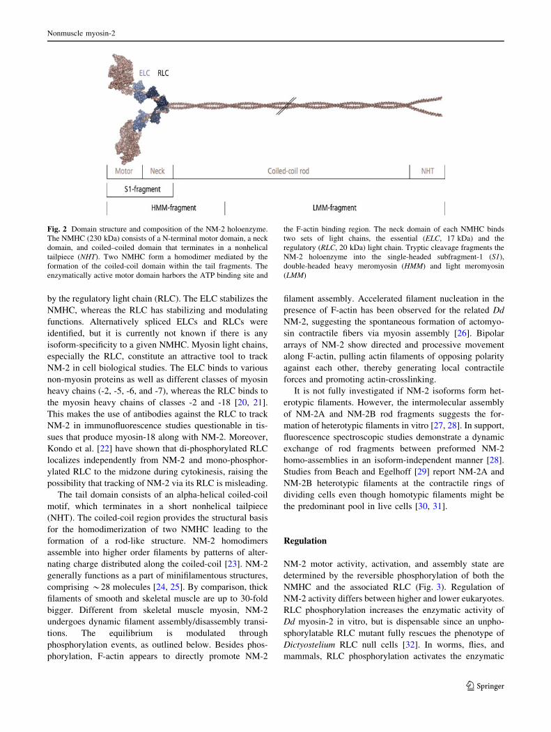
Regulation
NM-2 motor activity, activation, and assembly state are
determined by the reversible phosphorylation of both the
NMHC and the associated RLC (Fig. 3). Regulation of
NM-2 activity differs between higher and lower eukaryotes.
RLC phosphorylation increases the enzymatic activity of
Dd myosin-2 in vitro, but is dispensable since an unpho-
sphorylatable RLC mutant fully rescues the phenotype of
Dictyostelium RLC null cells [32]. In worms, flies, and
mammals, RLC phosphorylation activates the enzymatic
Fig. 2 Domain structure and composition of the NM-2 holoenzyme.
The NMHC (230 kDa) consists of a N-terminal motor domain, a neck
domain, and coiled–coiled domain that terminates in a nonhelical
tailpiece (NHT). Two NMHC form a homodimer mediated by the
formation of the coiled-coil domain within the tail fragments. The
enzymatically active motor domain harbors the ATP binding site and
the F-actin binding region. The neck domain of each NMHC binds
two sets of light chains, the essential (ELC, 17 kDa) and the
regulatory (RLC, 20 kDa) light chain. Tryptic cleavage fragments the
NM-2 holoenzyme into the single-headed subfragment-1 (S1),
double-headed heavy meromyosin (HMM) and light meromyosin
(LMM)
Nonmuscle myosin-2
123
activity of the NM-2 holoenzyme and triggers the assembly
in higher order filaments and hence actomyosin-mediated
contractility [33]. At the amino acid level, the highly con-
served residues S19 and T18 of the RLC constitute the
primary and secondary phosphorylation site, respectively.
In vitro, mono-phosphorylation of S19 enhances myosin
ATPase activity, motor activity, and filament assembly [34–
37]. This is discussed in greater detail below. Simultaneous
di-phosphorylation of T18 and S19 further enhances the
actin-activated ATPase activity and filament assembly [38,
39]. The activation of NM-2 by phosphorylation of the
associated RLC controls assembly and activation of the
holoenzyme to produce force on F-actin and serves as an
indicator for active NM-2 in cellular studies.
RLC kinases include Rho effector and myosin light
chain kinases (MLCK) [33, 40–42]. Regulated by respec-
tive upstream signals, phospho-signaling cascades
converge either in Ca2?/calmodulin- or Rho signaling. The
former activates MLCK [33], while the latter activates
downstream effectors of the Rho family of guanosine tri-
phosphatases (GTPase). The small GTPases RhoA and
Cdc42 mediate the activation of Rho kinase (ROCK) and
related effector kinases such as citron kinase, as well as the
myotonic dystrophy kinase-related Cdc-42-binding kinase
(MRCK) [43–45]. In contrast to MLCK, all other RLC
kinases identified so far are not RLC-specific and act on a
subset of cellular targets.
Protein kinase C (PKC) phosphorylates residues S1, S2,
and T9 of the RLC. Phosphorylation of these sites
decreases both the actin-activated ATPase activity and the
affinity of MLCK for the RLC, thereby preventing NM-2
activation [46, 47]. Whereas the inhibitory S1/S2 phos-
phorylation is associated with mitotic arrest and stress fiber
disassembly in live cells, inhibitory triple-phosphorylation
of the RLC is not regarded as an important regulatory
mechanism [48, 49].
RLC dephosphorylation is exclusively accomplished by
myosin light chain phosphatase (MLCP). MLCP itself is
highly regulated by numerous signaling loops including
RhoA-ROCK signaling. RhoA-ROCK activity enhances
RLC phosphorylation, both by inhibiting MLCP activity
through the inhibitory phosphorylation of its regulatory
myosin-binding subunit (MYPT) and direct RLC phos-
phorylation [50]. In agreement with the absence of RLC-
phosphorylation as an important regulatory mechanism in
Dictyostelium, no apparent orthologs of RhoA, ROCK, or
MYPT have been identified [33].
Several reports implicate NMHC phosphorylation to
regulate filament dynamics. Isoform-specific phosphoryla-
tion sites span the coiled-coil region and the NHT [51–53].
In vitro, kinases involved in NMHC phosphorylation
include PKC, casein kinase 2 (CK2), and the ion channel
kinases TRPM6 and TRPM7 [52, 54, 55]. Specific NMHC
phosphatases have not yet been identified.
Studies with recombinant NM-2A and -2B tail domains
suggest that NMHC phosphorylation inhibits filament for-
mation by shifting the monomer-filament equilibrium
towards the monomeric pool [56]. CK2-dependent
Fig. 3 Regulation of mammalian NM-2 enzymatic activity and
assembly state. RLC kinases promote the conformational change of
the inhibited (10S) to the extended NM-2 conformation (6S). The
inhibited conformation is assembly-incompetent, the extended con-
former assembly-competent. MLCP activity shifts the equilibrium
towards the inhibited conformation. The transition to the extended
conformation triggers the activation of NM-2 ATPase activity and
promotes the assembly of NM-2 homodimers into bipolar filaments.
NMHC phosphorylation or binding of the calcium-binding protein
Mts1 promotes NM-2 filament disassembly. NMHC phosphorylation
impairs Mts1 binding. Mechanisms underlying NMHC dephospho-
rylation are unknown
S. M. Heissler, D. J. Manstein
123
phosphorylation of S1943 of NM-2A inhibits binding of
Mts1 (also known as S100A4), a metastasis-associated
protein. Mts1 promotes the disassembly of NM-2 filaments
in an isoform-specific manner by sequestration of NM-2A
in the disassembled state [56, 57]. In vitro, spectroscopic
studies indicate that Mts1 promotes NM-2A rod fragments
to disassemble from preformed hetero-assemblies of NM-2A
and NM-2B [28].
Live cell studies on human carcinoma cells producing
phosphomimetic NM-2A mutants S1943E and S1943D
reveal increased migration rates, cell protrusions, and focal
adhesions, when compared to wild-type NM-2A or the non-
phosphorylatable NM-2A mutant S1943A [58]. Moreover,
NMHC-2A phosphorylation during epithelial–mesenchy-
mal transition (EMT) promotes enhanced motility and
invasiveness of mesenchymal cells, possibly by a redistri-
bution of NM-2 from posterior to anterior regions [59]. A
phosphorylation-dependent turnover from distal to anterior
regions of the lamellum has been reported for mutant
NM-2A, either lacking the NHT or carrying the S1943A
mutation [51]. NMHC phosphorylation hence prevents
over-accumulation and mislocalization of NM-2 isoforms.
The phosphorylation-dependent NM-2 turnover is required
for its intracellular redistribution and the well-organized
spatial and temporal controlled formation of local con-
tractile actomyosin modules [51, 60–62]. Even though
NM-2 filament formation in live cells is less well under-
stood, NMHC phosphorylation appears to form a viable
basis for the local fine-tuning of filament formation.
Autoinhibition
Another regulatory mode of controlling NM-2 activity is
mediated by the protein’s intrinsic ability to adopt an
autoinhibitory conformation [63, 64]. This feature seems to
be conserved among conventional myosins since it has also
been described for smooth, cardiac, and skeletal muscle
myosin [63, 65]. However, the molecular mechanism
underlying the conversion of the inactive state to the active
state remains to be resolved. Electron microscopic studies
of unphosphorylated NM-2A homodimers reveal intramo-
lecular head-to-head as well as head-to-tail interdomain
interactions, bringing the two motor domains in close
proximity [63, 65]. This conformation establishes contacts
between the actin binding region of one head (blocked
head) with the converter region of the second head (free
head). This conformation impairs actin binding to the
blocked head. In turn, contacts between the blocked head
and the converter region of the free head inhibit the cata-
lytic activity of the free head by blocking the nucleotide
binding site. This double-negative feedback mechanism
inactivates the enzymatic activity of both heads [63]. This
structural model is supported by kinetic studies that dem-
onstrate the autoinhibitory conformation to be
enzymatically inactive, hence preventing constitutive
activation [66, 67]. RLC-phosphorylation relieves the
autoinhibited conformation, thereby promoting the adap-
tion of a kinetically active and assembly-competent
extended NM-2 conformation (Fig. 3). Sedimentation
assays attribute faster sedimentation velocities to the
compact conformer (10S) and slower sedimentation coef-
ficients to the extended molecule (6S) [68]. The 10S
conformer is proposed to constitute an assembly-competent
NM-2 pool in equilibrium with NM-2 filaments [69]. The
strictly regulated interconversion between the 10S con-
former and filaments possibly reflects the spatiotemporal
control of myosin-mediated contractility [69].
Kinetic and mechanical properties
A detailed kinetic analysis of NM-2 isoforms can be per-
formed with recombinant constructs typically produced in the
baculovirus/Sf9 system. Constructs comprising the motor
domain or fusions of the motor domain with an artificial lever
arm are well suited for studying the kinetic properties. These
myosin fragments are constitutively active and display the
enzymatic properties of the phosphorylated holoenzyme [70].
Mechanical parameters like load dependence and proces-
sivity were determined using HMM fragments that require
RLC phosphorylation for full activation [71, 72].
Comparative analysis of the kinetic parameters of fully
activated mammalian NM-2 isoforms reveals subtle differ-
ences in the rate and equilibrium constants that determine the
ATPase cycle (Table 1) [9–11]. These differences are
responsible for the distinct enzymatic properties of NM-2
isoforms, reflecting their functional divergence and cellular
roles. General features of NM-2 isoforms are a slow actin-
activated ATP turnover and a low degree of coupling between
the actin and nucleotide binding sites. Moreover, NM-2A and
-2B show apparent second-order rate binding constants for
ATP that are much smaller in the presence of F-actin [9–11].
An important functional property of NM-2 isoforms is that
ADP binds to actin-bound NM-2 heads several times faster
than does ATP [the ratio of the second-order ADP and ATP
binding rate constants (k1AD/K1k12) is approximately 20 in
NM-2A, 10 in NM-2B, and 2.5 in NM-2C]. This feature
provides a basis for efficient substrate inhibition by ADP, thus
modulating the duty ratio and sliding velocity of NM-2 iso-
forms. NM-2 isoforms are amongst the slowest myosins
characterized, in terms of the velocity at which they trans-
locate actin filaments in the in vitro motility assay. NM-2A
propels actin filaments 2–3 times faster than NM-2C or -2B
[11, 73, 74]. The duty ratio is isoform-dependent: whereas
NM-2A shows a low duty ratio similar to smooth and skeletal
Nonmuscle myosin-2
123
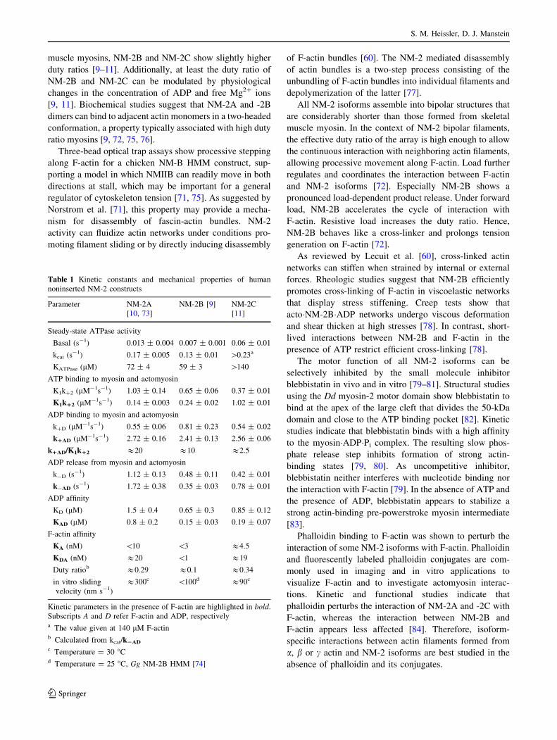
muscle myosins, NM-2B and NM-2C show slightly higher
duty ratios [9–11]. Additionally, at least the duty ratio of
NM-2B and NM-2C can be modulated by physiological
changes in the concentration of ADP and free Mg2? ions
[9, 11]. Biochemical studies suggest that NM-2A and -2B
dimers can bind to adjacent actin monomers in a two-headed
conformation, a property typically associated with high duty
ratio myosins [9, 72, 75, 76].
Three-bead optical trap assays show processive stepping
along F-actin for a chicken NM-B HMM construct, sup-
porting a model in which NMIIB can readily move in both
directions at stall, which may be important for a general
regulator of cytoskeleton tension [71, 75]. As suggested by
Norstrom et al. [71], this property may provide a mecha-
nism for disassembly of fascin-actin bundles. NM-2
activity can fluidize actin networks under conditions pro-
moting filament sliding or by directly inducing disassembly
of F-actin bundles [60]. The NM-2 mediated disassembly
of actin bundles is a two-step process consisting of the
unbundling of F-actin bundles into individual filaments and
depolymerization of the latter [77].
All NM-2 isoforms assemble into bipolar structures that
are considerably shorter than those formed from skeletal
muscle myosin. In the context of NM-2 bipolar filaments,
the effective duty ratio of the array is high enough to allow
the continuous interaction with neighboring actin filaments,
allowing processive movement along F-actin. Load further
regulates and coordinates the interaction between F-actin
and NM-2 isoforms [72]. Especially NM-2B shows a
pronounced load-dependent product release. Under forward
load, NM-2B accelerates the cycle of interaction with
F-actin. Resistive load increases the duty ratio. Hence,
NM-2B behaves like a cross-linker and prolongs tension
generation on F-actin [72].
As reviewed by Lecuit et al. [60], cross-linked actin
networks can stiffen when strained by internal or external
forces. Rheologic studies suggest that NM-2B efficiently
promotes cross-linking of F-actin in viscoelastic networks
that display stress stiffening. Creep tests show that
acto�NM-2B�ADP networks undergo viscous deformation
and shear thicken at high stresses [78]. In contrast, short-
lived interactions between NM-2B and F-actin in the
presence of ATP restrict efficient cross-linking [78].
The motor function of all NM-2 isoforms can be
selectively inhibited by the small molecule inhibitor
blebbistatin in vivo and in vitro [79–81]. Structural studies
using the Dd myosin-2 motor domain show blebbistatin to
bind at the apex of the large cleft that divides the 50-kDa
domain and close to the ATP binding pocket [82]. Kinetic
studies indicate that blebbistatin binds with a high affinity
to the myosin�ADP�Pi complex. The resulting slow phos-
phate release step inhibits formation of strong actin-
binding states [79, 80]. As uncompetitive inhibitor,
blebbistatin neither interferes with nucleotide binding nor
the interaction with F-actin [79]. In the absence of ATP and
the presence of ADP, blebbistatin appears to stabilize a
strong actin-binding pre-powerstroke myosin intermediate
[83].
Phalloidin binding to F-actin was shown to perturb the
interaction of some NM-2 isoforms with F-actin. Phalloidin
and fluorescently labeled phalloidin conjugates are com-
monly used in imaging and in vitro applications to
visualize F-actin and to investigate actomyosin interac-
tions. Kinetic and functional studies indicate that
phalloidin perturbs the interaction of NM-2A and -2C with
F-actin, whereas the interaction between NM-2B and
F-actin appears less affected [84]. Therefore, isoform-
specific interactions between actin filaments formed from
a, b or c actin and NM-2 isoforms are best studied in the
absence of phalloidin and its conjugates.
Table 1 Kinetic constants and mechanical properties of human
noninserted NM-2 constructs
Parameter NM-2A
[10, 73]
NM-2B [9] NM-2C
[11]
Steady-state ATPase activity
Basal (s-1) 0.013 ± 0.004 0.007 ± 0.001 0.06 ± 0.01
kcat (s-1) 0.17 ± 0.005 0.13 ± 0.01 [0.23a
KATPase (lM) 72 ± 4 59 ± 3 [140
ATP binding to myosin and actomyosin
K1k?2 (lM-1s-1) 1.03 ± 0.14 0.65 ± 0.06 0.37 ± 0.01
K1k12 (lM-1s-1) 0.14 ± 0.003 0.24 ± 0.02 1.02 ± 0.01
ADP binding to myosin and actomyosin
k?D (lM-1s-1) 0.55 ± 0.06 0.81 ± 0.23 0.54 ± 0.02
k1AD (lM-1s-1) 2.72 ± 0.16 2.41 ± 0.13 2.56 ± 0.06
k1AD/K1k12 &20 &10 &2.5
ADP release from myosin and actomyosin
k-D (s-1) 1.12 ± 0.13 0.48 ± 0.11 0.42 ± 0.01
k-AD (s-1) 1.72 ± 0.38 0.35 ± 0.03 0.78 ± 0.01
ADP affinity
KD (lM) 1.5 ± 0.4 0.65 ± 0.3 0.85 ± 0.12
KAD (lM) 0.8 ± 0.2 0.15 ± 0.03 0.19 ± 0.07
F-actin affinity
KA (nM) \10 \3 &4.5
KDA (nM) &20 \1 &19
Duty ratiob &0.29 &0.1 &0.34
in vitro sliding
velocity (nm s-1)
&300c \100d &90c
Kinetic parameters in the presence of F-actin are highlighted in bold.
Subscripts A and D refer F-actin and ADP, respectivelya The value given at 140 lM F-actinb Calculated from kcat/k2AD
c Temperature = 30 �Cd Temperature = 25 �C, Gg NM-2B HMM [74]
S. M. Heissler, D. J. Manstein
123
Development
NM-2 isoforms display a vast tissue distribution and most
cells express a set of isoforms, but no consistent expression
and intramolecular localization pattern has emerged [5, 31,
85]. Certain cell types predominantly or exclusively
express one particular NM-2 isoform. For example, plate-
lets and spleen produce exclusively NM-2A [85, 86],
whereas neuronal tissues such as the cerebellum and the
spinal cord are enriched in NM-2B [86, 87].
Their functional divergence allows NM-2 isoforms to
assume distinct roles at specific developmental stages, as
seen in mouse models. Germ line ablation of NMHC-2A
causes embryonic death by day E6.5 due to defects in cell–
cell adhesion, visceral endoderm formation, failure to
organize normal germ layers, and the resulting impairment
of the embryo to undergo gastrulation [12, 88]. Caused by
cardiac and brain defects, NMHC-2B ablation results in
embryonic lethality between day E14.5 and birth [89, 90].
Possibly due to the delayed MYH14 expression in mouse
embryonic development starting at day 10.5, NMHC-2C
knockout mice show no obvious phenotype and survive to
adulthood [86]. Mass spectroscopic analysis of adult mouse
tissues reveals that overall most tissues produce signifi-
cantly less NM-2C than NM-2A and -2B [86]. In contrast,
NM-2C forms 15–45 % of the total NM-2 pool in trans-
formed cells and cell lines such as the monkey kidney
fibroblast cell line COS-7 and the colon adenocarcinoma
cell line HT29 [86]. The simultaneous production of rela-
tively low amounts of NM-2C along with high amounts of
NM-2A and NM-2B in murine tissue and organs, such as
the adult cerebellum, the cerebral cortex, the spinal cord,
and kidneys, raises the question to what extent NM-2 iso-
forms can functionally replace each other [86]. Three
factors—the total NM-2 content in the tissue, their motor
activity, and scaffolding properties—appear to determine
the extent to which one isoform can substitute for another
[12]. Comparison of various phenotypes of genetically
modified mice models suggests that isoform-specific
enzymatic properties are less susceptible to substitution
than tail domain-mediated scaffolding properties, indicat-
ing the capacity for partial compensation [12].
NM-2B and NM-2C exist in the form of several splice
variants. All three NM-2B splice forms are produced in
adult mouse brain in a spatially restricted manner [91]. The
importance of their spatial and temporal splice heteroge-
neity is reflected by the neuron-specific expression of
NM-2B1 and -2B2 during rodent brain development [19].
NM-2B1 ablation causes the abnormal migration of facial
neurons and is associated with the development of hydro-
cephalus during mouse embryogenesis [91]. NM-2B
mRNA is predominantly detected in various regions of the
embryonic and neonatal brain, whereas the NM-2B2
mRNA level is low. Postnatal up-regulation of NM-2B2
mRNA is observed during dendritogenesis and synapto-
genesis in cerebellar Purkinje cells [19, 92]. NM-2B2
ablation results in abnormal maturation of Purkinje cells in
the developing mouse cerebellum, as manifested by a
motor impaired phenotype [91]. The available kinetic and
mechanic data for baculovirus-expressed constructs of
NM-2B1 indicate enhanced actin-activated steady-state
ATPase activity and in vitro translocation of actin fila-
ments, when compared to the noninserted splice form
NM-2B. Both NM-2B and NM-2B1 are regulated by RLC
phosphorylation [74]. In contrast, NM-2B2 appears to lack
actin-activated ATPase activity, motor activity, and regu-
lation by RLC phosphorylation [93].
Both the noninserted and the C1 inserted splice variants
of NM-2C are ubiquitous in their tissue distribution,
whereas the expression of the C2 inserted gene product is
confined to neuronal tissues [5, 94]. The unbalanced
splicing of NMHC-2C with the prevalent production of the
noninserted NM-2C splice form in human myostonic dys-
trophy type (DM1) muscle, in combination with the down-
regulation of both the MYH14 transcript and protein levels,
promotes the development of DM1 histopathological fea-
tures [95]. NM-2C1 is the only splice variant found in
tumor cell lines [94]. Comparisons of numerous human
tumor and nontumor cell lines, which were derived from
the same tissue, indicate increased levels of NM-2C1
production in tumor cells [18, 94]. In the human A549 lung
tumor cell line, small interfering RNA (siRNA) silencing
of NM-2C1 delays cell proliferation by interfering with a
late step in cytokinesis [94]. Reintroduction of NM-2C1
can rescue the phenotype. The noninserted splice form
NM-2C can partially compensate the decreased prolifera-
tion rate, while NM-2A or -2B overproduction is
ineffective [94].
In contrast to the equivalent NM-2B splice variant,
recombinant HMM constructs of NM-2C2 with an expan-
ded loop-2 are constitutively active and do not require RLC
phosphorylation [18].
Cell adhesion and morphogenesis
Morphogenesis involves the translation of biochemical
signaling pathways into forces that move cells. NM-2-
mediated contraction and adhesive forces control embry-
onic epithelial morphogenesis and organogenesis.
Moreover, NM-2 motor activity is at least in part respon-
sible for the cytoskeletal reorganization during epithelial
morphogenesis that determines cell intercalation, invagi-
nation, shape, and rotation [96].
Gastrulation in Drosophila encompasses active cell
shape changes that lead to the formation of ectoderm,
Nonmuscle myosin-2
123
endoderm, and mesoderm layers. Gastrulation is followed
by germ-band extension, which leads to an anterior–pos-
terior axis elongation of the epithelial layer that forms the
thorax and the abdomen of the embryo. All stages of gas-
trulation in Drosophila require the polarized distribution of
NM-2 and adhesion remodeling [96]. Before gastrulation,
the embryo forms a single layer of cells arranged in a
cylindrical shape and NM-2 localizes to the inner surface.
At the beginning of gastrulation, RhoA signaling leads to
an accumulation of NM-2 to the apical sites of the con-
stricting cells, and actomyosin-mediated compression
pushes the inner portion of the cells inwards, thereby cre-
ating a furrow that invaginates [97]. During dorsal closure
of the epithelium, a late event in gastrulation, NM-2
localizes to the leading edge where it creates a tension
force that pulls adjacent cells together as it contracts.
Studies by Franke et al. [97, 98] suggest that NM-2, in
either the leading edge cells or the underlying layer, is
sufficient for dorsal closure. Germ-band extension in
Drosophila is realized by the NM-2-driven disassembly of
adherens junctions and planar junction remodeling, pro-
cesses required for cell intercalation and hence anterior–
posterior axis elongation [99]. Deletion of zipper, the gene
encoding the Drosophila NM-2 heavy chain, is lethal
because of failure in dorsal closure [97]. In analogy to the
function of zipper in the model organism Drosophila,
NM-2A knockout mice die because of defects in the vis-
ceral endoderm development and the failure of the embryo
to undergo gastrulation [1]. NM-2B ablation causes spe-
cific defects in cardiac and brain organogenesis [90, 91].
Neural tube formation in vertebrates and Xenopus
depends on cell shape changes via the apical positioning of
actomyosin in neurepithelial cells [100, 101]. The actin
binding protein Shroom3 localizes to the apical tip of
adherens junctions and the apical junction complex (AJC)
and directs the spatial recruitment of ROCK as well as the
assembly of an actomyosin network associated with the AJC.
ROCK-induced actomyosin contractility further mediates
the Shroom3-induced apical constriction [100, 101]. Inter-
estingly, ROCK is activated by the small G-protein Rap1 and
not RhoA, which suggests the Rho-ROCK complex and
Shroom3 work in separate pathways that converge to
mediate constriction [100, 101]. Furthermore, Shroom3 is a
regulator of the microtubule cytoskeleton, suggesting that
the coordinated activity of the actin and the microtubule
cytoskeleton are essential during epithelial morphogenesis
in the developing vertebrate [102].
The lineage commitment of mesenchymal stem cells
(MSC) and precursor cells is controlled by Rho-ROCK
signaling and NM-2 activity. Regulating factors are
extracellular matrix (ECM) stiffness and cellular conflu-
ence [97]. The mechanical properties of the ECM
significantly determine cell fate: soft matrices are
neurogenic, stiffer matrices are myogenic, and rigid
matrixes are osteogenic [103]. Stiff substrates promote
focal adhesion growth and elongation, and actin assembly
follows the trends in adhesion assembly [103]. NM-2
directly promotes the assembly of focal adhesion and
senses cortical actin structures linked to focal adhesions,
thereby providing force transmission from the cell to the
ECM [104, 105]. Therefore, prominent adhesions of stiff
substrates are correlated to increased cytoskeletal tension
through actomyosin-mediated contractility, which gener-
ates high tension forces that pull on the surface and
promote differentiation towards the osteoblast lineage [97,
103]. Overexpression of either Rho or ROCK stimulates
actomyosin contractility and supports differentiation to
osteoblasts [97]. Chemical inhibition of NM-2 or MLCK
blocks all elasticity-directed lineage specification on any
substrate [103]. NM-2 exerts force through focal adhesions
in mechanisms of matrix sensing, hence contributing to
elasticity-driven lineage specification [103].
Cellular confluence promotes the commitment of precur-
sor cells: sparse MSC densities promote the commitment
towards osteoblasts, whereas confluent MSC differentiate to
adipocytes [106]. Inhibition of actomyosin filament forma-
tion triggers preconfluent human MCS to adipogenesis
instead of osteogenesis [105]. Single MSC plated on small
substrate areas show a round morphology and undergo
adipocytic differentiation. On large substrate areas,
cells retain an elongated shape that triggers osteogenesis
[105, 106].
Cell shape changes are linked to Rho-ROCK signaling
and hence the commitment of MSC. Inactive Rho-GDP is
the predominant Rho species in confluent or rounded MSC
and promotes adipogenesis and chondrogenesis [106]. A
round cell shape decreases the area with a rigid surface and
prevents the cell from generating actomyosin-mediated
tension and contractility [97]. Consistently, constitutively
active Rho inhibits adipocyte differentiation. Overexpres-
sion of Rac, which opposes the actions of Rho-ROCK
signaling, inhibits cytoskeletal contraction and promotes
lineage commitment to adipocytes and chondrocytes [97,
106]. Active Rho-GTP in spread cells activates ROCK and
filament formation. Actomyosin-mediated contractility
inhibits adipogenesis and chondrogenesis and promotes
osteogenesis [97, 106].
Besides the role in differentiation of MSC, NM-2 reg-
ulates the survival threshold of human and mouse
embryonic stem cells (ES) [107]. ES show increased sur-
vival after treatment with Y-27632, an inhibitor of ROCK
[108]. Genetic or pharmacological inhibition of NM-2
enhances the survival and self-renewal of pluripotent stem
cells and is associated with an increased expression level of
self-renewal regulators such as Nanog and Oct3/4 [107].
Similarly, enhanced survival is associated with murine ES
S. M. Heissler, D. J. Manstein
123
lacking NM-2A [107]. In contrast, NM-2B-ablated ES
show survival rates comparable to those of wild-type cells,
indicating distinct functions of NM-2 isoforms in ES cell
death [107]. NM-2 also regulates the cell–cell adhesion of
human and mouse ES cells via a Rho-ROCK signaling
pathway [109]. ROCK inhibition reveals that myosin-
mediated cell–cell contacts are dispensable for maintaining
the pluripotent function of ES [109]. In this context, the
cell–cell contact-free growth of ES plated on E-cadherin-
coated plates may account for the modulation of ROCK
signaling, since both proteins mutually control cell adhe-
sion [109].
In contrast, myosin-mediated cell–cell adhesions and
tension generation of NM-2A on actin filaments, which are
linked to the E-cadherin/beta-catenin complex, are required
to maintain the adhesion complex in the developing mouse
embryo [1]. Amongst other abnormalities, NM-2A-ablated
embryos and ES demonstrate a loss in cell–cell adhesion in
combination with a decrease in E-cadherin and b-catenin
localization at cell–cell adhesion sites [1]. The defect in
cell–cell adhesion causes cells to detach from the surface of
embryoid bodies and to migrate out from the cell cluster,
whereas wild-type embryoid bodies retain a cohesive
morphology [1]. Embryonic lethality of NM-2A-ablated
mice may be caused not only by cell–cell adhesion
impairment but also by defects in ES differentiation, as
outlined above [107]. Loss of cell–cell adhesion in NM-
2B-ablated mice is the cause of hydrocephalus [110]. The
absence of NM-2B in the apical border of the cells lining
the spinal canal enable the underlying neuroepithelial cells
to invade the canal, thereby interrupting the cerebral spinal
fluid flow [110].
Cell Migration
Directed cell migration is an essential process in the
development and maintenance of multicellular organisms
and is associated with cellular functions such as immunity,
wound and tissue repair, angiogenesis, and normal and
cancerous motility. Cell migration requires front-back
polarization, membrane protrusion, adhesion formation and
disassembly, cell body translocation, and rear retraction
and is associated with dynamic interactions between NM-2,
F-actin, the microtubule network, and focal adhesions [111,
112]. The coordinated adhesion assembly at the front and
disassembly at the rear between the cell and a substrate is a
prerequisite for cell migration [42]. ECM-cell adhesions
are force-sensing integrin-based assemblies that provide a
mechanical link between the actomyosin cytoskeleton and
the ECM. The formation of nascent focal adhesions is
NM-2 independent, whereas the formation, growth, and
maintenance of mature focal adhesions require NM-2
motor activity and actomyosin contractility [42, 113].
During all stages of the migratory process, NM-2 isoforms
orchestrate the dynamic spatial and temporal reorganiza-
tion of the actin and to a lesser extent microtubule
cytoskeleton [114]. This requires a capacity for local
restricted self-organization, including the simultaneous
performance of discrete sets of tasks in response to external
trigger events during interphase and a distinct precisely
timed and highly synchronized set of functions during
mitosis. At the heart of the ability to perform these inde-
pendent tasks within the cytosolic compartment is the
occurrence of two cytosolic actin isoforms, namely b-cys-
actin and c-cys-actin. Post-translational modifications and
the interaction with actin binding proteins can amplify the
diversity of the cytosolic actin isoforms. Tropomyosin
(Tm) isoforms were recognized to be of particular impor-
tance for the spatial and temporal dynamics of NM-2-actin
interactions in nonmuscle cells. Moreover, it has been
demonstrated that the activity of myosin motor domains are
differentially regulated by the Tm isoform composition of
actin filaments. Thus, elevated production of Tm5NM1 in
neuroepithelial cells was shown to promote stress fiber
formation, cell spreading, and decreased motility.
Increased TmBr3 levels induce lamellipodial formation,
faster motility, and a reduction in the formation of stress
fibers. Incorporation of Tm5NM1 into stress fibers specif-
ically recruits NM-2A into these structures, while NM-2B
becomes enriched at the cell periphery [115].
Migrating cells form actin-based cytoskeletal extensions
consisting of distinct substructures, designated lamellipo-
dium and lamellum. Both substructures differ in dynamic
properties and protein composition. The lamellipodium
contains a dense dendritic actin network and dynamic focal
contacts. The polymerization of actin filaments with their
plus ends oriented towards the plasma membrane is bal-
anced by a myosin-powered, rearward movement of the
lamellum actin meshwork known as retrograde flow. The
lamellum is less dynamic than the lamellipodium and is
characterized by linear actin bundles and mature adhesion
sites [116, 117]. In general, NM-2 promotes F-actin
anterograde flow in the cell body and retrograde flow in the
lamellum [118, 119]. Behind the lamellum, which typically
spans a broad area, actin bundles and meshwork move
towards the cell front to create a ‘convergence zone’,
where retrograde and anterograde actin motions merge and
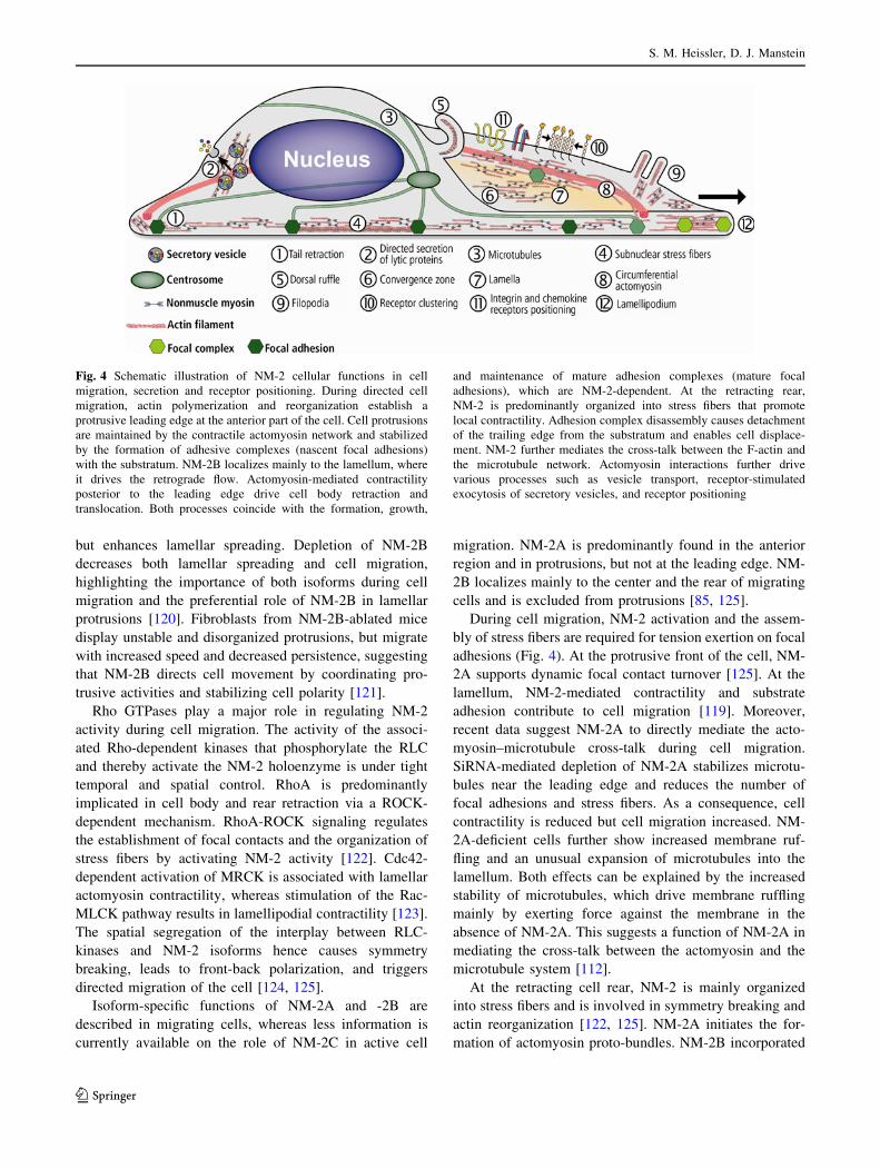
NM-2 is concentrated (Fig. 4).
Isoform-specific roles become prominently evident in
processes such as the lamellar spreading of MDA-MB-231
breast cancer cells on an extracellular matrix, where NM-2
is recruited to the lamellar margin in a phosphorylation-
dependent manner. Pharmacologic inhibition of either
NM-2 or MLCK is associated with decreased migratory
speed. SiRNA depletion of NM-2A impairs cell migration
Nonmuscle myosin-2
123
but enhances lamellar spreading. Depletion of NM-2B
decreases both lamellar spreading and cell migration,
highlighting the importance of both isoforms during cell
migration and the preferential role of NM-2B in lamellar
protrusions [120]. Fibroblasts from NM-2B-ablated mice
display unstable and disorganized protrusions, but migrate
with increased speed and decreased persistence, suggesting
that NM-2B directs cell movement by coordinating pro-
trusive activities and stabilizing cell polarity [121].
Rho GTPases play a major role in regulating NM-2
activity during cell migration. The activity of the associ-
ated Rho-dependent kinases that phosphorylate the RLC
and thereby activate the NM-2 holoenzyme is under tight
temporal and spatial control. RhoA is predominantly
implicated in cell body and rear retraction via a ROCK-
dependent mechanism. RhoA-ROCK signaling regulates
the establishment of focal contacts and the organization of
stress fibers by activating NM-2 activity [122]. Cdc42-
dependent activation of MRCK is associated with lamellar
actomyosin contractility, whereas stimulation of the Rac-
MLCK pathway results in lamellipodial contractility [123].
The spatial segregation of the interplay between RLC-
kinases and NM-2 isoforms hence causes symmetry
breaking, leads to front-back polarization, and triggers
directed migration of the cell [124, 125].
Isoform-specific functions of NM-2A and -2B are
described in migrating cells, whereas less information is
currently available on the role of NM-2C in active cell
migration. NM-2A is predominantly found in the anterior
region and in protrusions, but not at the leading edge. NM-
2B localizes mainly to the center and the rear of migrating
cells and is excluded from protrusions [85, 125].
During cell migration, NM-2 activation and the assem-
bly of stress fibers are required for tension exertion on focal
adhesions (Fig. 4). At the protrusive front of the cell, NM-
2A supports dynamic focal contact turnover [125]. At the
lamellum, NM-2-mediated contractility and substrate
adhesion contribute to cell migration [119]. Moreover,
recent data suggest NM-2A to directly mediate the acto-
myosin–microtubule cross-talk during cell migration.
SiRNA-mediated depletion of NM-2A stabilizes microtu-
bules near the leading edge and reduces the number of
focal adhesions and stress fibers. As a consequence, cell
contractility is reduced but cell migration increased. NM-
2A-deficient cells further show increased membrane ruf-
fling and an unusual expansion of microtubules into the
lamellum. Both effects can be explained by the increased
stability of microtubules, which drive membrane ruffling
mainly by exerting force against the membrane in the
absence of NM-2A. This suggests a function of NM-2A in
mediating the cross-talk between the actomyosin and the
microtubule system [112].
At the retracting cell rear, NM-2 is mainly organized
into stress fibers and is involved in symmetry breaking and
actin reorganization [122, 125]. NM-2A initiates the for-
mation of actomyosin proto-bundles. NM-2B incorporated
Fig. 4 Schematic illustration of NM-2 cellular functions in cell
migration, secretion and receptor positioning. During directed cell
migration, actin polymerization and reorganization establish a
protrusive leading edge at the anterior part of the cell. Cell protrusions
are maintained by the contractile actomyosin network and stabilized
by the formation of adhesive complexes (nascent focal adhesions)
with the substratum. NM-2B localizes mainly to the lamellum, where
it drives the retrograde flow. Actomyosin-mediated contractility
posterior to the leading edge drive cell body retraction and
translocation. Both processes coincide with the formation, growth,
and maintenance of mature adhesion complexes (mature focal
adhesions), which are NM-2-dependent. At the retracting rear,
NM-2 is predominantly organized into stress fibers that promote
local contractility. Adhesion complex disassembly causes detachment
of the trailing edge from the substratum and enables cell displace-
ment. NM-2 further mediates the cross-talk between the F-actin and
the microtubule network. Actomyosin interactions further drive
various processes such as vesicle transport, receptor-stimulated
exocytosis of secretory vesicles, and receptor positioning
S. M. Heissler, D. J. Manstein
123
into these bundles stabilizes and enlarges them, thereby
promoting the formation of extended rears [126].
NM-2B forms large and stable adhesion and actomyosin
bundles that locally inhibit protrusions and adhesion turn-
over [125]. NM-2B might not be unique in its ability to
form and stabilize a contractile rear because migrating B16
melanoma cells, which naturally produce NM-2C but not
NM-2B, show normal front-back polarization, suggesting
that the isoforms play analogous roles in creating a cell
back [125].
The interplay between focal adhesion and actomyosin
dynamics result in a specific balance between migration
and adhesion, which determines the migration velocity.
The size and density of adhesions decrease when NM-2
activity is inhibited by blebbistatin and increase upon
MLCP inhibition. Increased NM-2 activity is associated
with increased migratory speed [127]. In addition, early
adhesion-site formation has the same periodicity as myo-
sin-dependent edge retractions, suggesting a mechanical
relationship between edge retractions and early adhesion-
site formation [128, 129].
Experiments on the dynamics of adhesions-associated
NM-2A and -2B filaments indicate isoform-specific func-
tions during adhesion formation, maturation, and turnover.
Actomyosin bundles containing solely NM-2A mediate
initial adhesion maturation. The associated adhesion sites
turn over in parallel with the actomyosin bundle. Incor-
poration of NM-2B into the bundles enlarges and stabilizes
adhesions and abolishes dynamic adhesion turnover [126].
In contrast, NM-2A containing bundles at anterior parts of
protrusions disassemble as the protrusions evolve. Incor-
poration of NM-2B in these bundles stabilizes them,
possibly reflecting the higher actin affinity and duty ratio of
NM-2B compared to NM-2A [126].
Cytokinesis
Cytokinesis begins shortly after the onset of sister chro-
matid separation during the anaphase of mitosis. It
comprises the final events in the cell cycle with the posi-
tioning and constriction of a contractile ring, followed by
abscission, and the cutting of the midbody channel that
forms the final bridge between the dividing daughter cells.
An important role for NM-2 isoforms in these events is
suggested by a large number of results. Deletion of mhcA
in Dictyostelium abolishes cytokinesis [14]. Microinjec-
tions of pan-NM-2 antibodies or siRNA-mediated
knockdowns prevent furrow ingression [130, 131]. NM-2B
depletion in mice leads to defects in myocyte cytokinesis
[90]. Specific NM-2 isoforms play an essential role during
several morphological stages of cytokinesis, with their
action again critically depending on the spatiotemporal
regulation of RLC phosphorylation in higher eukaryotic
cells [20].
The contractile ring that is formed during cytokinesis is
composed of specific NM-2 and actin isoforms as well as
associated regulatory and scaffolding proteins. These pro-
teins assemble in the equatorial cortex after the selection
and positioning of the cleavage plane by the microtubule
cytoskeleton [132]. The contractile ring forms perpendic-
ular and equatorially at the cell cortex to the anaphase
spindle. During interphase, actomyosin-containing stress
fibers disassemble and relocalize to the nascent cleavage
furrow, where they provide the force that drives furrow
ingression and constriction [133, 134]. The contractile ring
is a highly dynamic structure that assembles and disas-
sembles during each cell cycle. Cortical flow has been
implicated in the transport of NM-2, F-actin, and other
proteins to the contractile ring during its formation, with
NM-2 motor activity also providing the force that drives
the flow [135–137]. As suggested by Levayer et al. [138],
actomyosin accumulation at the equator is promoted by
two synergetic mechanisms that result in a centripetal
actomyosin flow: the central spindle-dependent NM-2
activation (RhoA-dependent) promotes the recruitment of
NM-2 at the equator, whereas astral microtubules inhibit
(Rho-dependent) NM-2 recruitment to the cell periphery.
In some cells, NM-2 localizes to the ring prior to
F-actin, suggesting that NM-2 directly contributes to actin
assembly. However, NM-2 enzymatic activity is not
required for the recruitment of either myosin or F-actin to
the contractile ring [81, 139, 140]. Chemical inhibition of
NM-2 motor activity inhibits cytokinesis but does not
interfere with the equatorial localization of either actin or
myosin, even though actin turnover is reduced. However,
actin itself is highly dynamic in the contractile ring and
dissociates from the equator in control cells, whereas it
accumulates in blebbistatin-treated cells, which has been
attributed to the lack in myosin motor activity [141, 142].
These findings indicate that NM-2 enzymatic activity is not
required for the assembly of the equatorial cortex, but is
essential for actin retention and its dynamic turnover [139,
141, 142]. Similarly, the ability of NM-2 to translocate
actin appears not to be required for constriction of the
cleavage furrow. Rather, the role of NM-2 in vertebrate cell
cytokinesis involves the generation of tension to resist
expansion of the contractile ring by binding and cross-
linking of actin filaments [143].
The phosphorylation status of the NM-2 associated RLC
changes during the cell-cycle of higher eukaryotes: Upon
mitotic exit, S19 of the RLC becomes rapidly phosphory-
lated [33, 144, 145]. Phospho-specific antibodies indicate
that the cleavage furrow is enriched in RLC phosphory-
lated NM-2 holoenzymes [146]. Overexpression of a
nonphosphorylatable RLC disrupts cytokinesis by
Nonmuscle myosin-2
123
producing an abnormal, distorted cleavage furrow, which
leads to a failure to complete cytokinesis [147].
MLCK, MLCP, and the two RhoA-dependent kinases,
ROCK and citron kinase, localize to the cleavage furrow
and regulate NM-2 activity during cytokinesis [148–150].
RhoA activation and its accumulation at the contractile ring
are indispensable for furrow formation and ingression
[151]. Both RhoA activity and the position of the cleavage
furrow are mediated by the central spindle, suggesting a
link between microtubule organization and RhoA activa-
tion at the equator [60, 144]. RhoA activation at the
cleavage furrow leads to the temporal recruitment of its
effector kinases: ROCK is recruited during late anaphase
and stays at the furrow during cytokinesis, suggesting that
the RhoA-ROCK pathway plays a role in contractile ring
formation and cleavage furrow constriction [152]. Citron
kinase colocalizes to the cortex of the cleavage furrow
during telophase and cytokinesis and is involved in the
stabilization of NM-2 binding partners and abscission, as
outlined below [140, 150]. Inhibition studies in Drosophila
suggest that citron kinase is dispensable for initiation and
constriction of the cleavage furrow [153, 154]. Activated
MLCP accumulates at the cleavage furrow and indirectly
enhances the amount of RLC phosphorylation [155].
MLCK is recruited during late anaphase and telophase and
might be involved in both the assembly of the contractile
ring and its constriction [148, 149].
Besides the regulation of NM-2 activity via the phos-
phorylation of its RLC, regulatory and scaffolding proteins
interact with NM-2 at the cleavage furrow and appear to be
involved in its recruitment. Filamentous SEPT2 directly
binds NM-2, which links the former to F-actin [133]. This
interaction is required for NM-2 activation in interphase
and cytokinesis [133]. Disruption of the interaction is
associated with cleavage furrow instability and decreased
RLC phosphorylation. SEPT2-containing filaments possi-
bly form a scaffold that brings NM-2 and its associated
RLC kinases in close proximity, thereby ensuring maxi-
mum NM-2 activation during the final stages of cytokinesis
[133]. Another regulator of cytokinesis, anillin, binds to the
phosphorylated NM-2 holoenzyme, F-actin, and septins
[156]. RhoA activity recruits anillin to the equatorial cortex
early in cytokinesis, where it organizes the contractile ring
[156]. As part of the contractile ring, anillin restricts NM-2
contractility to the cleavage furrow during late stages of
cytokinesis [156]. NM-2 leaves the contractile ring late in
cytokinesis and anillin persists at the contracted furrow,
where it is required for abscission [153, 156].
NM-2C1 is implicated in abnormal cytokinesis in can-
cerous cells and localizes to the midbody, whereas NM-2A
is distributed throughout the cell and concentrated at the
two opposite poles of the dividing daughter cells during the
late stages of cytokinesis [94].
Vesicle transport, endocytosis, and exocytosis
Collective findings suggest NM-2A and -2B to be involved
in intracellular membrane fission of Golgi-derived vesicles
and their transport between different compartments [157–
160]. NM-2A transiently localizes with membranes of the
trans-Golgi network (TGN) during vesicle budding and is
found on a specific subset of Golgi-derived vesicles [161–
163]. Proteolytic cleavage experiments suggest that
NM-2A binds via its tail domain to Golgi stacks. This
interaction is abolished in the presence of NMHC phos-
phorylation by CK2 [164]. These findings indicate that
NM-2A is tethered to the Golgi membrane via its tail,
while its motor domain interacts with F-actin [164]. The
directed movement along F-actin might therefore extend
Golgi membrane tubules or transport vesicles away from
the Golgi complex [164]. This model is supported by
studies showing that NM-2 is involved in retrograde
transport of vesicles from the Golgi complex to the ER
[159]. Recent work by Miserey-Lenkei et al. [157] dem-
onstrates NM-2A and -2B to trigger fission of Rab6
transport carriers from the Golgi complex by interacting
with Rab6 and F-actin. The GTPase Rab6 directly binds to
the coiled-coil of NM-2 in a GTP-dependent manner,
thereby recruiting NM-2 isoforms to the Golgi membrane
[157]. Pharmacological or genetic depletion of NM-2 or
actin polymerization is associated with a phenotype that
produces long tubule stalks that radiate from the Golgi
complex and fail to undergo fission [157]. NM-2A- and
-2B and Rab6 localize to fission sites of these tubular
precursors. Actin is recruited to the assembly sites where it
is required for the detachment of Rab-6 positive transport
cargoes from the stalks [157, 165]. Inhibition of either
NM-2 or Rab6 impairs both the fission of Rab6 cargos from
Golgi membranes as well as the trafficking of anterograde
and retrograde cargo from the Golgi [157].
Recent work by Tang et al. [166] demonstrates NM-2A
to participate in the formation of autophagosomes, organ-
elles that capture cellular components and deliver them to
the lysosomes for degradation. Agt1 kinase plays a crucial
role in the induction of autophagosome formation. In
Drosophila, overexpression of Agt1 results in aberrant cell
morphology and triggers the reorganization of the actin
cytoskeleton, mediated by activation of zipper [166, 167].
The Agt1/Ulk1 signaling pathway activates the kinases
sqa/ZIPK in Drosophila and humans, respectively, which
phosphorylate the zipper or NM-2A-associated RLC during
starvation-induced autophagy. Consequently, knockdowns
of either ZIPK or NM-2A lead to a decrease in the size and
number of autophagosomes [166]. Activated NM-2 con-
trols autophagosome formation by interacting with the
transmembrane protein Atg9, as well as trafficking of
Atg9-containing membranes from the TGN to the sites of
S. M. Heissler, D. J. Manstein
123
autophagosome nucleation [166, 168]. This leads to the
speculation that zipper/NM-2A may either act as a
molecular motor that actively shuttles Atg9-containing
vesicles between the TGN and the forming autophagosome
or forms a complex with Rab6 at the TGN that promotes
the fission of Atg9-containing vesicles [166].
During exocytosis, secretory vesicles derived from the
ER or the Golgi network fuse with the plasma membrane
and release their content into the extracellular space.
Recent work has identified NM-2 isoforms to be involved
at different stages of exocytosis. Confocal intravital
microscopy on submandibular salivary glands of live
rodents indicates that the b-adrenergic receptor-stimulated
exocytosis of secretory vesicles is dependent on actomyo-
sin activity [169]. Agonist-induced stimulation recruits
both NM-2A and -2B onto the surface of fusing granules,
where they function as part of the machinery that regulates
the collapse of the granules after fusion with the apical
plasma membrane [169]. In this context, the authors
speculate that F-actin serves as a platform to recruit NM-2
to form a contractile scaffold that generates the force
required for the collapse of the secretory granules [169].
Another example of NM-2-dependent exocytosis occurs in
natural killer cells, which are lymphocytes of the innate
immune system and important for defense against cancer
and viral infection [170]. Natural killer cell cytotoxicity
involves the formation of an immunological synapse
between the natural killer cell and the target cell through
which lytic granules are delivered to the target cells via
exocytosis [171]. NM-2A inhibition or knockout blocks a
step between the formation of mature synapses and lytic
granule fusion with the cell membrane and promotes lytic
granule exocytosis [170, 171].
Activation of the insulin receptor increases glucose
transporter type 4 (GLUT4) vesicle exocytosis in adipo-
cytes via a NM-2A-dependent mechanism. Insulin
stimulates the MLCK-mediated RLC phosphorylation
thereby triggering the translocation of NM-2A to the
plasma membrane. There, the phosphorylated NM-2A
holoenzyme exhibits a dual role in insulin-stimulated glu-
cose uptake by facilitating GLUT4 vesicle fusion and
regulating GLUT4 activity [172]. Insulin-stimulation does
not change the localization of NM-2B, implicating that the
two isoforms have different functions in adipocytes [172].
Collective findings implicate NM-2 isoforms to regulate
the dynamic opening and closing as well as the size of the
fusion pore during exocytosis [173]. Even though the exact
role is not yet established, several studies have demon-
strated NM-2 isoforms to control vesicle cargo-discharge
kinetics by altering fusion pore conductance and gating in
numerous cell types, including pancreatic b-cells (NM-2A-
dependent), chromaffin cells, and neurons [174–176]. After
the fusion of the vesicle with the membrane, actin
polymerizes and coats the vesicle. Vesicle coating is
independent from NM-2 activity and may reflect an early
step in endocytotic recovery in some cells [177]. NM-2A
directly affects post-fusion dynamics by regulating fusion
pore opening and expansion in cells [173, 175, 177].
Chemical inhibition of either NM-2A or MLCK activity
causes the closure of the fusion pore, indicating that NM-
2A enzymatic activity is necessary to maintain fusion pore
opening in pancreatic acinar cells [177]. Concomitantly,
activated NM-2 slows down fusion pore closure upon
cargo discharge during kiss-and-run exocytosis in neuro-
endocrine PC12 cells, possibly by modifying the
subplasmalemmal actin cortex [178]. These findings sug-
gest that NM-2 activity controls the amount of hormone
released from vesicles in neuroendocrine cells by directly
influencing the duration of fusion pore opening [178].
Different from full fusion exocytosis, NM-2 does not
control the expansion of the fusion pore during kiss-and
run exocytosis, where the fusion pore is resealed before
complete dilation and cargo is not completely released
[178].
Besides exocytosis, NM-2 participates in endocytosis
and phagocytosis. Both processes describe the internali-
zation of extracellular material by invagination of the
plasma membrane to create an endocytic vesicle which
enters the endosomal pathway.
The actin cytoskeleton is the key structure during
receptor-mediated phagocytosis and involved in the for-
mation and closure of the phagocytic cup [179]. Even
though the role of NM-2 in this process has not yet been
fully investigated, NM-2 mediated contractile activity is
required during phagocytic cup assembly, squeezing and
closure during the receptor-mediated ingestion [180, 181].
Several studies demonstrate the impact of cell type,
receptor, and engulfed particle on downstream signaling
pathways that recruit a special set of kinases, including
Rho/Rac/Cdc42-dependent kinases to the nascent phago-
somes where they regulate NM-2 activity as well as actin
nucleation [179, 182].
Olazabal and coworkers demonstrated that the Rho-
ROCK-phosphorylated NM-2A holoenzyme is required for
F-actin recruitment to the phagocytic cup in complement
receptor 3 (CR3)-, but not FcgR-mediated phagocytosis
[181, 183]. During FcgR-mediated phagocytosis by mac-
rophages, NM-2-mediated contractile activity promotes
binding between the FcgR and ligands to facilitate the
efficient extension and subsequent closure of phagocytic
cups [180].
During retinal pigment epithelial phagocytosis of pho-
toreceptor outer segments, the receptor tyrosine kinase
Merkt is required for the spatial relocalization of NM-2A
and -2B from the cell periphery to the phagosome [179].
Further, Merkt triggers the assembly and activation of the
Nonmuscle myosin-2
123
actomyosin complex at the ingestion that promotes the
engulfment of photoreceptor outer segments [179]. NM-2
inhibition by blebbistatin or siRNA depletion of both
NM-2A and -2B leads to a reduction in the number of
ingested phagosomes, suggesting that both isoforms func-
tion in the phagocytic trafficking of photoreceptor outer
segments [179].
Internalization of the chemokine receptor CXCR4 upon
engagement by its agonists is facilitated by NM-2A in
T-lymphocytes [184]. The agonist-mediated receptor
endocytosis is inhibited by overexpression of NM-2A tail
domain [184]. This study favors a model in which NM-2A
serves as an adaptor protein that couples the membrane
receptor to the endocytic machinery, thereby triggering the
formation and uptake of CXCR4-bearing clathrin-coated
endocytic vesicles [184]. NM-2A also mediates the cyto-
kine interferon-gamma-induced endocytosis of tight
junction proteins [185]. In this context, the RhoGEF-
mediated spatial regulation of zipper has been shown to
play a role in the initiation of E-cadherin endocytosis in
Drosophila [186].
Viral infection
Novel roles of NM-2 during several stages of viral infec-
tion emerge. As part of the viral entry machinery, NM-2
functions as herpes simplex virus type-1 (HSV-1) cellular
entry receptor by directly associating with the viral enve-
lope glycoprotein B (gB) on the surface of naturally
permissive target cells [187]. The initiation of HSV-1 entry
induces the cell-surface expression of NM-2 via a MLCK-
dependent redistribution of cytosolic NM-2A [187]. Both
antibody blockage and knockdown of NM-2A in permis-
sive target cells inhibit HSV-1 infectivity whereas the
overexpression of NM-2A in relatively HSV-1-resistant
cell lines causes a high susceptibility to HSV-1 infection
[187]. As a functional gB receptor, NM-2A mediates the
broad HSV-1 infectivity by its ubiquitous expression in
various human tissues and makes it a medicinally relevant
drug target [187]. After HSV-1 entry into the host cells,
viral nucleocapsids move to the nucleus and the viral genes
are transcribed and translated. Late in infection, replicated
DNA is packed in capsids. During viral egress, the capsids
move from the nucleus to extracellular spaces [188].
Studies by Van Leeuwen et al. [189] implicate NM-2A to
play a role in viral transport during herpes virus replication
and viral egress. HSV-1 infection leads to the accumulation
of cytoplasmic NM-2A in a perinuclear cluster, where it
colocalizes with VP22, a major viral tegument protein.
These perinuclear clusters are proposed to be possible viral
assembly compartments where VP22 is incorporated into
assembling virions. Pharmacological inhibition of NM-2A
retards the perinuclear accumulation of VP22 clusters and
the release of virus to the extracellular space with minor
effect on the yield of cell-associated virus. These findings
suggest a role of NM-2A during viral transport and egress
[189]. This idea is supported by the observation that HSV-1
infection induces the formation of long plasma membrane
protrusions that establish contacts with adjacent cells.
NM-2 filaments run through the protrusions, and VP22-
containing particles align and progress along these exten-
sions to accumulate at the extremities of contact forming
adjacent cells [189]. Similar, protrusions such as filopodia
support the viral infection pathway of murine leukemia
virus (MLV). Lehman et al. [190] have shown that an actin
cytoskeleton and NM-2-mediated MLV surfing along fil-
opodia towards viral entry sites at the cell body of
permissive cells promotes MLV infectivity. Consequently,
pharmacological inhibition of NM-2 disrupts viral surfing
and reduces the viral infectivity [190].
Different from HSV1, bleb-associated macropinocytosis
is the predominant mode of Kaposi sarcoma-associated
herpes virus (KSHV) entry in its permissive target cells
[191]. KSHV infection triggers the phosphorylation of
C-Cbl. Phosphorylated C-Cbl associates with NM-2A and
F-actin and is recruited to membrane blebs. The association
with actomyosin leads to the C-Cbl-mediated ubiquitina-
tion of both NM-2A and actin. Actomyosin-mediated
contractility possibly accelerates bleb retraction with the
macropinosomes along with the viral particles. Concomi-
tantly, blebbistatin treatment of the cells or shRNA
knockout of C-Cbl causes defects in myosin-dependent
blebbing and retraction during KSHV entry [191].
Diseases
Mutations, alternative splicing, and misregulation of MYH9
and MYH14 and the associated changes in NM-2A and
NM-2C are linked to the onset and progression of a number
of serious human diseases. In contrast, disease-related
MYH10 mutations have not so far been characterized. Only
indirect links exist between MYH10 expression and disease
processes, such as scar tissue formation following myo-
cardial infarction, demyelination, and the inherited
neurodegenerative disease, juvenile-onset neuronal ceroid
lipofuscinosis (JNCL). JNCL is the most common form
(1:12.500) of a genetically heterogeneous group of rare
disorders known collectively as the neuronal ceroid lipo-
fuscinosis (NCLs), or Batten disease. Classical JNCL,
caused by CLN3 mutation, is a lysosomal storage disorder
with onset between 4 and 8 years of age. The disease is
characterized by accumulation of autofluorescent storage
material and neurodegeneration. Symptoms include sei-
zures, motor and cognitive regression, and progressive
S. M. Heissler, D. J. Manstein
123
vision loss leading to complete blindness [192]. A direct
functional and physical interaction between CLN3 and
NM-2B has been linked to the role of CLN3 in mediating
anterograde and retrograde trafficking [193]. The affected
transport pathway connects the Golgi network, endosomes,
autophagosomes, lysosomes, and the plasma membrane.
In neural tissue, the inhibition of NM-2B by blebbistatin
or knock-down of MYH10 by lentiviral shRNA promotes
remyelination. The myelin-forming cells in the CNS are
formed by differentiation of oligodendrocyte precursor
cells (OPC) into myelinating mature oligodendrocytes
(OL). Similar, after a demyelinating insult, remyelination
involves OPC proliferation, their migration into the lesion,
and differentiation into OL [194, 195]. Both migration and
formation of myelin lamellae involve contributions from
cytoskeletal motors [196, 197]. Recent results indicate that
NM-2B critically contributes to these processes [198, 199].
After myocardial infarction, MYH10 expression is
upregulated in myofibroblasts during the early stages of
cardiac remodeling. While invasion of the activated myo-
fibroblasts into the damaged area is beneficial during early
stages, their abundance has been linked to the formation of
non-functional scar-tissue at later stages [200, 201]. The
tight spatio-temporal control of NM-2B activity with the
aid of specific inhibitors of myosin motor function [202,
203] therefore holds the promise that an over-shooting of
the invasion of myofibroblasts can be prevented and an
optimal ratio of myofibroblasts to myoblasts can be
established [204].
A spectrum of autosomal-dominant disorders related to
MYH9 mutations are subsumed under the collective term
MYH9-related diseases. As many as 40 different mutations
have been mapped throughout the motor and the tail
domain of NMHC-2A [205, 206]. Disease phenotype and
genotypes are associated with congenital macrothrombo-
cytopenia and the onset of clinically variable symptoms
like deafness, progressive nephritis, and presenile cataracts
[205]. A large case series of patients with MYH9-related
diseases demonstrates that the site of MYH9 mutation is a
determinant of the clinical symptoms and features of the
disease [205]. Patients carrying mutations within the cat-
alytic NM-2A motor domain (R702C/H) develop severe
thrombocytopenia, nephritis, and deafness before the age
of 40 years [205]. Patients with mutations within the tail
domain (e.g., D1424H/N/Y, E1841 K, R1933X, V1516 M)
show no defects of clinical relevance [205, 207]. As
demonstrated by Zhang and coworkers, studies of mouse
models with MYH9-related diseases manifest similar phe-
notypes as observed in humans and hence serve as a good
model system to study NM-2A and associated diseases
[208].
Mutations in NM-2C are associated with hereditary
deafness (DFNA4) [209]. Genome-wide linkage analysis
identified an autosomal-dominant mutation which causes a
complex phenotype associated with peripheral neuropathy,
myopathy, hoarseness, and hearing loss [210]. Addition-
ally, aberrant splicing of MYH14 and the unbalanced
expression of NM-2C splice-variants contribute to the
molecular pathogenesis of DM1 [95].
Cancer metastasis and invasion require NM-2 mediated
cell migration, contractility and cell adhesion dynamics
[211]. NM-2 is directly and indirectly involved in cancer
cell motility through overexpression and overactivation.
Both mechanisms enhance cancerous cell migration,
thereby contributing to tumor invasion and metastasis [212,
213]. NM-2A overexpression correlates with increasing
numbers of metastatic lymph nodes, poor cancer differen-
tiation, and advanced tumor stages of esophageal squamous
cancer. SiRNA depletion of NM-2A in cancer cell line
KYSE-510 results in increased cell–matrix adhesion,
decreased cell motility, and reduced metastatic behavior
[13].
In different carcinoma cell lines, ROCK overactivation
causes enhanced RLC phosphorylation and direct activa-
tion of NM-2 driven cell migration [42, 214]. Cancerous
breast cell migration and invasion is attenuated after either
blebbistatin treatment or depletion of NMHC-2A and
NMHC-2B [120, 214]. In contrast, non-cancerous mam-
mary epithelial cell lines continue migration when treated
with blebbistatin [112].
As well as overactivation through RLC phosphorylation,
the direct interaction of NM-2A with the calcium-binding
EF hand protein MtsI drives cancerous cell migration.
Upregulation of Mts1 is associated with cancerous cell
migration, metastatic progression, increased angiogenesis,
and tumor invasion in numerous cell types. The direct
interaction between Mts1 and NM-2A at the leading edge
of migrating cells promotes directional motility, possibly
by enhancing the NM-2A filament turnover. The associated
increase in myosin-driven cell motility contributes to the
increased metastatic capacity of cancer cells and highlights
the importance of NM-2 isoforms during physiological
homeostasis [215]. As reviewed by Helfman and cowork-
ers, MtsI has been shown to interact with actin, nonmuscle
tropomyosin, and p53, suggesting a role for MstI in cell
motility and cytoskeletal rearrangements [216].
Conclusion and outlook
The steadily expanding research on NM-2 has elucidated
important functions of the family members in nearly all
aspects of normal and aberrant cell physiology. Pivotal
roles of NM-2 during development have been investigated
and highlight the impact of NM-2 isoforms during
embryogenesis and organogenesis. Tasks of NM-2 during
Nonmuscle myosin-2
123
cell migration and cytokinesis have been established that
highlight the importance of the spatiotemporal regulation
of the holoenzyme by kinases and phosphatases through
major signaling pathways. In this context, NM-2 functions
need to be addressed in a three-dimensional environment.
New roles of the family members in endo- and exo-cytosis
as well as other intracellular transport processes are
emerging and will be the focus of future research. Despite
our vast knowledge of NM-2 functions in general, many
isoform-specific functions and interactions with partner
proteins remain undefined. Questions that remain to be
addressed include details of the interplay with the micro-
tubule system, membrane interactions, and the cellular
function of arguably the least-characterized family member
NM-2C.
Acknowledgments We thank Neil Billington for the model of
NM-2 in the inhibited state. This work was supported by the European
Union Seventh Framework Programme (FP7/2007-2011) under grant
agreement no. 228971 (Molecular Motors-based Nano Devices—
MONAD) and by ‘‘Deutsche Forschungsgemeinschaft’’ Grant MA
1081/11-2 to D.J.M.
Open Access This article is distributed under the terms of the
Creative Commons Attribution License which permits any use, dis-
tribution, and reproduction in any medium, provided the original
author(s) and the source are credited.
References
1. Conti MA, Even-Ram S, Liu C, Yamada KM, Adelstein RS
(2004) Defects in cell adhesion and the visceral endoderm fol-
lowing ablation of nonmuscle myosin heavy chain II-A in mice.
J Biol Chem 279:41263–41266
2. Tullio AN, Accili D, Ferrans VJ, Yu ZX, Takeda K, Grinberg A,
Westphal H, Preston YA, Adelstein RS (1997) Nonmuscle
myosin II-B is required for normal development of the mouse
heart. Proc Natl Acad Sci USA 94:12407–12412
3. Kohler S, Schaller V, Bausch AR (2011) Collective dynamics of
active cytoskeletal networks. PLoS ONE 6:e23798
4. Berg JS, Powell BC, Cheney RE (2001) A millennial myosin
census. Mol Biol Cell 12:780–794
5. Golomb E, Ma X, Jana SS, Preston YA, Kawamoto S, Shoham
NG, Goldin E, Conti MA, Sellers JR, Adelstein RS (2004)
Identification and characterization of nonmuscle myosin II-C, a
new member of the myosin II family. J Biol Chem
279:2800–2808
6. Leal A, Endele S, Stengel C, Huehne K, Loetterle J, Barrantes
R, Winterpacht A, Rautenstrauss B (2003) A novel myosin
heavy chain gene in human chromosome 19q13.3. Gene
312:165–171
7. Toothaker LE, Gonzalez DA, Tung N, Lemons RS, Le Beau
MM, Arnaout MA, Clayton LK, Tenen DG (1991) Cellular
myosin heavy chain in human leukocytes: isolation of 50 cDNA
clones, characterization of the protein, chromosomal localiza-
tion, and upregulation during myeloid differentiation. Blood
78:1826–1833
8. Simons M, Wang M, McBride OW, Kawamoto S, Yamakawa K,
Gdula D, Adelstein RS, Weir L (1991) Human nonmuscle
myosin heavy chains are encoded by two genes located on
different chromosomes. Circ Res 69:530–539
9. Wang F, Kovacs M, Hu A, Limouze J, Harvey EV, Sellers JR
(2003) Kinetic mechanism of non-muscle myosin IIB: func-
tional adaptations for tension generation and maintenance. J Biol
Chem 278:27439–27448
10. Kovacs M, Wang F, Hu A, Zhang Y, Sellers JR (2003) Functional
divergence of human cytoplasmic myosin II: kinetic characteriza-
tion of the non-muscle IIA isoform. J Biol Chem 278:38132–38140
11. Heissler SM, Manstein DJ (2011) Comparative kinetic and
functional characterization of the motor domains of human
nonmuscle myosin-2C isoforms. J Biol Chem 286:21191–21202
12. Wang A, Ma X, Conti MA, Adelstein RS (2011) Distinct and
redundant roles of the non-muscle myosin II isoforms and
functional domains. Biochem Soc Trans 39:1131–1135
13. Xia ZK, Yuan YC, Yin N, Yin BL, Tan ZP, Hu YR (2011)
Nonmuscle myosin IIA is associated with poor prognosis of
esophageal squamous cancer. Dis Esophagus. doi:10.1111/j.
1442-2050.2011.01261.x
14. Manstein DJ, Titus MA, De Lozanne A, Spudich JA (1989)
Gene replacement in Dictyostelium: generation of myosin null
mutants. EMBO J 8:923–932
15. Young PE, Richman AM, Ketchum AS, Kiehart DP (1993)
Morphogenesis in Drosophila requires nonmuscle myosin heavy
chain function. Genes Dev 7:29–41
16. Sellers JR (2000) Myosins: a diverse superfamily. Biochim
Biophys Acta 1496:3–22
17. Takahashi M, Kawamoto S, Adelstein RS (1992) Evidence for
inserted sequences in the head region of nonmuscle myosin
specific to the nervous system. Cloning of the cDNA encoding
the myosin heavy chain-B isoform of vertebrate nonmuscle
myosin. J Biol Chem 267:17864–17871
18. Jana SS, Kim KY, Mao J, Kawamoto S, Sellers JR, Adelstein RS
(2009) An alternatively spliced isoform of non-muscle myosin
II-C is not regulated by myosin light chain phosphorylation.
J Biol Chem 284:11563–11571
19. Itoh K, Adelstein RS (1995) Neuronal cell expression of inserted
isoforms of vertebrate nonmuscle myosin heavy chain II-B.
J Biol Chem 270:14533–14540
20. Conti MA, Kawamoto S, Adelstein RS (2007) Non-muscle
myosin II. In: Coluccio LM (ed) Myosins: a superfamily of
molecular motors. Springer, Berlin, pp 223–264
21. Tan I, Yong J, Dong JM, Lim L, Leung T (2008) A tripartite
complex containing MRCK modulates lamellar actomyosin
retrograde flow. Cell 135:123–136
22. Kondo T, Isoda R, Uchimura T, Sugiyama M, Hamao K, Hosoya H
(2012) Diphosphorylated but not monophosphorylated myosin II
regulatory light chain localizes to the midzone without its heavy
chain during cytokinesis. Biochem Biophys Res Commun
417:686–691
23. McLachlan AD, Karn J (1982) Periodic charge distributions in
the myosin rod amino acid sequence match cross-bridge spac-
ings in muscle. Nature 299:226–231
24. Verkhovsky AB, Borisy GG (1993) Non-sarcomeric mode of
myosin II organization in the fibroblast lamellum. J Cell Biol
123:637–652
25. Niederman R, Pollard TD (1975) Human platelet myosin II.
In vitro assembly and structure of myosin filaments. J Cell Biol
67:72–92
26. Mahajan RK, Pardee JD (1996) Assembly mechanism of
Dictyostelium myosin II: regulation by K?, Mg2?, and actin
filaments. Biochemistry 35:15504–15514
27. Murakami N, Kotula L, Hwang YW (2000) Two distinct
mechanisms for regulation of nonmuscle myosin assembly via
the heavy chain: phosphorylation for MIIB and mts 1 binding for
MIIA. Biochemistry 39:11441–11451
S. M. Heissler, D. J. Manstein
123
28. Mitsuhashi M, Sakata H, Kinjo M, Yazawa M, Takahashi M
(2011) Dynamic assembly properties of nonmuscle myosin II
isoforms revealed by combination of fluorescence correlation
spectroscopy and fluorescence cross-correlation spectroscopy.
J Biochem 149:253–263
29. Beach JR, Egelhoff TT (2009) Myosin II recruitment during
cytokinesis independent of centralspindlin-mediated phosphor-
ylation. J Biol Chem 284:27377–27383
30. Kolega J (1998) Cytoplasmic dynamics of myosin IIA and IIB:
spatial ‘sorting’ of isoforms in locomoting cells. J Cell Sci
111(Pt 15):2085–2095
31. Kelley CA, Sellers JR, Gard DL, Bui D, Adelstein RS, Baines
IC (1996) Xenopus nonmuscle myosin heavy chain isoforms
have different subcellular localizations and enzymatic activities.
J Cell Biol 134:675–687
32. Ostrow BD, Chen P, Chisholm RL (1994) Expression of a
myosin regulatory light chain phosphorylation site mutant
complements the cytokinesis and developmental defects of
Dictyostelium RMLC null cells. J Cell Biol 127:1945–1955
33. Matsumura F (2005) Regulation of myosin II during cytokinesis
in higher eukaryotes. Trends Cell Biol 15:371–377
34. Scholey JM, Taylor KA, Kendrick-Jones J (1980) Regulation of
non-muscle myosin assembly by calmodulin-dependent light
chain kinase. Nature 287:233–235
35. Somlyo AP, Somlyo AV (2003) Ca2? sensitivity of smooth
muscle and nonmuscle myosin II: modulated by G proteins,
kinases, and myosin phosphatase. Physiol Rev 83:1325–1358
36. Sellers JR, Pato MD, Adelstein RS (1981) Reversible phos-
phorylation of smooth muscle myosin, heavy meromyosin, and
platelet myosin. J Biol Chem 256:13137–13142
37. Kim KY, Kovacs M, Kawamoto S, Sellers JR, Adelstein RS
(2005) Disease-associated mutations and alternative splicing
alter the enzymatic and motile activity of nonmuscle myosins II-
B and II-C. J Biol Chem 280:22769–22775
38. Umemoto S, Bengur AR, Sellers JR (1989) Effect of multiple
phosphorylations of smooth muscle and cytoplasmic myosins on
movement in an in vitro motility assay. J Biol Chem 264:1431–
1436
39. Ikebe M, Koretz J, Hartshorne DJ (1988) Effects of phosphor-
ylation of light chain residues threonine 18 and serine 19 on the
properties and conformation of smooth muscle myosin. J Biol
Chem 263:6432–6437
40. Kamm KE, Stull JT (2001) Dedicated myosin light chain
kinases with diverse cellular functions. J Biol Chem 276:4527–
4530
41. Amano M, Chihara K, Nakamura N, Fukata Y, Yano T, Shibata
M, Ikebe M, Kaibuchi K (1998) Myosin II activation promotes
neurite retraction during the action of Rho and Rho-kinase.
Genes Cells 3:177–188
42. Sandquist JC, Swenson KI, Demali KA, Burridge K, Means AR
(2006) Rho kinase differentially regulates phosphorylation of
nonmuscle myosin II isoforms A and B during cell rounding and
migration. J Biol Chem 281:35873–35883
43. Yamashiro S, Totsukawa G, Yamakita Y, Sasaki Y, Madaule P,
Ishizaki T, Narumiya S, Matsumura F (2003) Citron kinase, a
Rho-dependent kinase, induces di-phosphorylation of regulatory
light chain of myosin II. Mol Biol Cell 14:1745–1756
44. Riento K, Ridley AJ (2003) Rocks: multifunctional kinases in
cell behaviour. Nat Rev Mol Cell Biol 4:446–456
45. Wilkinson S, Paterson HF, Marshall CJ (2005) Cdc42-MRCK
and Rho-ROCK signalling cooperate in myosin phosphorylation
and cell invasion. Nat Cell Biol 7:255–261
46. Ikebe M, Reardon S (1990) Phosphorylation of bovine platelet
myosin by protein kinase C. Biochemistry 29:2713–2720
47. Nishikawa M, Sellers JR, Adelstein RS, Hidaka H (1984) Pro-
tein kinase C modulates in vitro phosphorylation of the smooth
muscle heavy meromyosin by myosin light chain kinase. J Biol
Chem 259:8808–8814
48. Beach JR, Licate LS, Crish JF, Egelhoff TT (2011) Analysis of
the role of Ser1/Ser2/Thr9 phosphorylation on myosin II
assembly and function in live cells. BMC Cell Biol 12:52
49. Komatsu S, Ikebe M (2007) The phosphorylation of myosin II at
the Ser1 and Ser2 is critical for normal platelet-derived growth
factor induced reorganization of myosin filaments. Mol Biol
Cell 18:5081–5090
50. Kimura K, Ito M, Amano M, Chihara K, Fukata Y, Nakafuku M,
Yamamori B, Feng JH, Nakano T, Okawa K, Iwamatsu A,
Kaibuchi K (1996) Regulation of myosin phosphatase by Rho
and Rho-associated kinase (Rho-kinase). Science 273:245–248
51. Breckenridge MT, Dulyaninova NG, Egelhoff TT (2009) Mul-
tiple regulatory steps control mammalian nonmuscle myosin II
assembly in live cells. Mol Biol Cell 20:338–347
52. Clark K, Middelbeek J, Dorovkov MV, Figdor CG, Ryazanov
AG, Lasonder E, van Leeuwen FN (2008) The alpha-kinases
TRPM6 and TRPM7, but not eEF-2 kinase, phosphorylate the
assembly domain of myosin IIA, IIB and IIC. FEBS Lett
582:2993–2997
53. Ronen D, Rosenberg MM, Shalev DE, Rosenberg M, Rotem S,
Friedler A, Ravid S (2010) The positively charged region of the
myosin IIC non-helical tailpiece promotes filament assembly.
J Biol Chem 285:7079–7086
54. Kelley CA, Kawamoto S, Conti MA, Adelstein RS (1991)
Phosphorylation of vertebrate smooth muscle and nonmuscle
myosin heavy chains in vitro and in intact cells. J Cell Sci Suppl
14:49–54
55. Even-Faitelson L, Ravid S (2006) PAK1 and aPKCzeta regulate
myosin II-B phosphorylation: a novel signaling pathway regu-
lating filament assembly. Mol Biol Cell 17:2869–2881
56. Dulyaninova NG, Malashkevich VN, Almo SC, Bresnick AR
(2005) Regulation of myosin-IIA assembly and Mts1 binding by
heavy chain phosphorylation. Biochemistry 44:6867–6876
57. Li ZH, Spektor A, Varlamova O, Bresnick AR (2003) Mts1
regulates the assembly of nonmuscle myosin-IIA. Biochemistry
42:14258–14266
58. Dulyaninova NG, House RP, Betapudi V, Bresnick AR (2007)
Myosin-IIA heavy-chain phosphorylation regulates the motility
of MDA-MB-231 carcinoma cells. Mol Biol Cell 18:3144–3155
59. Beach JR, Hussey GS, Miller TE, Chaudhury A, Patel P,
Monslow J, Zheng Q, Keri RA, Reizes O, Bresnick AR, Howe
PH, Egelhoff TT (2011) Myosin II isoform switching mediates
invasiveness after TGF-{beta}-induced epithelial-mesenchymal
transition. Proc Natl Acad Sci USA 108:17991–17996
60. Lecuit T, Lenne PF, Munro E (2010) Force generation, trans-
mission, and integration during cell and tissue morphogenesis.
Annu Rev Cell Dev Biol 157–84
61. Clark K, Middelbeek J, Lasonder E, Dulyaninova NG, Morrice NA,
Ryazanov AG, Bresnick AR, Figdor CG, van Leeuwen FN (2008)
TRPM7 regulates myosin IIA filament stability and protein local-
ization by heavy chain phosphorylation. J Mol Biol 378:790–803
62. Even-Faitelson L, Rosenberg M, Ravid S (2005) PAK1 regu-
lates myosin II-B phosphorylation, filament assembly,
localization and cell chemotaxis. Cell Signal 17:1137–1148
63. Jung HS, Komatsu S, Ikebe M, Craig R (2008) Head-head and
head-tail interaction: a general mechanism for switching off
myosin II activity in cells. Mol Biol Cell 19:3234–3242
64. Burgess SA, Yu S, Walker ML, Hawkins RJ, Chalovich JM,
Knight PJ (2007) Structures of smooth muscle myosin and
heavy meromyosin in the folded, shutdown state. J Mol Biol
372:1165–1178
65. Wendt T, Taylor D, Messier T, Trybus KM, Taylor KA (1999)Visualization of head–head interactions in the inhibited state of
smooth muscle myosin. J Cell Biol 147:1385–1390
Nonmuscle myosin-2
123
66. Cross RA, Jackson AP, Citi S, Kendrick-Jones J, Bagshaw CR
(1988) Active site trapping of nucleotide by smooth and non-
muscle myosins. J Mol Biol 203:173–181
67. Ankrett RJ, Rowe AJ, Cross RA, Kendrick-Jones J, Bagshaw
CR (1991) A folded (10 S) conformer of myosin from a striated
muscle and its implications for regulation of ATPase activity.
J Mol Biol 217:323–335
68. Craig R, Smith R, Kendrick-Jones J (1983) Light-chain phos-
phorylation controls the conformation of vertebrate non-muscle
and smooth muscle myosin molecules. Nature 302:436–439
69. Milton DL, Schneck AN, Ziech DA, Ba M, Facemyer KC,
Halayko AJ, Baker JE, Gerthoffer WT, Cremo CR (2011) Direct
evidence for functional smooth muscle myosin II in the 10S self-
inhibited monomeric conformation in airway smooth muscle
cells. Proc Natl Acad Sci USA 108:1421–1426
70. Anson M, Geeves MA, Kurzawa SE, Manstein DJ (1996)
Myosin motors with artificial lever arms. EMBO J 15:6069–
6074
71. Norstrom MF, Smithback PA, Rock RS (2010) Unconventional
processive mechanics of non-muscle myosin IIB. J Biol Chem
285:26326–26334
72. Kovacs M, Thirumurugan K, Knight PJ, Sellers JR (2007) Load-
dependent mechanism of nonmuscle myosin 2. Proc Natl Acad
Sci USA 104:9994–9999
73. Wang F, Harvey EV, Conti MA, Wei D, Sellers JR (2000) A
conserved negatively charged amino acid modulates function in
human nonmuscle myosin IIA. Biochemistry 39:5555–5560
74. Pato MD, Sellers JR, Preston YA, Harvey EV, Adelstein RS
(1996) Baculovirus expression of chicken nonmuscle heavy
meromyosin II-B. Characterization of alternatively spliced iso-
forms. J Biol Chem 271:2689–2695
75. Rosenfeld SS, Xing J, Chen LQ, Sweeney HL (2003) Myosin IIb is
unconventionally conventional. J Biol Chem 278:27449–27455
76. Kovacs M, Toth J, Nyitray L, Sellers JR (2004) Two-headed
binding of the unphosphorylated nonmuscle heavy meromyo-
sin.ADP complex to actin. Biochemistry 43:4219–4226
77. Haviv L, Gillo D, Backouche F, Bernheim-Groswasser A (2008)
A cytoskeletal demolition worker: myosin II acts as an actin
depolymerization agent. J Mol Biol 375:325–330
78. Norstrom M, Gardel ML (2011) Shear thickening of F-actin
networks crosslinked with non-muscle myosin IIB. Soft Matter
2011:3228–3233
79. Kovacs M, Toth J, Hetenyi C, Malnasi-Csizmadia A, Sellers JR
(2004) Mechanism of blebbistatin inhibition of myosin II. J Biol
Chem 279:35557–35563
80. Ramamurthy B, Yengo CM, Straight AF, Mitchison TJ, Swee-
ney HL (2004) Kinetic mechanism of blebbistatin inhibition of
nonmuscle myosin IIb. Biochemistry 43:14832–14839
81. Straight AF, Cheung A, Limouze J, Chen I, Westwood NJ,
Sellers JR, Mitchison TJ (2003) Dissecting temporal and spatial
control of cytokinesis with a myosin II Inhibitor. Science
299:1743–1747
82. Allingham JS, Smith R, Rayment I (2005) The structural basis of
blebbistatin inhibition and specificity for myosin II. Nat Struct
Mol Biol 12:378–379
83. Takacs B, Billington N, Gyimesi M, Kintses B, Malnasi-
Csizmadia A, Knight PJ, Kovacs M (2010) Myosin complexed
with ADP and blebbistatin reversibly adopts a conformation
resembling the start point of the working stroke. Proc Natl Acad
Sci USA 107:6799–6804
84. Diensthuber RP, Muller M, Heissler SM, Taft MH, Chizhov I,
Manstein DJ (2011) Phalloidin perturbs the interaction of human
non-muscle myosin isoforms 2A and 2C1 with F-actin. FEBS
Lett 585:767–771
85. Maupin P, Phillips CL, Adelstein RS, Pollard TD (1994) Dif-
ferential localization of myosin-II isozymes in human cultured
cells and blood cells. J Cell Sci 107(Pt 11):3077–3090
86. Ma X, Jana SS, Conti MA, Kawamoto S, Claycomb WC,
Adelstein RS (2010) Ablation of nonmuscle myosin II-B and
II-C reveals a role for nonmuscle myosin II in cardiac myocyte
karyokinesis. Mol Biol Cell 21:3952–3962
87. Rochlin MW, Itoh K, Adelstein RS, Bridgman PC (1995)
Localization of myosin II A and B isoforms in cultured neurons.
J Cell Sci 108(Pt 12):3661–3670
88. Wang A, Ma X, Conti MA, Liu C, Kawamoto S, Adelstein RS
(2010) Nonmuscle myosin II isoform and domain specificity
during early mouse development. Proc Natl Acad Sci USA
107:14645–14650
89. Ma X, Kawamoto S, Hara Y, Adelstein RS (2004) A point
mutation in the motor domain of nonmuscle myosin II-B impairs
migration of distinct groups of neurons. Mol Biol Cell
15:2568–2579
90. Takeda K, Kishi H, Ma X, Yu ZX, Adelstein RS (2003) Abla-
tion and mutation of nonmuscle myosin heavy chain II-B results
in a defect in cardiac myocyte cytokinesis. Circ Res 93:330–337
91. Ma X, Kawamoto S, Uribe J, Adelstein RS (2006) Function of
the neuron-specific alternatively spliced isoforms of nonmuscle
myosin II-B during mouse brain development. Mol Biol Cell
17:2138–2149
92. Miyazaki T, Watanabe M, Yamagishi A, Takahashi M (2000)
B2 exon splicing of nonmuscle myosin heavy chain IIB is dif-
ferently regulated in developing and adult rat brain. Neurosci
Res 37:299–306
93. Kim KY, Kawamoto S, Bao J, Sellers JR, Adelstein RS (2008)
The B2 alternatively spliced isoform of nonmuscle myosin II-B
lacks actin-activated MgATPase activity and in vitro motility.
Biochem Biophys Res Commun 369:124–134
94. Jana SS, Kawamoto S, Adelstein RS (2006) A specific isoform
of nonmuscle myosin II-C is required for cytokinesis in a tumor
cell line. J Biol Chem 281:24662–24670
95. Rinaldi F, Terracciano C, Pisani V, Massa R, Loro E, Vergani L,
Di Girolamo S, Angelini C, Gourdon G, Novelli G, Botta A
(2012) Aberrant splicing and expression of the non muscle
myosin heavy-chain gene MYH14 in DM1 muscle tissues.
Neurobiol Dis 45:264–271
96. Pouille PA, Ahmadi P, Brunet AC, Farge E (2009) Mechanical
signals trigger myosin II redistribution and mesoderm invagi-
nation in Drosophila embryos. Sci Signal 2:ra16
97. Patwari P, Lee RT (2008) Mechanical control of tissue mor-
phogenesis. Circ Res 103:234–243
98. Franke JD, Montague RA, Kiehart DP (2005) Nonmuscle
myosin II generates forces that transmit tension and drive con-
traction in multiple tissues during dorsal closure. Curr Biol
15:2208–2221
99. Bertet C, Sulak L, Lecuit T (2004) Myosin-dependent junction
remodelling controls planar cell intercalation and axis elonga-
tion. Nature 429:667–671
100. Haigo SL, Hildebrand JD, Harland RM, Wallingford JB (2003)
Shroom induces apical constriction and is required for hinge-
point formation during neural tube closure. Curr Biol 13:2125–
2137
101. Hildebrand JD (2005) Shroom regulates epithelial cell shape via
the apical positioning of an actomyosin network. J Cell Sci
118:5191–5203
102. Lee C, Scherr HM, Wallingford JB (2007) Shroom family
proteins regulate gamma-tubulin distribution and microtubule
architecture during epithelial cell shape change. Development
134:1431–1441
S. M. Heissler, D. J. Manstein
123
103. Engler AJ, Sen S, Sweeney HL, Discher DE (2006) Matrix
elasticity directs stem cell lineage specification. Cell
126:677–689
104. Beningo KA, Dembo M, Kaverina I, Small JV, Wang YL (2001)
Nascent focal adhesions are responsible for the generation of
strong propulsive forces in migrating fibroblasts. J Cell Biol
153:881–888
105. McBeath R, Pirone DM, Nelson CM, Bhadriraju K, Chen CS
(2004) Cell shape, cytoskeletal tension, and RhoA regulate stem
cell lineage commitment. Dev Cell 6:483–495
106. Cristancho AG, Lazar MA (2011) Forming functional fat: a
growing understanding of adipocyte differentiation. Nat Rev
Mol Cell Biol 12:722–734
107. Walker A, Su H, Conti MA, Harb N, Adelstein RS, Sato N
(2010) Non-muscle myosin II regulates survival threshold of
pluripotent stem cells. Nat Commun 1:71
108. Watanabe K, Ueno M, Kamiya D, Nishiyama A, Matsumura M,
Wataya T, Takahashi JB, Nishikawa S, Muguruma K, Sasai Y
(2007) A ROCK inhibitor permits survival of dissociated human
embryonic stem cells. Nat Biotechnol 25:681–686
109. Harb N, Archer TK, Sato N (2008) The Rho-Rock-Myosin
signaling axis determines cell–cell integrity of self-renewing
pluripotent stem cells. PLoS One 3:e3001
110. Ma X, Bao J, Adelstein RS (2007) Loss of cell adhesion causes
hydrocephalus in nonmuscle myosin II-B-ablated and mutated
mice. Mol Biol Cell 18:2305–2312
111. Lauffenburger DA, Horwitz AF (1996) Cell migration: a phys-
ically integrated molecular process. Cell 84:359–369
112. Even-Ram S, Doyle AD, Conti MA, Matsumoto K, Adelstein
RS, Yamada KM (2007) Myosin IIA regulates cell motility and
actomyosin-microtubule crosstalk. Nat Cell Biol 9:299–309
113. Delorme-Walker VD, Peterson JR, Chernoff J, Waterman CM,
Danuser G, Dermardirossian C, Bokoch GM (2011) Pak1 reg-
ulates focal adhesion strength, myosin IIA distribution, and actin
dynamics to optimize cell migration. J Cell Biol 193:1289–1303
114. Vicente-Manzanares M, Ma X, Adelstein RS, Horwitz AR
(2009) Non-muscle myosin II takes centre stage in cell adhesion
and migration. Nat Rev Mol Cell Biol 10:778–790
115. Bryce NS, Schevzov G, Ferguson V, Percival JM, Lin JJ,
Matsumura F, Bamburg JR, Jeffrey PL, Hardeman EC, Gunning
P, Weinberger RP (2003) Specification of actin filament func-
tion and molecular composition by tropomyosin isoforms. Mol
Biol Cell 14:1002–1016
116. Geiger B, Spatz JP, Bershadsky AD (2009) Environmental sensing
through focal adhesions. Nat Rev Mol Cell Biol 10:21–33
117. Humphrey D, Duggan C, Saha D, Smith D, Kas J (2002) Active
fluidization of polymer networks through molecular motors.
Nature 416:413–416
118. Gupton SL, Anderson KL, Kole TP, Fischer RS, Ponti A,
Hitchcock-DeGregori SE, Danuser G, Fowler VM, Wirtz D,
Hanein D, Waterman-Storer CM (2005) Cell migration without
a lamellipodium: translation of actin dynamics into cell move-
ment mediated by tropornyosin. J Cell Bioly 168:619–631
119. Ponti A, Machacek M, Gupton SL, Waterman-Storer CM,
Danuser G (2004) Two distinct actin networks drive the pro-
trusion of migrating cells. Science 305:1782–1786
120. Betapudi V, Licate LS, Egelhoff TT (2006) Distinct roles of
nonmuscle myosin II isoforms in the regulation of MDA-MB-
231 breast cancer cell spreading and migration. Cancer Res
66:4725–4733
121. Lo CM, Buxton DB, Chua GC, Dembo M, Adelstein RS, Wang
YL (2004) Nonmuscle myosin IIb is involved in the guidance of
fibroblast migration. Mol Biol Cell 15:982–989
122. Chrzanowska-Wodnicka M, Burridge K (1996) Rho-stimulated
contractility drives the formation of stress fibers and focal
adhesions. J Cell Biol 133:1403–1415
123. Tan I, Leung T (2009) Myosin light chain kinases: division of
work in cell migration. Cell Adh Migr 3:256–258
124. Kolega J (2003) Asymmetric distribution of myosin IIB in
migrating endothelial cells is regulated by a rho-dependent
kinase and contributes to tail retraction. Mol Biol Cell
14:4745–4757
125. Vicente-Manzanares M, Koach MA, Whitmore L, Lamers ML,
Horwitz AF (2008) Segregation and activation of myosin IIB
creates a rear in migrating cells. J Cell Biol 183:543–554
126. Vicente-Manzanares M, Newell-Litwa K, Bachir AI, Whitmore
LA, Horwitz AR (2011) Myosin IIA/IIB restrict adhesive and
protrusive signaling to generate front-back polarity in migrating
cells. J Cell Biol 193:381–396
127. Gupton SL, Waterman-Storer CM (2006) Spatiotemporal feed-
back between actomyosin and focal-adhesion systems optimizes
rapid cell migration. Cell 125:1361–1374
128. Giannone G, Dubin-Thaler BJ, Dobereiner HG, Kieffer N,
Bresnick AR, Sheetz MP (2004) Periodic lamellipodial con-
tractions correlate with rearward actin waves. Cell 116:431–443
129. Giannone G, Dubin-Thaler BJ, Rossier O, Cai Y, Chaga O, Jiang
G, Beaver W, Dobereiner HG, Freund Y, Borisy G, Sheetz MP
(2007) Lamellipodial actin mechanically links myosin activity
with adhesion-site formation. Cell 128:561–575
130. Shelton CA, Carter JC, Ellis GC, Bowerman B (1999) The
nonmuscle myosin regulatory light chain gene mlc-4 is required
for cytokinesis, anterior-posterior polarity, and body morphol-
ogy during Caenorhabditis elegans embryogenesis. J Cell Biol
146:439–451
131. Mabuchi I, Okuno M (1977) The effect of myosin antibody on
the division of starfish blastomeres. J Cell Biol 74:251–263
132. Robinson DN, Spudich JA (2000) Towards a molecular under-
standing of cytokinesis. Trends Cell Biol 10:228–237
133. Joo E, Surka MC, Trimble WS (2007) Mammalian SEPT2 is
required for scaffolding nonmuscle myosin II and its kinases.
Dev Cell 13:677–690
134. Glotzer M (2001) Animal cell cytokinesis. Annu Rev Cell Dev
Biol 17:351–386
135. DeBiasio RL, LaRocca GM, Post PL, Taylor DL (1996) Myosin
II transport, organization, and phosphorylation: evidence for
cortical flow/solation-contraction coupling during cytokinesis
and cell locomotion. Mol Biol Cell 7:1259–1282
136. Zhou M, Wang YL (2008) Distinct pathways for the early
recruitment of myosin II and actin to the cytokinetic furrow. Mol
Biol Cell 19:318–326
137. Uehara R, Goshima G, Mabuchi I, Vale RD, Spudich JA, Griffis
ER (2010) Determinants of myosin II cortical localization dur-
ing cytokinesis. Curr Biol 20:1080–1085
138. Levayer R, Lecuit T (2012) Biomechanical regulation of con-
tractility: spatial control and dynamics. Trends Cell Biol 61–81
139. Miyauchi K, Yamamoto Y, Kosaka T, Hosoya H (2006) Myosin
II activity is not essential for recruitment of myosin II to the
furrow in dividing HeLa cells. Biochem Biophys Res Commun
350:543–548
140. Dean SO, Spudich JA (2006) Rho Kinase’s role in myosin
recruitment to the equatorial cortex of mitotic Drosophila S2
cells Is for myosin regulatory light chain phosphorylation. PLoS
ONE 1:e131
141. Guha M, Zhou M, Wang YL (2005) Cortical actin turnover
during cytokinesis requires myosin II. Curr Biol 15:732–736
142. Murthy K, Wadsworth P (2005) Myosin-II-dependent localiza-
tion and dynamics of F-actin during cytokinesis. Curr Biol
15:724–731
143. Ma X, Kovacs M, Conti MA, Wang A, Zhang Y, Sellers JR,
Adelstein RS (2012) Nonmuscle myosin II exerts tension but
does not translocate actin in vertebrate cytokinesis. Proc Nat
Acad Sci USA 109:4509–4514
Nonmuscle myosin-2
123
144. Barr FA, Gruneberg U (2007) Cytokinesis: placing and making
the final cut. Cell 131:847–860
145. Katti C, Dalal JS, Dose AC, Burnside B, Battelle BA (2009)
Cloning and distribution of myosin 3B in the mouse retina:
differential distribution in cone outer segments. Exp Eye Res
89:224–237
146. Matsumura F, Ono S, Yamakita Y, Totsukawa G, Yamashiro S
(1998) Specific localization of serine 19 phosphorylated myosin
II during cell locomotion and mitosis of cultured cells. J Cell
Biol 140:119–129
147. Komatsu S, Yano T, Shibata M, Tuft RA, Ikebe M (2000)
Effects of the regulatory light chain phosphorylation of myosin
II on mitosis and cytokinesis of mammalian cells. J Biol Chem
275:34512–34520
148. Chew TL, Wolf WA, Gallagher PJ, Matsumura F, Chisholm RL
(2002) A fluorescent resonant energy transfer-based biosensor
reveals transient and regional myosin light chain kinase acti-
vation in lamella and cleavage furrows. J Cell Biol 156:543–553
149. Poperechnaya A, Varlamova O, Lin PJ, Stull JT, Bresnick AR
(2000) Localization and activity of myosin light chain kinase
isoforms during the cell cycle. J Cell Biol 151:697–708
150. Eda M, Yonemura S, Kato T, Watanabe N, Ishizaki T, Madaule
P, Narumiya S (2001) Rho-dependent transfer of Citron-kinase
to the cleavage furrow of dividing cells. J Cell Sci 114:3273–
3284
151. Piekny A, Werner M, Glotzer M (2005) Cytokinesis: welcome
to the Rho zone. Trends Cell Biol 15:651–658
152. Kosako H, Goto H, Yanagida M, Matsuzawa K, Fujita M,
Tomono Y, Okigaki T, Odai H, Kaibuchi K, Inagaki M (1999)
Specific accumulation of Rho-associated kinase at the cleavage
furrow during cytokinesis: cleavage furrow-specific phosphor-
ylation of intermediate filaments. Oncogene 18:2783–2788
153. Naim V, Imarisio S, Di Cunto F, Gatti M, Bonaccorsi S (2004)
Drosophila citron kinase is required for the final steps of cyto-
kinesis. Mol Biol Cell 15:5053–5063
154. D’Avino PP, Savoian MS, Glover DM (2004) Mutations in
sticky lead to defective organization of the contractile ring
during cytokinesis and are enhanced by Rho and suppressed by
Rac. J Cell Biol 166:61–71
155. Kawano Y, Fukata Y, Oshiro N, Amano M, Nakamura T, Ito M,
Matsumura F, Inagaki M, Kaibuchi K (1999) Phosphorylation of
myosin-binding subunit (MBS) of myosin phosphatase by Rho-
kinase in vivo. J Cell Biol 147:1023–1038
156. Straight AF, Field CM, Mitchison TJ (2005) Anillin binds
nonmuscle myosin II and regulates the contractile ring. Mol Biol
Cell 16:193–201
157. Miserey-Lenkei S, Chalancon G, Bardin S, Formstecher E,
Goud B, Echard A (2010) Rab and actomyosin-dependent fis-
sion of transport vesicles at the Golgi complex. Nat Cell Biol
12:645–649
158. DePina AS, Wollert T, Langford GM (2007) Membrane asso-
ciated nonmuscle myosin II functions as a motor for actin-based
vesicle transport in clam oocyte extracts. Cell Motil Cytoskel-
eton 64:739–755
159. Duran JM, Valderrama F, Castel S, Magdalena J, Tomas M,
Hosoya H, Renau-Piqueras J, Malhotra V, Egea G (2003)
Myosin motors and not actin comets are mediators of the actin-
based Golgi-to-endoplasmic reticulum protein transport. Mol
Biol Cell 14:445–459
160. Neco P, Giner D, Viniegra S, Borges R, Villarroel A, Gutierrez
LM (2004) New roles of myosin II during vesicle transport and
fusion in chromaffin cells. J Biol Chem 279:27450–27457
161. Ikonen E, de Almeid JB, Fath KR, Burgess DR, Ashman K,
Simons K, Stow JL (1997) Myosin II is associated with Golgi
membranes: identification of p200 as nonmuscle myosin II on
Golgi-derived vesicles. J Cell Sci 110(Pt 18):2155–2164
162. Stow JL, Fath KR, Burgess DR (1998) Budding roles for myosin
II on the Golgi. Trends Cell Biol 8:138–141
163. Musch A, Cohen D, RodriguezBoulan E (1997) Myosin II is
involved in the production of constitutive transport vesicles
from the TGN. J Cell Biol 138:291–306
164. Fath KR (2005) Characterization of myosin-II binding to Golgi
stacks in vitro. Cell Motil Cytoskeleton 60:222–235
165. Valente C, Polishchuk R, De Matteis MA (2010) Rab6 and
myosin II at the cutting edge of membrane fission. Nat Cell Biol
12:635–638
166. Tang HW, Chen GC (2011) Unraveling the role of myosin in
forming autophagosomes. Autophagy 7:778–779
167. Tang HW, Wang YB, Wang SL, Wu MH, Lin SY, Chen GC
(2011) Atg1-mediated myosin II activation regulates auto-
phagosome formation during starvation-induced autophagy.
EMBO J 30:636–651
168. Bialik S, Pietrokovski S, Kimchi A (2011) Myosin drives
autophagy in a pathway linking Atg1 to Atg9. EMBO J
30:629–630
169. Masedunskas A, Sramkova M, Parente L, Sales KU, Amorn-
phimoltham P, Bugge TH, Weigert R (2011) Role for the
actomyosin complex in regulated exocytosis revealed by intra-
vital microscopy. Proc Natl Acad Sci USA 108:13552–13557
170. Sanborn KB, Rak GD, Maru SY, Demers K, Difeo A, Marti-
gnetti JA, Betts MR, Favier R, Banerjee PP, Orange JS (2009)
Myosin IIA associates with NK cell lytic granules to enable their
interaction with F-actin and function at the immunological
synapse. J Immunol 182:6969–6984
171. Andzelm MM, Chen X, Krzewski K, Orange JS, Strominger JL
(2007) Myosin IIA is required for cytolytic granule exocytosis in
human NK cells. J Exp Med 204:2285–2291
172. Fulcher FK, Smith BT, Russ M, Patel YM (2008) Dual role for
myosin II in GLUT4-mediated glucose uptake in 3T3-L1 adi-
pocytes. Exp Cell Res 314:3264–3274
173. Thorn P (2009) New insights into the control of secretion.
Commun Integr Biol 2:315–317
174. Mochida S, Kobayashi H, Matsuda Y, Yuda Y, Muramoto K,
Nonomura Y (1994) Myosin II is involved in transmitter release
at synapses formed between rat sympathetic neurons in culture.
Neuron 13:1131–1142
175. Neco P, Fernandez-Peruchena C, Navas S, Gutierrez LM, de
Toledo GA, Ales E (2008) Myosin II contributes to fusion pore
expansion during exocytosis. J Biol Chem 283:10949–10957
176. Wilson JR, Ludowyke RI, Biden TJ (2001) A redistribution of
actin and myosin IIA accompanies Ca(2?)-dependent insulin
secretion. FEBS Lett 492:101–106
177. Bhat P, Thorn P (2009) Myosin 2 maintains an open exocytic
fusion pore in secretory epithelial cells. Mol Biol Cell 20:1795–
1803
178. Aoki R, Kitaguchi T, Oya M, Yanagihara Y, Sato M, Miyawaki
A, Tsuboi T (2010) Duration of fusion pore opening and the
amount of hormone released are regulated by myosin II during
kiss-and-run exocytosis. Biochem J 429:497–504
179. Strick DJ, Feng W, Vollrath D (2009) Mertk drives myosin II
redistribution during retinal pigment epithelial phagocytosis.
Invest Ophthalmol Vis Sci 50:2427–2435
180. Araki N (2006) Role of microtubules and myosins in Fc gamma
receptor-mediated phagocytosis. Front Biosci 11:1479–1490
181. Olazabal IM, Caron E, May RC, Schilling K, Knecht DA,
Machesky LM (2002) Rho-kinase and myosin-II control phag-
ocytic cup formation during CR, but not FcgammaR,
phagocytosis. Curr Biol 12:1413–1418
182. Hall AB, Gakidis MA, Glogauer M, Wilsbacher JL, Gao S, Swat
W, Brugge JS (2006) Requirements for Vav guanine nucleotide
exchange factors and Rho GTPases in FcgammaR- and com-
plement-mediated phagocytosis. Immunity 24:305–316
S. M. Heissler, D. J. Manstein
123
183. Caron E, Hall A (1998) Identification of two distinct mecha-
nisms of phagocytosis controlled by different Rho GTPases.
Science 282:1717–1721
184. Rey M, Valenzuela-Fernandez A, Urzainqui A, Yanez-Mo M,
Perez-Martinez M, Penela P, Mayor F Jr, Sanchez-Madrid F
(2007) Myosin IIA is involved in the endocytosis of CXCR4
induced by SDF-1alpha. J Cell Sci 120:1126–1133
185. Utech M, Ivanov AI, Samarin SN, Bruewer M, Turner JR,
Mrsny RJ, Parkos CA, Nusrat A (2005) Mechanism of IFN-
gamma-induced endocytosis of tight junction proteins: myosin
II-dependent vacuolarization of the apical plasma membrane.
Mol Biol Cell 16:5040–5052
186. Levayer R, Pelissier-Monier A, Lecuit T (2011) Spatial regu-
lation of dia and myosin-II by RhoGEF2 controls initiation of
E-cadherin endocytosis during epithelial morphogenesis. Nat
Cell Biol 13:734
187. Arii J, Goto H, Suenaga T, Oyama M, Kozuka-Hata H, Imai T,
Minowa A, Akashi H, Arase H, Kawaoka Y, Kawaguchi Y
(2010) Non-muscle myosin IIA is a functional entry receptor for
herpes simplex virus-1. Nature 467:859–862
188. Johnson DC, Baines JD (2011) Herpesviruses remodel host
membranes for virus egress. Nat Rev Microbiol 9:382–394
189. van Leeuwen H, Elliott G, O’Hare P (2002) Evidence of a role
for nonmuscle myosin II in herpes simplex virus type 1 egress.
J Virol 76:3471–3481
190. Lehmann MJ, Sherer NM, Marks CB, Pypaert M, Mothes W
(2005) Actin- and myosin-driven movement of viruses along
filopodia precedes their entry into cells. J Cell Biol 170:317–325
191. Valiya Veettil M, Sadagopan S, Kerur N, Chakraborty S,
Chandran B (2010) Interaction of c-Cbl with myosin IIA regu-
lates bleb associated macropinocytosis of Kaposi’s sarcoma-
associated herpesvirus. PLoS Pathog 6:e1001238
192. Cotman SL, Staropoli JF (2012) The juvenile batten disease
protein, CLN3, and its role in regulating anterograde and ret-
rograde post-Golgi trafficking. Clin Lipidol 7:79–91
193. Getty AL, Benedict JW, Pearce DA (2011) A novel interaction
of CLN3 with nonmuscle myosin-IIB and defects in cell motility
of Cln3(-/-) cells. Exp Cell Res 317:51–69
194. Stangel M, Trebst C (2006) Remyelination strategies: new
advancements toward a regenerative treatment in multiple
sclerosis. Curr Neurol Neurosci Rep 6:229–235
195. Stangel M, Hartung HP (2002) Remyelinating strategies for the
treatment of multiple sclerosis. Prog Neurobiol 68:361–376
196. Bauer NG, ffrench-Constant C (2009) Physical forces in mye-
lination and repair: a question of balance? J Biol 8:78
197. Bauer NG, Richter-Landsberg C, Ffrench-Constant C (2009)
Role of the oligodendroglial cytoskeleton in differentiation and
myelination. Glia 57:1691–1705
198. Wang H, Tewari A, Einheber S, Salzer JL, Melendez-Vasquez
CV (2008) Myosin II has distinct functions in PNS and CNS
myelin sheath formation. J Cell Biol 182:1171–1184
199. Kippert A, Fitzner D, Helenius J, Simons M (2009) Actomyosin
contractility controls cell surface area of oligodendrocytes.
BMC cell biol 10:71
200. Santiago JJ, Dangerfield AL, Rattan SG, Bathe KL, Cunnington
RH, Raizman JE, Bedosky KM, Freed DH, Kardami E, Dixon
IM (2010) Cardiac fibroblast to myofibroblast differentiation in
vivo and in vitro: expression of focal adhesion components in
neonatal and adult rat ventricular myofibroblasts. Dev Dyn
239:1573–1584
201. Ma X, Takeda K, Singh A, Yu ZX, Zerfas P, Blount A, Liu C,
Towbin JA, Schneider MD, Adelstein RS, Wei Q (2009) Con-
ditional ablation of nonmuscle myosin II-B delineates heart
defects in adult mice. Circ Res 105:1102–1109
202. Chinthalapudi K, Taft MH, Martin R, Heissler SM, Preller M,
Hartmann FK, Brandstaetter H, Kendrick-Jones J, Tsiavaliaris
G, Gutzeit HO, Fedorov R, Buss F, Knolker H-J, Coluccio LM,
Manstein DJ (2011) Mechanism and specificity of penta-
chloropseudilin-mediated inhibition of myosin motor activity.
J Biol Chem 286:29700–29708
203. Preller M, Chinthalapudi K, Martin R, Knolker H-J, Manstein DJ
(2011) Inhibition of myosin ATPase activity by halogenated
pseudilins: a structure-activity study. J Med Chem 54:3675–3685
204. Fedorov R, Bohl M, Tsiavaliaris G, Hartmann FK, Taft MH,
Baruch P, Brenner B, Martin R, Knolker H-J, Gutzeit HO,
Manstein DJ (2009) The mechanism of pentabromopseudilin
inhibition of myosin motor activity. Nat Struct Mol Biol
16:80–88
205. Pecci A, Panza E, Pujol-Moix N, Klersy C, Di Bari F, Bozzi V,
Gresele P, Lethagen S, Fabris F, Dufour C, Granata A, Doubek
M, Pecoraro C, Koivisto PA, Heller PG, Iolascon A, Alvisi P,
Schwabe D, De Candia E, Rocca B, Russo U, Ramenghi U,
Noris P, Seri M, Balduini CL, Savoia A (2008) Position of
nonmuscle myosin heavy chain IIA (NMMHC-IIA) mutations
predicts the natural history of MYH9-related disease. Hum
Mutat 29:409–417
206. Burt RA, Joseph JE, Milliken S, Collinge JE, Kile BT (2008)
Description of a novel mutation leading to MYH9-related dis-
ease. Thromb Res 122:861–863
207. Pecci A, Panza E, De Rocco D, Pujol-Moix N, Girotto G, Podda
L, Paparo C, Bozzi V, Pastore A, Balduini CL, Seri M, Savoia A
(2010) MYH9 related disease: four novel mutations of the tail
domain of myosin-9 correlating with a mild clinical phenotype.
Eur J Haematol 84:291–297
208. Zhang Y, Conti MA, Malide D, Dong F, Wang A, Shmist YA,
Liu C, Zerfas P, Daniels MP, Chan CC, Kozin E, Kachar B,
Kelley MJ, Kopp JB, Adelstein RS (2012) Mouse models of
MYH9-related disease: mutations in nonmuscle myosin II-A.
Blood 119:238–250
209. Donaudy F, Snoeckx R, Pfister M, Zenner HP, Blin N, Di Stazio
M, Ferrara A, Lanzara C, Ficarella R, Declau F, Pusch CM,
Nurnberg P, Melchionda S, Zelante L, Ballana E, Estivill X, Van
Camp G, Gasparini P, Savoia A (2004) Nonmuscle myosin
heavy-chain gene MYH14 is expressed in cochlea and mutated
in patients affected by autosomal dominant hearing impairment
(DFNA4). Am J Hum Genet 74:770–776
210. Choi BO, Hee Kang S, Hyun YS, Kanwal S, Park SW, Koo H,
Kim SB, Choi YC, Yoo JH, Kim JW, Park KD, Choi KG, Ja
Kim S, Zuchner S, Chung KW (2011) A complex phenotype of
peripheral neuropathy, myopathy, hoarseness, and hearing loss
is linked to an autosomal dominant mutation in MYH14. Hum
Mutat 32:669–677
211. Ivkovic S, Beadle C, Noticewala S, Massey SC, Swanson KR,
Toro LN, Bresnick AR, Canoll P, Rosenfeld SS (2012) Direct
inhibition of myosin II effectively blocks glioma invasion in the
presence of multiple motogens. Mol Biol Cell 23:533–542
212. Xia ZK, Yuan YC, Yin N, Yin BL, Tan ZP, Hu YR (2011)
Nonmuscle myosin IIA is associated with poor prognosis of
esophageal squamous cancer. Dis Esophagus. DOI: 10.1111/
j.1442-2050.2011.01261.x
213. Derycke L, Stove C, Vercoutter-Edouart AS, De Wever O, Dolle
L, Colpaert N, Depypere H, Michalski JC, Bracke M (2011) Therole of non-muscle myosin IIA in aggregation and invasion of
human MCF-7 breast cancer cells. Int J Dev Biol 55:835–840
214. Kim JH, Adelstein RS (2011) LPA(1)-induced migration
requires nonmuscle myosin II light chain phosphorylation in
breast cancer cells. J Cell Physiol 226:2881–2893.
215. Boye K, Maelandsmo GM (2010) S100A4 and metastasis: a
small actor playing many roles. Am J Pathol 176:528–535
216. Helfman DM, Kim EJ, Lukanidin E, Grigorian M (2005) The
metastasis associated protein S100A4: role in tumour progres-
sion and metastasis. Br J Cancer 92:1955–1958
Nonmuscle myosin-2
123


