Embed Size (px)
Citation preview
ELSEVIER
Nonpredictive Value of Fibrosis in Dilated Cardiomyopathy ‘Ikeated with Metoprolol
Gianfkanco Sinagra, MD, Serena Rakar, MD, Massixno Zeccbin, MD, Rossana Bussani, MD? Furio Silvestri, MDT Fabio Bassan, MD? Dario Gregori, PhD,$ Andrea Fkrkan, MD,
Andrea Di Lenarda, MD, Gabriele Secoli, MD, Gerardina Lardieri, MD, and Fulvio Camerini, MD
Cardiology Department and *Morbid Anutomy Department, Ospeahle Ma&ore and University, Trieste, Italy; #Depamnent of Statistics, Pennsylvania State University, State College, Pennsylvania
Therapy with /3-adrenergic blocking agents has been advocated as a potential useful approach in heart failure. Recent studies suggest that histologic parameters may be helpful in assessing the effectiveness of @-blocker treatment in dilated catdiomyopathy @CM). In order to predict the response to p-blockers in DCM, fibrous tissue was evahtated at endomyocatdial biopsy (EMB) in 45 patients (pts) with a mean leti ventricular ejection fraction of 0.28 f 0.07, who were successively long-term treated with metoprolol (M) (mean dosage 138 f 26 mg/die).
EMB was performed from left (n = 32) or right (n = 13) ventricle by means of a Ring’s bioptome or the Cotdis adaptation of this instrument.
QuantiIication of fibrous tissue was performed at 9 x magnification and with a computerized mor- phommic system. Qualitative evahuuion at light micmscopy distinguished four types of fibrosis: pericel- h&u, perivascular, focal, and endocanlial. Wume fraction of fibrous tissue ranged from 1.3 to 35.5% (mean 12.1 f 9.3%) and was not significantly correlated with any clinical variable considered.
After 24 f 12 months of treatment, 25 pts were considered improved (group A), whereas the remaining 20 pts were considered not improved (group B), according to criteria based on ejection fraction, left ventricular end-diastolic diameter, filling pattern at Doppler-Echocardiography, cardio- thoracic ratio, NYHA functional class, and exercise duration at ergometric test.
Volume fraction of fibrous tissue did not differ significantly between the two groups (group A = 12.1 f 9.1%; group B.= 11.3 f 9.6%;p = NS). Dominant pericellular type of fibrosis was equally distributed between the two groups (group A = 9125 pts, 36% ; group B = 10120 pts, 50%), whereas a perivascular and/or focal replacement fibrosis was more frequent in group A (group A = 10120 pts, 50%; group B = 2/20 pts, 10%; p = .05, OR 5.55 at univariate analysis). At multivariate analysis mean aortic blood pressure was the only variable discriminating the two groups; the type of fibrosis, although not statistically significant, maintained a high value of odds-ratio (5.23).
In conclusion, extent of total fibrosis assessed by EMB may range widely in patients with JXM, is not correlated with the most important clinical variables, and is not predictive of long-term re- sponse to &blocker treatment. Otherwise, prevalent perivascular and/or focal replacement fibrosis could be associated with a higher probability of improvement after long-term P-blocker treatment. Cardiovasc path01 19%:5:21-28
. . .- __^^_ :cepted June 7, 1995. Dilated cardiomyopathy (DCM) is a heart muscle disease of
ManUScnpt WXlved JuIIe I, 15ry3; ac Address for reprints: G. Sinagra, MD, CwyIVay5 “-4:-‘-y Dept, Ospedale Mag- unknown origin, increasingly recognized in the general popu-
niore and University, Pizza Gsuedde Thte, 1-3412s 3992327; fax: (046) 761637.‘
1 Italy; telephone: (040) lation and characterized by dilation of ventricular cavities, increased cardiac mass, and depressed systolic function (l-3).
Cardiovascular FWhology Vol. 5, No. 1, January/February 1996:21-28 0 1996 by Elsevier Science Inc. 1054-8807/96/$15.00 655 Avenue of the Americas, New York, NY 10010 SSDI 1054-8807(95)00056-B
22 SINAORA ET AL. Cardiovasc Pafhol Vol. 5, No. I NONPREDICTIVE VALUE OF FIBROSIS IN DCM January/February 199621-28
Its histologic findings are suggestive, but not specific (4-10) and are usually quite similar to those of other myocardial dis- eases characterized by hypertrophy and dilation. The most frequently observed histologic feature is hypertrophy of myo- fibers associated with thinned myocytes with hypertrophic nuclei. Interstitial or replacement fibrosis and hypertrophy of smooth muscle cells in the subendocardium may be pres- ent (7) as well as a slight or moderate lymphocytic infiltrate (10,ll).
6 months or less in case of intolerance, death or heart transplantation.
“Improvement” after long-term treatment with metoprolol was defined as follows:
1. an increase in left ventricular ejection fraction of 210 units combined with 1 major criterion, i.e.: l a decrease in left ventricular end-diastolic diameter
210% l regression of “restrictive” filling pattern (E-deceleration
time from 6120 to >120 msec) or with 2 of the minor criteria, i.e.: l decrease in at least 1 NYHA functional class l decrease in cardio-thoracic ratio 210% l increase in exercise duration b2 minutes
2. an increase in LVEF of >/5 point combined with 1 major and 2 minor criteria.
An increase of fibrous tissue has been described (12), and the fibrosis extent has been correlated with natural history (10,13) and with effectiveness of therapy (14,15).
Therapy with fi-adrenergic blocking agents has been pro- posed as a potentially useful approach, and several reports suggest that chronic P-blockade, most often with the 01 selec- tive agent metoprolol, may improve hemodynamic and clini- cal function in patients with DCM (16-22). In clinical trials the majority of patients (pts) tolerate P-blockers if carefully titrated and a variable percentage of treated pts (about 50%) may show a remarkable improvement after weeks or months, while the remaining are stable or deteriorate (23).
So tir the characterization of responders to P-blocker treat- ment represents a major unsolved problem and it is unclear at present which are the clinical, anatomic, and hemodynamic characteristics of patients who improve.
Aim of the study was to assess whether the extent and the characteristics of the fibrous tissue present in endomyocardial biopsies (EMB) of pts atfected by DCM could be a predictor of efficacy of long-term P-blocker treatment.
Materials and Methods From October 1987 to July 1993, 155 pts with DCM un-
derwent catheterization and EMB to exclude active myocar- ditis and specific heart muscle diseases. DCM was diagnosed according to the WHO criteria (24), in the presence of a depressed left ventricular ejection fraction (LVEF) and in ab- sence of significant (>50%) coronary artery stenosis and of other specific heart muscle diseases.
An alcohol intake ,100 g/die in the previous 6 months or a history of hypertension (>170/100 mm Hg) was considered as an exclusion criterion.
All 155 consecutive patients underwent a test-dose of 5 mg of metoprolol b.i.d.: 6 pts did not tolerate the drug. Out of the remaining 149 pts, 96 had a follow up of at least 24 f 6 months.
Forty-five patients satisfied the inclusion criteria for the study defined as follows:
l Left ventricular ejection fraction <0.40; l EMB adequate specimen (area >l mm2, available spec-
imen 32, interval between EMB and begin of treatment less than 12 months);
l longterm treatment with metoprolol for at least 24 f
Clinical data, evaluated at the time of diagnosis and dur- ing follow up, included functional status (NYHA functional class) and signs of cardiac heart failure. Cardiothoracic ratio was obtained from the chest x-rays. All pts underwent M-mode, two dimensional Doppler and Color-Doppler echocardio- graphic study.
All 45 pts, at the time of diagnosis, underwent a hemo- dynamic study with left and right ventriculograPhy and coro- nary angiography. Endomyoca&al biopsy wus perfbrmed dur- ing diagnostic heart catheterization, from right (n = 13) or left (n = 32) ventricle, by means of a King’s endomyocardial bioptome or the Cordis adaptation of this instrument (Cordis Corp., Miami, FL), using a femoral approach. Biopsies ranged from four to five specimens in each patient. Tissues were fixed immediately in 10% neutral fbrmalin, then processed through graded ethanol solutions, cleared in xylol and paraffin embedded. Paraffin sections were cut, on a base sledge microtome (Microm-H&I-400), at 2 Hrn mean thick- ness, automatically mounted (resin without) and stained with Azan-Mallory.
Histologic sections were analysed by a microscopist for quantitative evaluation and two pathologists for qualitative analysis, all blind of clinical data. Morpbom&ic analysis was limited only to myocardial fibrous tissue (25).
Quantification of fibrous tissue was performed at 9X magnification by light microscope and with a computerized morphometric system (Olympus CUE-2 Program; Olympus AH-3), with a Sony camera CUE version I-IV and a monitor (Hantarex) connected to a computer provided with “the pic- ture analyser CUE-2 program” (Olympt@. This program identifies fibrous tissue on the ground of gray levels; for each fragment total area, fibrosis volume fract&n and mu- lar volume fraction were determined.
Qualitative evaluation of the type of fibrosis was assessed by light microscopy. Four cellular, perivascular, fo
mc Path01 Vol. 5, No. I SINAGRA ET AL. ,/February 1996:21-28 NONPREDICTIVE VALUE OF FIBROSIS IN DCM
23
24 SINAORA ET AL. NONPRBDICTTVE VALUE OF FIBROSIS IN DCM
Cardiovasc Path1 Vol. 5, No. I January/February 199&21--28
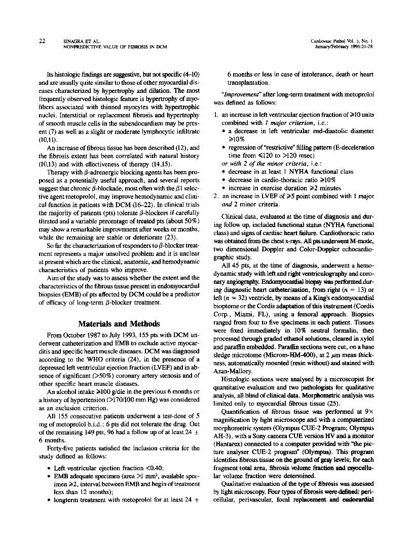
(26,27) (Figure 1 A-C). Pericellular type consists in a net- work encircling and separating individual myocardial fibers; perivascular is an increase of normal perivascular collage- nous tissue, delineating groups of muscle fibers into bundle or fascicles; replacement focal fibrosis refers to microscopic scars in a position previously occupied by muscle cells.
Endocardial fibrosis was excluded from the analysis (25), whereas perivascular and focal replacement have been con- sidered together.
Statistical Analysis To differentiate between the two groups regarding the clin-
ical variables at baseline, we performed a one-way ANOVA for continuous variables and a x2 test for categorical vari- ables. The same analysis was performed to detect a relation among clinical variables and histologic parameters (percent- age and type of fibrosis).
In order to predict improvement from baseline character- istics in patients treated with @-blockers, a univariate logistic regression analysis was performed as a first step.
Relative estimate risk for each variable was expressed and was considered significant when p < .05.
As suggested by Hosmer (28) variables with a univariate odds ratio significance lower than .25 were considered for
the stepwise multivariate analysis in a way to contml the poten- tial confounding effect of other variables.
Forty-five pts were considered for the present analysis: 37 males, 8 females, mean age 42.8 f 13.5 years (range 12-66 yrs). At 2-year follow-up, 25 pts (56%) were classified as im- proved (group A) and 20 pts (44%) as not improved (group B); five pts included in group B underwent cardiac transplan- tation during the follow-up.
Mean daily dosage of metoprolol was 138 + 26 mg (range 50-300 mg).
History of slight hypertension @ = .0006), more advanced age (p = 02) higher values of mean aortic pressure (p = 007) and of heart rate @ = .05) were signi&antly more represented in improved patients (Table 1).
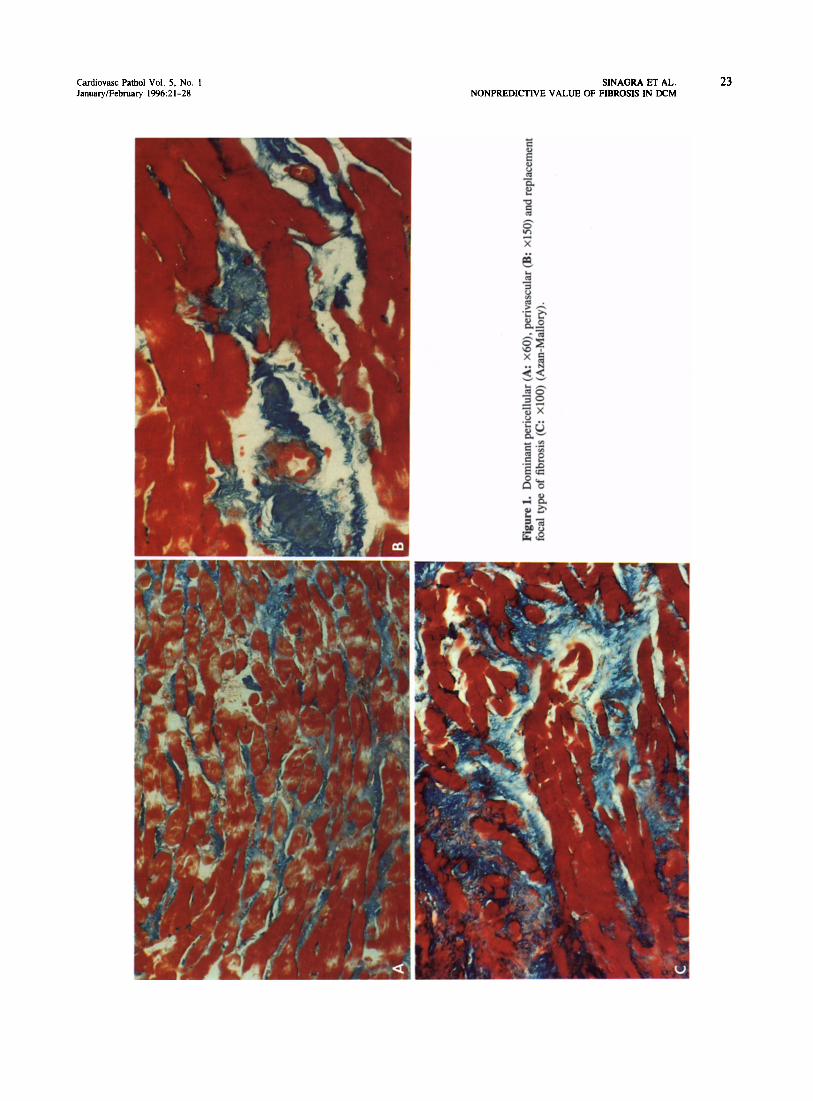
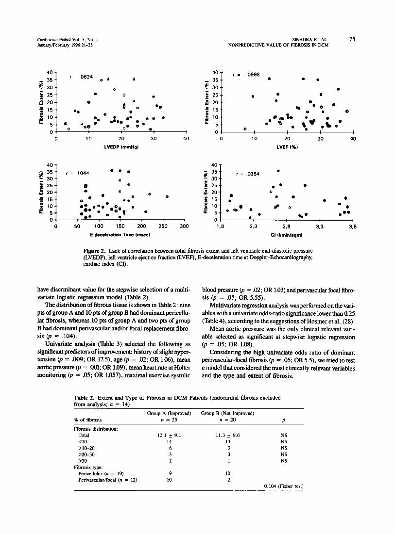
No significant relation was found among clinical variables and histologic parameters (Figure 2).
In the whole population, volume fraction of fibrous tissue ranged from 1.3% to 35.5% (mean 12.15 f 9.3%) and it was notsignificanttydi~~tbetweenthetwo~(l2.1 If: 9.1% in group A versus 11.3 rt 9.6% in group B; p = NS). The percentage of fibrous tissue, tested at di&ent levels, did not
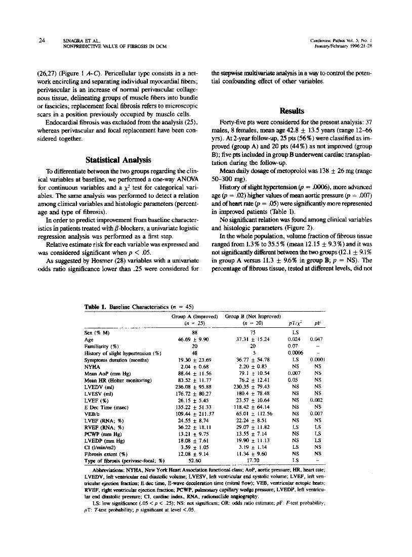
Table 1. Baseline Characteristics (n = 45) Group A (Improved) Group B (Not Improved)
(II = 25) (n = 20) PW PF Sex (7% M) 88 75 LS - Age 46.69 f 9.90 37.31 f 15.24 0.024 0.047 Familiarity ( W) 20 20 0.07 -
History of slight hypertension (X) 48 5 o.ooo6 --’ Symptoms duration (months) 19.30 f 23.69 36.77 f 54.78 LS 0.0001 NYHA 2.04 f 0.68 2.20 f 0.83 NS NS Mean AoP (mm Hg) 88.44 f 11.56 79.1 f 10.54 0.007 NS Mean HR (Hotter monitoring) 83.52 f 11.77 76.2 f 12.41 0.05 NS LVEDV (ml) 236.08 f 95.88 230.35 f 79.43 NS NS LVESV (ml) 176.72 f 80.27 180.4 i 78.48 NS NS LVEF (96) 26.15 f 5.45 23.57 f 10.64 NS 0.002 E Dee Time (msec) 135.22 f 51.33 118.42 f 64.14 NS NS VEBlb 109.44 f 211.37 65.01 f 112.56 NS 0.007 LVEF (RNA; %) 24.55 f 8.74 22.24 f 8.51 NS NS RVEF (RNA; $6) 36.22 f 18.11 29.07 f 11.82 LS Ls PCX+‘P (mm Hg) 13.21 f 9.75 13.55 f 7.14 NS LS LVEDP (mm Hg) 18.08 rt 7.61 19.90 f 11.13 NS Ls CI (l/min/nQ) 3.59 f 1.05 3.19 * 1.14 LS NS Fibrosis extent (96) 12.08 f 9.14 11.34 f 9.60 NS NS Type of fibrosis (perivasc-focal; %) 52.60 17.70 LS -
Abbteviatioos: NYHA, New York Heart Association functional class; AoP, aortic pressure; HR, heart rate; LVEDV, left ventricular end diastolic volume; LVESV, left ventricular end systolic volume; LVEF, left ven- tricular ejection fraction; E dec time, E-wave decderation time (mitral flow); VEB, ventricular cctopic beats; RVEF, right ventricular ejection fraction; PCWP, pulmonary capillary wedge pressure; LVEDP, left ventricu- lar end diastolic pressure; CI, cardiac index, RNA, radionuclide angiograpby.
LS: low significance (.05 < p < .25); NS: not significant; OR: odds ratio estimate; pF: F-test probability; pT: T-test probability; p significant at level <.05.
Cardiovasc PathoI Vol. 5, No. 1 Jsauary/Febraary 1996~21-28
SWAGRA ET AL. 25 NONPREDICTIVE VALUE OF FIBROSIS IN DCM
4”’ 4u - = .0624 r = - r ,096s 35 -. 00 0 35 -* 0 0
z 30-v 0 z 30-e
0
5 g l
25 -* 0 0 . 25 -. l l 0
z 0 Y 20-. 0
8 O* ; 20.. l
.f 15 -. %I
’ 00 .f 15 -. B
$ 0 00 0 0
lo-. 0 b eo l $ lo-.
u. @e
5 -. @ eoO @ OhQ e:.
O0
OQ u. 5-e 0 ot * 0. I Oi 0 i 0 10 20
LVEDP tmmHg)
30 40 0 10 20 30 40
LVEF I%)
40 T 40 -r g 35s’ r = 1044 00 0 3 35-. r = .0254 0
z 30.. 0 30-. 8 25-a 8 *
f Q 25-’ 0 . 0
j 20-e . 0
. e 5 20-. 0
2
15-s
a e 0
8 0 00
.f
15-e
e 0
0 0
- $ 10.. -. l ;/.oe0e 0
.”
l . : k ‘O-. OQ ’ 0 d 0 l
5 0 get
0 5.. . 0. . 0 8 @be
l eo 01 4 0-t b. 1 I 0 50 100 150 200 250 300 1.8 203 2.8 3.3 3.8
Edeceleration Time bnsecl Cl Wnin/sqm~
Figure 2. Lack of correlation between total fibrosis extent and left ventricle end-diastolic pressure (LVEDP), left ventricle ejection fraction (LVEF), E-deceleration time at Doppler-Echocardiography, cardiac index (CI).
have discrminant value for the stepwise selection of a multi- blood pressure (p = .02; OR 1.03) and perivascular focal fibro- variate logistic regression model (Table 2). sis (p = .05; OR 5.55).
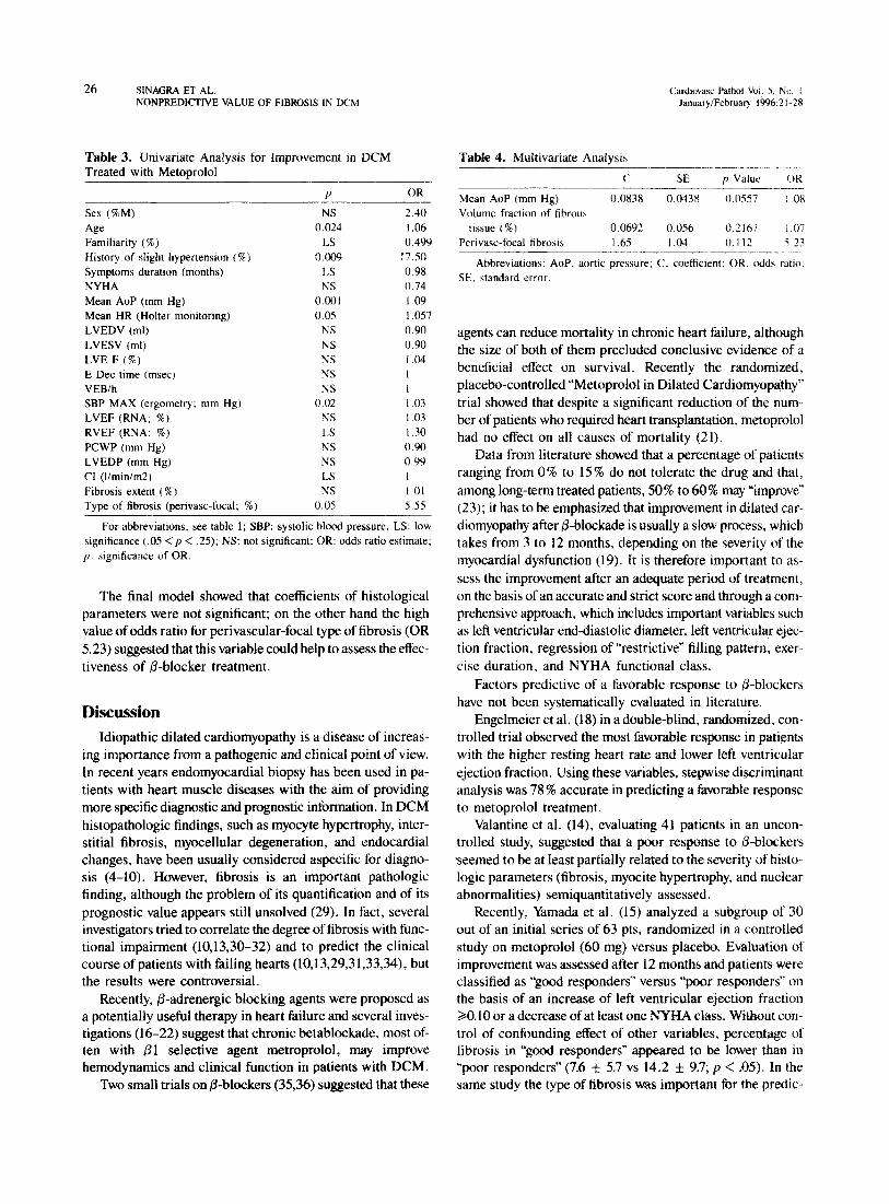
The distribution of fibrous tissue is shown in Table 2: nine pts of group A and 10 pts of group B had dominant pericellu- lar fibrosis, whereas 10 pts of group A and two pts of group B had dominant perivascular and/or focal replacement fibro- sis @ = .104).
Univariate analysis (Table 3) selected the following as significant predictors of improvement: history of slight hyper- tension @ = .009; OR 17.Q age @ = 02; OR 1.06), mean aortic pressure @ = .OOl; OR 1.09)) mean heart rate at Holter monitoring @ = .05; OR 1.057), maximal exercise systolic
Multivariate regression analysis was performed on the vari- ables with a univariate odds-ratio significance lower than 0.25 (Table 4), according to the suggestions of Hosmer et al. (28).
Mean aortic pressure was the only clinical relevant vari- able selected as significant at stepwise logistic regression @ = .05; OR 1.08).
Considering the high univariate odds ratio of dominant perivascular-focal fibrosis @ = .05; OR 5.5), we tried to test a model that considered the most clinically relevant variables and the type and extent of fibrosis.
Table 2. Extent and Type of Fibrosis in DCM Patients (endocardial fibrosis excluded from analysis; n = 14)
Group A (Improved) Group B (Not Improved) % of fibrosis n = 25 II = 20 P
Fibrosis distribution: Total 12.1 f 9.1 11.3 f 9.6 NS <lO 14 13 NS >lO-20 6 3 NS >20-30 3 3 NS >30 2 1 NS
Fibrosis type: Pericellular (n = 19) 9 10 Perivascular/focal (n = 12) 10 2
0.104 (Fisher test)
26 SINACXA ET AL. Cardwass Pathoi Vol. S, No. / NONPREDKITVE VALUE OF FIBROSIS IN DCM January/February 1996:21- ?X
Table 3. Univariate Analvsis for Imnrovement in DCM Treated with Metoprolol -
Table 4. Multivariate Analvsit,
..--__ ~- d
C SE p Value OR P OR
.--- Mean AoP (mm Hg) 0.0838 o.O43g---- o.oss7 !.OX
Sex (%M) NS 2.40 Volume fraction of fibrous Age 0.024 1.06 tissue (%) 0.0692 0.056 0.21h7 i.07
Familiarity (%) 13 0.499 Perivasc-focal fibrosis 1.65 I .04 0.1 1: 5 23 History of slight hypertension (%) Symptoms duration (months) NYHA
0.009 1,s NS
17.50 0.98 0.74
-----_______ Abbreviations: AoP, aortic pressure: C, coeffictent: OR. odds ratio.
SE. standard error
Mean AoP (mm Hg) 0.001 1.09 Mean HR (Holter monitoring) 0.0s I.057 LVEDV (ml) NS 0.90 LVESV (ml) NS 0.90 LVE F (F) NS 1.04 E Dee time (msec) NS I VEBlh NS I SBP MAX (ergometry: mm Hg) 0.02 1.03 LVEF (RNA; I) NS I .03 RVEF (RNA; %) 1.S 1.30 PCWP (mm Hg) NS 0.90 LVEDP (mm Hg) NS 0.99 CI (I/min/m2) LS I Fibrosis extent (%) NS 1.01 Type of fibrosis (perivasc-focal; %) 0.05 5.55
For abbreviations, see table I; SBP: systolic blood pressure. LS: low significance (.05 < p < .25); NS: not significant; OR: odds ratio estimate; p: significance of OR.
The final model showed that coefficients of histological parameters were not significant; on the other hand the high value of odds ratio for perivascular-focal type of fibrosis (OR 5.23) suggested that this variable could help to assess the effec- tiveness of P-blocker treatment.
Discussion Idiopathic dilated cardiomyopathy is a disease of increas-
ing importance from a pathogenic and clinical point of view. In recent years endomyocardial biopsy has been used in pa- tients with heart muscle diseases with the aim of providing more specific diagnostic and prognostic information. In DCM histopathologic findings, such as myocyte hypertrophy, inter- stitial fibrosis, myocellular degeneration, and endocardial changes, have been usually considered aspecific for diagno- sis (4-10). However, fibrosis is an important pathologic finding, although the problem of its quantification and of its prognostic value appears still unsolved (29). In fact, several investigators tried to correlate the degree of fibrosis with func- tional impairment (10,13,30-32) and to predict the clinical course of patients with failing hearts (10,13,29,31,33,34), but the results were controversial.
Recently, @-adrenergic blocking agents were proposed as a potentially useful therapy in heart failure and several inves- tigations (16-22) suggest that chronic betablockade, most of- ten with Pl selective agent metroprolol, may improve hemodynamics and clinical function in patients with DCM.
Two small trials on P-blockers (3536) suggested that these
agents can reduce mortality in chronic heart failure, although the size of both of them precluded conclusive evidence of a beneficial effect on survival. Recently the randomized, placebo-controlled “Metoprolol in Dilated Cardiomyopathy” trial showed that despite a significant reduction of the num- ber of patients who required heart transplantation, metoprolol had no effect on all causes of mortality (21).
Data from literature showed that a percentage of patients ranging from 0% to 15 % do not tolerate the drug and that, among long-term treated patients, 50% to 60% may “improve” (23); it has to be emphasized that improvement in dilated car- diomyopathy after P-blockade is usually a slow process, which takes from 3 to 12 months, depending on the severity of the myocardial dysfunction (19). It is therefore important to as- sess the improvement after an adequate period of treatment, on the basis of an accurate and strict score and through a com- prehensive approach, which includes important variables such as left ventricular end-diastolic diameter, left ventricular ejec- tion fraction, regression of “restrictive” filling pattern, exer cise duration, and NYHA functional class.
Factors predictive of a favorable response to &blockers have not been systematically evaluated in literature.
Engelmeier et al. (18) in a double-blind, randomized, con- trolled trial observed the most favorable response in patients with the higher resting heart rate and lower left ventricular ejection fraction. Using these variables, stepwise discriminant analysis was 78 % accurate in predicting a favorable response to metoprolol treatment.
Valantine et al. (14), evaluating 41 patients in an uncon- trolled study, suggested that a poor response to P-blockers seemed to be at least partially related to the severity of histo- logic parameters (fibrosis, myocite hypertrophy, and nuclear abnormalities) semiquantitatively assessed.
Recently, Yamada et al. (15) analyzed a subgroup of 30 out of an initial series of 63 pts, randomized in a controlled study on metoprolol (60 mg) versus placebo. Evaluation of improvement was assessed after 12 months and patients were classified as “‘good responders” versus “poor responders” on the basis of an increase of left ventricular ejection fraction 20.10 or a decrease of at least one NYHA class. Without con- trol of confounding effect of other variables, percentage of fibrosis in “good responders” appeared to be lower than in “poor responders” (7.6 + 5.7 vs 14.2 f 9.7; p < .05). In the same study the type of fibrosis was important for the predic-
Cardiovasc Pathol Vol. 5, No. I January/February 1996:21-28
tion of the effectiveness of long term P-blocker therapy. In fact dominant interfascicular fibrosis was significantly more frequent in “good responders”, whereas dominant intercellu- lar fibrosis was more frequent in “poor responders” (x2 = 11.8; p < .OOl).
It has however to be emphasized that in this study the num- ber of patients was small (30 pts), the follow-up relatively short (1 year), the dosage of &blockers not individualized (60 mg) and some criteria of improvement (NYHA functional class) at least partially subjective.
On the contrary, our patients had a longer follow-up (at least 2 years) and were treated with individualized dosage of metoprolol (between 50 and 300 mg/die); moreover, the criteria of improvement were strictly defined and based on several parameters, such as left ventricular ejection fraction, left ventricular end diastolic diameter, exercise duration, car- diothoracic ratio, evaluation of mitral flow at Echo-Doppler, and NYHA functional class.
In our study, no relation between clinical-instrumental parameters and extent or type of fibrosis was observed. Dom- inant perivascular-focal fibrosis, according to Yamada et al. (15), was associated with a favorable outcome, whereas the volume fraction of fibrous tissue was of no value for the predic- tion of response to P-blockers.
At univariate analysis dominant perivascular-focal fibro- sis showed a high odds ratio for improvement (p .05; OR 5.5), which persisted at multivariate logistic regression analysis (OR 5.23) despite the absence of a strict coefficient significance.
The study has some limitations such as a relatively small number of patients and raises some methodological problems on quantitative assessment of fibrosis at endomyocardial biopsy.
According to previous studies (31), we considered both right and left ventricles biopsies for analysis. The risk of a biased underestimation on final results is unlikely due to ho- mogeneous distribution of right side biopsies between sub- groups.
In our experience as in other morphometric studies (12, 37-41), another problem was that the percentage of fibrous tissue ranges widely. Baandrup et al. (40) and Schwarz et al. (41) comparing biopsies of the same patients showed a great variability in the estimation of fibrous tissue with a coefficient of variance more than 40 % . According to these authors we think that “the degree of fibrosis cannot be determined with accuracy when endomyocardial biopsy technique is used.”
In conclusion, as suggested by Yonesaka (4), the histologic assessment of interstitial fibrous tissue even when quantita- tively studied by morphometric techniques (10) does not neces- sarily allow a clear and unambiguous differentiation of vari- ous clinical subgroups, whereas a potentially higher predictive value may be attributed to a relatively simple parameter such as the “type” of fibrous tissue.
It is possible that a better standardization of procedures and evaluation of other variables, such as fiber diameter and
SINAGRA ET AL. 27 NONPREDICTIVE VALUE OF FIBROSIS IN DCM
myofibrillar volume fraction, could further contribute to the knowledge of the disease and help in patient management.
1.
2.
3.
4.
5.
6.
7. 8.
9.
10.
11.
12.
13.
14.
15.
16.
17.
18.
References Roberts WC. Defining idiopathic dilated cardiomyopathy: a courtroom discussion. Am J Cardiol 1989;63:893-896. Codd MB, Sugrue DD, Gersh BJ, Melton LJ: Epidemiology of idio- pathic dilated and hypertrophic cardiomyopathy. Circulation 1989; 80564-572. Lilienfeld DE, Spratka, JM, Pham DL, Baxter J. Morbidity from con- gestive and hypertrophic cardiomyopathy in the Minneapolis-St. Paul Metropolitan Area: 1979-1984. Cardiology 1992;80:71-76. Yonesaka S, Becker AE. Dilated cardiomyopathy: diagnostic accuracy of endomyocardial biopsy. Br Heart J 1987;58:156-161. Davies MJ. The cardiomyopathies: a review of terminology, pathol- ogy, and pathogenesis. Histopathology 1984;8:363-393. Camerini F, Salvi A, Sinagra G. Endomyocardial biopsy in dilated car- diomyopathy and myocarditis: which role? Int J Cardiol 1991;31: 1-8. Olsen EGJ. Endomyocardial biopsy. Br Heart J 1978$X95-98. Ferrans, UJ, Roberts WC. Myocardial biopsy: a useful diagnostic pro- cedure or only a research tool? Am J Cardiol 1978;41:%5-967. Olsen EGJ. Special investigation of CGCM: endomyocardial biopsies (morphologic analysis). Postgrad Med J 1978;54:486-490. Mall G, Schwarz F, Derks H. Clinicopathologic correlations in con- gestive cardiomyopathy-a study on endomyocardial biopsies. Virchows Arch (Path01 Anat) 1982;397:67-82. Tazelaar HD, Billingham M. Leukocytic intiltrates in idiopathic dilated cardiomyopathy. A source. of confusion with active myocarditis. Am J Surg Path01 1986;10:405-412. Unverferth DV, Baker PB, Swift SE, et al. Extent of myocardial fibro- sis and cellular hypertrophy in dilated cardiomyopathy. Am J Cardiol 1986;57:816-820. Figulla HR, Rahlf G, Nieger M, Luig H, Kreuzer H. Spontaneous hemodynamic improvement or stabilization and associated biopsy findings in patients with congestive cardiomyopathy. Circulation 1985;71:1095-1104. Valantine HA, Billingham ME, Heilbnmn SM, et al. Response to P-blockers in dilated cardiomyopathy predicted by myocardial biopsy. Circulation 1986;72:1174. Yamada Y, Fukunami M, Ohmuri M, et al. Which subgroup of patients with dilated cardiomyopathy would benefit from long-term @-blocker therapy? A histologic viewpoint. J Am Co11 Cardiol 1992;21:628-633. n Waagstein F, Hjalmarson A, Vamauskas E, Wallentin I. Effect of chronic p-adrenergic receptor blockade in congestive cardiomyopathy. Br Heart J 1975;37:1022-1036. Swedberg K, Hjalmarson .& Waagstein F, Wallentin I. Beneficial effects of long-term P-blockade in congestive cardiomyopathy. Br Heart J 1980;44:117-133. Engelmeier RS, O’Connel JB, Walsh R, Rad N, Scanlon PF, Gunnar RM. Improvement in symptoms and exercise tolerance by metoprolol in patients with dilated cardiomyopathy: a double-blind, randomized, placebo-controlled trial. Circulation 1985;72:536-546.
0 19. Waagstein F, Caidahl K, Wallentin I, Bergh CH, Hjahnarson A. Long-
term r%bloclutde in congestive cardiomyopathy: effects of short- and long- term metoprolol treatment followed by withdrawal and readministra- tion of metoprolol. Circulation 1989;80:551-563.
20. Anderson JL, Gilbert EM, O’Connel JB, et al. Long-term (2 year) beneficial elfects of &adrenergic blockade with bucindolol in patients with idiopathic dilated cardiomyopathy. J Am Co11 Cardiol 1991; 17:1373-1381.
21. Waagstein F, Bristow MR. Swedbetg K, et al. Beneficial effects of
28 SINAORA ET AL. NONPREDICTIVE VALUE OF FIBROSIS IN DCM
Cardim-asc Path01 Vol. 5. No. I Janwy/Fehwy 1994:21-28
22.
23.
24.
25.
26.
27.
28.
29.
30.
31.
32.
metopm~ol in idbpaw dihted ctardiomyopathy. Results of the metopmlol in dilated cardiow (MIX) trial. A randomized, double-blind, placebo-controlled multicenter study. Lancet 1993;342:1441-1446. Sinagra G, Ferkan A, Di Lena&t A, et al. I Betabloccanti nella terapia della cardiomiopatia dilatativa: revisione delle letterature ed esperienza clinica su 67 pazienti. G Ital Cardiol 1992;22:%9-989. Eichhorn EJ. The paradox of &adrenergic blockade for the manage- ment of congestive heart failure. Am J Med 1992;92:527-538. Report of the WHO/ISFC task force on the definition and classification of cardiomyopathies. Br Heart J 1980,44:672-673. Baa&up U, Olsen EGJ. Critical analysis of endomyocardial biopsies from patients suspected of having cardiomyopathy. I: Morphologic and morphomehc aspects. Br Heart J 1981;45:475-486. Hauck AJ, Edwards WD. Histopathologic examination tissues obtained by endommi biopsy. In: Fmvles RE, eds. Cardiac Biopsy. New York: Futura, 1992:98-100. Anderson KR, Sutton MGS, Lie JT. Histopatbologic types of cardiac fibrosis in myocardial disease. J Path01 1979;128:79-85. Hosmer DV, Lemeshow S. Applied Logistic Regression. New York: John Wiley and Sons, 1989:108. Schwarz F, Mall G, Z.&e H, et al. Determinants of survival in patients with congestive cardiomyopathy: quantitative morphologic findings and left ventricular hemodynamics. Circulation 1984;70:923-928.
Nakayama Y, Shimuzi G, Hirota Y, et al. Functional and hystopatho- k&correlation in patients with dilated cardiomyopathy: an integrated evah&on by muitivariate analysis. J Am Coil Catdid 1987,10:186-192.
Baa&up U, Florio RA, Rehan M, Richardson PJ, Olsen GJ. Critical analysis of endomyucardial biopsies from patients suspected of having cardiomyopathy. II: Comparison of histology and clinic&hemodynamic information. Br Heart J 1981;45:487-493.
Unverferth DV, Magorien RD, Kolibash AJ, et al. Biochemical and histo-
33.
34.
35.
36.
37.
38.
39.
40.
41.
logic correlates of ventricular end-diastolic pressure. int J Cardiol 1981;1:133-142. Unverferth DV, Magorien RD, Moeschberger ML, Baker P, Fetters JK, Leier CV. Factors infIuencing the l-year mortality of dilated cardiomy- opathy. Am J Cardiol 1984;54:147-152. Popma JJ, Cigarroa RG, Buja LM, Hillis LD. Diagnostic and prognos- tic utility of right-sided catheterization and endomyocardial biopsy in idiopathic dilated cardiomyopathy. Am J Cardiol 1989;63:955-958. Anderson JL, Lutz JR, Gilbert EM, et al. A randomized trial of low- dose B-blockade therapy for idiopathic dilated cardiomyopathy. Am J Cardiol 1985;55:471-475. Swedberg K, Hjalmarson A, Waagstein F, Waltentin I. Prolongation of survival in congestive cardiomyopathy by p-receptor blockade. Lancet 1979;1:1374-1376. Meckel CR, Wilson JE, Sears TD, Rogers JG, Goaley TJ, McManus BM. Myocardi fibrosis in endomyocardial biopsy specimens: do differ- ent bioptomes affect estimations? Am J Cardiovasc Pathol 1989;2:309-313. Hess OM, Schneider J, Koch R, Bamert C, Grimm J, Krayenbuehl HP. Diastolic function and myocardial structure in patients with myocardial hypertrophy. Special reference to normalized viscoelastic data. Circu- lation 1981;63:360-371. Kunkel B, Lapp H, Kober G, Kaltenbach M. Light microscopic evalua- tion of mycmxrdial biopsies. In: Kaltenbach, Loogen, Olsen, eds. Berlin- Heidelberg/New York; Springer, 1978:63-70. Baa&up U, Florio RA, Olsen EG. Do endomyocardial biopsies repre- sent the morphology of the rest of the myocardium? A quantitative light microscopic study of single versus multiple biopsies with the King’s bi- optome. Eur Heart J 1982;3:171-178. Schwarz F. Mall G, Z&e H, et al. Quantitative morphologic lindings of the myocardium in idiopathic dilated cardiomyopathy. Am J Cardiol 1983;51:501-506.