Embed Size (px)
Citation preview

This Provisional PDF corresponds to the article as it appeared upon acceptance. Fully formattedPDF and full text (HTML) versions will be made available soon.
Norepinephrine stimulates progesterone production in highly estrogenic bovinegranulosa cells cultured under serum-free, chemically defined conditions
Reproductive Biology and Endocrinology 2012, 10:95 doi:10.1186/1477-7827-10-95
Carla A Piccinato ([email protected])Luis H Montrezor ([email protected])
Cristhianna AV Collares ([email protected])Alessandra A Vireque ([email protected])
Alzira AM Rosa e Silva ([email protected])
ISSN 1477-7827
Article type Research
Submission date 28 July 2012
Acceptance date 20 November 2012
Publication date 22 November 2012
Article URL http://www.rbej.com/content/10/1/95
This peer-reviewed article can be downloaded, printed and distributed freely for any purposes (seecopyright notice below).
Articles in RB&E are listed in PubMed and archived at PubMed Central.
For information about publishing your research in RB&E or any BioMed Central journal, go to
http://www.rbej.com/authors/instructions/
For information about other BioMed Central publications go to
http://www.biomedcentral.com/
Reproductive Biology andEndocrinology
© 2012 Piccinato et al.This is an open access article distributed under the terms of the Creative Commons Attribution License (http://creativecommons.org/licenses/by/2.0),
which permits unrestricted use, distribution, and reproduction in any medium, provided the original work is properly cited.

Norepinephrine stimulates progesterone production
in highly estrogenic bovine granulosa cells cultured
under serum-free, chemically defined conditions
Carla A Piccinato1,4,*
Email: [email protected]
Luis H Montrezor2,3
Email: [email protected]
Cristhianna AV Collares2
Email: [email protected]
Alessandra A Vireque2,4
Email: [email protected]
Alzira AM Rosa e Silva5
Email: [email protected]
1 Hospital Israelita Albert Einstein, São Paulo, Brazil
2 Department of Physiology, School of Medicine of Ribeirão Preto, Universidade
de São Paulo, São Paulo, Brazil
3 Barão de Mauá University, Ribeirão Preto, São Paulo, Brazil
4 Department of Gynecology and Obstetrics, School of Medicine of Ribeirão
Preto, Universidade de São Paulo, São Paulo, Brazil
5 Department of Physiological Science, Biological Sciences Institute,
Universidade de Brasília, Brasília, Brazil
* Corresponding author. Hospital Israelita Albert Einstein, São Paulo, Brazil
Abstract
Background
Since noradrenergic innervation was described in the ovarian follicle, the actions of the
intraovarian catecholaminergic system have been the focus of a variety of studies. We aimed
to determine the gonadotropin-independent effects of the catecholamine norepinephrine (NE)
in the steroid hormone profile of a serum-free granulosa cell (GC) culture system in the
context of follicular development and dominance.

Methods
Primary bovine GCs were cultivated in a serum-free, chemically defined culture system
supplemented with 0.1% polyvinyl alcohol. The culture features were assessed by hormone
measurements and ultrastructural characteristics of GCs.
Results
GCs produced increasing amounts of estradiol and pregnenolone for 144h and maintained
ultrastructural features of healthy steroidogenic cells. Progesterone production was also
detected, although it significantly increased only after 96h of culture. There was a highly
significant positive correlation between estradiol and pregnenolone production in high E2-
producing cultures. The effects of NE were further evaluated in a dose–response study. The
highest tested concentration of NE (10 (−7) M) resulted in a significant increase in
progesterone production, but not in estradiol or pregnenolone production. The specificity of
NE effects on progesterone productio n was further investigated by incubating GCs with
propranolol (10 (−8) M), a non-selective beta-adrenergic antagonist.
Conclusions
The present culture system represents a robust model to study the impact of intrafollicular
factors, such as catecholamines, in ovarian steroidogenesis and follicular development. The
results of noradrenergic effects in the steroidogenesis of GC have implications on
physiological follicular fate and on certain pathological ovarian conditions such as cyst
formation and anovulation.
Keywords
Norepinephrine, Granulosa cells, Progesterone, Estradiol, Catecholamines, PVA, Bovine,
Steroidogenesis.
Background
The intraovarian regulatory system that modulates the response to gonadotropins and ovarian
functions is complex. Neurotransmitters, such as catecholamines, have been considered as
intraovarian regulators due to the identification of intrinsic catecholamine synthesis and the
presence of extensive sympathetic innervation in the mammalian ovary [1-4]. In fact, the
mammalian ovary is richly supplied by sympathetic fibers that innervate the interstitium and
the perifollicular regions of developing follicles, including the external thecal layer, but do
not reach the GC layer [3]. Although some minor species differences may exist regarding
localization, distribution, and density, the sympathetic innervation of the ovary is overall
similar in many animals [2,3,5], including ruminants [6]. Among the catecholamines,
norepinephrine (NE) is the most abundant neurotransmitter released by sympathetic nerves in
the mammalian ovary [7].
The proposed mechanism of action for NE in the ovary involves binding to ovarian
adrenergic receptors [8,9], induction of cAMP, leading to follicular development [10]. In
addition, few studies have evaluated the effects of NE on the synthesis of the key ovarian

hormones progesterone (P4) and estradiol (E2). Norepinephrine has been previously shown to
induce P4 production in the bovine corpus luteum [11,12]. However, the effects of NE in E2
production by ovarian follicular granulosa cells (GCs) have not been demonstrated in
mammalian species, despite the known elevation of catecholamine levels in the estrogenic
pre-ovulatory phase of the rodent estrous cycle [13] and the presence of high concentrations
of NE in the follicular fluid and follicular wall of both, small antral and pre-ovulatory
follicles of cows [14]. Although NE is known to stimulate ovarian steroidogenesis via a
gonadotropin-mediated mechanism [9,13], there is little evidence of a direct mechanism of
action of NE on specific steroid hormone synthesis. Moreover, the effects of NE in the highly
steroidogenic growing follicle have not yet been studied. The lack of an appropriate model
for testing both gonadotropin-dependent and -independent NE effects possibly explains our
limited understanding of this physiological situation.
The main physiological characteristic of healthy, non-atretic growing follicles is their ability
to maintain steroidogenic activity towards follicular dominance. Conversely, the unhealthy
atretic follicle shows low E2 production and high P4 concentration in the follicular fluid [15].
Although in vitro studies involving cultured GCs have expanded our knowledge on follicular
steroidogenesis and development, only GCs in serum-free cultures maintained morphological
characteristics of healthy non-atretic growing follicles and maintained their main ability to
produce high levels of E2 [16,17]. Furthermore, serum-free cultures represent a robust model
in which intraovarian local factors can be precisely and directly evaluated, without the
confounding interference of steroids and growth factors.
Although some evidence exists for the role of catecholamines in stimulating ovarian
steroidogenesis, it is proposed to study the effects of NE on GC steroidogenesis. Our first
objective was to characterize a serum-free bovine GC culture under a chemically defined
medium supplemented with polyvinyl alcohol (PVA). Our second and main objective was to
identify the effects of various physiological concentrations of NE on the synthesis of P4, E2,
and pregnenolone (P3) and so, infer how NE would affect follicular fate in serum-free,
gonadotropin-independent conditions.
Methods
Culture of bovine granulosa cells
Bovine ovaries from animals from the slaughterhouse were transported on PBS at 37°C,
rinsed with 70% ethanol, and rinsed in alpha-MEM (alpha-Minimum Essential Medium,
Gibco-BRL, Grand Island NY, USA). Follicles that showed a well vascularized capsule,
contained clear follicular fluid, and were between 3 and 5mm in diameter were selected.
Thus, most follicles were expected to be non-atretic and to have emerged after a new
follicular wave, although prior to the selection of the single dominant follicle. To harvest the
GCs, the selected follicles were either aspirated with a 27G syringe or bisected (and gently
washed 3 times) in supplemented alpha-MEM. Cells were washed twice using low-speed
centrifugation (500g) followed by resuspension in alpha-MEM, and the number and viability
of GCs were determined by Trypan blue exclusion. Cell culture procedures were based on a
previous work [16], with modifications. Briefly, 500,000 cells/well were seeded into 24-well
culture plates in alpha-MEM supplemented with 10mM sodium bicarbonate, 20mM Hepes,
antibiotics (1000 U/ml penicillin and 1000 μg/ml streptomycin), 1.4 ng/ml sodium selenite,
5μg/ml transferrin, 0.1 μg/ml insulin, 10 ng/ml human recombinant IGF-1, and 11mM non-

essential amino acids. All above reagents were obtained from Gibco BRL (Grand Island NY,
USA). In addition, 0.1% PVA (Sigma Chemical Co, St. Louis, MO, USA) was added to
adjust oncotic pressure of the medium (usually achieved with the addition of serum) and to
avoid steroid contaminants of other serum replacers [18,19]. The E2 precursor,
androstenedione (A4), cannot be synthesized by GCs and was also added to the medium (10-7
M, Sigma Chemical Co, St. Louis, MO, USA) in physiological conditions. The plates were
incubated with 5% CO2 at 37°C for 144h (6 days) with change of 70% of the medium every
48h. Media from all experiments were collected and stored at −20°C for posterior analysis of
steroid hormones and cholesterol. All experiments were done in accordance to the
institutional Research Ethics and Animal Care Committee at School of Medicine of Ribeirão
Preto (São Paulo University).
Experimental design
A total of 3 different sets of culture experiments were designed to determine the effects of
norepinephrine in regulating steroid hormone production in bovine GCs. First we
characterized the synthesis of steroid hormones and ultrastructural features of the GCs in the
chemically defined medium supplemented with PVA. A time-course analysis for E2, P3, and
P4 production, as well as cholesterol and A4 concentration and for the E2:P4 ratio was done
at 0, 48, 96, and 144h. Based on these results we selected only high E2 cultures to better
represent GC from non-atretic growing follicles for the following experiments. Bovine
growing follicles are characterized by an increasing production of E2 whereas a pronounced
P4 production is only observed before ovulation [15]. Next, dose–response and time-course
experiments evaluated NE effects on estrogenically active CGs by the addition of 3 levels of
NE concentration (0, 10-9
, 10-8
, and 10-7
M NE [Hipolabor, Belo Horizonte, Brazil]) at
different time points: 0, 48, and 96h. Physiological NE concentrations were chosen based on
previous studies [14]. Additionally, we evaluated whether the effects of NE occurred via
beta-adrenergic receptors in the cultured GCs at 48h by using 10-8
M propranolol
hydrochloride diluted in water (Wyeth-Ayerst Pharmaceuticals, Maidenhead, UK) as a
specific beta-adrenergic antagonist. Propranolol was added to the cultures 20 min before NE
challenge. Sample collection for all experiments occurred after 1h of NE challenge at the
specified time points.
Hormone and cholesterol assays
Concentrations of E2, A4, P4, and P3 in culture media of GCs were measured with
radioimmunoassay using the method described by [20] and modified in our laboratory. The
antibodies were generously donated by Dr. Bélanger and Dr. Labrie, CHUL, Quebec, CA.
Adequacy of method was evaluated by the parallelism of the slopes from serially diluted
medium with the standard curve of each steroid hormone after natural log-logit
transformation. The intra-assay and inter-assay coefficients of variation were respectively
<5% and <10% for hormone assay. Concentration of total cholesterol in the culture media
was evaluated by Trinder-enzymatic assay (COD-ANA Colesterol Liquiform, Labtest
Diagnostica SA, Lagoa Santa, Brazil).
Ultrastructural analysis of granulosa cells
For ultrastructural analysis, GCs were harvested and cultivated as described in the previous
section, but at a greater density (106 viable cells/well), in a 6-well plate (Nunc, Roskilde,
Denmark) until 96h (when results from hormone production better represented the growing

healthy GC). Cells were washed twice in PBS and 0.1M cacodylate buffer (pH 7.4). A total
of 20 μl of fixation solution (2% glutaraldehyde, 0.05% calcium chloride, and 2%
formaldehyde) was added to the wells for 2h at room temperature. Following fixation,
samples were washed twice with 0.1M cacodylate buffer and postfixed in 1% osmium
tetroxide, rinsed in double distilled water, dehydrated in increasing concentrations of ethanol,
rinsed with acetone, and embedded in resin (Embed 812, EMS, Fort Washington, PA, USA).
The resin blocks were cut by ultra-microtomy into 0.5 μm slices. Cellular ultrastructure was
observed by a Phillips EM 208 electron microscope (Eindhoven, Netherlands).
Statistical analysis
All data analyses were performed using SAS software version 6.11 (Statistical Analysis
System Institute Inc., Cary, NC, USA). Regression analysis with repeated measurements was
performed using MIXED procedures of SAS. The response variables were the production of
E2, P4, and P3, the concentration of A4 and cholesterol, and E2:P4 ratio. Final production of
hormones (E2, P4, and P3) by GCs was calculated by adding up the concentrations measured
at each time. Since only 70% of the medium was changed every time, mathematical
calculation were performed to remove the influence of the remaining medium when
calculating steroid production over time. In brief, the remaining medium left in the wells was
accounted by subtracting 30% of the hormone measured at the previous time in the
calculation of the hormone production of the subsequent time point. Based on the
characterization experiments, we were able to separate cell cultures into estrogenically active
(here called high E2-producing cultures) and estrogenically inactive (low E2-producing
cultures) cultures. These two subgroups were included in the statistical model. Thus, the
statistical model included experimental culture (estrogenically active or inactive), time, and
replicate. Autoregressive correlation coefficient structure for unequally spaced time data was
applied when evaluating all time points. Pearson correlation coefficients were used to
compare accumulated production levels between hormones that were synthesized by GCs.
The statistical model for the analysis of the effects of different NE levels (0, 10-9
, 10-8
, and
10-7
M) did not used the last time point (144h), based on the results of the GC model
characterization experiments. The addition or not of propranolol (at time 48h) was also
included in a last regression analysis.
For all experiments, the results represented the least squares means of hormone concentration
± the SEM of at least seven (characterization experiments) or five (NE and propranolol
experiments) different experiments performed in triplicate wells. Multiple paired
comparisons were adjusted according to Tukey-Kramer. In all analysis, the level of
significance was set at p<0.05.
Results
Characterization of bovine granulose cells cultured in a chemically defined
medium
Ultrastructural features of granulosa cells
After initial seeding in medium supplemented with 0.1% PVA, bovine GCs aggregated into
attached clusters within the first 12h (data not shown). Cluster formation allowed interactions

between cells possibly favoring GC polyhedral shape, as well as their high nucleus/cytoplasm
ratio and dispersed cytoplasmic lipid droplets, for up to 96h (Figure 1A). Smooth and rough
endoplasmic reticulum and free ribosomes were abundant in the cytoplasm of clustered cells,
and endoplasmic reticulum was frequently observed in the periphery of the cytoplasm.
Mitochondria with tubulovesicular cristae and lipid droplets were randomly distributed
through the cytoplasm (Figure 1A and C). Figure 1 (A and C) depicts a high
nucleus/cytoplasm ratio cell that was frequently observed in the ultra-thin slices. In contrast,
elongated cells (suggestive of fibroblast-like cells, previously described as cells that sustain
the cluster of differentiated GC [21]), were not frequently detected and had a low
nucleus/cytoplasm ratio and greater amount of lipid droplets (Figure 1B). Cells were attached
to each other via desmosomes and tight junctions (Figure 1D).
Figure 1 Electron micrographs of a bovine granulosa cells. Electron micrographs of a
bovine granulosa cells with high nuclear:cytoplasm ratio at 0h of culture (A; 0h of culture;
magnification: x 6800) and elongated cell with low nuclear:ctoplasm ratio that was randomly
found in the culture (B; 96h of culture; magnification: x 2720). Note the high concentration
of lipid droplets and smaller nucleus in the elongated cell. Bovine granulosa cells presented
polyhedral shape, dispersed lipid droplets, and mitochondrias with tubovesicular cristae. (C;
48h; magnification bar: x 8500). Neighboring cells within the cluster had rough endoplasmic
reticulum, free ribosomes and cellular communication described as tight junctions (D; 48h;
magnification bar: x 12500). mi = mitochondria; li= lipid droplets; n=nucleus; c= cytoplasm,
rer= rough endoplasmic reticulum; r= ribosomes, arrow=tight junctions
Steroidogenic capacity of granulosa cells
The steroidogenic capacity towards follicular dominance of GCs in chemically defined
medium with PVA was evaluated based on the production of E2, P4, and P3, and on the
E2:P4 ratio. Overall, GCs produced increasing amounts of E2 over time. The large variation
in the level of E2 production among culture experiments allowed for the classification of two
distinct groups of GC cultures: high E2 (E2 ≥ 1ng/ml from 48h and after) and low E2
producers (E2 < 1ng/ml at 48h and after). Such classification permitted the selection of more
estrogenically active cultures that better represented the healthy follicle growing towards
deviation/dominance. Figure 2A shows E2 production of both groups of GCs cultured under
defined conditions.
Figure 2 Estradiol, progesterone, and pregnenolone production by bovine granulosa
cells. Time-effect on production of estradiol (A), progesterone (B), and pregnenolone (ng/ml)
(C) by bovine granulosa cells cultivated under chemically defined conditions supplemented
with PVA. Values are least square means ± SEM (n=7 independent cultures). a, b
unlike
superscripts differ P<0.05
Estradiol production in high E2-producing cultures reached 1.80 ± 0.25, 3.13 ± 0.25, and 3.84
± 0.29 ng/ml at 48, 96, and 144h, respectively. The low E2-producing group presented
significantly lower E2 concentrations at all evaluated time points (0.04 ± 0.34, 0.029 ± 0.34,
0.13 ± 0.34 ng/ml; P<0.0001). An interaction between groups and time was noted for E2
production (p=0.0038), indicating that the culture of cells with low E2 production did not
change over time.
Progesterone production was also greater in high E2-producing cultures (Figure 2B,
P=0.014). There was an increase of P4 production over time (P<0.001), and an interaction

between time and culture groups was detected (P=0.009). Progesterone production in high
E2-producing cultures at 48, 96, and 144h reached 52.1 ± 25.3, 143.2 ± 25.1, and 243.2 ±
28.9 ng/ml, respectively; whereas low E2-producing cultures had lower P4 production
(respectively, 29.7 ± 31.5, 43.16 ± 32.9, and 57.2 ± 34.1 ng/ml). However, there was no
difference between high E2 cultures and low E2 cultures at 48h (P=0.58). Also, in high E2-
producing cultures, P4 production significantly increased only after 96h. Regarding the E2:P4
ratio, only a tendency of culture group effect was detected (P=0.055; data not shown). High
E2-producing cultures tended to have greater E2:P4 ratio than low E2-producing cultures at
48h (P=0.07) and 96h (P=0.09), but not at 144h (P=0.42).
Similarly, results of P3 production from GCs under our research conditions indicated an
effect of culture groups (P<0.023), time (P<0.0001), and their interaction (P=0.001). Results
shown in Figure 2C indicated that P3 production was greater in high E2-producing cultures at
96h (11.24 ± 1.62 vs. 5.19 ± 2.13 ng/ml; p=0.036) and 144h (20.33 ± 1.89 vs. 7.35 ± 2.21
ng/ml; p=0.0003), but not at 48h (4.39 ± 1.64 vs. 5.71 ± 2.04 ng/ml; P>0.05) when compared
to low E2-producing cultures.
Interestingly, while there was a significant high positive correlation between E2 and P3
production in high E2-producing cultures (ρ=0.767, P<0.0001), no correlation was detected
in low E2-producing cultures (ρ=−0.052, P=0.82). No significant correlation was detected
between E2 and P4 production in low E2-producing cultures (ρ=0.37, >0.05) or high E2-
producing cultures (ρ=0.482, P>0.05). A positive correlation between P4 and P3 production
was detected in high E2-producing cultures (ρ=0.482, P=0.003) and also in low E2 cultures
(ρ=0.497, P=0.015). Correlation data can be found in Table 1.
Table 1 Correlation and significance between hormone productions
Group Estradiol Progesterone Pregnenolone
High E2 GC
Estradiol -- 0.31 (0.07) 0.77 (<.0001)
Progesterone 0.31 (0.070) -- 0.48 (0.003)
Pregnenolone 0.77 (<.0001) 0.48 (0.003) --
Low E2 GC
Estradiol -- 0.37 (0.09) 0.05 (0.82)
Progesterone 0.37 (0.09) -- 0.49 (0.015)
Pregnenolone 0.05 (0.82) 0.49 (0.015) --
Correlations and significance between estradiol, progesterone, and pregnenolone productions
(ng/ml) by high and low estrogen-producing granulosa cells cultivated under chemically
defined conditions
Cholesterol and androstenedione concentration
Cholesterol and A4 were included in the culture medium as precursors of the main follicular
hormones. Cholesterol concentrations did not change during 144h of culture and no
difference was detected between high and low E2-producing cells (P>0.05). High E2 cultures
showed cholesterol concentrations of 64.2 ± 24.5, 42.2 ± 16.3, 64.1 ± 16.2, and 41.7 ± 19.2
μg/ml at times 0h, 48h, 96h, and 144h, respectively; whereas low E2 cultures showed 64.2 ±
24.5, 16.6 ± 24.9, 20.1 ± 30.2, and 25.0 ± 30.5 μg/ml. Similarly, A4 concentrations did not
change over time nor between groups (P>0.05). Androstenedione concentrations in high E2

cultures were 46.9 ± 11.7, 27.3 ± 9.22, 33.4 ± 9.2, and 40.5 ± 10.1 ng/ml at times 0h, 48h,
96h, and 144h, respectively. In low E2-producing cells, concentrations of A4 were 46.9 ±
11.7, 53.33 ± 9.1, 57.0 ± 11.9, and 52.0 ± 12.2 ng/ml at the same respective times.
Effects of norepinephrine on the steroidogenesis of high E2-producing
granulosa cells
Based on our results we established that the high E2 in vitro model represents the in vivo GCs
of a follicle growing towards dominance. The effects of different NE concentrations (0, 10-9
,
10-8
, 10-7
M) on steroid hormone production were then evaluated in the high E2-producing
cells for 96h and are shown in Tables 2, 3 and 4. An effect of time was found (P=0.001), but
not of NE concentration (P=0.82) or of time*concentration interaction (P=0.72) on E2
production. Progesterone production tended to be affected by NE concentration (P=0.07), but
not by time (P=0.61) nor by the interaction between concentration and time (P=0.14). At 48h,
the lower concentration of NE (NE 10-9
M) had no effect on P4 production (P=0.16), while
the higher concentrations of NE (10-8
and 10-7
M) increased P4 production (P=0.03 and
P=0.0007, respectively) when compared to control. A difference in P4 production was also
detected between the lower concentration of NE (10-9
M) and the higher concentration (10-
7M, P=0.03). Similarly to what was observed with E2 production, there was an effect of time
(P<0.0001), but not of NE concentration (P=0.29) or time by NE concentration (P= 0.91) on
P3 production (Table 4).
Table 2 Time and dose-dependent effects of norepinephrine on estradiol production
Time (h) Norepinephrine (M)
0 10-9
10-8
10-7
0 0.11 ± 0.4a 0.11 ± 0.4
a 0.11 ± 0.4
a 0.11 ± 0.4
a
48 1.83 ± 0.4b 2.61 ± 0.42
b 2.39 ± 0.4
b 2.1 ± 0.38
b
96 2.25 ± 0.5b 2.25 ± 0.42
b 2.37 ± 0.4
b 2.60 ± 0.4
b
Time and dose-dependent effects of norepinephrine on estradiol (ng/ml) production by highly
estrogenic bovine granulosa cells cultivated with 0.1% PVA supplementation a, b
Means within a column with unlike superscripts differ (P < 0.05); values are least square
means ± SEM (n=5 independent cultures)
Table 3 Time and dose-dependent effects of norepinephrine on progesterone production
Time (h) Norepinephrine (M)
0 10-9
10-8
10-7
0 46.9 ± 38.5a 46.9 ± 38.5
a 46.9 ± 38.5
a 46.9 ± 38.5
a
48 37.0 ± 6.0ab,A
125.5 ± 43.0ab,AB
171.0 ± 40.5b,BC
251.0 ± 38.5b,C
96 139.9 ± 49.7b 175.0 ± 43.0
b 188.3 ± 43.0
b 154.8 ± 43.0
ab
Time and dose-dependent effects of norepinephrine on progesterone (ng/ml) production by
highly estrogenic bovine granulosa cells cultivated with 0.1% PVA supplementation a, b
Means within a column with unlike superscripts differ (P < 0.05); A, B,C
Means within a
row with unlike superscripts differ (P < 0.05); values are least square means ± SEM (n=5
independent cultures)

Table 4 Time and dose-dependent effects of norepinephrine on pregnenolone
production
Time (h) Norepinephrine (M)
0 10-9
10-8
10-7
0 2.1 ± 1.2a 2.1 ± 1.2
a 2.1 ± 1.2
a 2.1 ± 1.2
a
48 2.5 ± 1.6a 4.7 ± 1.4
a 4.6 ± 1.3
a 4.4 ± 1.2
a
96 9.0 ± 1.6b 12.4 ± 1.4
b 10.6 ± 1.4
b 11.9 ± 1.4
b
Time and dose-dependent effects of norepinephrine on pregnenolone (ng/ml) production by
highly estrogenic bovine granulosa cells cultivated with 0.1% PVA supplementation a, b
Means within a column with unlike superscripts differ (P < 0.05); values are least square
means ± SEM (n=5 independent cultures)
Cholesterol was already present in the medium and A4 was included in the culture medium.
Both steroids are precursors of the main hormones produced by GCs. According to our
results, the concentrations of steroid hormone precursor cholesterol and A4 did not change
upon NE challenge (P>0.05) nor after 96h of culture (P>0.05) (data not shown).
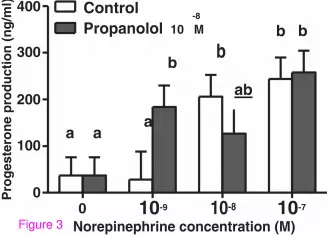
Effects of propranolol on the blockage of NE-mediated steroidogenesis of high
E2-producing granulosa cells
The specificity of NE effects on steroidogenesis was investigated by incubating GCs with
propranolol (10-8
M), a non-selective beta-adrenergic antagonist. The greatest accumulation of
P4 in media in the presence of NE was observed at 48h of incubation. Consequently, we
investigated the effects of propranolol only in regard to P4 production at 48h (Figure 3). At
the dose tested, propranolol failed to reverse the effects of NE on P4 secretion (P=0.46),
regardless of NE concentration. Unexpectedly, when GCs were exposed to the lower level of
NE, propranolol tended to enhance the stimulatory effect of NE (P=0.05). Similarly to our
previous results, there was an overall effect of NE levels (P=0.005) on P4 production. There
was a ~5.6 fold increase in P4 production (see Figure 3) in the presence of 10-8
NE, with no
change by propranolol addition. Similarly, regardless of propranolol addition, a 6.6 fold
increase in P4 production was detected in the presence of 10-7
NE (P= 0.009 and P=0.002,
respectively).
Figure 3 Propranolol effect on norepinephrine-induced progesterone production. Effect
of propranolol on norepinephrine-induced progesterone production (ng/ml) in highly
estrogenic bovine granulosa cells cultivated under chemically defined conditions
supplemented with PVA for 48h. Values are least square means ± SEM (n=5 independent
cultures). a, b
unlike superscripts differ P<0.05
Discussion
The actions of the intraovarian catecholaminergic system have been the focus of a number of
studies since the discovery of noradrenergic innervation in the ovarian follicle. The present
investigation describes the effects of NE in the steroid hormone profile of a serum-free GC
culture system in the context of follicular development and dominance. Our initial aims were
to evaluate how effectively the system maintains GC features and follicular steroidogenesis
and also, to identify the beta-adrenergic modulation of hormone production. The high E2-
producing culture described here represents the ovarian follicle that is growing towards

dominance, given that such culture system had greater production of P3, high positive
correlation between pro-dominance hormones E2 and P3, and tended to have high E2:P4
ratio. In this physiologically relevant system, it was possible to observe NE stimulation on P4
production, which suggests a pro-luteinization function of NE in the growing follicle.
To our knowledge, the present study is the first to examine the effects of NE on
gonadotropin-independent steroidogenesis by primary serum-free GC cultures with high E2
production. The finding that NE is a modulator of follicular steroidogenesis is relevant for a
better understanding of local intrafollicular regulatory mechanisms because NE is released by
terminal innervations in the outer theca cells and there is evidence of de novo synthesis of NE
by oocytes [1] and interstitial neuron-like cells [22]. Furthermore, NE binds to adrenergic
receptors which are mostly concentrated in other compartments, such as at interstitial and
theca cells [23-25]. Thus, NE has the potential to function as an intrafollicular paracrine
regulator of GCs. Our finding that NE increased P4 production by bovine GCs in a dose-
dependent manner agrees with previous reports that exogenous delivering of NE to cows
during the luteal phase [26] and in vitro to granulosa-luteal cells [12] enhanced P4
production. However, in both in vivo and in vitro studies mentioned above, GCs were
evaluated during the luteal phase. In cows, during this phase, circulating levels of P4 are
already high and follicular E2 production is low. Moreover, these models also presented
interfering factors (gonadotropins and serum contaminants, respectively) that prevent a
proper evaluation of gonadotropin-independent hormone production. These limitations
highlighted the need for a proper and controlled in vitro model that allows the investigation
of the effects of NE and other putative intraovarian factors in the follicles in a more
physiologically relevant state in regards to the modulation of follicular development and
dominance.
Steroid hormone production by GCs may vary under different culture conditions. Most
serum-based GC culture systems are not adequate for studying the actions of intrafollicular
factors because the serum itself already contains an undefined combination of hormones,
nutrients, and both growth and attachment factors that frequently leads to GC proliferation
[27] and/or luteinization [19]. In fact, E2 production, a pro-dominance marker of GC,
declines during culture in serum-coated dishes [18,21]. In the serum-free culture system
described here, GCs cultivated under chemically defined medium supplemented with PVA
continued to secrete E2 throughout the 144h of culture under gonadotropin-independent
conditions. High E2 production by GCs cultured in the presence of bovine serum albumin or
other serum replacers has also been described in earlier studies [16,28]. Although attempts to
propagate bovine GCs using serum replacers have succeeded in some cases [16,28,29], some
of those serum replacers were contaminated with steroids and other interfering factors [19].
Previous experiments from our group suggest that replacement with PVA (and additional
defined supplements) allows for greater and earlier E2 production by GCs when compared
with bovine serum albumin, a widely used serum replacer [18].
Pregnenolone is an important intermediary metabolite for E2 production and is synthesized at
both follicular compartments: granulosa and theca cell [30]. However, only theca cells
express the enzymes that convert P3 into androgens, which, in turn, are the direct precursors
of E2. Also, the concomitant expression of aromatase (the main E2 synthesizing enzyme) and
of cytochrome P450 11A1 (enzyme that converts cholesterol in P3) have been associated to
healthy growing follicles greater than 4mm in diameter [31]. The present investigation did
not use the expression of steroidogenic enzymes as markers of healthy growing follicles,
however a parallel evaluation is proposed here based on the production of E2 and P3, which

are the resulting hormones synthesized by these key steroidogenic enzymes associated to
growing follicles. In fact, P3 production is a useful marker of active follicular steroidogenesis
[30], and an important characteristic of the serum-free culture model described here is the
constant increase in P3 and a high significant correlation between E2 and P3 production.
In contrast to findings reported from other bovine GC primary cultures [28,29,32] and even
buffalo GC serum-free culture [33], P4 was low during all time points evaluated in our cell
cultures and a significant increase was observed only after 96h. A low although increasing P4
production is an important feature of the ideal GC culture system that represents
physiological events of an E2 active follicle that is growing towards dominance. Also, the
lack of positive correlation between E2 and P4 production reinforces the adequacy of the
present culture system.
Our ultrastructural findings show that GCs cultured under the present conditions preserve
morphological characteristics similar to those of in vivo [34] and in vitro [21] E2-producing
GCs. Because cellular architecture directly reflects cellular function, culture systems that
preserve normal GC morphology are more physiologically relevant [21]. For instance, the
maintenance of epithelial characteristics is only possible due to the presence of gap and tight
intercellular junctional complexes responsible for intercellular communication [35]. Tight
junctions were commonly observed in the GCs in our culture system. Furthermore, elongated
mitochondria with tubulovesicular cristae, cytoplasmic lipid droplets, and abundant
rough/smooth endoplasmic reticulum were also detected in the GCs and are consistent with
features of cells involved in cholesterol and steroid metabolism [34]. Taken together, the
steroidogenic profile and the ultrastructural findings of the GC reveal the functional status of
the present culture conditions, suggesting direct similarities to the follicle in vivo that is
growing towards dominance.
Norepinephrine has frequently been studied as an intraovarian modulator of gonadotropins
[36,37], prostaglandin [12], and oxytocin [38]. The purpose of the present study, however,
was to examine the direct and gonadotropin-independent actions of NE. Our results show a
dose-dependent effect of NE in P4 production of high E2-producing bovine GCs. In
agreement with this finding, dose-dependent increments in P4 and cAMP were observed in a
rat GC model in response to the non-selective beta-adrenergic agonist isoproterenol [39].
Similarly, catecholamines promoted P4 production in in vitro porcine GCs [40]. In addition,
isoproterenol induced expression of the P4 synthesizing enzyme, 3-beta-hydroxysteroid
dehydrogenase, in primary cultures of porcine GC [41]. However, both rat and porcine in
vitro systems used serum supplementation and consequently, GCs tended to luteinize
spontaneously. Thus, those systems present limitations on evaluating the adrenergic effects in
the health growing follicle. In contrast, beta-adrenergic agonists minimally stimulated P4
production in hypophysectomized rats [37], perhaps because of in vivo interfering factors.
To further substantiate our findings, we evaluated the ability of the non-selective beta-
adrenergic antagonist, propranolol, to reverse NE-induced P4 production. It is well
established that propranolol acts through beta-1 and beta-2 adrenergic receptors to block
catecholaminergic actions. Unexpectedly, propranolol was not able to block P4 production in
the lower molar concentrations despite being added at 10-fold molar excess in relation to NE.
Although it is possible that propranolol was not added in sufficiently high concentration, the
lack of effect seems surprising considering that propranolol reverses isoproterenol-induced
increments in P4 production in porcine GCs [40], as well as adrenaline (epinephrine)-
dependent P4 stimulation in human GCs [42]. Although an extended dose–response

experiment still need to be performed in order to optimize propranolol blockage in the present
conditions, a possible explanation to the lack of propranolol effect could be the involvement
of alpha-adrenergic receptors mediating NE effects in GCs. Most studies about the effects of
intraovarian NE in the presence of non-selective adrenergic agonists and antagonists have
suggested that beta-2 adrenergic receptors mediate the effects of NE on P4 production by
GCs and luteal cells [9,36,37]. However, NE may also modulate steroidogenesis via alpha-
adrenergic receptors [9], or interactions between alpha- and beta-adrenergic receptors. Since
most NE-mediated P4 responses have been described in luteal cells or during the luteal phase,
we speculate that GCs from developing growing follicles (and not from luteal or luteinized
tissues) may have a greater response to NE via alpha-adrenergic receptors. This might be a
distinct mechanism from the one previously described in luteal cells. However, this
hypothesis still needs to be formally tested in future investigations.
Conclusions
In conclusion, we employed a strictly defined culture system supplemented with PVA based
on a GC culture system described previously [16]. This model is valuable for studying
intraovarian factors without the interfering effects of serum or contaminated serum replacers.
Our results indicate that catecholamines such as NE may be paracrine/juxtacrine regulators of
steroidogenesis and consequently folliculogenesis, by modulating P4 synthesis, and not E2, in
the growing follicle. The fate of the follicle could be affected by NE via P4 accumulation that
disrupts the follicular balance between E2 and P4, and consequently favors luteinization. The
latter observation suggests a role of catecholamines as pro-luteinization factors in the
developing follicle, in addition to their already described function in luteal cells. Further
understanding of the mechanisms of action of catecholamines in the bovine follicle, as well
as the role of the adrenergic system during follicular development, will shed light on the
pathogenesis of certain ovarian diseases related to dysfunction of the intraovarian adrenergic
systems, such as cystic [14] and anovulatory conditions.
Competing interests
The authors declare that they have no competing interests.
Authors’ contributions
CAP carried out the cell cultures, radioimmunoassay and, microscopy, performed the
statistical analysis, and drafted the manuscript. CAVC participated in the cell culture and
writing. AAV participated in the radioimmunoassay. LHM participated in the design of the
study and cell cultures. AAMRS conceived the study, and participated in its design and
coordination. All authors read and approved the final manuscript.
Acknowledgments
The authors gratefully acknowledge Dr. Bèlanger and Dr. Labrie (CHUL, Quebec, Canada)
for kindly donating the antibodies for RIA. Thanks to FAPESP for financial support.

References
1. Mayerhofer A, Smith GD, Danilchik M, Levine JE, Wolf DP, Dissen GA, Ojeda SR:
Oocytes are a source of catecholamines in the primate ovary: Evidence for a cell-cell
regulatory loop. Proc Natl Acad Sci USA 1998, 95:10990–10995.
2. Schultea TD, Dees WL, Ojeda SR: Postnatal-development of sympathetic and sensory
innervation of the rhesus-monkey ovary. Biol Reprod 1992, 47:760–767.
3. Lawrence IE, Burden HW: The origin of the extrinsic adrenergic-innervation to the rat
ovary. Anat Rec 1980, 196:51–59.
4. Greiner M, Paredes A, Rey-Ares V, Saller S, Mayerhofer A, Lara HE: Catecholamine
uptake, storage, and regulated release by ovarian granulosa cells. Endocrinology 2008,
149:4988–4996.
5. Sporrong B, Kannisto P, Owman C, Sjoberg NO, Walles B: Histochemistry and
ultrastructure of adrenergic and acetylcholinesterase-containing nerves supplying
follicles and endocrine-cells in the guinea-pig ovary. Cell Tissue Res 1985, 240:505–511.
6. Kaleczyc J, Majewski M, Lakomy M, Sienkiewicz W: Occurrence and coexistence of
some neuropeptides in nerve-fibers supplying the bovine ovary and its extrinsic blood-
vessels. Folia Histochem Cytobiol 1995, 33:163–169.
7. Mayerhofer A, Frungieri MB, Bulling A, Fritz S: Sources and function of neuronal
signalling molecules in the gonads. Medicina (B Aires) 1999, 59:542–545.
8. Re G, Badino P, Novelli A, Canese MG, Girardi C: Identification of beta-adrenoceptor
subtypes in bovine ovarian and myometrial cell-membranes. Br Vet J 1995, 151:567–578.
9. Fohr KJ, Mayerhofer A, Sterzik K, Rudolf M, Rosenbusch B, Gratzl M: Concerted action
of human chorionic-gonadotropin and norepinephrine on intracellular-free calcium in
human granulosa-lutein cells - evidence for the presence of a functional alpha-
adrenergic receptor. J Clin Endocrinol Metab 1993, 76:367–373.
10. Mayerhofer A, Dissen GA, Costa ME, Ojeda SR: A role for neurotransmitters in early
follicular development: induction of functional follicle-stimulating hormone receptors in
newly formed follicles of the rat ovary. Endocrinology 1997, 138:3320–3329.
11. Kotwica J, Skarzynski D, Bogacki M: Effect of catecholamines on the secretory
function of the bovine corpus luteum. Reprod Domest Anim 1995, 30:218–223.
12. Skarzynski DJ, Okuda K: Different actions of noradrenaline and nitric oxide on the
output of prostaglandins and progesterone in cultured bovine luteal cells. Prostaglandins
Other Lipid Mediat 2000, 60:35–47.
13. Ferruz J, Barria A, Galleguillos X, Lara HE: Release of norepinephrine from the rat
ovary - local modulation by gonadotropins. Biol Reprod 1991, 45:592–597.

14. Paredes AH, Salvetti NR, Diaz AE, Dallard BE, Ortega HH, Lara HE: Sympathetic
nerve activity in normal and cystic follicles from isolated bovine ovary: local effect of
beta-adrenergic stimulation on steroid secretion. Reprod Biol Endocrinol 2011, 9:66–73.
15. Evans ACO: Characteristics of ovarian follicle development in domestic animals.
Reprod Domest Anim 2003, 38:240–246.
16. Gutierrez CG, Campbell BK, Webb R: Development of a long-term bovine granulosa
cell culture system: induction and maintenance of estradiol production, response to
follicle-stimulating hormone, and morphological characteristics. Biol Reprod 1997,
56:608–616.
17. Silva JM, Price CA: Effect of follicle-stimulating hormone on steroid secretion and
messenger ribonucleic acids encoding cytochromes P450 aromatase and cholesterol
side-chain cleavage in bovine granulosa cells in vitro. Biol Reprod 2000, 62:186–191.
18. Montrezor LH, Piccinato CA, Silva A: Polyvinyl alcohol is effective in a defined media
long-term bovine granulosa cell culture in the maintaince of 17 beta-estradiol
production. Biol Reprod Supl 2002, 66:280.
19. Mingoti GZ, Garcia JM, Rosa-e-Silva AAM: Steroidogenesis in cumulus cells of bovine
cumulus-oocyte-complexes matured in vitro with BSA and different concentrations of
steroids. Anim Reprod Sci 2002, 69:175–186.
20. Belanger A, Caron S, Picard V: Simultaneous radioimmunoassay of progestins,
androgens and estrogens in rat testis. J Steroid Biochem Mol Biol 1980, 13:185–190.
21. Gutierrez CG, Glazyrin AL, Robertson GW, Campbell BK, Gong JG, Bramley TA, Webb
R: Ultra-structural characteristics of bovine granulosa cells associated with
maintenance of oestradiol production in vitro. Mol Cell Endocrinol 1997, 134:51–58.
22. Anesetti G, Lombide P, D'Albora H, Ojeda SR: Intrinsic neurons in the human ovary.
Cell Tissue Res 2001, 306:231–237.
23. Aguado LI, Petrovic SL, Ojeda SR: Ovarian beta-adrenergic receptors during the
onset of puberty - characterization, distribution, and coupling to steroidogenic
responses. Endocrinology 1982, 110:1124–1132.
24. Luck MR, Munker M: Beta-adrenoceptors mediate the catecholamine-induced
simulation of oxytocin secretion from cultures bovine granulose-cells. Reprod Fertil Dev
1991, 3:715–723.
25. Itoh MT, Ishizuka B: Alpha(1)-adrenergic receptor in rat ovary: presence and
localization. Mol Cell Endocrinol 2005, 240:58–63.
26. Battista PJ, Poff JP, Deaver DR, Condon WA: Biogenic-amine regulation of bovine
luteal progesterone production in vivo. J Reprod Fertil 1987, 80:517–522.

27. Savion N, Lui GM, Laherty R, Gospodarowicz D: Factors controlling proliferation and
progesterone production by bovine granulosa-cells in serum-free medium. Endocrinology 1981, 109:409–420.
28. Rouillier P, Matton P, Dufour M, Sirard MA, Guilbault LA: Steroid production, cell
proliferation, and apoptosis in cultured bovine antral and mural granulosa cells:
Development of an in vitro model to study estradiol production. Mol Reprod Dev 1998,
50:170–177.
29. Roberts AJ, Echternkamp SE: In-vitro production of estradiol by bovine granulosa-
cells - evaluation of culture condition, stage of follicular development, and location of
cells within follicles. Biol Reprod 1994, 51:273–282.
30. Fortune JE: Bovine theca and granulosa-cells interact to promote androgen
production. Biol Reprod 1986, 35:292–299.
31. Xu ZZ, Garverick HA, Smith GW, Smith MF, Hamilton SA, Youngquist RS: Expression
of follicle-stimulating-hormone and luteinizing-hormone receptor messenger
ribonucleic-acids in bovine follicles during the first follicular wave. Biol Reprod 1995,
53:951–957.
32. Luo WX, Wiltbank MC: Distinct regulation by steroids of messenger RNAs for FSHR
and CYP19A1 in bovine granulosa cells. Biol Reprod 2006, 75:217–225.
33. Shanmugam M, Pandita S, Palta P: Effects of FSH and LH on steroid production by
buffalo (Bubalus bubalis) granulosa cells cultured in vitro under serum-free conditions. Reprod Domest Anim 2010, 45:922–926.
34. Amsterdam A, Rotmensch S: Structure-function relationships during granulose-cell
interaction. Endocr Rev 1987, 8:309–337.
35. Rodgers RJ, van Wezel IL, Irving-Rodgers HF, Lavranos TC, Irvine CM, Krupa M:
Roles of extracellular matrix in follicular development. J Reprod Fertil 1999, 54:343–352.
36. Aguado LI, Ojeda SR: Ovarian adrenergic-nerves play a role in maintaining
preovulatory steroid-secretion. Endocrinology 1984, 114:1944–1946.
37. Adashi EY, Hsueh AJW: Stimulation of b2- adrenergic responsiveness by follicle-
stimulating hormone in rat granulosa cells in vitro. Endocrinology 1981, 108:2170–2178.
38. Skarzynski DJ, Kobayashi S, Okuda K: Influence of nitric oxide and noradrenaline on
prostaglandin F-2 alpha-induced oxytocin secretion and intracellular calcium
mobilization in cultured bovine luteal cells. Biol Reprod 2000, 63:1000–1005.
39. Kliachko S, Zor U: Increase in catecholamine-stimulated cAMP and progesterone
synthesis in rat granulosa cells during culture. Mol Cell Endocrinol 1981, 23:23–32.
40. Wiesak T, Przala J, Muszynska A, Hunter MG: Effect of catecholamines and FSH on
progesterone secretion by pig granulosa cells. Endocrinol Exp 1990, 24:449–456.

41. Li XM, Juorio AV, Chedrese PJ, Murphy BD: Alterations in 3-beta-hydroxysteroid
dehydrogenase (3β-HSD) messenger-RNA accumulation induced by beta-adrenergic
stimulation in cultured porcine granulosa cells. Gen Pharmacol 1993, 24:715–720.
42. Webley GE, Luck MR, Hearn JP: Stimulation of progesterone secretion by cultured
human granulosa-cells with melatonin and catecholamines. J Reprod Fertil 1988, 84:669–
677.

Figure 1

Figure 2
400 Control-8
Propanolol 10 M b b
300 bb
200ab
aa a
100
Pro
ge
ste
ron
e p
rod
uc
tio
n (
ng
/ml)
00 10-9 10-8 10-7
Norepinephrine concentration (M)Figure 3


