Embed Size (px)
Citation preview

EXPERIMENTAL STUDY
Norepinephrine, tri-iodothyronine and insulin upregulateglyceraldehyde-3-phosphate dehydrogenase mRNA duringbrown adipocyte differentiationIsabel Barroso, Begona Benito, Custodia Garcıa-Jimenez, Arturo Hernandez, Marıa Jesus Obregon andPilar Santisteban
Instituto de Investigaciones Biomedicas ‘Alberto Sols’, Consejo Superior de Investigaciones Cientıficas, Universidad Autonoma de Madrid,Arturo Duperier 4, E-28029-Madrid, Spain
(Correspondence should be addressed to P Santisteban, Instituto de Investigaciones Biomedicas, CSIC/UAM, Arturo Duperier 4, 28029-Madrid,Spain; Email: [email protected])
AbstractGlyceraldehyde-3-phosphate dehydrogenase (GAPDH) gene expression was studied in differentiatingbrown adipocytes. Northern blot analysis showed that GAPDH mRNA levels increased duringdifferentiation of precursor cells into mature adipocytes, mainly in the initial stages of the differentiationprocess. Insulin, tri-iodothyronine (T3) and norepinephrine, the main regulators of brown adiposetissue function, upregulated GAPDH mRNA levels, whereas retinoic acid inhibited them. The effect ofinsulin was present on all culture days examined, was time- and dose-dependent, and was exertedthrough its own receptors, as demonstrated by comparing insulin and insulin-like growth factor(IGF)-I and -II potencies in this system. Using the transcriptional inhibitor, actinomycin D, wedemonstrated that T3, and to a lesser extent insulin, stabilized GAPDH mRNA. Experiments withcycloheximide indicated that both hormones require de novo protein synthesis to achieve their effects.Using cAMP analogs, we showed that the effect of norepinephrine is probably exerted through thissecond messenger. Co-operation was elucidated between norepinephrine- and insulin-mediatedinduction of GAPDH mRNA levels. In summary, we have demonstrated that GAPDH mRNA issubjected to multifactorial regulation in differentiating brown adipocytes that includes differentiationof precursor cells and the lipogenic/lipolytic regulators of the tissue.
European Journal of Endocrinology 141 169–179
Introduction
Glyceraldehyde-3-phosphate dehydrogenase (GAPDH)(EC 1.2.1.12) was first discovered as an abundant,glycolytic enzyme that catalyzes the conversion ofglyceraldehyde 3-phosphate to 1,3-diphosphoglycerate.Recent evidence demonstrated that GAPDH also parti-cipates in many other cellular processes (1). Althoughthis enzyme has been, for some years, considered to be anon-regulated protein, expression of the GAPDH genevaries in different circumstances such as hypoxia (2),transition during the cell cycle (3), or exposure to thetoxic compound, tetrachlorodibenzo-p-dioxin (TCDD) (4).The changes observed in response to insulin, serum, orepidermal growth factor have led investigators toquestion the use of GAPDH as a suitable control ingene regulation studies (5). More recently, other agentssuch as vitamin D3 (6) or glucose (7) have been added tothe growing list of GAPDH gene regulators. However,transcription factors that activate the GAPDH genepromoter remain obscure, with the exception of aprotein that mediates the insulin response (8, 9). In
adipose tissue, GAPDH is the rate-limiting enzyme forproviding lipogenic substrates in the glycolytic pathway.Spiegelman & Green demonstrated the GAPDH proteinlevels increase during adipose differentiation (10).Finally, GAPDH gene expression is upregulated in amodel of genetically obese rats (11).
Brown adipose tissue (BAT) provides heat in faculta-tive thermogenesis, and participates in the regulation ofthe energy balance in the body. In so doing, BATaccounts for the expression of the tissue-specificuncoupling protein 1 (UCP 1) that dissipates energyby uncoupling oxidative phosphorylation (12). Twoother uncoupling proteins, UCP 2 and UCP 3, whichhave a wider tissular expression, have recently beencloned (13–15). BAT is subjected to a complexneurohormonal regulation, exerted initially by thesympathetic nervous system (SNS) (16, 17) and thyroidhormones (18). Insulin (19) and retinoids (20) havealso been identified as important regulators of brownadipocyte function. This tissue is closely linked toglucose/lipid metabolism, as demonstrated by threelines of evidence. First, glucose and fatty acids are the
European Journal of Endocrinology (1999) 141 169–179 ISSN 0804-4643
q 1999 Society of the European Journal of Endocrinology Online version via http://www.eje.org

energy sources of BAT (21). Secondly, as fatty acids arethe main energy source of BAT, lipid metabolism isessential in brown adipocytes; in these cells, bothlipogenesis and lipolysis are active, and the balancebetween them is carefully maintained by an accurateregulation of lipogenesis- and lipolysis-related enzymes(22). Thirdly, BAT ablation results in obesity andgeneral metabolic disorders (23). For these reasons,brown adipocytes constitute an excellent model withwhich to study the regulation of enzymes related to lipidmetabolism.
We have chosen primary cultures of brown pre-adipocytes with the capacity to differentiate, in culture,into mature adipocytes. These cultures offer theadvantage of working in a defined medium, in whichit is possible to study the function of a single factor. Inaddition, they are subjected to the same regulators that
are effective in vivo, and their results are easier tointerpret, as secondary effects and tissue or blood-borneinteractions are not present. Before reaching conflu-ence, precursor cells have the morphology of mesenchy-mal-type cells. Once confluent, the preadipocytesdifferentiate into mature cells: they accumulate lipidsin the cytosol, acquire the morphology typical ofadipocytes, and become responsive to hormones thatregulate lipid metabolism (24). The acquisition of theBAT phenotype in these cells is reflected by theexpression of the differentiation marker, UCP 1 (25, 26).
In the present report, we show that GAPDH mRNAlevels increase in parallel with brown adipocytedifferentiation. A physiological dose of insulin inducesGAPDH transcript on whichever culture day it is added.Tri-iodothyronine (T3), another key regulator of BATfunction, also upregulates GAPDH mRNA in this
170 I Barroso and others EUROPEAN JOURNAL OF ENDOCRINOLOGY (1999) 141
Figure 1 GAPDH mRNA levels during brown preadipocyte differentiation: effect of insulin. (A) Preadipocytes were grown and differentiatedinto mature adipocytes as described in Materials and methods, and total RNA was extracted at the days indicated in the figure, whichrepresent different stages of the differentiation process: day 4, preadipocytes; day 6, differentiating adipocytes; day 8, mature adipocytes.Half of the plates (+) received 4 nmol/l insulin for the last 24 h; the remaining plates (-) were incubated in the absence of insulin for the last24 h. A representative Northern blot hybridized with the GAPDH probe and the corresponding ethidium bromide staining are shown. Thegraph represents the quantitation of GAPDH mRNA levels by densitometric analysis. Data are given as the mean 6 S.D. from threeindependent experiments. Ins, insulin. (B) Brown adipocytes at day 7 after seeding were maintained in insulin-depleted medium for 24 h andthen stimulated with 40 nmol/l insulin and total RNA was extracted at the times indicated. A representative Northern blot hybridized with theGAPDH probe, together with the ethidium bromide staining of the gel are shown. Densitometric quantitation was obtained; the mean 6 S.D.from three different experiments are represented.

system. Both hormones require de novo protein synthesisand their effects are due, in part, to the stabilizationof GAPDH transcripts. Norepinephrine, the mainregulator of BAT function, also induces GAPDH geneexpression, and could be acting through the cAMPsignaling pathway, as cAMP analogs reproduce theinduction obtained with norepinephrine. Finally, whennorepinephrine, insulin and T3 were added together indifferent combinations, an apparent co-operationbetween norepinephrine and insulin, but not T3, wasobserved. Overall, our data demonstrate that GAPDHmRNA increases concomitantly with brown adipocytedifferentiation and exhibits a multifactorial control inthis culture system.
Materials and methods
Materials
Tissue culture media were purchased from Gibco(Uxbridge, England), and newborn calf serum wasfrom Flow (Paisley, Scotland). Guanidine HCl, 3-(N-morpholino) propanesulfonic acid (MOPS), insulin, T3,norepinephrine, 8-Br-cAMP, all-trans retinoic acid,cycloheximide and actinomycin D were provided bySigma (St Louis, MO, USA). Insulin-like growth factors(IGFs)-I and -II were obtained from Amgen (ThousandsOaks, CA, USA). Nytran membranes were fromSchleicher and Schuell (Keene, NH, USA) and therandom primer kit was Pharmacia (Uppsala, Sweden).[a-32P]dCTP was purchased from ICN (Irvine, CA, USA,and X-Omat radiographic films from Kodak (Rochester,NY, USA).
Cell isolation and culturePrecursor cells were obtained from interscapular brownadipose tissue of 20-day-old rats as described previously(24), but a hypo-osmotic shock was not administered.After collagenase digestion, and separation of matureadipocytes by flotation and filtration, precursor cellswere obtained by centrifugation. Precursor cellsobtained from the same animal were divided betweentwo culture flasks (25 cm2; Nunclonc, Nunc, Kamstrup,Denmark) containing 5 ml DMEM medium supplemen-ted with 10% newborn calf serum, 4 nmol/l insulin,10 mmol/l HEPES, 15 mmol/l sodium ascorbate, 50 IUpenicillin, and 50 mg/ml streptomycin, and grown at37 8C in an atmosphere of 5% CO2 and 95% humidity.Insulin was omitted in the experiments with insulin,IGF-I or IGF-II. Cells were washed on day 1, and theculture medium was changed on days 1, 3, 5 and 7 afterseeding. During this period, cells proliferate until theyreach confluence (days 4–5) and differentiate thereafter(days 5–9). The differentiation process was monitoredby accumulation of lipid droplets in the cytosol.Experiments were performed during differentiation(4–9 days after seeding). Cells were exposed to insulin,
T3, norepinephrine, 8-Br-cAMP, or all-trans retinoic acidat the concentrations and times indicated for eachexperiment. In some experiments, cells were grown inhypothyroid medium, which was obtained by treatmentof serum with resin Dowex AG1X8 (Bio-Rad, Richmond,CA, USA). Other experiments were carried out with cellsgrowing in a medium depleted of most growth factorsand hormones, which was obtained by additionaltreatment of serum with charcoal (27). When indicated,cycloheximide and actinomycin D were used in doses of5 mg/ml. The day indicated in the figures represents theday of harvesting.
RNA extraction and Northern blot analysis
At the termination of the experiments, total RNA wasisolated by treatment of the cells with hot guanidine HClextraction buffer (28). Samples of 25 mg total RNA wereelectrophoresed in 1% agarose gels containing 2.2 mol/lformaldehyde. RNA was blotted on nylon filters (Nytran,Schleicher and Schuell), following the manufacturer’srecommendations. Hybridization and washing stepswere carried out as described elsewhere (29). A 1.0 kbPst I fragment obtained from a human GAPDH cDNA (8)was used as probe. It hybridizes specifically with a unique1.3 kb mRNA. The GAPDH probe was labeled by randomoligopriming to a specific activity of 1 × 10⁹ c.p.m./mgDNA. Ethidium bromide staining of ribosomal 18S wasused to correct for variations in sample loading: thelevels of GAPDH mRNA were visualized by autoradio-graphy, quantitated by densitometric scanning, andcorrected by the corresponding densitometric value ofribosomal 18S. The results are expressed as an intensitypercentage compared with the maximum expression ofeach experiment. Each value represents the mean 6 S.D.of three independent experiments.
Results
GAPDH gene expression is activated duringbrown adipocyte differentiation. Effect ofinsulin
After 4–5 days in culture, preadipocytes reach con-fluence and begin to express the marker of brownadipocyte differentiation, UCP 1 (26). From that day on,lipogenic differentiation begins, as we observed by lipidaccumulation in the cytosol (data not shown), and cellsare fully differentiated by days 7–8. We first studiedwhether the differentiation process affected GAPDHmRNA levels. To demonstrate this, precursor cells weredifferentiated in the absence of insulin, a knownregulator of the GAPDH gene in other systems, andGAPDH mRNA levels were analyzed (Fig. 1A). Asshown in this figure, GAPDH transcript was alreadydetectable in brown preadipocytes (day 4). A low, butreproducible increase in GAPDH mRNA levels was
GAPDH gene expression in brown adipocytes 171EUROPEAN JOURNAL OF ENDOCRINOLOGY (1999) 141

detected in the early stages of lipogenic differentiation(from days 4 to 6), with no other significant change inthe latest phases of differentiation (days 6 to 8). Thisearly onset of GAPDH transcript during lipogenicactivation is in agreement with similar results obtainedin white adipose tissue (30).
Insulin has a key role in adipose cells, where itactivates differentiation by triggering lipogenesis (19).In addition, it regulates GAPDH gene expression in avariety of systems (31, 32), including white adipocytes(8, 11). GAPDH regulation in differentiating brownadipocytes was studied in the presence and absence ofinsulin, to discriminate the effect due to insulin fromthat due to the differentiation process. Figure 1A showsthat insulin (black bars) induced GAPDH mRNA levelson all culture days examined, indicating that its effecton GAPDH was direct and not a consequence ofactivated differentiation.
The effect of insulin was time-dependent (Fig. 1B).Cells at day 7 after seeding were grown in the absence ofinsulin for 24 h and then incubated with 4 nmol/linsulin for the times indicated in the figure. An effect ofinsulin was already observed within 2 h of treatment,and then GAPDH mRNA levels increased slowly, reachinga maximum effect by 24 h. Insulin also induced GAPDH
transcript in a dose-dependent manner. Cells at day 7after seeding were depleted of insulin for 24 h and thentreated with 0.4, 4 or 40 nmol/l insulin for a further24 h. As may be seen in Fig. 2, insulin induced GAPDHmRNA with the minimum dose used (0.4 nmol/l,which is a physiological dose for this tissue) (lane 8)and maximum effect was achieved with 40 nmol/linsulin.
Insulin may exert its effects by acting through its ownreceptors or by activating IGF-I or IGF-II receptors. Tocharacterize further the insulin regulation of GAPDH inthis system, we compared the effects mediated byinsulin, IGF-I and IGF-II on GAPDH gene expression.Cells at day 7 after seeding were incubated with insulin-free medium for 24 h and then treated with sub-,physiological or supra-physiological doses of all threepeptides, and GAPDH mRNA levels were analyzed(Fig. 2). Whereas IGF-I and IGF-II in supra-physiologicaldoses (100 nmol/l) efficiently induced GAPDH, an effectof insulin was observed with all doses used, even withphysiological amounts (0.4 nmol/l). This findingdemonstrates that insulin induces GAPDH mRNA inbrown adipocytes by activating its own receptors.However, we cannot exclude the possibility that higherdoses of insulin could activate IGF receptors, as even the
172 I Barroso and others EUROPEAN JOURNAL OF ENDOCRINOLOGY (1999) 141
Figure 2 The effect of insulin is dose-dependent and takes place through itsown receptor. Cells at day 7 after seedingwere grown in the absence of insulin for24 h and then incubated with sub-,physiological or supra-physiologicaldoses of insulin, IGF-I or IGF-II for afurther 24 h. Total RNA was prepared andhybridized with a specific probe forGAPDH. A representative Northern blot isshown, and the ethidium bromide stainingcontrol. GAPDH mRNA levels werequantitated by densitometry of threeindependent autoradiograms and dataare represented as mean 6 S.D.

highest dose of insulin tested here (40 nmol/l) did notattain a plateau effect.
The effect of insulin on GAPDH transcript was seenbetter during the lipogenic differentiation period(between days 5 and 9), so this was the period examinedin subsequent experiments.
T3 induction of GAPDH mRNA levels indifferentiating brown adipocytes
Because thyroid hormones are known to be one of themain regulators of BAT function (18) and T3 has beenreported to affect gene expression in this tissue (33–35),we investigatedwhether this hormone had any effect onGAPDH mRNA levels in this system. Cells at day 6 afterseeding were grown either in control medium or inhypothyroid medium for 24 h. Cells were then treatedwith 10 or 50 nmol/l T3 for different times. As shown inFig. 3, GAPDH mRNA levels were decreased slightly incells grown in the absence of thyroid hormones (lane 1compared with lane 2). In these cells, T3 treatment notonly fully restored GAPDH expression, but activated it towell above control levels. The maximum effect wasachieved with 10 nmol/l T3 for 24 h, whereas higherdoses (such as 50 nmol/l T3) did not increase the response.
T3 and insulin stabilize GAPDH mRNA andrequire de novo protein synthesis
To examine more closely the mechanism by whichinsulin and T3 stimulate GAPDH mRNA, we adoptedtwo different approaches. In the first, cells were inducedwith insulin or T3 in the presence of the inhibitor oftranscription, actinomycin D. Cells at day 6 after seedingwere induced to achieve maximal induction withoptimal doses of T3 or insulin (10 nmol/l T3 or40 nmol/l insulin respectively) for 24 h, and thenwere treated with 5 mg/ml actinomycin D during thetimes indicated in Fig. 4. The results obtained show thatthe half-life of GAPDH mRNA increased from 7 h to 15 hin cells stimulated with T3, and to a lesser extent (from7 h to 9 h) in cells treated with insulin. These dataindicate that T3 induces GAPDH mainly throughstabilization of its mRNA. With regard to insulin, theobserved increase in GAPDH mRNA produced by thishormone was much greater than its effect on thestabilization of GAPDH transcript. This observationsuggests a direct effect of insulin on the transcriptionalrate of GAPDH, which has already been reported inother systems (32, 36).
In the second approach, cells were induced withinsulin or T3 in the presence or not of 5 mg/ml
GAPDH gene expression in brown adipocytes 173EUROPEAN JOURNAL OF ENDOCRINOLOGY (1999) 141
Figure 3 Effect of T3 (T3) on GAPDHmRNA. Brown adipocytes at day 6 afterseeding were grown either in control (C)medium or maintained for 24 h in amedium depleted of thyroid hormones(H). Cells were then treated with 10 nmol/lor 50 nmol/l T3 for the times indicated inthe figure. A representative Northern blot,the corresponding ethidium bromidestaining and the quantitation of thedensitometric scanning from threeindependent experiments are shown.Data are means 6 S.D.

cycloheximide, an inhibitor of de novo protein synthesis.When used, cycloheximide was added 30 min before thehormones. Cells maintained in the absence of thyroidhormones or insulin for 12 h were incubated at day 5 ofdifferentiation with 10 nmol/l T3, or at day 6 of differ-entiation with 40 nmol/l insulin for a further 12 h. Cyclo-heximide was able to inhibit the expected induction
produced both by T3 (Fig. 5, lanes 2 and 4), and byinsulin (lanes 5 and 6), indicating that de novo proteinsynthesis is required in the induction of GAPDH mRNAmediated by both hormones.
Norepinephrine stimulates GAPDH mRNAlevels in brown adipocytesNorepinephrine, released at the SNS terminals, isconsidered to be the main regulator of BAT thermogenicfunction (16). After adrenergic stimulation, norepin-ephrine triggers lipolysis and thermogenesis in brownadipocytes (16, 26). A cross-talk between norepinephrineand T3 pathways in this tissue has been extensivelyreported (37–39). In addition, there is a classicalantagonism between norepinephrine and insulinactions, norepinephrine being considered as a lipolyticenzyme and insulin as a lipogenic one (40). To study theeffect of norepinephrine, in addition to its possibleinteractions with insulin, T3, or both, cells at day 6 (inthe case of insulin), or at day 7 (for T3 or norepinephrinetreatments) were maintained for 24 h in control,hypothyroid medium, or in a medium depleted of mostgrowth factors present in the serum. Cells were thentreated with 4 nmol/l insulin for 24 h, or with 10 mmol/lnorepinephrine or 10 nmol/l T3 for the last 4 h, so thatall samples were collected at day 8 of culture. As shownin Fig. 6A, norepinephrine clearly stimulated GAPDHmRNA in control medium, its effect being similar to theinduction achieved by insulin. When both hormoneswere added together, no increase in GAPDH mRNAlevels was observed (lane 4).
In the absence of thyroid hormones (hypothyroidmedium), the basal levels of GAPDH mRNA were lowerthan in the control medium (lane 1 compared with lane5), because of the positive effect of T3 on this gene.Insulin induction in the hypothyroid medium was aspotent as in the control medium, but the action ofnorepinephrine was lower (lane 3 compared with lane7), which may suggest a permissive effect of T3 onnorepinephrine action. The addition of both insulin andnorepinephrine slightly increased the inductionobtained with insulin alone (compare lanes 6 and 8).This effect was better observed when cells were depletedof most growth factors present in the serum (depletedmedium). As shown in Fig. 6B, the addition ofnorepinephrine enhanced the effect of insulin (comparelanes 2 and 4), but T3 treatment did not further increasethis response. Again, insulin (lane 2) and T3 alone (lane6) induced GAPDH mRNA levels, but the addition ofboth hormones did not modify the effect obtained withinsulin alone. From these results, we conclude thatmaximal induction of GAPDH is achieved when insulinand norepinephrine are present together, which maysuggest a co-operation between them. Concerning T3,the results shown do not suggest a co-operation of thishormone with either norepinephrine or insulin, but
174 I Barroso and others EUROPEAN JOURNAL OF ENDOCRINOLOGY (1999) 141
Figure 4 T3 and insulin stabilize GAPDH mRNA. Brown adipocytes atday 6 after seeding were cultured in control medium or in a mediumsupplemented for the last 24 h with 40 nmol/l insulin or 10 nmol/l T3 toachieve maximal induction of GAPDH mRNA levels (which was takenas 100%). At this time, actinomycin D (5mg/ml) was added, total RNAwas obtained after 4, 6 or 24 h, and Northern blot performed. Thelogarithmic representation of GAPDH mRNA levels relative to theamount present before actinomycin D treatment is shown. Data aregiven as means 6 S.D. from three different experiments.
Figure 5 T3 and insulin induction of GAPDH mRNA levels requiresde novo protein synthesis. Brown adipocytes were cultured in amedium depleted of insulin or thyroid hormones for 12 h. After thistime, cells were treated with 10 nmol/l T3 or 40 nmol/l insulin for 12 hin the presence or absence of 5 mg/ml cycloheximide (CHX). Whenused, CHX was added 30 min before the hormones. T3 treatmentwas performed from days 5 and 6, and insulin induction betweendays 6 and 7. Total RNA was obtained and assayed for GAPDHgene expression. A representative Northern blot and the corre-sponding ethidium bromide staining are shown.

rather reflect that thyroid hormones regulate GAPDHthrough a different pathway.
cAMP analogs increase GAPDH mRNA levelsAs the effects of norepinephrine are mainly mediatedthrough stimulation of adrenergic receptors, followedby an increase in intracellular cAMP concentrations(17, 26, 37, 41) we tested whether norepinephrineincreases GAPDH mRNA through this second messen-ger, using the cAMP analog, 8-Br-cAMP (1 mmol/l).Cells at day 8 after seeding were treated with 1 mmol/l8-Br-cAMP and a time-course experiment was per-formed. As shown in Fig. 7, GAPDH mRNA levelsincreased two- to threefold in the presence of thiscompound, with a maximum effect observed after 24 hof incubation. The same data were obtained when cells
were treated with forskolin (data not shown). Weconclude that an increase in intracellular levels ofcAMP in our system positively regulated the expressionof the GAPDH gene.
Retinoic acid decreases GAPDH mRNA levels inbrown adipocytes
Retinoic acid is involved in a wide range of functions inthe body. Its role in adipose differentiation is contro-versial and seems to be strictly dependent on the doseused (42, 43). In this tissue, retinoic acid regulates theexpression of thermogenic genes such as UCP (20, 44),and lipogenesis-related proteins such as malic enzyme(45), or Spot 14 (34), either positively or negatively. Theeffect of retinoic acid on GAPDH mRNA was tested incells growing either in control or hypothyroid medium
GAPDH gene expression in brown adipocytes 175EUROPEAN JOURNAL OF ENDOCRINOLOGY (1999) 141
Figure 6 Effect of norepinephrine (NE) on GAPDH mRNA. Interaction among insulin (INS)-, norepinephrine- and T3 (T3)-mediated effectson GAPDH. Cells were cultured in control medium until day 6 (in the case of insulin) or day 7 (in the case of T3 and norepinephrine). For thenext 24 h, they were grown in control (C) or hypothyroid (H) medium, or in a medium depleted of most growth factors (D). (A) RepresentativeNorthern blot and its ethidium bromide control when cells were grown in C or H medium and treated with 4 nmol/l insulin for 24 h, or 10mmol/lnorepinephrine for the last 4 h. (B) Corresponding Northern blot when the same experimental approach was performed in cells depleted ofmost growth factors (D serum). The graphs represent the quantitation of GAPDH mRNA by densitometer analysis from three independentautoradiograms and the data are given as means 6 S.D.

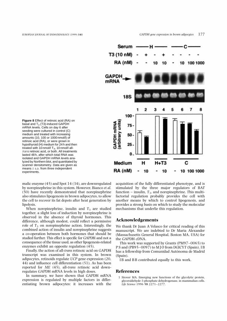
for 48 h in the presence or absence of 10 nmol/l T3,all-trans retinoic acid, or both, at the doses indicated inFig. 8. As shown in this figure, retinoic acid slightlyinhibited GAPDH transcript (lanes 5–8) only in highdoses, this effect being better observed in the absence ofthyroid hormones (hypothyroid medium) (lane 1compared with lane 3). When retinoic acid was addedtogether with T3, the induction observed with the latterwas only weakly inhibited by the retinoic acid (lane 2compared with lane 4).
DiscussionGAPDH has been traditionally considered as a house-keeping gene, the levels of which would remain stableunder different manipulations. Despite its widespreaduse as an internal control, GAPDH gene expressionvaries in different situations (3–9). This enzyme has akey role in adipocytes, in which glycolysis is an
important metabolic pathway for providing lipogenicsubstrates. As demonstrated in differentiating whiteadipocytes (10) and in adipose tissue derived fromgenetically obese rats (11, 30), upregulation of GAPDHparallels the acquisition of the full lipogenic phenotype.In this report, we have studied GAPDH regulation inbrown preadipocytes. When grown under specificconditions, these cells undergo both thermogenic andlipogenic differentiation programs (24, 26) and becomeresponsive to hormones that regulate lipid metabolism.
As expected, GAPDH transcript increased in parallelwith differentiation of the cells, mainly between days 4and 6, when they had not yet acquired full lipogeniccapacity. In later stages, GAPDH mRNA levels remainedunaltered unless the cells received a lipogenic orlipolytic stimulus. This early onset of GAPDH has beenreported in white adipose tissue (46) and, in BAT,precedes the activation of other lipogenesis-relatedenzymes such as malic enzyme (33) or Spot 14 (34). Asthe regulation of GAPDH transcript is seen better duringlipogenic differentiation (days 5–9), the effect of the mainregulators of the tissue was examined in that period.
Insulin, acting through its own receptors, upregu-lated GAPDH mRNA levels on whichever culture day itwas examined. It stabilized GAPDH transcript, andprobably stimulated the transcriptional rate of the gene.This latter mechanism of action has been reported inadipocyte (8) and non-adipocyte (36) cell lines, and twodifferent insulin response elements have been found inthe human gene promoter (32). When cycloheximidewas used, the inhibitor blocked the induction observedwith insulin, suggesting that short-lived proteins couldbe required for either the transcription or the stabiliza-tion process.
In addition to insulin, GAPDH appears to be a targetfor T3, norepinephrine and retinoic acid in these cells.The role of T3 in BAT is well known (18, 47): it affectsUCP 1 gene expression (35), and interacts withnorepinephrine and retinoic acid (20, 35). In thisreport, we have shown for the first time that T3 increasesGAPDH mRNA levels in differentiating brown adipo-cytes. Another lipogenic gene, malic enzyme (ME), issubjected to a similar regulation by T3 in this tissue(33). The mechanism used by T3 for regulating bothgenes is also very similar, as T3 stabilizes GAPDH mRNAand requires de novo protein synthesis, as was the casefor malic enzyme. However, the existence of a thyroidhormone response element – exhaustively described forME (48) – in the rat GAPDH promoter cannot beexcluded.
Norepinephrine, considered to be the main physiolo-gical regulator of BAT, affects proliferation (49) anddifferentiation (17, 26) of brown preadipocytes. We haveshown here that norepinephrine, probably throughcAMP, upregulates GAPDH mRNA levels in differentiat-ing brown adipocytes. This finding could be controversialin light of the well known role of norepinephrine asactivator of lipolysis: two lipogenesis-related proteins,
176 I Barroso and others EUROPEAN JOURNAL OF ENDOCRINOLOGY (1999) 141
Figure 7 Effect of 8-Br-cAMP on GAPDH transcript. Time course ofGAPDH mRNA induction by 8-Br-cAMP. Cells were grown in controlmedium until day 8 and then treated with 1 mmol/l 8-Br-cAMP. TotalRNA was obtained after 1, 3, 6 and 24 h and hybridized with GAPDHprobe. A representative Northern blot and the correspondingethidium bromide staining are shown. Quantitation by densitometeranalysis from three independent experiments is represented. Dataare given as means6S.D.
malic enzyme (45) and Spot 14 (34), are downregulatedby norepinephrine in this system. However, Bianco et al.(50) have recently demonstrated that norepinephrinealso stimulates lipogenesis in brown adipocytes, to allowthe cell to recover its fat depots after heat generation bylipolysis.
When norepinephrine, insulin and T3 are studiedtogether, a slight loss of induction by norepinephrine isobserved in the absence of thyroid hormones. Thisdifference, although modest, could reflect a permissiverole of T3 on norepinephrine action. Interestingly, thecombined action of insulin and norepinephrine suggestsa co-operation between both hormones that should bestudied further. This effect is specific for GAPDH and not aconsequence of the tissue used, as other lipogenesis-relatedenzymes exhibit an opposite regulation (45).
Finally, the action of all-trans retinoic acid on GAPDHtranscript was examined in this system. In brownadipocytes, retinoids regulate UCP gene expression (20,44) and influence cell differentiation (51). As has beenreported for ME (45), all-trans retinoic acid down-regulates GAPDH mRNA levels in high doses.
In summary, we have shown that GAPDH mRNAexpression is regulated by multiple factors in differ-entiating brown adipocytes: it increases with the
acquisition of the fully differentiated phenotype, and isstimulated by the three major regulators of BATfunction – insulin, T3, and norepinephrine. This multi-factorial regulation probably provides the cell withanother means by which to control lipogenesis, andprovides a strong basis on which to study the molecularmechanisms that underlie this regulation.
AcknowledgementsWe thank Dr Juan A Velasco for critical reading of thismanuscript. We are indebted to Dr Maria Alexander(Massachusetts General Hospital, Boston MA, USA) forthe GAPDH cDNA.
This work was supported by Grants (PM97–0065) toP S and (PB95–0097) to M J O from DGICYT (Spain). I Bhas a fellowship from Comunidad Autonoma de Madrid(Spain).
I B and B B contributed equally to this work.
References1 Sirover MA. Emerging new functions of the glycolytic protein,
glyceraldehyde-3-phosphate dehydrogenase, in mammalian cells.Life Science 1996 58 2271–2277.
GAPDH gene expression in brown adipocytes 177EUROPEAN JOURNAL OF ENDOCRINOLOGY (1999) 141
Figure 8 Effect of retinoic acid (RA) onbasal and T3 (T3)-induced GAPDHmRNA levels. Cells on day 6 afterseeding were cultured in control (C)medium and treated with increasingamounts (10, 100 or 1000 nmol/l) ofretinoic acid (RA), or were grown inhypothyroid (H) medium for 24 h and thentreated with 10 nmol/l T3, 10 nmol/l all-trans retinoic acid, or both. All treatmentslasted 48 h, after which total RNA wasisolated and GAPDH mRNA levels ana-lyzed by Northern blot, and quantitated byscanner densitometry. Data are given asmeans 6 S.D. from three independentexperiments.

2 Graven KK, Troxler RF, Kornfeld H, Panchenko MV & Farber HW.Regulation of endothelial cell glyceraldehyde-3-phosphatedehydrogenase expression by hypoxia. Journal of BiologicalChemistry 1994 269 24446–24453.
3 Mansur NR, Meyer-Siegler K, Wurzer JC & Sirover MA. Cell cycleregulation of the glyceraldehyde-3-phosphate dehydrogenase/uracil DNA glycosylase gene in normal human cells. Nucleic AcidsResearch 1993 21 993–998.
4 McNully SE & Toscano WA. Transcriptional regulation ofglyceraldehyde-3-phosphate dehydrogenase by 2,3,7,8-tetra-chlorodibenzo-p-dioxin. Biochemical and Biophysical ResearchCommunications 1995 212 165–171.
5 Vincent S, Marty L & Fort P. S26 ribosomal protein RNA: aninvariant control for gene regulation experiments in eucaryoticcells and tissues. Nucleic Acids Research 1993 21 1498.
6 Desprez PY, Pujol D & Saez S. Glyceraldehyde-3-phosphatedehydrogenase (GAPDH, E.C. 1.2.12.) gene expression in twomalignant human mammary epithelial cell lines: BT-20 andMCF7. Regulation of gene expression by 1,25-dihydroxyvitaminD3 (1,25-(OH)2D3. Cancer Letters 1992 64 219–224.
7 Roche E, Assimacopoulos-Jeannet F, Witters LA, Perruchoud B,Yaney G, Corkey B et al. Induction by glucose of genes coding forglycolytic enzymes in a pancreatic b-cell line (INS-1). Journal ofBiological Chemistry 1997 272 3091–3098.
8 Alexander M, Curtis G, Avruch J & Goodman HM. Insulinregulation of protein biosynthesis in differentiated 3T3adipocytes. Journal of Biological Chemistry 1985 260 11978–11985.
9 Nasrin N, Buggs C, Kong XF, Carnazza J, Goebl M & Alexander-Bridges M. DNA-binding properties of the product of the testis-determining gene and a related protein. Nature 1991 354 317–332.
10 Spiegelman BM & Green H. Control of specific protein biosynthesisduring the adipose conversion of 3T3 cells. Journal of BiologicalChemistry 1980 255 8811–8818.
11 Rolland V, Dugail I, Le Liepvre X & Lavau M. Evidence of increasedglyceraldehyde-3-phosphate dehydrogenase and fatty acidsynthetase promoter activities in transiently transfected adipo-cytes from genetically obese rats. Journal of Biological Chemistry1995 270 1102–1106.
12 Ricquier D & Bouillaud F. The brown adipose tissue mitochondrialuncoupling protein. In Brown Adipose Tissue, pp 86–104. EdsP Trayhurn & DG Nicholls. London: Edward Arnold Publishers,1986.
13 Fleury C, Neverova M, Collins S, Raimbault S, Champigny O,Levi MC et al. Uncoupling protein-2: a novel gene linked to obesityand hyperinsulinemia. Nature Genetics 1997 15 269–272.
14 Boss O, Samec S, Paoloni GA, Rossier C, Dulloo A, Seydoux J et al.Uncoupling protein-3: a new member of the mitochondrialcarrier family with tissue-specific expression. FEBS Letters 1997408 39–42.
15 Gong D-W, He Y, Karas M & Reitman M. Uncoupling protein-3 is amediator of thermogenesis regulated by thyroid hormone, b3-adrenergic agonists, and leptin. Journal of Biological Chemistry1997 272 24129–24132.
16 Himms-Hagen J. Brown adipose tissue metabolism and thermo-genesis. Annual Review of Nutrition 1985 5 69–94.
17 Cannon B & Nedergaad J. Adrenergic regulation of brownadipocyte differentiation. Biochemical Society Transactions 199624 407–412.
18 Silva JE. Thyroid hormone control of thermogenesis and energybalance. Thyroid 1995 5 481–492.
19 McCormack JG. The regulation of fatty acid synthesis in brownadipose tissue by insulin. Progess in Lipid Research 1982 21 195–223.
20 Rabelo R, Reyes C, Schifman A & Silva JE. A complex retinoic acidresponse element in the uncoupling protein gene defines a novelrole for retinoids in thermogenesis. Endocrinology 1996 1373488–3496.
21 Himms-Hagen J. Brown adipose tissue thermogenesis: inter-disciplinary studies. FASEB Journal 1990 4 2890–2898.
22 Himms-Hagen J. Brown adipose tissue metabolism. In Obesity, pp15–34. Eds P Bjorntorp & BN Brodoff. New York: Lippincott,1992.
23 Lowell BB, S-Susulic V, Hamann A, Lawitts JA, Himms-Hagen J,Boyer BB et al. Development of obesity in transgenic mice aftergenetic ablation of brown adipose tissue. Nature 1993 366 740–742.
24 Nechad M, Kuusela P, Carneheim C, Bjorntorp P, Nedergaad J &Cannon B. Development of brown fat cells in monolayer culture.Experimental Cell Research 1983 149 105–118.
25 Silva JE & Rabelo R. Regulation of the uncoupling protein geneexpression. European Journal of Endocrinology 1997 136 251–264.
26 Rehmark S, Nechad M, Herron D, Cannon B & Nedergaad J. a-and b- adrenergic induction of the expression of the uncouplingprotein thermogenin in brown adipocytes differentiated inculture. Journal of Biological Chemistry 1990 265 16464–16471.
27 Samuels HH, Stanley F & Casanova J. Depletion of L-3,5,30-triiodothyronine and L-thyroxine in euthyroid calf serum for usein cell culture studies of the action of thyroid hormone.Endocrinology 1979 105 80–85.
28 Herron D, Nechad M, Rehmark S, Nelson BD, Nedergaad J &Cannon B. Effects of cholera toxin on gene expression in brownpreadipocytes differentiating in culture. American Journal ofPhysiology 1989 257 C920–C925.
29 Maniatis T, Fritsch EF & Sambrook J. In Molecular Cloning – ALaboratory Manual. Cold Spring Harbor: Cold Spring HarborLaboratory, 1982.
30 Dugail I, Quignard-Boulange A, Le Liepvre X, Ardouin B &Lavau M. Gene expression of lipid storage-related enzymes inadipose tissue of the genetically obese Zucker rat. BiochemicalJournal 1992 281 607–611.
31 Kormanec J, Lempel’ova A, Novakova R, Rezuchova, B &Homerova D. Expression of the Streptomyces aureofaciens glycer-aldehyde-3-phosphate dehydrogenase gene (gap) is developmen-tally regulated and induced by glucose. Microbiology 1997 1433555–3561.
32 Alexander MC, Lomanto M, Nasrin N & Ramadka C. Insulinstimulates glyceraldehyde-3-phosphate dehydrogenase geneexpression through cis-acting DNA sequences. Proceedings of theNational Academy of Sciences of the USA 1988 85 5092–5096.
33 Garcıa-Jimenez C, Hernandez A, Obregon MJ & Santisteban P.Malic enzyme gene expression in differentiating brown adipo-cytes: regulation by insulin and triiodothyronine. Endocrinology1993 132 1537–1543.
34 Perez-Castillo A, Hernandez A, Pipaon C, Santos A & Obregon MJ.Multiple regulation of S14 gene expression during brown fatdifferentiation. Endocrinology 1993 133 545–552.
35 Rabelo R, Schifman A, Rubio A, Sheng X & Silva JE. Delineation ofthyroid hormone responsive sequences within a critical enhancerin the rat uncoupling protein gene. Endocrinology 1995 1361003–1013.
36 Ortiz L, Zannini M, Di Lauro R & Santisteban P. Transcriptionalcontrol of the fork-head thyroid transcription factor TTF-2 bythyrotropin, insulin, and insulin-like growth factor I. Journal ofBiological Chemistry 1997 272 23334–23339.
37 Bianco AC, Kieffer JD & Silva JE. Adenosine 30,50-monophosphateand thyroid hormone control of uncoupling protein messengerribonucleic acid in freshly dispersed brown adipocytes. Endocrin-ology 1992 130 2625–2633.
38 Rubio A, Raasmaja A, Maia AL, Kim KR & Silva JE. Effects ofthyroid hormone on norepinephrine signaling in brown adiposetissue. I. Beta 1- and beta 2-adrenergic receptors and cyclicadenosine 30,50-monophosphate generation. Endocrinology 1995136 3267–3276.
39 Rubio A, Raasmaja A & Silva JE. Thyroid hormone andnorepinephrine signaling in brown adipose tissue. II: Differentialeffects of thyroid hormone on beta 3-adrenergic receptors inbrown and white adipose tissue. Endocrinology 1995 136 3277–3284.
178 I Barroso and others EUROPEAN JOURNAL OF ENDOCRINOLOGY (1999) 141

40 El Hadri K, Charon C, Pairault J, Hauguel-de Mouzon S, Quignard-Boulange A & Feve B. Down-regulation of b3-adrenergic receptorexpression in rat adipose tissue during the fasted/fed transition:evidence for a role of insulin. Biochemical Journal 1997 323 359–364.
41 Ma SWY & Foster DO. Potentiation of in vivo thermogenesis in ratbrown adipose tissue by stimulation of alpha 1-adrenoreceptors isassociated with increased release of cyclic AMP. Canadian Journalof Physiology and Pharmacology 1984 62 943–948.
42 Kamei Y, Kawada T, Mizukami J & Sugimoto E. The prevention ofadipose differentiation of 3T3-L1 cells caused by retinoic acid iselicited through retinoic acid receptor alpha. Life Science 1994 55PL307–312.
43 Safonova I, Darimont C, Amri EZ, Grimaldi P, Ailhaud G, Reichert Uet al. Retinoids are positive effectors of adipose cell differentiation.Molecular and Cellular Endocrinology 1994 104 201–211.
44 Puigserver P, Vazquez F, Bonet ML, Pico C & Palou A. In vivo and invitro induction of brown adipocyte uncoupling protein (thermo-genin) by retinoic acid. Biochemical Journal 1996 317 827–833.
45 Hernandez A, Garcıa-Jimenez C, Santisteban P & Obregon, MJ.Regulation of malic-enzyme-gene expression by cAMP andretinoic acid in differentiating brown adipocytes. European Journalof Biochemistry 1993 215 285–290.
46 Dugail I, Quignard-Boulange A, Bazin R, Le Liepvre X & Lavau M.Adipose-tissue-specific increase in glyceraldehyde-3-phosphatedehydrogenase activity and mRNA amounts in suckling pre-obese
Zucker rats. Effect of weaning. Biochemical Journal 1988 254483–487.
47 Bianco AC & Silva JE. Intracellular conversion of thyroxine totriiodothyronine is required for the optimal thermogenic functionof brown adipose tissue. Journal of Clinical Investigation 1987 79295–300.
48 Petty KJ, Desvergne B, Mitshuhashi T & Nikodem V. Identificationof a thyroid hormone response element in the malic enzyme gene.Journal of Biological Chemistry 1990 265 7395–7400.
49 Garcia B & Obregon MJ. Norepinephrine potentiates the mitogeniceffect of growth factors in quiescent brown preadipocytes:relationship with uncoupling protein messenger ribonucleicacid expression. Endocrinology 1997 138 4227–4233.
50 Bianco AC, Carvalho SD, Carvalho CRF, Rabelo R & Moriscot AS.Thyroxine 50-deiodination mediates norepinephrine inducedlipogenesis in dispersed brown adipocytes. Endocrinology 1998139 571–578.
51 Safonova I, Reichert U, Shroot B, Ailhaud G & Grimaldi P. Fattyacids and retinoids act synergistically on adipose cell differentia-tion. Biochemical and Biophysical Research Communications 1994204 498–504.
Received 8 February 1999Accepted 27 April 1999
GAPDH gene expression in brown adipocytes 179EUROPEAN JOURNAL OF ENDOCRINOLOGY (1999) 141


