Embed Size (px)
Citation preview

Novel Anti-Inflammatory Properties of theAngiogenesis Inhibitor VasostatinRainer Huegel1,3, Paula Velasco1,3, Maria De La Luz Sierra2, Enno Christophers1, Jens M. Schroder1,Thomas Schwarz1, Giovanna Tosato2 and Bernhard Lange-Asschenfeldt1
Endothelial cells are critically involved in the pathogenesis of inflammation, which is characterized byvasopermeability, plasma leakage, leukocyte recruitment, and neovascularization. Therefore, inhibitors ofendothelial cell function could reduce inflammation. In this study, we evaluated the effects of the angiogenesisinhibitor vasostatin on inflammations induced by contact hypersensitivity reactions in mouse ears. Vasostatin-treated mice revealed significantly reduced edema formation, resulting from lower plasma leakage andinhibition of inflammation-associated vascular remodeling. Intravital microscopy studies of inflamed earsshowed a decrease in the fraction of rolling leukocytes in vasostatin-treated mice, and Lycopersicon esculentumlectin-perfused ears revealed fewer leukocytes adherent to the vessel wall. The inflammatory infiltrate fromvasostatin-treated mice was characterized by fewer CD8þ T cells, neutrophils, and macrophages compared tothe saline-treated animals. In a modified Miles assay, vasostatin inhibited vascular endothelial growth factor-A-induced permeability, and inflamed ear tissues from vasostatin-treated mice expressed significantly reducedlevels of the vascular destabilizer angiopoietin-2. These results reveal a previously unrecognized anti-inflammatory property of the angiogenesis inhibitor vasostatin, and suggest that vasostatin is a potentialcandidate drug for the treatment of inflammation.
Journal of Investigative Dermatology (2007) 127, 65–74. doi:10.1038/sj.jid.5700484; published online 3 August 2006
INTRODUCTIONThe vascular endothelium is an important component of theinflammatory response and manifests striking changes duringinflammation. As a result, vascular hyperpermeability, tissueedema, and perivascular inflammatory infiltrate characterizeinflamed tissues. Moreover, endothelial cells are an impor-tant source of proinflammatory and proangiogenic factors.Therefore, the increased vascular surface area resulting frominflammation-associated angiogenesis is an important com-ponent of inflammations, as it translates to an extendedcapacity for cytokine production, adhesion molecule expres-sion, and leukocyte recruitment (Jackson et al., 1997). In fact,there is accumulating experimental evidence that angiogen-esis and inflammation are closely interlinked (Braverman andSibley, 1982; Jackson et al., 1997; Lange-Asschenfeldt et al.,2002; Oura et al., 2002; Kunstfeld et al., 2004).
The experimental contact hypersensitivity (CHS) reactionis a model for an acute inflammatory response that isaccompanied by a robust increase in the index of proliferat-ing endothelial cells (Polverini et al., 1977). This model hasbeen utilized to demonstrate that angiogenesis factorsmodulate inflammation. For instance, mice that overexpressthe proangiogenic factors, vascular endothelial growth factor-A (VEGF-A) (Kunstfeld et al., 2004) or placental growth factor(Oura et al., 2002), react with an exaggerated ear swellingresponse associated with increased edema formation andvascular remodeling. In contrast, the CHS response inplacental growth factor-deficient mice is much less pro-nounced (Oura et al., 2002). Meanwhile, mice that lack theangioinhibitor thrombospondin-2 exhibited a prolonged andpersistent inflammation accompanied by enlarged vessels(Lange-Asschenfeldt et al., 2002). The positive correlationbetween enhanced vascularity and increased inflammatoryresponses and the abundance of factors that modulateangiogenesis known to be upmodulated during inflammatoryresponses (Detmar et al., 1994; Brown et al., 1995; Dvoraket al., 1995; Schroder and Mochizuki, 1999; Kuroda et al.,2001; Oura et al., 2002; Scott et al., 2002) confirm that theendothelium is active and relevant in the pathophysiology ofinflammatory diseases.
However, studies addressing the physiological role ofangiogenesis inhibitors during inflammation and potentialtherapeutic approaches with these factors in inflammationremain scant. In the past years, it has been demonstrated thatblocking VEGF-A signaling resulted in suppressed edema
& 2006 The Society for Investigative Dermatology www.jidonline.org 65
ORIGINAL ARTICLE
Received 28 January 2006; revised 16 May 2006; accepted 30 May 2006;published online 3 August 2006
1Department of Dermatology, University Hospital Schleswig-Holstein,Campus Kiel, Kiel, Germany and 2Experimental Transplantation andImmunology Branch, Center for Cancer Research, National Cancer Institute,National Institutes of Health, Bethesda, Maryland, USA
Correspondence: Dr Bernhard Lange-Asschenfeldt, Departmentof Dermatology, University Hospital Schleswig-Holstein, Campus Kiel,Schittenhelmstrasse 7, Kiel 24105, Germany.E-mail: [email protected]
3These authors contributed equally to this work.
Abbreviations: Ang, angiopoietin; CHS, contact hypersensitivity; EBD, Evansblue dye; PBS, phosphate-buffered saline; VEGF, vascular endothelial growthfactor
formation in experimental inflammations. Furthermore, fol-lowing treatment with the angiogenesis inhibitors TNP-470,caplostatin, and angiostatin, vascular leakage was reduced inexperimental inflammations. In this study, we assessed theeffect of a treatment regimen with the naturally occurringangiogenesis inhibitor vasostatin (Pike et al., 1998, 1999) inan experimental CHS reaction in mice sensitized againstoxazolone.
Vasostatin, an endothelial cell inhibitory factor originallyisolated in culture supernatants of Epstein–Barr virus-immor-talized cells, consists of the amino-terminal domain ofcalreticulin inclusive of residues 1–180. Vasostatin inhibitsthe proliferation of endothelial cells stimulated with basicfibroblast growth factor and VEGF-A and suppresses neo-vascularization and tumor growth in vivo. (Pike et al., 1998,1999). Here, we report that systemic treatment withrecombinant vasostatin resulted in a diminished inflamma-tory response in the ear skin of mice. We observed reducededema formation, leukocyte recruitment, and vascularremodeling following vasostatin treatment. Moreover,vasostatin potently inhibited VEGF-A-induced permeabilityin vivo and inhibited the upregulation of the vasculardestabilizer angiopoietin (Ang)-2 observed in the inflamedlesions. Our study reveals a novel function for the angiogen-esis inhibitor vasostatin as an anti-inflammatory substance,suggesting that therapeutic strategies using vasostatin andother angiogenesis antagonists could represent a novelapproach for the treatment of inflammations.
RESULTSVasostatin reduced experimentally induced cutaneousinflammation and vascular leakage
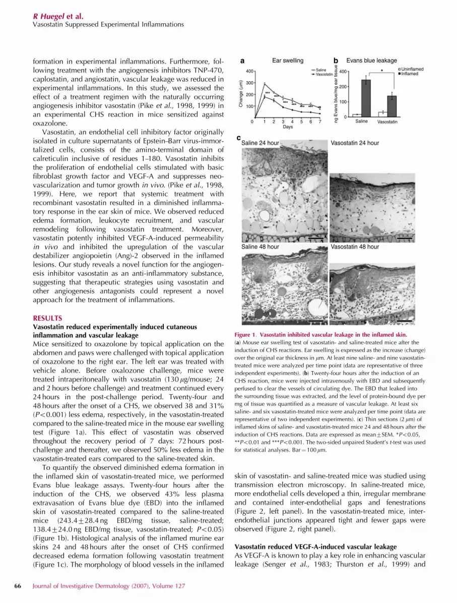
Mice sensitized to oxazolone by topical application on theabdomen and paws were challenged with topical applicationof oxazolone to the right ear. The left ear was treated withvehicle alone. Before oxalozone challenge, mice weretreated intraperitoneally with vasostatin (130 mg/mouse; 24and 2 hours before challenge) and treatment continued every24 hours in the post-challenge period. Twenty-four and48 hours after the onset of a CHS, we observed 38 and 31%(Po0.001) less edema, respectively, in the vasostatin-treatedcompared to the saline-treated mice in the mouse ear swellingtest (Figure 1a). This effect of vasostatin was observedthroughout the recovery period of 7 days: 72 hours post-challenge and thereafter, we observed 50% less edema in thevasostatin-treated ears compared to the saline-treated skin.
To quantify the observed diminished edema formation inthe inflamed skin of vasostatin-treated mice, we performedEvans blue leakage assays. Twenty-four hours after theinduction of the CHS, we observed 43% less plasmaextravasation of Evans blue dye (EBD) into the inflamedskin of vasostatin-treated compared to the saline-treatedmice (243.4728.4 ng EBD/mg tissue, saline-treated;138.4724.0 ng EBD/mg tissue, vasostatin-treated; Po0.05)(Figure 1b). Histological analysis of the inflamed murine earskins 24 and 48 hours after the onset of CHS confirmeddecreased edema formation following vasostatin treatment(Figure 1c). The morphology of blood vessels in the inflamed
skin of vasostatin- and saline-treated mice was studied usingtransmission electron microscopy. In saline-treated mice,more endothelial cells developed a thin, irregular membraneand contained inter-endothelial gaps and fenestrations(Figure 2, left panel). In the vasostatin-treated mice, inter-endothelial junctions appeared tight and fewer gaps wereobserved (Figure 2, right panel).
Vasostatin reduced VEGF-A-induced vascular leakage
As VEGF-A is known to play a key role in enhancing vascularleakage (Senger et al., 1983; Thurston et al., 1999) and
400 400
200
100
Ear swelling
Saline 24 hour
Saline 48 hour
Vasostatin 24 hour
Vasostatin 48 hour
Evans blue leakage
Change (�m
)
ng E
vans
blue/m
g e
ar
tissu
e
300
200
100
00
1 2Days3 4 5 6
Saline
Saline
Vasostatin
Vasostatin
UninflamedInflamed
7
a
c
b
Figure 1. Vasostatin inhibited vascular leakage in the inflamed skin.
(a) Mouse ear swelling test of vasostatin- and saline-treated mice after the
induction of CHS reactions. Ear swelling is expressed as the increase (change)
over the original ear thickness in mm. At least nine saline- and nine vasostatin-
treated mice were analyzed per time point (data are representative of three
independent experiments). (b) Twenty-four hours after the induction of an
CHS reaction, mice were injected intravenously with EBD and subsequently
perfused to clear the vessels of circulating dye. The EBD that leaked into
the surrounding tissue was extracted, and the level of protein-bound dye per
mg of tissue was quantified as a measure of vascular leakage. At least six
saline- and six vasostatin-treated mice were analyzed per time point (data are
representative of two independent experiments). (c) Thin sections (2 mm) of
inflamed skins of saline- and vasostatin-treated mice 24 and 48 hours after the
induction of CHS reactions. Data are expressed as mean7SEM. *Po0.05,
**Po0.01 and ***Po0.001. The two-sided unpaired Student’s t-test was used
for statistical analyses. Bar¼ 100mm.
66 Journal of Investigative Dermatology (2007), Volume 127
R Huegel et al.Vasostatin Suppressed Experimental Inflammations

inflammation (Brown et al., 1995; Kunstfeld et al., 2004), weinvestigated whether vasostatin controls plasma extravasationdirectly by inhibiting VEGF-A-induced vascular permeability.For this purpose, we quantified the extravasated EBD in
response to intradermally injected VEGF-A in the skin using amodified Miles assay. Vasostatin diminished VEGF-A-in-duced vascular hyperpermeability in the skin of mice by37% (12.771.8 mg EBD, saline-treated; 8.070.4 mg EBD,vasostatin-treated; Po0.05), whereas there was no differencein the leakage induced by intradermal injections of saline(3.270.5mg EBD, saline-treated; 1.970.5mg EBD, vasostatin-treated) (Figure 3).
Vasostatin decreased the vascularity in the inflamed skinof mice
Forty-eight hours following the onset of an inflammation, weobserved fewer blood vessels in the inflamed ears ofvasostatin-treated mice when compared to saline-treated mice(Figure 4a). Computer-assisted morphometric image analysesof the panendothelial cell marker CD31-immunostained bloodvessels revealed a significant decrease in the number of vesselsper mm ear length in the vasostatin-treated compared tothe saline-treated inflamed skins (50.871.4 blood vessels(BV)/mm, saline-treated; 43.273.1 BV/mm, vasostatin-treated;
Saline 24 hour
Saline 24 hour
Vasostatin 24 hour
Vasostatin 24 hour
Figure 2. Vasostatin suppressed the formation of inter-endothelial gaps
during an inflammation. Twenty-four hours after the induction of CHS
reactions, the skin of vasostatin- and saline-treated mice was harvested and
processed for electron microscopy. Representative images showing that in
(left panel) saline-treated mice endothelial cells display a thin, irregular
plasma membrane (fenestrations indicated by the arrows), and inter-
endothelial cell junctions (indicated by the asterisks) are discontinuous. In
contrast, in (right panel) vasostatin-treated mice, inter-endothelial junctions
(indicated by arrowhead) appear tight and fewer gaps and fenestrations are
observed. (upper panel) Bar¼ 1 mm and (lower panel) bar¼ 200 nm.
VEGF NaCI
Vas
osta
tinS
alin
e SalineVasostatin
�g E
vans
blu
e ex
trac
ted
NaCI VEGF165
16
12
8
4
0
ab
Figure 3. Vasostatin inhibited VEGF-A induced vascular leakage in the skin.
For the modified Miles assay, EBD was intravenously injected in (a, upper
panel) saline-treated and (a, lower panel) vasostatin-treated mice. After
10 minutes, NaCl alone and human VEGF165 (200 ng) were separately
injected intradermally into the back skin. After 30 minutes, the skin area
containing the extravasated protein-bound dye was excised and the dye was
extracted from the tissue. Dye concentrations were measured at 620 nm using
a spectrophotometer. (b) The values obtained are expressed as total
nanograms of EBD extracted, and are a measure of the total amount of
protein-bound dye that extravasated in response to VEGF-A or NaCl
treatment. At least five saline- and five vasostatin-treated mice were used for
the experiments. Data are representative of two independent experiments.
Data are expressed as mean7SEM. *Po0.05. The two-sided unpaired
Student’s t-test was used for statistical analyses. Bar¼ 5 mm
Saline
Vessel density Vessel size distribution
Vasostatin
<100 100–500 500–2,000
SalineVasostatin
SalineVasostatin
60
40
20
0
30
20
10
0Ves
sel/
mm
ear
leng
th
Ves
sel/
mm
ear
leng
th
Vessel area (�m2)
b c
Figure 4. Decreased vascular remodeling in the inflamed skin following
vasostatin treatment. (a) Cryostat sections stained with the pan endothelial
cell marker CD31 to visualize vessels of (a, left panel) saline-treated and
(a, right panel) vasostatin-treated inflamed ear skins 48 hours following
antigen challenge. (b) Computer-assisted morphometric image analyses based
on these sections revealed a significant decrease in the number of blood
vessels per mm ear length following vasostatin treatment 48 hours after the
onset of the inflammation. (c) Vessel size distribution analyses were
performed to compare the frequency of vessels according to their size. At least
six saline- and six vasostatin-treated mice were used for these experiments.
Data are representative of two independent experiments. Data are expressed
as mean7SEM. *Po0.05. The two-sided unpaired Student’s t-test was used
for statistical analyses. Bar¼ 200mm.
www.jidonline.org 67
R Huegel et al.Vasostatin Suppressed Experimental Inflammations

Po0.05) (Figure 4b). We then performed vessel size distribu-tion analysis, where we compared the total number of vesselsgrouped according to the size of the vessels. This analysisrevealed significantly fewer vessels (27%), with average vesselsize ranging in size between 500 and 2,000mm2 (9.870.9 BV/mm, saline-treated; 7.170.2 BV/mm, vasostatin-treated;Po0.05) (Figure 4c). The vessels that fall into this size rangeare arterioles and post-capillary venules. As CD31 alsopositively stains lymphatic endothelial cells, we could notexclude a vasostatin-mediated effect on the lymphaticvasculature. However, we observed no difference in theaverage size, density, or size distribution of lymphatic vesselsas judged by LYVE-1-stained sections between the groups (datanot shown), confirming that vasostatin inhibited blood vascularremodeling during CHS.
Vasostatin diminished the inflammatory infiltrate in theinflamed skin
The infiltrating cells in the inflamed skin of vasostatin- andsaline-treated mice consisted mainly of neutrophil granulo-cytes, macrophages, and lymphocytes. Analysis of theinflammatory infiltrate using tissue sections immunostained
with antibodies directed against Ly6-G, to identify neutrophilgranulocytes revealed a significant decrease in the number ofneutrophils in vasostatin-treated mice compared to saline-treated mice at 24 hours (437724 cells/mm, vasostatin-treated and 592744 cells/mm, saline-treated; Po0.01) and48 hours (330729 cells/mm, vasostatin-treated and502735 cells/mm, saline-treated; Po0.01) following antigenchallenge (Figure 5a and b). Tissue sections immunostainedwith antibodies directed against F4/80 used to identifymacrophages revealed a significant decrease in the numberof macrophages in vasostatin-treated mice compared tosaline-treated mice at 24 hours (101715 cells/mm, vasosta-tin-treated and 214715 cells/mm, saline-treated; Po0.001)and 48 hours (11375 cells/mm, vasostatin-treated and245721 cells/mm, saline-treated; Po0.001) (Figure 5c andd). Additionally, CD8 (CD8þ T cells) immunostains revealeda significant decrease in the number of infiltrating CD8þ
T cells per mm ear length (2874 cells/mm, vasostatin-treatedand 4775 cells/mm, saline-treated; Po0.05) 48 hours fol-lowing antigen challenge. No difference was observed in thenumber of CD4þ T cells in the inflamed skin of both groupsor in the number of neutrophils, macrophages, CD8þ T cells,
Saline 24 hour
Saline 24 hour
Vasostatin 24 hour
Vasostatin 24 hour
SalineVasostatin
SalineVasostatin
700
600
500
400
300
200
100
0
300
250
200
150
100
50
0
24 hours
Cel
ls/m
m e
ar le
ngth
Cel
ls/m
m e
ar le
ngth
48 hours
24 hours 48 hours
a
c d
b
***
**
**
***
Figure 5. Vasostatin diminished inflammatory infiltrate in the inflamed skin. (a) Immunohistochemical stains of inflamed ear skin of vasostatin- and
saline-treated mice 24 hours after the induction of CHS reactions using an antibody directed against Ly6-G to visualize neutrophils. (b) Based on these stains,
computer-assisted image analyses were performed to determine the number of infiltrating neutrophils in the inflamed skin of vasostatin-treated mice compared
to saline-treated mice 24 and 48 hours following the onset of the inflammation. (c) Immunohistochemical stains of vasostatin- and saline-treated mice
24 hours into the inflammation using an antibody directed against F4/80 to visualize macrophages. (d) We performed computer-assisted image analyses based
on the F4/80-stained sections to quantify the number of macrophages in the vasostatin- and saline-treated mice 24 and 48 hours after the induction of CHS
reactions. At least six saline- and six vasostatin-treated mice were used for the experiments. Data are expressed as mean7SEM. **Po0.01 and ***Po0.001.
The two-sided unpaired Student’s t-test was used for statistical analyses. Bar¼ 100mm.
68 Journal of Investigative Dermatology (2007), Volume 127
R Huegel et al.Vasostatin Suppressed Experimental Inflammations

and CD4þ T cells in the uninflamed skin of saline- andvasostatin-treated mice.
Vasostatin diminished leukocyte–endothelium interaction ininflamed skin
As the proangiogenic factor VEGF-A (Detmar et al., 1998)and the antiangiogenic factor thrombospondin-2 (Lange-Asschenfeldt et al., 2002) have been shown to influence theleukocyte–endothelial interaction, we performed intravitalmicroscopy of blood vessels in uninflamed and inflamed skinof mice treated with vasostatin or saline. The edema in theinflamed ears of the mice in both groups made it difficult toobtain reliable measurements at 24 hours post-challenge. Wetherefore selected a time point of 6 hours following antigenchallenge, as our previous experiments indicated changes inleukocyte adhesion as early as 2 hours following the onset ofinflammation, while maintaining optimal visualization of theleukocytes in vivo. After injection of Rhodamine-6G,intravital microscopy revealed no difference in the baseline-rolling fraction in the uninflamed skin of the vasostatin- andsaline-treated mice. However, the rolling fraction of leuko-cytes in the inflamed cutaneous blood vessels of vasostatin-treated mice was decreased by 40% when compared to theinflamed skin of saline-treated mice (4675%, vasostatin-treated and 6973%, saline-treated; Po0.01) (Figure 6a). Inaddition, whole mounts of inflamed ear skin after vascular
perfusion with the lectin Lycopersicon esculentum, whichbinds to N-acetyl-D-glucosamine residues on the luminalsurface of vascular endothelial cells and to leukocytes(Thurston et al., 1996), revealed decreased inflammatorycells adherent to the endothelium of the blood vessels in thevasostatin-treated compared to the saline-treated mice24 hours following antigen challenge (Figure 6b), confirmingthe data obtained from the intravital studies. We thereforetested whether vasostatin directly influenced the expressionlevel of intercellular adhesion molecule, vascular celladhesion molecule, E-selectin, and VE-cadherin on thesurface of tumor necrosis factor-a-stimulated human dermalmicrovascular endothelial cells in vitro using FACS analyses.Although the stimulated human dermal microvascularendothelial cells had increased levels of the tested celladhesion molecules, vasostatin at 10 or 100mg/ml had nosignificant effect on the cell-surface expression of the testedcell adhesion molecules.
Vasostatin blocked the upregulation of Ang-2 in the inflamedear skinVEGF-A (Senger et al., 1983; Thurston et al., 1999) is a potentinducer of vascular hyperpermeability, whereas Ang-1 hasbeen shown to inhibit vascular leakage (Thurston et al., 1999,2000; Gamble et al., 2000). Therefore, we analyzed theexpression pattern of these factors as well as the Ang-1antagonist Ang-2 using quantitative real-time reverse tran-scription-PCR (RT-PCR). We found no difference in theexpression pattern of VEGF-A or Ang-1 mRNA between thevasostatin- and saline-treated mice (Figure 7). However, wefound a more than 2-fold increase in the relative transcriptlevel of Ang-2 mRNA in the inflamed skin of saline-treatedmice when compared to the vasostatin-treated mice(1.970.08, vasostatin-treated and 5.770.02, saline-treated;Po0.001). There was no difference in the relative transcriptlevel of Ang-2 mRNA expression in the uninflamed skin ofeither group (2.470.28, vasostatin-treated and 1.970.08,
Rolling fraction (%)
80
60
40
20
0
SalineVasostatin
Saline 24 hour Vasostatin 24 hour
a
b
Figure 6. Diminished leukocyte–endothelial cell interaction following
vasostatin treatment. (a) Six hours after the induction of CHS reactions,
Rhodamine-6G was intravenously administered to visualize leukocytes in
vivo using intravital microscopy. The rolling fraction of leukocytes in post-
capillary venules in the skin of vasostatin- and saline-treated mice was
determined. At least four saline- and four vasostatin-treated mice were used
for the experiments. (b) Twenty-four hours after the onset of the inflammation,
vasostatin- and saline-treated mice were perfused with L. esculentum lectin
and whole mounts of the ears were used to visualize leukocytes adherent
to the wall of blood vessels (arrowheads). Four saline-treated and four
vasostatin-treated mice were used in the experiment. Data are expressed
as mean7SEM. **Po0.01. The two-sided unpaired Student’s t-test was
used for statistical analyses. Bar¼25 mm.
7
6
5
4
3
2
1
0
VEGF-A Ang-1 Ang-2
Saline Vasostatin-treated
Rel
ativ
e tr
ansc
ript l
evel
Saline-treated Vasostatin-treated Saline-treated Vasostatin-treated
UninflamedInflamed
Figure 7. Vasostatin inhibited inflammation-associated Ang-2 upregulation.
The mRNA expression of VEGF-A, Ang-1, and Ang-2 was measured in the
skin of saline-treated and vasostatin-treated mice 24 hours after the onset
of CHS reactions using real-time RT-PCR analyses. Four saline- and four
vasostatin-treated mice were used for the experiment. Data are expressed
as mean7SEM. ***Po0.001. The two-sided unpaired Student’s t-test
was used for statistical analyses.
www.jidonline.org 69
R Huegel et al.Vasostatin Suppressed Experimental Inflammations

saline-treated) (Figure 6). Interestingly, elicitation of CHSresulted in a 2-fold increase of Ang-2 mRNA expression insaline- but not in vasostatin-treated mice.
DISCUSSIONVasostatin treatment during experimental cutaneous CHSresponses resulted in a striking anti-inflammatory effectevidenced by suppressed vasopermeability, edema forma-tion, inflammatory cell influx, and angiogenesis. To ourknowledge, this is the first description of an angiogenesisinhibitor with broad-range anti-inflammatory action. Previousreports have demonstrated suppressed edema formationduring CHS reactions following the functional blockage ofthe proangiogenic factor VEGF-A (Kunstfeld et al., 2004) andreduced vascular leakage in CHS reactions in the presence ofthe angiogenesis inhibitor TNP-470, caplostatin, and angio-statin (Satchi-Fainaro et al., 2005).
Vascular integrity is fundamental in maintaining a physicalbarrier between blood components and the underlying tissue.During inflammation, plasma leakage is thought to occurprimarily through intercellular gaps that form betweenendothelial cells or through the vesiculovacuolar organelle.Plasma components are able to cross from the lumen into thebasement membrane through intercellular gaps or throughthe direct transendothelial channel created by the vesiculo-vacuolar organelles at sites of augmented vascular perme-ability (Thurston et al., 1996; Dvorak and Feng, 2001).Transmission electron microscopy studies of the inflamedears of vasostatin-treated mice revealed morphologicalfeatures associated with reduced vascular hyperpermeability.Dermal capillaries and venules in the inflamed ear skin ofvasostatin-treated mice contained fewer gaps and the inter-endothelial junctions in the vessels appeared tight comparedto the inflamed skin of saline-treated mice (Figure 2), asrevealed by transmission electron microscopy.
In our hands, vasostatin suppressed VEGF-A-inducedhyperpermeability in vivo as revealed by using a modifiedMiles assay, but did not influence the expression of VEGF-Ain inflamed tissue as shown by real-time RT-PCR analyses.However, vasostatin completely blocked the upregulation ofAng-2 mRNA in vivo during the inflammation and thisobservation could, at least in part, explain the increasedvascular integrity in the inflamed tissue following vasostatintreatment. Ang-2 is a vascular destabilizer that, similar toVEGF-A, has been shown to disrupt endothelial adherensjunctions, thereby contributing to inter-endothelial gapformation (Carter, 2001) and is believed to be involved inthe formation of intercellular openings in tumor vessels,leading to their leaky nature (Hashizume et al., 2000).Moreover, Ang-2 is potently upregulated in the synovialtissue in rheumatoid arthritis (Scott et al., 2002) and inpsoriatic lesions (Kuroda et al., 2001), highlighting animportant role of Ang-2 in inflammatory diseases. In contrast,the vascular stabilizer Ang-1 has been demonstrated to play acrucial role in controlling vascular leakage. For example,mice with targeted epidermal overexpression of Ang-1 wereresistant to leakage induced by histamine, serotonin, andmustard oil (Thurston et al., 1999), and adenoviral injection
of Ang-1 resulted in the inhibition of plasma leakagenormally induced by VEGF-A and mustard oil (Thurstonet al., 2000). Moreover, co-overexpression of Ang-1 withVEGF-A reversed the enhanced baseline leakage seen in thetransgenic mice that overexpressed VEGF-A alone (Thurstonet al., 1999). Importantly, Ang-2 is a competitive antagonistto Ang-1. Therefore, vasostatin could protect the interactionbetween Ang-1 on vascular supporting cells and Tie-2 on endo-thelial cells, promoting vascular quiescence and protectingagainst plasma leakage.
In agreement with its known activity as an angiogenesisantagonist, vasostatin treatment resulted in reduced inflam-mation-associated vascular remodeling. Size distributionanalyses revealed a decrease in the frequency of vesselsranging in size between 500 and 2,000mm2. In the skin, themajority of vessels in this size range are post-capillaryvenules (Braverman, 2000) and our data indicate thatvasostatin treatment resulted in fewer enlarged post-capillaryvenules. This observation is in agreement with increasingevidence, suggesting that inflammation-associated angiogen-esis consists predominantly of the enlargement of existingvessels as opposed to the growth of new capillaries (Braver-man and Sibley, 1982; Lange-Asschenfeldt et al., 2002; Ouraet al., 2002; Kunstfeld et al., 2004).
Remodeled venules are not only sites of focal plasmaleakage but also of extensive inflammatory cell recruitment.The infiltration of leukocytes into inflamed tissues requires achain of molecular events that mediate the rolling, adhesion,and extravasation of these cells in post-capillary venules(Springer, 1994; Butcher and Picker, 1996). Using intravitalmicroscopy, we found that the fraction of rolling leukocytes inthe inflamed skin of vasostatin-treated mice was significantlydecreased when compared to saline-treated mice, although nodifference was observed in the uninflamed skin of the twogroups. Concomitantly, we observed fewer neutrophils, macro-phages, and CD8þ T cells infiltrating the inflamed skinfollowing vasostatin treatment. This contributed to the attenu-ated inflammation as these inflammatory cells are importantsources of cytokines, chemokines, and growth factors thatpromote and perpetuate the inflammatory response.
The connection between angiogenesis inhibitors and theleukocyte–endothelial cell interaction is only beginning to beappreciated. Transgenic VEGF-A overexpression led to en-hanced rolling of leukocytes in cutaneous post-capillaryvenules, and this effect was obliterated following systemictreatment with antibodies directed against P- and E-selectin(Detmar et al., 1998). Likewise, mice that lack the endogenousangiogenesis inhibitor thrombospondin-2 are characterized byenhanced leukocyte rolling and an increased number ofP-selectin-positive blood vessels (Lange-Asschenfeldt et al.,2002). Recently, the endogenous angiogenesis inhibitorangiostatin was found to inhibit the recruitment and adhesionof neutrophils and macrophages to the endothelium and theirtransmigration in vitro (Chavakis et al., 2005). In accordance,vasostatin-treated mice showed reduced leukocyte–endothelialcell interaction and diminished recruitment and transmigrationof inflammatory cells into inflamed tissue. However, we foundno significant change in the expression level of several cell
70 Journal of Investigative Dermatology (2007), Volume 127
R Huegel et al.Vasostatin Suppressed Experimental Inflammations

adhesion molecules on the surface of tumor necrosis factor-a-stimulated endothelial cells following vasostatin treatment. Yet,the regulation of cell adhesion molecules by vasostatin cannotbe ruled out as leukocyte recruitment is not restricted to thetested molecules. In fact, the Angs have been shown to inducethe expression of platelet-activating factor and the b2-integrincomplex on neutrophils, all of which are involved in theadhesion of these cells to the endothelium (Lemieux et al.,2005). As vasostatin inhibited inflammation-associated Ang-2upregulation, this could provide a mechanism by whichvasostatin suppressed the rolling fraction of leukocytes andthe number of neutrophils infiltrating the inflamed skin.
Angiogenesis, whether vascular remodeling or neovascu-larization, is an important component of inflammatorydiseases like rheumatoid arthritis and psoriasis. In rheumatoidarthritis, the newly sprouted vasculature supports the devel-opment of pathological tissue that invades the healthy tissueand destroys the joints. Moreover, vascular hyperpermeabilityresulting in gross tissue edema is a hallmark of many diseaseslike contact dermatitis and uticaria. Current therapeuticoptions are mainly restricted to immunosuppressive therapiesusing corticosteroid treatment or application of other immuno-suppressive agents (topical and systemic). These treatmentregimens are associated with substantial side effects includingsuper-infection and atrophy of the skin, and long-term use canresult in the development of malignant lesions.
Our results strongly suggest that the development oftreatment options using angiogenesis inhibitors might benefitinflammatory diseases because of the observed suppression ofvascular leakage, leukocyte recruitment, and angiogenesisfollowing vasostatin treatment. Vasostatin is small, soluble,and easy to produce. The recombinant protein seems to bestable for more than 9 months in aqueous solutions, and theeffective dose of vasostatin (Pike et al., 1998) is much lowerthan other angiogenesis inhibitors making vasostatin anattractive therapeutic candidate. Additional studies are neces-sary to evaluate the long-term effects of vasostatin treatment,the possibility of topical application of vasostatin, and whetherit would benefit chronic inflammatory conditions.
MATERIALS AND METHODSAnimals
Eight- to 10-week-old female friends virus B mice (Charles River
Wiga GmbH, Sulzfeld, Germany) were used for experiments. Mice
were anesthetized with a mixture of ketamine (800 mg/10 g body
weight ketamine; Sigma-Aldrich, Taufkirchen, Germany) and avertin
(0.5 mg/10 g body weight 2,2,2,-tribromoethanol in 2.5% Rtamyl
alcohol; Sigma-Aldrich) as described (Lange-Asschenfeldt et al.,
2002), and the government of the state of Schleswig-Holstein,
Germany, approved all animal procedures.
Induction of CHS reactions in mice
CHS reactions were induced in the ear skin of 8- to 10-week-old
female friends virus B mice as described previously (Galli and
Hammel, 1984; Lange-Asschenfeldt et al., 2002). Mice were
sensitized by topical application of a 2% oxazolone (4-ethoxy-
methylene-2-phenyl-2-oxazoline-5-one; Sigma-Aldrich) solution in
acetone/olive oil (4:1 (vol/vol)) to the shaved abdomen (50 ml) and to
each paw (5ml). Five days after sensitization, the right ears were
challenged by topical application of 20 ml of a 1% oxazolone
solution, whereas the left ears were treated with the vehicle alone.
The thickness of vehicle- and oxazolone-treated ears (at least six
animals per time point) was measured using a dial thickness gage
(Mitutoyo, Japan) as described (Lange-Asschenfeldt et al., 2002). The
increase in ear thickness over the baseline levels, defined as the
thickness before the induction of the inflammation, was used as a
parameter for the extent of inflammation (Gad et al., 1986). Data are
expressed as mean7SE of the mean. The two-sided unpaired
Student’s t-test was used for statistical analyses.
Vasostatin treatment
Eight- to 10-week-old mice received intraperitoneal injection of
either 200 ml of 0.9% saline or were treated with the same volume
saline containing 130 mg of vasostatin (5.2 mg/kg body weight) 24
and 2 hours before challenge and every 24 hours during the post-
challenge period. Recombinant vasostatin was produced as de-
scribed (Pike et al., 1998). The purified protein was tested for
endotoxin by the Limulus Amebocyte Lysate kinetic-QCL assay and
was found to contain less than 5 endotoxin units/10 mg protein.
Evans blue permeability assay and modified Miles assay
Twenty-four hours after challenge, EBD (100 ml of a 1% solution in
0.9% NaCl; Sigma-Aldrich) was injected into the retro-orbital plexus
of anesthetized vasostatin- and saline-treated friends virus B mice.
Fifteen minutes after the injection, mice were perfused as described
(Thurston et al., 1999). Ears were removed, dried, weighed, and
incubated in 1 ml formamide (Sigma-Aldrich) at room temperature
for 5 days to extract the Evans blue from the tissue. Dye
concentration was measured at 620 nm using a spectrophotometer
and the value obtained was reported as ng/mg of ear tissue. For the
modified Miles assay, 24 hours after challenge, EBD was injected
into the retro-orbital plexus of anesthetized vasostatin- and saline-
treated friends virus B mice. After 10 minutes, 50 ml of 0.9%
NaCl alone and human VEGF165 (200 ng in 50 ml of saline;
Peprotech, London, UK) was injected intradermally into the back
skin as described previously (Miles and Miles, 1952; Claffey et al.,
1996; Oura et al., 2002). After 30 minutes, the area of skin that
included the blue spot resulting from leakage of the dye was
removed. EBD was extracted from the skin by incubation with
formamide for 5 days at room temperature, and the absorbance
of extracted dye was measured at 620 nm using a spectrophoto-
meter (Thurston et al., 1999). Data are expressed as mean7SEM.
The two-sided unpaired Student’s t-test was used for statistical
analyses.
L. esculentum lectin perfusions
Vascular perfusions were performed as described previously
(Thurston et al., 1996). Briefly, 24 hours after the onset of an
inflammation, 8-week-old vasostatin- or saline-treated mice were
anesthetized as described above, and 200 ml (1 mg/ml) of biotiny-
lated L. esculentum lectin (Vector Laboratories, Burlingame, CA) was
injected into the retro-orbital venous plexus. After this intravenous
injection, the animals were perfused via the left ventricle with
fixative (1% paraformaldehyde and 0.5% glutaraldehyde in phos-
phate-buffered saline (PBS), pH 7.4) followed by perfusion with PBS.
www.jidonline.org 71
R Huegel et al.Vasostatin Suppressed Experimental Inflammations

Ears were removed, stained using the Vectastain Elite ABC kit (Vector
Laboratories), and examined by light microscopy.
Histology and immunohistochemistry
Mice were killed 24 and 48 hours after oxazolone challenge. Ears
were embedded in optimal cutting temperature compound (Sakura
Finetek Europe BV, Zoeterwoude, The Netherlands) and were frozen
on liquid nitrogen. Hematoxylin/eosin stains were performed
following standard protocols. Immunohistochemical stains were
performed on 10 mm cryostat sections as described (Streit et al.,
2000), using monoclonal rat antibodies against CD31 (clone MEC
13.3), Ly6-G (clone RB6-8C5), CD4 (clone H129.19), CD8 (clone
53–6.7), (all from BD Biosciences Pharmingen, Erembodem,
Belgium) or against F4/80 (clone CI: A3-1; Caltag Laboratories
GmbH, Hamburg, Germany), with the corresponding biotinylated
secondary antibody and the horseradish-peroxidase-conjugated
Vectastain Elite ABC kit (Vector Laboratories).
Transmission electron microscopy
Twenty-four and 48 hours after oxazolone challenge, ears of
vasostatin- and saline-treated mice were processed for electron
microscopy in a standard manner (Eady, 1985). Ears were subjected
to Karnovsky’s fixative that was then substituted by cacodylate
buffer. Specimens were post-fixed in 1.3% osmium tetroxide in
distilled water. Subsequent staining was performed with 2% uranyl
acetate in 50% ethanol. After dehydration in a graded alcohol series,
ethanol was substituted by propylene oxide and the ears were
embedded in Epon (Agar Scientific Ltd, Stansted, UK). Ultrathin
sections were double-stained with uranyl-acetate and lead citrate.
For microscopy and photographs, a Zeiss electron microscope (EM S
10; Leo, Oberkochen, Germany) was used.
Computer-assisted morphometric vessel analyses
CD31-labeled sections (at least six per time point) were examined
using an Axiotech 100 Upright Microscope (Zeiss, Gena, Germany),
and images were captured with a PowerShot G2 digital camera
(Canon Deutschland GmbH, Krefeld, Germany). Morphometric
analyses of digital images were performed using the IP-LAB software
(Scanalytics Inc., Fairfax, VA). At least three individual fields per
section were examined at original magnification � 100, and the
number of vessels per mm ear length (Kunstfeld et al., 2004) and the
size of vessels were measured as described previously (Streit et al.,
1999). The determination of vessel density, that is, the number of
vessels per tissue area unit, has been frequently used to characterize
tumor and wound angiogenesis (Streit et al., 1999). However, the
marked increase of tissue area in inflamed mouse skin, resulting from
increased vascular leakage and edema formation, prevented a
meaningful analysis of vessel densities in our study. For this reason, a
modified method has been used to analyze vascularity in inflamed
murine skin, where the total number of vessels per mm of ear length
between the groups is compared (Kunstfeld et al., 2004). In order to
determine which type of vessels were mainly affected in response to
vasostatin treatment, we performed size distribution analyses. In
these analyses, we grouped the total number of vessels according to
their size. Capillaries are represented by the vessel population with a
size under 100mm2. Smaller post-capillary venules and arterioles are
found in the group of vessels with a size ranging between 100 and
500mm2. Larger post-capillary venules and arterioles are represented
by the group of vessels with a size of 500–2,000 mm2. Data are
expressed as mean7SEM. The two-sided unpaired Student’s t-test
was used to analyze differences in the number of vessels per mm ear
length and the average vessel size.
Computer-assisted morphometric inflammatory cell analysis
CD4-, CD8-, Ly6-G-, and F4/80-labeled sections (at least six per time
point) were examined using an Axiotech 100 Upright Microscope
(Zeiss), and images were captured with a PowerShot G2 digital
camera (Canon Deutschland GmbH). Morphometric analyses of
digital images were performed using the IP-LAB software (Scanaly-
tics). At least three individual fields per section were examined at
original magnification � 100, and the number of immunoreactive
cells per mm ear length was determined as a measure of cellular
infiltration. Data are expressed as mean7SEM. The two-sided
unpaired Student’s t-test was used to analyze differences in the
inflammatory cell number.
Intravital microscopy of cutaneous blood vessels
The animal preparation and the technical and experimental aspects
of the customized intravital microscopy apparatus have been
described previously (von Andrian and M’Rini, 1998). Briefly,
6 hours after challenge, 8-week-old friends virus B mice were
treated with vasostatin or saline and anesthetized with a mixture of
ketamine and avertin as described above. The fluorescent dye
Rhodamine-6G (20 mg/kg in PBS; Sigma-Aldrich) was administered
intravenously to visualize circulating leukocytes by using a VE-100
SIT Video Enhancement System (DAGE-MTI of MC Inc., Michigan
City, IN). At least three superficially located post-capillary and small
collecting venules in the ear skin were randomly chosen and
recorded during 1–3 minutes intervals to assess the baseline rolling
interactions of leukocytes. The rolling fraction was calculated as the
number of rolling leukocytes multiplied by 100 divided by the total
leukocyte flux as described (von Andrian and M’Rini, 1998). Data
are expressed as mean7SEM. The two-sided unpaired Student’s
t-test was used to analyze differences in the rolling fraction.
FACS analysis
Human dermal microvascular endothelial cells were isolated from
neonatal foreskins and cultivated as described (Richard et al., 1998).
Human dermal microvascular endothelial cells were seeded onto
10 cm dishes and starved overnight in endothelial basal media
(Cambrex Bioscience, Walkersville, MD) containing 2% fetal bovine
serum. Cells were washed several times with PBS and pre-treated
with endothelial basal media with 0.1% BSA with or without
vasostatin (at 10 or 100mg/ml) for 1 hour at 371C before stimulation
with tumor necrosis factor-a (20 ng/ml, R&D Systems, Minneapolis,
MN). After 6 hours of incubation, the cells were washed with ice-
cold PBS and detached with 1 mM EDTA in PBS. Cell-surface staining
was performed with anti-human vascular cell adhesion molecule
mAb (BD Pharmingen, Rockville, MD), anti-human mAb CD54/
intercellular adhesion molecule, anti-human mAb CD62e/E-selectin
(both Lab Vision, Freemont, CA), anti-human mAb CD144/VE-
cadherin (R&D Systems, Minneapolis, MN) and corresponding
isotypes (1:10 dilution) at 41C for 30 minutes in 1% fetal bovine
serum in PBS. After washing, the cells were incubated with an Alexa
fluor-conjugated anti-mouse mAb (1:200 dilution) for 30 minutes
(Molecular Probes, Carlsbad, CA). Following a final washing step,
72 Journal of Investigative Dermatology (2007), Volume 127
R Huegel et al.Vasostatin Suppressed Experimental Inflammations

the cells were subjected to flow cytometry analysis. Data were
collected using a FACScalibur cytofluorometer (Becton Dickinson, San
Jose, CA) and analyzed using CELLQuest software (Becton Dickinson).
Quantification of mRNA expression using real-time RT-PCR
Real-time RT-PCR analyses were performed in a fluorescence
temperature cycler (LightCycler, Roche Applied Science, Mannheim,
Germany) as described previously (Harder and Schroder, 2002).
Briefly, total RNA from inflamed and uninflamed mouse skin from
both vasostatin- and saline-treated mice 24 hours after antigen
challenge was extracted using Tri-reagent (Sigma-Aldrich), treated
with RNase-Free DNase (Qiagen, Germany), and 2 mg of total RNA
was reverse-transcribed using the First Strand cDNA Synthesis Kit for
RT-PCR (Roche Applied Science). The cDNA corresponding to 50 ng
of RNA served as a template in a 20 ml reaction containing 4 mM
MgCl2, 0.5 mM each primer, and 2ml LightCycler-FastStart DNA
Master SYBR Green I mixture (Roche Applied Science).
Samples were incubated for an initial denaturing at 951C for
10 minutes, followed by 45 cycles, each cycle consisting of 951C for
15 seconds, 601C (touchdown of 11C/cycle from 66 to 601C) for
5 seconds, and 721C for 10 seconds. Cycle-to-cycle fluorescence
emission readings were monitored at 721C at the end of each cycle
and analyzed using LightCycler Software (Roche Applied Science).
Melting curves were generated after each run to confirm amplifica-
tion of specific transcripts. All quantifications were normalized to the
housekeeping gene porphobilinogen deaminase. Standard
curves were obtained for each primer set with serial dilutions of
cDNA. Data are expressed as mean7SEM. The two-sided
unpaired Student’s t-test was used to analyze differences in the
mRNA levels.
Primer sequences:
Mouse porphobilinogen deaminase (Wang et al., 2004): FP,
50-CGG CCA CAA CCG CGG AAG AA-30 and RP, 50-GTC TCC CGT
GGT GGA CAT AGC AAT GA-30.
Mouse Ang-1 (Elson et al., 2001): FP, 50-GCA AAT GCG CTC
TCA TGC TA-30 and RP, 50- GGA GTA ACT GGG CCC TTT GAA-30.
Mouse Ang-2 (Elson et al., 2001): FP, 50-TGA CAG CCA CGG
TCA ACA AC-30 and RP, 50-ACG GAT AGC AAC CGA GCT CTT-30.
Mouse VEGF-A (Kunstfeld et al., 2004): FP, 50-CAT CTT CAA
GCC GTC CTG TGT-30 and RP, 50-CAG GGC TTC ATC GTT ACA
GCA-30; annealing temperature 601C for all primers.
CONFLICT OF INTERESTThe US Government holds a patent on vasostatin; Dr Tosato is named as anInventor.
ACKNOWLEDGMENTSWe thank D. Sicks, A. Preschke, and R. Rhode for technical assistance and DrJ. Harder and Dr M. Weichenthal for helpful discussions. This work wassupported by the Deutsche Forschunsgemeinschaft (DFG) (La1219/2-1), theDeutscher Akademischer Austauschdienst (DAAD) (R.H. A/03/35561), theHensel-Stiftung from the University of Kiel, Germany, and the Werner andKlara Kreitz Foundation (B.L.-A.).
REFERENCES
Braverman IM (2000) The cutaneous microcirculation. J Investig DermatolSymp Proc 5:3–9
Braverman IM, Sibley J (1982) Role of the microcirculation in the treatmentand pathogenesis of psoriasis. J Invest Dermatol 78:12–7
Brown LF, Olbricht SM, Berse B, Jackman RW, Matsueda G, Tognazzi KAet al. (1995) Overexpression of vascular permeability factor (VPF/VEGF)and its endothelial cell receptors in delayed hypersensitivity skinreactions. J Immunol 154:2801–7
Butcher EC, Picker LJ (1996) Lymphocyte homing and homeostasis. Science272:60–6
Carter WB (2001) HER2 signaling-induced microvessel dismantling. Surgery130:382–7
Chavakis T, Athanasopoulos A, Rhee JS, Orlova V, Schmidt-Woll T, BierhausA et al. (2005) Angiostatin is a novel anti-inflammatory factor byinhibiting leukocyte recruitment. Blood 105:1036–43
Claffey KP, Brown LF, del Aguila LF, Tognazzi K, Yeo K-T, Manseau EJ et al.(1996) Expression of vascular permeability factor/vascular endothelialgrowth factor by melanoma cells increases tumor growth, angiogenesis,and experimental metastasis. Cancer Res 56:172–81
Detmar M, Brown LF, Claffey KP, Yeo KT, Kocher O, Jackman RW et al.(1994) Overexpression of vascular permeability factor/vascular endothe-lial growth factor and its receptors in psoriasis. J Exp Med 180:1141–6
Detmar M, Brown LF, Schon MP, Elicker BM, Velasco P, Richard L et al.(1998) Increased microvascular density and enhanced leukocyte rollingand adhesion in the skin of VEGF transgenic mice. J Invest Dermatol111:1–6
Dvorak AM, Feng D (2001) The vesiculovacuolar organelle (VVO). A newendothelial cell permeability organelle. J Histochem Cytochem49:419–32
Dvorak HF, Detmar M, Claffey KP, Nagy JA, Van De Water L, Senger DR(1995) Vascular permeability factor/vascular endothelial growth factor:an important mediator of angiogenesis in malignancy and inflammation.Int Arch Allergy Immunol 107:233–5
Eady R (1985) Transmission electron microscopy. In: Methods in skin research(Skerrow C, Skerrow C, eds), Chichester: John Wiley and Sons, 1–36
Elson DA, Thurston G, Huang LE, Ginzinger DG, McDonald DM, Johnson RSet al. (2001) Induction of hypervascularity without leakage or inflamma-tion in transgenic mice overexpressing hypoxia-inducible factor-1alpha.Genes Dev 15:2520–32
Gad SC, Dunn BJ, Dobbs DW, Reilly C, Walsh RD (1986) Development andvalidation of an alternative dermal sensitization test: the mouse earswelling test (MEST). Toxicol Appl Pharmacol 84:93–114
Galli SJ, Hammel I (1984) Unequivocal delayed hypersensitivity in mast cell-deficient and beige mice. Science 226:710–3
Gamble JR, Drew J, Trezise L, Underwood A, Parsons M, Kasminkas L et al.(2000) Angiopoietin-1 is an antipermeability and anti-inflammatoryagent in vitro and targets cell junctions. Circ Res 87:603–7
Harder J, Schroder JM (2002) RNase 7, a novel innate immunedefense antimicrobial protein of healthy human skin. J Biol Chem277:46779–84
Hashizume H, Baluk P, Morikawa S, McLean JW, Thurston G, Roberge S et al.(2000) Openings between defective endothelial cells explain tumorvessel leakiness. Am J Pathol 156:1363–80
Jackson JR, Seed MP, Kircher CH, Willoughby DA, Winkler JD (1997) Thecodependence of angiogenesis and chronic inflammation. FASEB J11:457–65
Kunstfeld R, Hirakawa S, Hong YK, Schacht V, Lange-Asschenfeldt B,Velasco P et al. (2004) Induction of cutaneous delayed-type hypersensi-tivity reactions in VEGF-A transgenic mice results in chronic skininflammation associated with persistent lymphatic hyperplasia. Blood104:1048–57
Kuroda K, Sapadin A, Shoji T, Fleischmajer R, Lebwohl M (2001) Alteredexpression of angiopoietins and Tie2 endothelium receptor in psoriasis.J Invest Dermatol 116:713–20
Lange-Asschenfeldt B, Weninger W, Velasco P, Kyriakides TR, von AndrianUH, Bornstein P et al. (2002) Increased and prolonged inflammation andangiogenesis in delayed-type hypersensitivity reactions elicited in theskin of thrombospondin-2-deficient mice. Blood 99:538–45
Lemieux C, Maliba R, Favier J, Theoret JF, Merhi Y, Sirois MG (2005)Angiopoietins can directly activate endothelial cells and neutrophils topromote proinflammatory responses. Blood 105:1523–30
www.jidonline.org 73
R Huegel et al.Vasostatin Suppressed Experimental Inflammations

Miles AA, Miles EM (1952) Vascular reaction to histamine, histamine-liberator, and leukotaxine in the skin of guinea pigs. J Physiol (London)118:228–57
Oura H, Silva J, Velasco P, Brown LF, Carmeliet P, Detmar M (2002) A criticalrole of placental growth factor in the induction of inflammation andedema formation. Blood 5:560–7
Pike SE, Yao L, Jones KD, Cherney B, Appella E, Sakaguchi K et al. (1998)Vasostatin, a calreticulin fragment, inhibits angiogenesis and suppressestumor growth. J Exp Med 188:2349–56
Pike SE, Yao L, Setsuda J, Jones KD, Cherney B, Appella E et al. (1999)Calreticulin and calreticulin fragments are endothelial cell inhibitors thatsuppress tumor growth. Blood 94:2461–8
Polverini PJ, Cotran RS, Sholley MM (1977) Endothelial proliferation in thedelayed hypersensitivity reaction: an autoradiographic study. J Immunol118:529–32
Richard L, Velasco P, Detmar M (1998) A simple Immunomagnetic protocolfor the selective isolation and long-term culture of human dermalmicrovascular endothelial cells. Exp Cell Res 240:1–6
Satchi-Fainaro R, Mamluk R, Wang L, Short SM, Nagy JA, Feng D et al. (2005)Inhibition of vessel permeability by TNP-470 and its polymer conjugate,caplostatin. Cancer Cell 7:251–61
Schroder JM, Mochizuki M (1999) The role of chemokines in cutaneousallergic inflammation. Biol Chem 380:889–96
Scott BB, Zaratin PF, Colombo A, Hansbury MJ, Winkler JD, Jackson JR (2002)Constitutive expression of angiopoietin-1 and -2 and modulation of theirexpression by inflammatory cytokines in rheumatoid arthritis synovialfibroblasts. J Rheumatol 29:230–9
Senger DR, Galli SJ, Dvorak AM, Perruzzi CA, Harvey VS, Dvorak HF (1983)Tumor cells secrete a vascular permeability factor that promotesaccumulation of ascites fluid. Science 219:983–5
Springer TA (1994) Traffic signals for lymphocyte recirculation and leukocyteemigration: the multistep paradigm. Cell 76:301–14
Streit M, Riccardi L, Velasco P, Brown LF, Hawighorst T, Bornstein Pet al. (1999) Thrombospondin-2: a potent endogenous inhibitorof tumor growth and angiogenesis. Proc Natl Acad Sci USA 96:14888–14893
Streit M, Velasco P, Riccardi L, Spencer L, Brown LF, Janes L et al. (2000)Thrombospondin-1 suppresses wound healing and granulation tissueformation in the skin of transgenic mice. EMBO J 19:3272–82
Thurston G, Baluk P, Hirata A, McDonald DM (1996) Permeability-relatedchanges revealed at endothelial cell borders in inflamed venules bylectin binding. Am J Physiol 271:H2547–62
Thurston G, Rudge JS, Ioffe E, Zhou H, Ross L, Croll SD et al. (2000)Angiopoietin-1 protects the adult vasculature against plasma leakage.Nat Med 6:460–3
Thurston G, Suri C, Smith K, McClain J, Sato TN, Yancopoulos GD et al.(1999) Leakage-resistant blood vessels in mice transgenically over-expressing angiopoietin-1. Science 286:2511–4
von Andrian UH, M’Rini C (1998) In situ analysis of lymphocyte migration tolymph nodes. Cell Adhes Commun 6:85–96
Wang C, Gao D, Vaglenov A, Kaltenboeck B (2004) One-stepreal-time duplex reverse transcription PCRs simultaneously quantifyanalyte and housekeeping gene mRNAs. Biotechniques 36:508–16,518–509
74 Journal of Investigative Dermatology (2007), Volume 127
R Huegel et al.Vasostatin Suppressed Experimental Inflammations


![Broad-Spectrum G Protein-Coupled Receptor Antagonist, [D-Arg1,D-Trp5,7,9,Leu11]SP: A Dual Inhibitor of Growth and Angiogenesis in Pancreatic Cancer](https://img.pdfslide.net/doc/110x75/6344725bf474639c9b046127/broad-spectrum-g-protein-coupled-receptor-antagonist-d-arg1d-trp579leu11sp.jpg)


