Embed Size (px)
Citation preview

1
Novel mechanism of hydrogen sulfide-induced guinea pig urinary bladder smooth 1
muscle contraction: The role of BK channels and cholinergic neurotransmission 2
3
4
Vítor S. Fernandes#, Wenkuan Xin#, and Georgi V. Petkov* 5
6
7
Department of Drug Discovery and Biomedical Sciences, South Carolina College of 8
Pharmacy, University of South Carolina, Columbia, South Carolina, USA. 9
10
11
Running Head: H2S and detrusor function 12
13
14
15
*Correspondence to: 16
Georgi V. Petkov, PhD 17
Professor 18
Department of Drug Discovery and Biomedical Sciences 19
South Carolina College of Pharmacy 20
University of South Carolina 21
Coker Life Sciences Building, Room 609D 22
715 Sumter St, Columbia, SC-29208 23
Phone: 803-777-1891; Fax: 803-777-8356; E-mail: [email protected] 24
25
Articles in PresS. Am J Physiol Cell Physiol (May 6, 2015). doi:10.1152/ajpcell.00021.2015
Copyright © 2015 by the American Physiological Society.

2
Abstract 26
Hydrogen sulfide (H2S) is a key signalling molecule regulating important physiological 27
processes, including smooth muscle function. However, the mechanisms underlying H2S-28
induced detrusor smooth muscle (DSM) contractions are not well understood. This study 29
investigates the cellular and tissue mechanisms by which H2S regulates DSM contractility, 30
excitatory neurotransmission, and large-conductance voltage- and Ca2+-activated K+ (BK) 31
channels in freshly-isolated guinea pig DSM. We used a multidisciplinary experimental 32
approach including isometric DSM tension recordings, colorimetric ACh measurement, Ca2+ 33
imaging, and patch-clamp electrophysiology. In isolated DSM strips, the novel slow release 34
H2S donor, P-(4-methoxyphenyl)-p-4-morpholinylphosphinodithioic acid morpholine salt 35
(GYY4137), significantly increased the spontaneous phasic and nerve-evoked DSM 36
contractions. The blockade of neuronal voltage-gated Na+ channels or muscarinic ACh 37
receptors with tetrodotoxin or atropine, respectively, reduced the stimulatory effect of 38
GYY4137 on DSM contractility. GYY4137 increased ACh release from bladder nerves, 39
which was inhibited upon blockade of L-type voltage-gated Ca2+ channels with nifedipine. 40
Furthermore, GYY4137 increased the amplitude of the Ca2+ transients and basal Ca2+ levels 41
in isolated DSM strips. GYY4137 reduced the DSM relaxation induced by the BK channel 42
opener, NS11021. In freshly-isolated DSM cells, GYY4137 decreased the amplitude and 43
frequency of transient BK currents recorded in a perforated whole cell configuration and 44
reduced the single BK channel open probability measured in excised inside-out patches. 45
GYY4137 inhibited spontaneous transient hyperpolarizations and depolarized the DSM cell 46
membrane potential. Our results reveal the novel findings that H2S increases spontaneous 47
phasic and nerve-evoked DSM contractions by activating ACh release from bladder nerves in 48
combination with a direct inhibition of DSM BK channels. 49
Key Words: Detrusor smooth muscle, GYY4137, acetylcholine, Ca2+ transients 50
51

3
Introduction 52
Detrusor smooth muscle (DSM) is the major component of the urinary bladder wall. 53
Coordinated complex mechanisms involving the contraction and relaxation of DSM facilitate 54
bladder voiding and filling phases (1). DSM contractions in rodents are induced by two main 55
neurotransmitters, ACh and ATP, which are released from parasympathetic nerve endings 56
(1). However, in humans, evidence points to ACh as the major neurotransmitter triggering 57
DSM voiding contractions (7, 34). In experimental animals and humans, activation of 58
muscarinic ACh receptors (mAChRs) depolarizes the DSM cell membrane potential, 59
enhances action potential generation, promotes influx of Ca2+ via L-type voltage-gated Ca2+ 60
(CaV) channels, and leads to increased DSM contractility (6, 14, 41). Recently, it was 61
demonstrated that mAChR activation leads to an inhibition of large conductance voltage- and 62
Ca2+-activated K+ (BK) channels in rat and human DSM cells via a Ca2+-dependent 63
mechanism. This suggests the existence of a functional link between the mAChRs and BK 64
channels in the DSM of the urinary bladder (35, 36). 65
The BK channels are highly expressed in DSM and have been recognized as key 66
regulators of DSM excitability and contractility (38, 39). These K+ channels contribute to 67
maintenance of the resting membrane potential and modulation of the repolarization phase of 68
spontaneous action potentials that determine the DSM spontaneous phasic contractions (38, 69
39). Pharmacological inhibition of the BK channels with iberotoxin, charybdotoxin or 70
paxilline enhances the DSM excitability and contractility (14, 16, 21, 50, 51) whereas BK 71
channel pharmacological activators, such as NS11021 and NS1619, reduce the generation of 72
spontaneous action potentials and related DSM phasic contractions (21, 24, 28, 32), 73
confirming the important regulatory role of the BK channels (38, 39). 74
Hydrogen sulfide (H2S) is an important signalling molecule, exerting a wide range of 75
biological effects in mammalian tissues (47), and is proposed to function as a neuromodulator 76

4
(26). It has been suggested that H2S regulates Ca2+ homeostasis in neuronal cells via 77
activation of L-type CaV channels, and thereby regulates the neurotransmitter release (12, 27, 78
52). In the central nervous system, H2S promotes synaptic release of glutamate (3). 79
Furthermore, it has been proposed that H2S enhances ACh released in central preganglionic 80
terminals, and thus regulates gastrointestinal function (42). 81
In the lower urinary tract, H2S is primarily synthetized via cystathionine γ–lyase, 82
although cystathionine β–synthase has also been found to be expressed (9-11). Since H2S and 83
its synthases are present in the lower urinary tract tissues of various species, it is believed that 84
endogenous H2S might play a role in the physiological function of the bladder and/or in 85
pathological conditions such as overactive bladder (11). It has been suggested that in DSM, 86
H2S induces concentration-dependent contraction by stimulating tachykinins release and 87
activation of NK1 and NK2 receptors (37). In the ureter and bladder neck, it is believed that 88
H2S induces smooth muscle relaxation by stimulating capsaicin sensitive primary afferents 89
which release inhibitory neuropeptides (8, 10). Besides these few recent reports, there is no 90
information about the role of H2S in the cholinergic neurotransmission or BK channel activity 91
in DSM. 92
In the current study, for the first time we investigated H2S regulatory mechanisms 93
controlling ACh release and BK channel activity in freshly-isolated guinea pig DSM strips 94
and cells. We employed the novel slow-release H2S donor, P-(4-methoxyphenyl)-p-4-95
morpholinylphosphinodithioic acid morpholine salt (GYY4137). GYY4137 is a water soluble 96
compound that slowly releases H2S in both aqueous solutions in vitro and after intravenous or 97
intraperitoneal administration in vivo (30, 48). GYY4137 is a novel H2S donor that better 98
reflects the endogenous physiological release of H2S and it is much more stable compared to 99
the other known H2S donors. Thus, GYY4137 represents an improved pharmacological tool 100
for study of the physiological effects of H2S (30, 48). 101

5
102
Materials and Methods 103
Ethical approval: Experimental procedures were carried out in accordance with the Animal 104
Use Protocol #1747 reviewed and approved by the University of South Carolina Institutional 105
Animal Care and Use Committee. 106
Animal housing, euthanasia, and DSM tissue harvesting: A total of 42 adult male Hartley-107
Albino guinea pigs (Charles River Laboratories, Raleigh, NC), with weight average of 108
448±11 g were used in this study. The guinea pigs were housed within the Animal Resource 109
Facilities at the University of South Carolina. The animals had free access to food and water, 110
and were exposed to 12 h light/dark cycles. The guinea pigs were euthanized by CO2 111
inhalation using an automated CO2 delivery system (SmartBoxTM, Euthanex Corp., Palmer, 112
PA). Upon thoracotomy, the bladders were cut open above the bladder neck and transferred 113
to a Petri dish containing dissection solution. The urothelium and lamina propria were 114
carefully removed. DSM strips (5-6 mm long and 2-3 mm wide) were prepared for Ca2+ 115
imaging experiments, isometric DSM tension recordings, and DSM single cell isolation. 116
Isometric DSM tension recordings: The isometric DSM tension recording experiments 117
were conducted as previously described (22, 51). Briefly, DSM strips were attached to an 118
isometric force-displacement transducer and suspended in a 10 ml temperature-controlled 119
(37°C) water bath containing physiological saline solution (PSS) and aerated with 95% O2-120
5% CO2 (pH 7.4). DSM strips were stretched to 10 mN of initial tension and washed with 121
fresh PSS every 15 min during an equilibration period of 45–60 min. Nerve-evoked DSM 122
contractions were generated by applying electrical field stimulation (EFS), using a pair of 123
platinum electrodes mounted in the tissue baths parallel to the DSM strips. The EFS pulses 124
were generated using a PHM-152I stimulator (MED Associates, St. Albans, VT) and 125
delivered rectangular pulses with the following parameters: 0.75 ms pulse width, 20 V pulse 126
6
amplitude, 3 s stimuli duration and, polarity was reversed for alternating pulses. 127
Ca2+ transients and global Ca2+ level measurements: DSM strips were pinned on a Sylgard 128
base with a window of 3 mm in diameter in a glass bottom Petri dish. DSM preparations were 129
carefully stretched and were allowed to equilibrate for 30 min in PSS at 35ºC. Next, 130
preparations were incubated in PSS containing 8 µM of fura-2 AM for 2 h at 35ºC in a dark 131
room. The fura-2 AM solution was then removed and strips were washed three times with 132
PSS. The recording chamber was mounted on the stage of an inverted fluorescent microscope 133
(OLYMPUS IX81) equipped with a 40x objective. Images were captured on a Hamamatsu 134
C10600 camera connected to a computer running the MetaFluor 7.7.2.0 software (Molecular 135
Devices, Union City, CA). Next, DSM strips loaded with fura-2 were excited at 340 nm and 136
380 nm wavelengths light for 20 ms with 0.6 s intervals. Relative changes in [Ca2+]i were 137
expressed as the emission intensity ratio (F340/F380). The Ca2+ transients and global Ca2+ 138
levels were acquired using ImageJ, and the Ca2+ transients were picked up with a threshold of 139
20% above the basal Ca2+ level. All Ca2+ imaging experiments were carried out at room 140
temperature (22–23°C). 141
ACh measurements: Total ACh was measured with a choline/ACh assay kit (ab65345, 142
Abcam) using the colorimetric method following the manufacturer’s instructions. Briefly, the 143
samples were collected from the incubation medium containing the DSM strips, before 144
(control) and after GYY4137 stimulation and kept at -20 ºC. Fifty µl sample was then mixed 145
with 50 μl of the reaction mixture in a 96 well plate and allowed to incubate at room 146
temperature and protected from the light for 30 min. ACh is converted to choline by 147
acetylcholinesterase. The absorbance of 570 nm light by the solution was measured using a 148
ELx 808 Ultra Microplate Reader (BioTek Instruments Inc., Winooski, VT). The total 149
choline concentration ([Cho]) was calculated as a product of the amount of choline (ACho) in 150
the sample well determined from the standard curve and the volume of sample (Sv) used in 151

7
the reaction ([ACh]=ACho/Sv) and was expressed as nmol/ml. 152
DSM single cell isolation: DSM cells were isolated as previously described (22, 51). Briefly, 153
DSM strips were placed in 2 ml dissection solution supplemented with 1 mg/ml BSA, 1 154
mg/ml papain, and 1 mg/ml dithiothreitol and incubated for 12-18 min at 37°C. The DSM 155
strips were then transferred to 2 ml dissection solution supplemented with 1 mg/ml BSA, 0.5 156
mg/ml type II collagenase, 0.5 mg/ml trypsin inhibitor, and 100 µM CaCl2 and incubated at 157
37°C for 12–15 min. After the incubation, DSM strips were washed with fresh dissection 158
solution containing BSA 1 mg/ml. Individual cells were released from the tissue by passing 159
the enzyme-treated DSM strips through a Pasteur pipette. 160
Patch-clamp electrophysiological recordings: Patch-clamp electrophysiological 161
experiments were performed as previously described (22, 32, 51). Briefly, several drops of 162
the dissection solution containing freshly-isolated DSM cells were placed into a recording 163
chamber and were allowed to adhere to the glass bottom for at least 20 min. Next, the cells 164
were washed several times with extracellular solution. The amphotericin-B perforated whole 165
cell recording method was employed to measure transient BK currents (TBKCs) and resting 166
membrane potential. Whole cell currents were recorded using an Axopatch 200B amplifier, 167
Digidata 1440A, and pCLAMP version 10.2 software (Molecular Devices). An eight-pole 168
Bessel filter 900CT/9L8L (Frequency Devices, Ottawa, IL) was used to filter the recorded 169
currents. The patch-clamp pipettes were prepared from borosilicate glass (Sutter Instruments, 170
Novato, CA) and pulled using a Narishige PP-830 vertical puller (Narishige Group, Tokyo, 171
Japan). Then they were fire-polished with a Microforge MF-830 (Narishige Group) to give a 172
final tip resistance of 3-6 MΩ. TBKCs were recorded at -20 mV. Resting membrane potential 173
was recorded using the current-clamp mode of the patch-clamp technique (Ih=0). Single BK 174
channel recordings were conducted on excised patches using the inside-out configuration at 175
+60 mV. All single channel experiments were carried out with a symmetrical solution 176

8
containing 140 mM KCl and ~300 nM free [Ca2+] for pipette and bath compartments, as 177
previously described (32). The final tip resistance of the electrodes for single channel 178
recording was 6-15 MΩ. All patch-clamp experiments were carried out at room temperature 179
(22–23°C). 180
Chemicals and solutions: Dissection solution contained (in mM): 80 monosodium 181
glutamate, 55 NaCl, 6 KCl, 10 glucose, 10 HEPES, and 2 MgCl2, pH 7.3, adjusted with 182
NaOH. The Ca2+-containing PSS was prepared daily and had (in mM): 119 NaCl, 4.7 KCl, 24 183
NaHCO3, 1.2 KH2PO4, 2.5 CaCl2, 1.2 MgSO4, and 11 glucose, and was aerated with 95% O2-184
5% CO2 to get pH 7.4. The extracellular solution for whole cell patch-clamp experiments had 185
(in mM): 134 NaCl, 6 KCl, 1 MgCl2, 2 CaCl2, 10 glucose, and 10 HEPES, pH adjusted to 7.4 186
with NaOH. The pipette solution for whole cell patch-clamp experiments had (in mM): 110 187
potassium aspartate, 30 KCl, 10 NaCl, 1 MgCl2, 10 HEPES, and 0.05 EGTA, pH adjusted to 188
7.2 with NaOH and supplemented with freshly dissolved (every 1–2 h) 200 µg/ml 189
amphotericin-B. The symmetrical K+ solution for single channel recording had (in mM): 140 190
KCl, 1.08 MgCl2, 5 EGTA, and 3.16 CaCl2, pH adjusted to 7.2 with NaOH. The free Ca2+ 191
concentration of ~300 nM was calculated using the WEBMAXC Standard 192
(http://www.stanford.edu/~cpatton/webmaxcS.htm, Chris Patton). Papain was purchased 193
from Worthington (Lakewood, NJ); dithiothreitol, collagenase type II, tetrodotoxin (TTX) 194
and N'-[3,5-Bis (trifluoromethyl)phenyl]-N-[4-bromo-2- (2H-tetrazol-5-yl-phenyl]thiourea 195
(NS11021) from Sigma-Aldrich (St. Louis, MO); BSA, amphotericin-B and atropine from 196
Thermo Fisher Scientific (Fair Lawn, NJ); and P-(4-methoxyphenyl)-p-4-197
morpholinylphosphinodithioic acid morpholine salt (GYY4137) from Tocris (Bristol, UK). 198
Stock solution of GYY4137 was dissolved in DMSO and the final concentration of DMSO in 199
the bath did not exceed 0.06%. 200
Data analysis and statistics: MiniAnalysis software (Synaptosoft, Inc., Decatur, GA) was 201

9
used to analyse DSM phasic contraction amplitude and muscle force integral (determined by 202
integrating the area under the curve of the phasic contractions). Data were normalized to the 203
spontaneous contractions prior the addition of the first concentration of GYY4137 (taken as 204
100%). The 5 min recordings prior to the addition of each concentration of GYY4137 were 205
analysed. For the EFS-induced contractions, the contraction amplitude and muscle force were 206
normalized to the values at frequency of 50 Hz under control conditions (taken as 100%). The 207
TBKC amplitude and frequency, and the spontaneous transient hyperpolarizations were also 208
analysed using MiniAnalysis software. Data were normalized to the TBKCs prior the 209
addition of GYY4137 (taken as 100%). The whole cell current-clamp (Ih=0) recordings were 210
analysed using version 10.2 of the Clampfit software (Molecular Devices) and the last 5 min 211
of the stable recordings prior to the application of GYY4137 were used as a control. The BK 212
channel open probability (NPo) were obtained using the build-in algorithm in Clampfit, 213
which calculates open probability as (To)/(To + Tc), where To is the total open time and Tc 214
is the total closed time during the recording interval. Single-channel events were analyzed 215
over 10 min recording prior to and after addition of GYY4137. The NPo was normalized to 216
the control values and taken as 100%. GraphPad Prism 4.03 software (GraphPad Software, 217
Inc., La Jolla, CA) was used for statistical analysis. Sensitivity to GYY4137 was expressed as 218
pD2, and calculated using a computerized nonlinear regression analysis (GraphPad Software). 219
The data are summarized as mean±SEM of n=number of DSM cells or strips and N=number 220
of guinea pigs, and compared using unpaired or paired Student's t-test, as indicated. 221
Differences in the rate of ACh release were analysed using one-way ANOVA analysis of 222
variance following by Bonferroni post hoc test. A P value of <0.05 was considered 223
statistically significant. 224
225
226

10
Results 227
GYY4137 increases spontaneous phasic contractions in freshly-isolated DSM strips: To 228
examine the effects of the H2S donor, GYY4137, on DSM contractility we applied 229
cumulative concentrations of GYY4137 (0.1 nM-10 µM) on freshly-isolated DSM strips 230
exhibiting spontaneous phasic contractions. GYY4137 significantly increased the 231
spontaneous phasic contraction amplitude (pD2=7.4±0.2 and Emax=477.5±94.4%) and muscle 232
force integral (pD2=7.0±0.3 and Emax=625.1±164%) in a concentration-dependent manner 233
(n=12, N=8, P<0.05; Figs. 1 & 2). Because H2S increases the cholinergic neurotransmission 234
in type I glomus cells (31), we sought to investigate the effects of GYY4137 on DSM 235
contractility in the presence of TTX (1 µM), a blocker of neuronal voltage-gated Na+ 236
channels, which blocks the propagation of the nerve impulse. TTX (1 µM) significantly 237
decreased the GYY4137 stimulatory effects on DSM phasic contraction amplitude 238
(Emax=189.8±13.2%; n=7, N=4, P<0.05; Fig. 1A and B) and muscle force integral 239
(Emax=242.4±39.9%; n=7, N=4, P<0.05; Fig. 1A and C). These results suggest that H2S has a 240
pre-junctional effect and may function as a neuromodulator in the urinary bladder. 241
GYY4137 increases the contractility of freshly-isolated DSM strips in an mAChR-242
dependent manner: Activation of mAChRs by ACh plays a key role in triggering bladder 243
voiding contractions (7, 34). Therefore, we next investigated whether mAChRs are involved 244
in the GYY4137-evoked DSM contractions. DSM strips were pretreated with atropine, a 245
mAChR antagonist. In the presence of atropine (1 µM), GYY4137 increased the phasic 246
contraction amplitude and muscle force integral to Emax=184.2±12.9% and 240.3±35.5%, 247
respectively (n=7, N=4, P<0.05; Fig. 2). 248
We further investigated the effects of GYY4137 on the nerved-evoked DSM contractions. 249
GYY4137 (3 µM) caused a small but statistically significant increase of the EFS-induced 250
DSM contractions. The contraction amplitude and force at 50 Hz were increased to 251

11
111.5±1.4% and 114.0±1.1%, respectively (n=8, N=5, P<0.05; Fig. 3). Atropine (1 µM) 252
inhibited the EFS-induced DSM contraction amplitude and force to 45.4±4.2% and 253
26.4±4.1% of the control values at 50 Hz, respectively. In the presence of 1 µM atropine, 254
GYY4137 (3 µM) did not have any significant effects on the EFS-induced DSM contractions 255
(n=6, N=4, P>0.05; Fig. 3D-E). Collectively, these results suggest that H2S enhances DSM 256
contractility in a mAChR-dependent manner. 257
GYY4137 increases neuronal ACh release in freshly-isolated DSM strips through a 258
mechanism involving influx of Ca2+ via L-type CaV channels: To further investigate the 259
role of H2S on the ACh release from bladder nerves, we measured the amount of ACh 260
released from DSM strips using the colorimetric method. GYY4137 increased the ACh 261
release in a concentration-dependent manner (1 nM-3 µM). GYY4137 (1 µM) increased the 262
ACh release from 0.0028±0.0006 nmol/ml, under control conditions, to 0.0187±0.0025 263
nmol/ml (n=5, N=5, P<0.05; Fig. 4). Nifedipine (1 µM), an L-type CaV channel blocker, 264
significantly decreased the GYY4137 (1 µM)-induced ACh release to 0.0069±0.0007 265
nmol/ml (n=5, N=5, P<0.05; Fig. 4). These results suggest that in the bladder H2S increases 266
the ACh release through a mechanism involving Ca2+ influx via L-type CaV channels. 267
GYY4137 increases spontaneous Ca2+ transients and basal Ca2+ levels in freshly-isolated 268
DSM strips: We further investigated the effects of GYY4137 on spontaneous Ca2+ transients 269
and basal Ca2+ levels of fura-2 loaded DSM isolated strips. DSM strips generated 270
spontaneous Ca2+ transients with a mean frequency of 0.76±0.06 min-1, amplitude (F340/F380) 271
of 0.18±0.04, and a basal F340/F380 of 0.66±0.09 under control conditions. GYY4137 (3 µM) 272
increased spontaneous Ca2+ transient amplitude and basal Ca2+ levels to 0.29±0.06 and 273
0.79±0.11 (12 traces; n=6, N=6, P<0.05; Fig. 5), respectively, but it did not have any 274
significant effect on the Ca2+ transient frequency (12 traces; n=6, N=6, P>0.05; Fig. 5). 275
GYY4137 attenuates DSM relaxation induced by BK channel pharmacological 276

12
activation with NS11021: BK channels are considered the most important physiologically-277
relevant K+ channels that regulate DSM function (38, 39). Thus, we sought to investigate the 278
role of BK channels in the H2S-induced DSM contractions. We constructed concentration-279
response curves for NS11021, a selective BK channel opener, in the absence or presence of 280
GYY4137 (3 µM). NS11021 (10 µM) decreased the spontaneous phasic contraction 281
amplitude and muscle force integral to 21.9±3.5% and 15.9±4.1%, respectively (n=8, N=5; 282
Fig. 6). GYY4137 (3 µM), significantly reduced the DSM relaxation effect of NS11021 and 283
caused a rightward shift of the concentration-response curves. In the presence of GYY4137 284
(3 µM), NS11021 (10 µM) reduced the DSM spontaneous phasic contraction amplitude and 285
muscle force integral to 42.0±5.8% and 29.9±3.7%, respectively (n=8, N=5, P<0.05; Fig. 6). 286
These results suggest that GYY4137-induced DSM contractions involve interactions with the 287
BK channels. 288
GYY4137 reduces the amplitude and frequency of TBKCs in freshly-isolated DSM 289
cells: To further investigate the effects of H2S on BK channels, TBKCs were recorded using 290
the amphotericin-B perforated whole cell patch-clamp technique at a holding potential of -20 291
mV. DSM cells exhibited TBKCs with a mean frequency and amplitude of 0.66±0.1 Hz and 292
39.6±10.9 pA, respectively, and an average cell capacitance of 31.8±2.3 pF (n=7, N=7). 293
GYY4137 (3 µM) significantly decreased the frequency and amplitude of TBKCs to 294
52.3±8.8% and 84.4±5.2% of the control values, respectively (n=7, N=7, P<0.05; Fig. 7). 295
These results suggest that H2S inhibits TBKC activity in DSM cells. 296
GYY4137 decreases the single BK channel open probability in excised patches from 297
freshly-isolated DSM cells: To determine whether H2S directly modulates the open 298
probability of BK channels, single BK channel activity was recorded in excised membrane 299
patches using the inside-out configuration of the patch-clamp technique at +60 mV. 300
GYY4137 (3 µM) decreased the BK channel open probability to 44.8±12.6% of the control 301

13
values (n=7, N=7; P<0.05; Fig. 8). Under control conditions, the single BK channel current 302
amplitude was 10.8±3.2 pA, and GYY4137 (3 µM), did not change the amplitude of the BK 303
channel currents, 10.5±2.6 pA (n=7, N=7; Fig. 8A and C). The single channel recordings 304
suggest that H2S directly inhibits BK channel activity. 305
GYY4137 depolarizes the resting membrane potential in freshly-isolated DSM cells: BK 306
channels have a key role in maintaining DSM cell excitability (38, 39). For this reason, we 307
sought to determine the effects of GYY4137 on the DSM cell resting membrane potential. 308
DSM cell membrane potential was recorded using the amphotericin-B perforated whole cell 309
patch-clamp technique in current-clamp mode. Under control conditions, DSM cells 310
exhibited a membrane potential average of -25.1±3.6 mV with mean cell capacitance of 311
35.4±2.9 pF (n=9, N=9). GYY4137 (3 µM) caused a small but statistically significant 312
depolarization of DSM cell membrane potential to -22.3±3.6 mV (n=9, N=9, P<0.05; Fig. 9). 313
Five of the 9 cells tested exhibited spontaneous transient hyperpolarizations. GYY4137 (3 314
µM) decreased the amplitude and the frequency of the spontaneous transient 315
hyperpolarizations to 68.4±9.3% and 71.3±9.7% (n=5, N=5, P<0.05; Fig. 9) of the control 316
values, respectively. These results indicate that GYY4137 depolarizes DSM cell resting 317
membrane potential, and thereby increases DSM cell excitability. 318
319
Discussion 320
The present study used an innovative multi-level experimental designed to investigate 321
the role of H2S in guinea pig DSM function. Our results provide compelling evidence for a 322
novel regulatory mechanism by which H2S induces DSM excitability and contractility. H2S: 323
(1) promotes neuronal ACh release through a mechanism involving influx of Ca2+ via L-type 324
CaV channels; (2) inhibits BK channels in DSM cells and reduces single BK channel open 325
probability in excised patches from DSM cells; (3) inhibits the BK channel-mediated 326

14
spontaneous transient hyperpolarizations and depolarizes the DSM cell membrane potential; 327
(4) increases the amplitude of Ca2+ transients and the basal Ca2+ levels; and (5) increases 328
DSM phasic contractions. 329
In DSM, the myogenic nature of the spontaneous phasic contractions is determined by 330
the spontaneous action potentials (14, 15). The influx of Ca2+ via L-type CaV channels 331
initiates the depolarization phase of DSM action potentials thereby increasing the global 332
intracellular Ca2+ concentration which triggers DSM phasic contractions (14, 15). The 333
repolarization phase of DSM action potentials is initiated upon activation of the BK channels, 334
which is associated with a reduction in intracellular Ca2+ and DSM relaxation (38). Thus, the 335
amplitude of DSM phasic contractions is directly related to Ca2+ transients and cell 336
membrane depolarization (4, 15, 18). Our results obtained from isometric DSM tension 337
recordings showed a concentration-dependent increase in DSM spontaneous phasic 338
contraction amplitude and muscle force integral induced by the H2S donor, GYY4137 (Figs. 339
1 & 2). In agreement with our data, others have shown that low concentrations of H2S 340
significantly increase the smooth muscle contractions in the gastrointestinal tract (13, 53) and 341
coronary vessels (40). 342
In the present study, we used a multi-level approach spanning the molecular, cellular, 343
and tissue levels to reveal the precise mechanism of H2S-induced DSM contractions. The 344
various techniques and approaches have different advantages and disadvantages. For 345
example, the isometric DSM tension recordings and ACh release measurements were 346
conducted at physiological temperature (~37ºC) whereas the patch-clamp electrophysiology 347
and Ca2+-imaging experiments were done at room temperature (~21ºC). At physiological pH 348
(~7.4) and physiological temperature (~37ºC), 1 mM GYY4137 releases ~4 µM of H2S in 349
vitro (30). 350
Increasing evidence for the role of H2S as a neuromodulator has emerged in literature 351

15
(26). In our study, the neuronal voltage-gated Na+ channel blocker, TTX, reduced the ability 352
of GYY4137 to stimulate DSM spontaneous phasic contractions (Fig. 1). Our results are in 353
line with previous observations showing that TTX partially reduces the contractile effect of 354
NaHS in the rat urinary bladder (37). This suggests a possible presynaptic effect of H2S in the 355
regulation of the neurotransmitter release, which contributes to the increase in DSM 356
spontaneous phasic contractions. 357
The relative contributions of cholinergic and purinergic pathways to DSM voiding-358
contractions appear to vary depending on the species and disease states under 359
pathophysiological conditions (2). ACh is considered to play the primary excitatory role for 360
physiological bladder contraction (7, 34). In our study, the blockade of mAChRs with 361
atropine significantly reduced the stimulatory effect of GYY4137 on DSM spontaneous 362
phasic contractions suggesting that H2S increases DSM contractility in a mAChR-dependent 363
manner (Fig. 2). Furthermore, our data demonstrate that GYY4137 increases the amplitude 364
and muscle force of the nerve-evoked DSM contractions (Fig. 3). Consistently, in the 365
presence of atropine, GYY4137 did not produce any significant change on the nerve-evoked 366
purinergic DSM contractions. We further provided direct evidence that GYY4137 facilitated 367
the neuronal ACh release in a concentration-dependent manner (Fig. 4). Collectively, our 368
findings suggest that H2S increases the ACh release from bladder nerve terminals, leading to 369
enhancement of DSM contractions. In agreement with our data, others have reported that H2S 370
increases ACh release in frog neuromuscular junctions (43), and in the central preganglionic 371
nerve terminals of gastrointestinal smooth muscle from mouse (42). 372
In the synaptic junctions of the urinary bladder nerves, neuronal L-type CaV channel 373
activation play a key role facilitating cholinergic neurotransmission (45). In the current study, 374
we used nifedipine, which by blocking the extracellular Ca2+ influx in the bladder nerve 375
terminals, decreased the ability of GYY4137 to induce neuronal ACh release (Fig. 4). This 376

16
indicates that H2S promotes neuronal ACh release in a Ca2+-dependent manner. Similarly, 377
previous studies have demonstrated that H2S raises ACh release in a Ca2+-dependent manner 378
in type I glomus cells (31), in neurons (12, 52), astrocytes (33) and microglia (29). 379
In the urinary bladder, the activation of mAChRs by ACh depolarizes the DSM cell 380
membrane potential, and so promotes the generation of Ca2+ transients leading to overall 381
enhancement of DSM contractility (17, 18, 54). Recently, it has been shown that in DSM 382
cells type-3 mAChRs are functionally coupled to the BK channels, which mediate the 383
mAChR-induced membrane depolarization (35, 36). Our results obtained from Ca2+ imaging 384
demonstrate that GYY4137 increased the basal intracellular Ca2+ concentrations and the 385
amplitude of Ca2+ transient in freshly-isolated DSM strips (Fig. 5). 386
Since part of the GYY4137-induced DSM contractions is TTX- and atropine-387
insensitive, we further investigated the possible post-synaptic effects of GYY4137 on DSM 388
contractility. H2S has been reported to inhibit whole cell BK channel currents in type I 389
glomus cells of the carotid body of mice, rats, and humans (31, 46). BK channels that are 390
highly expressed in DSM cells, but not in the bladder innervating neurons (49), are critical 391
regulators of the DSM function (38, 39). NS11021 is a potent and specific BK channel 392
opener that is very selective for the channel at concentrations <10 µM. Pharmacological 393
activation of BK channels with NS11021 has been previously shown to decrease DSM 394
spontaneous phasic contractions (28). Our results demonstrated that GYY4137 significantly 395
attenuates the inhibitory effects of the BK channel opener NS11021 on DSM contractility 396
(Fig. 6). As the effects of NS11021 were mitigated by GYY4137 within the range of 397
concentrations selective for the BK channel, this supports a modulatory role for these 398
channels in the response induced by H2S. 399
Recent evidence suggests that mAChRs are linked to BK channels to enhance DSM 400
excitability (35, 36). Specifically, activation of mACRs with carbachol leads to an inhibition 401

17
of the spontaneous transient hyperpolarizations and depolarizes DSM cell membrane 402
potential via a Ca2+-dependent mechanism (35, 36). To further examine the mechanism 403
involved in the effects of H2S on BK channels activity, we performed patch-clamp 404
electrophysiology on freshly-isolated DSM cells. This approach helped us to separate the H2S 405
stimulatory effects caused by neuronal ACh release from the direct DSM effects involving 406
BK channel inhibition. 407
Our patch-clamp data on single DSM cells revealed that GYY4137 decreases the 408
TBKCs amplitude and frequency (Fig. 7). This further suggests a modulatory role for the BK 409
channels in the cellular mechanisms of GYY4137-induced DSM contractions. TBKCs are 410
mediated solely by the BK channels and are generated by “Ca2+ sparks”, rapid and localized 411
Ca2+ releases from the ryanodine receptors of the sarcoplasmic reticulum (19, 20, 39). The 412
inhibition of TBKCs leads to cell membrane potential depolarization and subsequent 413
activation of L-type CaV channels in DSM (38, 39). In comparison, BK channel 414
pharmacological inhibition with iberiotoxin or paxilline suppresses the TBKCs, depolarizes 415
the DSM resting membrane potential in humans (21, 50), guinea pigs (19), and rats (23), thus 416
causing activation of L-type CaV channels and DSM contractions. 417
To assess the potential direct interactions between H2S and BK channels, we further 418
studied the effects of GYY4137 on single BK channel activity in excised patches from DSM 419
cells. Our single BK channel recordings demonstrated that GYY4137 reduced the BK 420
channel open probability, suggesting a direct modulation of the BK channel by H2S (Fig. 8). 421
These results are in agreement with those reported in glomus cells of rats and humans, where 422
BK channel activity is acutely inhibited by H2S in single channel recordings from excised 423
membrane patches using the inside-out configuration (46). We further examined the effects 424
of GYY4137 on the membrane potential (Fig. 9). Our results demonstrated that GYY4137 425
depolarizes the DSM cell membrane potential and decreased the amplitude and frequency of 426

18
the spontaneous transient hyperpolarizations, which are known to be mediated by the BK 427
channels (38, 39). Because the BK channel is considered to be the most important and 428
physiologically-relevant channel controlling DSM function (38, 39), small changes in BK 429
channel activity are linked to significant changes in DSM contractility as observed in the 430
present study. This further supports a direct inhibitory effect of H2S on BK channel activity. 431
Indeed, our results agree with previous reports showing that H2S directly inhibits BK 432
channels in various types of cells treated with NaHS or gas-bubbled solutions containing 433
dissolved H2S gas (31, 46). Contrary to our findings, other study showed that H2S augments 434
whole cell currents in mouse tracheal smooth muscle (25) and increases BK channels open 435
probability in rat pituitary tumor cells (44). The H2S effects have been demonstrated to be 436
cell/tissue specific (47). Thus, potential differential regulatory H2S effects on BK channel 437
activity can be explained by differences in BK channel splice variants, differential BK 438
channel regulatory mechanisms, or BK channel-indirect mechanisms (38, 47). 439
Since in the cytosol H2S is able to dissociate to HS-, it is not clear if H2S and/or HS- are 440
the effective agents. However, it has been shown that the negatively charged C-terminal site 441
of the BK channel could prevent the binding of HS- (5), thus supporting H2S as a direct acting 442
molecule on the BK channel activity. 443
In summary, our results provide evidence for a novel regulatory pathway in DSM 444
whereby H2S promotes neuronal ACh release via mechanisms dependent on neuronal Ca2+ 445
influx, and a direct inhibition of DSM BK channels. Both mechanisms work in concert to 446
enhance DSM excitability and contractility (Fig. 10). 447
448
19
Acknowledgements 449
We would like to thank Drs. Shankar Parajuli, Kiril Hristov, and Mr. Aaron Provence for 450
their critical evaluation of our manuscript and Dr. James Hardin for his help with statistical 451
analysis. 452
453
#Vitor S. Fernandes and Wenkuan Xin contributed equality to this study. 454
455
Grants 456
This study was supported by a grant from the National Institutes of Health R01 DK084284 to 457
Georgi V. Petkov. Vítor S. Fernandes is a research fellow (SFRH/BD/68460/2010) of 458
Fundação para a Ciência e Tecnologia, Ministério da Educação e Ciência, Portugal. 459
460
V.S.F. current address: Departamento de Fisiología, Facultad de Farmacia, Universidad 461
Complutense de Madrid, Madrid, Spain. 462
463

20
References 464
465
1. Andersson KE, and Arner A. Urinary bladder contraction and relaxation: 466 physiology and pathophysiology. Physiol Rev 84: 935-986, 2004. 467 2. Andersson KE, and Wein AJ. Pharmacology of the lower urinary tract: basis for 468 current and future treatments of urinary incontinence. Pharmacol Rev 56: 581-631, 2004. 469 3. Austgen JR, Hermann GE, Dantzler HA, Rogers RC, and Kline DD. Hydrogen 470 sulfide augments synaptic neurotransmission in the nucleus of the solitary tract. J 471 Neurophysiol 106: 1822-1832, 2011. 472 4. Brading AF. Spontaneous activity of lower urinary tract smooth muscles: correlation 473 between ion channels and tissue function. J Physiol 570: 13-22, 2006. 474 5. Carvacho I, Gonzalez W, Torres YP, Brauchi S, Alvarez O, Gonzalez-Nilo FD, 475 and Latorre R. Intrinsic electrostatic potential in the BK channel pore: role in determining 476 single channel conductance and block. J Gen Physiol 131: 147-161, 2008. 477 6. Chess-Williams R, Chapple CR, Yamanishi T, Yasuda K, and Sellers DJ. The 478 minor population of M3-receptors mediate contraction of human detrusor muscle in vitro. J 479 Auton Pharmacol 21: 243-248, 2001. 480 7. Ekman M, Andersson KE, and Arner A. Signal transduction pathways of 481 muscarinic receptor mediated activation in the newborn and adult mouse urinary bladder. 482 BJU Int 103: 90-97, 2009. 483 8. Fernandes VS, Ribeiro AS, Barahona MV, Orensanz LM, Martínez-Sáenz A, 484 Recio P, Martínez AC, Bustamante S, Carballido J, García-Sacristán A, Prieto D, and 485 Hernández M. Hydrogen sulfide mediated inhibitory neurotransmission to the pig bladder 486 neck: role of KATP channels, sensory nerves and calcium signaling. J Urol 190: 746-756, 487 2013. 488 9. Fernandes VS, Ribeiro AS, Martínez MP, Orensanz LM, Barahona MV, 489 Martínez-Sáenz A, Recio P, Benedito S, Bustamante S, Carballido J, García-Sacristán 490 A, Prieto D, and Hernández M. Endogenous hydrogen sulfide has a powerful role in 491 inhibitory neurotransmission to the pig bladder neck. J Urol 189: 1567-1573, 2013. 492 10. Fernandes VS, Ribeiro AS, Martínez P, López-Oliva ME, Barahona MV, 493 Orensanz LM, Martínez-Sáenz A, Recio P, Benedito S, Bustamante S, García-Sacristán 494 A, Prieto D, and Hernández M. Hydrogen sulfide plays a key role in the inhibitory 495 neurotransmission to the pig intravesical ureter. PloS one 9: e113580, 2014. 496 11. Gai JW, Wahafu W, Guo H, Liu M, Wang XC, Xiao YX, Zhang L, Xin ZC, and 497 Jin J. Further evidence of endogenous hydrogen sulphide as a mediator of relaxation in 498 human and rat bladder. Asian J Androl 15: 692-696, 2013. 499 12. Garcia-Bereguiain MA, Samhan-Arias AK, Martin-Romero FJ, and Gutierrez-500 Merino C. Hydrogen sulfide raises cytosolic calcium in neurons through activation of L-type 501 Ca2+ channels. Antioxid Redox Signal 10: 31-42, 2008. 502 13. Han YF, Huang X, Guo X, Wu YS, Liu DH, Lu HL, Kim YC, and Xu WX. 503 Evidence that endogenous hydrogen sulfide exerts an excitatory effect on gastric motility in 504 mice. Eur J Pharmacol 673: 85-95, 2011. 505 14. Hashitani H, and Brading AF. Electrical properties of detrusor smooth muscles 506 from the pig and human urinary bladder. Br J Pharmacol 140: 146-158, 2003. 507 15. Hashitani H, Brading AF, and Suzuki H. Correlation between spontaneous 508 electrical, calcium and mechanical activity in detrusor smooth muscle of the guinea-pig 509 bladder. Br J Pharmacol 141: 183-193, 2004. 510 16. Heppner TJ, Bonev AD, and Nelson MT. Ca2+-activated K+ channels regulate 511
21
action potential repolarization in urinary bladder smooth muscle. Am J Physiol Cell Physiol 512 273: C110-117, 1997. 513 17. Heppner TJ, Bonev AD, and Nelson MT. Elementary purinergic Ca2+ transients 514 evoked by nerve stimulation in rat urinary bladder smooth muscle. J Physiol 564: 201-212, 515 2005. 516 18. Heppner TJ, Werner ME, Nausch B, Vial C, Evans RJ, and Nelson MT. Nerve-517 evoked purinergic signalling suppresses action potentials, Ca2+ flashes and contractility 518 evoked by muscarinic receptor activation in mouse urinary bladder smooth muscle. J Physiol 519 587: 5275-5288, 2009. 520 19. Herrera GM, Heppner TJ, and Nelson MT. Voltage dependence of the coupling of 521 Ca2+ sparks to BKCa channels in urinary bladder smooth muscle. Am J Physiol Cell Physiol 522 280: C481-490, 2001. 523 20. Herrera GM, and Nelson MT. Differential regulation of SK and BK channels by 524 Ca2+ signals from Ca2+ channels and ryanodine receptors in guinea-pig urinary bladder 525 myocytes. J Physiol 541: 483-492, 2002. 526 21. Hristov KL, Chen M, Kellett WF, Rovner ES, and Petkov GV. Large-conductance 527 voltage- and Ca2+-activated K+ channels regulate human detrusor smooth muscle function. 528 Am J Physiol Cell Physiol 301: C903-912, 2011. 529 22. Hristov KL, Chen M, Soder RP, Parajuli SP, Cheng Q, Kellett WF, and Petkov 530 GV. KV2.1 and electrically silent KV channel subunits control excitability and contractility of 531 guinea pig detrusor smooth muscle. Am J Physiol Cell Physiol 302: C360-372, 2012. 532 23. Hristov KL, Cui X, Brown SM, Liu L, Kellett WF, and Petkov GV. Stimulation of 533 β3-adrenoceptors relaxes rat urinary bladder smooth muscle via activation of the large-534 conductance Ca2+-activated K+ channels. Am J Physiol Cell Physiol 295: C1344-1353, 2008. 535 24. Hristov KL, Parajuli SP, Soder RP, Cheng Q, Rovner ES, and Petkov GV. 536 Suppression of human detrusor smooth muscle excitability and contractility via 537 pharmacological activation of large conductance Ca2+-activated K+ channels. Am J Physiol 538 Cell Physiol 302: C1632-1641, 2012. 539 25. Huang J, Luo YL, Hao Y, Zhang YL, Chen PX, Xu JW, Chen MH, Luo YF, 540 Zhong NS, Xu J, and Zhou WL. Cellular mechanism underlying hydrogen sulfide induced 541 mouse tracheal smooth muscle relaxation: role of BKCa. Eur J Pharmacol 741: 55-63, 2014. 542 26. Kimura H. Physiological role of hydrogen sulfide and polysulfide in the central 543 nervous system. Neurochem Int 63: 492-497, 2013. 544 27. Kimura H, Nagai Y, Umemura K, and Kimura Y. Physiological roles of hydrogen 545 sulfide: synaptic modulation, neuroprotection, and smooth muscle relaxation. Antioxid Redox 546 Signal 7: 795-803, 2005. 547 28. Layne JJ, Nausch B, Olesen SP, and Nelson MT. BK channel activation by 548 NS11021 decreases excitability and contractility of urinary bladder smooth muscle. Am J 549 Physiol Regul Integr Comp Physiol 298: R378-384, 2010. 550 29. Lee SW, Hu YS, Hu LF, Lu Q, Dawe GS, Moore PK, Wong PT, and Bian JS. 551 Hydrogen sulphide regulates calcium homeostasis in microglial cells. Glia 54: 116-124, 552 2006. 553 30. Li L, Whiteman M, Guan YY, Neo KL, Cheng Y, Lee SW, Zhao Y, Baskar R, 554 Tan CH, and Moore PK. Characterization of a novel, water-soluble hydrogen sulfide-555 releasing molecule (GYY4137): new insights into the biology of hydrogen sulfide. 556 Circulation 117: 2351-2360, 2008. 557 31. Li Q, Sun B, Wang X, Jin Z, Zhou Y, Dong L, Jiang LH, and Rong W. A crucial 558 role for hydrogen sulfide in oxygen sensing via modulating large conductance calcium-559 activated potassium channels. Antioxid Redox Signal 12: 1179-1189, 2010. 560

22
32. Malysz J, Afeli SA, Provence A, and Petkov GV. Ethanol-mediated relaxation of 561 guinea pig urinary bladder smooth muscle: involvement of BK and L-type Ca2+ channels. Am 562 J Physiol Cell Physiol 306: C45-58, 2014. 563 33. Nagai Y, Tsugane M, Oka J, and Kimura H. Hydrogen sulfide induces calcium 564 waves in astrocytes. FASEB J 18: 557-559, 2004. 565 34. Ochodnicky P, Uvelius B, Andersson KE, and Michel MC. Autonomic nervous 566 control of the urinary bladder. Acta Physiol (Oxf) 207: 16-33, 2013. 567 35. Parajuli SP, Hristov KL, Cheng Q, Malysz J, Rovner ES, and Petkov GV. 568 Functional link between muscarinic receptors and large-conductance Ca2+-activated K+ 569 channels in freshly isolated human detrusor smooth muscle cells. Pflugers Arch 467: 665-570 675, 2015. 571 36. Parajuli SP, and Petkov GV. Activation of muscarinic M3 receptors inhibits large-572 conductance voltage- and Ca2+-activated K+ channels in rat urinary bladder smooth muscle 573 cells. Am J Physiol Cell Physiol 305: C207-214, 2013. 574 37. Patacchini R, Santicioli P, Giuliani S, and Maggi CA. Hydrogen sulfide (H2S) 575 stimulates capsaicin-sensitive primary afferent neurons in the rat urinary bladder. Br J 576 Pharmacol 142: 31-34, 2004. 577 38. Petkov GV. Central role of the BK channel in urinary bladder smooth muscle 578 physiology and pathophysiology. Am J Physiol Regul Integr Comp Physiol 307: R571-R584, 579 2014. 580 39. Petkov GV. Role of potassium ion channels in detrusor smooth muscle function and 581 dysfunction. Nat Rev Urol 9: 30-40, 2012. 582 40. Ping NN, Li S, Mi YN, Cao L, and Cao YX. Hydrogen sulphide induces 583 vasoconstriction of rat coronary artery via activation of Ca2+ influx. Acta Physiol (Oxf) 2015. 584 41. Schneider T, Fetscher C, Krege S, and Michel MC. Signal transduction underlying 585 carbachol-induced contraction of human urinary bladder. J Pharmacol Exp Ther 309: 1148-586 1153, 2004. 587 42. Sha L, Linden DR, Farrugia G, and Szurszewski JH. Hydrogen sulfide selectively 588 potentiates central preganglionic fast nicotinic synaptic input in mouse superior mesenteric 589 ganglion. J Neurosci 33: 12638-12646, 2013. 590 43. Sitdikova GF, Gerasimova EV, Khaertdinov NN, and Zefirov AL. Role of cyclic 591 nucleotides in effects of hydrogen sulfide on the mediator release in frog neuromuscular 592 junction. Neurochem J 3: 283-287, 2009. 593 44. Sitdikova GF, Weiger TM, and Hermann A. Hydrogen sulfide increases calcium-594 activated potassium (BK) channel activity of rat pituitary tumor cells. Eur J Pharmacol 459: 595 389-397, 2010. 596 45. Somogyi GT, Zernova GV, Tanowitz M, and de Groat WC. Role of L- and N-type 597 Ca2+ channels in muscarinic receptor-mediated facilitation of ACh and noradrenaline release 598 in the rat urinary bladder. J Physiol 499 ( Pt 3): 645-654, 1997. 599 46. Telezhkin V, Brazier SP, Cayzac SH, Wilkinson WJ, Riccardi D, and Kemp PJ. 600 Mechanism of inhibition by hydrogen sulfide of native and recombinant BKCa channels. 601 Respir Physiol Neurobiol 172: 169-178, 2010. 602 47. Wang R. Physiological implications of hydrogen sulfide: a whiff exploration that 603 blossomed. Physiol Rev 92: 791-896, 2012. 604 48. Wei WB, Hu X, Zhuang XD, Liao LZ, and Li WD. GYY4137, a novel hydrogen 605 sulfide-releasing molecule, likely protects against high glucose-induced cytotoxicity by 606 activation of the AMPK/mTOR signal pathway in H9c2 cells. Mol Cell Biochem 389: 249-607 256, 2014. 608 49. Werner ME, Knorn AM, Meredith AL, Aldrich RW, and Nelson MT. Frequency 609
23
encoding of cholinergic- and purinergic-mediated signaling to mouse urinary bladder smooth 610 muscle: modulation by BK channels. Am J Physiol Regul Integr Comp Physiol 292: R616-611 624, 2007. 612 50. Xin W, Cheng Q, Soder RP, Rovner ES, and Petkov GV. Constitutively active 613 phosphodiesterase activity regulates urinary bladder smooth muscle function: critical role of 614 KCa1.1 channel. Am J Physiol Renal Physiol 303: F1300-1306, 2012. 615 51. Xin W, Li N, Cheng Q, and Petkov GV. BK Channel-Mediated Relaxation of 616 Urinary Bladder Smooth Muscle: A Novel Paradigm for Phosphodiesterase Type 4 617 Regulation of Bladder Function. J Pharmacol Exp Ther 349: 56-65, 2014. 618 52. Yong QC, Choo CH, Tan BH, Low CM, and Bian JS. Effect of hydrogen sulfide 619 on intracellular calcium homeostasis in neuronal cells. Neurochem Int 56: 508-515, 2010. 620 53. Zhao P, Huang X, Wang ZY, Qiu ZX, Han YF, Lu HL, Kim YC, and Xu WX. 621 Dual effect of exogenous hydrogen sulfide on the spontaneous contraction of gastric smooth 622 muscle in guinea-pig. Eur J Pharmacol 616: 223-228, 2009. 623 54. Zhu HL, Brain KL, Aishima M, Shibata A, Young JS, Sueishi K, and Teramoto 624 N. Actions of two main metabolites of propiverine (M-1 and M-2) on voltage-dependent L-625 type Ca2+ currents and Ca2+ transients in murine urinary bladder myocytes. J Pharmacol Exp 626 Ther 324: 118-127, 2008. 627 628
629

24
Figure Legend 630
631
Figure 1. GYY4137 increased spontaneous phasic contractions in freshly-isolated DSM 632
strips, an effect that was significantly inhibited by TTX. (A) Original isometric DSM 633
tension recordings showing that the neuronal voltage-gated Na+ channel inhibitor, TTX (1 634
µM), decreased the stimulatory effects of GYY4137 on DSM spontaneous phasic 635
contractions. (B & C) Summary data indicating that TTX (1 µM) decreased the GYY4137-636
induced DSM spontaneous phasic contraction amplitude (B) and muscle force integral (C); 637
(n=12, N=8, in the absence of TTX; n=7, N=4 in the presence of TTX; asterisks indicate 638
unpaired Student's t-test: *P<0.05, **P<0.01, GYY4137 vs. GYY4137 + TTX). 639
640
Figure 2. Pharmacological inhibition of mAChR with atropine led to a decrease in the 641
stimulatory effect of GYY4137 on DSM spontaneous phasic contractions. (A) Original 642
isometric DSM tension recordings showing that the mAChR inhibitor, atropine (1 µM), 643
reduced the ability of GYY4137 to enhance DSM spontaneous phasic contractions. (B & C) 644
Summary data indicating that atropine (1 µM) decreased the stimulatory effect of GYY4137 645
on DSM spontaneous phasic contraction amplitude (B), and muscle force integral (C); (n=12, 646
N=8, in the absence of atropine; n=7, N=4, in the presence of atropine; asterisks indicate 647
unpaired Student's t-test: *P<0.05, **P<0.01; GYY4137 vs. GYY4137 + atropine). 648
649
Figure 3. GYY4137 increased the nerve-evoked DSM contractions. (A) Original isometric 650
DSM tension recordings showing that GYY4137 (3 µM) increased the EFS-induced 651
contractions. (B & C) Summary data indicating that GYY4137 (3 µM) increased the nerve-652
evoked DSM contraction amplitude (B) and muscle force integral (C); (n=8, N=5; asterisks 653
indicate paired Student's t-test: *P<0.05, **P<0.01, ***P<0.001; EFS vs. EFS + GYY4137). 654

25
(D & E) Summary data showing a lack of GYY4137 effect on the purinergic component of 655
the nerve-evoked DSM contractions amplitude (D), and muscle force integral (E); (n=6, N=4; 656
asterisks indicate paired Student's t-test: *P<0.05, **P<0.01, ***P<0.001, control vs. 657
atropine). 658
659
Figure 4. GYY4137 promoted neuronal ACh release. Summary data showing that in the 660
absence of stimulation by GYY4137, the rate of ACh release did not change during the 661
experimental time frame (time controls) (n=5, N=5). GYY4137 increased the ACh release in 662
DSM strips in a concentration-dependent manner (n=5, N=5; asterisks indicate one-way 663
ANOVA: **P<0.01, ***P<0.001; time control vs GYY4137). Nifedipine (1 µM), an L-type 664
CaV channel blocker, reduced the GYY4137-induced ACh release; (n=5, N=5; the number 665
sign indicate one-way ANOVA: #P<0.05, ##P<0.01, ###P<0.001; GYY4137 vs. GYY4137 + 666
nifedipine). 667
668
Figure 5. GYY4137 increased spontaneous Ca2+ transients and basal Ca2+ levels in 669
freshly-isolated DSM strips. (A) Sequence of Ca2+ images at intervals of 0.6 s showing Ca2+ 670
transients generated within a DSM strip before and after application of GYY4137 (3 µM). 671
(B) An original recording of spontaneous Ca2+ transients from freshly-isolated DSM strips 672
illustrating that GYY4137 (3 µM) increased the amplitude of the Ca2+ transients and basal 673
intracellular Ca2+ levels. (a & b) Insets depicting Ca2+ transients on an expanded time scale 674
before (a), and after application of 3 µM GYY4137 (b). (C & D) Summary data indicating 675
that GYY4137 (3 µM) increased the spontaneous Ca2+ transient amplitude (C), and basal 676
intracellular Ca2+ levels (D); (12 traces; n=6, N=6; asterisks indicate paired Student's t-test: 677
**P<0.01, control vs. GYY4137). GYY = GYY4137; ns = non-significant. 678
679

26
Figure 6. GYY4137 reduced the relaxation effects of NS11021, a BK channel selective 680
opener, on the DSM spontaneous phasic contraction. (A) Original isometric DSM tension 681
recordings showing the relaxation effects of NS11021 (100 nM-30 µM) on the spontaneous 682
phasic contractions of DSM isolated strips in the absence or presence of 3 µM GYY4137. (B 683
& C) Summary data indicating that GYY4137 (3 µM) reduced the relaxation effects of 684
NS11021 on spontaneous phasic contraction amplitude (B) and muscle force integral (C) of 685
DSM strips; (n=8, N=5 in the absence of GYY4137; n=8, N=5 in the presence of GYY4137; 686
asterisks indicate unpaired Student's t-test: *P<0.05, **P<0.01; NS11021 vs. NS11021 + 687
GYY4137). 688
689
Figure 7. GYY4137 decreased TBKC activity in freshly-isolated DSM cells. (A) An 690
original recording showing that GYY4137 (3 µM) decreased the amplitude and frequency of 691
TBKCs in a single freshly-isolated DSM cell. Insets depicting TBKCs on an expanded time 692
scale before and after GYY4137 (3 µM) application. (B) Summary data showing that 693
GYY4137 (3 µM) significantly decreased the amplitude and frequency of TBKCs; (n=7, 694
N=7; asterisks indicate paired Student's t-test: *P<0.05, **P<0.01, control vs. GYY4137). 695
696
Figure 8. GYY4137 decreased single BK channel open probability in excised patches 697
from freshly-isolated DSM cells. (A) An original single BK channel recording in an excised 698
patch using inside-out configuration at +60 mV. (B & C) Summary data illustrating a 699
significant decrease in the channel open probability following application of 3 µM GYY4137 700
(B), and the lack of effect on single BK channel current amplitude (C); (n=7, N=7; asterisks 701
indicate paired Student's t-test: **P<0.01, control vs. GYY4137). GYY = GYY4137. 702
703
Figure 9. GYY4137 depolarized DSM cells membrane potential. (A) An original current-704
27
clamp recording illustrating that GYY4137 (3 µM) decreases the spontaneous transient 705
hyperpolarizations and depolarizes DSM cell membrane potential. Portions of the recording 706
are shown on an expanded time scale showing the control and the decrease in the amplitude 707
and frequency of the spontaneous transient hyperpolarizations following GYY4137 708
application. (B) Summary data showing that GYY4137 (3 µM) significantly depolarizes 709
DSM cell membrane potential; (n=9, N=9; asterisks indicate paired Student's t-test: 710
**P<0.01, control vs. GYY4137). GYY = GYY4137; ns = non-significant. 711
712
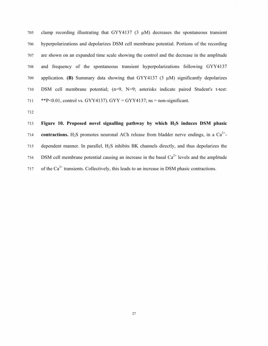
Figure 10. Proposed novel signalling pathway by which H2S induces DSM phasic 713
contractions. H2S promotes neuronal ACh release from bladder nerve endings, in a Ca2+-714
dependent manner. In parallel, H2S inhibits BK channels directly, and thus depolarizes the 715
DSM cell membrane potential causing an increase in the basal Ca2+ levels and the amplitude 716
of the Ca2+ transients. Collectively, this leads to an increase in DSM phasic contractions. 717

1
Figure 1
GYY4137 (log[M])
5 m
N
10 min
-10 -9 -8 -7 -6 -5
TTX (1 M)
-9 -8-7
-6 -5
5 m
N
10 min
-10
GYY4137 (log[M])
-10 -9 -8 -7 -6 -50
150
300
450
600
* ********
TTX + GYY4137GYY4137
* ** **
GYY4137 log[M]
Am
plitu
de (%
)
-10 -9 -8 -7 -6 -50
200
400
600
800 TTX + GYY4137GYY4137
* * * ** * *
GYY4137 log[M]
Mus
cle
Forc
e (%
)
A
B C

Figure 2
GYY4137 (log[M])
5 m
N
10 min
-10 -9 -8 -7 -6 -5
Atropine (1 M)
-10 -9 -8 -7 -6 -50
150
300
450
600 Atropine + GYY4137GYY4137
* ** ***** ** * *
GYY4137 log[M]
Am
plitu
de (%
)
-10 -9 -8 -7 -6 -50
200
400
600
800
* * * * *
GYY4137Atropine + GYY4137
* * *
GYY4137 log[M]
Mus
cle
Forc
e (%
)
A
B
C

Figure 3
0 10 20 30 40 500
30
60
90
120
****
ControlGYY4137
****** ***
***
EFS (Hz)
Mus
cle
Forc
e (%
)
0 10 20 30 40 500
30
60
90
120
*
ControlGYY4137
*** ***
************
***
EFS (Hz)
Am
plitu
de (%
)GYY4137 (3 M)
0.5 2 3.5 5 7.5 10 12.5 20 30 40 5015EFS (Hz)
3 min
20 m
N
0.5 2 3.5 5 7.5 10 12.5 20 30 40 5015
0 10 20 30 40 500
30
60
90
120
*
ControlAtropine
*** *********** ***
Atropine + GYY4137
*
EFS (Hz)
Am
plitu
de (%
)
0 10 20 30 40 500
30
60
90
120
****
ControlAtropine
***** *** *** ***
Atropine + GYY4137
**
EFS (Hz)
Mus
cle
Forc
e (%
)
A
B C
D E

Figure 4
0.000
0.005
0.010
0.015
0.020
0.025
GYY4137Nifedipine (1 M) + GYY4137
*** ** #
ns
Time
1 nM 30 nM 1 M 3 M
Time Control
ns
###
##**
20 40 60 800
[GYY]
(min)
[AC
h] n
mol
/ml

Figure 5
GYY4137 (3 M)
ControlF340/F380
High[Ca2+]i
Low[Ca2+]i a
b
2 min
0.2
(F34
0/F38
0)
GYY4137 (3 M)
a b
0.0
0.1
0.2
0.3
0.4
Control GYY (3 M)
**
Ca2+
Tra
ns. A
mp.
(F34
0/F38
0)
0.2
0.4
0.6
0.8
1.0
Control GYY (3 M)
**
Bas
al C
a2+ L
evel
(F34
0/F38
0)
A
B
C
D

Figure 6
NS11021 (log[M])
5 m
N
10 min
GYY4137 (3 M)
-7-6
-5
5 m
N
10 min
NS11021 (log[M])
-6.5-5.5 -4.5
-7 -6.5 -6 -5.5-5
-4.5
-7 -6 -5 -40
25
50
75
100GYY4137 + NS11021NS11021
*
**
**
**
NS11021 log[M]
Am
plitu
de (%
)
-7 -6 -5 -40
25
50
75
100GYY4137 + NS11021NS11021
*****
*
NS11021 log[M]
Mus
cle
Forc
e (%
)
A
B C

Figure 7
GYY4137 (3 M)
50 p
A
2 min
50 p
A
17 s
B
0
20
40
60
80
100
**
Amplitude Frequency
*
TBK
Cs
(%)
A

1
Figure 8
GYY4137 (3 M)
Control
5 pA
4 s
C
C
O
O
0
25
50
75
100
**
GYY (3 M)
NP O
(%)
A
B C ns
0
3
6
9
12
15
Control GYY (3 M)Sing
le c
hann
el c
urre
nt
ampl
itude
(pA)

Figure 9

Figure 10
?


