Embed Size (px)
Citation preview

REVIEW
Presynaptic nicotinic receptors: a dynamic anddiverse cholinergic filter of striatal dopamineneurotransmission
R Exley and SJ Cragg
Department of Physiology, Anatomy and Genetics, University of Oxford, Oxford, UK
The effects of nicotine on dopamine transmission from mesostriatal dopamine neurons are central to its reinforcing properties.Only recently however, has the influence of presynaptic nicotinic receptors (nAChRs) on dopaminergic axon terminals withinstriatum begun to be understood. Here, rather than simply enhancing (or inhibiting) dopamine release, nAChRs perform therole of a presynaptic filter, whose influence on dopamine release probability depends on presynaptic activity in dopaminergicas well as cholinergic neurons. Both mesostriatal dopaminergic neurons and striatal cholinergic interneurons play key roles inmotivational and sensorimotor processing by the basal ganglia. Moreover, it appears that the striatal influence of dopamineand ACh cannot be fully appreciated without an understanding of their reciprocal interactions. We will review the powerfulfiltering by nAChRs of striatal dopamine release and discuss its dependence on activity in dopaminergic and cholinergicneurons. We will also review how nicotine, acting via nAChR desensitization, promotes the sensitivity of dopamine synapses toactivity. This filtering action might provide a mechanism through which nicotine promotes how burst activity in dopamineneurons facilitates goal-directed behaviour and reinforcement processing. More generally, it indicates that we should notrestrict our view of presynaptic nAChRs to simply enhancing neurotransmitter release. We will also summarize currentunderstanding of the forms and functions of the diverse nAChRs purported to exist on dopaminergic axons. A greaterunderstanding of nAChR form and function is imperative to guide the design of ligands with subtype-selective efficacy forimproved therapeutic interventions in nicotine addiction as well as Parkinson’s disease.
British Journal of Pharmacology (2008) 153, S283–S297; doi:10.1038/sj.bjp.0707510; published online 26 November 2007
Keywords: acetylcholine; nicotine; a6-containing nicotinic receptor; b2-containing nicotinic receptor; dopamine neuron;burst firing; striatum; cholinergic interneuron; Parkinson’s disease; smoking
Abbreviations: ACh, acetylcholine; CNS, central nervous system; CPu, caudate–putamen; a-CtxMII, a-conotoxin MII; GABA,g-aminobutyric acid; KO, knockout; mAChR, muscarinic acetylcholine receptor; MSN, medium spiny neuron;NAc, nucleus accumbens; nAChR, nicotinic acetylcholine receptor; SNc, substantia nigra pars compacta; TAN,tonically active neuron; VTA, ventral tegmental area
Introduction: nicotinic acetylcholine receptors andthe striatum
Nicotinic acetylcholine receptors (nAChRs) are important
modulators of neuronal excitability throughout the central
nervous system (CNS). The most widely observed role of
nAChRs in the CNS is to influence the release of neuro-
transmitters through presynaptically located ‘heterorecep-
tors’. Presynaptic nAChRs have been found to modify the
release of nearly every neurotransmitter examined through
mechanisms that include modifying preterminal membrane
excitability, transmembrane Ca2þ permeability or intracellular
Ca2þ signalling, as reviewed elsewhere (Dani, 2001; Dajas-
Bailador and Wonnacott, 2004; Dani and Bertrand, 2007).
nAChRs can also participate in fast postsynaptic neurotrans-
mission as a small excitatory input in several areas including
hippocampus, and subcortical areas including ventral teg-
mental area (Dani, 2001; Mansvelder et al., 2002; Mameli-
Engvall et al., 2006). However, this role is far from equivalent
to the role of nAChRs in fast excitatory synaptic neuro-
transmission outside the CNS, for example, as primary
mediators of postjunctional excitability in ganglia and
neuromuscular junctions, and is no match within the CNS
for the postsynaptic effects of excitatory glutamatergic
transmission. By contrast, the modulation of neurotransmitter
release by presynaptically located nAChRs is among the most
powerful yet seen for any receptor family, and it is this
presynaptic role for nAChRs that we will discuss further in
this review.Received 13 July 2007; revised 11 September 2007; accepted 19 September
2007; published online 26 November 2007
Correspondence: Dr SJ Cragg, Department of Physiology, Anatomy and
Genetics, University of Oxford, Sherrington Building, Oxford OX1 3PT, UK.
E-mail: [email protected]
British Journal of Pharmacology (2008) 153, S283–S297& 2008 Nature Publishing Group All rights reserved 0007–1188/08 $30.00
www.brjpharmacol.org

Neuronal nAChRs belong to a superfamily of ligand-gated
ion channels, which include g-aminobutyric acid (GABA;
A and C), serotonin and glycine receptors, and have cation-
selective permeability through receptor-channel complexes
formed by pentameric oligomers (Sargent, 1993; McGehee
and Role, 1995; Karlin, 2002; Dani and Bertrand, 2007). In
the CNS, nAChRs are formed from a portfolio of some 12
different a- and b-subunits (a2–a10 and b2–b4) (Corringer
et al., 2000; Le et al., 2002). The association of different
subunits can confer distinct structural and functional
properties to the resultant nAChR, including different
agonist affinity, and kinetics of activation, closure, desensi-
tization, resensitization and even internalization (Ramirez-
Latorre et al., 1996; Chavez-Noriega et al., 1997; Fenster et al.,
1997; Quick and Lester, 2002; Giniatullin et al., 2005;
Nashmi and Lester, 2006). The potential neuron-specific
expression of the diverse array of possible conformations of
nAChRs could offer varied and discrete neuromodulatory
roles to nAChRs in specific neurons. However, the discrete
function(s) that might be offered by the expression of any
one of the heteromeric nAChR subtypes in situ remain
largely unresolved. Moreover, in most neurons, there is
coexpression of multiple diverse nAChR subtypes (Gotti
et al., 2006), which could therefore have complex outcomes
on neurotransmitter release probability. It remains a key
challenge to identify the form and function of nAChR types
differentially expressed in each brain region and neuron
type. If this challenge can be met, we could gain valuable
insights into the underlying computational function afforded
by presynaptic nAChRs, and moreover, into which of these
receptors might be affected or lend itself to a target for drug
design in human neurological or neuropsychiatric disorders
(Sher et al., 2004). Indeed, nicotinic ligands have already
been suggested to hold promise for pharmacological inter-
vention in a large range of disorders, including Parkinson’s
disease, Alzheimer’s disease, epilepsy, schizophrenia and
nicotine addiction (Dani, 2001; Quik and McIntosh, 2006;
Dani and Bertrand, 2007). Significant progress has been
made in identifying the nAChRs expressed by dopamine
neurons and in dopamine axon terminal fields within
striatum as will be reviewed here; intriguingly however, the
details of their influence on dopamine function remains
poorly defined.
The influence of presynaptic nAChRs on dopamine
neurotransmission in the striatum is particularly important
as a model system for nAChR function for several reasons.
Anatomically, the striatum is a large subcortical structure
that contains the densest innervation of dopaminergic and
cholinergic axons seen anywhere in the brain (Bjorklund and
Lindvall, 1984; Butcher and Woolf, 1984; Parent et al., 2000;
Zhou et al., 2002). Functionally, both dopaminergic and
cholinergic systems are central to the role of the striatum,
which, as the main input nucleus to the basal ganglia, is
central to the processing of motivational and sensorimotor
information and to the dysfunction seen in Parkinson’s
disease and drug addiction. Moreover, dopaminergic axons
contain a large diversity of subtypes of heteromeric nAChRs
(see later for review of up to seven proposed types), that as a
population permits striatal ACh to operate a sophisticated
and powerful neuromodulatory control over the release of
striatal dopamine (Zhou et al., 2001; Rice and Cragg, 2004;
Zhang and Sulzer, 2004). In overview, nAChRs do not simply
facilitate presynaptic excitability and/or neurotransmitter
release, but rather more dynamically, they act in a capacity as
a presynaptic filter to differentially govern how variable
activity in dopamine neurons is reported to the striatum by
the release of dopamine. More intriguing yet, the cholinergic
interneurons that supply the acetylcholine (ACh) tone at
striatal nAChRs themselves undergo phasic changes in
activity that are time-locked to the phasic changes in
dopamine neuron activity. Without appreciating the con-
current activity in dopaminergic and striatal cholinergic
neurons, the consequences on striatal signal integration of
neuronal activity in either population alone can only be
poorly calculated. Thus, the striatum provides an outstanding
example of how we should not simply consider the actions
of presynaptic nAChRs to be tonic enablers of neurotrans-
mitter release. Both the outcome of nAChR activity and the
underlying nAChR activity itself are highly dynamic. These
insights require us to rethink our understanding of the scope
of the role of presynaptic nAChRs.
Here, we will review our current understanding of the
presynaptic nAChR control of dopamine neurotransmission
in the striatum. We will discuss the dynamic dependence of
nAChR control on the state of presynaptic activity in both
the output (dopamine) and input (ACh) synapses. We will
also review how the actions of nicotine on striatal dopamine
neurotransmission contribute a link and not a contradiction
between nicotine-induced nAChR desensitization and
postulated mechanisms of drug reinforcement. In addition,
we will discuss the known diversity and function of nAChR
subunits and pentamers within dopaminergic axon terminal
fields. Finally, we will assess whether the field is yet ripe for
therapeutic exploitation and conclude with some of the
many outstanding questions in nAChR research.
Dopamine and acetylcholine systems in thestriatum
The dorsal striatum and ventral striatum collectively parti-
cipate in a large range of motivational, associative- and
sensorimotor-related brain functions (for example, Albin
et al., 1989; Haber et al., 2000; Gerdeman et al., 2003; Voorn
et al., 2004; Everitt and Robbins, 2005; and Schultz, 2006),
among which are the following commonly attributed (and
not necessarily mutually exclusive) processes: natural re-
inforcement, drug reinforcement, conditioned reinforcement,
reward prediction, reward-prediction errors, appetitive beha-
viour, motivational values, stimulus significance, motor
response selection, procedural or stimulus–response learning,
habit formation and task set-shifting. Dopamine inputs to
striatum arise from midbrain dopamine neurons located in
the ventral tegmental area (VTA) and substantia nigra pars
compacta (SNc), which project in a topographic pattern to
differentially innervate the ventral striatum (nucleus
accumbens (NAc)) and dorsal striatum (caudate–putamen
(CPu)), respectively (Bjorklund and Lindvall, 1984; Gerfen
et al., 1987; McFarland and Haber, 2000; Voorn et al., 2004).
The dopaminergic innervation to striatum forms large, highly
Presynaptic nAChRs filter dopamine releaseR Exley and SJ CraggS284
British Journal of Pharmacology (2008) 153 S283–S297

branched arbours with a high density of axonal varicosities
(Pickel et al., 1981; Bouyer et al., 1984; Doucet et al., 1986;
Sesack et al., 1994; Descarries et al., 1996; Descarries and
Mechawar, 2000) and forms dopaminergic synapses at an
incidence estimated at 1 in every 10–20mm3 in the rat
(Descarries et al., 1996; Arbuthnott and Wickens, 2007).
Although dopamine axons can form synapses, dopamine
receptors and the dopamine uptake transporter are found
extrasynaptically (Nirenberg et al., 1996, 1997; Pickel, 2000),
and dopamine can spill over from sites of release to mediate
extrasynaptic or ‘volume’ transmission (Fuxe and Agnati,
1991; Garris et al., 1994; Gonon, 1997; Cragg and Rice, 2004).
The location of dopamine synapses on the necks of spines on
GABAergic medium spiny projection neurons (MSNs) adja-
cent to corticostriatal glutamatergic input (Freund et al., 1984;
Smith and Bolam, 1990; Groves et al., 1994), and the presence
of D1-like and/or D2-like dopamine receptors on MSNs, striatal
interneurons as well as dopamine axons in striatum (Gerfen,
1992; Sesack et al., 1994; Hersch et al., 1995; Surmeier et al.,
1996; Alcantara et al., 2003), ensures dopamine is well placed
to modulate striatal function.
The cholinergic input to the striatum arises solely from
cholinergic striatal interneurons (Woolf, 1991; Contant
et al., 1996; Calabresi et al., 2000). Like the mesostriatal
dopamine neurons, striatal ACh interneurons (or ‘tonically
active neurons’, TANs) (Wilson et al., 1990; Aosaki et al.,
1995; Bennett and Wilson, 1999; Zhou et al., 2002) appear
pivotal for signalling unexpected primary rewards as well as
the learning and signalling of environmental cues that
predict reward (or more generally, events of high salience)
(Calabresi et al., 2000; Schultz, 2002; Berridge and Robinson,
2003; Centonze et al., 2003; Wickens et al., 2003; Wise,
2004). These large, aspiny neurons form a small fraction
(B2–5%) of the total number of neurons in the neostriatum
(Oorschot, 1996; Descarries and Mechawar, 2000; Zhou et al.,
2002), but they provide an extensive axonal arborization
within dorsal and ventral striatum reminiscent of dopami-
nergic arbours (Bolam et al., 1984; Graybiel et al., 1986;
Phelps and Vaughn, 1986; Zahm and Brog, 1992; Contant
et al., 1996; Holt et al., 1997; Calabresi et al., 2000; Descarries
and Mechawar, 2000; Zhou et al., 2001, 2002, 2003). The
density of ACh varicosities is estimated as similar to
dopamine varicosities (Descarries and Mechawar, 2000),
and both dopaminergic and ACh arbours are denser in the
striatum than elsewhere in the brain. Furthermore, like
dopaminergic neurons, striatal cholinergic interneurons
form synapses primarily onto distal dendrite shafts and
spine necks (Bolam et al., 1984; Phelps et al., 1985).
Interestingly, the majority of cholinergic varicosities
(490%) may not form synapses (Descarries et al., 1997;
Descarries and Mechawar, 2000), indicating that ACh like DA
may also influence striatal function primarily via extra-
synaptic, ‘volume’ transmission (Descarries et al., 1997).
Cholinergic receptors in the striatum are of both the
metabotropic muscarinic (muscarinic acetylcholine receptor
(mAChR)) and ionotropic nicotinic (nAChR) families.
Muscarinic receptor types, M1, M2 and M4, appear to be
the dominant muscarinic striatal subtypes (Zhang et al.,
2002; Zhou et al., 2003), with evidence for modulation of
excitability of striatal neurons that include MSNs (Calabresi
et al., 2000), corticostriatal terminals (Malenka and Kocsis,
1988; Pakhotin and Bracci, 2007; Surmeier et al., 2007),
GABAergic and cholinergic interneuron outputs (Koos and
Tepper, 2002; Zhang et al., 2002), and cholinergic interneurons
(Yan and Surmeier, 1996; Bernard et al., 1998; Calabresi et al.,
1998). There is no convincing evidence that mAChRs are
present on dopamine axon terminals to modulate dopamine
release, although indirect striatal circuits may play a role in
mAChR-mediated ACh-dopamine interactions (Zhang et al.,
2002; Zhou et al., 2003). By contrast, the role of nicotinic
AChRs appears less widespread across neuron types within
striatum generally; there is some evidence for nAChR
modulation of GABAergic interneurons (Koos and Tepper,
2002). Most strikingly however, nAChRs located on dopa-
minergic axon terminals (Jones et al., 2001) play a major role
in the modulation of striatal dopamine release by endogenous
ACh (Zhou et al., 2001; Rice and Cragg, 2004; Zhang and
Sulzer, 2004).
Acetylcholine-dopamine crosstalk: antagonistic or cooperative?
Apart from their separate actions within striatum, interac-
tions between dopamine and ACh are fundamental to the
operation of the striatum. There is a long-standing hypothesis
of an antagonistic balance between dopamine and ACh (for
reviews see Calabresi et al., 2000; Zhou et al., 2002; Pisani
et al., 2003; Centonze et al., 2003). The ACh/dopamine
balance hypothesis arose from the alleviation of the
debilitating motor dysfunctions of Parkinson’s disease by
pro-dopaminergic treatments on the one hand, and anti-
cholinergic on the other (Barbeau, 1962; Pisani et al., 2003).
This hypothesis is borne out to some extent on the
postsynaptic cellular level: dopamine and ACh can have
opposing effects on acute excitability of striatal output
neurons as well as on long-term corticostriatal plasticity
(Calabresi et al., 1998, 2000, 2007; Kerr and Wickens, 2001;
Reynolds et al., 2001; Reynolds and Wickens, 2002; Centonze
et al., 2003; Pisani et al., 2003; Zhou et al., 2003; Morris et al.,
2004). Note that this long-term plasticity in corticostriatal
synaptic efficacy is thought to represent reward-related
learning (including the acquisition of incentive value by
previously neutral stimuli, learning of stimulus–response
associations, and development of habits including addiction
at the synaptic level) (Reynolds et al., 2001; Reynolds and
Wickens, 2002; Gerdeman et al., 2003; Robinson and
Berridge, 2003; Robinson and Kolb, 2004) and that this
learning subsequently governs the likelihood that reward-
related signals are translated into contextually appropriate
behavioural responses (reviewed elsewhere, for example,
Wickens et al., 2003; Pisani et al., 2005).
This antagonism is less simple on a presynaptic level
however. ACh and dopamine interact directly at a presynaptic
level. Dopamine can either limit or promote ACh release:
local and systemic striatal D1 agonists can enhance striatal
ACh release in vivo, while D2 agonists reduce (DeBoer and
Abercrombie, 1996; Ikarashi et al., 1997). The availability of
dopamine in the striatum appears, however, to promote the
long-term acquisition of conditioned inhibition or ‘pauses’
in ACh interneuron activity (Maurice et al., 2004; Reynolds
et al., 2004) that are thought to signal reward-related
Presynaptic nAChRs filter dopamine releaseR Exley and SJ Cragg S285
British Journal of Pharmacology (2008) 153 S283–S297

information (see later section). Moreover, as we will see, ACh
acting at nAChRs can either enhance or inhibit dopamine
release within striatum.
Key to the debate of how ACh and dopamine interact is
how this interaction functions during the context of
physiologically relevant and highly dynamic neuron activity.
Clearly, dopamine and ACh may act in opposition (see
above). Yet, these systems do not necessarily oppose each
other during the context of physiologically relevant changes
in dopamine and ACh neurons; the balance of their actions
in a physiological context will depend on the dynamic
changes in neuron activity and the consequent availability
of each neurotransmitter (Cragg, 2006). Importantly, there
are coincident changes in the physiological activity of
dopaminergic and cholinergic neurons, which together with
their consequences for ACh-dopamine interactions indicate
a powerful functional cooperativity between dopaminergic
and cholinergic systems. It is important that we first consider
the underlying physiology in dopamine and ACh neurons
before we can begin to appreciate the presynaptic function of
striatal nAChRs.
Coincident activity in acetylcholine and dopamine neurons
Both dopamine neurons and striatal cholinergic interneurons
participate in reward-related signalling and reinforcement
learning by carrying information about the predicted
availability, or errors in the prediction as well as the receipt,
of primary rewards (Aosaki et al., 1994; Knowlton et al., 1996;
Jog et al., 1999; Matsumoto et al., 1999; Reynolds and
Wickens, 2000, 2002; Reynolds et al., 2001; Packard and
Knowlton, 2002; Schultz, 2002, 2007; Centonze et al., 2003;
Gerdeman et al., 2003; Schultz et al., 2003; Wickens et al.,
2003). These systems do not by contrast signal the hedonic
value of rewards (see Berridge and Robinson, 1998, 2003;
Schultz, 2002; Pecina et al., 2003; Robinson et al., 2005),
which may be processed by other regions, for example,
human orbitofrontal cortex (Kringelbach, 2005). Rather, the
reward-related functions of DA and ACh neurons are
hypothesized to inform the corticostriatal system of the
discrepancy between the prediction of a reward and its actual
occurrence. In simple form, if the reward obtained after an
action or environmental cue is fully predicted, then the
reward prediction error signalled by these neurons at the
time of the reward is zero; if however, the reward is
unpredicted or greater (or less) than predicted, these neurons
signal a positive (or negative) prediction error. These signals
can then be used to teach or update the value of that
predictor (action or cue), and thus, the likelihood that the
action will be repeated or the cue attended to in the future.
This underlies reinforcement learning, which ultimately
allows an animal to respond or perform optimally in the
same environment in the future.
The characteristic responses of dopamine neurons and
striatal ACh neurons to such unexpected primary rewards
and conditioned reward-predicting cues in the environment
can be summarized as follows. Mesostriatal dopamine
neurons signal these events by a switch from tonic firing
rates (typically 2–5 Hz) to either a phasic increase (burst, 15–
100 Hz) or a decrease (pause) according to the presentation or
omission, respectively, of a reward (Hyland et al., 2002;
Schultz, 2002; Fiorillo et al., 2003; Tobler et al., 2003, 2005;
Morris et al., 2004; Bayer and Glimcher, 2005). The responses
of striatal cholinergic interneurons (TANs) in contrast, usually
(but see Ravel et al., 2001; Yamada et al., 2004) involves a brief
pause in tonic rates of activity following reward-related events
whether ‘rewarding’ or ‘punishing’ (Aosaki et al., 1994; Shimo
and Hikosaka, 2001; Morris et al., 2004; Apicella, 2007).
Moreover, these transient reward-related responses of dopa-
mine and ACh neurons appear time-locked with each other:
when recorded in the same task, reward-related bursts in
dopamine neurons occur simultaneously with pauses in ACh
neurons (similar latency and duration, approximately 100–
200ms) (Figure 1) (Morris et al., 2004). The coincident timing
of these events points to a dynamism in the interaction of
dopamine and ACh in striatum. Notably, it suggests that if we
are to appreciate the function of presynaptic nAChRs in
striatum, we must consider highly variable changes
in dopamine neuron activity and the coincident reduction in
ACh tone that will result from a pause in ACh neuron activity.
In other words, we should not confine our understanding of
nAChR function to any single effect of ACh; rather, we should
consider a range of possible effects of ACh across the range
of firing frequencies observed in dopamine neurons, and
furthermore, understand the outcome of a loss of resting
ACh tone.
Presynaptic nAChRs filter the dynamic probabilityof dopamine release according to dopamineneuron activity
How a given pattern and frequency of incoming dopamine
neuron activity is relayed into striatal dopamine release
transients will depend on the processing of dopamine
neuron activity at the release site. Dopamine release does
Figure 1 Synchronization of dopaminergic and cholinergic neuronresponses to behavioural events. Mean responses of population ofdopamine neurons (DAN, grey) and cholinergic interneurons(tonically active neuron (TAN), black) to a visual cue (left) andreward (right) presented to Macaque monkeys in an instrumentalconditioning task at t¼0 (arrow). The increase in dopamine neuronsfiring coincides with the same latency and duration as the pause intonically active cholinergic interneuron firing. Population responsesare baseline-subtracted, averaged for all probabilities in the experi-ment, and normalized to the peak of each response (for DANs) orthe maximum trough (for TANs). The different scales in each panelreveal different degrees of changes for positive and negative errors(see text for discussion of negative prediction errors). Figure takenfrom (Cragg, 2006) and originally adapted from (Morris et al., 2004)with permission.
Presynaptic nAChRs filter dopamine releaseR Exley and SJ CraggS286
British Journal of Pharmacology (2008) 153 S283–S297

not depend linearly on the frequency of activity in
dopamine neurons (Chergui et al., 1994) owing to use-
dependent short-term changes, or ‘plasticity’, in dopamine
release probability (Cragg, 2003; Montague et al., 2004) as
well as neuromodulation by dopamine and ACh transmitters
acting at auto- and heteroreceptors on dopaminergic axons
(Kennedy et al., 1992; Benoit-Marand et al., 2001; Schmitz
et al., 2002, 2003; Rice and Cragg, 2004; Zhang and Sulzer,
2004).
Ligands for nAChRs (including nicotine) have long been
known to influence striatal dopamine release (Di Chiara and
Imperato, 1988; Dajas-Bailador and Wonnacott, 2004).
Importantly, we now appreciate that the control of dopa-
mine release probability by presynaptic nAChRs and en-
dogenous ACh in striatum is dynamic and multifaceted
(Zhou et al., 2001; Rice and Cragg, 2004; Zhang and Sulzer,
2004). Typically, dopamine release in striatum is accompa-
nied by a use-dependent, short-term depression of release
probability at rapidly successive pulses (Abeliovich et al.,
2000; Cragg, 2003; Montague et al., 2004). Studies in striatal
slices, using fast (subsecond) voltammetric detection of
dopamine, indicate that the ACh released from sponta-
neously active striatal cholinergic interneurons (Bennett and
Wilson, 1999; Zhou et al., 2001, 2003) acts at b2-subunit-
containing (b2*)-nAChRs on striatal dopamine axon
terminals to maintain a high probability of dopamine release
evoked by single action potentials (Zhou et al., 2001; Rice
and Cragg, 2004). ACh thus contributes to consequent short-
term depression. A reduction in ACh action at nAChRs, for
example, using competitive antagonists, reduces initial
dopamine release probability (Zhou et al., 2001; Rice and
Cragg, 2004), and as a direct consequence, short-term
depression becomes relieved (Cragg, 2003; Rice and Cragg,
2004). Thus, inhibition of nAChRs can suppress release by
single stimuli but correspondingly facilitate release by a burst
(Figure 2) (Rice and Cragg, 2004).
Moreover, this reorganization of dopamine release prob-
abilities by nAChR inhibition depends on the frequency of
dopamine neuron activity (Rice and Cragg, 2004; Zhang and
Sulzer, 2004): the shorter the interpulse interval, that is, the
higher the dopamine neuron frequency, the greater the relief
from short-term depression. In other words, antagonism of
nAChR activity reduces initial dopamine release probability
but in turn permits a high-frequency pass filtering that
facilitates burst release. Consequently, reduced nAChR
activity can (1) enhance dopamine signals by dopamine
neuron bursts at high frequencies that is those that
accompany the presentation of rewards or conditioned
reward-predicting stimuli (20–100 Hz), while it can also (2)
diminish further the reduced dopamine signals accompany-
ing reductions in dopamine neuron activity, as occurs at
times of omission of expected rewards (Rice and Cragg, 2004;
Cragg, 2006). Thus, a major impact of this filtering will be an
increase in contrast in dopamine signals when dopaminergic
neurons switch from tonic activity to either a high
frequency, reward-related burst or a pause (Cragg, 2006).
Turning the nAChR filter on and off: a pause in TANs and ACh
tone?
These data suggest that a reduction in endogenous ACh tone
at striatal nAChRs following a TAN pause would similarly
enhance the contrast in dopamine signalling when dopa-
mine neurons concurrently change firing rate (Figure 2). A
pause response of the TANs to reward presentation- and
omission-related events could amplify both of the bipolar
effects of these cues on dopamine neuron activity (bursts and
pauses) (Cragg, 2006). These findings suggest that synchro-
nization of phasic changes in activity in dopamine and
acetylcholine neurons could serve to promote the sensitivity
of dopamine synapses to dopamine neuron activity and
enhance the contrast between dopamine transients released
by different activities, bursts or pauses (Cragg, 2006). These
data illustrate the importance of considering the dynamic
activity not only in the dopamine neurons, but also in the
TANs when considering the function of presynaptic nAChRs
on dopamine signalling. The function of presynaptic
nAChRs on striatal dopamine transmission (for example,
during reward-related tasks) might be completely different in
Figure 2 Cartoon of outcome of nicotinic acetylcholine receptors (nAChRs) active and inactive on dopamine transients released by burst-likeand nonburst activity in dopamine neurons. Left, tonic acetylcholine (ACh) tone at nAChRs on dopaminergic axon terminals. Right, nAChR toneswitched off (or reduced) by either a pause in ACh interneuron activity, nAChR desensitization by nicotine or block by nAChR antagonists.
Presynaptic nAChRs filter dopamine releaseR Exley and SJ Cragg S287
British Journal of Pharmacology (2008) 153 S283–S297

the presence of tonic TAN activity (and nAChR tone) versus a
concomitant TAN pause.
Turning the nAChR filter on and off: the effects of nicotine via
nAChR desensitization
Nicotine has long been known to enhance the net release of
dopamine in vivo measured over a timescale of minutes with
microdialysis after system injection (Di Chiara and Imperato,
1988; Corrigall et al., 1992, 1994; Nisell et al., 1994b), and
nicotine also acts as a secretagogue at high concentrations
(mM) to evoke release of preloaded [3H]-dopamine from
striatal synaptosomes (Kulak et al., 1997; Kaiser et al., 1998;
Grady et al., 2002; Dajas-Bailador and Wonnacott, 2004). But
until recently, much less has been known about how
nicotine governs the subsecond release of endogenous
striatal dopamine by dynamic patterns of physiological
action potentials and where endogenous striatal ACh tone
is intact. The effects of nicotine on these dynamic properties
of dopamine release at concentrations of nicotine seen in
smokers have recently been identified, and have simulta-
neously offered insights into several aspects of nicotine’s
properties ranging molecular through to systemic properties,
three of which (1–3) will be discussed here.
1. Desensitization versus agonism?. Nicotinic AChRs are
notoriously susceptible to desensitization, a reversible or
temporary inactivation of response, following sustained
administration of nicotinic receptor ligands including
nicotine (Pidoplichko et al., 1997; Zhou et al., 2001;
Mansvelder et al., 2002; Quick and Lester, 2002; Wooltorton
et al., 2003; Giniatullin et al., 2005). The molecular
mechanisms underlying nAChR desensitization remain in-
completely resolved; candidate mechanisms and their role in
modulating receptor function generally are reviewed else-
where (Quick and Lester, 2002; Giniatullin et al., 2005).
Nicotine, when applied to the brain at concentrations seen
in smokers, readily desensitizes b2*-nAChRs in the VTA and
in the striatum (Pidoplichko et al., 1997; Zhou et al., 2001;
Mansvelder et al., 2002). Note that although a7-receptors are
known to have the fastest onset of desensitization (at
saturating doses of agonist) of any nAChR, the low,
smoking-related concentrations of nicotine preferentially
desensitize the higher affinity b2*-nAChRs (see Quick and
Lester, 2002). It is this nAChR desensitization rather than
simple receptor activation that appears critical to the
outcome on dopamine neuron excitability and ultimately,
on presynaptic nAChR control of dopamine release in
striatum. In the VTA, a greater susceptibility to desensitiza-
tion of b2*- versus a7*-nAChRs reduces direct cholinergic
excitation of dopamine neurons as well as GABAergic
inhibition to dopamine neurons but appears to leave
relatively intact the a7*-nAChR activation of glutamatergic
input to dopamine neurons (Mansvelder et al., 2002). The
net effect of sustained nicotine in VTA is therefore an
enhanced excitability of dopamine neurons (Mansvelder and
McGehee, 2002; Mansvelder et al., 2002). In the striatum
however, where action potential-evoked dopamine release is
under the control of b2*- but not a7*-nAChRs (Zhou et al.,
2001; Rice and Cragg, 2004; Zhang and Sulzer, 2004),
nicotine-induced desensitization of tonically active striatal
nAChRs, reduces initial dopamine release probability, and
consequently, facilitates release by bursts in the same
manner as nAChR antagonists (Zhou et al., 2001; Rice and
Cragg, 2004; Zhang and Sulzer, 2004). Thus, nicotine
dramatically enhances how dopamine is released by re-
ward-related burst activity compared to non-reward-related
tonic activity (Figure 2).
Thus, desensitization of nAChRs plays an essential facili-
tatory role in both the VTA and directly within the striatum
in the ability of nicotine to enhance striatal dopamine
neurotransmission. We should not limit our appreciation of
the action of nicotine on the brain to its effects at nAChRs as
a simple agonist. Rather, loss of function of striatal nAChRs
can be a gain of function to dopamine transmission.
2. VTA versus striatum?. The desensitization of nAChRs by
nicotine in striatum also sheds light on why the striatum was
long overlooked as a site of action of nicotine; the long-held
notion was that the VTA but not the striatum was important
for nicotine’s effects on striatal dopamine transmission. For
example, previous experiments identified that local admin-
istration of nAChR blockers into the VTA but not striatum
prevents the self-administration of systemic nicotine
(Corrigall et al., 1994) or prevent increases in accumbal
dopamine measured with microdialysis following systemic
nicotine injection (Nisell et al., 1994b). Furthermore,
nicotine infused continuously into the VTA but not striatum
has been reported to sustain striatal dopamine release
(measured by microdialysis) (Nisell et al., 1994a). Recent
insights into nicotine’s desensitizing action within striatum
now reveal several possible alternative explanations for why
the effects of nicotine in the striatum may previously have
been underestimated. For example, since nicotine’s effects
within striatum appear to involve switching nAChRs off,
nAChR antagonists in striatum are unlikely to reverse
nicotine’s effects, but might in fact substitute for nicotine.
In contrast in the VTA, where the effects of nicotine require
both desensitization and agonism (Mansvelder et al., 2002),
nAChR antagonists would be expected to obscure nicotine
actions. Furthermore, since nicotine’s effects in striatum
involve enhancing signal contrast on a subsecond basis by
both reducing and enhancing striatal dopamine release
depending on dopamine neuron (and TAN) firing rate, it is
possible that the net effect of nicotine in striatum might be
an undetectable net zero change when measured over a long
microdialysis sample period. In addition, nicotine’s striatal
effects also depend on changes in underlying dopamine
neuron firing rate, for example, changes that accompany
nicotine reaching the VTA, and may not in contrast be
exposed in vivo by striatal administration only. The striatal
mechanisms reviewed here are distinct from (but additive
with) those actions of nicotine on dopamine neuron
excitability at the somatodendritic level. Together, axonal
effects in combination with somatodendritic effects
(Grenhoff et al., 1986; Pidoplichko et al., 1997; Mansvelder
et al., 2002; Champtiaux et al., 2003; Maskos et al., 2005;
Mameli-Engvall et al., 2006) will provide a two-step ampli-
fication by nicotine of dopamine signalling in striatum. Our
improved understanding of the consequences of nicotine in
Presynaptic nAChRs filter dopamine releaseR Exley and SJ CraggS288
British Journal of Pharmacology (2008) 153 S283–S297

striatum (via nAChR desensitization) on physiological sub-
second dopamine signals, now allows us to appreciate the
striatum as an important site for nicotine action.
3. Primary reinforcer or enhancer of secondary reinforce-
ment?. The striatal effects of nicotine also offer a neuro-
pharmacological correlate for some of nicotine’s less
well-understood psychopharmacological properties. A key
question in nicotine addiction is to what extent the
reinforcing effects of nicotine result from its intrinsic
reinforcing properties, that is, as a primary or unconditioned
reward, versus its ability to enhance the reinforcing efficacy
of other primary and secondary reinforcers (Caggiula et al.,
2001; Donny et al., 2003; Olausson et al., 2003). It has been
questioned whether the primary, or unconditioned, reinfor-
cing effects of nicotine are sufficient to explain the
maintenance of smoking behaviour in humans (Perkins
et al., 2003). Rather, sustained nicotine self-administration,
in rats as well as in human smokers, may be dependent on
associated environmental cues (for example, visual or
olfactory), which, through becoming conditioned reinfor-
cers, can take on critical incentive properties (Caggiula et al.,
2001). Notably, nicotine can noncontingently enhance the
reinforcing efficacy of previously neutral cues (Chaudhri
et al., 2003; Donny et al., 2003). A principal means through
which novel, behaviourally relevant stimuli and conditioned
cues are signalled to the brain is via a switch in dopamine
neuron firing activity to a brief high-frequency burst
(Schultz, 2002, 2007). Since nicotine via nAChR desensitiza-
tion in striatum enhances the dopamine signalling by burst
versus nonburst activity in dopamine neurons, the pharma-
cological effects of nicotine in striatum reviewed here may
therefore offer a neurochemical correlate for the nicotine
enhancement of the reinforcing efficacy of any reward-
related stimuli, including non-nicotine conditioned stimuli
(for example, predictive visual cues) that support nicotine
self-administration (Caggiula et al., 2001; Chaudhri et al.,
2003; Donny et al., 2003).
The diversity of nAChR subunits in dopamineneurons
What do we understand of the nAChRs responsible for the
control of dopamine neurotransmission by ACh and by
nicotine in striatum? Dopamine neurons express a large
range of nAChR subunits and several stoichiometric config-
urations are proposed to exist in dopamine axons and
regulate striatal dopamine neurotransmission.
To date, 12 mammalian subunits a2–a10 and b2–b4 have
been identified and cloned. These subunits can be organized
into subfamilies I–IV, according to their gene sequence and
structure (Corringer et al., 2000; Le et al., 2002). Only nAChR
subunits from subfamilies II (a7) and III (a2–a6, b2–b4) have
been identified in mammalian brain (Corringer et al., 2000;
Le et al., 2002). These subunits can form homomeric
pentamers (all one type of subunit, for example, a7) or
heteromeric pentamers, consisting of combinations of
various a- and b-type subunits. Dopamine neurons within
rodent SNc and VTA express mRNAs for the nAChR subunits
a4, a5, a6, b2 and b3, as well as lower levels of a3 and a7 and
lower levels yet of b4 (Azam et al., 2002). This diversity of
subunit expression has the potential to give rise to multiple
types of pentameric receptors in somatodendritic and axon
terminal regions of dopamine neurons with a consequent
myriad of corresponding functions.
The nAChRs present in both somatodendritic (VTA/SNc) as
well as axonal regions (striatum) are believed to be important
to the reinforcing effects of nicotine (Grenhoff et al., 1986;
Mansvelder and McGehee, 2002; Mansvelder et al., 2002;
Zhou et al., 2003; Rice and Cragg, 2004; Zhang and Sulzer,
2004; Ungless and Cragg, 2006; Mameli-Engvall et al., 2006).
However, there are differences in the nAChR subunits
present in axon terminals compared to somatodendritic
regions. Whereas a7- and b4-subunits are present in VTA/
SNc, they are not apparently transported to striatal dopami-
nergic axon terminals (Champtiaux et al., 2003; Quik et al.,
2005). The a7-subunit is, however, expressed within striatum
by non-dopaminergic neurons, although this is not dis-
cussed here. Furthermore, there is a lack of a3-subunits
within the striatum in rodents (although not in primates)
(Wonnacott et al., 2000; Zoli et al., 2002; Champtiaux et al.,
2003; Gotti et al., 2005; Quik et al., 2005). However, the a4-,
a5-, a6-, b2- and b3-subunits (and a3-subunits in primate) are
found at high density in dopamine axon terminals where
they can assemble as functional receptor channels in the
form of heteromers.
As heteromeric receptors, these subunits arrange to form
two a/b pairs; the interface of each of these pairs is
responsible for providing one of the two ACh-binding sites
at each nAChR (Gotti and Clementi, 2004). The two a/b pairs
that form the ligand-binding sites in dopamine axons in
striatum are suggested to be primarily a4/b2 and/or a6/b2
(and/or a3/b2 in primates) (Luetje, 2004; Salminen et al.,
2004; Quik et al., 2005). The fifth subunit in the pentamer
may consist of any other subunit, including a5 or b3. Several
mechanisms are known to control the stoichiometry of
pentameric nAChRs during assembly in transfected cell
expression systems: nAChR subunit composition and stoi-
chiometry may be influenced by cellular chaperone proteins
and intracellular cAMP levels as well as transfected subunit
ratio or exposure to nicotine (Zwart and Vijverberg, 1998;
Wanamaker et al., 2003; Kuryatov et al., 2005; Vallejo et al.,
2005; Exley et al., 2006). These mechanisms will not be
discussed further here. However, while we have some
understanding of how these characteristics arise in nAChRs
in non-neuronal expression systems in vitro, we are still
far from understanding their form and function in neurons
in vivo.
What is the stoichiometry and function of striatal nAChRs in
dopamine axons?
Our current appreciation of the nAChRs that participate in
the regulation of striatal dopamine transmission has arisen
from the development of specific ligands, subunit-specific
knockout (KO) mice and immunoprecipitation studies. We
will review our current understanding of the nAChRs present
on dopamine axons that is derived primarily from rodent
Presynaptic nAChRs filter dopamine releaseR Exley and SJ Cragg S289
British Journal of Pharmacology (2008) 153 S283–S297
studies with a few valuable insights from primate studies
where available.
The b2-subunit was the first shown to be crucial to the
reinforcing properties of nicotine in rats (Picciotto et al.,
1998). It was later demonstrated that b2-nAChR subunits are
widely expressed presynaptically on striatal dopamine axon
terminals (Jones et al., 2001). Subsequent immunoprecipita-
tion studies revealed the dominant presence of the b2-
subunit within the striatum: all nAChRs on dopamine
axon terminals are thought to contain the b2-subunit
(Champtiaux et al., 2003; Salminen et al., 2004). The
determination of the stoichiometry of these b2*-nAChRs
has since been particularly aided by the development of
a ligand, which can differentiate alternate a-subunit-con-
taining nAChRs. Specifically, the antagonist a-conotoxin MII
(a-CtxMII) (extracted from Conus snails) is selective for a3/
a6-subunit-containing nAChRs (Cartier et al., 1996; White-
aker et al., 2000; McIntosh et al., 2004). This ligand has been
used in studies of nicotine-evoked [3H]-dopamine release
from synaptosomes to identify two distinct classes of striatal
b2*-nAChRs, a-CtxMII-sensitive (a6/a3,b2*-nAChRs) and
a-CtxMII-resistant (non-a6/a3,b2*-nAChRs) (Kulak et al.,
1997; Kaiser et al., 1998; Quik et al., 2005). These studies
suggest that the a-CtxMII-resistant population of nAChRs
(that is, non-a6/a3,b2*) can account for up to 60% of
nicotine-evoked dopamine release from rodent striatal
synaptosomes (Kulak et al., 1997; Kaiser et al., 1998;
Salminen et al., 2007). However, the relative importance of
a6* versus non-a6-nAChRs in the regulation of DA release by
endogenous ACh varies greatly within different functional
subterritories of the striatum (Exley et al., in press).
Immunoprecipitation studies have contributed greatly in
defining nAChR subunits which could constitute these
different a6- and non-a6,b2*-nAChRs in striatal dopaminer-
gic terminals. These studies have shown that in addition to
the b2-subunit, both a4- and a6-subunits are also widely
present in striatum. In addition, a small portion of a4-
subunits are colocalized with a5-subunits (approximately
11%), whereas a6-subunits do not appear to be colocalized
with a5 (Champtiaux et al., 2003). The b3-subunit by
contrast, appears always to be colocalized with a6-subunits,
and a portion of this population can contain a4-subunits
(but see later) (Champtiaux et al., 2003; Cui et al., 2003; Gotti
et al., 2005). Together, these data have suggested the
presence of a4b2- and a4a5b2-nAChRs (a-CtxMII-resistant)
as well as a6b2b3- and a6a4b2b3-nAChRs (a-CtxMII-sensi-
tive) (Zoli et al., 2002; Champtiaux et al., 2003; Cui et al.,
2003; Luetje, 2004; Salminen et al., 2004, 2005; Gotti et al.,
2005). In the primate striatum, there is an additional a3b2*-
nAChR which also binds a-CtxMII (Cartier et al., 1996; Quik
et al., 2005).
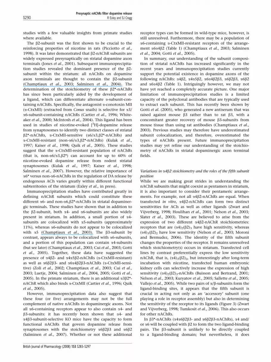
However, immunoprecipitation data also suggest that
these four (or five) arrangements may not be the full
complement of native nAChRs in dopaminergic axons. Not
all a6-containing receptors appear to also contain a4- and
b3-subunits: it has recently been shown that a4- and
a4b3-subunit-selective KO mice have the capacity to form
functional nAChRs that govern dopamine release from
synaptosomes with the stoichiometry a6b2b3 and a6b2
(Salminen et al., 2007). Whether or not these additional
receptor types can be formed in wild-type mice, however, is
still unresolved. Furthermore, there may be a population of
a6-containing a-CtxMII-resistant receptors of the arrange-
ment a6a4b2 (Table 1) (Champtiaux et al., 2003; Salminen
et al., 2004; Gotti et al., 2005).
In summary, our understanding of the subunit composi-
tion of striatal nAChRs has increased significantly in the
recent years and immunoprecipitation studies currently
support the potential existence in dopamine axons of the
following nAChRs: a4b2, a4a5b2, a6a4b2b3, a6b2b3, a6b2
and a6a4b2 (Table 1). Intriguingly however, we may not
have yet reached a completely accurate picture. One major
limitation of immunoprecipitation studies is a limited
capacity of the polyclonal antibodies that are typically used
to extract each subunit. This has recently been shown by
Gotti et al. (2005), who generated a new antiserum that was
raised against mouse b3 rather than to rat b3, with a
concomitant greater recovery of mouse b3-subunits from
mouse tissue than using rat antibodies (Champtiaux et al.,
2003). Previous studies may therefore have underestimated
subunit colocalization, and therefore, overestimated the
types of nAChRs present. Future immunoprecipitation
studies may yet refine our understanding of the stoichio-
metry of nAChRs in striatal dopaminergic axon terminal
fields.
Variations in a4b2 stoichiometry and the roles of the fifth subunit
position
While we are making great strides in understanding the
nAChR subunits that might coexist as pentamers in striatum,
it is also important to consider their pentameric arrange-
ments. For example, not all a4b2-nAChRs are equal. When
transfected in vitro, a4b2-nAChRs can form two distinct
sensitivities for ACh as well as other ligands (Zwart and
Vijverberg, 1998; Houlihan et al., 2001; Nelson et al., 2003;
Slater et al., 2003). These are believed to arise from the
expression of two different a4b2-nAChR stoichiometries:
receptors that are (a4)2(b2)3 have high sensitivity, whereas
(a4)3(b2)2 have low sensitivity (Nelson et al., 2003; Moroni
and Bermudez, 2006). The identity of the fifth subunit
changes the properties of the receptor. It remains unresolved
which stoichiometry(s) occurs in striatum. Transfected cell
lines by contrast preferentially express the low sensitivity
nAChR, that is, (a4)3(b2)2, but interestingly after long-term
incubation with nicotine, transfected human embryonic
kidney cells can selectively increase the expression of high
sensitivity (a4)2(b2)3-nAChRs (Buisson and Bertrand, 2001;
Nelson et al., 2003; Kuryatov et al., 2005; Sallette et al., 2005;
Vallejo et al., 2005). While two pairs of a/b-subunits form the
ligand-binding sites, it appears that the fifth subunit is
crucial in acting not only as an ‘accessory’ subunit (one
playing a role in receptor assembly) but also in determining
the sensitivity of the receptor to its ligands (Figure 3) (Zwart
and Vijverberg, 1998; Tumkosit et al., 2006). This also occurs
for other nAChRs.
In b3*-nAChRs (a4a6b2b3- and a6b2b3-nAChRs), a4 and/
or a6 will be coupled with b2 to form the two ligand-binding
pairs. The b3-subunit is unlikely to be directly coupled
to a ligand-binding domain; but nevertheless, it does
Presynaptic nAChRs filter dopamine releaseR Exley and SJ CraggS290
British Journal of Pharmacology (2008) 153 S283–S297

appear to influence receptor assembly and the binding
of a-CtxMII (Cui et al., 2003; Tumkosit et al., 2006). For
the a4a5b2 receptor, it appears that the a4/b2 subunits form
the ACh-binding site, while the a5-subunit acts as an
accessory to occupy the fifth position. Furthermore, inclu-
sion of chick a5-subunit in the human a4b2-nAChR (in
Xenopus oocytes) can increase receptor desensitization to
nicotine (Ramirez-Latorre et al., 1996). It is interesting to
note that inclusion of the human a5-subunit in a different
human nAChR, the a3b2 receptor, increases receptor
desensitization to nicotine, but also nicotine efficacy (Wang
et al., 1996).
In a6*-nAChRs, the a6-subunit predominantly forms a
a6/b2 interface, which binds a-CtxMII. However, in rodent
striatum, there is a small (B6%) portion of a6*-nAChRs,
which are a-CtxMII-resistant (Gotti et al., 2005). This
suggests that some nAChRs have a6-subunits that do not
form ligand-binding domains, but rather occupy the fifth
non-ACh-binding position. Might the a6-subunit, as seen
for the a4 and b2 subunits, be able to take on two different
types of roles? One role could confer ligand specificity at an
a6/b2 interface in the a6b2-, a6b2b3- and a6a4b2b3-nAChRs
(Cui et al., 2003; Salminen et al., 2007), while the other
could be to act as an accessory subunit at the fifth position
(Figure 3). In summary, even when stoichiometry in striatum
is known the nature of the ACh-binding site may depend not
only on the four subunits, which constitute the two binding
sites, but also the fifth non-binding subunit. We therefore
need to improve our understanding of the function and the
stoichiometry of the nAChRs that regulate dopamine release,
if we are to successfully exploit nAChRs in neurodegenera-
tion and addictive disorders.
�4
�4
�2/�4
�2
�2
�2/�3
α-CtxMII-resistant α-CtxMII-sensitive
Modulator Accessory/ Modulator
Accessory/ Modulator?
Accessory/ Modulator?
ACh binding site �4
�4 �2
�6
�4 �2 �6
�6
�2
�2�2
�3
�2
�5/�6
Figure 3 Nicotinic acetylcholine receptor (nAChR) stoichiometry, a4/b2-binding pairs and the role of the fifth subunit. Both a4/b2 and a6/b2pairs form ligand-binding domains. For a-conotoxin MII (a-CtxMII)-resistant receptors, the fifth subunit can modulate nAChR ligand affinity.Sensitivity to a-CtxMII is conferred at the a6/b2 domain only and will result from block of either one or two acetylcholine (ACh)-binding sitesavailable to activate the channel. Fifth subunits in a-CtxMII-sensitive receptors may be accessories or affinity modulators. Binding sites for AChare a-CtxMII-resistant (filled squares) or a-CtxMII-sensitive (unfilled squares).
Table 1 nAChRs proposed to be expressed on dopaminergic axon terminals in striatum
Species Region Receptor stoichiometry
a4b2 a3b2* a4a5b2 a6a4b2 a6a4b2b3 a6b2 a6b2b3
Mouse Striatum Champtiaux et al.(2003);
NA Champtiauxet al. (2003);
Champtiauxet al. (2003);
Champtiaux et al.(2003);
Champtiaux et al.(2003);
Champtiaux et al.(2003);
Gotti et al. (2005); Gotti et al.(2005);
Salminen et al.(2004)
Gotti et al. (2005); Salminen et al.(2004)
Gotti et al.(2005);
Salminen et al.(2004)
Salminen et al.(2004)
Salminen et al.(2004)
CPu NA Cui et al. (2003); Salminen et al.(2007)
Cui et al. (2003);
Salminen et al.(2007)
Salminen et al.(2007)
NAc NA Cui et al. (2003); Salminen et al.(2007)
Cui et al. (2003);
Salminen et al.(2007)
Salminen et al.(2007)
Rat Striatum Zoli et al. (2002) NA Zoli et al.(2002)
Zoli et al.(2002)
Zoli et al. (2002) Zoli et al. (2002) Zoli et al. (2002)
Primate Striatum Quik et al. (2005) Quik et al.(2005)
Quik et al. (2005) Quik et al. (2005)
Abbreviations: CPu, caudate–putamen (dorsal striatum); NAc, nucleus accumbens (ventral striatum); nAChR, nicotinic acetylcholine receptor. Due to the
detectable expression of a3-subunit only in primate but not rodent striatum, the expression of a3b2*-nAChRs are not applicable (NA) to rodents.
Table of studies citing the presence of each proposed nAChR subtype. Note that studies listed in ‘Striatum’ do not distinguish between ventral and dorsal territories
of striatum.
Presynaptic nAChRs filter dopamine releaseR Exley and SJ Cragg S291
British Journal of Pharmacology (2008) 153 S283–S297

Summary and perspective
Our understanding of presynaptic nAChR function in
mesostriatal dopamine neurons has been revised signifi-
cantly by studies in the last 4–5 years. We now understand
the need to appreciate how nAChRs can filter, rather than
simply enhance, neurotransmitter release from dopamine
neurons. It remains a key and open question as to whether
this filtering mode of action occurs for presynaptic nAChRs
on other neuron types, that is, whether this can be general-
ized to nAChR function across the brain. Furthermore, the
presynaptic filter on dopamine release probability operated
by presynaptic nAChRs in striatum depends on activity in
both dopamine and striatal cholinergic neurons. Given that
synchronized phasic changes in both dopaminergic and
cholinergic neurons appear to be the means through which
behaviourally relevant information is signalled, it is thus
imperative that we appreciate this dynamic interplay.
Specifically, we should consider the powerful outcome of
turning off nAChR tone during such a pause in cholinergic
interneurons, rather than limiting our perspective to the
response to nAChR activation. Together, these data suggest
that future approaches to finesse our understanding of the
function of presynaptic nAChRs on striatal dopaminergic
axons should aim to explore dopamine signals using
methods that can explore release on a timescale commensu-
rate with the dynamic properties of neuron activity and also
consider the outcome of turning ACh tone off. It is also
becoming clear that turning off nAChRs may be central to
the reinforcing action of nicotine. The desensitization of
nAChRs by nicotine is increasingly becoming appreciated as
fundamental to nicotine’s properties rather than a paradox-
ical synaptic observation at odds with our expectation of
how nicotine might be reinforcing.
We understand much less about why nAChRs are pro-
duced in such diverse forms and even exactly how many
diverse forms exist within any one neuron type. While we
have nonetheless made considerable progress in under-
standing the possible forms these nAChRs may take in
dopaminergic axons, we have yet to obtain insights in to
whether these different receptors have different functions in
the regulation of discrete dopamine signals by endogenous
ACh or by nicotine. We also have yet to establish what the
function of any one subtype may be or furthermore whether
there is functional redundancy. We have therefore still to
bridge a large gap between insights into subunit identity on
the one hand, and an understanding of subunit function in
the dynamic control of dopamine signalling by endogenous
ACh on the other. And yet, if we understood the form and
function of nAChR diversity generally, we would not only
better grasp the fundamental purpose of nAChRs in the
brain, but moreover, we could reveal major potential for
selective neuromodulation of targeted neuron types and
guide future drug design for targeted nAChR types that could
offer major potential therapeutic benefit to many disorders
of neuron function that have been suggested to include
nicotine addiction, Parkinson’s disease, Alzheimer’s disease,
epilepsy and schizophrenia.
What other unknowns lie between our current under-
standing of nAChR control of dopaminergic transmission
and an ideal goal of therapeutic exploitation, for example, in
smoking and Parkinson’s disease? To begin with, can this
potent presynaptic regulation of DA signalling by nAChRs
be demonstrated in behaving animals? To date, real-time
electrochemical techniques at carbon-fibre microelectrodes
within the striatum have identified that phasic-like DA
transients in reward-seeking, behaving animals accompany
reward prediction and positive prediction errors (for exam-
ple, Garris et al., 1999; Phillips et al., 2003; Stuber et al.,
2005); however, the effect of ACh on these transients is not
currently known. Furthermore, we will need to identify how
the adaptive and passive changes in nAChR expression that
are well documented after chronic nicotine (Buisson and
Bertrand, 2002; Parker et al., 2004; Lai et al., 2005; Pakkanen
et al., 2005, 2006; McCallum et al., 2006a, b; Quik et al., 2006;
Perry et al., 2007) or in Parkinson’s disease (Aubert et al.,
1992; Martin-Ruiz et al., 2000; Quik and Jeyarasasingam,
2000; Quik et al., 2001, 2002, 2005; Kulak et al., 2002a, b;
Bohr et al., 2005) might influence the underlying nAChR
function we would aim to modulate. We will also need a
better grasp of subunit-specific function if any nAChR
therapy is to achieve regional selectivity. We may still be
some way from realizing these potential opportunities for
therapeutic avenues in addictive and other neurological
disorders but the future now holds significant promise.
Acknowledgements
We thank Professor I Bermudez for comments on the
manuscript and acknowledge the Parkinson’s Disease Society
(UK) for grant support.
Conflict of interest
SJC has received grant support from Eli Lilly (UK).
References
Abeliovich A, Schmitz Y, Farinas I, Choi-Lundberg D, Ho WH,Castillo PE et al. (2000). Mice lacking alpha-synuclein displayfunctional deficits in the nigrostriatal dopamine system. Neuron25: 239–252.
Albin RL, Young AB, Penney JB (1989). The functional anatomy ofbasal ganglia disorders. Trends Neurosci 12: 366–375.
Alcantara AA, Chen V, Herring BE, Mendenhall JM, Berlanga ML(2003). Localization of dopamine D2 receptors on cholinergicinterneurons of the dorsal striatum and nucleus accumbens of therat. Brain Res 986: 22–29.
Aosaki T, Kimura M, Graybiel AM (1995). Temporal and spatialcharacteristics of tonically active neurons of the primate’sstriatum. J Neurophysiol 73: 1234–1252.
Aosaki T, Tsubokawa H, Ishida A, Watanabe K, Graybiel AM, KimuraM (1994). Responses of tonically active neurons in the primate’sstriatum undergo systematic changes during behavioral sensor-imotor conditioning. J Neurosci 14: 3969–3984.
Apicella P (2007). Leading tonically active neurons of the striatumfrom reward detection to context recognition. Trends Neurosci 30:299–306.
Arbuthnott GW, Wickens J (2007). Space, time and dopamine. TrendsNeurosci 30: 62–69.
Presynaptic nAChRs filter dopamine releaseR Exley and SJ CraggS292
British Journal of Pharmacology (2008) 153 S283–S297

Aubert I, Araujo DM, Cecyre D, Robitaille Y, Gauthier S, Quirion R(1992). Comparative alterations of nicotinic and muscarinicbinding sites in Alzheimer’s and Parkinson’s diseases. J Neurochem58: 529–541.
Azam L, Winzer-Serhan UH, Chen Y, Leslie FM (2002). Expression ofneuronal nicotinic acetylcholine receptor subunit mRNAs withinmidbrain dopamine neurons. J Comp Neurol 444: 260–274.
Barbeau A (1962). The pathogenesis of Parkinson’s disease: a newhypothesis. Can Med Assoc J 87: 802–807.
Bayer HM, Glimcher PW (2005). Midbrain dopamine neuronsencode a quantitative reward prediction error signal. Neuron 47:129–141.
Bennett BD, Wilson CJ (1999). Spontaneous activity of neostriatalcholinergic interneurons in vitro. J Neurosci 19: 5586–5596.
Benoit-Marand M, Borrelli E, Gonon F (2001). Inhibition ofdopamine release via presynaptic D2 receptors: time course andfunctional characteristics in vivo. J Neurosci 21: 9134–9141.
Bernard V, Laribi O, Levey AI, Bloch B (1998). Subcellular redistribu-tion of m2 muscarinic acetylcholine receptors in striatal inter-neurons in vivo after acute cholinergic stimulation. J Neurosci 18:10207–10218.
Berridge KC, Robinson TE (1998). What is the role of dopamine inreward: hedonic impact, reward learning, or incentive salience?Brain Res Brain Res Rev 28: 309–369.
Berridge KC, Robinson TE (2003). Parsing reward. Trends Neurosci 26:507–513.
Bjorklund A, Lindvall O (1984). Dopamine-containing systems in theCNS. In: Bjorklund A, Hokfelt T (eds). Handbook of ChemicalNeuroanatomy. Elsevier: New York, pp 55–122.
Bohr IJ, Ray MA, McIntosh JM, Chalon S, Guilloteau D, McKeith IGet al. (2005). Cholinergic nicotinic receptor involvement inmovement disorders associated with Lewy body diseases. Anautoradiography study using [(125)I]alpha-conotoxinMII in thestriatum and thalamus. Exp Neurol 191: 292–300.
Bolam JP, Wainer BH, Smith AD (1984). Characterization ofcholinergic neurons in the rat neostriatum. A combination ofcholine acetyltransferase immunocytochemistry, Golgi-impregna-tion and electron microscopy. Neuroscience 12: 711–718.
Bouyer JJ, Joh TH, Pickel VM (1984). Ultrastructural localization oftyrosine hydroxylase in rat nucleus accumbens. J Comp Neurol 227:92–103.
Buisson B, Bertrand D (2001). Chronic exposure to nicotineupregulates the human (alpha)4(beta)2 nicotinic acetylcholinereceptor function. J Neurosci 21: 1819–1829.
Buisson B, Bertrand D (2002). Nicotine addiction: the possible role offunctional upregulation. Trends Pharmacol Sci 23: 130–136.
Butcher LL, Woolf NJ (1984). Histochemical distribution of acetyl-cholinesterase in the central nervous system: clues to thelocalization of cholinergic neurons. In Bjorklund A, Hokfelt T,Kuhar MJ (eds). Classical Transmitters and Transmitter Receptors inthe CNS, Part II. Elsevier: Amsterdam, pp 1–50.
Caggiula AR, Donny EC, White AR, Chaudhri N, Booth S, Gharib MAet al. (2001). Cue dependency of nicotine self-administration andsmoking. Pharmacol Biochem Behav 70: 515–530.
Calabresi P, Centonze D, Gubellini P, Pisani A, Bernardi G (1998).Blockade of M2-like muscarinic receptors enhances long-termpotentiation at corticostriatal synapses. Eur J Neurosci 10: 3020–3023.
Calabresi P, Centonze D, Gubellini P, Pisani A, Bernardi G (2000).Acetylcholine-mediated modulation of striatal function. TrendsNeurosci 23: 120–126.
Calabresi P, Picconi B, Tozzi A, Di Filippo M (2007). Dopamine-mediated regulation of corticostriatal synaptic plasticity. TrendsNeurosci 30: 211–219.
Cartier GE, Yoshikami D, Gray WR, Luo S, Olivera BM, McIntosh JM(1996). A new alpha-conotoxin which targets alpha3beta2 nico-tinic acetylcholine receptors. J Biol Chem 271: 7522–7528.
Centonze D, Gubellini P, Pisani A, Bernardi G, Calabresi P (2003).Dopamine, acetylcholine and nitric oxide systems interact toinduce corticostriatal synaptic plasticity. Rev Neurosci 14: 207–216.
Champtiaux N, Gotti C, Cordero-Erausquin M, David DJ, PrzybylskiC, Lena C et al. (2003). Subunit composition of functionalnicotinic receptors in dopaminergic neurons investigated withknock-out mice. J Neurosci 23: 7820–7829.
Chaudhri N, Caggiula AR, Donny EC, Booth S, Gharib MA, ClementsL et al. (2003). Enhancement of reinforced operant responding bynicotine and cocaine in rats: analysis of dose and drug-con-tingency. Program no. 323.8. 2003 Abstract Viewer and ItineraryPlanner. Society for Neuroscience: Washington, DC, Online.
Chavez-Noriega LE, Crona JH, Washburn MS, Urrutia A, Elliott KJ,Johnson EC (1997). Pharmacological characterization of recombi-nant human neuronal nicotinic acetylcholine receptors h alpha 2beta 2, h alpha 2 beta 4, h alpha 3 beta 2, h alpha 3 beta 4, h alpha4 beta 2, h alpha 4 beta 4 and h alpha 7 expressed in Xenopusoocytes. J Pharmacol Exp Ther 280: 346–356.
Chergui K, Suaud-Chagny MF, Gonon F (1994). Nonlinear relation-ship between impulse flow, dopamine release and dopamineelimination in the rat brain in vivo. Neuroscience 62: 641–645.
Contant C, Umbriaco D, Garcia S, Watkins KC, Descarries L (1996).Ultrastructural characterization of the acetylcholine innervationin adult rat neostriatum. Neuroscience 71: 937–947.
Corrigall WA, Coen KM, Adamson KL (1994). Self-administerednicotine activates the mesolimbic dopamine system through theventral tegmental area. Brain Res 653: 278–284.
Corrigall WA, Franklin KB, Coen KM, Clarke PB (1992). Themesolimbic dopaminergic system is implicated in the reinforcingeffects of nicotine. Psychopharmacology (Berl) 107: 285–289.
Corringer PJ, Le NN, Changeux JP (2000). Nicotinic receptors at theamino acid level. Annu Rev Pharmacol Toxicol 40: 431–458.
Cragg SJ (2003). Variable dopamine release probability and short-term plasticity between functional domains of the primatestriatum. J Neurosci 23: 4378–4385.
Cragg SJ (2006). Meaningful silences: how dopamine listens to theACh pause. Trends Neurosci 29: 125–131.
Cragg SJ, Rice ME (2004). DAncing past the DAT at a DA synapse.Trends Neurosci 27: 270–277.
Cui C, Booker TK, Allen RS, Grady SR, Whiteaker P, Marks MJ et al.(2003). The {beta}3 nicotinic receptor subunit: a component of{alpha}-conotoxin MII-binding nicotinic acetylcholine receptorsthat modulate dopamine release and related behaviors. J Neurosci23: 11045–11053.
Dajas-Bailador F, Wonnacott S (2004). Nicotinic acetylcholinereceptors and the regulation of neuronal signalling. TrendsPharmacol Sci 25: 317–324.
Dani JA (2001). Overview of nicotinic receptors and their roles in thecentral nervous system. Biol Psychiatry 49: 166–174.
Dani JA, Bertrand D (2007). Nicotinic acetylcholine receptors andnicotinic cholinergic mechanisms of the central nervous system.Annu Rev Pharmacol Toxicol 47: 699–729.
DeBoer P, Abercrombie ED (1996). Physiological release of striatalacetylcholine in vivo: modulation by D1 and D2 dopaminereceptor subtypes. J Pharmacol Exp Ther 277: 775–783.
Descarries L, Gisiger V, Steriade M (1997). Diffuse transmission byacetylcholine in the CNS. Prog Neurobiol 53: 603–625.
Descarries L, Mechawar N (2000). Ultrastructural evidence for diffusetransmission by monoamine and acetylcholine neurons of thecentral nervous system. Prog Brain Res 125: 27–47.
Descarries L, Watkins KC, Garcia S, Bosler O, Doucet G (1996). Dualcharacter, asynaptic and synaptic, of the dopamine innervation inadult rat neostriatum: a quantitative autoradiographic andimmunocytochemical analysis. J Comp Neurol 375: 167–186.
Di Chiara G, Imperato A (1988). Drugs abused by humanspreferentially increase synaptic dopamine concentrations in themesolimbic system of freely moving rats. Proc Natl Acad Sci USA 85:5274–5278.
Donny EC, Chaudhri N, Caggiula AR, Evans-Martin FF, Booth S,Gharib MA et al. (2003). Operant responding for a visual reinforcerin rats is enhanced by noncontingent nicotine: implications fornicotine self-administration and reinforcement. Psychopharmacol-ogy (Berl) 169: 68–76.
Doucet G, Descarries L, Garcia S (1986). Quantification of thedopamine innervation in adult rat neostriatum. Neuroscience 19:427–445.
Everitt BJ, Robbins TW (2005). Neural systems of reinforcement fordrug addiction: from actions to habits to compulsion. Nat Neurosci8: 1481–1489.
Exley R, Clements MA, Hartung H, McIntosh JM, Cragg SJ (2007). a6-Containing nicotinic acetylcholine receptors dominate the
Presynaptic nAChRs filter dopamine releaseR Exley and SJ Cragg S293
British Journal of Pharmacology (2008) 153 S283–S297

nicotine control of dopamine neurotransmission in nucleusaccumbens. Neuropsychopharmacology (in press).
Exley R, Moroni M, Sasdelli F, Houlihan LM, Lukas RJ, Sher E et al.(2006). Chaperone protein 14-3-3 and protein kinase A increasethe relative abundance of low agonist sensitivity human alpha 4beta 2 nicotinic acetylcholine receptors in Xenopus oocytes.J Neurochem 98: 876–885.
Fenster CP, Rains MF, Noerager B, Quick MW, Lester RA (1997).Influence of subunit composition on desensitization of neuronalacetylcholine receptors at low concentrations of nicotine.J Neurosci 17: 5747–5759.
Fiorillo CD, Tobler PN, Schultz W (2003). Discrete coding of rewardprobability and uncertainty by dopamine neurons. Science 299:1898–1902.
Freund TF, Powell JF, Smith AD (1984). Tyrosine hydroxylase-immunoreactive boutons in synaptic contact with identifiedstriatonigral neurons, with particular reference to dendritic spines.Neuroscience 13: 1189–1215.
Fuxe K, Agnati LF (1991). Volume Transmission in the Brain. RavenPress: New York.
Garris PA, Ciolkowski EL, Pastore P, Wightman RM (1994). Efflux ofdopamine from the synaptic cleft in the nucleus accumbens of therat brain. J Neurosci 14: 6084–6093.
Garris PA, Kilpatrick M, Bunin MA, Michael D, Walker QD, Wight-man RM (1999). Dissociation of dopamine release in the nucleusaccumbens from intracranial self-stimulation. Nature 398: 67–69.
Gerdeman GL, Partridge JG, Lupica CR, Lovinger DM (2003). It couldbe habit forming: drugs of abuse and striatal synaptic plasticity.Trends Neurosci 26: 184–192.
Gerfen CR (1992). The neostriatal mosaic: multiple levels ofcompartmental organization. Trends Neurosci 15: 133–139.
Gerfen CR, Herkenham M, Thibault J (1987). The neostriatal mosaic:II. Patch- and matrix-directed mesostriatal dopaminergic and non-dopaminergic systems. J Neurosci 7: 3915–3934.
Giniatullin R, Nistri A, Yakel JL (2005). Desensitization of nicotinicACh receptors: shaping cholinergic signaling. Trends Neurosci 28:371–378.
Gonon F (1997). Prolonged and extrasynaptic excitatory action ofdopamine mediated by D1 receptors in the rat striatum in vivo.J Neurosci 17: 5972–5978.
Gotti C, Clementi F (2004). Neuronal nicotinic receptors: fromstructure to pathology. Prog Neurobiol 74: 363–396.
Gotti C, Moretti M, Clementi F, Riganti L, McIntosh JM, Collins ACet al. (2005). Expression of nigrostriatal alpha 6-containingnicotinic acetylcholine receptors is selectively reduced, but noteliminated, by beta 3 subunit gene deletion. Mol Pharmacol 67:2007–2015.
Gotti C, Zoli M, Clementi F (2006). Brain nicotinic acetylcholinereceptors: native subtypes and their relevance. Trends PharmacolSci 27: 482–491.
Grady SR, Murphy KL, Cao J, Marks MJ, McIntosh JM, Collins AC(2002). Characterization of nicotinic agonist-induced [3H]dopa-mine release from synaptosomes prepared from four mouse brainregions. J Pharmacol Exp Ther 301: 651–660.
Graybiel AM, Baughman RW, Eckenstein F (1986). Cholinergicneuropil of the striatum observes striosomal boundaries. Nature323: 625–627.
Grenhoff J, Aston-Jones G, Svensson TH (1986). Nicotinic effects onthe firing pattern of midbrain dopamine neurons. Acta PhysiolScand 128: 351–358.
Groves PM, Linder JC, Young SJ (1994). 5-hydroxydopamine-labeleddopaminergic axons: three-dimensional reconstructions of axons,synapses and postsynaptic targets in rat neostriatum. Neuroscience58: 593–604.
Haber SN, Fudge JL, McFarland NR (2000). Striatonigrostriatalpathways in primates form an ascending spiral from the shell tothe dorsolateral striatum. J Neurosci 20: 2369–2382.
Hersch SM, Ciliax BJ, Gutekunst CA, Rees HD, Heilman CJ, Yung KKet al. (1995). Electron microscopic analysis of D1 and D2 receptorproteins in the dorsal striatum and their synaptic relationshipswith motor corticostriatal afferents. J Neurosci 15: 5222–5237.
Holt DJ, Graybiel AM, Saper CB (1997). Neurochemical architectureof the human striatum. J Comp Neurol 384: 1–25.
Houlihan LM, Slater Y, Guerra DL, Peng JH, Kuo YP, Lukas RJ et al.(2001). Activity of cytisine and its brominated isosteres onrecombinant human alpha7, alpha4beta2 and alpha4beta4 nico-tinic acetylcholine receptors. J Neurochem 78: 1029–1043.
Hyland BI, Reynolds JN, Hay J, Perk CG, Miller R (2002). Firingmodes of midbrain dopamine cells in the freely moving rat.Neuroscience 114: 475–492.
Ikarashi Y, Takahashi A, Ishimaru H, Arai T, Maruyama Y (1997).Regulation of dopamine D1 and D2 receptors on striatalacetylcholine release in rats. Brain Res Bull 43: 107–115.
Jog MS, Kubota Y, Connolly CI, Hillegaart V, Graybiel AM(1999). Building neural representations of habits. Science 286:1745–1749.
Jones IW, Bolam JP, Wonnacott S (2001). Presynaptic localisation ofthe nicotinic acetylcholine receptor beta2 subunit immunoreac-tivity in rat nigrostriatal dopaminergic neurones. J Comp Neurol439: 235–247.
Kaiser SA, Soliakov L, Harvey SC, Luetje CW, Wonnacott S (1998).Differential inhibition by alpha-conotoxin-MII of the nicotinicstimulation of [3H]dopamine release from rat striatal synapto-somes and slices. J Neurochem 70: 1069–1076.
Karlin A (2002). Emerging structure of the nicotinic acetylcholinereceptors. Nat Rev Neurosci 3: 102–114.
Kennedy RT, Jones SR, Wightman RM (1992). Dynamic observationof dopamine autoreceptor effects in rat striatal slices. J Neurochem59: 449–455.
Kerr JN, Wickens JR (2001). Dopamine D-1/D-5 receptor activation isrequired for long-term potentiation in the rat neostriatum in vitro.J Neurophysiol 85: 117–124.
Knowlton BJ, Mangels JA, Squire LR (1996). A neostriatal habitlearning system in humans. Science 273: 1399–1402.
Koos T, Tepper JM (2002). Dual cholinergic control of fast-spikinginterneurons in the neostriatum. J Neurosci 22: 529–535.
Kringelbach ML (2005). The human orbitofrontal cortex: linkingreward to hedonic experience. Nat Rev Neurosci 6: 691–702.
Kulak JM, McIntosh JM, Quik M (2002a). Loss of nicotinic receptorsin monkey striatum after 1-methyl-4-phenyl-1,2,3,6-tetrahydro-pyridine treatment is due to a decline in alpha-conotoxin MIIsites. Mol Pharmacol 61: 230–238.
Kulak JM, Musachio JL, McIntosh JM, Quik M (2002b). Declines indifferent beta2* nicotinic receptor populations in monkey stria-tum after nigrostriatal damage. J Pharmacol Exp Ther 303: 633–639.
Kulak JM, Nguyen TA, Olivera BM, McIntosh JM (1997). Alpha-conotoxin MII blocks nicotine-stimulated dopamine release in ratstriatal synaptosomes. J Neurosci 17: 5263–5270.
Kuryatov A, Luo J, Cooper J, Lindstrom J (2005). Nicotine acts as apharmacological chaperone to up-regulate human alpha4beta2acetylcholine receptors. Mol Pharmacol 68: 1839–1851.
Lai A, Parameswaran N, Khwaja M, Whiteaker P, Lindstrom JM, Fan Het al. (2005). Long-term nicotine treatment decreases striatalalpha6* nicotinic acetylcholine receptor sites and function inmice. Mol Pharmacol 67: 1639–1647.
Le NN, Corringer PJ, Changeux JP (2002). The diversity of subunitcomposition in nAChRs: evolutionary origins, physiologic andpharmacologic consequences. J Neurobiol 53: 447–456.
Luetje CW (2004). Getting past the asterisk: the subunit compositionof presynaptic nicotinic receptors that modulate striatal dopaminerelease. Mol Pharmacol 65: 1333–1335.
Malenka RC, Kocsis JD (1988). Presynaptic actions of carbachol andadenosine on corticostriatal synaptic transmission studied in vitro.J Neurosci 8: 3750–3756.
Mameli-Engvall M, Evrard A, Pons S, Maskos U, Svensson TH,Changeux JP et al. (2006). Hierarchical control of dopamineneuron-firing patterns by nicotinic receptors. Neuron 50: 911–921.
Mansvelder HD, Keath JR, McGehee DS (2002). Synaptic mechanismsunderlie nicotine-induced excitability of brain reward areas.Neuron 33: 905–919.
Mansvelder HD, McGehee DS (2002). Cellular and synaptic mechan-isms of nicotine addiction. J Neurobiol 53: 606–617.
Martin-Ruiz CM, Piggott M, Gotti C, Lindstrom J, Mendelow AD,Siddique MS et al. (2000). Alpha and beta nicotinic acetylcholinereceptors subunits and synaptophysin in putamen from Parkin-son’s disease. Neuropharmacology 39: 2830–2839.
Presynaptic nAChRs filter dopamine releaseR Exley and SJ CraggS294
British Journal of Pharmacology (2008) 153 S283–S297

Maskos U, Molles BE, Pons S, Besson M, Guiard BP, Guilloux JP et al.(2005). Nicotine reinforcement and cognition restored by targetedexpression of nicotinic receptors. Nature 436: 103–107.
Matsumoto N, Hanakawa T, Maki S, Graybiel AM, Kimura M (1999).Role of [corrected] nigrostriatal dopamine system in learning toperform sequential motor tasks in a predictive manner.J Neurophysiol 82: 978–998.
Maurice N, Mercer J, Chan CS, Hernandez-Lopez S, Held J, Tkatch Tet al. (2004). D2 dopamine receptor-mediated modulationof voltage-dependent Naþ channels reduces autonomous activityin striatal cholinergic interneurons. J Neurosci 24: 10289–10301.
McCallum SE, Parameswaran N, Bordia T, Fan H, McIntosh JM, QuikM (2006a). Differential regulation of mesolimbic {alpha}3/{alpha}6beta2 and {alpha}4beta2 nicotinic acetylcholine receptorsites and function after long-term oral nicotine to monkeys.J Pharmacol Exp Ther 318: 381–388.
McCallum SE, Parameswaran N, Bordia T, Fan H, Tyndale RF,Langston JW et al. (2006b). Increases in alpha4* but not alpha3*/alpha6* nicotinic receptor sites and function in the primatestriatum following chronic oral nicotine treatment. J Neurochem96: 1028–1041.
McFarland NR, Haber SN (2000). Convergent inputs from thalamicmotor nuclei and frontal cortical areas to the dorsal striatum inthe primate. J Neurosci 20: 3798–3813.
McGehee DS, Role LW (1995). Physiological diversity of nicotinicacetylcholine receptors expressed by vertebrate neurons. Annu RevPhysiol 57: 521–546.
McIntosh JM, Azam L, Staheli S, Dowell C, Lindstrom JM, Kuryatov Aet al. (2004). Analogs of alpha-conotoxin MII are selective foralpha6-containing nicotinic acetylcholine receptors. Mol Pharma-col 65: 944–952.
Montague PR, McClure SM, Baldwin PR, Phillips PE, Budygin EA,Stuber GD et al. (2004). Dynamic gain control of dopaminedelivery in freely moving animals. J Neurosci 24: 1754–1759.
Moroni M, Bermudez I (2006). Stoichiometry and pharmacology oftwo human alpha4beta2 nicotinic receptor types. J Mol Neurosci30: 95–96.
Morris G, Arkadir D, Nevet A, Vaadia E, Bergman H (2004).Coincident but distinct messages of midbrain dopamine andstriatal tonically active neurons. Neuron 43: 133–143.
Nashmi R, Lester HA (2006). CNS localization of neuronal nicotinicreceptors. J Mol Neurosci 30: 181–184.
Nelson ME, Kuryatov A, Choi CH, Zhou Y, Lindstrom J (2003).Alternate stoichiometries of alpha4beta2 nicotinic acetylcholinereceptors. Mol Pharmacol 63: 332–341.
Nirenberg MJ, Chan J, Vaughan RA, Uhl GR, Kuhar MJ, Pickel VM(1997). Immunogold localization of the dopamine transporter: anultrastructural study of the rat ventral tegmental area. J Neurosci17: 5255–5262.
Nirenberg MJ, Vaughan RA, Uhl GR, Kuhar MJ, Pickel VM (1996).The dopamine transporter is localized to dendritic and axonalplasma membranes of nigrostriatal dopaminergic neurons.J Neurosci 16: 436–447.
Nisell M, Nomikos GG, Svensson TH (1994a). Infusion of nicotine inthe ventral tegmental area or the nucleus accumbens of the ratdifferentially affects accumbal dopamine release. Pharmacol Toxicol75: 348–352.
Nisell M, Nomikos GG, Svensson TH (1994b). Systemic nicotine-induced dopamine release in the rat nucleus accumbens isregulated by nicotinic receptors in the ventral tegmental area.Synapse 16: 36–44.
Olausson P, Jentsch JD, Taylor JR (2003). Repeated nicotine exposureenhances reward-related learning in the rat. Neuropsychopharma-cology 28: 1264–1271.
Oorschot DE (1996). Total number of neurons in the neostriatal,pallidal, subthalamic, and substantia nigral nuclei of the rat basalganglia: a stereological study using the cavalieri and opticaldisector methods. J Comp Neurol 366: 580–599.
Packard MG, Knowlton BJ (2002). Learning and memory functions ofthe Basal Ganglia. Annu Rev Neurosci 25: 563–593.
Pakhotin P, Bracci E (2007). Cholinergic interneurons control theexcitatory input to the striatum. J Neurosci 27: 391–400.
Pakkanen JS, Jokitalo E, Tuominen RK (2005). Up-regulation of beta2and alpha7 subunit containing nicotinic acetylcholine receptorsin mouse striatum at cellular level. Eur J Neurosci 21: 2681–2691.
Pakkanen JS, Stenfors J, Jokitalo E, Tuominen RK (2006). Effect ofchronic nicotine treatment on localization of neuronal nicotinicacetylcholine receptors at cellular level. Synapse 59: 383–393.
Parent A, Sato F, Wu Y, Gauthier J, Levesque M, Parent M (2000).Organization of the basal ganglia: the importance of axonalcollateralization. Trends Neurosci 23: S20–S27.
Parker SL, Fu Y, McAllen K, Luo J, McIntosh JM, Lindstrom JM et al.(2004). Up-regulation of brain nicotinic acetylcholine receptors inthe rat during long-term self-administration of nicotine:disproportionate increase of the alpha6 subunit. Mol Pharmacol65: 611–622.
Pecina S, Cagniard B, Berridge KC, Aldridge JW, Zhuang X (2003).Hyperdopaminergic mutant mice have higher ‘wanting’ but not‘liking’ for sweet rewards. J Neurosci 23: 9395–9402.
Perkins K, Sayette M, Conklin C, Caggiula A (2003). Placebo effects oftobacco smoking and other nicotine intake. Nicotine Tob Res 5:695–709.
Perry DC, Mao D, Gold AB, McIntosh JM, Pezzullo JC, Kellar KJ(2007). Chronic nicotine differentially regulates {alpha}6- and{beta}3-containing nicotinic cholinergic receptors in rat brain.J Pharmacol Exp Ther 322: 306–314.
Phelps PE, Houser CR, Vaughn JE (1985). Immunocytochemicallocalization of choline acetyltransferase within the rat neostria-tum: a correlated light and electron microscopic study ofcholinergic neurons and synapses. J Comp Neurol 238: 286–307.
Phelps PE, Vaughn JE (1986). Immunocytochemical localization ofcholine acetyltransferase in rat ventral striatum: a light andelectron microscopic study. J Neurocytol 15: 595–617.
Phillips PE, Stuber GD, Heien ML, Wightman RM, Carelli RM (2003).Subsecond dopamine release promotes cocaine seeking. Nature422: 614–618.
Picciotto MR, Zoli M, Rimondini R, Lena C, Marubio LM, Pich EMet al. (1998). Acetylcholine receptors containing the beta2 subunitare involved in the reinforcing properties of nicotine. Nature 391:173–177.
Pickel VM (2000). Extrasynaptic distribution of monoamine trans-porters and receptors. Prog Brain Res 125: 267–276.
Pickel VM, Beckley SC, Joh TH, Reis DJ (1981). Ultrastructuralimmunocytochemical localization of tyrosine hydroxylase in theneostriatum. Brain Res 225: 373–385.
Pidoplichko VI, DeBiasi M, Williams JT, Dani JA (1997). Nicotineactivates and desensitizes midbrain dopamine neurons. Nature390: 401–404.
Pisani A, Bonsi P, Centonze D, Gubellini P, Bernardi G, Calabresi P(2003). Targeting striatal cholinergic interneurons in Parkinson’sdisease: focus on metabotropic glutamate receptors. Neuropharma-cology 45: 45–56.
Pisani A, Centonze D, Bernardi G, Calabresi P (2005). Striatalsynaptic plasticity: implications for motor learning and Parkin-son’s disease. Mov Disord 20: 395–402.
Quick MW, Lester RA (2002). Desensitization of neuronal nicotinicreceptors. J Neurobiol 53: 457–478.
Quik M, Chen L, Parameswaran N, Xie X, Langston JW, McCallum SE(2006). Chronic oral nicotine normalizes dopaminergic functionand synaptic plasticity in 1-methyl-4-phenyl-1,2,3,6-tetrahydro-pyridine-lesioned primates. J Neurosci 26: 4681–4689.
Quik M, Jeyarasasingam G (2000). Nicotinic receptors and Parkinson’sdisease. Eur J Pharmacol 393: 223–230.
Quik M, McIntosh JM (2006). Striatal alpha6* nicotinic acetylcholinereceptors: potential targets for Parkinson’s disease therapy.J Pharmacol Exp Ther 316: 481–489.
Quik M, Polonskaya Y, Kulak JM, McIntosh JM (2001). Vulnerabilityof 125I-{alpha}-conotoxin MII binding sites to nigrostriataldamage in monkey. J Neurosci 21: 5494–5500.
Quik M, Polonskaya Y, McIntosh JM, Kulak JM (2002). Differentialnicotinic receptor expression in monkey basal ganglia: effects ofnigrostriatal damage. Neuroscience 112: 619–630.
Quik M, Vailati S, Bordia T, Kulak JM, Fan H, McIntosh JM et al.(2005). Subunit composition of nicotinic receptors in monkeystriatum: effect of treatments with 1-methyl-4-phenyl-1,2,3,6-tetrahydropyridine or L-DOPA. Mol Pharmacol 67: 32–41.
Presynaptic nAChRs filter dopamine releaseR Exley and SJ Cragg S295
British Journal of Pharmacology (2008) 153 S283–S297

Ramirez-Latorre J, Yu CR, Qu X, Perin F, Karlin A, Role L (1996).Functional contributions of alpha5 subunit to neuronal acetylcho-line receptor channels. Nature 380: 347–351.
Ravel S, Sardo P, Legallet E, Apicella P (2001). Reward unpredict-ability inside and outside of a task context as a determinant of theresponses of tonically active neurons in the monkey striatum.J Neurosci 21: 5730–5739.
Reynolds JN, Hyland BI, Wickens JR (2001). A cellular mechanism ofreward-related learning. Nature 413: 67–70.
Reynolds JN, Wickens JR (2000). Substantia nigra dopamine regulatessynaptic plasticity and membrane potential fluctuations in the ratneostriatum, in vivo. Neuroscience 99: 199–203.
Reynolds JN, Hyland BI, Wickens JR (2004). Modulation of anafterhyperpolarization by the substantia nigra induces pauses inthe tonic firing of striatal cholinergic interneurons. J Neurosci 24:9870–9877.
Reynolds JN, Wickens JR (2002). Dopamine-dependent plasticity ofcorticostriatal synapses. Neural Netw 15: 507–521.
Rice ME, Cragg SJ (2004). Nicotine amplifies reward-related dopa-mine signals in striatum. Nat Neurosci 7: 583–584.
Robinson S, Sandstrom SM, Denenberg VH, Palmiter RD (2005).Distinguishing whether dopamine regulates liking, wanting, and/or learning about rewards. Behav Neurosci 119: 5–15.
Robinson TE, Berridge KC (2003). Addiction. Annu Rev Psychol 54:25–53.
Robinson TE, Kolb B (2004). Structural plasticity associated withexposure to drugs of abuse. Neuropharmacology 47 (Suppl 1): 33–46.
Sallette J, Pons S, villers-Thiery A, Soudant M, Prado de CL,Changeux JP et al. (2005). Nicotine upregulates its own receptorsthrough enhanced intracellular maturation. Neuron 46: 595–607.
Salminen O, Drapeau J, McIntosh M, Collins AC, Marks MJ, Grady SR(2007). Pharmacology of {alpha}-conotoxin MII-sensitive subtypesof nicotinic acetylcholine receptors isolated by breeding of nullmutant mice. Mol Pharmacol 71: 1563–1571.
Salminen O, Murphy KL, McIntosh JM, Drago J, Marks MJ, CollinsAC et al. (2004). Subunit composition and pharmacology of twoclasses of striatal presynaptic nicotinic acetylcholine receptorsmediating dopamine release in mice. Mol Pharmacol 65:1526–1535.
Salminen O, Whiteaker P, Grady SR, Collins AC, McIntosh JM,Marks MJ (2005). The subunit composition and pharmacology ofalpha-Conotoxin MII-binding nicotinic acetylcholine receptorsstudied by a novel membrane-binding assay. Neuropharmacology48: 696–705.
Sargent PB (1993). The diversity of neuronal nicotinic acetylcholinereceptors. Annu Rev Neurosci 16: 403–443.
Schmitz Y, Benoit-Marand M, Gonon F, Sulzer D (2003). Presynapticregulation of dopaminergic neurotransmission. J Neurochem 87:273–289.
Schmitz Y, Schmauss C, Sulzer D (2002). Altered dopamine releaseand uptake kinetics in mice lacking D2 receptors. J Neurosci 22:8002–8009.
Schultz W (2002). Getting formal with dopamine and reward. Neuron36: 241–263.
Schultz W (2006). Behavioral theories and the neurophysiology ofreward. Annu Rev Psychol 57: 87–115.
Schultz W (2007). Behavioral dopamine signals. Trends Neurosci 30:203–210.
Schultz W, Tremblay L, Hollerman JR (2003). Changes in behavior-related neuronal activity in the striatum during learning. TrendsNeurosci 26: 321–328.
Sesack SR, Aoki C, Pickel VM (1994). Ultrastructural localization ofD2 receptor-like immunoreactivity in midbrain dopamine neu-rons and their striatal targets. J Neurosci 14: 88–106.
Sher E, Chen Y, Sharples TJ, Broad LM, Benedetti G, Zwart R et al.(2004). Physiological roles of neuronal nicotinic receptor sub-types: new insights on the nicotinic modulation of neurotrans-mitter release, synaptic transmission and plasticity. Curr Top MedChem 4: 283–297.
Shimo Y, Hikosaka O (2001). Role of tonically active neurons inprimate caudate in reward-oriented saccadic eye movement.J Neurosci 21: 7804–7814.
Slater YE, Houlihan LM, Maskell PD, Exley R, Bermudez I, Lukas RJet al. (2003). Halogenated cytisine derivatives as agonists at
human neuronal nicotinic acetylcholine receptor subtypes. Neuro-pharmacology 44: 503–515.
Smith AD, Bolam JP (1990). The neural network of the basal gangliaas revealed by the study of synaptic connections of identifiedneurones. Trends Neurosci 13: 259–265.
Stuber GD, Roitman MF, Phillips PE, Carelli RM, Wightman RM(2005). Rapid dopamine signaling in the nucleus accumbensduring contingent and noncontingent cocaine administration.Neuropsychopharmacology 30: 853–863.
Surmeier DJ, Ding J, Day M, Wang Z, Shen W (2007). D1 and D2dopamine-receptor modulation of striatal glutamatergic signalingin striatal medium spiny neurons. Trends Neurosci 30: 228–235.
Surmeier DJ, Song WJ, Yan Z (1996). Coordinated expression ofdopamine receptors in neostriatal medium spiny neurons.J Neurosci 16: 6579–6591.
Tobler PN, Dickinson A, Schultz W (2003). Coding of predictedreward omission by dopamine neurons in a conditioned inhibi-tion paradigm. J Neurosci 23: 10402–10410.
Tobler PN, Fiorillo CD, Schultz W (2005). Adaptive coding of rewardvalue by dopamine neurons. Science 307: 1642–1645.
Tumkosit P, Kuryatov A, Luo J, Lindstrom J (2006). Beta3 subunitspromote expression and nicotine-induced up-regulation of hu-man nicotinic alpha6* nicotinic acetylcholine receptors expressedin transfected cell lines. Mol Pharmacol 70: 1358–1368.
Ungless MA, Cragg SJ (2006). A choreography of nicotinic receptorsdirects the dopamine neuron routine. Neuron 50: 815–816.
Vallejo YF, Buisson B, Bertrand D, Green WN (2005). Chronicnicotine exposure upregulates nicotinic receptors by a novelmechanism. J Neurosci 25: 5563–5572.
Voorn P, Vanderschuren LJ, Groenewegen HJ, Robbins TW, PennartzCM (2004). Putting a spin on the dorsal-ventral divide of thestriatum. Trends Neurosci 27: 468–474.
Wanamaker CP, Christianson JC, Green WN (2003). Regulation ofnicotinic acetylcholine receptor assembly. Ann NY Acad Sci 998:66–80.
Wang F, Gerzanich V, Wells GB, Anand R, Peng X, Keyser K et al.(1996). Assembly of human neuronal nicotinic receptor alpha5subunits with alpha3, beta2, and beta4 subunits. J Biol Chem 271:17656–17665.
Whiteaker P, McIntosh JM, Luo S, Collins AC, Marks MJ (2000). 125I-alpha-conotoxin MII identifies a novel nicotinic acetylcholinereceptor population in mouse brain. Mol Pharmacol 57: 913–925.
Wickens JR, Reynolds JN, Hyland BI (2003). Neural mechanisms ofreward-related motor learning. Curr Opin Neurobiol 13: 685–690.
Wilson CJ, Chang HT, Kitai ST (1990). Firing patterns and synapticpotentials of identified giant aspiny interneurons in the ratneostriatum. J Neurosci 10: 508–519.
Wise RA (2004). Dopamine, learning and motivation. Nat RevNeurosci 5: 483–494.
Wonnacott S, Kaiser S, Mogg A, Soliakov L, Jones IW (2000).Presynaptic nicotinic receptors modulating dopamine release inthe rat striatum. Eur J Pharmacol 393: 51–58.
Woolf NJ (1991). Cholinergic systems in mammalian brain andspinal cord. Prog Neurobiol 37: 475–524.
Wooltorton JRA, Pidoplichko VI, Broide RS, Dani JA (2003).Differential desensitization and distribution of nicotinic acetyl-choline receptor subtypes in midbrain dopamine areas. J Neurosci23: 3176.
Yamada H, Matsumoto N, Kimura M (2004). Tonically active neurons inthe primate caudate nucleus and putamen differentially encodeinstructed motivational outcomes of action. J Neurosci 24: 3500–3510.
Yan Z, Surmeier DJ (1996). Muscarinic (m2/m4) receptors reduce N-and P-type Ca2þ currents in rat neostriatal cholinergic interneur-ons through a fast, membrane-delimited, G-protein pathway.J Neurosci 16: 2592–2604.
Zahm DS, Brog JS (1992). On the significance of subterritories inthe ‘accumbens’ part of the rat ventral striatum. Neuroscience 50:751–767.
Zhang H, Sulzer D (2004). Frequency-dependent modulation ofdopamine release by nicotine. Nat Neurosci 7: 581–582.
Zhang W, Yamada M, Gomeza J, Basile AS, Wess J (2002). Multiplemuscarinic acetylcholine receptor subtypes modulate striataldopamine release, as studied with M1-M5 muscarinic receptorknock-out mice. J Neurosci 22: 6347–6352.
Presynaptic nAChRs filter dopamine releaseR Exley and SJ CraggS296
British Journal of Pharmacology (2008) 153 S283–S297

Zhou FM, Liang Y, Dani JA (2001). Endogenous nicotinic cholinergicactivity regulates dopamine release in the striatum. Nat Neurosci 4:1224–1229.
Zhou FM, Wilson CJ, Dani JA (2002). Cholinergic interneuron characteri-stics and nicotinic properties in the striatum. J Neurobiol 53: 590–605.
Zhou FM, Wilson CJ, Dani JA (2003). Muscarinic and nicotiniccholinergic mechanisms in the mesostriatal dopamine systems.Neuroscientist 9: 23–36.
Zoli M, Moretti M, Zanardi A, McIntosh JM, Clementi F, Gotti C(2002). Identification of the nicotinic receptor subtypes expressedon dopaminergic terminals in the rat striatum. J Neurosci 22:8785–8789.
Zwart R, Vijverberg HP (1998). Four pharmacologically distinctsubtypes of alpha4beta2 nicotinic acetylcholine receptorexpressed in Xenopus laevis oocytes. Mol Pharmacol 54: 1124–1131.
Presynaptic nAChRs filter dopamine releaseR Exley and SJ Cragg S297
British Journal of Pharmacology (2008) 153 S283–S297


![Glutamatergic neurotransmission and synaptic plasticity: molecular, clinical, and phylogenetic aspects [Portuguese]](https://img.pdfslide.net/doc/110x75/634c6757738f1906320e170a/glutamatergic-neurotransmission-and-synaptic-plasticity-molecular-clinical-and.jpg)