Embed Size (px)
Citation preview

1
Novel Polypyridyl Ruthenium(II) Complexes Containing Oxalamidines as Ligands
M. Ruben§, S. Rau§, A. Skirl§, K.Krause§, H. Görls§, D. Walther§*, J. G. Vos&*
§ Friedrich-Schiller-Universität Jena, Institut für Anorganische und Analytische
Chemie, August-Bebel-Strasse 2 , 07743 Jena, Germany
& Inorganic Chemistry Research Centre, School of Chemical Sciences, Dublin City
University, Dublin 9, Ireland
Received:
Abstract
The complexes [Ru(bpy)2(H2TPOA)](PF6)2 ⋅ 4H2O, (1); [Ru(Me-bpy)2(H2TPOA)](PF6)2
⋅ 2H2O, (2); [Ru(bpy)2(H2TTOA)](PF6)2 ⋅ 2H2O, (3); [Ru(Me-bpy)2(H2TTOA)](PF6)2 ⋅
2H2O,
(4) and {[Ru(bpy)2]2(TPOA)}(PF6)2 ⋅ 2H2O, (5) (where bpy is 2,2´bipyridine; Me-bpy is 4,4´-
dimethyl-2,2´-bipyridine; H2TPOA is N, N´, N´´, N´´´- tetraphenyloxalamidine; H2TTOA is
N, N´, N´´, N´´´- tetratolyloxalamidine) have been synthesized and characterized by 1H-NMR,
FAB-MS, infrared spectroscopy and elemental analysis. The X-ray investigation shows the
coordination of the still protonated oxalamidine moiety via the 1,2−diimine unit. The dimeric
compound (5) could be separated in its diastereoisomers (5´) and (5´´) by repeated
recrystallisation. The diastereomeric forms exhibit different 1H-NMR spectra and slightly
shifted electronic spectra. Compared with the model compound [Ru(bpy)3]2+, the absorption
maxima of (1)–(5) are shifted to lower energies. The mononuclear complexes show Ru(III/II)-
couples at about 0.9 V vs SCE, while for the dinuclear complex two well defined metal based
redox couples are observed at 0.45 and 0.65 V indicating substantial interaction between the
two metal centres.
keywords: ruthenium complexes, oxalamidines, diastereomer separation, redox properties

2
1. Introduction
Oligonuclear polypyridyl Ru(II) complexes are currently being investigated in detail because
of their rich electrochemical and photophysical properties which render them very attractive
systems for modeling electron and energy transfer processes [1], which are known to play a
crucial role in biological processes such as respiration, photosynthesis and oxydative DNA
cleavage [2].
In most of the compounds described so far the Ru(II) metal center is bound to aromatic and
polyaromatic pyridyl compounds containing 1,2-diimine units [3]. Less attention has been
paid to compounds containing ligands in which the chelating 1,2−diimine unit is not part of an
aromatic system [4] [5]. In this contribution novel of ruthenium polypyridyl complexes
containing nonaromatic 1,2−diimines are further considered and we report our studies on
mono- and dinuclear Ru(II) complexes containing N, N`, N``, N````- tetraaryloxalamidines (
aryl = phenyl: H2TPOA; aryl = tolyl: H2TTOA). (For structures of these ligands see Figure
1)
((insert here: Figure 1))
The purpose of these investigations is to study the effect that these non-aromatic diimine
ligands have on the absorption spectra and the electrochemical properties of ruthenium
polypyridyl moieties. Of interest is also to determine the coordination mode of the ligands.
Since oxalamidines can be deprotonated [9] several coordination modes are possible for this
type of ligand. The acid-base properties of the ligands can in principle also be used to tune the
electronic properties of the ruthenium polypyridyl complexes obtained. The coordination
chemistry of these ligands with complex fragments such as Mo(CO)4; BR2 (R=Me); and
Cu(I)L [6 - 8] has already been reported.

3
2. Experimental Section
Materials: H2TPOA and H2TTOA were prepared according literature procedures [10].
RuCl3.xH2O was purchased from Strem Chemical and used without further purification. 2,2`-
bipyridine and 4,4`-dimethyl-2,2`-bipyridine were obtained from Aldrich.
Instrumentation and Measurements:1H-NMR spectra were obtained on a Bruker AC 200
MHz spectrometer and all spectra were referenced to TMS or deuteriated solvent as an
internal standard. UV-Vis spectra were recorded on a Shimadzu UV 3100 spectrometer using
Teflon stoppered quartz cells having a path length of 1 cm. FAB-MS data were obtained on a
Finnigan MAT SSQ 710 instrument using 2,4-dimethoxybenzylalcohol as matrix. Studies of
the acid base properties were carried out in a 50/50 %(v:v) mixture of acetonitrile and
Britton-Robinson buffer (0.04 M H3BO3; 0.04 M H3PO3; 0.04 M CH3COOH). This mixture
was used for all measurements and the pH was measured directly with an EDT
Microprocessor pH-meter calibrated with standard buffers of pH 4.0 and pH 7.0. The pKa
costants were obtained from the absorption spectra with the aid of a diagram ∆Abs% vs pH.
The electrochemical cell was a conventional three compartement cell. The reference electrode
was a saturated calomel electrode and the working electrode was 3 mm diameter teflon
shrouded glassy carbon electrode and a platinum gauze was used as the counter electrode. A
solution of 0.1 M tetraethylammonium perchlorate (TEAP) in acetonitrile was used as
electrolyte in all measurements. Cyclic voltammetry was carried out on a CH-instruments
model 660 Electrochemical Workstation interfaced to an Elonex PC466 personal computer.
Analytical HPLC experiments were carried out using a Waters HPLC system, consisting of a
model 501 pump, a 20µl injector loop, a Partisil SCX radial PAK cartridge mounted in a
radial compression Z module and a Waters 990 photodiode array detector. The system was
controlled by a NEC APC III computer. The detection wavelength was 290 nm. The mobile
phase used was 90:10 CH3CN:H2O containing 0.1 M LiClO4. Elemental analysis on C,H and

4
N were carried out at the Microanalytical Laboratory of the University College Dublin and at
the Friedrich-Schiller-University Jena.
Preparations: A typical protocol for preparation of compounds (1)-(4) is as follows (here
described for [Ru(bpy)2(H2TPOA)](PF6)2 ⋅ 4H2O, (1)): 0.416 g (0.8 mmol) of Ru(bpy)2Cl2 ⋅
2H2O were dissolved in 50 ml ethanol/water (95:5 v/v%) and subsequently 0.390 g (1.0
mmol) of H2TPOA was added. The mixture was refluxed for 24 h, during which the colour
changed from violet to deep red. After cooling to room temperature the solution was
evaporated to dryness. The resulting residue was redissolved in a small amount of acetonitrile
and purified using column chromatography (Al2O3; acetonitrile/toluene). The brick red main
band was collected and the complex precipitated by adding an excess of aqueous NH4PF6.
The precipitate was isolated, washed with diethylether and dried under vacuum. Alternatively
the pure compounds can be also obtained by fractional crystallisation from acetone/water.
[Ru(bpy)2(H2TPOA)](PF6)2 ⋅ 4H2O, (1), yield: 84 %; 1H-NMR [D6-DMSO, δ, ppm, 20°C]:
10.02 (s, NH, 2H); 9.23 (d, H6, 2H), 8.42 (d, H3, 2H); 8.24 (t, H4, 2H); 8.12 (d, H3`, 2H); 8.05
(t, H5, 2H); 7.71 (t, H4`, 2H); 7.47 (d, H6`, 2H), 7.19 (t, H5`, 2H); 6.96 (t, Harom, 4H); 6.83 (d,
Harom, 4H); 6.78 (t, Harom, 2H); 6.56; 6.49; 5.31 (dynamisystem, Harom, 10H); FAB-MS [dmba,
m/z]: 949 ([M+]- PF6-) ; 804 ([M+]- 2PF6
- ); IR [nujol; ν, cm-1]: 3382 (m, NH); 3075 (w, arom.
C-H); 1595 (s, C=N); 1448 (s, C=C), 842, 557 (s, PF6-); UV-VIS [acetonitrile, λMLCT, nm]:
469 ( ε= 13938 l cm-1 mol-1); CV [acetonitrile, 0,1 M TEAP vs SCE, ERu(III/II),V]: 0.94; Anal.:
calc. C, 48.89; H, 3.75; N, 9.92; found: C, 48.49; H, 3.95; N, 9.88.
[Ru(Me-bpy)2(H2TPOA)](PF6)2 ⋅ 2H2O, (2),: yield: 75 %; 1H-NMR [D6-DMSO, δ, ppm,
20°C] : 9.98 (s, NH, 2H); 9.32 (d, H6, 2H), 8.42 (d, H3, 2H); 8.11 (d, H3`, 2H); 7.96 (t, H5,
2H); 7.54 (d, H6`, 2H), 7.14 (t, H5`, 2H); 7.00 (t, Harom, 2H); 6.99 (t, Harom, 4H); 6.79 (d, Harom,
4H); 6.57; 6.45; 5.37 (dynamic system, Harom, 10H); 2.69 (s, CH3, 6H); 2.34 (s, CH3, 6H);

5
FAB-MS [dmba, m/z]: 1005 ([M+]- PF6-) ; 859 ([M+]- 2PF6
--H+ ); IR [nujol; ν, cm-1]: 3367
(m, NH); 3060 (w, arom. C-H); 2924 (w, C-H); 1619 (s, C=N); 1450 (s, C=C), 845, 558 (s,
PF6-); UV-VIS [acetonitrile, λMLCT, nm]: 476 ( ε= 11728 l cm-1 mol-1); ); CV[acetonitrile, 0,1
M TEAP vs SCE, ERu(III/II),V]: 0.90; Anal: calc.: C, 52.20; H, 4.03; N 9.78;; found: C, 51.88;
H, 4.92; N, 8.74.
[Ru(bpy)2(H2TTOA)](PF6)2 ⋅ 2H2O, (3), yield: 72%. 1H-NMR [D6-DMSO, δ, ppm, 20°C] :
9.95 (s, NH, 2H); 9.35 (d, H6, 2H), 8.36 (d, H3, 2H); 8.09 (m, H4 u. H3`, 4H); 7.98 (t, H5, 2H);
7.60 (t, H4`, 2H); 7.43 (d, H6`, 2H), 7.08 (t, H5`, 2H); 6.57 (dd, Harom, 8H); 6.08; 5.49;
(dynamic System, Harom, 8H); 2.00 (s, CH3, 6H); 1.80 (s, CH3, 6H); FAB-MS [dmba, m/z]:
1006 ([M+]- PF6-+ H+) ; 860 ([M+]- 2PF6
- ); IR [nujol; ν, cm-1]: 3366 (m, NH); 2923 (w, C-H)
; 1593 (s, C=N); 1463 (s, C=C), 841, 557 (s, PF6-); CV [acetonitrile, 0,1 M TEAP vs SCE,
ERu(III/II),V]: 0.96; UV-VIS [acetonitrile, λMLCT, nm]: 472( ε= 11274 l cm-1 mol-1); Anal: calc.
C, 50.42; H, 4.25; N, 9.44; found : C, 50.91; H, 4.94; N, 8.66.
[Ru(Me-bpy)2(H2TTOA)](PF6)2 ⋅ 2H2O, (4); yield: 45 %. 1H-NMR [D6-DMSO, δ, ppm,
20°C] : 9.56 (s, NH, 2H); 8.96 (d, H6, 2H), 8.23 (d, H3, 2H); 7.98 (d, H3`, 2H); 7.85 (t, H5,
2H); 7.18 (d, H6`, 2H), 7.05 (t, H5`, 2H); 6.80 (dd, Harom, 8H); 6.40 (dynamic System, m,
Harom, 8H); 2.62 (s, CH3, 6H); 2.26 (s,CH3,6H); 2.06 (s, CH3, 6H); 1.93 (s, CH3, 6H); FAB-
MS [dmba, m/z]: 1061 ([M+]- PF6-+H+) ; 1004 ([M+]- 2PF6
--3F- ); 915 ([M+]- 2PF6--H+); IR
[nujol; ν, cm-1]: 3371 (m, NH); 3029 (w, arom. C-H); 2924 (w, C-H); 1618 (s, C=N); 1450 (s,
C=C), 845, 558 (s, PF6-); CV [ acetonitrile, 0,1 M TEAP vs SCE, ERu(III/II),V]: 0.87; UV-VIS
[acetonitrile, λMLCT, nm]: 478 ( ε= 13983 l cm-1 mol-1); Anal. calc. C, 53.78; H, 4.51; N 9.29;;
found : C, 54.60; H 4.68; N, 8.76.
Preparation of {[Ru(bpy)2]2(TPOA)}(PF6)2 ⋅ 2H2O (5): 195 mg (0.5 mmol) of TPOA were
dissolved in 60 ml ethanol/water (50:50 v/v %). An excess of 0.671 g (0.8mmol) of
Ru(bpy)2Cl2.2H2O was added and the resulting solution was refluxed for 8 hours. The

6
reaction mixture was treated with an excess of aqueous NH4PF6. The precipitate was
redissolved in acetone/water and pure and mixed fractions of the meso-(∆Λ)−compound and
the unresolved (∆∆/ΛΛ) enantiomeric pair (5’ and 5’’) could be isolated subsequently by
fractional crystallisation. Yield: 1.3 g (84 %) of (5’ and 5’’).
(5´): 1H-NMR [D6-DMSO, δ, ppm, 20°C]: 8.78 (d, H6, 4H); 8.71 (d, H3, 4H); 8.56 (d, H3`,
4H); 8.18 (t, H4; 4H); 7.77 (t, H4`; 4H); 7.69 (t, H5, 4H); 7.30 (d, H6`, 4H); 7.14 (t, H5`, 4H);
6.68 (t, Harom, 4H); 6.53 (t, Harom, 8H); 5.97 (d, Harom, 8H); IR [nujol; ν, cm-1]: 3036(w, arom.
C-H); 1592 (s, C=N); 1489, 1444 (s, C=C); 842, 557 (PF6- ); UV-VIS [acetonitrile, λMLCT,
nm]: 520; CV [ acetonitrile, 0.1 M TEAP vs SCE, ERu(III/II), V]: 0.45; 0.64; Anal. calc. C,
51.43; H, 3.66; N, 10.91; found: C, 52.23; H, 3.42; N, 10.90.
(5´´): 1H-NMR [D6-DMSO, δ, ppm, 20°C] : 9.09 (d, H6, 4H); 8.90 (d, H3, 4H); 8.68 (d, H3`,
4H); 8.41 (t, H4; 4H); 8.12 (t, H5, 4H); 7.80 (t, H4`; 4H); 7.29 (d, H6`, 4H); 7.13 (t, H5`, 4H);
6.41 (t, Harom, 4H); 6.09 (t, Harom, 8H); 5.70 (d, Harom, 8H); IR [nujol; ν, cm-1]: 3036 (w, arom.
C-H); 1592 (s, C=N); 1489, 1444 (s, C=C); 842, 557 (PF6-); UV-VIS [acetonitrile, λMLCT,
nm]: 526; CV [acetonitrile, 0.1 M TEAP vs SCE, ERu(III/II), V]: 0.46; 0.65; Anal. calc.: C,
51.43; H, 3.66; N, 10.91;; found : C, 52.87; H, 3.98; N, 10.84.
Crystal structure determination of [(bpy)2Ru(H2TPOA)](PF6)2 ⋅ 4H2O (1)
X-ray diffraction was carried out on a Nonius Kappa CCD diffractometer, using graphite-
monochromated Mo-Kα radiation and φ-scan technique (∆φ = 1°, scan-range 180°, time/frame
= 30s) at 20° C. Data were corrected for Lorentz and polarization effects, but not for absorption
[13].
The structure was solved by direct methods (SHELXS [14]) and refined by full-matrix least
squares techniques against F2 (SHELXL-93 [15]). The hydrogen atoms (without the water
molecules) were located by difference Fourier synthesis and refined isotropically. All

7
nonhydrogen atoms were refined anisotropically. XP (SIEMENS Analytical X-ray Instruments,
Inc.) was used for structure representations.
3. Results and discussion
Synthesis and Structure of Mononuclear Complexes (1) - (4)
The synthesis of ruthenium oxalamidine compounds was accomplished using standard
methods. The nuclearity of resulting complexes could be governed by controlling the metal to
ligand ratio (See Figure 2). Nevertheless, in the case of the mononuclear compounds (1)- (4)
some dimer formation could not be prevented and a subsequently cleaning by column
chromatography was necessary. The 1H-NMR spectra do exhibit the expected pattern of the
bpy protons in a C2-symmetric Ru(bpy)2 environment. All protons of the bipyridine ligands
could be unambiguously attributed by 1H-1H- COSY-experiments.
(( insert here Figure 2 ))
The signals that can be attributed to the aromatic substituents of the coordinated oxalamidine
ligands are found at higher field than the bipyridine protons. At room temperature, only half
of the expected aromatic signals for (1)-(4) are well resolved peaks (See for example
compound (3) in Figure 3). Thus, compound (3) shows for half of the tolyl protons at room
temperature one well resolved AA`BB` spin system, while the other half is observed as a
broadened signal at about 6.0 ppm. Upon heating this signal becomes the expected AA’MM’
spin system at 5.49 and 6.08 ppm. This dynamic process is most likely explained by hindered
rotation of the aryl substituents on the oxalamidine ligands. Interestingly, this behaviour is not
observed in the binuclear compound (5) (vide infra). This can be explained by enhanced
crowding around the tetraphenyloxalamidine bridging ligand, not allowing for any rotation of
the aryl substituents around the C-N bound.

8
(( insert here Figure 3 ))
An important aspect of this study is establishing the coordination mode of the oxalamidine
ligand. The tetradentate nature of these ligands allows different coordination modes (see
figure 4) and possible deprotonation of the ligands also needs to be considered.
(( insert here Figure 4 ))
The presence of protonated secondary amino functions in the mononuclear complexes (1) -(4)
was established from 1H-NMR measurements, by infrared spectroscopy and by elemental
analysis. This protonation behaviour is contrary to that of triazole ligands, where a secondary
N-H function is being deprotonated upon coordination [11]. The composition of compounds
(1) - (4) was further confirmed by FAB-MS, in which the complexes exhibit characteristic
losses of the only electrostatically bound PF6-anions rendering the cationic complex fragment
as the most intense signal (see experimental part).
In order to establish the coordination mode of the ligand, the X-ray structure of (1) was
determined. Some relevant distances and angles are given in Table 1. As in solution, (1)
exhibits C2-symmetry in the solid state. The result of the X-ray-investigations depicted in
Figure 5 demonstrates clearly octahedral coordination of the Ru(II)-ion by two bipyridine
ligands and by one H2TPOA ligand. The latter ligand is coordinated via the 1,2−diimine unit.
The secondary amines are clearly protonated. The C-N(H)-bond length (dC-N= 1.355(5) Å )
reveals partial double bond character probably due to delocalisation of the double bound in
the amidine system, but the 1,2−diimine unit is clearly defined by its significantly shorter C-
N-distance (dC-N= 1.294(4) Å). This allows one to draw the unambigueous conclusion, that
the tetraphenyloxalamidine is coordinated via its 1,2−diimine system (C in Figure 4). It is

9
worth emphasizing that H2TPOA is bound there in its s-cis configuration while the free ligand
exists in crystal only as s-trans conformer [12].
(( insert here:Figure 5 ))
The Ru-N(bpy)-distances (dRu-N= 2.049(3) Å- 2.053(3) Å) are in the typical range of other
members of the Ru(bpy)2 (LL)-class [2], while the Ru-N(oxalamidine) distances are slightly
elongated (dRu-N=2.081(3) Å) indicating the weaker back bonding character of the
oxalamidine ligand or maybe a steric hindrance by the phenyl groups. Interestingly the Ru-N
distance in the, also neutral, dihydrazone type ligands are significantly shorter at 2.01 Å [4].
The N-Ru-N „bite“ angles show the anticipated values (α= 78.7(1) °) for the bipyridines,
while the „bite“ angle of the oxalamidine do not exceed α= 75.4(2) ° due to easier pinching of
the nonrigid oxalamidine system.
(( insert here: Table 1 ))
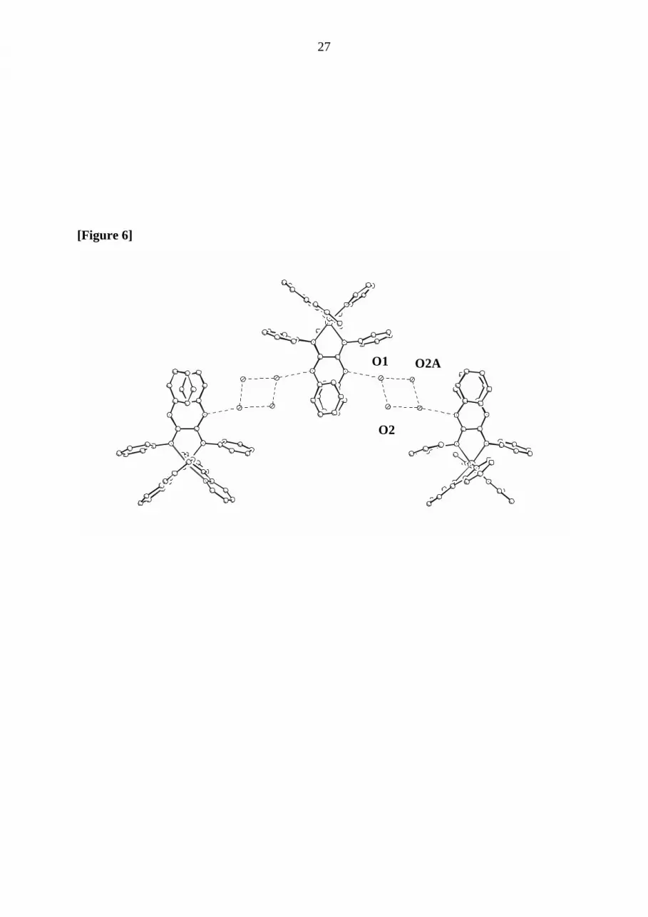
In addition, the X-ray investigations reveal intermolecular interactions. In the crystalline state
compound (1) forms a polymeric chain of alternating ordered complex cations whereby the
cations are interconnected by interaction of each N-H-function with a planar ring consisting of
four water molecules (see Figure 6). The distance dN(H)-O= 2.957(6) Å is in the typical range
of H-bonding distances, while the distances between the oxygen atoms within in the planar
water four ring are 2.778(5) Å and 2. 957(5) Å. Thus, highly ordered solvent molecules create
a supramolecular arrangement in the crystal .
((insert here: Figure 6 ))

10
It is worth mentioning that the position of the outwards directed phenyl rings seems to
suggests a π−π−interaction. But compared with literature values [16], the distance of dphenyl-
phenyl= 3.78 Å in the phenyl stacks indicates rather only simple crystal packing effects than
π−π−interaction as reason for this particular arrangement.
Synthesis and Structure of the Dinuclear Compound {[Ru(bpy)2]2(TPOA)}(PF6)2 . 2H2O,
(5)
The use of more as twofold excess of Ru(bpy)2-precursor yields the dinuclear compound (5)
in good yield. HPLC measurements indicate the presence of two compounds with a peak area
of 50:50 %. It was possible to separate these by recrystallisation in acetone/water (60/40
v/v%). Elemental analysis yielded identical composition for both compounds and was
indicative of a deprotonation of the tetraphenyloxalamidine ligand in both cases. The 1H-
spectra of the two fractions were recorded in d6-DMSO and are depicted in Figure 7. The
spectra are only slightly different and exhibit a quite simple pattern with only one set of
aromatic phenyl signals as expected for a high symmetrical complex. All peaks could be
assigned unequivocally with help of COSY experiments. No broad peaks as in the
mononuclear compounds appear in the spectra and the phenyl rings of the TPOA2- bridging
ligand show only one well-resolved set of two triplets and one doublet. This suggests that in
the dinuclear compound the aryl rings are not free to rotate. The protons of the bipyridine
ligands show the usual shifts and pattern.
(( insert here Figure 7 ))
On the basis of these observations it seems reasonable to assume that the two fractions are
optical isomers (5´) and (5´´). The presence of stereoisomers is related to the well-known
stereochemical problem of linking two metal ions with helical chirality by a bridging ligand

11
[17]. This causes the emergence of one meso-form (∆Λ) and one enantiomeric pair with
∆∆ and ΛΛ configuration. The two isolated diastereomeric isomers (5´) and (5´´) should
correspond to the meso-(∆Λ)−compound and to the unresolved (∆∆/ΛΛ) enantiomeric pair
(enantiomers are undistinguishable in 1H-NMR-spectroscopy) [18] but with the data available
it was not possible to assign the absolute configuration of (5´) and (5´´).
Absorption Spectra
The absorption spectra of compound (1)-(5) were recorded in methanol/ethanol and show the
typical features for members of the polypyridyl ruthenium (II) class. The results are
summarised in Table 3. The most significant feature of these spectra is a strong band in the
visible range due to dπ−π* -MLCT transitions [19].
(( insert Table 2 ))
In comparison to the model compound [Ru(bpy)3]2+ (λMLCT = 452 nm), the MLCT bands of
the oxalamidine complexes exhibit a shift to lower wavenumbers. Therefore, it can be
suggested that the investigated oxalamidines possess stronger σ-donor and weaker π-acceptor
properties than bipyridine. The presence of methylsubstituents in (2) and (4) has only a minor
influence on the absorption maxima. The MLCT-maximum of the dinuclear compound is
observed at lower energy than in the monomeric compounds. This reflects the deprotonation
of the bridging ligand and it is indicative of the stronger σ-donor and weakened π-acceptor
properties of the deprotonated bridge.
The possibility of tuning of the absorption spectra of the compounds (1) – (4) by changing the
acidity of the solution was investigated. Thus, Figure 8 shows the absorption spectra of
compound (1) in the range from pH = 3.08 to 12.05. In this range only one set of isobestic

12
points is found. The MLCT-band around λ = 470 nm collapses gradually upon deprotonation
and two new bands appear. The emerging band at λ = 505 nm can be assigned to a MLCT
transition in the deprotonated complex. The deprotonated tetraphenyloxalamidine ligand
should act as stronger σ-donor and increasing electron density around the metal shifts the
maximum of the MLCT-band to higher wavelengths. The band at λ = 380 nm might be
correspond to an additional MLCT band to either bpy or to H2TPOA.
From the absorption spectra it was possible to determine the pKa values for (1) – (4) (See
Table 2). The pKa is relatively insensitive to substitution changes on both the bpy ligand and
the oxalamidines. Only one protonation step could be observed in the range measured for all
complexes. This might suggest that both N-H functions are deprotonated at the same time or
that the second deprotonation is outside the range measured.
(( insert figure 8 ))
The two diastereomers (5´) and (5´´) show slightly different absorption spectra (see figure 9).
Potential differences in the electronic behaviour of diasteromeric and enantiomeric isomers of
polypyridyl ruthenium(II) complexes were recently subject of several investigations [20, 21].
Multinuclear ruthenium polypyridyl complexes will normally contain a manifold of
diastereomers and these studies are aimed at determining whether optical isomers have
significantly different photophysical properties. That is a rather important problem, because
constructing antenna systems from Ru-bipyridyl units demands the very strict control of the
photophysical properties of such light absorbing devices.
Surprisingly, the measurement of compounds (1)-(5) do not show any emission. Even chilling
down to 77 K in various solvents and extended change of the pH-value did not render the
expected emission. This is a unexpected result, because the appearance of a emission from a
long living 3MLCT state is to be seen as one of intrinsic characteristic for polypyridyl-Ru(II)

13
compounds. One possible explanation might be that the electron occupies an oxalamidine π*-
orbital instead of a bipy π*- orbital in the excited state. Thus, the inappropriateness of the
oxalamidine π*- orbitales to deliver a long living excited state could explain the absence of
emission. This assumption might be supported by the first reduction potentials for for
compounds (1) - (4) which are irreversible, in contrast to those of polypyridyl-Ru(II)
complexes.
(( insert Figure 9 ))
Electrochemistry
The Ru(III)/(II) potentials for the PF6-salts in 0.1 M solution of tetra-n-butylammonium
perchlorate in acetonitrile versus saturated calomel electrode are summarised in Table 3. In
comparison with [Ru(bpy)3]2+ (EIII/II = 1.23 V vs SCE) all redox Ru(III/II) couples of the
mononuclear complexes (1) -(4) are shifted to more negative potentials (EIII/II = 0.96 V - 0.87
V), in agreement with an increased electron density around the ruthenium ion caused by the
better σ -donor properties of the oxalamidine ligands. Methyl substitution on the bipyridine
ligands pushes the Ru(III/II) couples to somewhat more negative potentials consistent with
the results of before described absorption measurements. Substitution at the oxalamidine does
affect the oxidation couple hardly. The oxidation potentials for the two isomers of the dimeric
compound (5) differ only slightly. The oxidation of the dinuclear complexes results in two
one electron single waves, which are in both isomers separated by 190 mV. Using this value
the comproportionation constant Kcom for (5) was calculated following the relationship (at
T=298 K) [22]
Kcom= exp{∆E(mV)/25.69}= 1.63*103

14
This value points to a substantial electronic interaction between the two metal centres. The
oxidation of the first centre causes an increase in the charge of the complex and therefore the
second oxidation occurs at higher potential. In compound (5) the first oxidation couple occurs
at EIII/II = 0.45 - 0.46 V exhibiting the strong σ-donor capability of the deprotonated bridging
ligand. Thus, even the second oxidation occurs at more negative potential (EIII/II = 0.64/0.65
V) than the lowest of the mononuclear compounds, for which the oxidation potential is found
at about 0.90 V. This is most likely explained by the double negative charge on the bridging
ligand.
5. Conclusion
We have prepared series of novel mononuclear complexes of the type [Ru(bpy)2(LL)]2+ with
LL= H2TPOA, H2TTOA as members of a new class of non aromatic 1,2−diimine ligands in
polypyridyl Ruthenium(II) chemistry. The structure of the mononuclear compounds was
elucidated by 1H-NMR, FAB-MS and X-ray investigations. The coordination of the Ru(bpy)2-
moiety to the 1,2−diimine unit was assured unambiguously by X-ray structure determination
of compound [Ru(bpy)2(H2TPOA)](PF6)2. 4H2O, (1) . On supramolecular level, complex (1)
forms chainlike structures, whereby the complex cations are interconnected via their N-H
functions by four ordered water molecules. Surprisingly, the complexes (1)- (5) do not
exhibit any emission under any condition. The dinuclear compound
{[Ru(bpy)2]2TPOA}(PF6)2.2H2O, (5) was prepared and structurally characterised. We
succeeded in separating the two diastereomers by recrystalisation and could confirm small but
significant differences in electronic spectra.
Finally we propose that since they possess potential for H-bonding and intercalation by π-
stacking with biological substrates (e.g. DNA) the mononuclear complexes (1)- (4) can

15
become valuable building blocks in supramolecular architectures. Future research efforts will
follow this line.
6. Supplementary material
Further details of the crystal investigations are available on requests from the
Fachinformationszentrum Karlsruhe, Gesellschaft für wissenschaftlich – technische
Information mbH, D-76344 Eggenstein.Leopoldshafen, on quoting the depository number
CSD ###, the names of the authors, and the journal citation.
Acknowledgements
We would like to thank to Prof. U.-W. Grummt and Mrs. E. Backhaus for collaboration in
some experiments and for measuring several spectra. This work was financially supported by
the „Studienstiftung des deutschen Volkes“ by a doctoral scholarship for M.R. and S. R. and
by “Deutsche Forschungsgemeinschaft” (SFB 436).
References
[1] (a) J.-P. Collin, P. Gavina, P. Heitz, J.-P. Sauvage, Eur. J. Inorg. Chem. 1 (1998) 1.(b)
A. Livoreil, J.-P. Sauvage, N. Armaroli, V. Balzani, L. Flamigni, B. Ventura, J.Am.
Chem. Soc. 119 (1997) 12114. c) V. Balzani, A. Juris, M. Venturi, S. Campagna, S.
Serroni, Chem. Rev. 96 (1996) 759.
[2] (a) R.E. Holmlin, P.J. Dadliker, J.K. Barton, Angew. Chem., Int. Ed. Engl. 36 (1997)
2714. (b) A. Magnuson, Y. Frapart, M. Abrahamson, O. Horner, B. Åckermark, L.
Sun, J.-J. Girerd, L. Hammarström, S. Styring, J. Am. Chem. Soc. 121 (1999) 89.
[3] J.-P. Sauvage, J.-P. Collin, J.-C. Chambron, S. Guillerez, C. Coudret, V. Balzani,
F. Barigelletti, L. DeCola, L. Flamigni, Chem. Rev. 94 (1994) 993.
16
[4] J. A. Bolger, G. Ferguson, J. P. James, C. Long, P. McArdle, J. G. Vos, J. Chem.
Soc. Dalton Trans. (1993) 1577.
[5] A. Juris, V. Balzani, F. Barigelletti, S. Campagna, P. Belser, A. v. Zelewsky, Coord.
Chem. Rev. 84 (1988) 85.
[6] M. Döring, H. Görls, R. Beckert, Z. Anorg. Allg. Chemie 620 (1994) 551.
[7] J. Trofimenko, J. Am. Chem. Soc. J. 89 (1967) 7014.
[8] E. Papafil, A. Papafil, A. Kleinstein, I. Gabe, V. Macovei, Analele Stiint. Univ. “Al.
I; Cuza” Iasi, Sect. I 10c(2) (1964) 115.
[9] D. Lindauer, R. Beckert, M. Döring, P. Fehling, H. Goerls, J. Prakt. Chem. 337 (1995)
143.
[10] K. Bauer, Chem. Ber. 40 (1907) 2655.
[11] (a) R. Hage, J. G. Haasnoot, J. Reedijk, R. Wang, J. G. Vos, Inorg. Chem. 30 (1991)
3263. (b) R. Hage, Coordination Chemistry Reviews 111 (1991) 161.
[12] S. Vorwerk, Diploma thesis (1992) , Friedrich-Schiller-University Jena
[13] Z. Otwinowski, W. Minor, „Processing of X-Ray Diffraction Data Collected in
Oscillation Mode“, in Methods in Enzymology, Vol. 276, Macromolecular
Crystallography, Part A, edited by C.W. Carter & R.M. Sweet, Academic Press, pp.
307-326.
[14] G.M. Sheldrick, Acta Crystallogr. Sect. A 46 (1990) 467.
[15] G.M. Sheldrick, SHELXL-97, University of Göttingen, Germany, (1993)
[16] C. A. Hunter, J. K. M. Sanders, J. Am. Chem. Soc. 112 (1990) 5525.
[17] A. von Zelewsky, Chimia 48 (1993) 331.
[18] B. Testa, “Grundlagen der Organischen Stereochemie”, Verlag Chemie,
Weinheim, (1983) pp.158
[19] K. Kalanyanasundaram, „Photochemistry of Polypyridine and Porphyrin Complexes“,
Academic Press, London, (1992)

17
[20] (a) X. Hua, A. v. Zelewsky, Inorganic Chem. 30 (1991) 3796. (b) K. Wärnmark, J.
Thomas, O. Heyke, J.-M. Lehn, J. Chem. Soc., Chem. Commun. (1994) 2075 (c) F.R.
Keene, Coord. Chem. Rev. 166 (1997) 121. (d) F.R. Keene, Chem. Soc. Rev. 27
(1998) 185.
[21] (a) S. Serroni, G. Denti, S. Campagna, M. Ciano, V. Balzani, J. Am. Chem. Soc. 114
(1992) 2944; (b) S. Serroni, G. Denti, S. Campagna, A. Juris, M. Ciano, V. Balzani,
Angew. Chem., Int. Ed. Engl. 31 (1992) 1493; (c) T. J. Rutherford, O. Van Gijte; A.
Kirsch-De Mesmaker, F. R. Keene, Inorg. Chem. 1997, 36, 4465.
[22] G. Giuffrida, S. Campagna, Coord. Chem. Rev. 135/136 (1994) 517.

18
Capture of Figures
Figure 1: Structure of the ligands
Figure 2: Synthesis of mononuclear complexes (1) - (4) and dimeric complex (5)
Figure 3: 1H-NMR spectra of [(bpy)2Ru(H2TTOA)](PF6)2 . 2H2O, (3), at 293 K and 383 K
exhibiting the dynamic behaviour of the aromatic protons of the
tetratolyloxalamidine
Figure 4: Possible coordination modes of the oxalamidine ligand towards the Ru(bpy)2-
moiety
Figure 5: Drawing of the X-ray structure of [(bpy)2Ru(H2TPOA)](PF6)2 . 4H2O, (1), (anions
and solvent molecules are omitted for reasons of clarity)
Figure 6: Supramolecular structure of [(bpy)2Ru(H2TPOA)](PF6)2 . 4H2O, (1) (anions are
omitted for reasons of clarity)
Figure 7: 1H-NMR spectra of the two diastereomers (5´) (above) and (5´´) (below) in D6-
DMSO at 293 K
Figure 8: pH-dependence of the absorption spectra of [Ru(bpy)2(H2TPOA)](PF6)2 . 4 H2O,
(1) in the range from pH 3.08 to 12.05
Figure 9: UV-VIS spectra of compounds (5`) (- -)and (5´´) (-)

19
Table 1: Selected bound lengths (Å ) and angles (°) for [Ru(bpy)2(H2TPOA)]( PF6)2 . 4H2O,
(1)
Ru-N(4)
2.049(3)
N(4)-Ru-N(4A)
173.8(2)
Ru-N(3) 2.053(3) N(4)-Ru-N(3A) 97.0(1) Ru-N(1) 2.081(3) N(4)-Ru-N(3) 78.7(1) N(1)-C(1) 1.294(4) N(3A)-Ru-N(3) 91.6(2) N(2)-C(1) 1.355(5) N(4)-Ru-N(1) 85.7(1) C(1)-C(1A) 1.503(6) N(4A)-Ru-N(1) 99.3(1) N(2)-O(1) 2.957(6) N(3A)-Ru-N(1) 171.9(1) O(1)-O(2) 2.778(5) N(3)-Ru-N(1) 96.5(1) O(1)-O(2A)
2.862(5) N(1)-Ru-N(1A) 75.4(2)

20
Table 2: Absorption maxima and electrochemical data of the complexes (1) - (5) in
acetonitril; pKa values for (1) – (4)
compound λMLCT ε
[nm] [l cm-1mol-1]
Ru(III/II)
V
pKa
[Ru(bpy)3]2+ [5]
452 1.46 .104
1.26
-
[Ru(bpy)2(H2TPOA)]2+ (1) 469 1.39 .104 0.94 9.19
[Ru(Me-bpy)2(H2TPOA)]2+ (2) 476 1.17. 104 0.90 9.33
[Ru(bpy)2(H2TTOA)]2+ (3) 472 1.12 .104 0.96 9.94
[Ru(Me-bpy)2(H2TTOA)]2+ (4) 478 1.40 .104 0.87 10.15
{[Ru(bpy)2]2(TPOA)}2+ (5)
(5´) 520 (n. n.) 0.45/0.64 -
(5´´) 526 (n. n.) 0.46/0.65 -

21
Annex: Crystallographic data for [Ru(bpy)2(H2TPOA)]( PF6)2 . 4H2O, (1)
empiric Formula
C46H38F12N8P2Ru * 4H2O
formula weight [g mol-1]crystal size 1159.87 space group C2/c crystal size[mm] 0.20 x 0.20 x 0.10 crystal colour red-brown a (Å) 23.037(5) b (Å) 12.739(3) c (Å) 18.550(4) β ° 114.04(3) temperature [K] 293 volume [Å 3] 4971(1) Z 4 density (calc.) [g cm-3] 1.550 Θ range for data collection [°] 3.10 to 23.26 Limiting indices 0 ≤ h ≤ 25, 0 ≤ k ≤ 14, -20 ≤ l ≤ 18 reflections collected 5951 Rint 0.034 independent reflections 3572 observed reflections [I > 2σ (I)] 3344 absorption coefficient [cm-1] 4.76 Parameters/restrains 461/12 Final R indices [I > 2σ (I)] R1=0.038
wR2=0.095 R indices (all data) R1=0.055
wR2=0.104 Goodness-of-fit F2 1.009 Largest diff. Peak and hole [eÅ -3] 0.556 and –0.450

22
List of Figures [Figure 1]
Ar = Ph H2TPOAAr = Tol H2TTOA
N
N N
NAr
Ar Ar
Ar
H H

23
[Figure 2]
R
R
Ru
Cl
Cl
HN
N
N
NH
Ph
Ph
+
Ru
R
R
N
N N
NRu
Ar
ArAr
2+
(PF6)2
Ar
Ar
Ar Ar
Ar
N
NHN
NH
R
R
Ru
2+
(PF6)2
1:1
2:1
(i) ethanol/H2O
(ii) NH4PF6
1 R = H Ar = Ph2 R = Me Ar = Ph3 R = H Ar = Tol4 R = Me Ar = Tol
R = H ; Me
5 R = H Ar = Ph
R
R
N
N
2
2
2
N
N
N
N
N
N
Ph
Ph
2

24
[Figure 3]
Coalescence of the AA´MM´system
AA´BB´system

25
[Figure 4]
2+
(bpy)2RuN
N NH
HN
Ar
ArAr
Ar Ar
Ar Ar
Ar
HN
NH N
N(bpy)2Ru
2+ 2+
(bpy)2Ru
+
(bpy)2Ru
N
N NH
Ar
ArAr
Ar
N
N NH
N
Ar
Ar Ar
Ar
N N
Ar
Ar Ar
Ar
NN
N
(bpy)2Ru
0
A B C
D E
H

26
[Figure 5]
27
[Figure 6]
O1
O2
O2A

28
[Figure 7]
6.0 6.57.0 5 57.58.08.5 9.0 ( ppm

29
[Figure 8]
absorption [a.u] 1.00
0.80
0.60
0.40
0.20
300 400 700600500

30
[Figure 9]
absorption [a.u.] 1
0 300 400 500 wavelength
[nm]

31
Pictogramm (for the list of content)
2
Ru
N
N
22N
NRu
2
N NAr Ar
N NAr Ar
2 PF6
Novel polypyridyl Ruthenium(II) complexescontaining oxalamidines as ligands were prepared and fully characterized. Their acidbase and electrochemical properties arediscussed. The dimeric complex could beseparated in its diastereomeres. M. Ruben, S. Rau, A. Skirl, K. Krause,H. Görls, D. Walther, J. G. Vos


![Fulvene−Ruthenium and Cp−Ruthenium Complexes via [2 + 2 + 1] Cyclotrimerization of Phenylacetylene with [RuCl(Tp)(1,5-cod](https://img.pdfslide.net/doc/110x75/63571599ea2708a6d301e835/fulveneruthenium-and-cpruthenium-complexes-via-2-2-1-cyclotrimerization-1701042340.jpg)


