Embed Size (px)
Citation preview

Identifying Neurospora Clock Genes 173
173
From: Methods in Molecular Biology, vol. 362: Circadian Rhythms: Methods and ProtocolsEdited by: E. Rosato © Humana Press Inc., Totowa, NJ
12
Novel Strategies for Identification of Clock Genesin Neurospora With Insertional Mutagenesis
Kruno Sveric, Moyra Mason, Till Roenneberg, and Martha Merrow
SummaryAs the molecular mechanism of circadian clocks has reached high complexity, the
fungal model system, Neurospora crassa, is increasingly important for clock research. Itoffers the possibility of extensive biochemical experimentation and thorough descrip-tion of circadian properties. Realization of the full potential is dependent on efficient,high-throughput methods. We have combined several protocols to develop abundant andinexpensive production of mutants, and subsequent identification of the affected gene.We applied a novel screening protocol and, after screening several hundred mutants,identified a known clock gene, frequency. Furthermore, the methods described here caneasily be adapted to various insertional constructs (e.g., those with alternative selectionmarkers or that facilitate overexpression) or combined with strains carrying clock-regu-lated reporter genes.
Key Words: Neurospora crassa; fungi; circadian; mutagenesis; clock; entrainment.
1. IntroductionNeurospora has been a model organism for basic research on genetics for
more than half a century. “She” is the mother of the “one gene, one enzyme”hypothesis (1). Accordingly, there are a number of well-described and easyways in which to make mutants. Probably the most common methods includeultraviolet-light and chemically induced mutagenesis (see Note 1) of asexualspores, or conidia. Insertional mutagenesis, however, offers the possibility forrapid identification of the mutation through the rescue of the tagged DNA. Ourmethod uses transformation of a resistance gene (to “Basta”) into conidia viaelectroporation. The mutants are selected for growth, screened for circadianproperties, and “purified” to homokaryons. Once strains are confirmed to havesingle insertion sites, the location of the insertion is identified by a combina-

174 Sveric et al.
tion of mapping, partial cloning, and polymerase chain reaction (PCR). Theidentification of an insertion in a known clock gene has confirmed the generalapproach.
2. Materials1. Solid minimal media: 1X Vogel’s salts (2; see also Chapter 32), 2% glucose,
with 2% agar (Difco).2. 1 M Sorbitol.3. 10X FIGS: 1 M L-sorbose, 10 mM D-fructose, 10 mM glucose.4. Bottom agar: 1X Vogel’s salts, 1.5% agar (Difco), 0.3% L-arginine, 1.5% Basta
(also called ignite, glufosinate, or phosphinothricin), 1X FIGS. Add FIGS as asterile solution following autoclaving of the rest of the formulation. About 25 mLof bottom agar is added per 87-mm Petri dish.
5. Top agar: 1X Vogel’s salts, 1.0% agar (Difco), 1 M sorbitol, 1X FIGS. Add FIGSas a sterile solution following autoclaving the rest of the ingredients.
6. Slants containing minimal media and Basta: 70 × 10 mm glass test tubes with0.5 mL minimal media and 0.4% Basta, autoclaved and cooled slanted so thatthe agar solidifies at an angle, leaving an increased surface area for growth.
7. Race tube media (3): 1X Vogel’s salts, 0.3% glucose, 0.5% L-arginine, 10 mg/mLbiotin, 2% agar (Difco).
8. Iodoacetate media (4): 0.1X Westergaard’s salts (5; see Note 2), 0.1% sucrose,2% agar (Difco), 1 mM iodoacetate. Add iodoacetate from a sterile 100 mM stocksolution after autoclaving the rest of the formulation.
9. Sorbose media: 1X Vogel’s salts, 0.05% glucose, 0.05% fructose, 2% sorbose,2% agar.
10. Liquid minimal media: 1X Vogel’s salts, 2% glucose.11. Cetyltrimethylammoniumbromide (CTAB) buffer: 2% CTAB, 100 mM Tris-HCl,
pH 8.0, 1.4 M NaCl, 20 mM EDTA, 1% sodium bisulfite.12. C/IAA: 24:1 v/v, chloroform:isoamyl alcohol.13. Primers: bar2.1, TCA AGC ACG GGA ACT GG; bar2.2, CAG CCT GCC GGT
ACC GC; T7, TAA TAC GAC TCA CTA TAG GG; T3, AAT TAA CCC TCACTA AAG GG.
3. MethodsThe protocols described include (1) preparation of electrocompetent conidia;
(2) their transformation and selection; (3) an example of a screening protocol;(4) purifying homokaryons; and (5) methods for rescuing the inserted DNA foridentification of the mutation.
3.1. Preparation of Electrocompetent Cells
1. Grow the conidia on about 100 mL of solid minimal media after it has been auto-claved in a large (500-mL or 1-L) flask.

Identifying Neurospora Clock Genes 175
Fig. 1. Transformation of Neurospora with a plasmid that yields Basta-resistantcolonies. (A) pKSbar2 was used for insertional mutagenesis. It is the standardBluescript vector pKS containing a Basta resistance cassette (see Note 5). (B) Two (ormore, as shown here) days following transformation, resistant colonies are picked andtransferred to slants.
2. Harvest by adding 50 mL of sterile 1 M sorbitol and gently mixing on a shaker for5 to 10 min (see Note 3).
3. Filter the conidia through several layers of sterile cheesecloth to remove hyphae.4. Centrifuge the conidia at 2000g for 10 min at 4°C and resuspend in 50 mL of 1 M
sorbitol. This process is repeated four times. The concentration of conidia is mea-sured and adjusted to 2.5 × 10⁹/mL (see Note 4). Keep on ice until ready for use.
3.2. Transformation and Selection
1. Combine 40 μL of the conidial suspension (about 108 conidia) and 1 μL of linear-ized pKSbar2 (1 to 5 μg; see Note 5 and Fig. 1A) in a 1.5-mL microcentrifugetube. Incubate on ice for 5 min. Linearized plasmid is inserted more efficiently inthe genome than circularized plasmid (6).
2. Place this suspension in an ice-cold, 0.2-cm-wide cuvet (previously stored in a–20°C freezer) and electroporate using a Bio-Rad Gene Pulser with settings of1.5 kV/cm, 25 μF, and 600 Ω (7).
3. Recover the cells by gently combining with 1 mL of 1 M sorbitol for 10 to 20 minon ice.
4. Dilute 10 μL of this solution in 10 mL of molten top agar, held at 50°C, andimmediately pour over a Petri dish with bottom agar containing the selectiveagent Basta.
5. At 30°C, colonies will appear after 2 d (Fig. 1B). Pick colonies with a sterileneedle and place them on slants containing minimal media and Basta. A negativecontrol plate, containing conidia electroporated with no added DNA, should yieldno colonies.

176 Sveric et al.
3.3. Screening Mutants
Following 7 d of growth on slants, the mutant colonies will have developedsufficient conidia for inoculation of race tubes for screening. For functionalscreening, Basta is generally excluded from the media. As for protocols, wenoted that—to this point—Neurospora clock mutagenesis screens have usedonly constant conditions, thus limiting the results to the discovery of mutantswith an altered free-running period or arrhythmicity. Screens utilizing entrain-ment theoretically allow for the possibility of identifying “phase of entrain-ment” mutants (such as so-called larks and owls). Some of these could have anormal free-running period and would be missed if the screen is performed inconstant conditions, although they are potentially interesting mutants. Wetherefore determined to perform this screen under entraining conditions. Fur-thermore, as we are interested in looking for novel clock genes, and all theknown Neurospora clock genes are involved in light signaling, we additionallyintroduced two unusual features: first, we screened in temperature cycles indarkness, and second, the cycle was 16 h long. With an amplitude of 5°C(between 22 and 27°C), with 8 h at both high and low temperatures, the bdstrain entrains (Fig. 2; see Note 6), with the onset of conidiation occurring 2 to3 h into the warm phase (8). In a cycle just 1 h shorter, entrainment occurs inonly a few cases; rather, a free-run with relative coordination is observed. Thus,a 16-h cycle is close to the limit of the range of entrainment (9), and representsa challenge to the clock system.
1. Inoculate mutants and controls using a single race tube per isolate. Germinateovernight in the laboratory under constant light and transfer the tubes to thetemperature cycle setup (8), screening under the entrainment protocol for about 1 wk.Mark the conidiation band on each race tube daily and finally analyze the circadianparameters with the CHRONO program (10).
2. Screen mutants with an abnormal phenotype (early or late conidiation comparedwith controls, or arrhythmicity) for a second time, using a pair of race tubes permutant. About 10% of all isolates are usually rescreened.
3. To characterize the phenotype further, screen for free-running period under con-stant darkness and phase of entrainment in light–dark cycles.
3.4. Purifying Homokaryons
Once an interesting mutant is confirmed, its rescue is initiated. Most Neuro-spora conidia have two to three nuclei, so the transformed conidia can be het-erokaryons. Growth on selective media (see Note 7) generally favorsBasta-resistant nuclei, but mutants still must be purified to homokaryons. Thereare at least two methods that can be used for this purpose.

Identifying Neurospora Clock Genes 177
Fig. 2. Entrainment in short temperature cycles. In cycles with equal periods ofcold (22°C) and warm (27°C) incubation, the bd strain entrains when the cycle length(T) is equal to or greater than 16 h. In a 15-h cycle (T = 15), stable entrainment rarelyoccurs, but sometimes relative coordination is observed. In still shorter cycles—butstill substantially longer than half of the free-running period—the data show almost afree run, despite the imposition of a temperature cycle (a 14-h cycle is shown here).
3.4.1. Plating Assay
1. Suspend conidia to a concentration of about 1000 per mL.2. Combine 100-μL aliquots (i.e., 100 conidia) with 10 mL of top agar. Pour the top
agar over plates that already contain bottom agar with and without Basta.3. Grow colonies for 2 to 3 d. Compare the number of colonies on several sets of
plates with and without the selective agent. If the numbers are equal, then theconidia are homokaryotic. If they are skewed (more colonies growing on plateswith no selection), pick resistant colonies, grown on fresh slants, and reassay bythis plating method until equal numbers of colonies grow with and without selec-tive pressure.

178 Sveric et al.
3.4.2. Induction of Microconidia
Neurospora can be induced to produce microconidia, which contain a singlenucleus.
1. Boil cellophane in 1% KOH to remove impurities, rinse thoroughly to removesalts, and autoclave between layers of filter paper.
2. Inoculate the heterokaryotic mutant strain underneath a layer of cellophane ontoiodoacetate media (4). This is accomplished by puncturing the cellophane with asterile needle in the center of the plate, and then inoculating through the hole.Incubate the Petri dishes in humidified conditions at 22 to 25°C for 10 d in thelaboratory.
3. Remove the cellophane and wash the surface of the agar with 1 mL of sterilewater prior to returning the dishes to the humidified incubation chamber for addi-tional 24 h.
4. Harvest the microconidia with sterile water and plate on sorbose medium. Addi-tional microconidia will develop with another 24 h incubation.
3.5. Rescuing Insertion to Identify Mutation
Once an interesting mutation is purified to homokaryonicity, identifying thegene that carries the insertion is initiated. An initial step in this regard is todetermine how many insertions are found per mutant. This could be deter-mined either by performing a sexual cross and following transmission of thetrait using classical genetics, or more simply, with a Southern blot (describedhere).
1. Inoculate the conidia of each mutant line into liquid minimal medium. Afterachieving a tissue mass of about 1 cm3, blot the mycelia dry on paper towels andfreeze in a –70°C freezer.
2. Grind the mycelia in liquid nitrogen using a mortar and pestle with the aid of apinch of sand and suspend the powder in 0.5 mL of CTAB buffer.
3. Vortex and incubate at 60°C for 30 min.4. Add 0.5 mL of C/IAA and extract the genomic DNA for 5 min with mixing.5. Spin at 10,000g for 30 min at room temperature.6. Collect the supernatant in a clean tube and repeat steps 4 and 5.7. Add 1 μL of RNase A (10 mg/mL) and incubate the mixture for 10 min at 37°C.8. Precipitate the DNA with isoproponal at room temperature for 30 min. Collect
the precipitate by centrifugation for 10 min at 10,000g, and wash it once with70% ice-cold ethanol. After drying the pellet, the DNA can be suspended in50 μL water and quantitated by standard methods.
9. Digest 5 to 10 μg of genomic DNA with EcoRI, which does not cut within thepSKbar2 plasmid, thus yielding a single band per insertion.
10. Following digestion, separate the DNA on a 1% agarose gel, and blot accordingto standard methods.
Identifying Neurospora Clock Genes 179
11. Using primers bar2.1 and bar2.2 and the pKSbar2 plasmid as template DNA,amplify the Basta resistance cassette by PCR.
12. Label the PCR product by random priming using either radioactivity ordigoxygenin, and then use it to probe the blot.
Insertional mutagenesis often yields a single insertion, but there can also beseveral (see Fig. 3). In the latter case, there may be multiple insertions spreadthroughout the genome or at a single site. Mutants with a single insertion are suit-able for rescue. To identify the insertion, we used a combination method, startingwith size selection of genomic fragments, ligation, and PCR. The fragments areligated into a vector and the mixture is subjected to PCR using known sequencesfrom the Basta resistance gene and within the vector (see Fig. 4 and Note 8).
1. Map the area of the insertion by standard methods (i.e., Southern blot), digestingthe genomic DNA of the mutant with several restriction enzymes, which do notcut within the inserted plasmid, in conjunction with HindIII, which is known tocut within the Basta resistance gene (the minimal fragment that must be insertedfor the selection). The primary goal is to identify a fragment that runs from theknown HindIII site in the resistance gene (see Fig. 1) into the disrupted gene. Afragment with two distinct sticky ends (only one of which is HindIII) must beidentified. Also, the size of the fragment should be big enough to deliver sequenceinformation but small enough to ligate efficiently.
2. Gel-purify the restricted fragments of genomic DNA that run with the appropri-ate molecular weight (see Note 9) and ligate them overnight into a plasmid vec-tor (i.e., pSK-II) using standard methods (see Note 10).
3. Use 1 μL of the ligation reaction as the DNA template for a PCR reaction, whichemploys a primer for the Basta resistance gene on one end (bar2.1), and a primer
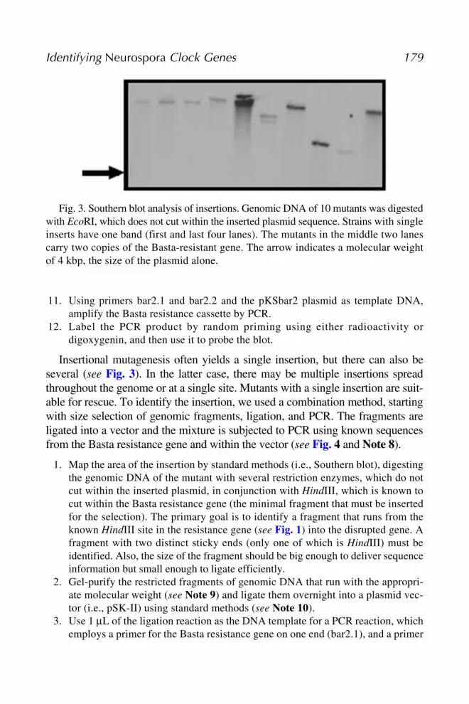
Fig. 3. Southern blot analysis of insertions. Genomic DNA of 10 mutants was digestedwith EcoRI, which does not cut within the inserted plasmid sequence. Strains with singleinserts have one band (first and last four lanes). The mutants in the middle two lanescarry two copies of the Basta-resistant gene. The arrow indicates a molecular weightof 4 kbp, the size of the plasmid alone.

180 Sveric et al.
Fig. 4

Identifying Neurospora Clock Genes 181
for either T3 or T7 sites on the other. Thanks to the mapping work, the orienta-tion of the fragment with respect to the vector is known, as well as the predictedsize of the PCR product.
4. Utilize 0.5 μL of the first PCR as a template for a second PCR reaction, using aprimer nested in the Basta resistance gene (bar2.2). A major fragment of the expectedsize should be apparent. This can be purified and sequenced. The product can becompared to the known plasmid sequence, as well as to the mapped DNA.
3.6. Case Study
One test of a mutagenesis protocol is the rediscovery of previously describedmutant alleles. On screening the first 800 Basta-resistant strains, a mutant(KOMO 303) showed a phase of entrainment that was opposite that of wild-type in the 16-h temperature cycle protocol (22 to 27°C). In a light–dark cycleusing 3 μmol of photons/m2/s, it completely failed to entrain, and in constantdarkness, it was arrhythmic (see Fig. 5). These phenotypes resemble the nullfrequency mutant, frq9, and also the white collar (wc) mutants. The wc mutantsare blind for light-induced mycelial carotenoids, in addition to having clockdefects (11,12).
1. We determined that the mutation was not likely in the wc genes by a simple light-induced carotenoid assay using mycelial pads, showing near-normal levels. Thefrq locus was investigated for insertion of the Basta-resistance cassette withSouthern analysis, revealing insertion in the open reading frame, in approximatelythe same location as the frq9 mutation is found (13). Furthermore, the Southernanalysis showed a clear double insertion (i.e., two copies) of the drug resistancemarker at this site.
2. One of the first insertions, KOMO 58, rescued as described in Subheading 3.5.,was further characterized by amplifying the insertion site from the genomicmutant DNA. Primers were designed using the genomic DNA sequence pre-dicted to flank the insertion. A positive result (a robust fragment of an appropri-ate molecular weight) would indicate that the drug-resistance marker is insertedwith no large deletions or translocations. A negative result (no fragment) wouldindicate that genomic DNA was lost in the recombination leading to insertion, orthat a translocation occurred. These possibilities can be finally determined with
Fig. 4. (opposite page) Flow diagram of insertion rescue protocol. (A) The areasurrounding the insertion is mapped by restriction digestion and Southern blotting. (B)The genomic DNA is digested and the desired fragment is enriched by size selection.(C) The pool of fragments is ligated into pSK-II. (D) The appropriate ligation productis amplified with nested polymerase chain reactions, and gel-purified. (E) The frag-ment is sequenced. RE, restriction endonuclease site; XbaI and HindIII, restrictionenzymes; T3 and T7, standard RNA polymerase sites used for primer binding; bar2.1and bar2.2, primers specific for the Basta-resistance gene, used in this protocol.

182 Sveric et al.
Fig. 5. A frequency mutant was generated by insertional mutagenesis. Race tubesand double plots for a frq mutant generated in this study are shown here. The mutant iscompared with the bd strain in constant darkness, a 16-h temperature cycle (TC), anda 12-h light cycle (LC). For each condition a control and a mutant race tube is shown,as well as a sample double plot. The gray areas in the plots indicate the cold (TC) orthe dark phase (LC). Note the phase of entrainment of KOMO303 in the temperaturecycle is approximately opposite that of bd. Furthermore conidiation is not stimulatedby (or driven by) the onset of the cold phase. KOMO303 completely fails to entrain inlight cycles. These phenotypes are consistent with those for the frq9 strain (8).KOMO303 was determined to be a frq mutant by Southern blotting (data not shown).
Southern blot or by rescuing the other end of the insertion. In either of the lattercases, the mutant is much less interesting, as it is almost impossible to determinewhere the mutant phenotype derives from. In the case of KOMO 58, the fragmentgenerated in the PCR reaction was larger than would have been predicted. Sequenceanalysis showed that there was a duplication in the inserted plasmid (an extra copyof the ampicillin resistance gene was present), whereas the genomic DNA wasintact except for a few nucleotides at the junctions.
3. To finally determine that the insertion is acting through a specific gene, the geneshould be knocked out (13) and the null mutant strain then assayed for clockcharacteristics. If the gene is essential, an overexpression construct can be engi-neered using the inducible qa-2 promoter (14) and transformed ectopically. Aftergrowing in the precence of an inducer, functional tests can be carried out in itsabsence.

Identifying Neurospora Clock Genes 183
4. Notes1. Mutagenesis with ultraviolet light can be accomplished by diluting conidia in a
sterile Petri dish and placing on a transilluminator for increasing amounts of timeuntil 70% of the conidia do not survive (15). Chemical mutagenesis uses stan-dard methods as described (16).
2. The formulation for 1X Westergard’s media is: 10 mM KNO3, 74 mM KH2PO4,2 mM MgSO4·7H2O, 20 mM NaCl, 2.5 mM CaCl2, and 0.5 mL of the traceelements used in preparation of Vogel’s media. Whereas the stock solution is usu-ally made up to 5X, the concentration required for microconidia production is 0.1X.
3. The conidia are hydrophobic; this step wets the conidia so that they will stay insolution better.
4. The concentration of conidia is determined by measuring the optical density at420 nm. An optical density of 1.0 = 2.86 × 10⁶/mL.
5. The plasmid, pKSbar2, was donated by U. Schulte. For its construction,pBARKS1 (17) was cut with XbaI and SpeI restriction enzymes. Ends were filledand the fragment containing the Bar gene (for resistance to Basta) was insertedinto pBluescript II KS, cut with the enzymes HincII and SmaI.
6. The bd gene is essential for formation of a clear conidiation band, which is thecircadian phenotype under analysis.
7. We grow our selected transformants on solid media for convenience. Immediatetransfer to liquid media, instead, results in more rapid growth (conidia can out-grow the liquid media in 2 d rather than 1 wk), thus decreasing the time neededfor this step. Liquid media may also promote homokaryon formation.
8. We have developed this method to avoid certain pitfalls, including the possibleloss of the ampicillin-resistance gene on insertion; otherwise we could simply cutthe mutant DNA with EcoRI and circularize, transform bacteria, and select forampicillin-resistant plasmids.
9. The fragment of interest is copurified together with many unrelated genomicDNA fragments, which will contribute to background. These “contaminating”fragments can be minimized by cutting with additional enzymes (those that donot disrupt the identified fragment, i.e., that cut outside this region). There willbe fewer nonspecific fragments, and they will have different restriction sites,thus forming fewer ligation products that will amplify in the PCR reaction.The enzymes for this job are already known from the mapping work. Thismust be done strategically, as it could—under certain circumstances—actuallyincrease contaminating fragments. See ref. 18 for help with the equations.
10. We have found that the efficiency of this ligation reaction is poor. By using PCRto amplify the ligation product we need only one side joined, increasing the effi-ciency of this step.
AcknowledgmentsWe are indebted to Ulrich Schulte for generously supplying us with the plas-
mid for insertion as well as the selective agent, Basta. We also thank Cornelia

184 Sveric et al.
Bösl and Shahana Sultana for sharing data and advice in the course of thiswork. Moyra Mason was supported by and Erasmus/Socrates grant from theEU (academic year 2000–2001). Our work is additionally supported by theDeutsche Forschungsgemeinschaft, the 5th Framework Programme of the Euro-pean Union (“BrainTime”), the Dr.-Meyer-Struckmann-Stiftung and theEppendorf Company, Hamburg, Germany.
References1. Beadle, G. W., and Tatum, E. L. (1942) Genetic control of biochemical reactions
in Neurospora. Proc. Natl. Acad. Sci. USA 27, 499–506.2. Vogel, H. J. (1964) Distribution of lysine pathways among fungi: evolutionary
implications. Amer. Nat. 98, 435–446.3. Merrow, M., Roenneberg, T., Macino, G., and Franchi, L. (2001) A fungus among
us: the Neurospora crassa circadian system. Semin. Cell Dev. Biol. 12, 279–285.4. Pandit, A., and Maheshwari, R. (1993) A simple method of obtaining pure micro-
conidia in Neurospora crassa. Fungal Genet. Newsl. 40, 63.5. Westergaard, M., and Hirsh, H. M. (1954) Environmental and genetic control of
differentiation in Neurospora. Proc. Symp. Colson Res. 7, 171–183.6. Paietta, J. V., and Marzluf, G. A. (1985) Gene disruption by transformation in
Neurospora crassa. Mol. Cell. Biol. 5, 1554–1559.7. Margolin, B. S., Freitag, M., and Selker, E. U. (1997) Improved plasmids for gene
targeting at the his-3 locus of Neurospora crassa by electroporation. FungalGenet. Newsl. 44, 34–36.
8. Merrow, M., Brunner, M., and Roenneberg, T. (1999) Assignment of circadianfunction for the Neurospora clock gene frequency. Nature 399, 584–586.
9. Aschoff, J., and Pohl, H. (1978) Phase relations between a circadian rhythm andits zeitgeber within the range of entrainment. Naturwiss. 65, 80–84.
10. Roenneberg, T., and Taylor, W. (2000) Automated recordings of bioluminescencewith special reference to the analysis of circadian rhythms. Meth. Enzymol. 305,104–119.
11. Degli-Innocenti, F., and Russo V. E. (1984) Isolation of new white collar mutantsof Neurospora crassa and studies on their behavior in the blue light-induced for-mation of protoperithecia. J. Bacteriol. 159, 757–761.
12. Crosthwaite, S. K.,. Dunlap, J. C, and Loros, J. J. (1997) Neurospora wc-1 and wc-2:Transcription, photoresponses, and the origin of circadian rhythmicity. Science 276,763–769.
13. Aronson, B. D., Johnson, K. A., and Dunlap J. C. (1994) The circadian clocklocus frequency: a single ORF defines period length and temperature compensa-tion. Proc. Natl. Acad. Sci. USA. 91, 7683–7687.
14. Aronson, B. D., Johnson, K. A., Loros, J. J., and Dunlap, J. C. (1994) Negativefeedback defining a circadian clock: autoregulation of the clock gene frequency.Science 263, 1578–1584.

Identifying Neurospora Clock Genes 185
15. Feldman, J. F., and Hoyle, M. N. (1974) A direct comparison between circadianand noncircadian rhythms in Neurospora crassa. Plant Physiol. 53, 928–930.
16. Ferket, K. K. A., Levery, S. B., Park, C., Cammue, B. P., and Thevissen, K. (2003)Isolation and characterization of Neurospora crassa mutants resistant to antifun-gal plant defensins. Fungal Genet. Biol. 40, 176–185.
17. Pall, M. L., and Brunelli, J. P. (1993) A series of six compact fungal transforma-tion vectors containing polylinkers with multiple unique restriction sites. FungalGenet. Newsl. 40, 59–62.
18. Ausubel, F. M., Brent, R., Kingston, R. E., et al. (eds.) (2002) Current Protocolsin Molecular Biology, John Wiley and Sons, New York.


