Embed Size (px)
Citation preview

Epilepsy & Behavior 62 (2016) 104–114
Contents lists available at ScienceDirect
Epilepsy & Behavior
j ourna l homepage: www.e lsev ie r .com/ locate /yebeh
Occipital and occipital “plus” epilepsies: A study of involvedepileptogenic networks through SEEG quantification
Angela Marchi a,b, Francesca Bonini a,b, Stanislas Lagarde a,b, Aileen McGonigal a,b, Martine Gavaret a,b,Didier Scavarda c, Romain Carron d, Sandrine Aubert a, Nathalie Villeneuve a, Samuel Médina Villalon a,Christian Bénar b, Agnes Trebuchon a,b, Fabrice Bartolomei a,b,⁎a APHM, Timone Hospital, Clinical Neurophysiology and Epileptology Department, Marseille 13005, Franceb Aix-Marseille Université, Institut de Neuroscience des Systèmes, UMR_S 1106, Marseille 13005, Francec APHM, Timone Hospital, Paediatric Neurosurgery Department, Marseille 13005, Franced APHM, Timone Hospital, Functional and Stereotactical Neurosurgery Department, Marseille 13005, France
⁎ Corresponding author at: Service deNeurophysiologieSaint-Pierre, 13005 Marseille, France. Tel.: +33 49138583
E-mail address: [email protected] (F. Barto
http://dx.doi.org/10.1016/j.yebeh.2016.06.0141525-5050/© 2016 Elsevier Inc. All rights reserved.
a b s t r a c t
a r t i c l e i n f oArticle history:Received 19 March 2016Revised 14 May 2016Accepted 16 June 2016Available online xxxx
Compared with temporal or frontal lobe epilepsies, the occipital lobe epilepsies (OLE) remain poorly character-ized. In this study, we aimed at classifying the ictal networks involving OLE and investigated clinical features ofthe OLE network subtypes. We studied 194 seizures from 29 consecutive patients presenting with OLE andinvestigated by stereoelectroencephalography (SEEG). Epileptogenicity of occipital and extraoccipital regionswas quantified according to the ‘epileptogenicity index’ (EI) method. We found that 79% of patients showedwidespread epileptogenic zone organization, involving parietal or temporal regions in addition to the occipitallobe. Two main groups of epileptogenic zone organization within occipital lobe seizures were identified: apure occipital group and an occipital “plus” group, the latter including two further subgroups, occipitotemporaland occipitoparietal. In 29% of patients, the epileptogenic zone was found to have a bilateral organization. Themost epileptogenic structure was the fusiform gyrus (mean EI: 0.53). Surgery was proposed in 18/29 patients,leading to seizure freedom in 55% (Engel Class I). Results suggest that, in patient candidates for surgery, themajority of cases are characterized by complex organization of the EZ, corresponding to the occipital plus group.
© 2016 Elsevier Inc. All rights reserved.
Keywords:Occipital lobeFocal epilepsyEpilepsy surgeryEpileptogenicity index
1. Introduction
Occipital lobe epilepsies (OLE) are rare (1–8% of epilepsy surgeryseries) [1–4] and challenging. The reported success rate of surgery islower compared with that of temporal lobe epilepsy and may rangefrom 25 to 90% [4–7] depending on studies. There are relatively fewstudies of homogeneous surgical populations with OLE, particularlyinvolving invasive recordings [4,7–15]. Indeed, as most of the anatomi-cal borders in the posterior cortex are almost arbitrary, some authorshave studied OLE in the context of posterior cortex epilepsies (PCE),encompassing a group of epilepsies originating from the occipital,parietal, and posterior–temporal lobes [5,16–18]. Furthermore, mostseries on OLE refer only to patients who have undergone epilepsysurgery, thus potentially limited to a subset of patients with a morefocal form within the larger clinical spectrum of OLE. Most of theoccipital cortex is buried, and its high connectivity facilitates rapidand widespread propagation of an epileptic discharge. These intrinsic
Clinique, CHU Timone, 264 Rue3; fax: +33 491385826.lomei).
characteristics hinder identification and localization of focal epileptiformabnormalities using scalp electrodes. Therefore, a noninvasive presurgicalassessment of OLE is often not sufficient to precisely define the epilepto-genic zone (EZ) (region of primary organization of ictal discharge). In ad-dition, the EZ often extends beyond the epileptogenic lesion [11,18,19].Thus, invasive recordings with stereoelectroencephalography (SEEG)might be needed, including those in lesional cases. Attempts to classi-fy occipital lobe seizures on anatomical or anatomoclinical groundshave been performed in the past. Within occipital seizures, some au-thors have distinguished mesial versus lateral types, occipital versusoccipitotemporal types, or occipital versus occipital plus types, basingsuch classification on clinical features, scalp-EEG features, seizure spreadon invasive EEG, or surgical outcome [1,3,6,8,13,20].
From a functional standpoint, an anatomical definition of occipitalseizures as entities confined within the occipital lobe boundaries willbe inevitably unsatisfactory. Indeed, epileptic seizures, as a pathologicalbut dynamic event, follow existing integrated neural pathways notlimited by classical anatomic boundaries.
In this study, we aimed at delineating the organization of neuralnetworks of seizures arising fromtheoccipital lobe, using SEEG recordings.In particular, we intended to determine whether subgroups of OLE

Table 1Main clinical features of the studied population. Abbreviations: F = female; M=male; N= no; Y = yes; DNET= dysembryoplastic neuroepithelial tumor; FCD= focal cortical dysplasia; HME= hemimegalencephaly; L = left; R = right; GK=gamma-knife; B = bilateral epileptogenic zone organization; EZ = epileptogenic zone; SVH = simple visual hallucination; CVH = complex visual hallucination.
Patient Sex Age atonset(year)
Age atrecordings(year)
Etiology Sideof EZ
Aura Subtype Surgery Follow-up(year)
Engel
Class
1 F 8 17 FCD R 1. Amaurosis2. SVH (vision of a moved image)
Occipital pure Y 9 I
2 F 21 30 Heterotopia B 1. SVH2. déjà-vu/déjà vecu/taste or smell
Occipital pure N – –
3 F 10 15 Heterotopia L SVH (multicolored ball moving over the upper quadrant of CVH) Occipitotemporal N – –4 F 5 7 HME L Fear Occipital pure Y 2 IA5 M 2 29 Surgical scar B 1. SVH (flashing lights over the inferior quadrant of CVH)
2. Visual illusion (dyschromatopsia)Occipitoparietal Y 6 III
6 M 18 24 Heterotopia/polymicrogyria R 1. Thoracic pressure-like sensation/nausea/déjà-vu/déjà vecu2. Blurring vision
Occipital pure Y 0,5 IA
7 M 0.66 7 FCD R Not reported Occipital pure Y 1 IA8 M 7 15 DNET R 1. SVH (unformed vision)
2. CVH (cars of videogame)/visual illusion (room moving from R to L)3. Fear
Occipitoparietal Y 3 IA
9 F 5 19 FCD B (R) 1. SVH (colored bright ball)2. Visual illusion (loss of stereoscopic vision)3. Feeling of estrangement/depersonalization
Occipitoparietal N – –
10 M 18 49 Polymicrogyria/double cortex R 1. Blurred vision2. SVH (black spots moving from contralateral to ipsilateral hemifield)3. Feeling of being moved to the right side4. Feeling of warmth
Occipitotemporal N – –
11 F 6 15 Cryptogenic B (R) Amaurosis Occipitoparietal Y 9 II12 M 3 25 FCD R 1. Blurred vision
2. Visual illusion (objects around him moving)Occipitotemporal Y 2 III
13 F 11 25 Dilatation ventricular occipital horn R 1. Blurred vision2. Vertigo/somatosensory (sensation of limpness right arm)3. Feeling of eyes being pulled to the right
Occipitoparietal N – –
14 F 2.5 50 Prenatal ischemic sequel B SVH (stars over contralateral visual hemifield) Occipitoparietal Y 12 IV15 F 1.5 21 FCD L Feeling of emptiness Occipitotemporal GK 7 IV
(continued on next page) 105A.M
archietal./Epilepsy&
Behavior62
(2016)104–114

Table 1 (continued)
Patient Sex Age atonset(year)
Age atrecordings(year)
Etiology Sideof EZ
Aura Subtype Surgery Follow-up(year)
Engel
Class
16 F 9 22 FCD L 1. SVH (black and white ball moving from L to R)2. Feeling of being forced to look on the R
Occipital pure Y 2 IV
17 F 11 18 Perinatal ischemic sequel L 1. SVH (flashes)2. déjà-vu
Occipital pure Y 1 I A
18 M 1 14 Prenatal ischemic sequel B 1. SVH (undefined vision over the L hemifield)2. Spatial disorientation3. Nausea/feeling of warmth
Occipital pure N – –
19 F 16 42 FCD B (R) 1. Visual illusions (palinopsia)/dyschromatopsia2. Amaurosis3. déjà-vu/déjà-vécu
Occipitoparietal GK 5 IB
20 F 8 29 FCD R 1. CVH (scenes)2. Feeling of estrangement/depersonalization/dreamy state
Occipitotemporal Y 0,5 IA
21 F 20 50 Cryptogenic R 1. CVH (scenes)2. déja-vu3. Vertigo/unpleasant sensation/thirst
Occipitotemporal N – –
22 F 7 23 FCD R 1. CVH (colored cars)2. SVH (moving image in front of her/lights over the CVH)
Occipitotemporal Y 3 IB
23 M 1 18 FCD R 1. CVH (upper part of a blue man with a dog-head) over the CVH2. Visual illusion (kaleidoscopic vision/the furniture around appears strange/metachromopsia)3. Dreamy state
Occipital pure Y 5 IC
24 F 3 31 DNET R SVH (colored ball) Occipital pure GK 3 IV25 M 4 32 FCD B 1. SVH (spots)
2. Visual illusion (macropsia)3. Feeling of eyes being pulled to one side (“like by a lover”)4. Feeling of fatigue
Occipital pure N – –
26 F 10 21 Posterior cortical atrophy L 1. SVH (bright-colored rotating ball over the inferior quadrant of contralateral visual hemifield)2. Feeling of pression on the eyeballs3. Fear/feeling of stress
Occipitotemporal N – –
27 F 12 46 Cryptogenic B 1. SVH (little white squares with red lines moving from R to L side)2. Visual illusion
Occipitotemporal N – –
28 M 10 18 FCD L Feeling of being abruptly angry Occipital pure N – –29 F 7 26 Cryptogenic L 1. Visual illusions (feeling of objects approaching to her/metachromopsia)
2. SVH (bright flashes or circles moving from the CVH to HVH)3. Feeling of right eye moving4. Unilateral asomatognosia (R hemibody)
Occipital pure Y 3 II
106A.M
archietal./Epilepsy&
Behavior62
(2016)104–114

107A. Marchi et al. / Epilepsy & Behavior 62 (2016) 104–114
seizuresmight be definedbasedon the involvement at seizure onset of oc-cipital or extraoccipital cortices, whether parietal or temporal. To this aim,we used an objective measure of the EZ, the “epileptogenicity index” (EI)[21]. Secondary objectives included the investigation of the relationshipbetween the type of epileptogenic network and clinical data includingictal semiology, age at epilepsy onset, epilepsy duration, etiology, and sur-gical prognosis.
2. Patients and methods
2.1. Patient selection and SEEG recording
Twenty-nine consecutive patients undergoing presurgical evalua-tion for drug-resistant OLEwere selected from a series of three hundredpatients in whom intracerebral recordings had been performedbetween 2001 and 2015 (Timone Hospital, Marseille, France).
Selection was based on the results of SEEG recordings showing in-volvement of occipital cortex at ictal onset. Prior to selection for SEEG,all patients underwent noninvasive assessment for drug-resistant partialepilepsy including detailed clinical history, neurological examination,neuropsychological evaluation, long-termvideo-EEG recording, structuralmagnetic resonance imaging (MRI), interictal 18-fluorodeoxyglucose-positron emission tomography (18FDG-PET), and ictal single photonemission computed tomography (SPECT) when possible.
The SEEG recording was carried out during long-term video-EEGmonitoring in order to record several of the patient's habitual seizures,following complete or partial withdrawal of antiepileptic drugs. TheSEEG exploration was performed using intracerebral multiple-contact
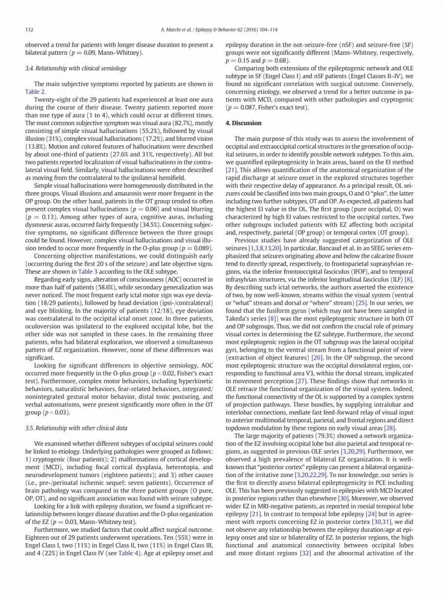
Fig. 1. Values of the epileptogenicity index (EI) computed from sampled regions. (A) Mean aaveraged over the 29 patients. Mean and standard deviation of IE values obtained from thegroup (C) for each structure. Black columns: occipitoparietal subgroup. Gray columns: occipilateral occipital gyri; ODL = occipital dorsolateral regions.
electrodes (Dixi Medical (France) or Alcis (France); 10–15 contacts,length: 2 mm, diameter: 0.8 mm, 1.5 mm apart) placed intracraniallyaccording to Talairach's stereotactic method [21].
The decision about anatomic positioning of electrodes wasestablished in each patient based upon available noninvasive data andhypotheses of localization of the epileptogenic zone. Postoperative com-puterized scan (CT) orMRIwas performed in order to verify the absenceof bleeding and the position of each recording lead. Subsequently, CTscan/MRI data fusion was performed in order to accurately identifyand locate each contact along the electrode trajectory.
We considered as anterior limits of the occipital lobe on the lateralsurface, the upper end of the parietal occipital fissure and thepreoccipital notch. The mesial occipital surface was defined as thecuneus and the lingual gyrus. The inferior limit of the mesial surfacewas considered the collateral sulcus; its anterior limits the parietal–occipital fissure and a line extending from it to the preoccipital notch.Its superior limit was the border between the mesial surface and thelateral convexity. The remainder of the occipital lobe was considered‘lateral’ occipital cortex [1].
All patients had spatial sampling of dorsomesial areas of occipitallobe (cuneus, CU; mesial part of occipitoparietal junction, OPJ),ventromedial areas (lingual gyrus, LG), dorsolateral areas (occipitalpole, ODL), ventrolateral areas (lateral occipital gyri, LOG), and basalregions (posterior part of fusiform gyrus, FG) [22]. Some patients hadonly one part of these regions explored, but the majority of patientsbenefited from complete exploration of occipital regions.
Extraoccipital regionswere explored in all patients, including at leastone temporal structure (temporal pole, mesial temporal areas, and/or
nd standard deviation of IE values obtained from all the different explored brain regionsoccipital explored regions averaged over the O pure group patients (B) and the O-plustal–temporal subgroup. Cu = cuneus; GL = lingual gyrus; GF = fusiform gyrus; LOG =

Fig. 2. Summarized comparative profiles of epileptogenicity in the three subtypes of occipital networks. For each subregion, we kept the highest EI value obtained in one occipital regionarea (CU, GL, ODL, LOG, Gfus, JPO), the highest EI obtained in one temporal area (Tmes, Tpi, NCT), and the highest EI values in one parietal region (pGC, SMG, BA7lat, BA7m) and frontalstructure (SMA, FEF).
Fig. 3. SEEG recordings of one seizure and representation of epileptogenicity index (EI) values, on the electrode contacts projected into the individual MRI in two patients with differentsubtypes of occipital epilepsies. Values close to the value 1 appear in red. A/ Example of a patient with pure occipital subtype (“O” group). B/ Example of a patient with occipitotemporalsubtype (“OT” group). For each group, we chose a representative patient for the epileptogenic index (EI) onto their individual 3D brain mesh. Cortical meshes were obtained from eachpatientMRI by FreeSurfer software. Then, we performed coregistration of the referenceMRIwith either a CT scan or anMRIwith the SEEG electrodes, using the SPM toolbox [http://www.fil.ion.ucl.ac.uk/spm/software/spm12/]. For patients with CT, the location of each contact was obtained by image processing (Matlab, Natick, MA)with custom code (Paz et al. in prep). Forthe patientwith postimplantationMRI, electrodes were placedmanually. After transformation to theMRI coordinate system, these electrodes were represented together with the corticalsurface in Matlab in a 3D plot. We reconstructed each SEEG electrode as black and white cylinders corresponding to the contacts and the insulated parts, respectively. As EI values werecomputed on a bipolar montage, they are drawn between each pair of contacts of the bipolar derivation. The diameter and position on the color scale are proportional to the normalized EIvalues. For electrode legend, see supporting information figure. (For interpretation of the references to color in this figure legend, the reader is referred to the web version of this article.)
108 A. Marchi et al. / Epilepsy & Behavior 62 (2016) 104–114

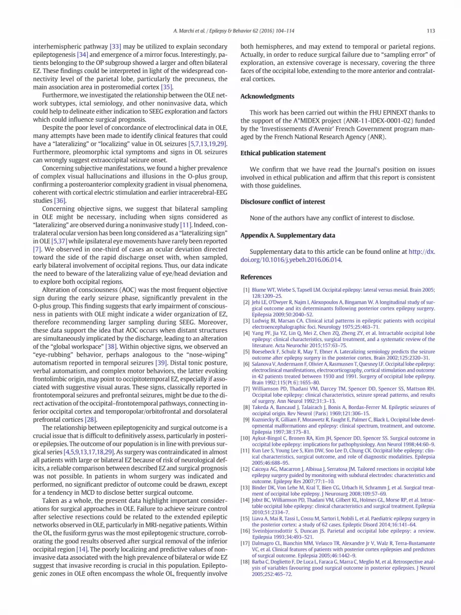
Fig. 4. SEEG recordings of one seizure and representation of epileptogenicity index (EI) values, on the electrode contacts projected into the individual MRI. Example of a patient with anoccipitoparietal subtype (“OP” group).
109A. Marchi et al. / Epilepsy & Behavior 62 (2016) 104–114
temporal neocortex) and one parietal area (posterior cingulategyrus, supramarginal gyrus, Brodmann area, BA 7 lateral, BA 7 mesial/precuneus), as well as one frontal premotor structure (supplementarymotor areas, lateral premotor cortex, frontal eye field), when sampled(see supporting information figure).
Fig. 5.A/Histogram showing the number of structures disclosing EI values ≥0.3 in thewhole pattwo main groups (O: occipital and O+: occipital “plus”); and D/ in the three subgroups (O: oc
Signalswere recorded on a 128-channel system (Natus/Deltamed™)sampled at 512 Hz and recorded on a hard disk (16 bits/sample) usingno digital filter. A high-pass filter (cutoff frequency equal to 0.16 Hzat −3 dB) was used to remove very slow variations that sometimescontaminate the baseline.
ient population. Box plots of theNEI ≥ 0.3 in B/ lesional andMRI-negative patients; C/ in thecipital; OP: occipitoparietal; OT: occipitotemporal).

110 A. Marchi et al. / Epilepsy & Behavior 62 (2016) 104–114
2.2. SEEG signal analysis: computation of the epileptogenicity index
The epileptogenicity index (EI) is intended to quantify two impor-tant features of SEEG signals recorded during the transition frompreictal to ictal activity: (i) the redistribution of signal energy fromlower frequency band (delta, theta, alpha) toward higher frequencyband (beta, gamma) and (ii) the delay of appearance of these high-frequency components in a given structure with respect to the firststructure, itself involved in a “rapid discharge mode” [21]. In thisrespect, the EI is based on the same information as that used in a visualreview of SEEG seizure recordings. A recent study reports a goodconcordance with visual estimation [23].
In practice, we used a semiautomatic approach: a user-friendlygraphical user interface allows the user to easily inspect and automati-cally validate detected change points, indicating the accurate onset ofrapid discharges. From this validation performed on a “structure-by-structure” basis, the EI was then computed. Epileptogenicity indexvalues are normalized with respect to the highest value across channels(ranging from 0 to 1). If there is no involvement of the brain structure,then EI = 0 whereas if the brain structure generates a rapid dischargeand if the delay with respect to seizure onset is minimal, then EI = 1.An EI between 0 and 1 corresponds to secondary involvement of theconsidered brain structure (for detailed methodology, see [21]).
A cutoff of EI ≥ 0.3 was used to define regions with highepileptogenicity, according to previous reports [24]. The EI values ob-tained in sampled cortical regionswere used to define different cerebralepileptogenic networks.
2.3. Statistical analysis
Statistical analysis was performed (i) to assess potential linksbetween the different epileptogenic zone (EZ) network subtypes(as defined by EI values) and bilateral EZ organization and someclinical parameters (age at epilepsy onset, epilepsy duration, etiology,semiological features) and (ii) to determine which one, among thesepotential factors, could influence postoperative prognosis.
A total of 194 seizureswere analyzed for EI determination. Statisticaltests were performed on EI values estimated and averaged from two orthree of the most representative seizures from each patient. Possiblecorrelation between epilepsy duration, age at epilepsy onset, and sizeof the EZ, as reflected by the number of structures disclosing highEI values (NEI ≥ 0.3), was investigated using a Spearman rank correla-tion coefficient. Statistical analysis was performed in order to comparethe data between different groups using a rank-analysis of variance(Kruskal–Wallis) for quantitative data. Bivariate analysis used the
Table 2Main reported subjective manifestations (n = 29 patients). Abbreviations: OT = occipitotemp
Auras OT OP
n° patients % n° patients
Total aura 9 100 7Total visual aura 8 88.8 7Simple visual hallucination 5 55.5 4Visual illusion 2 22.2 4Complex visual hallucination 3 33.3 1Blurring 2 22.2 1Amaurosis 0 0 2Image movement 5 55.5 1Colored image 5 55.5 1Controlateral visual hemifield 4 44.4 2Psychic 3 33.3 2Feeling of eye being pulled 0 0 2Fear 1 11.1 1Somatosensory 1 11.1 1Vertigo 1 11.1 1Viscerosensory symptoms 1 11.1 0Abdominal/cephalic 0 0 0
nonparametric Mann–Whitney test. Fisher's exact test was used forqualitative data. A p-value smaller or equal to 0.05 was considered tobe statistically significant.
3. Results
Table 1 summarizes the clinical characteristics of the 29 includedpatients. Surgical results are indicated in Table 4.
3.1. Classification of OLE seizure networks
Overall, 344 structures were investigated from a total of 305electrodes. Bilateral exploration, consisting of 1–7 electrodes placed inthe contralateral hemisphere, particularly in the posterior cortex, wasperformed in nineteen patients.
The EI values fromdifferent explored brain structureswere computedfrom all seizures and averaged from two to three representative seizuresin each patient. According to the inclusion criteria, all the patients hadmaximal EI values in the occipital lobe. Nonetheless, high values couldalso be observed in extraoccipital structures, notably temporal andparietal cortex.
The most epileptogenic structure in the occipital lobe was theoccipital part of the fusiform gyrus (mean EI value: 0.53), followed bythe occipital dorsolateral region and the lingual gyrus (mean EI: 0.32and 0.29, respectively), while the cuneus and the lateral occipital gyrishowed a lower profile of epileptogenicity (Fig. 1).
Mean EI values within all occipital areas were 0.30 (±0.13).Concerning extraoccipital regions, the most epileptogenic structurein the temporal lobe was the mesial temporal area (mean EI value:0.18 ± 0.2), while the lateral neocortex showed the lowest EI value(0.06 ± 0.12). In the parietal lobe, the highest epileptogenicity wasfound in the precuneus (mean EI value: 0.23 ± 0.27), while the lowestwas in the posterior cingulate gyrus (mean EI value: 0.09 ± 0.12).
To classify seizure networks, for each patient, we took the maximalvalues obtained in one occipital region (CU, LG, ODL, LOG, FG, OPJ),one temporal structure (temporal mesial areas, T pole, lateral neocor-tex), and one parietal area (mesial BA 7, lateral BA 7/precuneus,supramarginal gyrus, posterior cingulate gyrus). Structures disclosingEI values ≥0.3were considered to behighly epileptogenic. Subsequently,according to maximal EI values, we identified a pure occipital groupand an “occipital-plus” group, the latter including two subgroups(Figs. 2, 3, and 4). The pure occipital group corresponds to pure occipitalseizures (O group) and included thirteen patients in whom higherEI values were confined to occipital lobe structures (range: 0.76–1). Inthis group, the most epileptogenic occipital structure was the lingual
oral; OP = occipitoparietal; O = occipital.
O “pure” Total
% n° patients % n° patients %
100 12 92.3 28 96.5100 9 69.2 24 82.757.2 7 53.8 16 55.257.1 3 23.1 9 3114.3 1 7.7 5 17.214.3 1 7.7 4 13.828.6 1 7.7 3 10.314.3 3 23.1 9 3114.3 2 15.4 8 27.628.6 3 23.1 9 3128.6 5 38.5 10 34.528.6 2 15.4 4 13.814.3 1 7.7 3 10.314.3 1 7.7 3 10.314.3 0 0 2 6.90 0 0 1 3.40 1 7.7 1 3.4

Table 3Distribution of objective signs according to the subtype of occipital seizures. Abbreviations: OT = occipitotemporal; OP = occipitoparietal; O = occipital; IOC = impairment ofconsciousness; SGTCS = secondarily generalized tonic–clonic seizure.
OT OP O Total
n° patients (%) n° patients (%) n° patients (%) n° patients (%)
Early Late Early Late Early Late Early Late
Eye deviation contralateral 4 (44.4) 2 (22.2) 4 (57.1) 2 (28.6) 4 (30.8) 2 (15.4) 12 (41.4) 6 (20.7)Head deviation ipsilateral 4 (44.4) 1 (11.1) 2 (28.6) 1 (14.3) 3 (23.1) 1 (7.7) 9 (31) 3 (10.3)Head deviation contralateral 4 (44.4) 2 (22.2) 3 (42.9) 2 (28.6) 3 (23.1) 2 (15.4) 9 (31) 6 (20.7)Blinking 3 (33.3) 1 (11.1) 1 (14.3) 0 (0) 3 (23.1) 0 (0) 7 (24.1) 1 (3.4)Eye deviation ipsilateral 3 (33.3) 1 (11.1) 1 (14.3) 1 (14.3) 2 (15.4) 1 (7.7) 6 (20.7) 4 (13.8)Eyelid myoclonia 1 (11.1) 2 (22.2) 1 (14.3) 1 (14.3) 3 (23.1) 2 (15.4) 5 (17.2) 5 (17.2)Oculoclonic/oculogyric 0 (0) 0 (0) 1 (14.3) 1 (14.3) 2 (15.4) 2 (15.4) 3 (10.3) 3 (10.3)Complex motor behaviors⁎ 4 (44.4) 4 (44.4) 1 (14.3) 1 (14.3) 0 (0) 0 (0) 5 (17.2) 5 (17.2)Distal tonic posturing⁎ 3 (33.3) 2 (22.2) 0 (0) 2 (28.6) 0 (0) 3 (23.1) 3 (10.3) 7 (24.1)Proximal tonic posturing 1 (11.1) – 2 (28.6) – 0 (0) – 3 (10.3) –Unilateral Motor Signs 2 (22.2) 5 (55.5) 1 (14.3) 2 (28.6) 0 (0) 3 (23.1) 3 (10.3) 10 (34.5)Bilateral Motor Signs 1 (11.1) 2 (22.2) 2 (28.6) 1 (14.3) 0 (0) 0 (0) 3 (10.3) 3 (10.3)Vocalization 3 (33.3) – 0 (0) – 1 (7.7) – 4 (13.8) –Clonic 0 (0) 4 (44.4) 0 (0) 3 (42.9) 0 (0) 1 (7.7) 0 (0) 8 (27.6)SGTCS 0 (0) 2 (22.2) 0 (0) 2 (28.6) 0 (0) 2 (15.4) 0 (0) 6 (20.7)IOC 7 (77.7) 7 (77.7) 5 (71.4) 4 (57.1) 5 (38.5) 7 (53.9) 17 (58.6) 18 (62.1)Gestural automatisms 3 (33.3) 4 (44.4) 0 (0) 0 (0) 2 (15.4) 2 (15.4) 5 (17.2) 6 (20.7)Jargon 4 (44.4) 3 (33.3) 0 (0) 1 (14.3) 3 (23.1) 1 (7.7) 7 (24.1) 5 (17.2)Asymmetric facial contraction 4 (44.4) 1 (11.1) 0 (0) 1 (14.3) 2 (15.4) 1 (7.7) 6 (20.7) 3 (10.3)Eye rubbing 2 (22.2) – 1 (14.3) – 4 (30.8) – 7 (24.1) –Oroalimentary automatisms 3 (33.3) 4 (44.4) 0 (0) 0 (0) 2 (15.4) 3 (23.1) 5 (17.2) 7 (24.1)Ictal smile/laughing 1 (11.1) 1 (11.1) 1 (14.3) 0 (0) 3 (23.1) 0 (0) 5 (17.2) 1 (3.4)Verbal automatisms⁎ 3 (33.3) 4 (44.4) 0 (0) 0 (0) 0 (0) 0 (0) 3 (10.3) 4 (13.8)Autonomic signs 0 (0) 2 (22.2) 0 (0) 0 (0) 0 (0) 0 (0) 0 (0) 2 (6.9)“Chapeau de gendarme” 0 (0) 0 (0) 0 (0) 1 (14.3) 0 (0) 0 (0) 0 (0) 1 (3.4)
⁎ Significant difference in the OT group.
111A. Marchi et al. / Epilepsy & Behavior 62 (2016) 104–114
gyrus (range: 0.1–1), followed by the fusiform gyrus. The occipital-plusgroup (sixteen patients) included seizures with high EI values both inoccipital and extraoccipital cortices; within this group, two further sub-groups could be identified. The first subgroup (nine patients), besidesmaximal EI value in occipital cortex (range: 0.62–1), was characterizedby high EI value in temporal cortex (0.3–0.65), thus defining theoccipitotemporal seizures (OT group). The second subgroup (sevenpatients) corresponded to occipitoparietal seizures. It was characterizedby high EI values both in occipital (range: 0.65–1) and in parietal cortex(range: 0.38–0.87). Frontal involvement, as revealed by relative high
Table 4Etiology and surgical outcomes in the different subgroups. Abbreviations: MCD: malforma-tion of cortical development; DNET = dysembryoplastic neuroepithelial tumor; FCD =focal cortical dysplasia.
Total(n = 29)
O pure (12) OT(9) OP (7)
EtiologyMCD 20 10 (50) 6 (30) 4 (20)FCD 12 6 (50) 4 (33,3) 2(16,6)DNET 3 1 (33,3) 0(0) 2 (66,6)Nodular heterotopia 3 2(66,6) 1(33,3) 0(0)Other MCD 2 1(50) 1(50) 0(0)Others 5 2(40) 1(20) 2(40)Cryptogenic 4 1(25) 2(50) 1(25)
Age at onset mean ± st.dev (range)
8,2 ± 5,9(0,66–21)
7,6 ± 6,3(0,66–21)
9,9 ± 6,1(1,5–18)
7 ± 4,9(2–16)
Surgery 18 (62) 9(50) 4(22,2) 5(27,8)Gamma-knife 3 (16,6) 1 (5,6) 1(5,6) 1(5,6)Extended occipitallobectomy
6 (33,3) 1(5,6) 1(5,6) 3 (16,6)
Tailored resection 7 (38,9) 5 (27,7) 1(5,6) 1(5,6)Posterior disconnection 2 (11,1) 2 (11,1) 0 (0) 0 (0)
Engel ClassI 10 (55,6) 6 (33,3) 2 (11,1) 2 (11,1)II 2 (11,1) 1(5,6) 0 (0) 1(5,6)III 2 (11,1) 0 (0) 1(5,6) 1(5,6)IV 4 (22,2) 2 (11,1) 1(5,6) 1(5,6)
EI values, was observed only in the O and OP groups. Examples of O,OT, and OP groups of seizures are illustrated in Figs. 3 and 4.
3.2. Extension of epileptogenic networks
In the selected population, the median number of epileptogenicstructures (NEI ≥ 0.3) was two. Only six patients, all belonging to thepure O group, disclosed a unique epileptogenic structure (Fig. 5A).Most patients presented two or more epileptogenic structures, thusdisclosing a network organization of the EZ. In particular, patientsbelonging to the OP subgroup showed a significantly wider network(p b 0.0003, Kruskal–Wallis test) (Fig. 5C, D).
We then looked for a relationship between epilepsy duration andthe extent of the EZ. A nonparametric Spearman correlation foundno correlation between age at epilepsy onset and NEI ≥ 0.3 (p = 0.17)neither between epilepsy duration nor the extent of the EZ (p = 0.23)(Fig. 4).
In addition, patientswith normalMRI tended to have awider epilep-togenic network (NEI ≥ 0.3) than patients with focal lesions (MCD orothers) (p = 0.07, Mann–Whitney test) (Fig. 5B).
3.3. Bilateral involvement
Nine patients presented high EI values in both hemispheres atseizure onset. Bilateral EZ organization corresponded to two kindsof patterns, which we called the “simultaneous” pattern and the“ping-pong” pattern. The simultaneous pattern (six patients) describedseizures whose epileptogenic network involved both hemispheres atictal onset. The “ping-pong” pattern (seven patients) accounted for apatient whose seizures showed independent onset in the two hemi-spheres, not necessarily in homotopic regions. Four among these ninepatients presented both types of bilateral patterns, accounting for anoverlap group. We found a significant correlation between bilateral in-volvement of the EZ and the OP group (p b 0.03, Fisher's exact test).When analyzing ‘bilateral’ versus ‘nonbilateral’ involvement, we foundno significant correlation with NEI ≥ 0.3 (p = 0.2). Nonetheless, we
112 A. Marchi et al. / Epilepsy & Behavior 62 (2016) 104–114
observed a trend for patients with longer disease duration to present abilateral pattern (p= 0.09, Mann–Whitney).
3.4. Relationship with clinical semiology
The main subjective symptoms reported by patients are shown inTable 2.
Twenty-eight of the 29 patients had experienced at least one auraduring the course of their disease. Twenty patients reported morethan one type of aura (1 to 4), which could occur at different times.Themost common subjective symptomwas visual aura (82.7%), mostlyconsisting of simple visual hallucinations (55.2%), followed by visualillusion (31%), complex visual hallucinations (17.2%), and blurred vision(13.8%). Motion and colored features of hallucinations were describedby about one-third of patients (27.6% and 31%, respectively). All buttwo patients reported localization of visual hallucinations in the contra-lateral visual field. Similarly, visual hallucinations were often describedas moving from the contralateral to the ipsilateral hemifield.
Simple visual hallucinations were homogeneously distributed in thethree groups. Visual illusions and amaurosis were more frequent in theOP group. On the other hand, patients in the OT group tended to oftenpresent complex visual hallucinations (p = 0.06) and visual blurring(p = 0.13). Among other types of aura, cognitive auras, includingdysmnesic auras, occurred fairly frequently (34.5%). Concerning subjec-tive symptoms, no significant difference between the three groupscould be found. However, complex visual hallucinations and visual illu-sion tended to occur more frequently in the O-plus group (p = 0.089).
Concerning objective manifestations, we could distinguish early(occurring during the first 20 s of the seizure) and late objective signs.These are shown in Table 3 according to the OLE subtype.
Regarding early signs, alteration of consciousness (AOC) occurred inmore than half of patients (58.6%), while secondary generalization wasnever noticed. The most frequent early ictal motor sign was eye devia-tion (18/29 patients), followed by head deviation (ipsi-/contralateral)and eye blinking. In the majority of patients (12/18), eye deviationwas contralateral to the occipital ictal onset zone. In three patients,oculoversion was ipsilateral to the explored occipital lobe, but theother side was not sampled in these cases. In the remaining threepatients, who had bilateral exploration, we observed a simultaneouspattern of EZ organization. However, none of these differences wassignificant.
Looking for significant differences in objective semiology, AOCoccurred more frequently in the O-plus group (p b 0.02, Fisher's exacttest). Furthermore, complex motor behaviors, including hyperkineticbehaviors, naturalistic behaviors, fear-related behaviors, integrated/nonintegrated gestural motor behavior, distal tonic posturing, andverbal automatisms, were present significantly more often in the OTgroup (p b 0.03).
3.5. Relationship with other clinical data
We examined whether different subtypes of occipital seizures couldbe linked to etiology. Underlying pathologies were grouped as follows:1) cryptogenic (four patients); 2) malformations of cortical develop-ment (MCD), including focal cortical dysplasia, heterotopia, andneurodevelopment tumors (eighteen patients); and 3) other causes(i.e., pre-/perinatal ischemic sequel; seven patients). Occurrence ofbrain pathology was compared in the three patient groups (O pure,OP, OT), and no significant association was found with seizure subtype.
Looking for a link with epilepsy duration, we found a significant re-lationship between longer disease duration and the O-plus organizationof the EZ (p = 0.03, Mann–Whitney test).
Furthermore, we studied factors that could affect surgical outcome.Eighteen out of 29 patients underwent operations. Ten (55%) were inEngel Class I, two (11%) in Engel Class II, two (11%) in Engel Class III,and 4 (22%) in Engel Class IV (see Table 4). Age at epilepsy onset and
epilepsy duration in the not-seizure-free (nSF) and seizure-free (SF)groups were not significantly different (Mann–Whitney, respectively,p = 0.15 and p = 0.68).
Comparing both extensions of the epileptogenic network and OLEsubtype in SF (Engel Class I) and nSF patients (Engel Classes II–IV), wefound no significant correlation with surgical outcome. Conversely,concerning etiology, we observed a trend for a better outcome in pa-tients with MCD, compared with other pathologies and cryptogenic(p = 0.087, Fisher's exact test).
4. Discussion
The main purpose of this study was to assess the involvement ofoccipital and extraoccipital cortical structures in the generation of occip-ital seizures, in order to identify possible network subtypes. To this aim,we quantified epileptogenicity in brain areas, based on the EI method[21]. This allows quantification of the anatomical organization of therapid discharge at seizure onset in the explored structures togetherwith their respective delay of appearance. As a principal result, OL sei-zures could be classified into twomain groups, O and O “plus”, the latterincluding two further subtypes, OT and OP. As expected, all patients hadthe highest EI value in the OL. The first group (pure occipital, O) wascharacterized by high EI values restricted to the occipital cortex. Twoother subgroups included patients with EZ affecting both occipitaland, respectively, parietal (OP group) or temporal cortex (OT group).
Previous studies have already suggested categorization of OLEseizures [1,3,8,13,20]. In particular, Bancaud et al. in an SEEG series em-phasized that seizures originating above and below the calcarine fissuretend to directly spread, respectively, to frontoparietal suprasylvian re-gions, via the inferior frontooccipital fasciculus (IFOF), and to temporalinfrasylvian structures, via the inferior longitudinal fasciculus (ILF) [8].By describing such ictal networks, the authors asserted the existenceof two, by now well-known, streams within the visual system (ventralor “what” stream and dorsal or “where” stream) [25]. In our series, wefound that the fusiform gyrus (which may not have been sampled inTakeda's series [8]) was the most epileptogenic structure in both OTand OP subgroups. Thus, we did not confirm the crucial role of primaryvisual cortex in determining the EZ subtype. Furthermore, the secondmost epileptogenic region in the OT subgroup was the lateral occipitalgyri, belonging to the ventral stream from a functional point of view(extraction of object features) [26]. In the OP subgroup, the secondmost epileptogenic structure was the occipital dorsolateral region, cor-responding to functional area V3, within the dorsal stream, implicatedin movement perception [27]. These findings show that networks inOLE retrace the functional organization of the visual system. Indeed,the functional connectivity of the OL is supported by a complex systemof projection pathways. These bundles, by supplying intralobar andinterlobar connections, mediate fast feed-forward relay of visual inputto anteriormultimodal temporal, parietal, and frontal regions and directtopdown modulation by these regions on early visual areas [28].
The large majority of patients (79.3%) showed a network organiza-tion of the EZ involving occipital lobe but also parietal and temporal re-gions, as suggested in previous OLE series [3,20,29]. Furthermore, weobserved a high prevalence of bilateral EZ organization. It is well-known that “posterior cortex” epilepsy can present a bilateral organiza-tion of the irritative zone [3,20,22,29]. To our knowledge, our series isthe first to directly assess bilateral epileptogenicity in PCE includingOLE. This has been previously suggested in epilepsies withMCD locatedin posterior regions rather than elsewhere [30]. Moreover, we observedwider EZ in MRI-negative patients, as reported in mesial temporal lobeepilepsy [21]. In contrast to temporal lobe epilepsy [24] but in agree-ment with reports concerning EZ in posterior cortex [30,31], we didnot observe any relationship between the epilepsy duration/age at epi-lepsy onset and size or bilaterality of EZ. In posterior regions, the highfunctional and anatomical connectivity between occipital lobesand more distant regions [32] and the abnormal activation of the
113A. Marchi et al. / Epilepsy & Behavior 62 (2016) 104–114
interhemispheric pathway [33] may be utilized to explain secondaryepileptogenesis [34] and emergence of a mirror focus. Interestingly, pa-tients belonging to the OP subgroup showed a larger and often bilateralEZ. These findings could be interpreted in light of the widespread con-nectivity level of the parietal lobe, particularly the precuneus, themain association area in posteromedial cortex [35].
Furthermore, we investigated the relationship between the OLE net-work subtypes, ictal semiology, and other noninvasive data, whichcould help to delineate either indication to SEEG exploration and factorswhich could influence surgical prognosis.
Despite the poor level of concordance of electroclinical data in OLE,many attempts have been made to identify clinical features that couldhave a “lateralizing” or “localizing” value in OL seizures [5,7,13,19,29].Furthermore, pleomorphic ictal symptoms and signs in OL seizurescan wrongly suggest extraoccipital seizure onset.
Concerning subjectivemanifestations, we found a higher prevalenceof complex visual hallucinations and illusions in the O-plus group,confirming a posteroanterior complexity gradient in visual phenomena,coherent with cortical electric stimulation and earlier intracerebral-EEGstudies [36].
Concerning objective signs, we suggest that bilateral samplingin OLE might be necessary, including when signs considered as“lateralizing” are observed during a noninvasive study [11]. Indeed, con-tralateral ocular version has been long considered as a “lateralizing sign”in OLE [5,37]while ipsilateral eyemovements have rarely been reported[7]. We observed in one-third of cases an ocular deviation directedtoward the side of the rapid discharge onset with, when sampled,early bilateral involvement of occipital regions. Thus, our data indicatethe need to beware of the lateralizing value of eye/head deviation andto explore both occipital regions.
Alteration of consciousness (AOC) was the most frequent objectivesign during the early seizure phase, significantly prevalent in theO-plus group. This finding suggests that early impairment of conscious-ness in patients with OLE might indicate a wider organization of EZ,therefore recommending larger sampling during SEEG. Moreover,these data support the idea that AOC occurs when distant structuresare simultaneously implicated by the discharge, leading to an alterationof the “global workspace” [38]. Within objective signs, we observed an“eye-rubbing” behavior, perhaps analogous to the “nose-wiping”automatism reported in temporal seizures [39]. Distal tonic posture,verbal automatism, and complex motor behaviors, the latter evokingfrontolimbic origin,may point to occipitotemporal EZ, especially if asso-ciated with suggestive visual auras. These signs, classically reported infrontotemporal seizures and prefrontal seizures, might be due to the di-rect activation of the occipital–frontotemporal pathways, connecting in-ferior occipital cortex and temporopolar/orbitofrontal and dorsolateralprefrontal cortices [28].
The relationship between epileptogenicity and surgical outcome is acrucial issue that is difficult to definitively assess, particularly in posteri-or epilepsies. The outcomeof our population is in linewith previous sur-gical series [4,5,9,13,17,18,29]. As surgerywas contraindicated in almostall patients with large or bilateral EZ because of risk of neurological def-icits, a reliable comparison between described EZ and surgical prognosiswas not possible. In patients in whom surgery was indicated andperformed, no significant predictor of outcome could be drawn, exceptfor a tendency in MCD to disclose better surgical outcome.
Taken as a whole, the present data highlight important consider-ations for surgical approaches in OLE. Failure to achieve seizure controlafter selective resections could be related to the extended epilepticnetworks observed inOLE, particularly inMRI-negative patients.WithintheOL, the fusiform gyruswas themost epileptogenic structure, corrob-orating the good results observed after surgical removal of the inferioroccipital region [14]. The poorly localizing and predictive values of non-invasive data associatedwith the high prevalence of bilateral or wide EZsuggest that invasive recording is crucial in this population. Epilepto-genic zones in OLE often encompass the whole OL, frequently involve
both hemispheres, and may extend to temporal or parietal regions.Actually, in order to reduce surgical failure due to “sampling error” ofexploration, an extensive coverage is necessary, covering the threefaces of the occipital lobe, extending to themore anterior and contralat-eral cortices.
Acknowledgments
This work has been carried out within the FHU EPINEXT thanks tothe support of the A*MIDEX project (ANR-11-IDEX-0001-02) fundedby the ‘Investissements d'Avenir’ French Government program man-aged by the French National Research Agency (ANR).
Ethical publication statement
We confirm that we have read the Journal's position on issuesinvolved in ethical publication and affirm that this report is consistentwith those guidelines.
Disclosure conflict of interest
None of the authors have any conflict of interest to disclose.
Appendix A. Supplementary data
Supplementary data to this article can be found online at http://dx.doi.org/10.1016/j.yebeh.2016.06.014.
References
[1] BlumeWT,Wiebe S, Tapsell LM. Occipital epilepsy: lateral versusmesial. Brain 2005;128:1209–25.
[2] Jehi LE, O'Dwyer R, Najm I, Alexopoulos A, BingamanW. A longitudinal study of sur-gical outcome and its determinants following posterior cortex epilepsy surgery.Epilepsia 2009;50:2040–52.
[3] Ludwig BI, Marsan CA. Clinical ictal patterns in epileptic patients with occipitalelectroencephalographic foci. Neurology 1975;25:463–71.
[4] Yang PF, Jia YZ, Lin Q, Mei Z, Chen ZQ, Zheng ZY, et al. Intractable occipital lobeepilepsy: clinical characteristics, surgical treatment, and a systematic review of theliterature. Acta Neurochir 2015;157:63–75.
[5] Boesebeck F, Schulz R, May T, Ebner A. Lateralizing semiology predicts the seizureoutcome after epilepsy surgery in the posterior cortex. Brain 2002;125:2320–31.
[6] SalanovaV, Andermann F, Olivier A, Rasmussen T, Quesney LF. Occipital lobe epilepsy:electroclinicalmanifestations, electrocorticography, cortical stimulation and outcomein 42 patients treated between 1930 and 1991. Surgery of occipital lobe epilepsy.Brain 1992;115(Pt 6):1655–80.
[7] Williamson PD, Thadani VM, Darcey TM, Spencer DD, Spencer SS, Mattson RH.Occipital lobe epilepsy: clinical characteristics, seizure spread patterns, and resultsof surgery. Ann Neurol 1992;31:3–13.
[8] Takeda A, Bancaud J, Talairach J, Bonis A, Bordas-Ferrer M. Epileptic seizures ofoccipital origin. Rev Neurol (Paris) 1969;121:306–15.
[9] Kuzniecky R, Gilliam F, Morawetz R, Faught E, Palmer C, Black L. Occipital lobe devel-opmental malformations and epilepsy: clinical spectrum, treatment, and outcome.Epilepsia 1997;38:175–81.
[10] Aykut-Bingol C, Bronen RA, Kim JH, Spencer DD, Spencer SS. Surgical outcome inoccipital lobe epilepsy: implications for pathophysiology. Ann Neurol 1998;44:60–9.
[11] Kun Lee S, Young Lee S, Kim DW, Soo Lee D, Chung CK. Occipital lobe epilepsy: clin-ical characteristics, surgical outcome, and role of diagnostic modalities. Epilepsia2005;46:688–95.
[12] Caicoya AG, Macarron J, Albisua J, Serratosa JM. Tailored resections in occipital lobeepilepsy surgery guided bymonitoring with subdural electrodes: characteristics andoutcome. Epilepsy Res 2007;77:1–10.
[13] Binder DK, Von Lehe M, Kral T, Bien CG, Urbach H, Schramm J, et al. Surgical treat-ment of occipital lobe epilepsy. J Neurosurg 2008;109:57–69.
[14] Jobst BC, Williamson PD, Thadani VM, Gilbert KL, Holmes GL, Morse RP, et al. Intrac-table occipital lobe epilepsy: clinical characteristics and surgical treatment. Epilepsia2010;51:2334–7.
[15] Liava A, Mai R, Tassi L, CossuM, Sartori I, Nobili L, et al. Paediatric epilepsy surgery inthe posterior cortex: a study of 62 cases. Epileptic Disord 2014;16:141–64.
[16] Sveinbjornsdottir S, Duncan JS. Parietal and occipital lobe epilepsy: a review.Epilepsia 1993;34:493–521.
[17] Dalmagro CL, Bianchin MM, Velasco TR, Alexandre Jr V, Walz R, Terra-BustamanteVC, et al. Clinical features of patients with posterior cortex epilepsies and predictorsof surgical outcome. Epilepsia 2005;46:1442–9.
[18] Barba C, Doglietto F, De Luca L, Faraca G, Marra C, Meglio M, et al. Retrospective anal-ysis of variables favouring good surgical outcome in posterior epilepsies. J Neurol2005;252:465–72.

114 A. Marchi et al. / Epilepsy & Behavior 62 (2016) 104–114
[19] Tandon N, Alexopoulos AV, Warbel A, Najm IM, Bingaman WE. Occipital epilepsy:spatial categorization and surgical management. J Neurosurg 2009;110:306–18.
[20] Palmini A, Andermann F, Dubeau F, Gloor P, Olivier A, Quesney LF, et al.Occipitotemporal epilepsies: evaluation of selected patients requiring depthelectrodes studies and rationale for surgical approaches. Epilepsia 1993;34:84–96.
[21] Bartolomei F, Chauvel P, Wendling F. Epileptogenicity of brain structures in humantemporal lobe epilepsy: a quantified study from intracerebral EEG. Brain 2008;131:1818–30.
[22] Gavaret M, Trebuchon A, Bartolomei F, Marquis P, McGonigal A, Wendling F, et al.Source localization of scalp-EEG interictal spikes in posterior cortex epilepsiesinvestigated by HR-EEG and SEEG. Epilepsia 2009;50:276–89.
[23] Gollwitzer S, Valente I, Rodionov R, Scott C, Ritter LM, Wehner T, et al. Visual andsemiautomated evaluation of epileptogenicity in focal cortical dysplasias— an intra-cranial EEG study. Epilepsy Behav 2016;58:69–75.
[24] Bartolomei F, Cosandier-Rimele D, McGonigal A, Aubert S, Regis J, Gavaret M, et al.From mesial temporal lobe to temporoperisylvian seizures: a quantified study oftemporal lobe seizure networks. Epilepsia 2010;51:2147–58.
[25] Ungerleider LG, Haxby JV. ‘What’ and ‘where’ in the human brain. Curr OpinNeurobiol 1994;4:157–65.
[26] Ishai A, Ungerleider LG, Martin A, Haxby JV. The representation of objects in thehuman occipital and temporal cortex. J Cogn Neurosci 2000;12(Suppl. 2):35–51.
[27] Tootell RB, Mendola JD, Hadjikhani NK, Ledden PJ, Liu AK, Reppas JB, et al. Functionalanalysis of V3A and related areas in human visual cortex. J Neurosci 1997;17:7060–78.
[28] Catani M, Jones DK, Donato R, Ffytche DH. Occipito-temporal connections in thehuman brain. Brain 2003;126:2093–107.
[29] Appel S, Sharan A, Tracy J, Evans J, Sperling M. A comparison of occipital andtemporal lobe epilepsies. Acta Neurol Scand 2015;132:284–90.
[30] Aubert S, Wendling F, Regis J, McGonigal A, Figarella-Branger D, Peragut JC, et al. Localand remote epileptogenicity in focal cortical dysplasias and neurodevelopmentaltumours. Brain 2009;132:3072–86.
[31] Bartolomei F, Gavaret M, Hewett R, Valton L, Aubert S, Regis J, et al. Neural networksunderlying parietal lobe seizures: a quantified study from intracerebral recordings.Epilepsy Res 2011;93:164–76.
[32] Berlucchi G. Visual interhemispheric communication and callosal connections of theoccipital lobes. Cortex 2014;56:1–.
[33] Latini F, Hjortberg M, Aldskogius H, Ryttlefors M. The classical pathways of occipitallobe epileptic propagation revised in the light of white matter dissection. BehavNeurol 2015;2015:872645.
[34] Morrell F. Varieties of human secondary epileptogenesis. J Clin Neurophysiol 1989;6:227–75.
[35] Cavanna AE, Trimble MR. The precuneus: a review of its functional anatomy andbehavioural correlates. Brain 2006;129:564–83.
[36] Jonas J, Frismand S, Vignal JP, Colnat-Coulbois S, Koessler L, Vespignani H, et al. Righthemispheric dominance of visual phenomena evoked by intracerebral stimulation ofthe human visual cortex. Hum Brain Mapp 2014;35:3360–71.
[37] Munari C, Bonis A, Kochen S, PestreM, Brunet P, Bancaud J, et al. Eyemovements andoccipital seizures in man. Acta Neurochir 1984;33:47–52.
[38] Bartolomei F, McGonigal A, Naccache L. Alteration of consciousness in focal epilepsy:the global workspace alteration theory. Epilepsy Behav 2014;30:17–23.
[39] Meletti S, Cantalupo G, Stanzani-Maserati M, Rubboli G, Alberto TC. The expressionof interictal, preictal, and postictal facial-wiping behavior in temporal lobe epilepsy:a neuro-ethological analysis and interpretation. Epilepsy Behav 2003;4:635–43.


![[Ion channel dysfunction in pathogenesis of idiopathic epilepsies]](https://img.pdfslide.net/doc/110x75/634a831d9fa0ab3f6b080208/ion-channel-dysfunction-in-pathogenesis-of-idiopathic-epilepsies.jpg)


