Embed Size (px)
Citation preview
This article was downloaded by: [196.45.51.39]On: 23 July 2014, At: 03:37Publisher: Taylor & FrancisInforma Ltd Registered in England and Wales Registered Number: 1072954 Registeredoffice: Mortimer House, 37-41 Mortimer Street, London W1T 3JH, UK
Biocontrol Science and TechnologyPublication details, including instructions for authors andsubscription information:http://www.tandfonline.com/loi/cbst20
Occurrence and effectiveness of anindigenous strain of Myrotheciumroridum Tode: Fries as a bioherbicidefor water hyacinth (Eichhorniacrassipes) in NigeriaWahab O. Okunowoab, Akinniyi A. Osuntokia, Adedotun A.Adekunlec & George O. Gbenlea
a Department of Biochemistry, College of Medicine, University ofLagos, Lagos State, Nigeriab Department of Medicinal Chemistry, College of Pharmacy,University of Minnesota Minneapolis, MN, USAc Department of Botany, Faculty of Science, University of Lagos,Lagos State, NigeriaAccepted author version posted online: 02 Sep 2013.Publishedonline: 04 Oct 2013.
To cite this article: Wahab O. Okunowo, Akinniyi A. Osuntoki, Adedotun A. Adekunle & George O.Gbenle (2013) Occurrence and effectiveness of an indigenous strain of Myrothecium roridum Tode:Fries as a bioherbicide for water hyacinth (Eichhornia crassipes) in Nigeria, Biocontrol Science andTechnology, 23:12, 1387-1401, DOI: 10.1080/09583157.2013.839981
To link to this article: http://dx.doi.org/10.1080/09583157.2013.839981
PLEASE SCROLL DOWN FOR ARTICLE
Taylor & Francis makes every effort to ensure the accuracy of all the information (the“Content”) contained in the publications on our platform. However, Taylor & Francis,our agents, and our licensors make no representations or warranties whatsoever as tothe accuracy, completeness, or suitability for any purpose of the Content. Any opinionsand views expressed in this publication are the opinions and views of the authors,and are not the views of or endorsed by Taylor & Francis. The accuracy of the Contentshould not be relied upon and should be independently verified with primary sourcesof information. Taylor and Francis shall not be liable for any losses, actions, claims,proceedings, demands, costs, expenses, damages, and other liabilities whatsoever orhowsoever caused arising directly or indirectly in connection with, in relation to or arisingout of the use of the Content.

This article may be used for research, teaching, and private study purposes. Anysubstantial or systematic reproduction, redistribution, reselling, loan, sub-licensing,systematic supply, or distribution in any form to anyone is expressly forbidden. Terms &Conditions of access and use can be found at http://www.tandfonline.com/page/terms-and-conditions
Dow
nloa
ded
by [
196.
45.5
1.39
] at
03:
37 2
3 Ju
ly 2
014
RESEARCH ARTICLE
Occurrence and effectiveness of an indigenous strain of Myrotheciumroridum Tode: Fries as a bioherbicide for water hyacinth (Eichhornia
crassipes) in Nigeria
Wahab O. Okunowoa,b*, Akinniyi A. Osuntokia, Adedotun A. Adekunlec andGeorge O. Gbenlea
aDepartment of Biochemistry, College of Medicine, University of Lagos, Lagos State, Nigeria;bDepartment of Medicinal Chemistry, College of Pharmacy, University of Minnesota
Minneapolis, MN, USA; cDepartment of Botany, Faculty of Science, University of Lagos,Lagos State, Nigeria
(Received 27 May 2013; returned 25 June 2013; accepted 27 August 2013)
In a study to isolate fungal pathogens with potential for the biocontrol of waterhyacinth (Eichhornia crassipes), some lakes in the Lagos State and its environs,Nigeria, were surveyed for diseased water hyacinth (E. crassipes). The fungi presentin the diseased tissue were isolated and identified as: Aspergillus niger, Aspergillusflavus, Penicillium sp., Curvularia pallescens, Fusarium solani and Myrotheciumroridum. The pathogenicity of isolates of these fungi on fresh, non-diseased waterhyacinth plants was investigated. Myrothecium was the only species capable ofinducing disease symptoms. Necrosis was observed on water hyacinth leavesthree days post inoculation (DPI) with M. roridum (1 × 10⁶ spores/ml). The leavesand the petioles were withered at the end of day 24, and the disease incidence anddisease severity were 100% and 8.67%, respectively. Molecular analysis of theinternal transcribed spacer rDNA of the M. roridum isolate from water hyacinthshowed >98% homology to authenticated sequences of M. roridum. The isolate,deposited at the International Mycological Institute, UK, asM. roridum Tode: Fries(IMI 394934), possesses the level of virulence needed in a potential mycoherbicide foruse in the management of water hyacinth.
Keywords: biocontrol; mycoherbicide; fungi; pathogen
1. Introduction
Humans have facilitated the spread of water hyacinth, Eichhornia crassipes(Marts.) Solms-Laubach, from its native environment in South America to manyregions throughout the world because of its attractive flowers. It made its entryinto Nigerian waters via the Southwestern coastal border of Badagry around 1984(Oso, 1988).
Water hyacinth forms dense impenetrable mats that impede the recreationaluse of water and economic activities such as agricultural irrigation, navigation,fishing and power generation (loss of electricity production) (Mailu, 2001). Thesemats competitively exclude native aquatic plants and create good conditionsfor breeding disease vectors, particularly mosquitoes (Harley, 1990; Center, Hill,
*Corresponding author. Email: [email protected]
Biocontrol Science and Technology, 2013Vol. 23, No. 12, 1387–1401, http://dx.doi.org/10.1080/09583157.2013.839981
© 2013 Taylor & Francis
Dow
nloa
ded
by [
196.
45.5
1.39
] at
03:
37 2
3 Ju
ly 2
014
Cordo, & Julien, 2002). The losses caused by the weed in the several key sectors ofsome African countries is estimated to be in the order of billions of dollars (Mailu,2001). The negative socio-economic and environmental impacts of this weed in manyareas of the world are well documented in the literature (Mailu, 2001; Schmitz et al.,1993; Center et al., 2002). The indirect costs are enormous.
Various control measures such as manual, mechanical, chemical and biologicalcontrol are employed to check this water hyacinth. Some of these methods are,however, expensive and not environment-friendly. The use of conventional controlmeasures such as mechanical removal, chemical herbicides and classical biologicalcontrol using herbivorous insects are not entirely adequate and are probablyexpensive measures to apply on a large scale (Bateman, 2001).
Biocontrol involves the use of host-specific natural enemies to minimise thepopulation of a target pest. Several fungi and insects have been reported as controlagents for aquatic weeds such as water hyacinth (Bateman, 2001; Charudattan, 2001,1997; Coetzee, Hill, Julien, Center, & Cordo, 2009; Venter, Hill, Hutchinson, &Ripley, 2013). All biological control agents are specific on the target weed, generallypersist at the site of infestation and tend to be self-regulating. Therefore, biologicalcontrol is considered to be environmentally safe.
Several plant pathogens have been tested and developed as biocontrol agents forlarge scale field application and over 15 have been used for biological control ofweeds worldwide (Evans & Reeder, 2001). Several pathogens have also been testedfor water hyacinth control though no commercial mycoherbicide was eventuallydeveloped (Dagno, Lahlali, Diourte, & Jijakli, 2011; El-Morsy, 2004; Praveena,Naseema, & George, 2007; Shabana & Mohamed, 2005; Tessmann, Charudattan, &Preston, 2008).
However, the success of a fungal pathogen used as a biocontrol agent isinfluenced by environmental factors (Kirkpatrick, Templeton, TeBeest, & Smith Jr,1982; TeBeest & Templeton, 1985; Walker, 1981), and one of the goals of biocontrolstrategies is that potential biocontrol agents of pests should be isolated and studied inthe region of the origin where the target organisms were suppressed naturally (Hong,Ryu, Hyun, Uhm, & Kim, 2002). This work reported here was carried out to survey,isolate, evaluate and identify an indigenous fungal agent which is biologically activeunder Nigerian climatic conditions for use locally and regionally as a biocontrolagent for water hyacinth.
2. Materials and methods2.1. Survey for pathogens
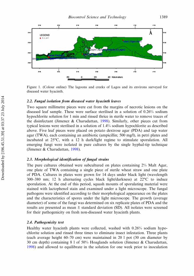
Field trips were undertaken to observe and examine waterways and lagoons ofBadagry, Mile 2, Lagos and Ogun River (Isheri) (Figure 1) and to collect fungalpathogens from diseased water hyacinth. Sampling was done randomly at eachsampling station: Lagos Lagoon, Mile 2, Ogun River and Badagry creeks using amotorised canoe at intervals of three months for a period of three years to collectfungal pathogens that attack the plant at various seasons of the year. The total areasurveyed was 125 km2, 44 km2, 300 km2 and 45 km2 in Badagry, Mile 2, Lagos andOgun River, respectively.
1388 W.O. Okunowo et al.
Dow
nloa
ded
by [
196.
45.5
1.39
] at
03:
37 2
3 Ju
ly 2
014
2.2. Fungal isolation from diseased water hyacinth leaves
Two square millimetre pieces were cut from the margins of necrotic lesions on thediseased leaf sample. These were surface sterilised in a solution of 0.26% sodiumhypochlorite solution for 1 min and rinsed thrice in sterile water to remove traces ofthe disinfectant (Jimenez & Charudattan, 1998). Similarly, other pieces cut fromtypical lesions were sterilised in a solution of 1.4% sodium hypochlorite as describedabove. Five leaf pieces were placed on potato dextrose agar (PDA) and tap wateragar (TWA), each containing an antibiotic (ampicillin; 500 mg/l), in petri plates andincubated at 25°C, with a 12 h dark/light regime to stimulate sporulation. Allemerging fungi were isolated in pure cultures by the single hyphal-tip technique(Jimenez & Charudattan, 1998).
2.3. Morphological identification of fungal strains
The pure cultures obtained were subcultured on plates containing 2% Malt Agar,one plate of TWA containing a single piece of sterile wheat straw and one plateof PDA. Cultures in plates were grown for 14 days under black light (wavelength300–380 nm; 12 h alternating cycles black light/darkness) at 22°C to inducesporulation. At the end of this period, squash mounts of sporulating material werestained with lactophenol stain and examined under a light microscope. The fungalpathogens were identified according to their morphological appearance on the platesand the characteristics of spores under the light microscope. The growth (averagediameter) of some of the fungi was determined on six replicate plates of PDA and theresults are presented as mean ± standard deviation (SD). All isolates were screenedfor their pathogenicity on fresh non-diseased water hyacinth plants.
2.4. Pathogenicity test
Healthy water hyacinth plants were collected, washed with 0.26% sodium hypo-chlorite solution and rinsed three times to eliminate insect infestation. Three plants(each average height 60–70 cm) were maintained in 20 l pot (30 cm diameter by30 cm depth) containing 8 l of 50% Hoaglands solution (Jimenez & Charudattan,1998) and allowed to equilibrate in the solution for one week prior to inoculation
Figure 1. (Colour online) The lagoons and creeks of Lagos and its environs surveyed fordiseased water hyacinth.
Biocontrol Science and Technology 1389
Dow
nloa
ded
by [
196.
45.5
1.39
] at
03:
37 2
3 Ju
ly 2
014
with the pathogen. Inoculum was formulated by harvesting fungal spores from PDAculture plates in sterile distilled water containing 0.1% v/v Tween 80 solution. Leavesand petioles of experimental plants were inoculated with 200 ml of 1 × 10⁶ spores/mlspore suspension containing 0.1% v/v Tween 80 using a hand-held low-pressureatomiser at a distance of 20 cm from the plant. The fungus was sprayed until run offon the leaves and stem of the plants. This experiment was conducted in six replicatepots and in three different experiments A, B and C in the University of Lagos.Control plants were also set up in the three different experiments by spraying leavesand petioles with sterile distilled water containing Tween 80 (0.1% v/v). Inoculatedand control plants were immediately covered with sterile polythene bags for 48 h tomaintain high relative humidity. The plants were then left in the open experimentalfield under the conditions of average temperature ranging between 24°C and 31°C,relative humidity between 68% in the night and 86% in the day and at an averagerainfall of 25 mm for the month. The average sunlight/intensity was 7 h per day.Plants were monitored at three-day intervals for symptoms development. Theisolates were ranked on the basis of the severity of the disease inflicted. Diseaseseverity was assessed according to Freeman and Charudattan (1984). Finally, thepathogens were reisolated and identified from the inoculated and dead plants as wellas from the control plants to fulfil Koch’s postulates.
2.5. Host range examination
Host range of the most pathogenic isolate from the pathogenicity trial was tested onseveral local and economically important agricultural crops under field conditions(described above). The plants were sprayed with 200 ml fungal suspension (1 × 10⁶
spores/ml in 0.1% v/v Tween 80 solution) and monitored for about three weeks fordisease development and host plant reactions. Three individual plants in each potwere examined in triplicate experimental pots. Disease symptom rating was assessedby visual examination as: − = not susceptible (leaves healthy, no disease symptomobserved), + = slightly susceptible (scanty leaf spotting or slight chlorosis nonecrosis), ++ = susceptible (leaf spots/leaf necrosis at 30–50% leaf is dead) and +++= highly susceptible (severe leaf spotting/necrosis at >50% leaf is dead).
2.6. Molecular characterisation of the pathogenic isolate
The pathogenic isolate which was tentatively identified as Myrothecium sp. wasfurther characterised by the Centre for Agriculture and Bioscience International(CABI), Egham, Surrey, UK using standard molecular identification technique toanalyse the ITS1 rDNA sequence. The sequence and the isolate were deposited,respectively, in the GenBank (accession no. GQ853401) and the CABI microbialcollection (Deposit no. IMI 394934).
2.7. Blast and phylogenetic analysis of the isolate
The ITS1 rDNA sequence of our isolate obtained from CABI was subjected tohomology analysis against the holdings of the GenBank using the software BLASTN2.2.28+ (Zhang, Schwartz, Wagner, & Miller, 2000) and the phylogenetic relation-ship among taxa (for >97% homology) were determined using the neighbour-joiningmethod (Saitou & Nei, 1987).
1390 W.O. Okunowo et al.
Dow
nloa
ded
by [
196.
45.5
1.39
] at
03:
37 2
3 Ju
ly 2
014
2.8. Data analysis
The data presented in this study are the results obtained from six replicatedeterminations and are expressed as mean ± SD. To determine the reproducibilityof the experimental results, disease progression between the different experimentswere compared using the general linear model (GLM) regression analysis with dayspost inoculation (DPI) set as continuous predictor. The analyses were done at criticalP value of 0.05 using the GraphPad Prism version 5.00 for Windows (GraphPadSoftware, San Diego, California USA).
3. Results3.1. Survey for pathogens
The continuous survey of the water bodies over the period of investigation showedthat water hyacinth was prevalent at different periods in the year, particularly in therainy season between late May and late September and absent in the dry season.Initial cursory observations in the field revealed that there was little evidence ofoccurrence of fungal pathogens on water hyacinth except during September toNovember when plants with unique pattern of infection were found in two of thefour sampling stations: Badagry Creeks (N6.41950° E2.86019°, N6.42066° E2.86630°and N6.41748° E2.87552°) and Ogun River (N6.64091° E3.8406°) (Figure 2A). Thedisease appeared as a leaf spot, with concentric rings rounded on the side facing thepetiole and narrowing towards the laminar tip. Older leaf spots turned necrotic withdark brown margins, with the centre of the spot containing white and black fungalspores. The diameter of each spot appeared to be proportional to the age of the spot.The disease was easy to identify as brownish necrotic leaf blight, forming massivebrownish patches on water hyacinth leaves in the field. The total area infected was0.0396 km2 in Badagry and 0.0074 km2 in Ogun River. The fungal prevalence ornumber of infection was 3.17 × 10⁻²% of the total number of plants in BadagryCreeks and 1.64 × 10⁻²% of the total number of plants surveyed in Ogun River. Therewere no infection in Mile 2 and Lagos Lagoon.
3.2. Fungal isolation from diseased water hyacinth leaves
Five different fungi (Fusarium sp., Aspergillus niger, Aspergillus flavus, Curvulariasp., Penicillium sp.) were isolated from water hyacinth leaf pieces sterilised with0.26% sodium hypochlorite and plated on PDA. These fungi grew out within 24 hwhile a Myrothecium sp. appeared within 36 h. From leaf pieces sterilised with 1.4%sodium hypochlorite solution, Myrothecium sp. appeared conspicuously on day 3,while the other fungi appeared between days 5 and 6. Of these organisms,Myrothecium sp. occurred most frequently (45%) of total isolation (350) followedby Fusarium sp. (30%), Curvularia sp. (14.7%), Penicillium sp. (5%), A. niger (3.5%)and A. flavus (1.8%). The use of TWA medium yielded Myrothecium sp. after 24 h,although this appeared as transparent hyphae on the medium as compared to thefluffy, whitish, conspicuous appearance on PDA. The other fungi such as Fusariumsp. and Curvularia sp. on TWA were not noticeable until the fourth or fifth day. Thegrowth of the other organisms appeared not to be well supported by TWA.
Biocontrol Science and Technology 1391
Dow
nloa
ded
by [
196.
45.5
1.39
] at
03:
37 2
3 Ju
ly 2
014
3.3. Fungal identification
Cultures of one of the isolates on PDA plates (using an 8 mm diameter cork borer)reached 56 ± 3 mm diameter in six days at 25°C and appeared brownish-black withirregular border and concentric zones. Conidia were slightly curved, septate and thecentral cells were broader than the end cells. The isolate was identified andauthenticated as Curvularia pallescens Boedijn by Dr Markus N. Thormann
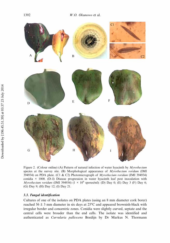
Figure 2. (Colour online) (A) Pattern of natural infection of water hyacinth by Myrotheciumspecies at the survey site. (B) Morphological appearance of Myrothecium roridum (IMI394934) on PDA plate. (C1 & C2) Photomicrograph of Myrothecium roridum (IMI 394934)conidia × 1000. (D–I) Disease progression in water hyacinth leaf post inoculation withMyrothecium roridum (IMI 394934) (1 × 10⁶ spores/ml): (D) Day 0; (E) Day 3 (F) Day 6;(G) Day 9; (H) Day 12; (I) Day 21.
1392 W.O. Okunowo et al.
Dow
nloa
ded
by [
196.
45.5
1.39
] at
03:
37 2
3 Ju
ly 2
014
(Northern Forestry Centre, Natural Resources Canada, 5320-12251, Edmonton, ABT6H 3S5, Canada).
Cultures of a second isolate on PDA reached 77.5 ± 4.2 mm diameter in six daysat 25°C, slightly whitish at first and later turning pinkish in colour. Conidia weresickle shaped and septate. The isolate was authenticated as Fusarium solani by Prof.A.A. Adekunle (Botany and Microbiology Department, University of Lagos).
The third isolate on PDA reached 77.13 ± 1.6 mm diameter in 14 days at 25°C.The isolate produced white, floccose colonies with sporodochia in dark green-to-black concentric rings (Figure 2B). Conidia were sub-hyaline and cylindrical withrounded ends (Figure 2C1 & C2). All characteristics were consistent with thedescription of Myrothecium roridum Tode ex Fr. (Ellis, 1971; Fitton & Holliday,1970). This was authenticated as M. roridum and was given the accession number(IMI 394934) at the Centre for Agriculture and Bioscience International (CABI),Egham Surrey, UK.
3.4. Pathogenicity screening
No disease symptoms were observed on water hyacinth plant infected withC. pallescens and F. solani 24 DPI. Of the six different fungal species tested fortheir ability to infect healthy water hyacinth plants in vitro, the result showed thatM. roridum was the only candidate which infected and produced disease symptomson water hyacinth leaves. The disease started as scanty patches which developed intopale-to-dark brown heavy necrotic spots on the leaves. The necrotic spots expandedin diameter between 5 and 10 mm. With disease progression, the necrotic spotscoalesced and the necrotic area increased. The resultant effect was a decrease in thegreen leaf area and leaf death (Figure 2D–I). The symptom produced in thepathogenicity test was similar to that seen in the field (Figure 2A).
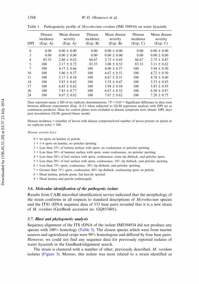
The M. roridum isolate was ranked on the basis of the severity of the damage itcaused (Table 1). The disease progression was monitored over time in terms ofdisease severity and disease incidence (Table 1). The disease incidence on day 4 wasgreater than 60% in experiments A–C, respectively, and 100% in these experimentson day 7. Similarly, the disease severity became prominent on day 4 in allexperiments and the mean values were greater or equal to 2.60. The average diseaseseverity on day 24 was maximum in experiment A and least in experiment C.However, regression analysis indicates that there was no significant difference in therate of disease progression in all experiments (F2,27 = 0.95, P = 0.4). This is anindication that the result is reproducible. Based on the result obtained, the isolate ofM. roridum (IMI 394934) was chosen for further study.
3.5. Host specificity test.
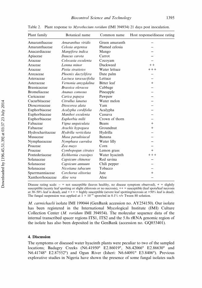
The host range plant response to M. roridum showed that 74.19% of the test plantswere not susceptible (Table 2). Slightly susceptible plants account for 16.13% andplants health status were not compromised. Duckweed was susceptible to the fungusresulting in necrosis and death of the plants. Water lettuce was highly susceptible.Water hyacinth was highly susceptible showing heavy leaf spotting and necrosis withmore than 50% of leaf area coalescing with a resultant death of the plant in less than21 days.
Biocontrol Science and Technology 1393
Dow
nloa
ded
by [
196.
45.5
1.39
] at
03:
37 2
3 Ju
ly 2
014
3.6. Molecular identification of the pathogenic isolate
Results from CABI microbial identification service indicated that the morphology ofthe strain conforms in all respects to standard descriptions of Myrothecium speciesand the ITS1 rDNA sequence data of 533 base pairs revealed that it is a new strainof M. roridum (GenBank accession no. GQ853401).
3.7. Blast and phylogenetic analysis
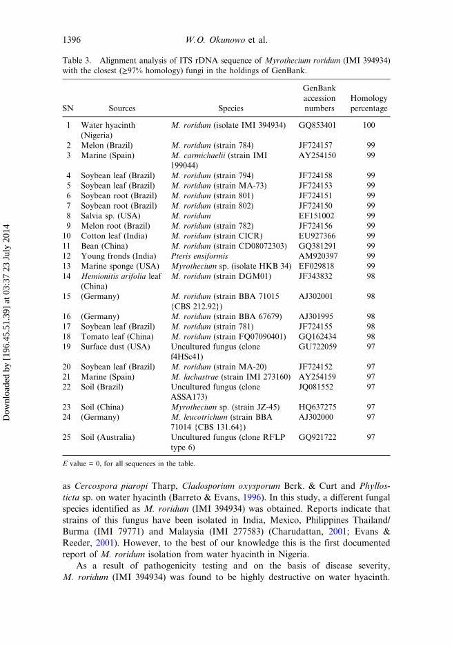
Sequence alignment of the ITS rDNA of the isolate IMI394934 did not produce anyspecies with 100% homology (Table 3). The closest species which were from marinesources and agricultural crops were 99% homologous and differed by four base pairs.However, we could not find any sequence data for previously reported isolates ofwater hyacinth in the GenBank/alignment search.
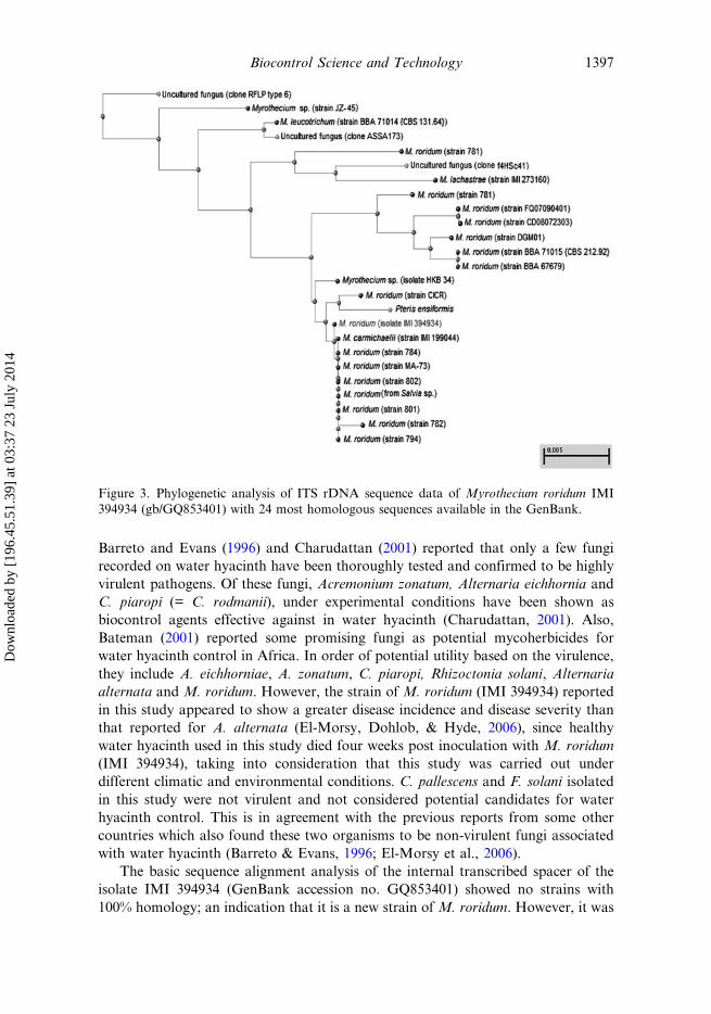
The strain is clustered with a number of other, previously described, M. roridumisolates (Figure 3). Moreso, this isolate was most related to a strain identified as
Table 1. Pathogenicity profile of Myrothecium roridum (IMI 394934) on water hyacinth.
DPI
Diseaseincidence(Exp. A)
Mean diseaseseverity(Exp. A)
Diseaseincidence(Exp. B)
Mean diseaseseverity(Exp. B)
Diseaseincidence(Exp. C)
Mean diseaseseverity(Exp. C)
0 0.00 0.00 ± 0.00 0.00 0.00 ± 0.00 0.00 0.00 ± 0.003 0.00 0.00 ± 0.00 0.00 0.00 ± 0.00 0.00 0.00 ± 0.004 83.33 2.60 ± 0.62 66.67 2.75 ± 0.45 66.67 2.75 ± 0.475 100 3.17 ± 0.72 83.33 3.00 ± 0.52 83.33 3.11 ± 0.627 100 4.17 ± 0.66 100 4.00 ± 0.57 100 3.94 ± 0.5010 100 5.00 ± 0.57 100 4.67 ± 0.51 100 4.72 ± 0.5011 100 5.17 ± 0.54 100 4.67 ± 0.51 100 4.78 ± 0.6014 100 5.83 ± 0.62 100 5.53 ± 0.47 100 5.33 ± 0.8317 100 6.83 ± 0.62 100 5.94 ± 0.54 100 5.83 ± 0.9320 100 7.83 ± 0.77 100 6.67 ± 0.52 100 6.50 ± 0.8724 100 8.67 ± 0.62 100 7.67 ± 0.62 100 7.28 ± 0.75
Data represent mean ± SD of six replicate determinations. *P < 0.05 = Significant difference in data existbetween different experiments (Exp. A–C) when subjected to GLM regression analysis with DPI set ascontinuous predictor. Data for control plants were excluded as disease symptoms were absent. DPI, dayspost inoculation; GLM, general linear model.
Disease incidence = (number of leaves with disease symptoms/total number of leaves present on plants insix replicate pots) × 100.
Disease severity keys:
0 = no spots on lamina or petiole.1 = 1–4 spots on lamina, no petiolar spotting.2 = Less than 25% of lamina surface with spots, no coalescence or petiolar spotting.3 = Less than 50% of laminar surface with spots, some coalescence, no petiolar spotting.4 = Less than 50% of leaf surface with spots, coalescence, some tip dieback, and petiolar spots.5 = Less than 50% of leaf surface with spots, coalescence, 10% tip dieback, and petiolar spotting.6 = Less than 75% spots, coalescence, 30% tip dieback, and petiolar spotting.7 = Greater than 75% spots, coalescence, 60% tip dieback, coaleascing spots on petiole.8 = Dead lamina, petiole green, but heavily spotted.9 = Dead lamina and petiole (submerged).
1394 W.O. Okunowo et al.
Dow
nloa
ded
by [
196.
45.5
1.39
] at
03:
37 2
3 Ju
ly 2
014
M. carmichaelii isolate IMI 199044 (GenBank accession no. AY254150). Our isolatehas been registered in the International Mycological Institute (IMI) CultureCollection Center (M. roridum IMI 394934). The molecular sequence data of theinternal transcribed spacer regions ITS1, ITS2 and the 5.8s rRNA genomic region ofthe isolate has also been deposited in the GenBank (accession no. GQ853401).
4. Discussion
The symptoms or diseased water hyacinth plants were peculiar to two of the sampledlocations; Badagry Creeks (N6.41950° E2.86019°, N6.42066° E2.86630° andN6.41748° E2.87552°) and Ogun River (Isheri: N6.64091° E3.8406°). Previousexplorative studies in Nigeria have shown the presence of some fungal isolates such
Table 2. Plant response to Myrothecium roridum (IMI 394934) 21 days post inoculation.
Plant family Botanical name Common name Host response/disease rating
Amaranthaceae Amaranthus viridis Green amaranth −Amaranthaceae Celosia argentea Plumed celosia −Anacardiaceae Mangifera indica Mango −Apiaceae Daucus carota Carrot −Araceae Colocasia esculenta Cocoyam −Araceae Lemna minor Duckweed ++Araceae Pistia stratiotes Water lettuce +++Arecaceae Phoenix dactylifera Date palm −Asteraceae Lactuca taraxacifolia Lettuce −Asteraceae Vernonia amygdalina Bitter leaf +Brassicaceae Brassica oleracea Cabbage −Bromeliaceae Ananas comosus Pineapple −Caricaceae Carica papaya Pawpaw −Cucurbitaceae Citrullus lanatus Water melon −Dioscoreaceae Dioscorea alata Yam −Euphorbiaceae Acalypha cordifolia Acalypha −Euphorbiaceae Manihot esculenta Cassava −Euphorbiaceae Euphorbia milii Crown of thorn −Fabaceae Vigna unguiculata Beans +Fabaceae Arachis hypogaea Groundnut +Hydrocharitaceae Hydrilla verticilata Hydrilla −Musaceae Musa paradisiacal Banana −Nymphaeaceae Nymphaea caerulea Water lilly −Poaceae Zea mays Corn −Poaceae Cymbopogun citrates Lemon grass +Pontederiaceae Eichhornia crassipes Water hyacinth +++Solanaceae Capsicum chinense Red savina −Solanaceae Capsicum annuum Chili pepper −Solanaceae Nicotiana tabacum Tobacco −Sparrmanniaceae Corchorus olitorius Jute +Xanthorrhoeaceae Aloe vera Aloe −
Disease rating scale: − = not susceptible (leaves healthy, no disease symptom observed), + = slightlysusceptible (scanty leaf spotting or slight chlorosis or no necrosis), ++ = susceptible (leaf spots/leaf necrosisat 30–50% leaf is dead), and +++ = highly susceptible (severe leaf spotting/necrosis at >50% leaf is dead).The fungal suspension was applied at 1 × 10⁻⁶ spore/ml in 0.1% v/v Tween 80 solution.
Biocontrol Science and Technology 1395
Dow
nloa
ded
by [
196.
45.5
1.39
] at
03:
37 2
3 Ju
ly 2
014
as Cercospora piaropi Tharp, Cladosporium oxysporum Berk. & Curt and Phyllos-ticta sp. on water hyacinth (Barreto & Evans, 1996). In this study, a different fungalspecies identified as M. roridum (IMI 394934) was obtained. Reports indicate thatstrains of this fungus have been isolated in India, Mexico, Philippines Thailand/Burma (IMI 79771) and Malaysia (IMI 277583) (Charudattan, 2001; Evans &Reeder, 2001). However, to the best of our knowledge this is the first documentedreport of M. roridum isolation from water hyacinth in Nigeria.
As a result of pathogenicity testing and on the basis of disease severity,M. roridum (IMI 394934) was found to be highly destructive on water hyacinth.
Table 3. Alignment analysis of ITS rDNA sequence of Myrothecium roridum (IMI 394934)with the closest (≥97% homology) fungi in the holdings of GenBank.
SN Sources Species
GenBankaccessionnumbers
Homologypercentage
1 Water hyacinth(Nigeria)
M. roridum (isolate IMI 394934) GQ853401 100
2 Melon (Brazil) M. roridum (strain 784) JF724157 993 Marine (Spain) M. carmichaelii (strain IMI
199044)AY254150 99
4 Soybean leaf (Brazil) M. roridum (strain 794) JF724158 995 Soybean leaf (Brazil) M. roridum (strain MA-73) JF724153 996 Soybean root (Brazil) M. roridum (strain 801) JF724151 997 Soybean root (Brazil) M. roridum (strain 802) JF724150 998 Salvia sp. (USA) M. roridum EF151002 999 Melon root (Brazil) M. roridum (strain 782) JF724156 9910 Cotton leaf (India) M. roridum (strain CICR) EU927366 9911 Bean (China) M. roridum (strain CD08072303) GQ381291 9912 Young fronds (India) Pteris ensiformis AM920397 9913 Marine sponge (USA) Myrothecium sp. (isolate HKB 34) EF029818 9914 Hemionitis arifolia leaf
(China)M. roridum (strain DGM01) JF343832 98
15 (Germany) M. roridum (strain BBA 71015{CBS 212.92})
AJ302001 98
16 (Germany) M. roridum (strain BBA 67679) AJ301995 9817 Soybean leaf (Brazil) M. roridum (strain 781) JF724155 9818 Tomato leaf (China) M. roridum (strain FQ07090401) GQ162434 9819 Surface dust (USA) Uncultured fungus (clone
f4HSc41)GU722059 97
20 Soybean leaf (Brazil) M. roridum (strain MA-20) JF724152 9721 Marine (Spain) M. lachastrae (strain IMI 273160) AY254159 9722 Soil (Brazil) Uncultured fungus (clone
ASSA173)JQ081552 97
23 Soil (China) Myrothecium sp. (strain JZ-45) HQ637275 9724 (Germany) M. leucotrichum (strain BBA
71014 {CBS 131.64})AJ302000 97
25 Soil (Australia) Uncultured fungus (clone RFLPtype 6)
GQ921722 97
E value = 0, for all sequences in the table.
1396 W.O. Okunowo et al.
Dow
nloa
ded
by [
196.
45.5
1.39
] at
03:
37 2
3 Ju
ly 2
014
Barreto and Evans (1996) and Charudattan (2001) reported that only a few fungirecorded on water hyacinth have been thoroughly tested and confirmed to be highlyvirulent pathogens. Of these fungi, Acremonium zonatum, Alternaria eichhornia andC. piaropi (= C. rodmanii), under experimental conditions have been shown asbiocontrol agents effective against in water hyacinth (Charudattan, 2001). Also,Bateman (2001) reported some promising fungi as potential mycoherbicides forwater hyacinth control in Africa. In order of potential utility based on the virulence,they include A. eichhorniae, A. zonatum, C. piaropi, Rhizoctonia solani, Alternariaalternata and M. roridum. However, the strain of M. roridum (IMI 394934) reportedin this study appeared to show a greater disease incidence and disease severity thanthat reported for A. alternata (El-Morsy, Dohlob, & Hyde, 2006), since healthywater hyacinth used in this study died four weeks post inoculation with M. roridum(IMI 394934), taking into consideration that this study was carried out underdifferent climatic and environmental conditions. C. pallescens and F. solani isolatedin this study were not virulent and not considered potential candidates for waterhyacinth control. This is in agreement with the previous reports from some othercountries which also found these two organisms to be non-virulent fungi associatedwith water hyacinth (Barreto & Evans, 1996; El-Morsy et al., 2006).
The basic sequence alignment analysis of the internal transcribed spacer of theisolate IMI 394934 (GenBank accession no. GQ853401) showed no strains with100% homology; an indication that it is a new strain of M. roridum. However, it was
Figure 3. Phylogenetic analysis of ITS rDNA sequence data of Myrothecium roridum IMI394934 (gb/GQ853401) with 24 most homologous sequences available in the GenBank.
Biocontrol Science and Technology 1397
Dow
nloa
ded
by [
196.
45.5
1.39
] at
03:
37 2
3 Ju
ly 2
014
99% homologous to a strain identified as the closely related species M. carmichaeliiisolate IMI 199044 (GenBank accession no. AY254150). This suggests that thedifference in homology is not sufficient to establish an unequivocal identification.
The paucity in the sequence data of the previous isolates of water hyacinth inSri Lanka; IMI 261802 (Hettiarachchi, Gunasekera, & Balasooriya, 1983), India(Ponnappa, 1970), Mexico/India/Philippines Thailand/Burma (IMI 79771) andMalaysia (IMI 277583) (Barreto & Evans, 1996; Evans & Reeder, 2001) made itimpossible to compare our isolate or perform phylogenetic relationship studiesamong isolates of M. roridum pathogenic to water hyacinth. Phylogenetic studieshave been done on Cercospora species pathogenic to water hyacinth (Tessmann,Charudattan, Kistler, & Rosskopf, 2001), such studies could offer some insights intobiogeographic hypothesis of Myrothecium on water hyacinth. Our isolate was able toweakly infect bean and groundnut; however, it is not clear if the isolates from bean,soybean and other agricultural crops can infect or be pathogenic to water hyacinth.
M. roridum has been previously reported as a pathogen of water hyacinth andsome other host plants including some economically important crops (Fish, Bruton,& Popham, 2012; Gaikwad, 1988; Hettiarachchi et al., 1983; Ponnappa, 1970). Thenon-host nonspecificity was confirmed in this study by the ability to cause slightdisease on bitter leaf, bean, groundnut, lemon grass and jute plants. The isolatestudied caused no disease symptoms in corn unlike the report of Gaikwad (1988),this may be due to the differences in the source and origin of the isolates. Severalstudies indicate that the difference in the source or origin of microorganisms affectstheir performance (Anuna & Akpapunam, 1995; Anuna, Sokari, & Akpapunam,1990; Okunowo & Osuntoki, 2007). We have previously reported its virulence onwater lettuce (Okunowo, Osuntoki, & Adekunle, 2011). The efficacy of the fungus inthe integrated management of water hyacinth is known to be enhanced by 2,4 D(Liyanage & Gunasekera, 1989). Hoagland, Weaver, and Boyette (2007) elucidatedsome possible strategies to reduce the non-target risk of a promising mycoherbicidalagent Myrothecium verrucaria which can be adapted to reduce the non-hostspecificity of M. roridum.
Conclusively, this study has isolated and identified a Nigerian indigenous strainof M. roridum, which is highly virulent to water hyacinth. This M. roridum isolatehas potential for application in the biocontrol of water hyacinth. However, since it isnot host specific, future studies should include extensive host range tests andstrategies to reduce its non-target risk.
AcknowledgementsThe authors are grateful to Raghavan Charudattan (Emeritus Professor, Plant PathologyDepartment, University of Florida, Gainsville, Florida, USA) and Hamed K. Abbas(Research Plant Pathologist/Lead Scientist, Biocontrol of Pest Research Unit, USDA-ARS,NBCL, Stoneville, MS, USA) for their helpful discussions and review of the draft version ofthe manuscript.
ReferencesAnuna, M. I., & Akpapunam, M. A. (1995). Quantitative analysis of alcohol types in
pineapple (Ananas comosus) L (L). Merr.) wine fermented by two strains of Saccharomycescerevisiae. Nigerian Food Journal, 13, 12–17.
1398 W.O. Okunowo et al.
Dow
nloa
ded
by [
196.
45.5
1.39
] at
03:
37 2
3 Ju
ly 2
014
Anuna, M. I., Sokari, T. G., & Akpapunam, M. A. (1990). Effect of source of yeast(Saccharomyces spp.) on alcohol content and quality of pineapple (Ananas comosus) wine.Discovery and Innovation, 2, 80–84.
Barreto, R. W., & Evans, H. C. (1996, January). Fungal pathogens of some Brazilian aquaticweeds and their potential use in biocontrol. Paper presented at the 9th internationalsymposium on Biological Control of Weeds, Stellenbosch, South Africa.
Bateman, R. (2001). IMPECCA: An international, collaborative program to investigate thedevelopment of a mycoherbicide for use against water hyacinth in Africa. In M. H. Julien,M. P. Hill, T. D. Center, & D. Jianqing (Eds.), Biological and integrated control of waterhyacinth, Eichhornia crassipes. Proceedings of the second meeting of the Global WorkingGroup for the biological and integrated control of water hyacinth (pp. 57–61), Beijing, China,October 9–12, 2000. ACIAR Proceedings No. 102.
Center, T. D., Hill, M. P., Cordo, H., & Julien, M. H. (2002). Water hyacinth. In R. VanDriesche, B. Blossey, M. Hoddle, S. Lyon, & R. Reardon (Eds.), Biological control ofinvasive plants in the Eastern United States (413 p). Morgantown, WV: USDA ForestService Publication Forest Health Technology Enterprise Team-2002-04. Retrieved fromhttp://dnr.state.il.us/stewardship/cd/biocontrol/4WaterHyacinth.html.
Charudattan, R. (2001). Biological control of water hyacinth by using pathogens: Opportun-ities, challenges, and recent developments. In M. H. Julien, M. P. Hill, T. D. Center, &D. Jianqing (Eds.), Biological and integrated control of water hyacinth, Eichhornia crassipes.Proceedings of the second meeting of the Global Working Group for the biological andintegrated control of water hyacinth (pp. 21–28), Beijing, China, October 9–12, 2000,ACIAR Proceedings No. 102.
Charudattan, R. (1997). Bioherbicides for the control of water hyacinth: Feasibility and needs.In: E. S. Delfosse & N. R. Spencer (Eds.), Proceedings of the international water hyacinthconsortium. Held at the World Bank, March 18–19, 1997. Washington, DC: World Bank.
Coetzee, J. A., Hill, M. P., Julien, M. H., Center, T. D., & Cordo, H. A. (2009). Eichhorniacrassipes. In R. Muniappan, G. V. P. Reddy, A. Raman, & V. P. Gandhi (Eds.), Weedbiological control with arthropods in the tropics (pp. 183–210). Cambridge: CambridgeUniversity Press.
Dagno, K., Lahlali, R., Diourté, M., & Jijakli, M. H. (2011). Effect of temperature and wateractivity on spore germination and mycelial growth of three fungal biocontrol agents againstwater hyacinth (Eichhornia crassipes). Journal of Applied Microbiology, 110, 521–528.doi:10.1111/j.1365-2672.2010.04908.x
Ellis, M. B. (1971). Myrothecium. In: Dematiaceous hyphomycetes. Kew, Surrey, England:Commonwealth Mycological Institute CAB.
El-Morsy, E. S. M. (2004). Evaluation of microfungi for the biological control water hyacinthin Egypt. Fungal Diversity, 16, 35–51. Retrieved from http://www.fungaldiversity.org/fdp/sfdp/16-12.pdf
El-Morsy, E. M., Dohlob, S. M., & Hyde, K. D. (2006). Diversity of Alternaria alternata acommon destructive pathogen of Eichhornia crassipes in Egypt and its potential use inbiological control. Fungal Divers, 23, 139–158. Retrieved from http://www.fungaldiversity.org/fdp/sfdp/23-8.pdf
Evans, H., & Reeder, R. (2001). Fungi associated with Eichhornia crassipes (water hyacinth)in the upper Amazon basin and prospects for their use in biological control. In M. H. Julien,M. P. Hill, T. D. Center, & D. Jianqing (Eds.), Biological and integrated control of waterhyacinth, Eichhornia crassipes. Proceedings of the second meeting of the Global WorkingGroup for the biological and integrated control of water hyacinth (pp. 62–70), Beijing, China,October 9–12, 2000, ACIAR Proceedings No. 102.
Fish, W. W., Bruton, B. D., & Popham, T. W. (2012). Cucurbit host range of Myrotheciumroridum isolated from watermelon. American Journal of Plant Sciences, 3, 353–359.doi:10.4236/ajps.2012.33042
Fitton, M., & Holliday, P. (1970). No. 253 in: CMI descriptions of pathogenic fungi andbacteria. Great Britain: The Eastern Press.
Freeman, T. E., & Charudattan, R. (1984). Cercospora rodmanii conway: A biocontrol agentfor water hyacinth. Florida, FL: Agricultural Experiment Stations, Institute of Food andAgricultural Sciences, University of Florida Gainesville.
Biocontrol Science and Technology 1399
Dow
nloa
ded
by [
196.
45.5
1.39
] at
03:
37 2
3 Ju
ly 2
014
Gaikwad, S. J. (1988). Host range studies of Myrothecium roridum Tode ex Fr the causalorganism of Myrothecium leaf spot disease of pearl millet. Punjabrao Krishi VidyapeethResearch Journal, 12, 162–163.
Harley, K. L. (1990). The role of biological control in the management of water hyacinth,Eichhornia crassipes. Biocontrol News and Information, 11, 11–22.
Hettiarachchi, S., Gunasekera, S. A., & Balasooriya, I. (1983). Leaf spot diseases of waterhyacinth in Sri Lanka. Journal of Aquatic Plant Management, 21, 62–65. Retrieved fromhttp://www.apms.org/japm/vol21/v21p62.pdf
Hoagland, R. E., Weaver, M. A., & Boyette, C. D. (2007). Myrothecium verrucaria fungus:A bioherbicide and strategies to reduce its non-target risks. Allelopathy Journal, 19, 179.Retrieved from http://www.ars.usda.gov/SP2UserFiles/Place/64022000/Publications/Boy-ette/Hoaglandetal.07AllelJ19-179-192.pdf
Hong, Y.-K., Ryu, K.-L., Hyun, J.-N., Uhm, J.-Y., & Kim, S.-C. (2002). Distribution andchanges in occurrence of fingerprint stem blight of Eleochariskuroguwai caused by Epicocco-sorus nematosporus in Korea. The Plant Pathology Journal, 18, 152–155. doi:10.5423/PPJ.2002.18.3.152
Jimenez, M. M., & Charudattan, R. (1998). Survey and evaluation of Mexican native fungi forpotential biocontrol of water hyacinth. Journal of Aquatic Plant Management, 36, 145–148.Retrieved from https://www.apms.org/wp/wp-content/uploads/2012/10/v36p145.pdf
Kirkpatrick, T. L., Templeton, E., TeBeest, D. O., & Smith Jr, R. J. (1982). Potential ofColletotrichum malvarum for biological control of prickly sida. Plant Disease, 66, 323–325.doi:10.1094/PD-66-323
Liyanage, N. P., & Gunasekera, S. A. (1989). Integration of Myrothecium roridum and 2, 4-Din water hyacinth management. Journal of Aquatic Plant Management, 27, 15–20. Retrievedfrom http://www.apms.org/japm/vol27/v27p15.pdf
Mailu, A. M. (2001). Preliminary assessment of the social, economic and environmentalimpacts of water hyacinth in Lake Victoria Basin and status of control biological andintegrated control of water hyacinth. In M. H. Julien, M. P. Hill, T. D. Center, & D.Jianqing (Eds.), Biological and integrated control of water hyacinth, Eichhornia crassipes.Proceedings of the second meeting of the Global Working Group for the biological andintegrated control of water hyacinth (pp. 130–139), Beijing, China, October 9–12, 2000,ACIAR Proceedings No. 102.
Okunowo, W. O., & Osuntoki, A. A. (2007). Quantitation of alcohols in orange winefermented by four strains of yeast. The African Journal of Biotechnology, 1, 95–100.Retrieved from http://www.academicjournals.org/ajbr/PDF/Pdf2007/Nov/Okunowo%20and%20Osuntoki.pdf
Okunowo, W. O., Osuntoki, A. A., & Adekunle, A. A. (2011). Myrothecium roridum Todeand its toxin shows potential for management of water lettuce. Phytopathology, 101, S131.Retrieved from http://www.apsnet.org/meetings/Documents/2011_Meeting_Abstracts/a11ma768.htm
Oso, B. A. (1988). Explorative studies for biological control agents of water hyacinth inNigeria. In O. L. Oke, A. M. A. Imevbore, & A. F. Titilola (Eds.), Water hyacinth menaceand resource. Proceedings of an international workshop held in Lagos Nigeria (pp. 129–136),August 7–12, 1988. Federal Ministry of Science and Technology Publication.
Ponnappa, K. M. (1970). On the pathogenicity of Myrothecium roridum–Eichhornia crassipesisolate. Hyacinth Control Journal, 8, 18–20.
Praveena, R., Naseema, A., & George, S. (2007). Effect of herbicides on Fusariumpallidoroseum – A potential biocontrol agent of water hyacinth [Eichhornia crassipes(Mart.) Solms]. Journal of Tropical Agriculture, 45, 55–57. Retrieved from http://www.jtropag.in/index.php/ojs/article/view/290/181
Saitou, N., & Nei, M. (1987). The neighbor-joining method: A new method for reconstructingphylogenetic trees. Molecular biology and evolution, 4, 406–425. Retrieved from http://mbe.oxfordjournals.org/content/4/4/406.full.pdf
Schmitz, D. C., Schardt, J. D., Leslie, A. J., Dray Jr, F. A., Osborne, J. A., & Nelson, B. V.(1993, October). The ecological impact and management history of three invasive alienaquatic plant species in Florida. Paper presented at the Biological Pollution: The Controland Impact of Invasive Exotic Species symposium held at Indianapolis, Indiana, IN, USA.
1400 W.O. Okunowo et al.
Dow
nloa
ded
by [
196.
45.5
1.39
] at
03:
37 2
3 Ju
ly 2
014
Shabana, Y. M., & Mohamed, Z. A. (2005). Integrated control of water hyacinth with amycoherbicide and a phenylpropanoid pathway inhibitor. Biocontrol Science and Techno-logy, 15, 659–669. doi:10.1080/09583150500135842
TeBeest, D. O., & Templeton, G. E. (1985). Mycoherbicides: Progress in the biological controlof weeds. Plant Disease, 69, 6–10.
Tessmann, D. J., Charudattan, R., Kistler, H. C., & Rosskopf, E. N. (2001). A molecularcharacterization of Cercospora species pathogenic to water hyacinth and emendation of C.piaropi Tharp. Mycologia, 93, 323–334. doi:10.2307/3761654
Tessmann, D. J., Charudattan, R., & Preston, J. F. (2008). Variability in aggressiveness,cultural characteristics, cercosporin production and fatty acid profile of Cercospora piaropi,a biocontrol agent of water hyacinth. Plant Pathology, 57, 957–966. doi:10.1111/j.1365-3059.2008.01867.x
Venter, N., Hill, M. P., Hutchinson, S.-L., & Ripley, B. S. (2013). Weevil borne microbescontribute as much to the reduction of photosynthesis in water hyacinth as does herbivory.Biological Control, 64, 138–142. doi:10.1016/j.biocontrol.2012.10.011
Walker, H. L. (1981). Factors affecting biological control of spurred anoda (Anoda cristata)with Alternaria macrospora. Weed Science, 29, 505–507. Retrieved from http://www.jstor.org/stable/4043339
Zhang, Z., Schwartz, S., Wagner, L., & Miller, W. (2000). A greedy algorithm for aligningDNA sequences. Journal of Computational Biology, 7, 203–214. doi:10.1089/10665270050081478
Biocontrol Science and Technology 1401
Dow
nloa
ded
by [
196.
45.5
1.39
] at
03:
37 2
3 Ju
ly 2
014


