Embed Size (px)
Citation preview
Molecular Immunology, Vol. 25, No. 4, pp. 337-344, 1988 Printed in Great Britain.
0161-5890/88 $3.00 + 0.00 Pergamon Press plc
OLIVE (OLEA EUROPEA) POLLEN ALLERGENS-II. ISOLATION AND CHARACTERIZATION OF TWO
MAJOR ANTIGENS
P. LAUZURICA, N. MARURI, B. GALOCHA, J. GONZALEZ, R. DIAZ, P. PALOMINO, D. HERNANDEZ, R. GARCiA and C. LAHOZ*
Department of Immunology, Fundaci6n JimCnez Diaz, Avda, Reyes Catolicos 2, 28040 Madrid, Spain
(Firs? received 8 April 1987; accepled in revised form 13 October 1987)
Abstract-A dialyzed extract of olive (Oleu europeu) pollen was fractionated by anion exchange chromatography on DEAE-Sepharose CL-6B using a discontinuous gradient of ammonium bicarbonate. The most important protein allergen was obtained from the 0.3 M fraction after gel filtration on Sephadex G-100 and separation by lentil-lectin Sepharose-4B. The major allergen of olive pollen was contained in the effluent and was designated Olea Antigen I. This material inhibited the RAST activity of 15 patients’ sera that were tested. Analytical IEF demonstrated a major band at pH 5.3 and two minor ones at pH 5.6 and 5.0. When these were run into SDS-polyacrylamide gel electrophoresis in a second dimension, all were separated into two bands of mol. wt 17 and 19 K. A second protein, which is the next most important allergen, Olea Antigen II, was obtained from the 0.5 M fraction by chromatofocusing in a 4-7 pH range followed by filtration on Bio-gel P-30. Olea Antigen II had a mol. wt of 8 K as assessed by SDS-PAGE. IEF analysis displayed one main band at pH 3.6 and two minor bands at pH 3.8 and 4.0, respectively. OL-f, an anti-Olea europea monoclonal antibody (MAb) previously reported by us Lauzurica et al. (1988) reacted with the 17 and 19 K antigens from the crude extract and with Olea Antigen I but not with Olea Antigen II.
INTRODUCTION
The isolation and characterization of highly purified IgE binding molecules are of great importance. These purified antigens can be used not only for diagnostic purposes but also as tools for dissecting cellular and humoral IgE response (Rawle et al., 1984; Ekramod- doullah et al., 19862~). The isolation of major antigens is thus the first step for studies leading to the identification of determinants related to the IgE response (Olson and Klapper, 1986; Elsayed et al.,
1986; Ekramoddoullah et al., 1986~) as well as for studying the relationship with molecules such as Class II and T cell receptors which could be involved
in the IgE response (Marsh et al., 1982; Marsh, 1986; Roebber et al., 1985).
Among the pollens of the Mediterranean area, Olea europea pollen is one of the more widely distrib- uted (Bousquet et al., 1985). Although several aller- gens have been identified (Vela et al., 1982; Blanca et
al., 1983) little information is available regarding the molecular characteristics of the isolated antigens.
In a previous report (Lauzurica et al., 1988) we analysed, by immunoblotting and CRIE, the major allergens found in the 0. europea crude extract and we described a monoclonal antibody (MAb) OL-I
which recognizes the 17 and 19 K bands from the whole crude extract.
*Author to whom correspondence should be addressed.
This paper describes the purification and partial
characterization of two proteins, considered as major allergens from 0. europea pollen. These proteins (Olea Antigen I and Olea Antigen II) are quite different in terms of their charge, size and allergenic activity. Monoclonal antibody OL-1 reacts only with protein Olea Antigen I.
MATERIALS AND METHODS
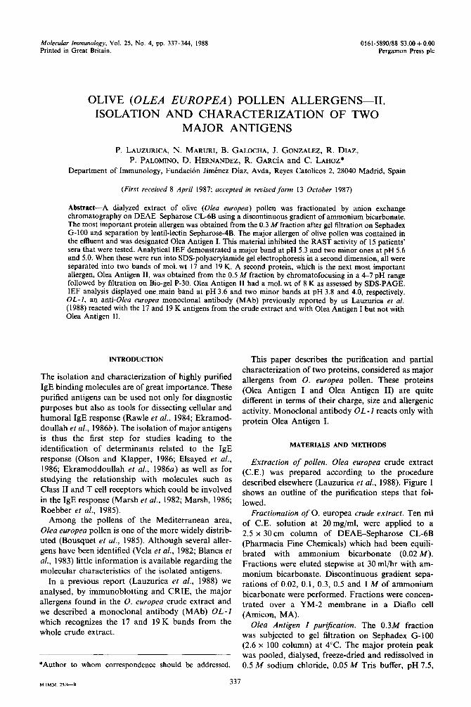
Extraction of pollen. Olea europea crude extract (C.E.) was prepared according to the procedure described elsewhere (Lauzurica et al., 1988). Figure 1 shows an outline of the purification steps that fol- lowed.
Fractionation of 0. europea crude extract. Ten ml of C.E. solution at 20mg/ml, were applied to a 2.5 x 30cm column of DEAE-Sepharose CLdB (Pharmacia Fine Chemicals) which had been equili- brated with ammonium bicarbonate (0.02 M). Fractions were eluted stepwise at 30 ml/hr with am- monium bicarbonate. Discontinuous gradient sepa- rations of 0.02, 0.1, 0.3, 0.5 and 1 M of ammonium bicarbonate were performed. Fractions were concen- trated over a YM-2 membrane in a Diaflo cell (Amicon, MA).
Olea Antigen Z purt$cation. The 0.3M fraction was subjected to gel filtration on Sephadex G-100 (2.6 x 100 column) at 4°C. The major protein peak was pooled, dialysed, freeze-dried and redissolved in 0.5 M sodium chloride, 0.05 M Tris buffer, pH 7.5,
M MM 25,4-a 337
338 P. LAUZURICA et al.
L Crude extract I ----_---___---__---__------~-
DEAE- Sephorose CL-66
Ion exchange chromatography ---__---___---___----_---- __-_
,!, .I, [ \ ? / 0.3M O.&M
--_-----_ Sephadex G - 100
Gel filtration
----_----_ PEE -94
Chromotofocusinq
------- -- Lentil-lectin
Affinity chromatography ---_-----
/+ Ebate
Olea Antigen I
- iozg;; p -3o-
Gel filtration --_ -- ----
*
Olea Antigen II
Fig. 1. Schematic diagram of the isolation of Olea Antigen I and Olea Antigen II.
containing 1 .O mM MnCl, and CaCl,. The sample was then applied to a lentil-lectin Sepharose4B (Pharmacia Fine Chemicals) 1 x 8 column which had previously been equilibrated with the same buffer solution, at a flow rate of 30 ml/hr at 4°C. The starting buffer was used during elution until no more U.V. absorbing material (280 nm) was detected in the effluent. Thereafter the column was washed with the same buffer containing 5% methyl-D-mannoside.
Olea Antigen II purtjication. Chromatofocusing
of the 0.5M fraction was performed on a 30ml (1 x 30) column of PBE 94 equilibrated with 0.025M
imidazole-HCl. The fraction was eluted using a polybuffer 74-Cl (Pharmacia, Sweden). The peak eluted at pH 4 was applied to a Bio-gel P-30 (LKB, Sweden).
Isoelectrofucusing (ZEF). Olea Antigen I and Olea Antigen II were labelled with ‘251 according to the method of Greenwood et al. (1963). Labelled Olea Antigen I and Olea Antigen II were dissolved in 100 ~1 of a sample buffer containing 8M urea, 1% Nonidet P-40, 10% 2-mercaptoethanol and 4% (w/v) Ampholine (LKB) mixture, pH 3.5510, 4-6 and 6-8 (1:4:4). After 3 min centrifugation at 11,600g to remove insoluble material, IEF was performed as described elsewhere (Rodriguez de Cordoba et al., 1984).
Sodium dodecyl sulphate (SDS) polyacrylamide gradient gel electrophoresis. SDS-PAGE was per- formed according to Laemmli (1970).
Two-dimensional eIectrophoresis. One strip from a running IEF gel described above was placed on top of a 7-15% polyacrylamide linear gradient and SDS- PAGE electrophoresis carried out as above. Gels were fixed in H,O : methanol : acetic acid (10 : 4: 1) and
analysed by autoradiography with Kodak X-OMAT GRS film.
Protein determination. The protein content of sam- ples was estimated by the Coomassie blue assay of Bradford (1976).
Solid-phase radioimmunoassays (RAST). This was performed by incubating the different allergenic frac- tions at serial dilutions in PBS with nitrocellulose
discs (5 mm diameter). The discs were incubated at 4°C for 18 hr. Allergen solutions were then with-
drawn and the discs washed with PBS. Unbound sites were blocked with 3% BSA (Sigma) in PBS contain- ing 0.1% Tween-20. Finally, the discs were washed 3 times with 0.1% Tween-20 PBS. After 2 hr of incu- bation with 100 ~1 of allergic sera, specific IgE was developed with ‘2SI-anti-human IgE (Pharmacia, Up- sala, Sweden).
Radioimmunoassay (RAST) inhibition analysis. The radioallergosorbent inhibition test was performed according to Gleich et al. (1975) with commercial discs coated with Olea europea pollen extract (Phar- macia, Upsala, Sweden). Five-fold serial dilutions of Olea Antigen I (200pg/ml) and crude extract (200pg/ml) were used as inhibitors.
ELBA. This was performed according to the method of Chapman et al. (1984). Olea Antigen I, Olea Antigen II and crude extract were bound to microtitre plates at a concn of 10 pg/ml and these were incubated with serial dilutions of MAb OL-1 supernatant. A goat anti-gamma mouse serum per- oxidase labelled (Tago, Burlingame, CA) was used as a second antibody.
Patients sera. Sera were selected from patients with documented positive clinical history and positive cutaneous tests to olive pollen. All patient’s sera were screened with Phadebast RAST and showed specific IgE binding (RAST class 34). Some sera were used as individual sera while others were pooled prior to
use.
RESULTS
Fractionation of Olea europea pollen proteins
Five fractions were obtained from the pollen ex-
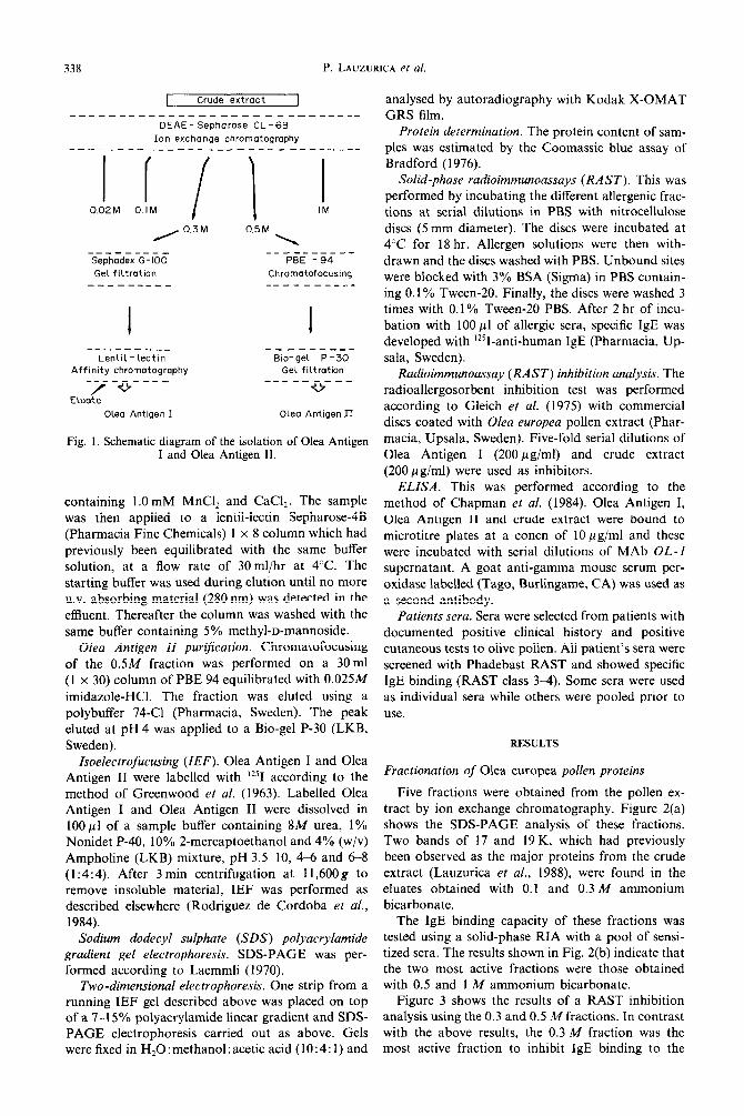
tract by ion exchange chromatography. Figure 2(a) shows the SDS-PAGE analysis of these fractions. Two bands of 17 and 19 K, which had previously been observed as the major proteins from the crude extract (Lauzurica et al., 1988), were found in the eluates obtained with 0.1 and 0.3 M ammonium bicarbonate.
The IgE binding capacity of these fractions was tested using a solid-phase RIA with a pool of sensi- tized sera. The results shown in Fig. 2(b) indicate that the two most active fractions were those obtained with 0.5 and 1 M ammonium bicarbonate.
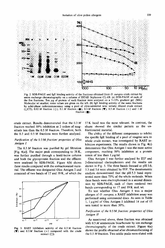
Figure 3 shows the results of a RAST inhibition analysis using the 0.3 and 0.5 M fractions. In contrast with the above results, the 0.3 M fraction was the most active fraction to inhibit IgE binding to the
Isolation of olive pollen allergens
Fig. 2. SDS-PAGE and IgE binding activity of the fractions obtained from 0. europea crude extract by anion exchange chromatography on a column of DEAE-Sepharose CL-6B. (a) SDS-PAGE of each of the five fractions. Ten pg of protein of each fraction were analysed on a 7-15% gradient gel. (MK) Molecular wt marker; some values are given on the left. (b) IgE binding activity of the same fractions by solid-phase radioimmunoassay using a pool of olive-sensitized sera: serially diluted crude extract (OCE), 0.02 M fraction (O), 0.1 M fraction (e), 0.3 M fraction (v), 0.5 M fraction (X ) and I M
fraction (W).
crude extract. Results demonstrated that the 0.3 M fraction reached 50% inhibition at 2 orders of mag- nitude less than the 0.5 M fraction. Therefore, both the 0.3 and 0.5 M fractions were further analysed.
Purification of the 0.3 M fraction: properties of Olea Antigen I
The 0.3 M fraction was purified by gel filtration [Fig. 4(a)]. The major peak corresponding to 38 K, was further purified through a lentil-lectin column and both the glycoprotein fraction and the effluent were analysed by SDS-PAGE. Figure 4(b) shows these results compared with the unfractionated mate- rial. The effluent was designated Olea Antigen I and consisted of two bands of 17 and 19 K, of which the
Fig. 3. RAST inhibition activity of the 0.3 M fraction (v) and 0.5M fraction (x ) compared with the crude
extract (0).
17 K band was the most relevant. In contrast, the eluate showed the similar pattern as the un- fractionated material.
The ability of the different components to inhibit the specific IgE binding of a pool of reaginic sera to whole crude extract, was investigated by RAST in- hibition experiments. The results shown in Fig. 4(c) demonstrate that Olea Antigen I was the most active component, reaching 50% inhibition at a protein concn of less than 1 pg/ml.
Olea Antigen I was further analysed by IEF and 2-dimensional electrophoresis and the results are shown in Fig. 5. The three bands focused at pH 5.0, 5.3 and 5.6 were obtained by IEF. The densitometric analysis demonstrated that the pH 5.3 band repre- sented more than 75% of the whole molecule. When these bands were electrophoresed in a second dimen- sion by SDS-PAGE, each of them rendered two bands corresponding to 17 and 19 K mol. wt.
To test whether Olea Antigen I was a major allergen of 0. europea, a RAST inhibition assay was performed using commercial discs. As seen in Table 1, 1 pg/ml of Olea Antigen I inhibited 14 out of 15 sera tested to more than 50%.
Purzjication of the 0.5 M fraction: properties of Olea Antigen II
As mentioned above, these fraction was obtained with 0.5 M ammonium bicarbonate by ion exchange chromatography of the crude extract. Figure 6(a) shows the profile obtained after chromatofocusing of the 0.5 M fraction. Two acidic peaks were eluted with
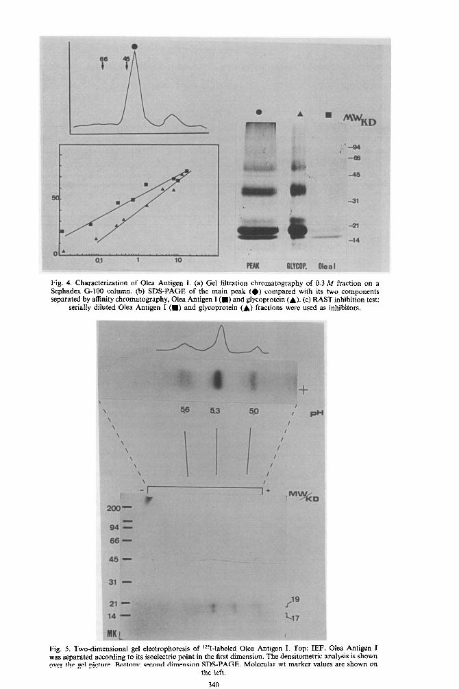
Fig. 4. Characterization of Olea Antigen I. (a) Gel filtration chromatography of 0.3 M fraction on a Sephadex G-100 column. (b) SDS-PAGE of the main peak (e) compared with its two components separated by affinity chromatography, Olea Antigen I (m) and glycoprotein (A). (c) RAST inhibition test:
serially diluted Olea Antigen I (m) and glycoprotein (A) fractions were used as inhibitors.
Fig. 5. Two-dimensional gel electrophoresis of L251-labeled Olea Antigen I. Top: IEF. Olea Antigen I was separated according to its isoelectric point in the first dimension. The densitometric analysis is shown over the gel picture. Bottom: second dimension SDS-PAGE. Molecular wt marker values are shown on
the ieft.
340
Isolation of olive pollen allergens 341
Table 1. RAST inhibition by Olea Antigen I of 15 individual allergic sera from olive-sensitized patients
N Sera Olea Antigen I Control % Inhibition
1 2147 11394 81.0 2 709 1652 57.0 3 1115 1963 43.0 4 805 2094 62.0 5 815 1878 67.0 6 1868 9093 80.0 7 1003 3222 69.0 8 708 2400 70.5 9 1143 3213 64.4
10 2009 5466 63.3 11 693 4839 85.7 12 560 5664 90.2 13 557 3800 85.7 14 704 3995 82.4 15 RR2 3500 75.0
One pg of Olea Antigen I was used on 0. europea-coated commercial discs from Pharmacia. Results are expressed in cpm. Control: results obtained using PBS instead of inhibitor.
% Inhibition = cpm control - cpm problem
cpm control x loo
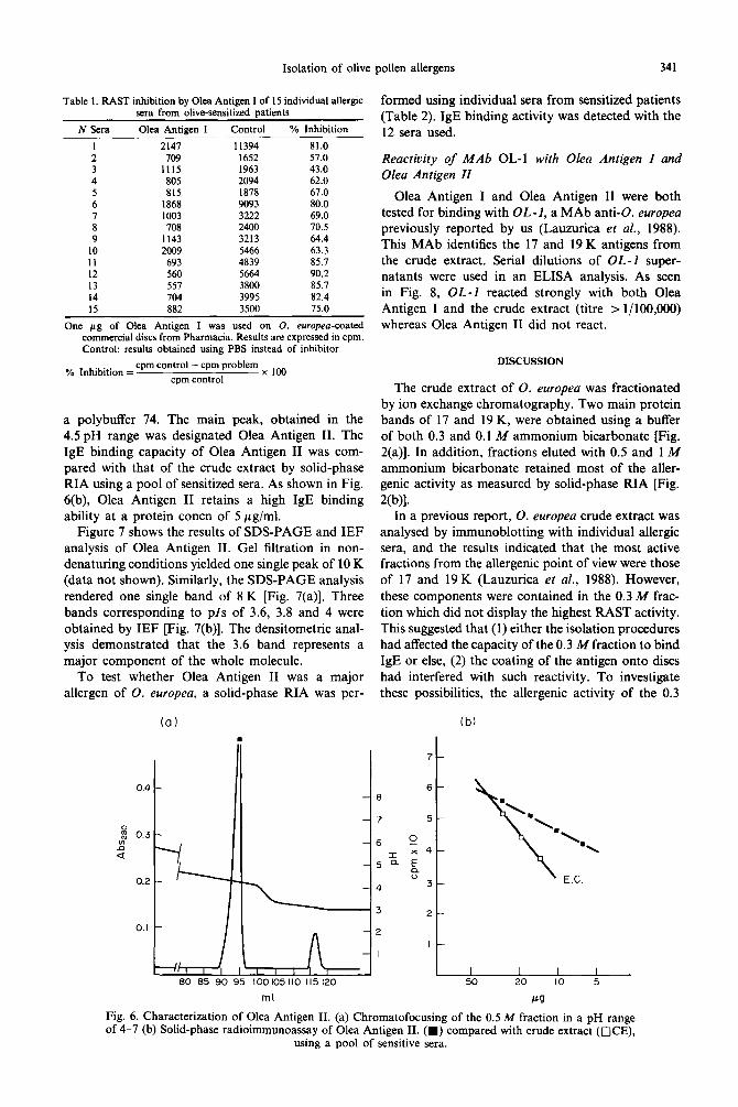
a polybuffer 74. The main peak, obtained in the 4.5 pH range was designated Olea Antigen II. The IgE binding capacity of Olea Antigen II was com- pared with that of the crude extract by solid-phase RIA using a pool of sensitized sera. As shown in Fig. 6(b), Olea Antigen II retains a high IgE binding ability at a protein concn of 5 pg/ml.
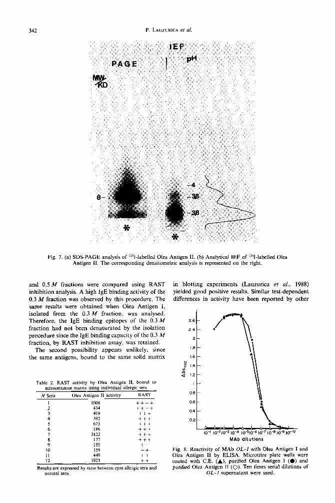
Figure 7 shows the results of SDS-PAGE and IEF analysis of Olea Antigen II. Gel filtration in non- denaturing conditions yielded one single peak of 10 K (data not shown). Similarly, the SDS-PAGE analysis rendered one single band of 8 K [Fig. 7(a)]. Three bands corresponding to pls of 3.6, 3.8 and 4 were obtained by IEF [Fig. 7(b)]. The densitometric anal- ysis demonstrated that the 3.6 band represents a major component of the whole molecule.
To test whether Olea Antigen II was a major allergen of 0. europea, a solid-phase RIA was pcr-
(a)
formed using individual sera from sensitized patients (Table 2). IgE binding activity was detected with the 12 sera used.
Reactivity of MAb OL-1 with Olea Antigen I and Olea Antigen II
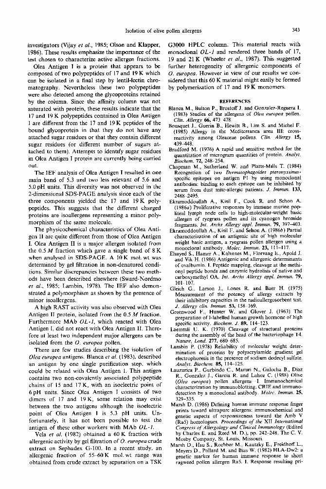
Olea Antigen I and Olea Antigen II were both tested for binding with OL-1, a MAb anti-O. europea previously reported by us (Lauzurica et al., 1988). This MAb identifies the 17 and 19 K antigens from the crude extract. Serial dilutions of OL-1 super- natants were used in an ELISA analysis. As seen in Fig. 8, OL-I reacted strongly with both Olea Antigen I and the crude extract (titre > l/100,000) whereas Olea Antigen II did not react.
DISCUSSION
The crude extract of 0. europea was fractionated by ion exchange chromatography. Two main protein bands of 17 and 19 K, were obtained using a buffer of both 0.3 and 0.1 M ammonium bicarbonate [Fig. 2(a)]. In addition, fractions eluted with 0.5 and 1 M ammonium bicarbonate retained most of the aller- genic activity as measured by solid-phase RIA [Fig.
W)l. In a previous report, 0. europea crude extract was
analysed by immunoblotting with individual allergic sera, and the results indicated that the most active fractions from the allergenic point of view were those of 17 and 19 K (Lauzurica et al., 1988). However, these components were contained in the 0.3 M frac- tion which did not display the highest RAST activity. This suggested that (1) either the isolation procedures had affected the capacity of the 0.3 M fraction to bind IgE or else, (2) the coating of the antigen onto discs had interfered with such reactivity. To investigate these possibilities, the allergenic activity of the 0.3
(b)
7
t I 6-
8
7 5-
$ s 0.3
- b -
s -6
-55 x 4- E E?
-4 3-
ml EJ
Fig. 6. Characterization of Olea Antigen II. (a) Chromatofocusing of the 0.5 M fraction in a pH range of 4-7 (b) Solid-phase radioimmunoassay of Olea Antigen II. (W) compared with crude extract (OCE),
using a pool of sensitive sera.
342 P. LAUZURICA et al.
Fig. 7. (a) SDS-PAGE analysis of ‘251-labelled Olea Antigen II. (b) Analytical IEF of ‘251-labelled Olea Antigen II. The corresponding densitometric analysis is represented on the right.
and 0.5 M fractions were compared using RAST inhibition analysis. A high IgE binding activity of the 0.3 M fraction was observed by this procedure. The same results were obtained when Olea Antigen I, isolated from the 0.3 M fraction, was analysed. Therefore, the IgE binding epitopes of the 0.3 M fraction had not been denaturated by the isolation procedure since the IgE binding capacity of the 0.3 M fraction, by RAST inhibition assay, was retained.
The second possibility appears unlikely, since the same antigens, bound to the same solid matrix
Table 2. RAST activity by Olea Antigen II, bound to nitrocellulose matrix using individual allergic sera
N Sera Olea Antigen II activity RAST
1 1006 ++++ 2 434 ++++ 3 419 +++ 4 392 +++ 5 673 +++ 6 186 +++ I 3122 +++ 8 111 +++ 9 180 ++
10 159 ++ 11 449 ++ 12 1925 ++
Results are expressed by ratio between cpm allergic sera and normal sera.
in blotting experiments (Lauzurica et al., 1988) yielded good positive results. Similar test-dependent differences in activity have been reported by other
2.6
2.4
2
I.6 -
1.6 -
g ,” 1.4-
: 1.2-
I-
0.6 -
0.6 -
IO-’ 10-210‘~ 10-4 IO-~@ 10-7 10-6 lo-9 lo-‘0
MAC dilutions
Fig. 8. Reactivity of MAb OL-l with Olea Antigen 1 and Olea Antigen II by ELISA. Microtitre plate wells were coated with C.E. (A), purified Olea Antigen I (0) and purified Olea Antigen II (0). Ten times serial dilutions of
OL-I supematant were used.
Isolation of olive
investigators (Vijay et al., 1985; Olson and Klapper,
1986). These results emphasize the importance of the test chosen to characterize active allergen fractions.
Olea Antigen I is a protein that appears to be composed of two polypeptides of 17 and 19 K which can be isolated in a final step by lentil-lectin chro- matography. Nevertheless these two polypeptides were also detected among the glycoproteins retained by the column. Since the affinity column was not saturated with protein, these results indicate that the 17 and 19 K polypeptides contained in Olea Antigen
I are different from the 17 and 19 K peptides of the bound glycoprotein in that they do not have any attached sugar residues or that they contain different
sugar residues (or different number of sugars at- tached to them). Attempts to identify sugar residues in Olea Antigen I protein are currently being carried out.
G3000 HPLC column. This material reacts with monoclonal OL-1 and rendered three bands of 17, 19 and 21 K (Wheeler et al., 1987). This suggested further heterogeneity of allergenic components of 0. europea. However in view of our results we con- sidered that this 60 K material might easily be formed by polymerization of 17 and 19 K monomers.
REFERENCES
Blanca M., Bulton P., Brostoff J. and Gonzalez-Reguera I. (1983) Studies of the allergens of Olea europea pollen. Clin. Allergy 66, 473478.
Bousquet J., Guerin B., Hewitt B., Lim S. and Michel F. (1985) Allergy in the Mediterranea area III: cross- reactivity among Oleaceae pollens. Clin. Allergy 15, 439448.
Bradford M. (1976) A rapid and sensitive method for the quantitation of microgram quantities of protein. Analyf. Biochem. 72, 248-254.
The IEF analysis of Olea Antigen I resulted in one main band of 5.3 and two less relevant of 5.6 and 5.0 pH units. This diversity was not observed in the 2-dimensional SDS-PAGE analysis since each of the three components yielded the 17 and 19 K poly- peptides. This suggests that the different charged proteins are isoallergens representing a minor poly- morphism of the same molecule.
Chapman M., Sutherland W. and Platts-Mills T. (1984) Recognition of two Dermatophagoides pleronyssinus- specific epitopes on antigen Pl by using monoclonal antibodies: binding to each epitope can be inhibited by serum from dust mite-allergic patients. J. Immun. 133, 248882495.
The physicochemical characteristics of Olea Anti- gen II are quite different from those of Olea Antigen I. Olea Antigen II is a major allergen isolated from the 0.5 M fraction which gave a single band of 8 K when analysed in SDS-PAGE. A 10 K mol. wt was determined by gel filtration in non-denatured condi- tions. Similar discrepancies between these two meth- ods have been described elsewhere (Sward-Nordmo et al., 1985; Lambin, 1978). The IEF also demon- strated a polymorphism as shown by the presence of minor isoallergens.
Ekramoddoullah A., Kisil F., Cook R. and Sehon A. (1986a) Proliferative responses by immune murine pop- liteal lymph node cells to high-molecular-weight basic allergen of ryegrass pollen and its cyanogen bromide fragments. Int. Archs Allergy appl. Immun. 19, 397403.
Ekramoddoullah A., Kisil F. and Sehon A. (19866) Partial characterization of an antigenic site of high molecular weight basic antigen, a ryegrass pollen allergen using a monoclonal antibody. Molec. Immun. 23, 11 l-1 17.
Elsayed S., Hamer A., Kalvenes M., Florvaag E., Apold J. and Vik H. (1986) Antigenic and allergenic determinants of ovalbumin. I. Peptide mapping, cleavage at the methi- onyl peptide bonds and enzymic hydrolisis of native and carboxymethyl OA. Int. Archs Allergy appl. Immun. 79, 101-107.
A high RAST activity was also observed with Olea Antigen II protein, isolated from the 0.5 M fraction. Furthermore MAb OL-1, which reacted with Olea Antigen I, did not react with Olea Antigen II. There- fore at least two independent major allergens can be isolated from the 0. europea pollen.
Gleich G., Larson J., Lones R. and Baer H. (1975) Measurement of the potency of allergy extracts by their inhibitory capacities in the radioallergosorbent test. J. Allergy clin. Immun. 53, 1588169.
Greenwood F., Hunter W. and Glover J. (1963) The preparation of I-labelled human growth hormone of high specific activity. Biochem. J. 89, 114123.
Laemmli U. K. (1970) Cleavage of structural proteins during the assembly of the head of the bacteriophage T4. Nature, Lond. 277, 68@685.
There are few studies describing the isolation of Olea europea antigens. Blanca et al. (1983) described an antigen by one single purification step, which could be related with Olea Antigen I. This antigen contains two non-covalently associated polypeptide chains of 15 and 17 K, with an isoelectric point of 6pH units. Since Olea Antigen I consists of two dimers of 17 and 19 K, some relation may exist between the two antigens although the isoelectric point of Olea Antigen I is 5.3 pH units. Un- fortunately, it has not been possible to test the antigen of these other workers with MAb OL-J.
Lambin P. (1978) Reliability of molecular weight deter- mination of proteins by polyacrylamide gradient gel electrophoresis in the presence of sodium dodecyl sulfate. Analyf. Biochem. 85, 114125.
Lauzurica P., Gurbindo C., Maruri N., Galocha B., Diaz R., Gonzalez J., Garcia R. and Lahoz C. (1988) Olive (Olea europea) pollen allergens-I. Immunochemical characterization by immunoblotting. CRIE and immuno- detection by a monoclonal antibody. Molec. Immun. 25, 329-335.
Vela et al. (1982) obtained a 60 K fraction with allergenic activity by gel filtration of 0. europea crude extract on Sephadex G-100. In a recent study, an allergenic fraction of 55560 K mol. wt range was obtained from crude extract by separation on a TSK
Marsh D. (1986) Defining human immune response finger prints toward ultrapure allergens: immunochemical and genetic aspects of responsiveness toward the Amb V (Ra5) homologues. Proceedings of the XII International Congress of Allergology and Clinical Immunology (Edited by Charles E. and Reed M. D.), pp. 242-248. The C. V. Mosby Company, St. Louis, Missouri.
Marsh D., Hsu S., Roebber M., Kautzky E., Freidhoff L., Meyers D., Pollard M. and Bias W. (1982) HLA-Dw2: a genetic marker for human immune response to short ragweed pollen allergen Ra5. I. Response resulting pri-
pollen allergens 343

344 P. LAUZURICA et al.
marily from natural antigenic exposure. J. exp. Med. 155, (RaSG) an Ra5 homologue from giant ragweed pollen. 1439-1451. J. Immun. 134, 3062-3069.
Olson J. and Klapper D. (1986) Two major human aller- genic sites on ragweed pollen allergen antigen E identified by using monoclonal antibodies. J. Immun. 136, 2109-2115.
Sward-Nordmo M., Wold J., Paulsen B. and Aukrust L. (1985) Purification and partial characterization of the allergen Ag-54 from Cladosporium herbarum. Int. Archs Allergy appl. Immun. 78, 249-255.
Rawle F., Mitchel E. and Platts-Mills T. (1984) T cells responses to the major allergen from the house dust mite Dermatophagoides pteronyssinus. Antigen Pl : comparison of patients with asthma, atopic dermatitis and perennial rhinitis. J. Immun. 133, 195-200.
Vela C., Platas C., Gurbindo C., Tricas L., Subiza E., Garcia R. and Lahoz C. (1982) Fractionation and biolog- ical characterization of Olea europea pollen extract. In?. Archs Allergy appl. Immun. 68, 289-294.
Rodriguez de Cordoba S., Rubinstein P. and Ferreira A. (1984) High resolution isoelectric focusing of immuno- precipitated proteins under denaturing conditions. A simple analytical method applied to the study of com- plement component polymorphisms. J. Immun. Mefh. 69, 165-170.
Vijay H., Young N., Jackson G., White G. and Bernstein I. (1985) Studies on the Alternaria allergens V. Comparative biochemical and immunological studies of three isolates Alternaria tenuis cultured on synthetic media. Inr. Archs Allergy appl. Immun. 18, 3742.
Roebber M., Klapper D., Goodfriend L. and Marsh D. (1985) Immunochemical and genetic studies of Amb.t.V
Wheeler A., Lauzurica P., Garcia R., Hickman B. and Lahoz C. (1987) Identification of a major allergen in olive tree (Olea europea) pollen extract. Rev. Esp. Alergol. Immun. Clin. 2, 228 (Abstr. 122).


