Embed Size (px)
Citation preview
Vol 44, No. 4;Apr 2014
301 Pretoria, South Africa
Application of microwave as an alternative home
pasteurization method for camel milk; microbiological,
physiochemical and biochemical study.
Ali Alkaladi1, Mohamed Afifi1,2* and Rania Kamal3
1Department of Biological Sciences, Faculty of Science, King Abdulaziz University, North
Campus, PO Box 11508, Jeddah, 21463, Saudi Arabia. 2Department of Biochemistry, Faculty of Veterinary Science, Zagazig University, Egypt. 3Food Control Department, Faculty of Veterinary Medicine, Zagazig University, Egypt.
* Corresponding Author
Mohamed Afifi
e-mail [email protected], [email protected]
tel. 00966509562637 Statement of novelty: This work was conducted to study the application of microwave in
pasteurization of the camel milk. For the first record the treatment of camel milk for 40s in microwave
is sufficient for complete pasteurization of it without adversely affect it's properties and components.
Abstract The present study aimed to compare the efficacy of microwave treatments of camel milk
with the standard milk heat pasteurization method in the terms of microbiological,
physiochemical and biochemical components changes. Fifty camel’s milk samples were
divided into 7 parts for each, one part examined raw the other was pasteurized 62.3 °C for 30
min and the rest were exposed to microwave for 10, 20 ,30, 40 and 50S. The results indicated
the insufficiency of heating camel milk at 62.3 °C for 30 m especially when it has a high
initial microbial count. Contrarily, microwave treatment at 40 seconds was enough to
destruct all tested microbial content of examined camel milk. Unfortunately, both heat
treatment and microwave adversely affect the vital component of the camel's milk as vitamin
C, E, glutathione (GSH), insulin and Immunoglobulin G, in addition to elevation of the
oxidative products as molondialdehyd (MDA) and nitric oxide
Keywords: Camel milk, Pasteurization, Microwave, microbiological and biochemical
properties
Introduction Peoples in many world countries are widely consuming camel milk especially in the arid and
semi-arid zones for its nutritional value, medicinal properties and availability. Many reports
had reported many health associated benefits of camel milk. It was found that camel milk
contains good qualities of lactoferrin, lactoperoxidase, lysozyme and other antibacterial and
antiviral protective proteins, which make it more superior over cow milk (Elagamy, 2000;
Mal & Pathak, 2010). Camel milk is considered to have anti-carcinogenic (Magjeed, 2005), a
protective effect against cisplatin-induced nephrotoxicity (Afifi, 2010), hypo-allergic (Shabo
et al., 2005) and anti-diabetic properties (Agrawal et al., 2003). A high content of
unsaturated fatty acids contributes to its overall dietary quality (Konuspayeva et al., 2008).
The low quantity of ß-casein and the lack of ß-lactoglobulin are linked to the hypo-allergic
Vol 44, No. 4;Apr 2014
302 Pretoria, South Africa
effect of camel milk (Konuspayeva et al., 2007). Camel’s milk is different from other
ruminants’ milk; having low cholesterol, low sugar, high minerals (sodium, potassium, iron,
copper, zinc and magnesium), high vitamin C, B2, A and E, low protein and high
concentrations of insulin. Moreover, camel milk can be consumed by lactase deficient
persons and those with weak immune systems (Afifi, 2010).
Since camel milk possess the all required nutrients for microbial growth, it required an
effective method for controlling microorganism before consumption. Pasteurization is the
process that uses relatively mild heat treatment to kill pathogens, and inactivate vegetative
bacteria and enzymes. Heat treatment of milk aims to extend the shelf life and improve the
quality of this complex biological fluid by reducing the microbial load and thus, minimizing
the risk of food borne infections (McKinnon et al., 2009).
Despite that pasteurized camel milk is produced and sold in a few countries including
Saudi Arabia, United Arab Emirates, Kazakhstan and Mauritania, (Wernery et al., 2005)
camel milk still to be widely consumed in its raw state. Therefore, this study aimed to find an
effective ways to pasteurize camel’s milk and assess their impact on the physiochemical,
nutritional, immunological, biochemical and microbial properties of camel’s milk.
Materials and methods Row milk supply
Fifty camel's milk samples (1750 mL each ) were collected under aseptic conditions in sterile
plastic bottles from different camels farms at Sharkia province, Egypt and transported in ice
poxes to the milk hygiene laboratory at Faculty of Veterinary Medicine, Zagazig University,
where every sample was divided into seven parts (250 ml each). The first part was untreated
to serve as control, the 2nd part was aseptically exposed to indirect heat treatment in water
path at 62.3 °C for 30 min then cooled directly to 10 degrees, the 3rd, 4th, 5th, 6th and 7th parts
were exposed to microwave (Triple Distribution System, model ME732K, Samsung,
Selangor, Malaysia) treatment for 10, 20, 30, 40 and 50 seconds respectively. After
treatment, each sample was thoroughly mixed before further analysis.
Microbiological examination
Fresh and treated samples were decimally diluted using peptone water before inoculation
onto plates. 3MTM petrifilmTM plates were used in this study to determine the total bacterial,
Staphylococcus aureus (St. aureus), total coliform and Escherichia coli (E. coli) and total
yeast and mold counts (3M Corporate Headquarters; St. Paul, MN, USA) (AOAC, 2003).
Briefly, 1 ml of appropriate dilution was inoculated onto petrifilm plate and the inoculum
was evenly distributed with a sterile plastic spreader. After one minute at ambient
temperature, each petrifilm plate was incubated aerobically according to manufacturer
recommendations, of each tested group. Following incubation, corresponding colonies on
each plate were counted according to the manufactures’ instructions.
Physiochemical examination of milk
pH values were determined using pH meter (Model HI, Hanna Instruments, Latina, Italy),
titrable acidity, specific gravity, total solids and ash contents were determined according to
the method of Association of Official Analytical Chemists (AOAC, 1990a,b,c,e). Fat content
was analyzed by Gerber method as described by James (1995). Protein content was estimated
according the method of British Standards Institution (BSI, 1990), Lactose content was
determined by the method of Lynch et al. (2007).
Biochemical assays
Vol 44, No. 4;Apr 2014
303 Pretoria, South Africa
Vitamin A was determined using Enzyme immunoassay kit (VitaKit ATM; SciMed
Technologies, Inc., Catalogue No. KTSP-71051), vitamin C was assayed colorimetric, firstly
the milk samples was cleared by mixing 600 µL milk with 100 µL 6N HCl, centrifuged for 5
min at 14,000 rpm, then 300 µL supernatant was neutralized with 50 µL 6 N NaOH, and the
supernatant was used for assay of vitamin C according to the method described by Arya
(2002), vitamin E was determined colorimetrically using the method of Rutkowski et al.
(2007). Zn and Se were determined by Flame atomic absorption spectrometric method in
accordance with BSI 11813 (2010), Malodialdehyde (MDA) and nitric oxide and reduced
glutathione were assayed using methods of Okhawa (1979), Montgomery and Dymock
(1961) and Sedlak and Lindsay (1968). IgG was determined by ELISA according to the
method described by Erhardt et al. (1992) and insulin was determined using ultraviolet-visible
absorption spectroscopy, according to Royatvand et al. (2013).
Statistical analysis
The data were statistically analyzed by SPSS version 20 statistical packages (IBM 1 New
Orchard Rd Armonk, NY 10504, USA). Data were presented as a mean ± SD, n = 10.
Statistical differences between groups were performed using student's t-test. Differences
considered significant when P < 0.05.
Results and Discussion Camel's milk has invaluable nutritional and medicinal values, as it contains high levels of
vitamins C, A, B2 and E; and very rich in magnesium and other trace elements as Se and Zn
(Afifi, 2010). Camel milk represents a good nutrition source for peoples live in hard
conditions as desert; as well, it contains a high concentration of non-specific low molecular
weight immunoglobulin G (IgG) which help in protecting them against different pathogens.
It contains high level of insulin that resist acidity, proteolysis and encapsulated in
nanoparticles (lipid vesicles) that make possible its passage through stomach and entry into
circulation (Ajamaluddin et al., 2012). Camel's milk is usually consumed raw, which
certainly constitutes a prominent health hazards for transporting many zoonotic milk-borne
pathogens. Heat treated camel milk sold on a limited scale in some countries as Kingdom of
Saudi Arabia, United Arab Emirates, Mauritania and Khazakhastan (Wernery et al., 2005). In
this study, we tested two ways for the camel's milk pasteurization. The first one is a
modification of the known pasteurization method where the milk is indirectly heated in a
water bath up to 62.3 °C for 30 min, and the second one is microwave treatment, using the
household device (2450MHZ), where the milk samples were exposed to ordinary
microwaves for 10, 20, 30, 40 and 50 seconds. Following treatments, microbiological,
physiochemical and biochemical properties of the milk were examined.
Microbiological parameters
There is no doubt that the major goal of milk heat treatment is making it safe for
consumption. Process of pasteurization can not kill all pathogens through elevated
temperature. Obtained results of the heat treated group of camel milk were in conformity
with the major goals of milk pasteurization. As at 62.3 °C/30 min, highly significant
reductions of all examined microbiological parameters were obtained (Table 1), although,
total elimination was not achieved. Staphylococcus aureus and coliform could be detected
after heat treatment at 62.3 °C/30m, in addition to a total bacterial count. This may be
attributed to high initial value of contaminating microorganisms. The automatic milking
Vol 44, No. 4;Apr 2014
304 Pretoria, South Africa
system of dairy cows is mostly responsible for the lower levels of contaminating
microorganisms which can get entrance to milk, and since she-camel milking is still
depending on hand milking, high initial level of contaminating microorganism is usually a
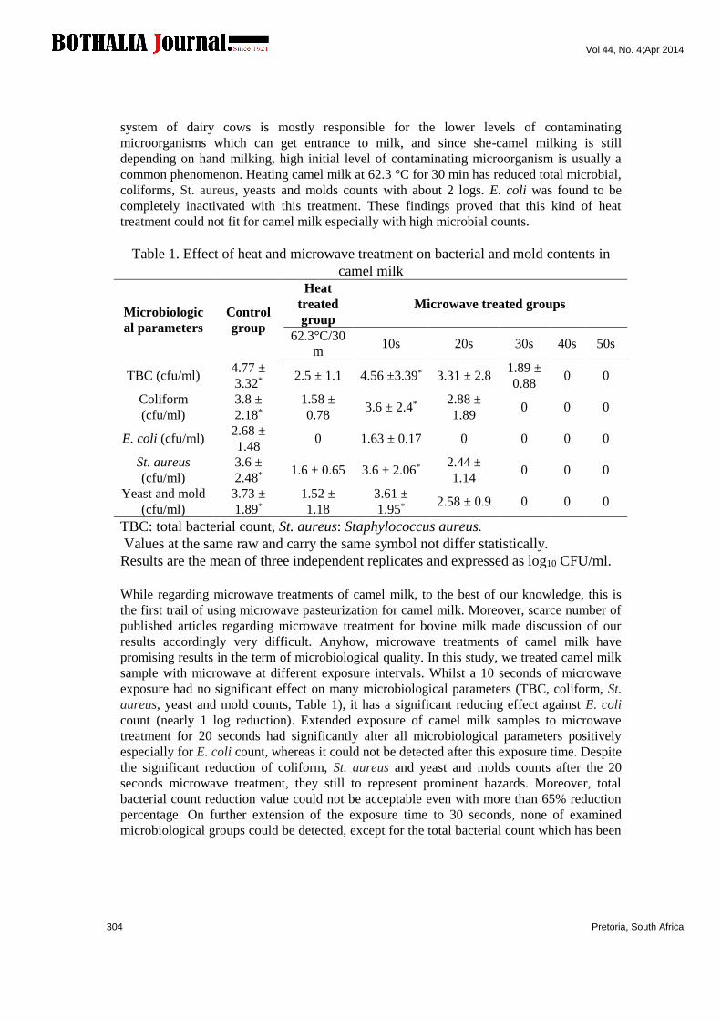
common phenomenon. Heating camel milk at 62.3 °C for 30 min has reduced total microbial,
coliforms, St. aureus, yeasts and molds counts with about 2 logs. E. coli was found to be
completely inactivated with this treatment. These findings proved that this kind of heat
treatment could not fit for camel milk especially with high microbial counts.
Table 1. Effect of heat and microwave treatment on bacterial and mold contents in
camel milk
Microwave treated groups
Heat
treated
group Control
group
Microbiologic
al parameters
50s 40s 30s 20s 10s 62.3°C/30
m
0 0 1.89 ±
0.88 3.31 ± 2.8 4.56 ±3.39* 2.5 ± 1.1
4.77 ±
3.32* TBC (cfu/ml)
0 0 0 2.88 ±
1.89 3.6 ± 2.4*
1.58 ±
0.78
3.8 ±
2.18*
Coliform
(cfu/ml)
0 0 0 0 1.63 ± 0.17 0 2.68 ±
1.48 E. coli (cfu/ml)
0 0 0 2.44 ±
1.14 3.6 ± 2.06* 1.6 ± 0.65
3.6 ±
2.48*
St. aureus
(cfu/ml)
0 0 0 2.58 ± 0.9 3.61 ±
1.95*
1.52 ±
1.18
3.73 ±
1.89*
Yeast and mold
(cfu/ml)
TBC: total bacterial count, St. aureus: Staphylococcus aureus.
Values at the same raw and carry the same symbol not differ statistically.
Results are the mean of three independent replicates and expressed as log10 CFU/ml.
While regarding microwave treatments of camel milk, to the best of our knowledge, this is
the first trail of using microwave pasteurization for camel milk. Moreover, scarce number of
published articles regarding microwave treatment for bovine milk made discussion of our
results accordingly very difficult. Anyhow, microwave treatments of camel milk have
promising results in the term of microbiological quality. In this study, we treated camel milk
sample with microwave at different exposure intervals. Whilst a 10 seconds of microwave
exposure had no significant effect on many microbiological parameters (TBC, coliform, St.
aureus, yeast and mold counts, Table 1), it has a significant reducing effect against E. coli
count (nearly 1 log reduction). Extended exposure of camel milk samples to microwave
treatment for 20 seconds had significantly alter all microbiological parameters positively
especially for E. coli count, whereas it could not be detected after this exposure time. Despite
the significant reduction of coliform, St. aureus and yeast and molds counts after the 20
seconds microwave treatment, they still to represent prominent hazards. Moreover, total
bacterial count reduction value could not be acceptable even with more than 65% reduction
percentage. On further extension of the exposure time to 30 seconds, none of examined
microbiological groups could be detected, except for the total bacterial count which has been
Vol 44, No. 4;Apr 2014
305 Pretoria, South Africa
reduced significantly (one log reduction). 40 seconds microwave exposure has reached with
microbiological quality of camel milk to nearly a sterile stage, as none of tested microbial
groups has yielded any count. Same phenomenon of microbial elimination was also
encountered at 50 seconds microwave exposure time. While there is no any work on
microwave pasteurization of camel's milk, its application for caw's milk was studied in the
last few years by some researches (Soler et al., 1995; Sieber et al., 1996; Asaad et al., 2013).
Microwave pasteurization of cow’s milk to reach 72°C for 15 seconds has reduced total
bacteria, psychotropic bacteria counts, E. coli ( Asaad et al., 2013). Microwave energy
inactivated milk pathogens as Yersinia enterocolitica, Listeria monocytogenes
Campylobacter jejuni, and, Enterococcus faecalis in cow's milk (Choi et al., 1993 a,b) and
decrease the count of milk spoilage bacteria (Géczi et al., 2013). Nasri et al. (2013)
investigated the lethal effect of microwave against Salmonella enterica serovar typhimurium
and found that microwave has a bactericidal effect and this would be explained by high
energy absorption by vital cell components (Rougier, 2003). Another theory (Khalil &
Villota, 1989) was linked to change in chemical structure of cellular lipids and protein.
Similar bactericidal effect was also reported against St. aureus and E. coli (Pothakamury et
al.,1995), Clostridium perfringens (Blanco & Dawson, 1974), Listeria monocytogenes
(Farber et al., 1998) and Lactobacillus plantarum (Shin and Pyun, 1997). Interestingly,
bactericidal effect of microwave in Nasri et al. (2013) trails was more prominently achieved
at 40 seconds exposure. These findings augmented our findings as the total elimination
(bactericidal effect) of microwave treatments was met at 40 seconds exposure. In another
study, Villamiel et al. (1996) used microwave to heat both bovine and ovine milk and they
recorded reduction in the total bacterial count (less than 2 log cfu/ml) using different
temperature/time combination. Clare et al. (2005) have reached to a sound conclusion based
on their bovine milk microwaving (continuous flow microwave system) and they stated that
microwave is a suitable sterilization method as milk was kept for 1 year without any sign of
microbial growth.
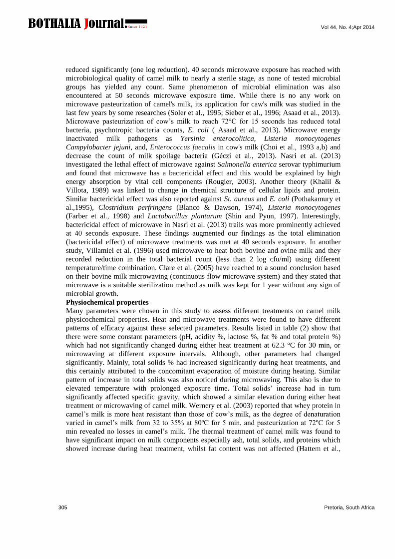
Physiochemical properties
Many parameters were chosen in this study to assess different treatments on camel milk
physicochemical properties. Heat and microwave treatments were found to have different
patterns of efficacy against these selected parameters. Results listed in table (2) show that
there were some constant parameters (pH, acidity %, lactose %, fat % and total protein %)
which had not significantly changed during either heat treatment at 62.3 °C for 30 min, or
microwaving at different exposure intervals. Although, other parameters had changed
significantly. Mainly, total solids % had increased significantly during heat treatments, and
this certainly attributed to the concomitant evaporation of moisture during heating. Similar
pattern of increase in total solids was also noticed during microwaving. This also is due to
elevated temperature with prolonged exposure time. Total solids’ increase had in turn
significantly affected specific gravity, which showed a similar elevation during either heat
treatment or microwaving of camel milk. Wernery et al. (2003) reported that whey protein in
camel’s milk is more heat resistant than those of cow’s milk, as the degree of denaturation
varied in camel’s milk from 32 to 35% at 80ºC for 5 min, and pasteurization at 72ºC for 5
min revealed no losses in camel’s milk. The thermal treatment of camel milk was found to
have significant impact on milk components especially ash, total solids, and proteins which
showed increase during heat treatment, whilst fat content was not affected (Hattem et al.,
Vol 44, No. 4;Apr 2014
306 Pretoria, South Africa
2011). Previous study showed that pasteurization of camel milk had very little effects on its
constituent (fat and protein) with slight increase in ash content (Wernery et al., 2005). Thus,
based on earlier report, which revealed that foodstuffs are less damaged with microwave
rather than regular heat treatments, microwave pasteurization looks promising for camel milk
(Albert et al., 2009).
Table 2. Physiochemical characters of camel milk
Microwave treated groups Heat
treated
group
Control
group
Paramet
ers
50s 40s 30s 20s 10s 62.3°C/30
m
6.3 ±
0.35
6.5 ±
0.5
6.2 ±
0.15
6.5 ±
0.3 6.3 ± 0.22 6.5 ± 0.21 6.4± o.2 pH
0.13 ±
0.011
0.14 ±
0.01
0.137 ±
0.12
0.13 ±
0.01
0.127 ±
0.02
0.12 ±
0.015
0.127 ±
0.015 Acidity
1.0197
± .007a
1.019 ±
.006ab
1.017 ±
0.006bc
1.016 ±
0.005cd
1.016 ±
0.001cd
1.016 ±
0.001cd
1.015 ±
0.002d
Specific
Gravity
0.7 ±
0.01a
0.69 ±
0.005ab
0.68 ±
0.01abc
0.67 ±
0.006bc
0.66 ±
0.005c
0.66 ±
0.03c
0.66 ±
0.02c Ash %
4.6 ±
0.09
4.57 ±
0.13
4.54 ±
0.17
4.5 ±
0.15 4.56 ± 0.1 4.5 ± 0.08
4.4 ±
0.09
Lactose
%
3.45 ±
0.06
3.42 ±
0.07
3.43 ±
0.06
3.41 ±
0.11
3.41 ±
0.052 3.47 ± 0.05 3.43 ±
0.06 Fat %
4.29 ±
0.1
4.25 ±
0.09
4.24 ±
0.1
4.2 ±
0.17
4.25 ±
0.06 4.16 ± 0.07
4.23 ±
0.06 Protein %
13.6 ±
2.5a
13.4 ±
1.3a
12.9 ±
1.5b
12.8 ±
1.3bc
12.5 ±
1.5cd 12.5 ± 1.3cd 12.3 ±
1.5d
Total
Solid
Each value represents M± SE of 10 samples. Means within the same raw carrying
different subscript (a,b,c,d) are significant at P˂0.05 Antioxidant and free radicals
Very little is known about the effect of heat on the antioxidant vitamins, minerals and thiol-
containing substances concentration of camel's milk. As it can be seen in table (3), vitamin C
and reduced glutathione are the most susceptible to degradation by heat. They were hardly
decreased in heat treatment in comparison to fresh milk. This degradation was certainly
attributed to either primary to oxidative effect or secondary to heat treatment (Wernery et al.,
2005). Conversely to Albert et al. (2009), the damage in vitamin C and GSH by microwave
pasteurization is less than that of heat treatment, may be due to less exposure time to heat in
microwave pasteurization. Fat-soluble vitamins A and E are relatively resistant to heat
treatment. A slight reduction was occurred in vitamin E in both treatment and this may be
caused by oxidation (Renner, 1983). While regarding mineral contents of camel milk, heat
generally has very little effect on camel's milk mineral content with exception of Cu and Zn
(Safaa et al., 2013; Wernery et al., 2003). Both Zn and Se were increased in our results by
heat or microwave treatment. Autoxidation may occur for milk lipids during processing with
formation of aldehyds, ketons and lactons (Frankel et al., 1987), trans fatty acids (Semma,
2002). Heat treatments significantly increased the concentrations of the oxidation markers as
Vol 44, No. 4;Apr 2014
307 Pretoria, South Africa
MDA in cow's milk compared to the fresh raw milk and correlated to the heating temperature
(Meshref & Al-Rowaily, 2008). Lipid peroxidation represented by MDA and protein
oxidation measured as nitric oxide were significantly increased by heat treatment and
correlated to the heating temperature in comparison with fresh raw milk, and the highest
increase was observed in microwave-pasteurized milk. MDA values for the microwave
heated milk was significantly different from all other heating methods (Meshref & Al-
Rowaily, 2008).
Table 3. Effect of heat and microwave treatment on camel milk antioxidants, free
radicals, insulin and IgG contents.
Microwave treated groups Heat
treated
group
Control
group
Parameter
s
50s 40s 30s 20s 10s 62.3°C/30
m
31 ±
2.9b
31.8 ±
3.3b
35.6 ±
2.2b
40.8 ±
3.2a
45.4 ±
4.6a
35.8 ±
3.2b
46.2 ±
9.7a
Vit.C
(mg/dl)
0.122
±0.0 3
0.124 ±
0.03
0.12 ±
0.007
0.128 ±
0.02
0.122 ±
0.022
0.126 ±
0.015
0.136 ±
0.0 2
Vit. A
(mg/L)
0.48 ±
0.02b
0.48 ±
0.01b
0.49 ±
0.02b
0.52 ±
0.02a
0.53 ±
0.01a
0.5 ±
0.02b
0.54 ±
0.03a
Vit. E
(mg/L)
112.3 ±
2.5c
125.7 ±
2.5b
130.3 ±
4ab
133 ±
16.5ab
140 ±
7.9a
132.3 ±
2.5ab
142.7 ±
3a
GSH
(μmol/ml
4.9 ±
0.7a
4.6 ±
0.6a
4.4 ±
0.7a
4.3 ±
0.7a 4 ± 0.6a 4.2 ±
0.64a
3.1 ±
0.23b Zn (PPM)
136.3 ±
1.5a
130 ±
4.5ab
127.3±
2.5ab
124.3 ±
1.5ab
123.7 ±
4.7ab
115.7 ±
12b
113.3 ±
20b Se (PPM)
0.46 ±
0.05a
0.41 ±
0.03b
0.38 ±
0.03bc
0.38 ±
0.02bc
0.352 ±
0.02c
0.22 ±
0.015e
0.172 ±
0.03f
MDA
(nmol/L)
0.55 ±
0.04a
0.49 ±
0.015b
0.40 ±
0.015cd
0.38 ±
0.01de
0.37 ±
0.015de
0.37 ±
0.01de
0.36 ±
0.01e
NO
(μmol/ml
37.4 ±
1.9a
37.6 ±
1.1a
38.8 ±
2a
39.2 ±
2.8a
40 ±
2.7a 40.4 ± 2a 41.2 ±
5.7a
Insulin
(μU/ml)
723.3 ±
25b
800 ±
43.6a
805 ±
43.6a
820 ±
10a
816.7 ±
15a
823.3 ±
25a
850 ±
50a IgG (μg/ml)
MDA; Malondialdehyde, GSH; Reduced glutathione, NO; Nitric Oxide. Each value
represents M± SE of 10 samples. Means within the same raw carrying different subscript (
a,b,c,d) are significant at P˂0.05
Insulin
Camel milk contains high levels of insulin (52 μU/ml) than cow milk (16.32 μU/ml) and
similar to human milk (60.23±41.05 μU/ml), so camel milk can be used for Diabetes mellitus
control (Agrawal et al., 2003). However, Wernery et al. (2006) reported that the insulin
content in camel milk is only slightly higher than in cow milk. Amel et al. (2012) found that
the pasteurization condition (63°C/30 min) not affected insulin level and its anti-diabetic
effect, but it decreased by boiling (100°C/30 min). At this study, insulin level was not
Vol 44, No. 4;Apr 2014
308 Pretoria, South Africa
affected by microwave pasteurization (Table 3), which certainly due loser expositor time.
Pasteurization, freeze drying or storage of camel milk at 4°C for 4 days as well as freezing at
-20 °C resulted in a statistically significant reduction in insulin concentrations; however, this
was minimal (Wernery et al., 2006).
Immunoglobulin G (IgG)
The importance of IgG in camel's milk has been emphasized by many authors to explain the
health related effects of camel milk (Farah, 1993; Konuspayeva et al., 2004). On average,
IgG concentration in camel colostrum seemed to be higher than that in other species (El-
Hatmi et al., 2006). Camel milk was found to be the richest source of IgG (Conesa et al.,
2008). Lactoproteins such as lactoferrin and IgG are known as health factors that play an
important role for milk consumers. In particular, camel milk, which is consumed in many
countries for its medicinal virtues, is renowned for being richer in some lactoproteins
compared with cow's milk (Konuspayeva et al., 2007). Camel milk IgG is a heat labile
compound and its denaturation midpoints for the mature milk heated for 30 min is 67·2°C
(Didier et al., 2006). The concentration of IgG in camel milk was slightly decreased during
heating at 62.3 °C for 30 mints. Whilst the detrimental effect of microwave treatment was
encountered only at exposure time for 50s (Table 3). Microwave pasteurization of milk was
reported to result in lower levels of denaturation of whey proteins compared to conventional
thermal processes and the denaturation of_ß-lactoglobulin was almost similar in both
processes (Villamiel et al., 1996). In general, Elagamy (2000) found that camel IgG is more
resistant to heating up to 100°C than that of cow’s milk.
Conclusion Microwave treatment at 40 sec can be used as an alternative pasteurization tools for camel
milk. Despite that microwave treatment likely affect the physiochemical properties of the
milk by the same degree as regular heat pasteurization, but the microwave method is more
advantageous in affecting the vital components in camel's milk.
Acknowledgments This work was funded by the Deanship of Scientific Research (DSR), King Abdulaziz
University, Jeddah, under grant No (965-005-D1434). The authors, therefore, acknowledge
with thanks DSR technical and financial support.
Author disclosures Ali Alkaladi, Mohamed Afifi, and Rania Kamal declare that they have no conflict of interest
References Afifi, E.M. (2010). Effect of Camel’s Milk on Cisplatin-Induced Nephrotoxicity in Swiss
Albino Mice. Amer J Biochem Biotechnol, 6, 141-147
Agrawal, R.P., Swami, S.C., Beniwal, R., Kochar, D.K., Sahani, M.S., Tuteja, F.C. &
Ghouri, S.K. (2003). Effect of camel milk on glycemic control, risk factors and diabetes
quality of life in type-1 diabetes: a randomised prospective controlled study. Journal of
Camel Practice and Research, 10,45–50.
Ajamaluddin, M., Abdulrahman, A., Ewa, S. & Jerzy, J. (2012). A study of the anti-diabetic
agents of camel milk. Int J Mol Med, 30, 585-592.
Albert,C.S., M´andoki, Z.S., Csapo-Kiss, Z.S. &Csapo, J. (2009). The effect of microwave
pasteurization on the composition of milk. Acta Univ. Sapientiae Aliment, 2,153-165.
Vol 44, No. 4;Apr 2014
309 Pretoria, South Africa
Amel, S., Touhami, K., Mongi, D., Aroua, A., Amel, D. & Omrane, B. (2012). Camel Milk
as Adjuvant to Treat Alloxan Diabetes: Effect of Heat Treatment on this Property. J
Diabetes Metab, 3,190-194.
AOAC. (1990a). Acidity of milk (Titrimetric method). In: Official Methods of Analysis, No.
947.05. Association of Official Analytical Chemists Inc. Virginia, USA.
AOAC. (1990b). Specific gravity of milk (pycnometer method). In: Official Methods of
Analysis, No.925.22. Association of Official Analytical Chemists Inc. Virginia, USA.
AOAC. (1990c).Solids (Total) in milk. In: Official Methods of Analysis, No.925.23.Association of Official Analytical Chemists Inc. Virginia, USA.
AOAC. (1990e). Ash of milk (Gravimetric method). In: Official Methods of Analysis. No.
945.46. Association of Official Analytical Chemists Inc: Virginia, USA.
AOAC. (2003). Official methods of analysis of AOAC International. 2nd revision (17th ed).
Gaithersburg, MD, USA: Association of Analytical communities.
Arya, S.P., Jain, P.& Mahajan, M. (2002). A new method for the ascorbic acid assay using
iron (II)-pyridine-2,6-dicarboxylic acid complex. Ann di Chim, 92, 1159-1164.
Asaad, R., Saeed, A. & Haider, A. (2013). Milk Flash Pasteurization by the Microwave and
Study its Chemical, Microbiological and Thermo Physical Characteristics. J Food Process
Technol, 4, 1-5.
Blanco, J.F. &Dawson, L.E. (1974). Survival of Clostridium perfringens on chicken cooked
with microwave energy. Poult Sci, 53, 1823-1830.
BSI. (2010). British Standards Institution. Milk, Dairy products, Food products, Food testing,
Chemical analysis and testing, Determination of content, Zinc, Atomic absorption
spectrophotometry, Flame atomic absorption spectrometry, Reproducibility.
BSI. (1990). Determination of the nitrogen content of liquid milk. In: Methods for Chemical
Analysis of Liquid Milk and Cream. BSI.1741, British Standards Institution, London, U.K.
Choi, H.K., Marth, E.H. & Vasavada, P.C. (1993a). Use of microwave energy to inactivate
Yersinia enterocolitica and Campylobacter jejuni in milk. Milchwissenschaft, 48, 134-136.
Choi, H.K., Marth, E.H. & Vasavada, P.C. (1993b). Use of microwave energy to inactivate
Listeria monocytogenes in milk. Milchwissenschaft, 48, 200-203.
Clare, D.A., Bang, W.S., Cartwright, G., Drake, M.A., Coronel, P. & Simunovic, J. (2005).
Comparison of Sensory, Microbiological, and Biochemical Parameters of Microwave versus
Indirect UHT Fluid Skim Milk During Storage. J Dairy Sci, 88,4172-4182.
Conesa, C., Sánchez, L., Rota, C., Pérez, M.D., Calvo, M., Farnaud, S. & Evans, R.W. 2008
Isolation of lactoferrin from milk of different species: Calorimetric and antimicrobial studies.
Comp Biochem Physiol B, 150, 131-139.
Didier, L., Annie, L., Halima, E. & Jean-Paul, R. (2006). Immunochemical quantification of
heat denaturation of camel ( Camelus dromedarius ) whey proteins. J Dairy Res, 73,1-9
Elagamy, E.I. (2000). Effect of heat treatment on camel milk proteins with respect to
antimicrobial factors: a comparison with cows' and buffalo milk proteins. Food Chem, 68,
227-232.
El-Hatmi, H., Levieux, A. & Levieux, D. (2006) Camel (Camelus dromedarius)
immunoglobulin G, α-lactalbumin, serum albumin and lactoferrin in colostrum and milk
during the early post partum period. J. Dairy Res, 73, 288-293.
Vol 44, No. 4;Apr 2014
310 Pretoria, South Africa
Erhard, M.H., Quistop, I., Vonschrummer, I., Jungling, A., Kaspers, B., Schmidi, P. & Kuhmann,
R. (1992). Development of specific enzyme linked immunosorbent antibody assay for detection of
immunoglobulin G,M&A using monoclonal antibodies. Poult Sci, 71, 302-310.
Farah, Z. (1993). Composition and characteristics of camel milk. J Dairy Res, 60, 603-626.
Farber, J.M., Aoust, J.Y.D., Diotte, M., Sewell, A., & Daley, E. (1998). Survival of
Listeria on raw whole chickens cooked in microwave ovens, J Food Prot, 61, 1465-
1469.
Frankel, E.N., Nash, A.M. & Synder, J.M. (1987). Methodology study to evaluate quality of
saybeans stored at different moisture levels. J Amer Oil Chem Soc, 64, 987-992.
Géczi, G., Horvath, M., Kaszab, T. & Alemany, G.G. (2013). No Major Differences Found
between the Effects of Microwave-Based and Conventional Heat Treatment Methods on
Two Different Liquid Foods. PLoS ONE, 8, e53720.
Hattem, H.E., Manal, A.N., Hanaa, S.S. & Elham, H.A. (2011). A study on the effect of
thermal treatments on composition and some properties of camel milk. J Brew Distill, 2, 51-
55.
James, C.S. (1995). Determination of the Fat Content of Dairy Products by the Gerber
Method. Analytical Chemistry of Food. Blackie Academic and Professional, an imprint of
Chapman and Hall, Glagow, U.K. pp. 93–5
Khalil, H. & Villota, R. (1989). The effect of microwave sublethal heating on the
ribonucleic acids of Staphylococcus aureus. J Food Prot, 52, 544-548.
Konuspayeva, G., Loiseau, G. & Faye, B. (2004). La plus-value “santé” du lait de chamelle
cru et fermenté: L’Expérience du Kazakhstan. In: Proc. 11th Rencontre autour des
Recherches sur les Ruminants, Institut National de la Recherche Agronomique. Paris, Dec.
8–9, 2004. Institut d’Elevage, Paris, France. 47-50.
Konuspayeva, G., Faye, B., Loiseau, G. & Levieux, D. (2007). Lactoferrin and
immunoglobin content in camel milk from Kazakhstan. J Dairy Sci, 90, 38-46.
Konuspayeva, G., Lemarie, E., Faye, B., Loiseau, G. & Montet, D. (2008). Fatty acid and
cholesterol composition of camel’s (Camelus bactrianus, Camelus dromedaries and hybrids)
milk in Kazakhstan. Dairy Science and Technology, 88, 327–340.
Lynch, J.M., Barbano, D.M. & Fleming, J.R. (2007). Determination of the lactose content of
fluid milk by spectrophotometric enzymatic analysis using weight additions and path length
adjustment: collaborative study. J AOAC Int, 90,196-216.
Magjeed, N.A. (2005). Corrective effect of milk camel on some cancer biomarkers in blood
of rats intoxicated with aflatoxin B1. Journal of the Saudi Chemical society, 9, 253–263.
Mal, G. & Pathak, K.M. (2010). Camel milk and milk products. SMSV' Dairy Year Book, pp.
97-103.
McKinnon, I.R., Yap, S.E., Augustin, M.A. & Hermar, Y. (2009). Diffusing-wave
spectroscopy investigation of heated reconstituted skim milks containing calcium chloride.
Food Hydrocolloid, 23, 1127-1133.
Meshref, A. & Al-Rowaily, A. (2008). Effect of Heating Treatments, Processing Methods
and Refrigerated Storage of Milk and Some Dairy Products on Lipids Oxidation. Pak J Nut,
7, 118-125.
Montgomery, H.A. & Dymock, J.F. (1961). The determination of nitrite in water. Analyst,
86, 414-416
Vol 44, No. 4;Apr 2014
311 Pretoria, South Africa
Nasri, K., Daghfous, D. & Landoulsi, A. (2013). Effects of microwave (2.45 GHz)
irradiation on some biological characters of Salmonella typhimurium. Comptes Rendus
Biologies, 336, 194-202.
Okhawa, H., Ohigni, N. & Yagi, K. (1979). Assay of lipid peroxides in animal tissues by
thiobarbituric acid reaction. Anal Biochem, 95, 351-358.
Pothakamury, U.R., Monslave-Gonzalez, A., Barbosa-Canovas, G.V. & Swanson, B.G.
(1995). Inactivation of Escherichia coli and Staphylococcus aureus in model foods by
pulsed electric field technology. Food Res Int, 28, 167-171.
Renner, E. (1983). Milk and diary products in human nutrition. Volkswirtsch Verl München,
291-296.
Rougier, C. (2003). Ètude des interactions entre la bactèrie Escherichia coli et les
micro-ondes appliquèes en mode discontinu dans des conditions faiblement thermiques.
PhD thesis, de l’universitè de Limoges, No 33.
Royatvand, S., Fallah, H.H., Ezzatpanah, H. & Sekehchi, M. (2013). Determination of
Insulin Concentration in Camel Milk Using UltraViolet –Visible Absorption Spectroscopy. J
Food Biosci Technol, 3, 53-60.
Rutkowski, M. & Krzysztof, G. (2007) Modifications of spectrophotometric methods for
antioxidative vitamins determination convenient in analytic practice. Acta Sci Pol Technol
Aliment, 6,17-28.
Safaa, O., Samia, H.A. & Salwa, M.K. (2013). Effect of Heat Treatment on Some Mineral
Status of Camel Milk. J Anim Sci, 2, 4-5.
Sedlak, J. & Lindsay, R.H. (1968). Estimation of total protein bound and non-protein
sulfhydryl groups in tissue with Ellmans reagent. Anal Biochem, 25, 293-98.
Semma, M. (2002). Trans fatty acids properties, benefit, and risks. J Health Sci, 48,7-13.
Shabo. Y., Barzel, R., Margoulis, M. & Yagil, R. (2005). Camel milk for food allergies in
children. Immunology and Allergy, 7, 796–798.
Shin, J.K. & Pyun, Y.R. (1997) Inactivation of lactobacillus plantarum by pulsed
microwave irradiation, J Food Sci, 62, 163-166.
Sieber, R., Eberhard, P. & Gallmann, P.U. (1996). Heat treatment of milk in domestic
microwave ovens. Int. Dairy J, 6, 231-246.
Soler, A., Ponsell, C., De Paz, M. & Nunez, M. (1995). The microbiological quality of milk
produced in the Balearic Islands. Int Dairy J, 5, 69-74.
Villamiel, M., Corzo, N., Martinez-Castro, I. & Olano, A. (1996). Chemical changes during
microwave treatment of milk. Food Chem, 56, 385–388.
Wernery, U., Hanke, B., Braun, F. & Johnson, B. (2003). The effect of heat treatment on
some camel milk constituents. Preliminary report. Milchwissenschaft, 58, 277-279.
Wernery, U., Johnson, B. & Abrahm, A. (2005) The effect of short-term heat treatment on
vitamin C concentration in camel milk. Milchwissenschaft, 60, 266-267.
Werney, U., Nagy, P., Bhai, I., Schiele, W. & Johnson, B. (2006) The effect of heat
treatment, pasteurization and different storage temperatures on insulin concentrations in
camel milk. Milchwissenschaft, 61, 25-28.