Embed Size (px)
Citation preview
1 23
Journal of Materials Science:Materials in Electronics ISSN 0957-4522 J Mater Sci: Mater ElectronDOI 10.1007/s10854-013-1164-8
Optical and magnetic properties of CuO/CuFe2O4 nanocomposites
M. M. Rashad, D. A. Rayan &A. A. Ramadan

1 23
Your article is protected by copyright and all
rights are held exclusively by Springer Science
+Business Media New York. This e-offprint is
for personal use only and shall not be self-
archived in electronic repositories. If you
wish to self-archive your work, please use the
accepted author’s version for posting to your
own website or your institution’s repository.
You may further deposit the accepted author’s
version on a funder’s repository at a funder’s
request, provided it is not made publicly
available until 12 months after publication.
Optical and magnetic properties of CuO/CuFe2O4 nanocomposites
M. M. Rashad • D. A. Rayan • A. A. Ramadan
Received: 11 December 2012 / Accepted: 26 February 2013
� Springer Science+Business Media New York 2013
Abstract CuO/c-CuFe2O4 nanocomposites have been
synthesized via the oxalate precursor route. Effect of syn-
thesis conditions on the crystal structure, microstructure,
magnetic and optical properties of the formed powders was
studied. The results indicated that pure CuO nanoparticles
were obtained from the oxalate precursor annealed at
600 �C for 2 h. However, substitution of Cu2? ion by Fe3?
ion (Cu1-XFeXO, where X = 0, 0.05, 0.1 and 0.2) led to
form of CuO/CuFe2O4 nanocomposites. The microstruc-
tures of the powders appeared as a monoclinic like shape.
Furthermore, the band gap energy of the obtained CuO
nanopowders was 1.41 eV and the value was slightly
decreased by Fe3? ion substitution. In addition, the formed
CuO particles had weak ferromagnetic characteristics.
However, the substitution Cu2? ion by Fe3? ion enhanced
the magnetic properties of the formed composite as the
result of increasing the CuFe2O4 phase formation. Hence,
the saturation magnetization was increased from 0.13 to
9.8 emu/g by increasing the Fe3? ion from 0 to 0.2.
1 Introduction
The focus on the correlation between the magnetic and
optical properties of magnetic-semiconductors composites
has a special attention in the recent years. Magnetic par-
ticles have the great potential applications in aircraft,
spacecraft, magnetic hard disks, telecommunication, mag-
netic recoding media and credit cards. On the other hand,
semiconductors have a wide range of applications in gas
sensing, catalysis, solar cells and electro-optical devices.
From this the point of view, copper oxide (CuO) as an
important p-type semiconductor with a narrow band gap
(1.2 eV), has been extensively studied because of its
diverse applications for heterogeneous catalysts, gas sen-
sors, optical switch, magnetic storage media, rectifiers,
lithium-ion electrode materials, field emission devices and
solar cells etc. [1–5].
However, the substitution of Cu2? ion by Fe3? ion leads
to form CuO/CuFe2O4 nanocomposite [6]. In particular,
copper ferrite has unique characteristics compared with the
other spinal ferrites. CuFe2O4 is known to exist in tetrag-
onal and cubic structures. Under slow cooling Cu-ferrite
crystallizes in a tetragonal structure with lattice parameter
ratio c/a of 1.06. Tetragonal Cu-ferrite phase has inverse
spinel structure with almost all Cu2? ions occupying
octahedral sublattice, whereas Fe3? ions divide equally
between the tetrahedral and octahedral sublattices. The
cations distribution in CuFe2O4 can be represented by
(CuxFe1-x)A(Cu1-xFe1?x)BO4, where x is the inversion
parameter and x = 0 and 1 stands for the inverse and
normal cases, respectively. The cubic structure is stable at
room temperature and transforms to tetragonal phase only
at a temperature of 360 �C and above due to Jahn–Teller
distortion. The distortion is directly related to the magnetic
properties. The cubic structure possesses a larger magnetic
moment than that of the tetragonal one, because there are
more cupric ions (Cu2?) at tetrahedral sites in cubic
structure as compared to that in the case of tetragonal
structure [7]. CuxFe3-xO4 has been chosen for its n-type
semiconducting property, as well as the possibility of
forming into nanocomposites thin films with CuO. By
M. M. Rashad (&) � D. A. Rayan
Central Metallurgical Research & Development Institute,
P.O. Box 87, Helwan 11421, Egypt
e-mail: [email protected]
A. A. Ramadan
Physics Department, Faculty of Sciences, Helwan University,
Helwan, Egypt
123
J Mater Sci: Mater Electron
DOI 10.1007/s10854-013-1164-8
Author's personal copy
changing the Cu and Fe contents, the material can be tuned
in a wide range of conductivities and altered from n- to
p-type [8].
The properties of the nanocomposites depend on the
microstructure which is related to the method of preparation.
The later plays a very important role with regard to the
chemical and structural properties of the composite. In fact,
the diverse available methods or routes to prepare magnetic-
CuO nanocomposites have been published. For example,
magnetic nanocomposites containing (NiFe2O4/CuO/FeO)
phases having particle size *17 nm were synthesized by a
sol–gel method [9]. Furthermore, CuO–CuFe2O4 nanocom-
posites have been synthesized through solid state reaction [6],
nitrate route [10] and sol gel method [3]. From our knowl-
edge, this is the first time for studying the optical and
magnetic properties of CuO–CuFe2O4 nanocomposite syn-
thesized via the oxalate precursor method. The oxalate pre-
cursor method involves the preparation of aqueous solution
containing cations, chelating of cations from solution by
adding carboxylic acid, followed by raising the temperature
of the solution until precursor formation. The precursor is
consequently calcined at low temperature, which is consid-
ered an advantage when compared to other methods men-
tioned earlier. The used method has similarities with the
combustion method, polymeric precursor method, acid sol
gel method (oxalate precursor, tartaric acid, lactic acid, etc.),
auto-combustion and also Pechini-type process (citrate pre-
cursor method). The carboxylic acid in this approach was not
only used to form stable complexes with starting metallic ions
but also as organic rich fuel. The process is simple, considers
an environmentally friendly method that offers scalability for
large scale production, uses low cost starting materials, low
synthesis temperature and yields homogeneous microstruc-
ture with narrow particle size distribution [11–13]. However,
it is necessary to enhance the magnetic properties of CuO for
magnetic storage media. Furthermore, the presence of
CuFe2O4 magnetic material during CuO as application pho-
tocatalyst helps in the regeneration of the catalyst after the
processing which can be easily removed from the solutions
[7]. The aim of the present work is to investigate the synthesis
of CuO–CuFe2O4 nanocomposites powders via the oxalate
precursor pathway. Moreover, the change in phase compo-
sition, microstructure, optical and magnetic properties of the
obtained materials is also investigated.
2 Experimental
The oxalate precursor method was applied for the prepa-
ration of CuO/CuFe2O4. Chemically grade copper chloride
CuCl2.2H2O, anhydrous ferric chloride FeCl3, and oxalic
acid H2C2O4 were used as starting materials. A certain
amount of oxalic acid related to stoichiometric ratio of
Cu2? ion was added gradually into the aqueous solution.
Than, the solution was stirred gently and evaporated at
80 �C till a clear, viscous gel was obtained, and then dried
at 110 �C for 24 h. The dry precursors were thermally
treated at a rate of 10 �C/min in static air in muffle furnace
with different maximum holding temperatures ranging
from 400 to 900 �C for time 2 h. To explore the effect of
substitution of Fe3? ion substituted CuO on the crystal
structure, microstructure and magnetic properties of the
formed particle, experiments were conducted at 600 �C for
2 h with different of Fe3? ion (CuXFe1-XO from 0 to 0.2)
where the molar ratio between the metal ion to oxalic acid
was 1:1 according to the following equations:
1� XCu2þ þ XFe2þ þ C2O2�4 CuC2O4ð Þ1�X FeC2O4ð ÞX
ð1ÞCuC2O4ð Þ1�X FeC2O4ð ÞXCuOþ CuFe2O4 þ 2CO2 þ 2CO
ð2Þ
The crystallite phases present in the different annealed
samples were identified by X-ray diffraction (XRD) on a
Brucker axis D8 diffractometer with crystallographic data
software Topas 2 using Cu-Ka (k = 1.5406 A) radiation
operating at 40 kV and 30 mA at a rate of 2�/min. The
diffraction data were recorded for 2h values between 20� and
80�. Fourier transform infrared spectroscopy (FTIR) was
conducted on a Thermo Electron Magna 760. Transmission
electron microscopy (TEM) of the particles were performed
using a JEOL-JEM-1230 microscope. The UV–VIS
absorption spectrum was recorded by a UV–VIS–NIR
spectrophotometer (Jasco-V-570 spectrophotometer, Japan).
The magnetic properties of the formed powders were
measured at room temperature using a vibrating sample
magnetometer (VSM; 9600-1 LDJ, USA) in a maximum
applied field of 16 kOe. From the obtained hysteresis
loops, the saturation magnetization (Ms), remanence mag-
netization (Mr) and the coercive field (Hc) were
determined.
3 Results and discussion
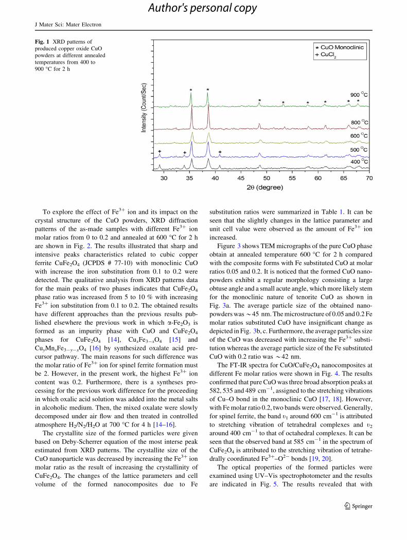
XRD patterns of the formed precursors using oxalic acid
molar ratio 1 related to Cu2? ions then annealed at different
temperatures from 400 to 900 �C for 2 h are given in
Fig. 1. It is clear that increasing the temperature led to
increase the crystallinity of copper (II) oxide CuO phase
formation. However, at low annealing temperature 400 and
500 �C, an impurity secondary phase of orthorhombic
eriochalacite CuCl2 (JCPDS # 76-0569) was formed with
tenorite monoclinic CuO phase ((JCPDS # 72-0629).
However, increasing the annealing temperature from 600 to
900 �C, single monoclinic CuO phase was indexed.
J Mater Sci: Mater Electron
123
Author's personal copy
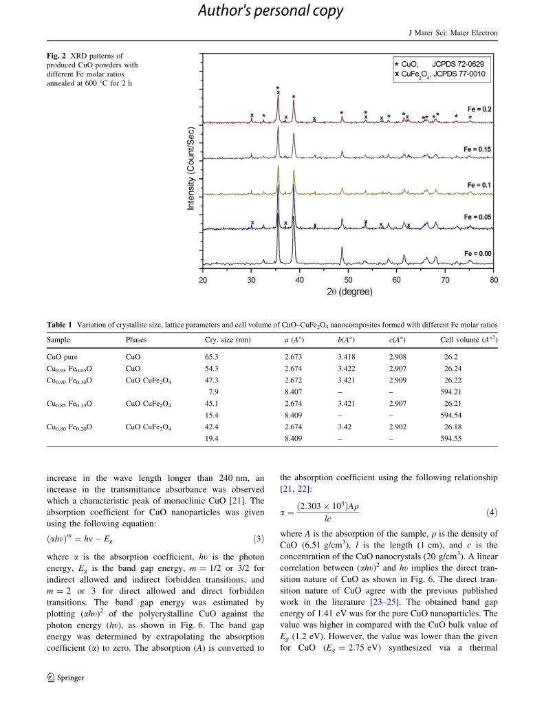
To explore the effect of Fe3? ion and its impact on the
crystal structure of the CuO powders, XRD diffraction
patterns of the as-made samples with different Fe3? ion
molar ratios from 0 to 0.2 and annealed at 600 �C for 2 h
are shown in Fig. 2. The results illustrated that sharp and
intensive peaks characteristics related to cubic copper
ferrite CuFe2O4 (JCPDS # 77-10) with monoclinic CuO
with increase the iron substitution from 0.1 to 0.2 were
detected. The qualitative analysis from XRD patterns data
for the main peaks of two phases indicates that CuFe2O4
phase ratio was increased from 5 to 10 % with increasing
Fe3? ion substitution from 0.1 to 0.2. The obtained results
have different approaches than the previous results pub-
lished elsewhere the previous work in which a-Fe2O3 is
formed as an impurity phase with CuO and CuFe2O4
phases for CuFe2O4 [14], CuxFe3-xO4 [15] and
CuyMnxFe3-y-xO4 [16] by synthesized oxalate acid pre-
cursor pathway. The main reasons for such difference was
the molar ratio of Fe3? ion for spinel ferrite formation must
be 2. However, in the present work, the highest Fe3? ion
content was 0.2. Furthermore, there is a syntheses pro-
cessing for the previous work difference for the proceeding
in which oxalic acid solution was added into the metal salts
in alcoholic medium. Then, the mixed oxalate were slowly
decomposed under air flow and then treated in controlled
atmosphere H2/N2/H2O at 700 �C for 4 h [14–16].
The crystallite size of the formed particles were given
based on Deby-Scherrer equation of the most interse peak
estimated from XRD patterns. The crystallite size of the
CuO nanoparticle was decreased by increasing the Fe3? ion
molar ratio as the result of increasing the crystallinity of
CuFe2O4. The changes of the lattice parameters and cell
volume of the formed nanocomposites due to Fe
substitution ratios were summarized in Table 1. It can be
seen that the slightly changes in the lattice parameter and
unit cell value were observed as the amount of Fe3? ion
increased.
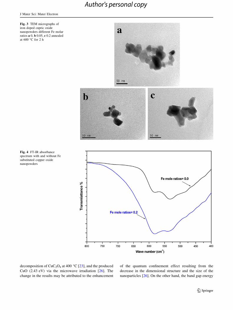
Figure 3 shows TEM micrographs of the pure CuO phase
obtain at annealed temperature 600 �C for 2 h compared
with the composite forms with Fe substituted CuO at molar
ratios 0.05 and 0.2. It is noticed that the formed CuO nano-
powders exhibit a regular morphology consisting a large
obtuse angle and a small acute angle, which more likely stem
for the monoclinic nature of tenorite CuO as shown in
Fig. 3a. The average particle size of the obtained nano-
powders was*45 nm. The microstructure of 0.05 and 0.2 Fe
molar ratios substituted CuO have insignificant change as
depicted in Fig. 3b, c. Furthermore, the average particles size
of the CuO was decreased with increasing the Fe3? substi-
tution whereas the average particle size of the Fe substituted
CuO with 0.2 ratio was *42 nm.
The FT-IR spectra for CuO/CuFe2O4 nanocomposites at
different Fe molar ratios were shown in Fig. 4. The results
confirmed that pure CuO was three broad absorption peaks at
582, 535 and 489 cm-1, assigned to the stretching vibrations
of Cu–O bond in the monoclinic CuO [17, 18]. However,
with Fe molar ratio 0.2, two bands were observed. Generally,
for spinel ferrite, the band t1 around 600 cm-1 is attributed
to stretching vibration of tetrahedral complexes and t2
around 400 cm-1 to that of octahedral complexes. It can be
seen that the observed band at 585 cm-1 in the spectrum of
CuFe2O4 is attributed to the stretching vibration of tetrahe-
drally coordinated Fe3?–O2- bonds [19, 20].
The optical properties of the formed particles were
examined using UV–Vis spectrophotometer and the results
are indicated in Fig. 5. The results revealed that with
Fig. 1 XRD patterns of
produced copper oxide CuO
powders at different annealed
temperatures from 400 to
900 �C for 2 h
J Mater Sci: Mater Electron
123
Author's personal copy
increase in the wave length longer than 240 nm, an
increase in the transmittance absorbance was observed
which a characteristic peak of monoclinic CuO [21]. The
absorption coefficient for CuO nanoparticles was given
using the following equation:
ðahvÞm ¼ hv� Eg ð3Þ
where a is the absorption coefficient, ht is the photon
energy, Eg is the band gap energy, m = 1/2 or 3/2 for
indirect allowed and indirect forbidden transitions, and
m = 2 or 3 for direct allowed and direct forbidden
transitions. The band gap energy was estimated by
plotting (aht)2 of the polycrystalline CuO against the
photon energy (ht), as shown in Fig. 6. The band gap
energy was determined by extrapolating the absorption
coefficient (a) to zero. The absorption (A) is converted to
the absorption coefficient using the following relationship
[21, 22]:
a ¼ ð2:303� 103ÞAqlc
ð4Þ
where A is the absorption of the sample, q is the density of
CuO (6.51 g/cm3), l is the length (1 cm), and c is the
concentration of the CuO nanocrystals (20 g/cm3). A linear
correlation between (aht)2 and ht implies the direct tran-
sition nature of CuO as shown in Fig. 6. The direct tran-
sition nature of CuO agree with the previous published
work in the literature [23–25]. The obtained band gap
energy of 1.41 eV was for the pure CuO nanoparticles. The
value was higher in compared with the CuO bulk value of
Eg (1.2 eV). However, the value was lower than the given
for CuO (Eg = 2.75 eV) synthesized via a thermal
Fig. 2 XRD patterns of
produced CuO powders with
different Fe molar ratios
annealed at 600 �C for 2 h
Table 1 Variation of crystallite size, lattice parameters and cell volume of CuO–CuFe2O4 nanocomposites formed with different Fe molar ratios
Sample Phases Cry. size (nm) a (A�) b(A�) c(A�) Cell volume (A�3)
CuO pure CuO 65.3 2.673 3.418 2.908 26.2
Cu0.95 Fe0.05O CuO 54.3 2.674 3.422 2.907 26.24
Cu0.90 Fe0.10O CuO CuFe2O4 47.3 2.672 3.421 2.909 26.22
7.9 8.407 – – 594.21
Cu0.85 Fe0.15O CuO CuFe2O4 45.1 2.674 3.421 2.907 26.21
15.4 8.409 – – 594.54
Cu0.80 Fe0.20O CuO CuFe2O4 42.4 2.674 3.42 2.902 26.18
19.4 8.409 – – 594.55
J Mater Sci: Mater Electron
123
Author's personal copy
decomposition of CuC2O4 at 400 �C [23], and the produced
CuO (2.43 eV) via the microwave irradiation [26]. The
change in the results may be attributed to the enhancement
of the quantum confinement effect resulting from the
decrease in the dimensional structure and the size of the
nanoparticles [26]. On the other hand, the band gap energy
Fig. 3 TEM micrographs of
iron doped cupric oxide
nanopowders different Fe molar
ratios a 0, b 0.05, c 0.2 annealed
at 600 �C for 2 h
Fig. 4 FT-IR absorbance
spectrum with and without Fe
substituted copper oxide
nanopowders
J Mater Sci: Mater Electron
123
Author's personal copy
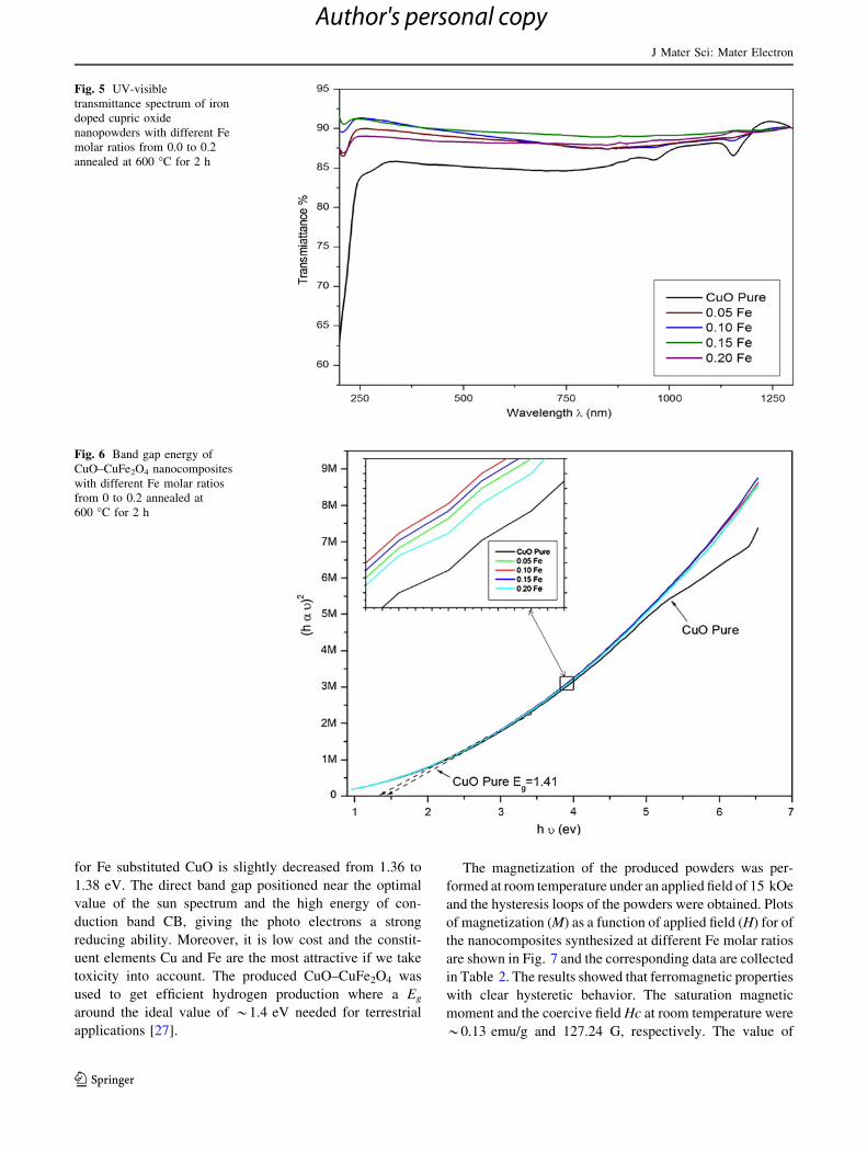
for Fe substituted CuO is slightly decreased from 1.36 to
1.38 eV. The direct band gap positioned near the optimal
value of the sun spectrum and the high energy of con-
duction band CB, giving the photo electrons a strong
reducing ability. Moreover, it is low cost and the constit-
uent elements Cu and Fe are the most attractive if we take
toxicity into account. The produced CuO–CuFe2O4 was
used to get efficient hydrogen production where a Eg
around the ideal value of *1.4 eV needed for terrestrial
applications [27].
The magnetization of the produced powders was per-
formed at room temperature under an applied field of 15 kOe
and the hysteresis loops of the powders were obtained. Plots
of magnetization (M) as a function of applied field (H) for of
the nanocomposites synthesized at different Fe molar ratios
are shown in Fig. 7 and the corresponding data are collected
in Table 2. The results showed that ferromagnetic properties
with clear hysteretic behavior. The saturation magnetic
moment and the coercive field Hc at room temperature were
*0.13 emu/g and 127.24 G, respectively. The value of
Fig. 5 UV-visible
transmittance spectrum of iron
doped cupric oxide
nanopowders with different Fe
molar ratios from 0.0 to 0.2
annealed at 600 �C for 2 h
Fig. 6 Band gap energy of
CuO–CuFe2O4 nanocomposites
with different Fe molar ratios
from 0 to 0.2 annealed at
600 �C for 2 h
J Mater Sci: Mater Electron
123
Author's personal copy
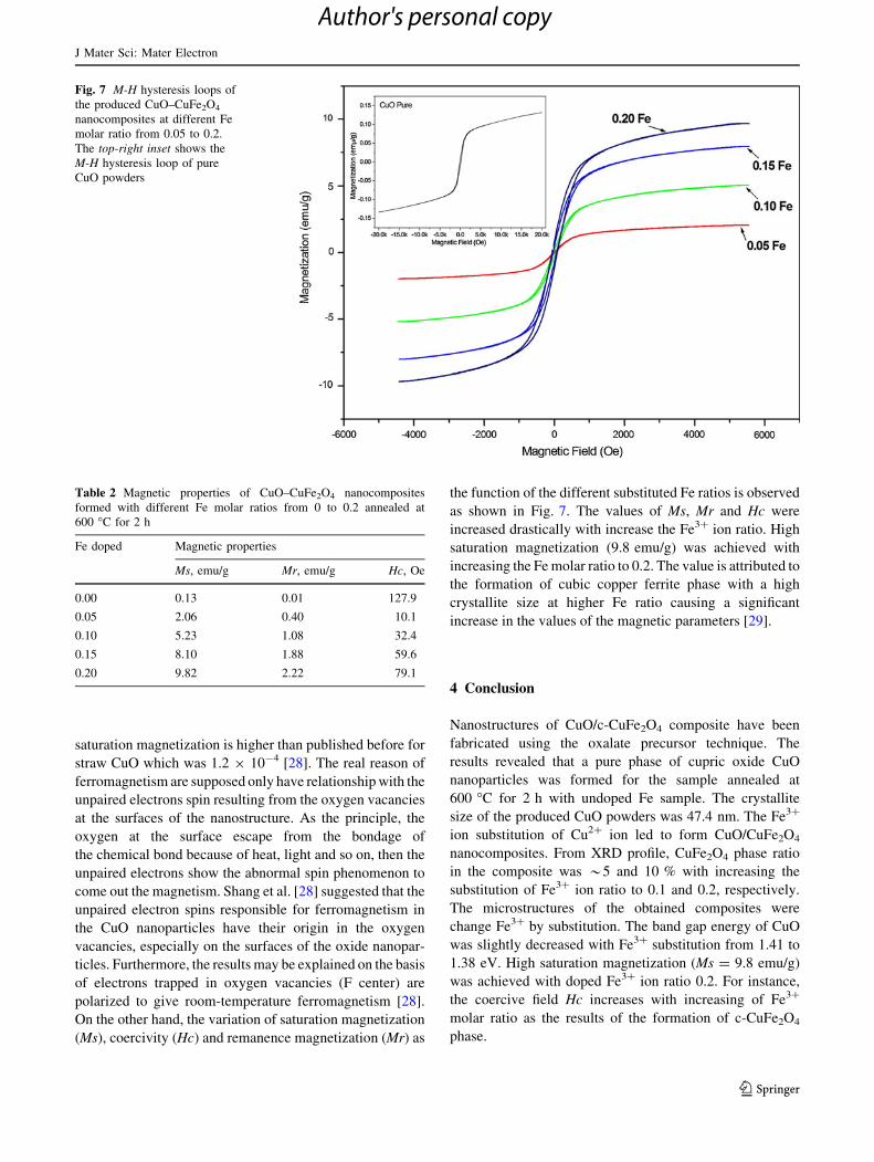
saturation magnetization is higher than published before for
straw CuO which was 1.2 9 10-4 [28]. The real reason of
ferromagnetism are supposed only have relationship with the
unpaired electrons spin resulting from the oxygen vacancies
at the surfaces of the nanostructure. As the principle, the
oxygen at the surface escape from the bondage of
the chemical bond because of heat, light and so on, then the
unpaired electrons show the abnormal spin phenomenon to
come out the magnetism. Shang et al. [28] suggested that the
unpaired electron spins responsible for ferromagnetism in
the CuO nanoparticles have their origin in the oxygen
vacancies, especially on the surfaces of the oxide nanopar-
ticles. Furthermore, the results may be explained on the basis
of electrons trapped in oxygen vacancies (F center) are
polarized to give room-temperature ferromagnetism [28].
On the other hand, the variation of saturation magnetization
(Ms), coercivity (Hc) and remanence magnetization (Mr) as
the function of the different substituted Fe ratios is observed
as shown in Fig. 7. The values of Ms, Mr and Hc were
increased drastically with increase the Fe3? ion ratio. High
saturation magnetization (9.8 emu/g) was achieved with
increasing the Fe molar ratio to 0.2. The value is attributed to
the formation of cubic copper ferrite phase with a high
crystallite size at higher Fe ratio causing a significant
increase in the values of the magnetic parameters [29].
4 Conclusion
Nanostructures of CuO/c-CuFe2O4 composite have been
fabricated using the oxalate precursor technique. The
results revealed that a pure phase of cupric oxide CuO
nanoparticles was formed for the sample annealed at
600 �C for 2 h with undoped Fe sample. The crystallite
size of the produced CuO powders was 47.4 nm. The Fe3?
ion substitution of Cu2? ion led to form CuO/CuFe2O4
nanocomposites. From XRD profile, CuFe2O4 phase ratio
in the composite was *5 and 10 % with increasing the
substitution of Fe3? ion ratio to 0.1 and 0.2, respectively.
The microstructures of the obtained composites were
change Fe3? by substitution. The band gap energy of CuO
was slightly decreased with Fe3? substitution from 1.41 to
1.38 eV. High saturation magnetization (Ms = 9.8 emu/g)
was achieved with doped Fe3? ion ratio 0.2. For instance,
the coercive field Hc increases with increasing of Fe3?
molar ratio as the results of the formation of c-CuFe2O4
phase.
Fig. 7 M-H hysteresis loops of
the produced CuO–CuFe2O4
nanocomposites at different Fe
molar ratio from 0.05 to 0.2.
The top-right inset shows the
M-H hysteresis loop of pure
CuO powders
Table 2 Magnetic properties of CuO–CuFe2O4 nanocomposites
formed with different Fe molar ratios from 0 to 0.2 annealed at
600 �C for 2 h
Fe doped Magnetic properties
Ms, emu/g Mr, emu/g Hc, Oe
0.00 0.13 0.01 127.9
0.05 2.06 0.40 10.1
0.10 5.23 1.08 32.4
0.15 8.10 1.88 59.6
0.20 9.82 2.22 79.1
J Mater Sci: Mater Electron
123
Author's personal copy
References
1. J.O. Park, K.Y. Rhee, S.J. Park, Appl. Surf. Sci. 256, 6945 (2010)
2. X. Wang, C. Hu, H. Liu, G. Du, X. He, Y. Xi, Sens. Actuators B:
Chem. 144, 220 (2010)
3. S.P. Meshram, P.V. Adhyapak, U.P. Mulik, D.P. Amalnerkar,
Chem. Eng. J. 204–206, 158 (2012)
4. Z. Yang, D. Wang, F. Li, D. Liu, P. Wang, X. Li, H. Yue, S.
Peng, D. He, Mater. Lett. 90, 4 (2013)
5. S. Rehman, A. Mumtaz, S.K. Hasanain, J. Nanopart. Res. 13,
2479 (2011)
6. K.L. Liu, S.L. Yuan, H.N. Duan, S.Y. Yin, Z.M. Tian, X.F.
Zheng, S.X. Huo, C.H. Wang, Mater. Lett. 64, 192 (2010)
7. M.M. Rashad, R.M. Mohamed, M.A. Ibrahim, L.F.M. Ismail,
E.A. Abdel-Aal, Adv. Powder Technol. 23, 315 (2012)
8. A. Chapelle, M.H. Yaacob, I. Pasquet, L. Presmanes, A. Barnabe,
Ph. Tailhades, J. Du Plessis, K.K. Zadeh, Sens. Actuators B:
Chem. 153, 117 (2011)
9. M. Srivastava, A.K. Ojha, S. Chaubey, J. Singh, J. Solid State
Chem. 183, 2669 (2010)
10. S.J. Stewart, G.F. Goya, G. Punte, R.C. Mercader, J. Phy. Chem.
Solids. 58, 73 (1997)
11. M.M. Rashad, Z.I. Zaki, H. El-Shall, J. Mater. Sci. 44, 2992
(2009)
12. R.M. Mohamed, M.M. Rashad, F.A. Haraz, W. Sigmund, J.
Magn. Magn. Mater. 322, 2058 (2010)
13. M.M. Rashad, J. Mater. Sci.: Mater. Electron. 23, 882 (2012)
14. C. Villette, Ph. Tailhades, A. Rousset, C.R. Acad, Sci. Paris 316,
1717 (1993)
15. E. Kester, B. Gillot, P. Perriat, Ph. Dufour, C. Villette, Ph.
Tailhades, A. Rousset, J. Solid State Chem. 126, 7 (1996)
16. B. Gillot, N. Nivoix, E. Kester, O. Nusillard, C. Villette, Ph.
Tailhades, A. Rousset, Mater. Chem. Phys. 48, 111 (1997)
17. M.A. Dar, Y.S. Kim, W.B. Kim, J.M. Sohn, H.S. Shin, Appl.
Surf. Sci. 254, 7477 (2008)
18. R.K. Selvan, C.O. Augustin, L.J. Berchmans, R. Saraswathi,
Mater. Res. Bull. 38, 41 (2003)
19. W. Ponhan, S. Maensiri, Solid State Sci. 11, 479 (2009)
20. M. Faisal, S.B. Khan, M.M. Rahman, A. Jamal, A. Umar, Mater.
Lett. 65, 1400 (2011)
21. S. N. Karthick, K. Prabakar, A. Subramania, Ji-Tae Hong, J.-J.
Jang and H.-J. Kim, Powder. Technol. 205, 36 (2011)
22. M.M. Rashad, R. Roshdi, K. El-Barawy, A.T. Kandil, Min.
Process. Ext. Metall. 120, 156 (2011)
23. X. Zhang, D. Zhang, X. Ni, H. Zheng, Solid State Electr. 52, 245
(2008)
24. C. Yang, X. Su, F. Xiao, J. Jian, J. Wang, Sens. Actuators B:
Chem. 158, 299 (2011)
25. Y. Li, X.-Y. Yang, J. Rooke, G.V. Tendeloo, B.-L. Su, J. Colloid
Interface Sci. 348, 303 (2010)
26. S.K. Maji, N. Mukherjee, A. Mondal, B. Adhikary, B. Karmakar,
J. Solid State Chem. 183, 1400 (2010)
27. A. Kezzim, N. Nasrallah, A. Abdi, M. Trari, Eng. Conv. Manag.
52, 2800 (2011)
28. D. Shang, K. Yu, Y. Zhang, J. Xu, J. Wu, Y. Xu, L. Li, Z. Zhu,
Appl. Surf. Sci. 255, 4093 (2009)
29. M. Srivastava, A.K. Ojha, S. Chaubey, P.K. Sharma, A.C.
Pandey, J. Alloys Compd. 494, 275 (2010)
J Mater Sci: Mater Electron
123
Author's personal copy


