Embed Size (px)
Citation preview

J. Insecf Physiol. Vol. 34, No. 4, pp. 337-345, 1988 Printed in Great Britain. All rights reserved
0022-1910/88 $3.00 -I- 0.00 Copyright Q 1988 Pergamon Press plc
OPTIMIZATION OF A MONOLAYER PHAGOCYTOSIS ASSAY AND ITS APPLICATION FOR STUDYING THE
ROLE OF THE PROPHENOLOXIDASE SYSTEM IN THE WAX MOTH, GALLERIA MELLONELLA
JAYNE L. BROOKMAN, NORMAN A. RATCLIFFE* and ANDREW F. ROWLEY Biomedical and Physiological Research Group, School of Biological Sciences, University College of
Swansea, Singleton Park, Swansea SA2 8PP. Wales
(Received 21 July 1987; revised 9 September 1987)
Abstract-The optimization of an assay for the measurement of in Crro phagocytosis in monolayers of Gull&~ mellonellu haemocytes is described. The effect of changing various parameters within the assay was tested and, of these, incubation temperature and calcium ion concentration were found to exert the major influence on the phagocytosis of erythrocytes by Galleria haemocytes. Further evidence is presented for the involvement of the prophenoloxidase system within the phagocytic process of insects. Laminarin, a /I-1,3-glucan which activates th,s enzyme cascade and stimulates the phagocytosis of bacteria by Galleria haemocytes, was shown to enhance the ingestion of human erythrocytes. This enhancement was not due to the laminarin acting as a bridging molecule between the test particle and the haemocyte surface since preincubation of erythrocytes and bacteria with laminarin failed to enhance phagocytosis. This apd other results strongly suggest that the stimulation of phagocytosis by the /3-1,3-glucan is due to the ac;vation of the prophenoloxidase cascade. This is further substantiated by treating bacteria with a laminarin- activated haemocyte lysate supernatant preparation containing various components of the prophenol- oxidase system, since these were phagocytosed to a greater extent than those pretreated and incubated with a non-activated preparation or with laminarin alone. These experiments further point to a possible opsonic function for the prophenoloxidase system in insects.
Key Word Index: Haemocytes, in vitro phagocytosis optimization, prophenoloxidase system, immuno- recognition, Gulleriu mellonella
INTRODUCTION
Phagocytosis is a vital process in the host defences of invertebrates against microbial infection. Factors in the blood of these animals, including agglutinins, have been shown to be important in the recognition of foreign material by the leucocytes which in turn leads to a more effective uptake of such particles (e.g. Renwrantz and Stahmer, 1983). It has been proposed that the prophenoloxidase system is also responsible, at least in part, for this recognition process in crus- taceacs (Smith and SdderhBll, 1983a,b; Sijderhall et al., 1986) and in insects (Ratcliffe ef al.. 1984; Leonard et al., 1985).
Activation of the prophenoloxidase system of ar- thropods generates not only melanin but also sticky proteins which apparently have opsonic activity (SaderhBll et al., 1984). Thus, there is significant enhancement of phagocytosis in vitro in the presence of the fi-1,3-glucan, laminarin, which is an activator of the prophenoloxidase system (Smith and SGderhBll, 1983a; Ratcliffe et al., 1984; Leonard et al., 1985). Concomitantly, inhibitors of the proph- enoloxidase complex abolish this stimulatory effect while carbohydrates incapable of activating this sys- tem also fail to enhance particle uptake (Smith and SBderhill, 1983a; Leonard et al., 1985).
Other explanations, not involving the prophenol- oxidase system, could account for the enhanced
*To whom correspondence should be addressed.
phagocytosis after exposure to laminarin. For exam- ple, as reported in human monocytes (Czop et al., 1985), the fi-1,3-glucan may bind to a receptor on the cell membrane to directly activate the cells. This possibility, however, has been eliminated by the work of Leonard et al. (1985) in which pretreatment of Galleriu haemocyte monolayers with laminarin, prior to overlaying with bacteria, failed to stimulate micro- bial ingestion by the blood cells. Alternatively, the laminarin may be acting as a bridging molecule, binding to both the cell membrane and the outside of the microorganism, thereby increasing phagocytosis by enhancing attachment.
The present study examines this latter bridging hypothesis for the mode of enhancement of phago- cytosis by laminarin using both human erythrocytes and bacteria as test particles in the assay. Erythro- cytes are particularly useful as they are less likely to have binding sites for laminarin on their cell mem- branes. In addition, since in preliminary experiments with erythrocytes, ingestion levels by the haemocytes were extremely low (about l.S%), we also describe the optimization of a phagocytosis assay in order to mimic more closely the immune potential of haemo- cytes in vivo (see Rabinovitch and De Stefano, 1970).
MATERIALS AND METHODS
Insects
Final-instar larvae of the waxmoth, Galleriu mel- lone&a (0.24-0.26 g). were used for all phagocytosis
337

338 JAYNE L. BROOKMAN et al.
experiments. These were reared as described pre- viously (Ratcliffe and Rowley, 1975).
Erythroeytes
Fixed human type-B erythrocytes were prepared from blood supplied by Singleton Hospital, Swansea, Wales. The erythrocytes were fixed for 12 h in 2.5% Tris-buffered glutaraldehyde (10 mM Tris-HCl, pH 7) and washed several times with 0.9% NaCI. Free aldehyde groups were blocked using 0.3 M ethanolamine and the cells were rewashed and resuspended in Grace’s Insect Medium or in a Tri-saline (0.075 M Tri-HCI, pH 6.5, adjusted to 440 mOsm kg-’ with NaCl). A concentration of I x IO” erythrocytes ml-’ was used to overlay haemocyte monolayers (erythrocyte: haemocyte ratio about 20: I).
Bacterial cultures
The type strain Bacillus cereus (NCIB 9373, Torry Research Station, Aberdeen, Scotland) was grown up in nutrient broth (Difco Ltd, England) with constant shaking for 18 h at 37°C. The bacteria were heat- killed (IOOC for 10 min), washed twice with 0.9% NaCl solution and resuspended in Tris-saline (see above). The suspension was then forced through a 26G sterile needle to break up any clumps of bacteria. A concentration of 2.5 x lo6 bacteria ml-i was used to overlay haemocyte monolayers (bacteria : haemocyte ratio about 50: 1).
Chemicals, solutions and glassware
A laminarin (Sigma) solution (1 mg ml-‘) was prepared using pyrogen-free water (Travenol Labora- tories) with heating (100°C for 5 min) to aid dis- solution. The final concentration of laminarin in experimental monolayers was 0.1 mg ml-‘.
Haemocyte monolayers were maintained in Grace’s Insect Medium or in Tris-saline (0.075 M Tri-HCI, pH 6.5, 440 mOsm kg-‘; referred to below as the monolayer buffer) which was prepared using pyrogen-free water and filter sterilized.
All glassware was washed in Decon (BDH, England) and heat-treated (18O‘C for 24 h) to ren- der it pyrogen-free as endotoxin has been shown to activate the prophenoloxidase system in crustaceans (Siiderhlll and Hall, 1984), and to enhance phago- cytosis of B. cereus in G. mellonella haemocyte mono- layers (Ratcliffe et al., 1984). Plastics were washed with E-toxa Clean (Sigma) to render them pyrogen- free.
Bleeding, haemocyte monolayer preparation and phagocytosis assal
The larvae were chilled, surface sterilized and then bled by proleg puncture into endotoxin-free 1 ml glass tubes filled with ice-cold Grace’s Insect Medium or monolayer buffer.
Monolayers were formed by pipetting about 5 x IO4 haemocytes, in 50~1, onto coverslips and allowing the cells to attach for 5min at room tem- perature before washing off non-adherent cells with Grace’s Insect Medium or monolayer buffer. The monolayers were then overlaid with erythrocytes or bacteria suspended in 25 ~1 Grace’s Insect Medium or overlay buffer (a T&-saline or varying compos-
ition, see below), incubated for 1 h, rinsed to remove any non-adherent particles and finally fixed in form- aldehyde vapour for 20min. The monolayers were then examined immediately or stored at 4°C until use. Haemocyte viability was tested after incubation with test particles by immersion of the monolayer in 0.2% nigrosine solution for 2min and rinsing in Grace’s Insect Medium or overlay buffer before fixation.
In each experiment, control monolayers were over- laid with bacteria or erythrocytes alone, while in experimental monolayers the test particles were sus- pended in overlay buffer containing 0.1 mg ml-’ laminarin. Monolayers were prepared in duplicate and examined using the phase-contrast optics of a Leitz SM-Lux microscope. The number of haemo- cytes phagocytic, the number of particles phago- cytosed per 100 haemocytes, the number of haemo- cytes showing attached particles, and the number of particles attached per 100 haemocytes were deter- mined for the different types of cells present on the monolayers. Monolayers were scored blind and at least 300 cells counted per monolayer. Intracellular bacteria were distinguished from extracellular forms by the criteria outlined by Rowley and Ratcliffe (1980).
The phagocytosis figures for the control and ex- perimental monolayers were compared using a paired t-test. Where appropriate, experimental monolayers using different treatments were compared using an independent t-test. The significance level was set at P ,< 0.05 in all cases.
Optimization of phagocytosis assa,v
Initial experiments using erythrocytes as test par- ticles showed that, although laminarin enhanced phagocytosis, the rates were very low compared with bacteria (Leonard et al., 1985). Attempts were there- fore made to optimize the assay by manipulating the incubation temperature and medium, calcium ion concentration and pH of the monolayers.
A. Incubation temperature
To determine the effect of temperature on rates of phagocytosis, cell monolayers were prepared using Grace’s Insect Medium and incubated at 23, 30, 35 or 40°C. As a result of these experiments, the tem- perature range was narrowed and monolayers incu- bated at 30, 31.5, 33 or 34.5”C. The optimal tem- perature of 31.5”C was then used for incubation in all subsequent experiments.
B. Incubation medium
In an attempt to improve the rate of phagocytosis obtained with erythrocytes, Grace’s Insect Medium was replaced by the monolayer and overlay buffers (detailed above and below, respectively) which pro- vided a simple system allowing manipulation of various physicochemical parameters in the mono- layers. The monolayer buffer was calcium free to reduce spreading of the haemocytes on the coverslip before addition of the test particles.
C. Calcium ion concentration of overlay buffer
In order to determine the effect of calcium ions (Ca2+ ) on the rate of phagocytosis, monolayers were prepared using calcium-free monolayer buffer

Phagocytosis and the prophenoloxidase system 339
(0.075 M Tri-HCI, pH 6.5, adjusted to 440mOsm kg-’ with NaCI). These were then overlaid with erythrocytes suspended in an identical Tris-saline (the overlay buffer) containing 0, 1, 10, or 30 mM Ca2+. The optimal Ca2+ concentration, lOmM, was used in the overlay buffer for all subsequent experiments.
D. pH of overlay buffer
To determine the effect of the pH of the overlay buffer on the phagocytosis rate, monolayers were prepared using Ca*+-free monolayer buffer, pH 6.5 (as in C above), and overlaid with erythrocytes suspended in an identical Tri-saline with 10 mM Ca2+ and at pH 6.5, 7.0 or 7.5. This relatively narrow pH range was used, as preliminary experiments showed that reduced levels of adherence of erythro- cytes to G. mellonella haemocytes occurred at pH 4-5, while cell degranulation and vacuolation increased in monolayers incubated with an overlay buffer at pH 8-9. In all subsequent experiments, a pH of 7.0 was employed for the overlay buffer.
E. pH of monolayer buffer
In order to determine the effect, if any, of changes in the pH of the monolayer buffer, three sets of monolayers were prepared using Ca2+-free mono- layer buffer at pH 6.5, 7.0 or 7.5. Control and experi- mental monolayers were overlaid with erythrocytes suspended in overlay buffer, pH 7.0, containing 10 mM Ca’+. In all subsequent experiments, a pH of 6.5 was employed for the monolayer buffer.
Bacteria as test particles
In a number of experiments with the Tris-saline system optimized above, erythrocytes were replaced by B. cereus as a test particle. This allowed com- parison to be made more closely with the results of Ratcliffe et al. (1984) and Leonard et at. (1985) who also used this bacteria1 species but who set up and incubated the Galleria haemocyte monolayers in Grace’s Insect Medium at 25-26°C. Monolayers were thus prepared using Ca2 + -free monolayer buffer, pH 6.5, overlaid with bacteria suspended in overlay buffer, containing 10 mM Ca’+, pH 7, and incubated at 25 or 31.5”C.
Preincubation of erythrocytes and bacteria with laminarin
To determine whether laminarin enhances phago- cytosis by crosslinking the phagocytic haemocytes with the test particles, erythrocytes or B. cereus were preincubated in Ca2+-free monolayer buffer, pH 6.5, with or without laminarin (0.1 mg ml-‘) for 1 h at 30°C. They were then washed and resuspended in overlay buffer (containing 10 mM Ca*+, pH 7), with or without laminarin and pipetted onto the haemocyte monolayers, and incubated for 1 h at 31.5”C.
Preincubation of bacteria with haemocyte lysate super- natant
In an attempt to detect any possible opsonic effects of the various components of the prophenoloxidase pathway, bacteria were suspended in a Galleria hae- mocyte lysate supernatant, prior to overlaying mono-
layers. The haemocyte lysate supernatant was pre- pared in 0.01 M sodium cacodylate buffer with 1OmM Ca2+, pH 7 as described by Leonard et al. (1985). Bacteria were preincubated for 1 h at 30°C in either haemocyte lysate supernatant with or without laminarin, or in cacodylate buffer with or without laminarin, and then overlaid without further rinsing onto haemocyte monolayers prepared as above in Ca2+-free monolayer buffer, pH 6.5. The activity of the haemocyte lysate supernatant preparations was routinely checked using a spot assay as described by Unestam and Nyhlen (1974). Cacodylate buffer was used to make the haemocyte lysate supernatant as initial experiments using a Tris buffer failed to give an active preparation.
RESULTS
Cell types and haemocyte viability in vitro
Plasmatocytes and granular cells in the ratio of about 1.5 : 1 made up >95% of the Galleria mono- layers. In all monolayer preparations, unless other- wise specified, the plasmatocyte and granular cell viabilities were >95% and > 85%, respectively, after the 1 h incubation period employed.
Phagocytosis of erythrocytes
Erythrocytes were phagocytosed by a significantly higher number of haemocytes in the presence of laminarin compared with the controls (P = 0.04). The percentage of haemocytes phagocytosing test particles was very low in both the control and experimental monolayers incubated in Grace’s Insect Medium before the optimization described below (0.7 and 1.5%, respectively; Table 1). The plasmatocytes were the major phagocytic cell-type, with very few granular cells ingesting any erythrocytes and neither haemocyte type phagocytosing more than one eryth- rocyte. Laminarin did not significantly increase the percentage of haemocytes with adherent erythrocytes nor the number of erythrocytes adhering per hundred plasmatocytes or granular cells (P > 0.14; Table 1).
Optimization of phagocytosis assay
A. Incubation temperature. The results of testing various incubation temperatures showed an increase in phagocytosis at 30 and 35°C compared with 23 and 40°C (4.6 and 3.0% compared with 1.6 and 1.7% of haemocytes in experimental monolayers phagocytic, respectively; Fig. 1A). At all temperatures, except 40°C (P = 0.43), the experimental monolayers gave a significantly higher rate of phagocytosis than the controls. When the 30-35°C temperature range was examined in more detail, the highest value for phago- cytosis of erythrocytes was 4.2% at 3 1.5”C (Fig. 1 B). Thus, an incubation temperature of 3 1.5”C was used for all subsequent experiments.
No statistically significant differences were seen in the percentage of cells showing adherent erythrocytes (P > 0.09), nor in the number of erythrocytes adher- ing per hundred haemocytes (P > 0.17), between the different temperatures in both the 2340°C and 30-35°C temperature ranges (results not shown‘).
B. Calcium ion concentration of overlay buffer. The effect of varying the calcium ion concentration

340 JAYNE L. BROOKMAN et al.
A 23-40% Range
23 30 35 40
Temperature ‘C
B 30-35’~ Range
0 Control
Experimental
30 31.5 33 34.5
Temperature ‘C
Figs lA,B. Effect of temperature on the in tdro phago- cytosis of human erythrocytes by G. mellonella haemocytes. Monolayers were formed in Grace’s Insect Medium and overlaid with erythrocytes suspended in Grace’s Insect Medium alone (control) or Grace’s Insect Medium plus laminarin (experimental). Values given are means + SE, n = 4 monolayers from duplicate experiments. *P 6 0.05
compared with Grace’s Insect Medium control.
(Ca”) of the overlay buffer on phagocytosis of human erythrocytes was tested, and it was found that this parameter also exerts a considerable influence on the phagocytic rate of Gderia haemocytes. The percentage phagocytosis in experimental monolayers with 10mM Ca2+ was about 3.8 times higher than that in a Ca2+-free buffer (5.3 and 1.4% respectively; Fig. 2). In experimental monolayers, the percentage phagocytosis recorded was also significantly lower at 30mM Ca2+ compared with 1OmM Ca2+ in the

Phagocytosis and the prophenoloxidase system 341
(65-+2)
,+ 0 Control
(90?1)
r
93f 1)
Y
~
fi
0 Experimental
(59 f 3)
Y
‘~ 0 1 10 30
Ca2’ Concentration mM
Fig. 2. Effect of calcium ion concentration on the in vitro phagocytosis of human erythrocytes by G. mellonella hae- mocytes. Monolayers were formed in Ca2+-free Tris-saline, pH 6.5 and overlaid with erythrocytes suspended in Tris-saline, pH 6.5 with different Ca2+ concentrations, with (experimental) or without (control) laminarin. Values given are means & SE, n = 6 monolayers from triplicate experi- ments. The values in parentheses are the granular cell viabilities following incubation (mean k SE n = 4).
*P 6 0.05 compared with Tris-saline controls.
overlay buffer (Fig. 2). This reduction was correlated with a drop in the granular cell viability from 85% in IO mM Ca’+ buffer to only 59% in 30 mM Ca2+ buffer (Fig. 2). Plasmatocyte viability, however, was retained at >95% with all Ca2+ levels. The changes in granular cell viability was not a result of changes in osmolality as the buffers were all adjusted to 440mOsm kg-‘. In all subsequent experiments, IOmM Ca’+ levels were used as standard.
C. pH of overlay buffer. Varying the pH of the overlay buffer showed that the number of haemocytes phagocytosing erythrocytes in the experimental monolayers was greatest at pH 7.0 with 5.9% of the haemocytes phagocytic compared with 4.2 and 4.1% at pH 6.5 and 7.5, respectively (Fig. 3A). In all subsequent experiments, a pH of 7.0 was employed for the overlay buffer.
D. pH of monolayer buffer. There was no significant difference in the percentage phagocytosis of erythro- cytes recorded, in experimental monolayers, when insects were bled into a monolayer buffer of pH 6.5 or 7.0 (Fig. 3B). At pH 7.5, however, the total percentage phagocytosis dropped significantly (P = 0.002) to 3.7% as compared with 5.9% at pH 6.5. A monolayer buffer of pH 6.5 was adopted for all subsequent experiments described below.
Bacteria as test particles
Using the conditions optimized above for human erythrocytes, (3 1.5”C incubation temperature, Ca’+-free monolayer buffer at pH 6.5, and 10 mM Ca’+ overlay buffer at pH 7.0), 8. cereus were tested as particles in the phagocytosis assay. The results showed that 22.6% of haemocytes ingested bacteria in experimental monolayers compared with 2.6% in
A Overlay Buffer
PH
0 Monolayer Buffer
6.5
0 Control
[=1 Experimental
I 7.0 7.5
PH
Figs 3A.B. Effect of pH of Tris overlay (Fig. 3A) and monolayer (Fig. 3B) buffers on the in vitro phagocytosis of human erythrocytes by G. mellonella haemocytes. (A). Monolayers were formed in Ca’+-free, Tris-saline. pH 6.5 overlaid with erythrocytes suspended in Tris-saline, 10 mM Ca’+, pH6.5, 7.0 or 7.5 with (experimental) or without (control) laminarin. (B). Monolayers were formed in Ca*+-free, Tris-saline, at pH 6.5, 7.0 or 7.5 and overlaid with erythrocytes suspended in Tris-saline with 10mM Ca*+, pH 7.0, with (experimental) or without (control) laminarin. Values given in Figs 3A and 3B are means f SE, n = 4 monolayers from duplicate experiments. *P < 0.05,
compared with Tris-saline controls.

342 JAYNE L. BROOKMAN et al.
Table 2. Effect of laminarin on the in vitro ohawcytosis of B. cereus by G. me//one/la haemocytes
Incubationt conditions
Percentage cells phagocytic
All haemocytes Plasmatocytes Granular cells No. of bacteria
ingested/l00 haemocytes
Tris-saline 2.6 k 0.31 4.0 i_ 0.3 0.6 + 0.2 2.7 f 0.2 Laminarin 22.6 i 2.0; 36.0 + 3.1* 1.8 kO.5 31.5 i 3.6’
tMonolayers formed in Ca *+-free Tris-saline. pH 6.5, overlaid with B. cereus suspended in Trissaline containing IO mM Ca’+ , pH 7.0 (control monolayers) or Tris-saline containing 0.1 mg ml -’ laminarin (experimental monolayers).
$Mean + SE, n = 4 monolayers from 2 experiments. *P Q 0.05 compared with controls.
Table 3. Effect of laminarin on the in Cm adherence of B. ceww by G. mellonella haemocytes
Incubation conditionst
Percentage cells with adherent bacteria
Plasmatocytes Granular cells
No. of bacteria adhering/l00 haemocytes
Plasmatocytes Granular cells
Control 27.3 + l.l$ 44.1 * 1.3 46.3 + 2.8 72.1 + 3.0 Laminarin 48.5 f 3.6’ 52.5 + 3.9 139.8? IO.]* 130.7 5 18.0*
tconditions as for Table 2. fMean f SE, n = 4 monolayers from 2 experiments *P < 0.05 compared with controls.
the controls (Table 2). In contrast to erythrocytes, the numbers of test bacteria ingested per hundred hae- mocytes in experimental monolayers was 3 1.5 which is higher than the overall percentage phagocytosis of 22.6% and also significantly higher than controls at 2.7% (P < 0.001). This indicates an average of about 1.4 bacteria ingested per plasmatocyte, with cells containing up to three internalized particles com- monly observed.
Table 3 shows the percentage of plasmatocytes and granular cells with adherent bacteria and the number of bacteria adhering per hundred cells. With plas- matocytes. the presence of laminarin enhanced both parameters with the numbers of bacteria adhering per hundred cells increasing about 3-fold compared with a 1.7-fold increase in the percentage of cells showing bacterial adherence. Similar trends were recorded with the granular cells, with the percentage of cells with adherent bacteria increasing slightly by 1.2-fold while the number of bacteria adhering per hundred cells increased 1.8-fold compared with the controls.
Previous measurements of the phagocytosis of B. cereus by G. mellonella haemocytes in vitro had been undertaken at 25-26°C in Grace’s Insect Medium (Ratcliffe et al., 1984; Leonard et al., 1985). There- fore, to compare the phagocytosis figures obtained with the present optimized Tris-saline buffer system
with those of the Grace’s Insect Medium system reported previously, the phagocytosis assay was repeated at 25 and 31.5”C in Tris-saline using one pooled blood sample to prepare both sets of mono- layers. In experimental monolayers, 11.3 + 0.7% (mean + SE; n = 4) of haemocytes phagocytosed bacteria at 25”C, compared with 20.1 k 0.2% (mean k SE) of haemocytes in monolayers incubated at 31.5”C. This compares with Leonard et al. (1985) who reported that 8.0 f 1.8% of G. mellonella hae- mocytes phagocytosed bacteria in the presence of laminarin at 26°C in Grace’s Insect Medium.
Preincubation of erythrocytes and bacteria with laminarin
Experiments in which erythrocytes and bacteria were preincubated with laminarin showed that such a treatment had no opsonizing effect (Tables 4 and 5). Direct binding between the haemocyte and the test particles via laminarin. therefore, was probably not occurring. Erythrocytes or bacteria overlaid onto monolayers in Tris-saline alone were phagocytosed to the same extent whether preincubated in laminarin or not (0.9 and 0.9% for erythrocytes; 1.7 and 1.4% for bacteria, respectively; Tables 4 and 5). This was also the case with the erythrocytes or bacteria re- suspended in Tri-saline plus laminarin before over-
Table 4. Effect of laminarin preincubation on the in uirro phagocytosls of human erythrocytes by G. mellonella haemocytes
Incubation conditionst Percentage cells phagocytic ._
Preincubatio” Incubation All haemocvtes Plasmatocvtes Granular cells
Tris-saline I. Tris-saline 0.9to.l$ 1.4 + 0.2 0.1 kO.1 2. Laminarin 4.2 f 0.2. 6.8 0.4 * 0.4 + 0.2
Laminarin 3. Tris-saline 0.9+0.1 1.4 * 0.1* 0. I f 0.1 4. Laminarin 4.3+o.i* 6.9kO.l’ 0.5 + 0.1
tMonolayers formed in Ca’+-free Tris-saline, pH 6.5. Erythrocytes were preincubated for 1 h at 30°C in Tris-saline, containing 1OmM Ca*+ (situation 1. 2), or in Tris-saline, IOmM Ca*+, plus laminarin (situation 3. 4). washed with the Tris-saline and then resuspended in Tris-saline with or without laminarin for incubation on haemocyte monolayers.
fMean f SE. n = 4 monolayers from 2 experiments. ‘P < 0.05 compared with appropriate controls.
Phagocytosis and the prophenoloxidase system 343
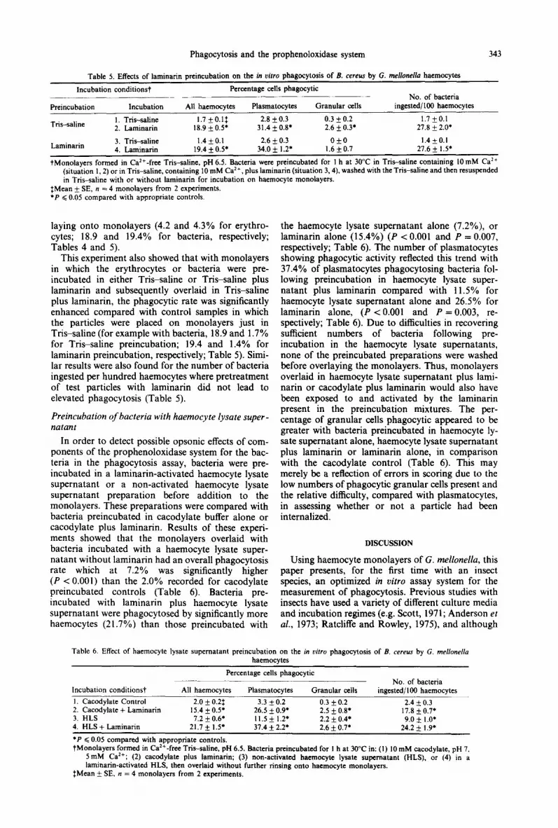
Table 5. Effects of laminarin preincubation on the in uirro phagocytosis of B. cereus by G. mellonel/a haemocytes
Incubation conditionst Percentage cells phagocytic ~_ No. of bacteria Preincubation Incubation All haemocytes Plasmatocytes Granular cells ingested/l00 haemocytes
Tris-saline I T&-saline 1.7 kO.l$ 2.8 f 0.3 0.3 * 0.2 1.7 f0.1 2. Laminarin 18.9 + 0.5’ 31.4 + 0.8. 2.6 +_ 0.3. 27.8 + 2.0.
Laminarin 3. T&-saline 1.4kO.l 2.6 + 0.3 Ok0 1.4 +0.1 4. Laminarin 19.4 * 0.5. 34.0 * 1.2. 1.6kO.7 27.6 f I .5’
tMonolayers formed in Ca ‘+-free Tris-saline, pH 6.5. Bacteria were preincubated for I h at 30°C in T&saline containing 10 mM Ca’+ (situation 1,2) or in Tris-saline, containing IO mM Ca2+, plus laminarin (situation 3,4), washed with the Tris-saline and then resuspended in Tris-saline with or without laminarin for incubation on haemocyte monolayers.
fMean + SE, n = 4 monolayers from 2 experiments. l P G 0.05 compared with appropriate controls.
laying onto monolayers (4.2 and 4.3% for erythro- cytes; 18.9 and 19.4% for bacteria, respectively; Tables 4 and 5).
This experiment also showed that with monolayers in which the erythrocytes or bacteria were pre- incubated in either Tri-saline or Tri-saline plus laminarin and subsequently overlaid in Tri-saline plus laminarin, the phagocytic rate was significantly enhanced compared with control samples in which the particles were placed on monolayers just in Tris-saline (for example with bacteria, 18.9 and 1.7% for Tris-saline preincubation; 19.4 and 1.4% for laminarin preincubation, respectively; Table 5). Simi- lar results were also found for the number of bacteria ingested per hundred haemocytes where pretreatment of test particles with laminarin did not lead to elevated phagocytosis (Table 5).
Preincubation of bacteria with haemocyte Iysate super- natant
In order to detect possible opsonic effects of com- ponents of the prophenoloxidase system for the bac- teria in the phagocytosis assay, bacteria were pre- incubated in a laminarin-activated haemocyte lysate supernatant or a non-activated haemocyte lysate supernatant preparation before addition to the monolayers. These preparations were compared with bacteria preincubated in cacodylate buffer alone or cacodylate plus laminarin. Results of these experi- ments showed that the monolayers overlaid with bacteria incubated with a haemocyte lysate super- natant without laminarin had an overall phagocytosis rate which at 7.2% was significantly higher (P < 0.001) than the 2.0% recorded for cacodylate preincubated controls (Table 6). Bacteria pre- incubated with laminarin plus haemocyte lysate supernatant were phagocytosed by significantly more haemocytes (21.7%) than those preincubated with
the haemocyte lysate supernatant alone (7.2%), or laminarin alone (15.4%) (P < 0.001 and P = 0.007, respectively; Table 6). The number of plasmatocytes showing phagocytic activity reflected this trend with 37.4% of plasmatocytes phagocytosing bacteria fol- lowing preincubation in haemocyte lysate super- natant plus laminarin compared with 11.5% for haemocyte lysate supernatant alone and 26.5% for laminarin alone, (P < 0.001 and P = 0.003, re- spectively; Table 6). Due to difficulties in recovering sufficient numbers of bacteria following pre- incubation in the haemocyte lysate supernatants, none of the preincubated preparations were washed before overlaying the monolayers. Thus, monolayers overlaid in haemocyte lysate supematant plus lami- narin or cacodylate plus laminarin would also have been exposed to and activated by the laminarin present in the preincubation mixtures. The per- centage of granular cells phagocytic appeared to be greater with bacteria preincubated in haemocyte ly- sate supernatant alone, haemocyte lysate supernatant plus laminarin or laminarin alone, in comparison with the cacodylate control (Table 6). This may merely be a reflection of errors in scoring due to the low numbers of phagocytic granular cells present and the relative difficulty, compared with plasmatocytes, in assessing whether or not a particle had been internalized.
DISCUSSION
Using haemocyte monolayers of G. mellonella, this paper presents, for the first time with an insect species, an optimized in vitro assay system for the measurement of phagocytosis. Previous studies with insects have used a variety of different culture media and incubation regimes (e.g. Scott, 1971; Anderson et al., 1973; Ratcliffe and Rowley, 1975), and although
Table 6. Effect of haemocyte lysate supematant preincubation on the in vim phagocytosis of B. cereus by G. me/lone//a haemocytes
Percentage cells phagocytic _..~._ No. of bacteria
Incubation conditionst All haemocytes Plasmatocytes Granular cells ingested/l00 haemocytes _ 1. Cacodylate Control 2.0 + 0.2i 3.3 f 0.2 0.3 f 0.2 2.4 f 0.3 2. Cacodylate + Laminarin 15.4 f 0.5. 26.5 f 0.9. 2.5 f O.g* 17.8 k 0.71 3. HLS 7.2 k 0.6* 11.5+ 1.21 2.2 f 0.4* 9.0 It 1 .O’ 4. HLS + Laminarin 21.7 f 1.5’ 37.4 f 2.21 2.6 k 0.7’ 24.2 & I .9’
l P Q 0.05 compared with appropriate controls. tMonolayers formed in Ca ‘*-free Tris-saline, pH 6.5. Bacteria preincubated for I h at 30°C in: (I) 10 mM cacodylate, pH 7.
5mM Ca’+; (2) cacodylate plus laminarin; (3) non-activated haemocyte lysate supematant (HLS), or (4) in a laminarin-activated HLS, then overlaid without further rinsing onto haemocyte monolayers.
$Mean + SE. n = 4 monolayers from 2 experiments.

344 JAYNE L. BROOKMAN er al.
some workers have considered a single aspect of the assay system and its effect on uptake of test particles by the haemocytes (e.g. Scott, 1971; Anderson et al.. 1973) no systematic study has to date been under- taken
The parameter which most markedly influenced the phagocytic rate was the incubation temperature. Pre- vious work by Ratcliffe et al. (1984) and Leonard er al. (1985) was performed at suboptimal temperatures for G. mellonella haemocytes, which accounts for the lower rates of phagocytosis observed by these authors compared with the results reported here. In the present study, the Galleria larvae were routinely maintained at 25-26”C, but, due to the high density and metabolic activity of the insects, the temperature often rose to 31-32°C in the culture jars. This corre- sponds, not surprisingly, to the optimum for phago- cytosis recorded here.
The calcium ion concentration of the overlay buffer was also important in the phagocytosis assay. It was shown that as the calcium ion concentration in- creased so the granular cell viability decreased. how- ever, since this cell type only has a limited phagocytic ability in vitro its viability does not greatly influence the overall phagocytic activity. This was largely de- termined by the main phagocytic cell type, the plas- matocyte, which maintained a viability of >95% at all calcium levels. The importance of calcium ions in other in ritro systems has been studied. Armstrong (1980) reported that adhesion and degranulation of Limulus haemocytes in vitro did not require the addition of extraneous calcium, but that haemocytes collected in 5 mM EDTA did not degranulate nor- mally due to excessive stabilization of the cells. G. mellonella haemocytes collected into an anticoagulant containing EDTA and allowed to form monolayers also showed reduced levels of cell adhesion and spreading in comparison with monolayers formed in the Ca’+-free monolayer buffer of the present study (unpublished observation). In addition, Renwrantz and Stahmer (1983) compared the phagocytosis of yeast cells by MJ~i1u.s edulis haemocytes in the pres- ence and absence of calcium ions. Considering the requirement for calcium ions shown by mammalian cell surface receptors when binding ligands (Kaplan, 1981) and the differences in the rates of phagocytosis recorded, they suggested that the M. edulis haemo- cytes possess calcium-dependent membrane-bound recognition molecules. A similar recognition event may be responsible for the increased level of phago- cytosis recorded for the experimental monolayers incubated with 10mM Ca*+ compared with those overlaid with calcium-free buffer.
This report also attempts to further investigate the involvement of the prophenoloxidase system in the non-self recognition process in arthropods. Previous studies by Smith and Siiderhall (1983a) in crus-
taceans, Ratcliffe et al. (1984) Leonard (1985) and Leonard et al. (1985) in insects, proposed that the enhancement of phagocytosis recorded in the pres- ence of laminarin was due to the activation of the prophenoloxidase system by this /?-1,3-glucan. The products of this activation include sticky proteins. which are probably opsonic, at least in crustaceans (Siiderhdll et al., 1984). An alternative explanation for the enhancement of phagocytosis with laminarin
is that this molecule acts as a bridge, binding to both the haemocyte and the test particle, enhancing con- tact and hence increasing phagocytosis. The use of erythrocytes as an alternative particle to bacteria in the assay, and the subsequent enhancement of phago- cytosis seen in the presence of laminarin, decreases the likelihood of laminarin acting as a bridging molecule. This is because, in contrast to erythrocytes, many bacteria have complex molecules such as lectins associated with their surfaces (Sharon et al., 1981) which are capable of binding carbohydrates, such as laminarin, and these may then form complexes with sugar residues on the haemocyte surface.
Additional proof negating the bridging hypothesis is provided by the preincubation of both erythrocytes and bacteria with laminarin and the subsequent lack of effect on the phagocytosis profile in vitro. This result, together with the inability of laminarin to enhance phagocytosis in monolayers pretreated with this p-1.3-glucan (Leonard et aI., 1985) implies that the mode of action of this molecule is via proph- enoloxidase activation.
The enhancement of phagocytosis in monolayers following preincubation of bacteria in a laminarin- activated haemocyte lysate supernatant in com- parison with bacteria preincubated in laminarin alone is also significant. This further indicates a possible opsonic function for components of the proph- enoloxidase system although the presence and func- tioning of other recognition molecules, such as agglu- tinins, cannot be totally disregarded at this stage. The increased levels of phagocytosis of bacteria pre- incubated in non-activated haemocyte lysate super- natant preparations, compared with cacodylate con- trols, may be due to the bacteria themselves activating the prophenoloxidase system, as has been found in several insect species (Leonard, 1985; Row- ley et ul.. 1986) or to a spontaneous activation within the preparations themselves.
The phagocytosis assay system developed in this work should prove of considerable use in the future in elucidating the link between humoral and cellular responses to non-self and for testing the function of purified cell and plasma components of the proph- enoloxidase cascade. The use of cell separation tech- niques (Mead et al.. 1986) will also allow monolayers to be formed from single or composite cell types as required and thus facilitate study of cell-cell cooper- ation events within the phagocytic process under optimized conditions.
Acknowledgements-We are grateful to Professor L. Renwrantz for his helpful discussion. to Mr L. O’Brien for rearing the insects and to the Science and Engineering Research Council for financial support (Grant No. GRIDI21684).
REFERENCES
Anderson R. S., Holmes B. and Good R. A. (1973) In vitro bactericidal capacity of Blaberus craniifer hemocytes. J. Invert. Path. 22, 127-135.
Armstrong P. B. (1980) Adhesion and spreading of Limulus blood cells on artificial surfaces. J. Cell Sci. 44, 243-262.
Czop J. K. and Austen K. F. (1985) Generation of leukot- rienes by human monocytes upon stimulation of their B-1.3-glucan receptor during phagocytosis. Proc. natn. Acad. Sci. U.S.A. 82, 2751-2755.

Phagocytosis and the prophenoloxidase system 345
Kaplan J. (1981) Polypeptide binding membrane receptors: Analysis and classification. Science 212, 14-20.
Leonard C. M. (1985) Studies on the Role of the Proph- enoloxidase Activating System in Non-self Recognition in Insect Immune Defences. Ph.D. Thesis, University of Wales.
Leonard C., Ratcliffe N. A. and Rowley A. F. (1985) The role of prophenoloxidase activation in non-self recog- nition and phagocytosis by insect blood cells. J. Inset/ Physiol. 31, 789-799.
Mead G. P., Ratcliffe N. A. and Renwrantz L. R. (1986) The separation of insect haemocyte types on Percoll gradients, methodology and problems. J. Insect Physiol. 32, 167- 177.
Rabinovitch M. and De Stefano M-J. (1970) Interactions of red cells with phagocytes of the wax-moth (GaNeria mellonella, Lepidoptera) and mouse. Exp. Cell Res. 59, 272-282.
Ratcliffe N. A., Leonard C. and Rowley A. F. (1984) Prophenoloxidase activation: nonself recognition and cell cooperation in insect immunity. Science 226, 557-559.
Ratcliffe N. A. and Rowley A. F. (1975) Cellular defence reactions of insect haemocytes in vitro. Phagocytosis in a new suspension culture system. J. Invert. Path. 26, 225-233.
Renwrantz L. and Stahmer A. (1983) Opsonizing properties of an isolated hemolymph agglutinin and demonstration of lectin-like recognition molecules at surface of hemo- cytes from Mytilus edulis. J. camp. Physiol. 149, 535-546.
Rowley A. F. and Ratcliffe N. A. (1980) Insect erythro- cyte agglutinins. In vitro opsonization experiments with Clitumnus extradentatus and Periplaneta americana hae- mocytes. Immunology 40, 483492.
Rowley A. F., Ratcliffe N. A., Leonard C. M., Richards E. H. and Renwrantz L. (1986) Humoral recognition
factors in insects, with particular reference to agglutinins and the prophenoloxidase system. In Hemocytic and Humoral Immunity in Arthropods (Ed. by Gupta A. P.), pp. 381406. Wiley Interscience, New York.
Scott M. T. (1971) Recognition of foreignness in in- vertebrates. II. In vitro studies of cockroach phagocytic haemocytes. Immunology 21, 817-828.
Sharon N., Eshdat Y., Silverblatt F. J. and Ofek I. (1981) Bacterial adherence to cell surface sugars. In Adhesion and Microorganism Pafhogenicity (Ed. by Elliott K.. O’Connor M. and Whelan J.), pp. 119-141. Pitman Medical. London.
Smith V. J. and Siiderhlll K. (1983a) b-1,3-glucan activa- tion of crustacean haemocytes in vitro and in viva. Biol. Bull. mar. biol. Lab., Woodr Hole 164, 299-314.
Smith V. J. and Soderhlll K. (1983b) Induction of de- granulation and lysis of haemocytes in the freshwater crayfish, Astacus asfacus by components of the pro- phenoloxidase activating system in vitro. Cell Tissue kes. 233, 295-303.
Soderhlll K. and Hall L. (1984) Lipopolysaccharide- induced activation of prophenoloxidase activating system in crayfish hemocyte lysate. Biochem. biophys. Acra 797, 99-104.
Soderhill K., Smith V. J. and Johansson M. W. (1986) Exocytosis and uptake of bacteria by isolated haemocyte populations of crustaceans: Evidence for cell co-operation in the defence reactions of arthropods. Cell Tissue Res. 245, 4349.
Siiderhall K., Vey A. and Ramstedt M. (1984) Haemocyte lysate enhancement of fungal spore encapsulation by crayfish haemocytes. Dev. Comp. Immun. 8, 23-29.
Unestam T. and Nyhlen L. (1974) Cellular and non-cellular recognition of and reactions to fungi in crayfish. Contemp. Top. Immunobiol. 4, 189-206.


