Embed Size (px)
Citation preview
Origin and Structural Evolution of the EarlyProliferating Oval Cells in Rat Liver
Sandor Paku,* Janos Schnur,† Peter Nagy,†‡ andSnorri S. Thorgeirsson‡
From the Joint Research Organization of the Hungarian
Academy of Sciences and Semmelweis University of Medicine,*
Budapest, Hungary; the First Institute of Pathology and
Experimental Cancer Research,† Semmelweis University of
Medicine, Budapest, Hungary; and the Laboratory of
Experimental Carcinogenesis,‡ National Cancer Institute,
National Institutes of Health, Bethesda, Maryland
We have analyzed the histological changes in rat liverafter 2-acetylaminofluorene (AAF) administration.The data demonstrate that AAF-induced oval cellswere preferentially generated by proliferation of theterminal biliary ductules that we suggest constitutethe primary hepatic stem cell niche. The oval cellsformed ductular structures, representing an exten-sion of the canals of Hering. This histological organi-zation provides continuous bile drainage of the hepa-tocytes and uninterrupted blood flow in thesinusoids. The oval cell ductules are surrounded by acontinuous basement membrane that is intermit-tently disrupted by processes of stellate cells thatform direct cell-cell contact with the oval cells. Al-though both AAF treatment and bile duct ligation re-sults in proliferation of biliary epithelial cells, themechanism(s) responsible for the proliferation of thebiliary epithelium seems to differ in the two models.In contrast to the biliary proliferation stimulated bybile ligation, AAF-induced oval cell proliferation aswell as the capacity of these cells to differentiate intohepatocytes, bile epithelial cells and possibly othercell lineages can be blocked by administration ofdexamethasone. (Am J Pathol 2001, 158:1313–1323)
Although a substantial amount of knowledge has beenaccumulated throughout the last 2 decades about liverstem cells,1,2 numerous aspects of this intriguing cellcompartment remain undefined. Indeed, there are con-flicting data on the exact location of liver stem cells andeven the growth pattern of these cells is not completelyunderstood. The proliferating oval cells—the progeny ofthe stem cells—always expand into liver parenchymefrom the portal area. Furthermore, selective damage ofthe periportal zone reduces oval cell proliferation.3 Theseobservations support the notion that the stem cells must
reside somewhere in the periportal region. The pheno-typic resemblance between the oval cells and biliaryepithelium suggests that they derive from the biliary tree,and terminal hepatic ductules (canals of Hering) thatconnect the most distal hepatocyte of the hepatic plate tothe interlobular bile ducts are thought to harbor the he-patic stem cells.4–7 However, there is no general agree-ment on this issue. In fact, potential candidates for thestem cells outside the biliary system have been pro-posed.8
In the absence of a specific marker for the hepaticstem cells, several investigators using different modelshave attempted to identify the stem cells by labeling thedividing cells in the early phase of oval cell expan-sion.4,8–10 However, most of the experimental protocolsfor the activation of the hepatic stem cell compartmentrequire a relatively long time and this may explain thedivergent results. The 2-acetylaminofluorene (AAF)/par-tial hepatectomy (PH) model of oval cell proliferation/differentiation has been extensively used to analyze thehepatic stem cell compartment during the last fewyears.11–13 We have recently modified the classicalAAF/PH model14 and demonstrated that after a singledose of AAF administration a notable cell proliferationtakes place in the periportal zone and at least some ofthese proliferating cells are the precursors of oval cells.Therefore AAF administration provides a uniquely fastand synchronized activation of the oval cell precursorswithout any major disruption of the hepatic structure. Wecould identify dividing cells in the interlobular bile ductsafter AAF treatment, whereas the exact nature of the restof the thymidine-labeled cells could not be unambigu-ously defined by traditional light microscopy.14
Biliary cell proliferation can also be induced in rats bythe ligation of the common bile duct (BDL).15,16 Thisreaction is, however, morphologically and phenotypicallyvery different from the oval cell proliferation. After BDL,proliferating biliary cells do not show any signs of differ-entiation into other cell types. Another difference betweenBDL- and AAF-induced biliary cell proliferation is illus-trated by selective inhibition of oval cell proliferation bydexamethasone.17
Supported by OTKAT 22737, T29006, and North American Treaty Orga-nization Collaborative Research grant HTECH-EV 973276.
Accepted for publication December 27, 2000.
Address reprint requests to Dr. Peter Nagy, National Cancer Institute,NIH, 37 Convent Dr. MSC 4255, Building 37, Room 3C28, Bethesda, MD20892-4255.
American Journal of Pathology, Vol. 158, No. 4, April 2001
Copyright © American Society for Investigative Pathology
1313
In the present work we have characterized the earlycellular events in the liver during the proliferative re-sponse induced by AAF or BDL. To obtain a more de-tailed morphological assessment, the samples were an-alyzed by, in addition to traditional light microscopy, bothconfocal and electron microscopy. Both AAF and BDLinduced an intense biliary cell proliferation. The fre-quency of dividing cells after AAF treatment was signifi-cantly higher in the terminal hepatic ductules. Morpho-logical analysis revealed that the early oval cells arestrictly confined to ductular structures surrounded bybasement membrane, representing an extension of thecanals of Hering.
Materials and Methods
Animal Experiments
Male F-344 rats (180 to 200 g) were used for all experi-ments and kept under standard conditions. The animalstudy protocols were conducted according to NIH guide-lines for animal care.
AAF/PH Experiment
AAF (1.5 mg) suspended in dimethyl-cellulose wasgiven to the rats on 4 consecutive days by gavage.Traditional 70% PH18 was performed on the fifth day,which was followed by five additional AAF treatments.Animals were sacrificed at the described time points (atleast three at each time point).
BDL
BDL was done according to Cameron and Oakley.19
The rats were sacrificed 48 hours after the operation.
Electron Microscopy
Preparation of liver tissue for electron microscopy wasdone by perfusing the livers under anesthesia (35 mg/kg,Nembutal; Serva, Heidelberg, Germany) via the portalvein with phosphate-buffered saline (PBS) for 10 minutesand with 2.5% glutaraldehyde in 0.05 mol/L Na-cacody-late (pH 7.2) for 15 minutes at room temperature. Liverswere cut into 1 3 3 mm pieces and immersed in 2.5%glutaraldehyde for 2 hours. The pieces were postfixedin 1% OsO4, 0.05% K-ferrocyanide in 0.05 mol/L Na-cacodylate for 1 hour, dehydrated in a graded series ofalcohol, contrasted en bloc with 2% uranylacetate, andembedded in Spurr’s mixture. Ultrathin sections werestained with lead citrate and examined on a PhilipsCM10 electron microscope.
Ultrastructural Analysis of 5-Bromo-29-Deoxy-Uridine (BrdU)-Labeled Periportal Cell
BrdU (100 mg/kg) was injected intraperitoneally after twodoses of AAF, 24 hours after the second treatment or 2
days after BDL. One hour after the injection, the animalswere anesthetized and perfused via the portal vein withPBS for 10 minutes, followed by 4% paraformaldehydefor 20 minutes. Livers were removed, cut into 5-mm thickslices and postfixed for 24 hours. The fixed tissues werewashed in PBS and immersed in 15% sucrose for 24hours followed by 30% sucrose for another 24 hours.Specimens were frozen in isopentane cooled by liquidnitrogen.
Cryosections (15 mm thick) were mounted on micro-scope slides coated with parlodion. The cryosectionswere rinsed in PBS and incubated for 20 minutes in 3 NHCl at room temperature. After washing in PBS, the sec-tions were incubated with monoclonal anti-BrdU antibody(diluted 1:100; Becton-Dickinson, Mountain View, CA) for3 hours and later with biotinylated anti-mouse antibody(Vector Laboratories, Burlingame, CA) for 2 hours. Thereaction was developed by an ABC reaction (Elite ABCKit, Vector Laboratories) using diaminobenzidine aschromogen. The sections were osmificated (1% OsO4 inPBS), dehydrated in graded series of ethanol, and em-bedded in Epon 812. Blocks were removed by immersingthe slides in liquid nitrogen. Semithin sections wereslightly stained by 0.5% toluidine blue (pH 8.5), portalareas containing BrdU-labeled cells were trimmed outand unstained ultrathin sections were analyzed on a Phil-ips CM 10 electron microscope. Labeled cells weredivided into three categories based on their localiza-tion: 1) cells residing outside the basement mem-branes in the periportal connective tissue; 2) cellsconfined within the basement membrane, as part ofbile ducts; and 3) cells of the canals of Hering. Thecells comprising the canals of Hering were in directcontact with a hepatocyte or were separated from ahepatocyte by only one biliary cell.
Immunofluorescent Analysis
Double Labeling for Laminin-Cytokeratin andLaminin-Desmin
Cryostat sections (6 mm) were fixed in acetone (220°Cfor 20 minutes) and incubated overnight with a mixture ofrabbit polyclonal anti-laminin antibody (diluted 1:50,Z0097; DAKO, Glostrup, Denmark) and fluorescein iso-thiocyanate-conjugated mouse monoclonal antibody di-rected against human cytokeratin 5, -6, -8, -17, and -19(diluted 1:10, M0859; DAKO) or anti-laminin and mousemonoclonal anti-desmin (diluted 1:50, M0724; DAKO),respectively. After washing with PBS the sections wereincubated for 60 minutes with tetramethylrhodamine Bisothiocyanate-conjugated anti-rabbit IgG (diluted 1:20,R0156; DAKO) for the laminin-cytokeratin double labelingor with the combination of the same tetramethylrhodam-ine B isothiocyanate-conjugated anti-rabbit IgG and flu-orescein isothiocyanate-conjugated anti-mouse IgG (di-luted 1:50, F5262; Sigma) for the simultaneous detectionof laminin and desmin.
1314 Paku et alAJP April 2001, Vol. 158, No. 4
Double Labeling for Laminin and a-Fetoprotein (AFP)
Liver samples were fixed in 10% formaldehyde, em-bedded, and cut. After deparaffinization and hydration,the sections were microwaved for 15 minutes in ethyl-enediaminetetraacetic acid buffer (pH 8.0), followed byincubation in 0.1% Triton X-100 in PBS for 20 minutes,and digested with Proteinase K (2 mg/ml, 15 minutes,37°C). The sections were washed with PBS and incu-bated overnight with a mixture of rabbit polyclonal anti-laminin (diluted 1:20, Z0097; DAKO) and goat anti-ratAFP no. 89 antibody (diluted 1:100; generous gift from Dr.Stewart Sell, Department of Pathology and LaboratoryMedicine, Albany Medical College, Albany, NY). Tetram-ethylrhodamine B isothiocyanate-conjugated anti-rabbitIgG (as described above), and in a second step, fluores-cein isothiocyanate-conjugated anti-goat IgG (diluted1:400, F7367; Sigma), were used as secondary antibod-ies. All samples were analyzed by confocal laser-scan-
ning microscopy using the Bio-Rad MRC-1024 system(Bio-Rad, Richmond, CA).
Results
BrdU Labeling of Dividing Cells after BDL andAAF Administration
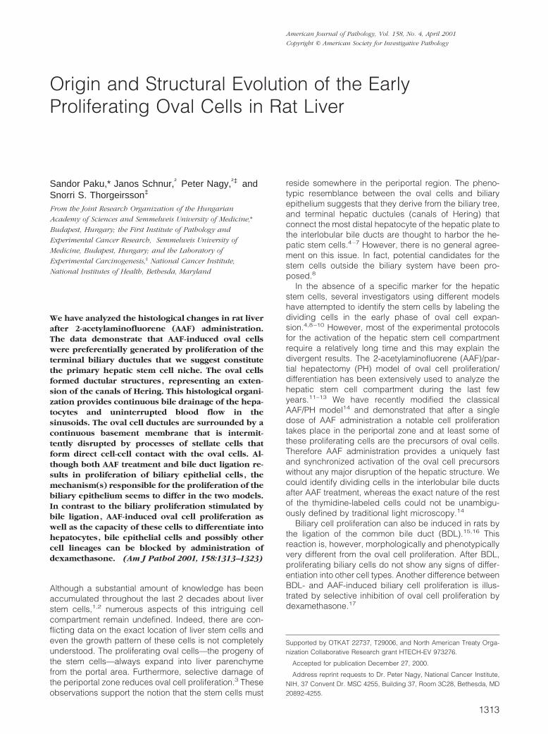
The ligation of the common bile duct resulted in a veryintensive biliary cell proliferation. The cells were labeledwith BrdU 48 hours after the ligation. In agreement withprevious results,20 most of the labeled cells were in theinterlobular bile ducts. Immunoelectron-microscopic analy-sis of the localization of labeled cells revealed ,6% of theBrdU-positive biliary epithelial cells in the canals of Hering(Figure 1A). Additionally, dividing inflammatory and fibro-blastic cells could be found in the periportal connectivetissue and a few labeled hepatocytes were also present.
Figure 1. A: Distribution of BrdU-labeled cells, between bile ducts and canals of Hering, analyzed by immunoelectronmicroscopy. The first two columns representthe distribution after two treatments with AAF, and the second two 48 hours after BDL. Total number of labeled cells analyzed was 141 in the AAF-treated groupand 143 in the BDL group. B: Immunoelectron micrograph 2 days after AAF treatment showing a canal of Hering consisting of four cells. Three of them are labeledby BrdU. Two cells of the canals are attached to the hepatocytes (H) (arrowheads). The fourth labeled cell (asterisk) is located in the periportal connectivetissue outside the basement membrane of the terminal hepatic ductule. Scale bar, 2 mm. C: Activated cell in the canal of Hering (He) 2 days after AAF treatment.The nucleus is enlarged and contains euchromatin and a prominent nucleolus (small arrow). Continuous basement membrane is visible on the connective tissueside of the cell (arrowheads). The bile ductule lumen is sealed by intercellular junctions (large arrows) (H, hepatocyte). Scale bar, 1 mm.
Two Distinct Forms of Biliary Proliferation 1315AJP April 2001, Vol. 158, No. 4
Similarly to our earlier observations,14 a single dose ofAAF resulted in cell proliferation in the periportal region.However, because the number of the BrdU-labeled cellswas higher after two doses of AAF, without any apparentadverse effects, we selected the 48-hour time point forthe quantitative electron-microscopic analysis. Approxi-mately 80% of the labeled biliary cells were inside thewell-defined interlobular bile ducts. However, 22% of theBrdU-positive epithelial cells were located in the canalsof Hering (Figure 1A). Only those biliary cells havingdirect connection with a hepatocyte or separated by onlyone interposed biliary cell from the hepatocyte werecounted as lining cells of the canals of Hering (Figure1B). It is possible that by using these strict criteria weunderestimated the number of labeled cells in the canalsof Hering. However, the observation that 22% of thelabeled biliary cells were in these structures can be takenas preferential labeling, considering that the overwhelm-ing majority of the biliary epithelial cells belong to theinterlobular and larger bile ducts.
After AAF treatment the terminal hepatic ductules weresurrounded, by an almost continuous basement mem-brane that terminated on a hepatocyte of the limiting plate(Figure 1C). In addition, administration of AAF also in-duced proliferation outside the biliary system. Mostly sin-gle cells residing in the periportal extracellular matrixwere labeled. Although the exact nature of these cellscould not be established by electron microscopy theymost likely represent mesenchymal cells.
Immunohistochemical Analysis of the Liver 2Days after AAF Administration
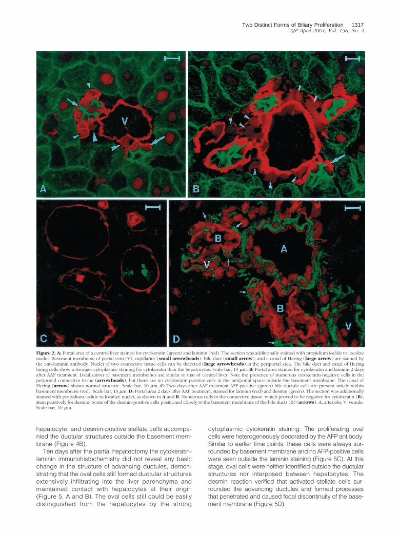
The cell density increased in the periportal space afterone or two doses of AAF, but the proliferating ductulesdid not infiltrate the liver lobules. To determine the char-acteristics of the proliferating cells in the periportalspace, double labeling for laminin/cytokeratin, laminin/desmin, and laminin/AFP was performed. Immunofluores-cent-stained liver sections were analyzed by confocalmicroscopy. The cytokeratin antibody gave a weak mem-brane and reticular cytoplasmic staining in the hepato-cytes, whereas producing a strong cytoplasmic reactionin the biliary epithelial cells. Therefore, the two cell pop-ulations could be easily distinguished. The laminin anti-body, a well-established marker of the basement mem-brane, sharply circumscribed the bile ducts and theblood vessels, whereas the liver acini were completelynegative. The terminal hepatic ductules were intenselydecorated by the laminin/cytokeratin double labeling. Inthe appropriate plane of the section, the basement mem-brane stained with laminin surrounded the bile ductulesand terminated on hepatocytes at the limiting plate (Fig-ure 2A). The basement membrane extended without in-terruption along the biliary ducts and ductules. If thenuclei were stained by propidium iodide in combinationwith the double-immunofluorescent reaction, increasedcell density was revealed periportally after AAF treatment(Figure 2, A and B). However, we did not observe cyto-keratin-labeled cells outside the basement membrane in
the periportal space. Combined laminin/AFP immunohis-tochemistry showed that basement membrane sur-rounded a few AFP-positive biliary cells after two doses ofAAF (Figure 2C). Similar to the cytokeratin reaction, noAFP-stained cells were observed outside the basementmembrane. Immunostaining with a desmin antibody re-vealed an increased number of activated stellate cellsoutside the basement membrane. These cells and theirprocesses showed a very intimate connection with theproliferating ductules (Figure 2D).
Morphological Alterations after PH inAAF-treated Rats
To study later events during stem cell activation, we usedthe AAF/PH model and the structural alterations wereanalyzed 3 days after the partial hepatectomy. By thistime the oval cells had extensively infiltrated the liverlobules. Electron microscopic examination revealed thatthe oval cells always formed ductules (Figure 3A). Thesegrowing ductules reached the sinusoids and passedalong or between them. During this process the sinusoidswere left intact, preserving their normal function. Thebasic structure of these oval cell ductules was not differ-ent from the normal canals of Hering except for the elon-gation. The ductules grew by the continuous proliferationof the ductular epithelial cells (Figure 3B), whereas thesides of these tubules were continuously sealed by des-mosomes. Also, the ductules always terminated at a he-patocyte (Figure 3C). This hepatocyte was probably theone connected with the original canal of Hering. In thisway the original connection between the bile canaliculiand the bile duct system was preserved throughout theregenerative process. The growing tubules were sur-rounded mostly by basement membrane that terminatedon the hepatocyte. However, there were segments alongthese growing ductules where no structured basementmembrane could be seen by electron microscope. Againno cells penetrating through the basement membranecould be observed. There were, however, plenty of pro-liferating cells outside the basement membrane withoutcharacteristic ultrastructural features. These cells did notshow epithelial phenotype or form desmosomes, yethad a very close contact with the expanding ductularepithelial cells. Higher magnification showed that smallprocesses of these nonepithelial cells appeared tohave direct cell-cell contact with the epithelial cells(Figure 3D).
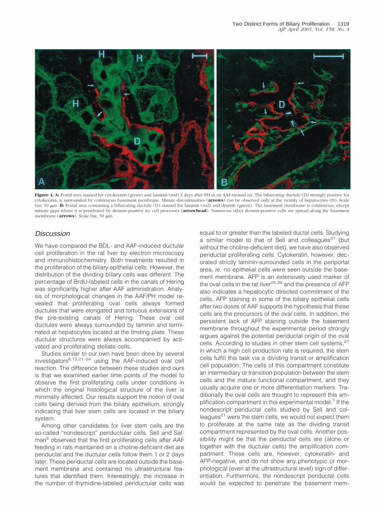
Immunohistochemical observations confirmed theelectron-microscopic data. The cytokeratin-stained ovalcells formed ductules that were elongations of the portallylocated canals of Hering. The tubular structure of theseductules with a central lumen was much more obvious inthe pictures generated by confocal microscopy. The ma-jor difference between the two methods was that, al-though the basement membrane could not be continu-ously followed along these ductules ultrastructurally, theimmunohistochemistry showed consistent laminin positiv-ity around them (Figure 4A). The cylinder formed by thebasement membrane had an open end plugged by a
1316 Paku et alAJP April 2001, Vol. 158, No. 4
hepatocyte, and desmin-positive stellate cells accompa-nied the ductular structures outside the basement mem-brane (Figure 4B).
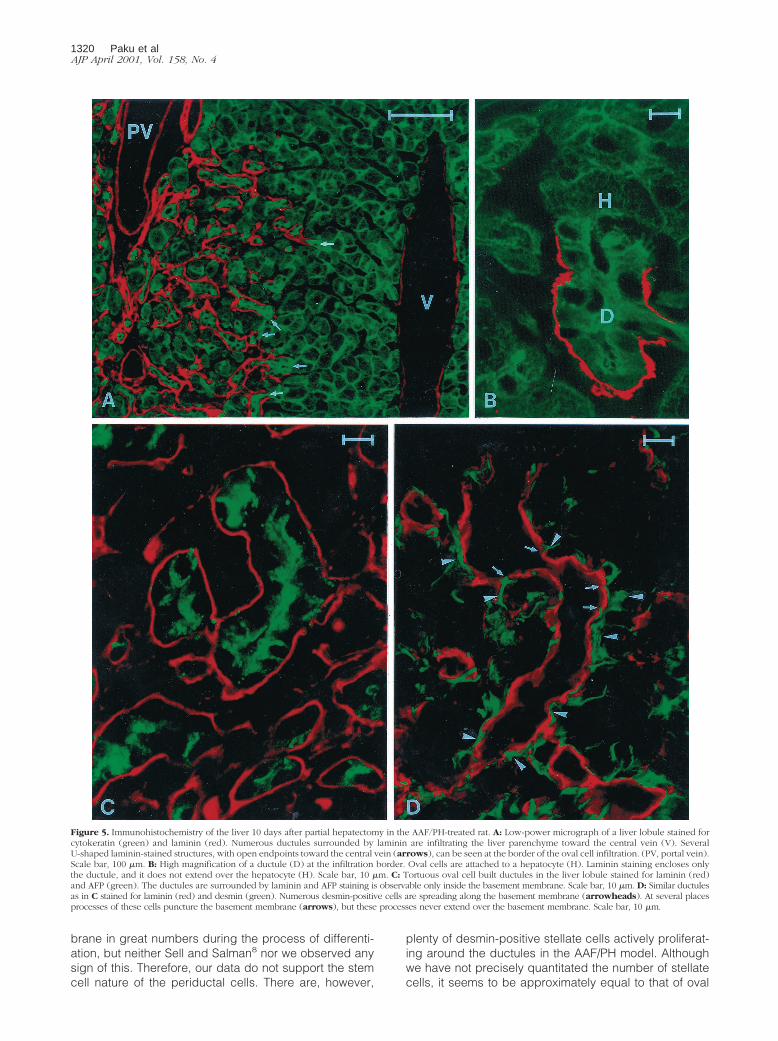
Ten days after the partial hepatectomy the cytokeratin-laminin immunohistochemistry did not reveal any basicchange in the structure of advancing ductules, demon-strating that the oval cells still formed ductular structuresextensively infiltrating into the liver parenchyma andmaintained contact with hepatocytes at their origin(Figure 5, A and B). The oval cells still could be easilydistinguished from the hepatocytes by the strong
cytoplasmic cytokeratin staining. The proliferating ovalcells were heterogeneously decorated by the AFP antibody.Similar to earlier time points, these cells were always sur-rounded by basement membrane and no AFP-positive cellswere seen outside the laminin staining (Figure 5C). At thisstage, oval cells were neither identified outside the ductularstructures nor interposed between hepatocytes. Thedesmin reaction verified that activated stellate cells sur-rounded the advancing ductules and formed processesthat penetrated and caused focal discontinuity of the base-ment membrane (Figure 5D).
Figure 2. A: Portal area of a control liver stained for cytokeratin (green) and laminin (red). The section was additionally stained with propidium iodide to localizenuclei. Basement membrane of portal vein (V), capillaries (small arrowheads), bile duct (small arrow), and a canal of Hering (large arrow) are stained bythe anti-laminin antibody. Nuclei of two connective tissue cells can be detected (large arrowheads) in the periportal area. The bile duct and canal of Heringlining cells show a stronger cytoplasmic staining for cytokeratin than the hepatocytes. Scale bar, 10 mm. B: Portal area stained for cytokeratin and laminin 2 daysafter AAF treatment. Localization of basement membranes are similar to that of control liver. Note the presence of numerous cytokeratin-negative cells in theperiportal connective tissue (arrowheads), but there are no cytokeratin-positive cells in the periportal space outside the basement membrane. The canal ofHering (arrow) shows normal structure. Scale bar, 10 mm. C: Two days after AAF treatment AFP-positive (green) bile ductule cells are present strictly withinbasement membrane (red). Scale bar, 10 mm. D: Portal area 2 days after AAF treatment, stained for laminin (red) and desmin (green). The section was additionallystained with propidium iodide to localize nuclei, as shown in A and B. Numerous cells in the connective tissue, which proved to be negative for cytokeratin (B),stain positively for desmin. Some of the desmin-positive cells positioned closely to the basement membrane of the bile ducts (B) (arrows). A, arteriole; V, venule.Scale bar, 10 mm.
Two Distinct Forms of Biliary Proliferation 1317AJP April 2001, Vol. 158, No. 4
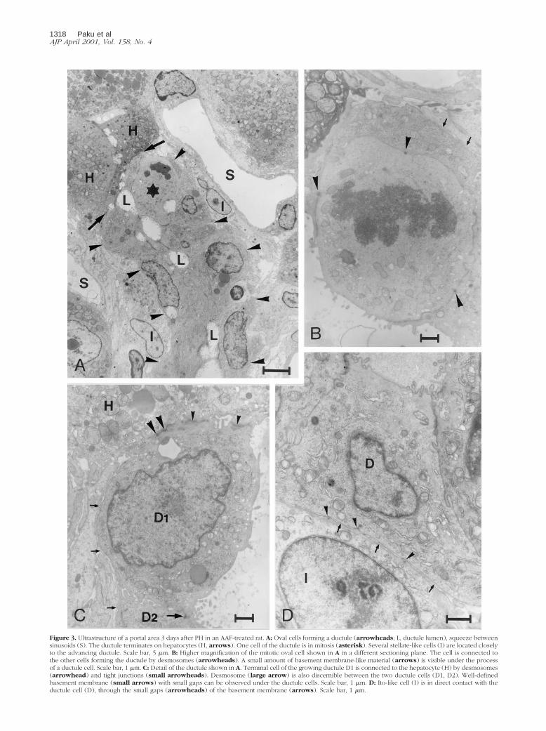
Figure 3. Ultrastructure of a portal area 3 days after PH in an AAF-treated rat. A: Oval cells forming a ductule (arrowheads; L, ductule lumen), squeeze betweensinusoids (S). The ductule terminates on hepatocytes (H, arrows). One cell of the ductule is in mitosis (asterisk). Several stellate-like cells (I) are located closelyto the advancing ductule. Scale bar, 5 mm. B: Higher magnification of the mitotic oval cell shown in A in a different sectioning plane. The cell is connected tothe other cells forming the ductule by desmosomes (arrowheads). A small amount of basement membrane-like material (arrows) is visible under the processof a ductule cell. Scale bar, 1 mm. C: Detail of the ductule shown in A. Terminal cell of the growing ductule D1 is connected to the hepatocyte (H) by desmosomes(arrowhead) and tight junctions (small arrowheads). Desmosome (large arrow) is also discernible between the two ductule cells (D1, D2). Well-definedbasement membrane (small arrows) with small gaps can be observed under the ductule cells. Scale bar, 1 mm. D: Ito-like cell (I) is in direct contact with theductule cell (D), through the small gaps (arrowheads) of the basement membrane (arrows). Scale bar, 1 mm.
1318 Paku et alAJP April 2001, Vol. 158, No. 4
Discussion
We have compared the BDL- and AAF-induced ductularcell proliferation in the rat liver by electron microscopyand immunohistochemistry. Both treatments resulted inthe proliferation of the biliary epithelial cells. However, thedistribution of the dividing biliary cells was different. Thepercentage of BrdU-labeled cells in the canals of Heringwas significantly higher after AAF administration. Analy-sis of morphological changes in the AAF/PH model re-vealed that proliferating oval cells always formedductules that were elongated and tortuous extensions ofthe pre-existing canals of Hering. These oval cellductules were always surrounded by laminin and termi-nated at hepatocytes located at the limiting plate. Theseductular structures were always accompanied by acti-vated and proliferating stellate cells.
Studies similar to our own have been done by severalinvestigators8,13,21–24 using the AAF-induced oval cellreaction. The difference between these studies and oursis that we examined earlier time points of the model toobserve the first proliferating cells under conditions inwhich the original histological structure of the liver isminimally affected. Our results support the notion of ovalcells being derived from the biliary epithelium, stronglyindicating that liver stem cells are located in the biliarysystem.
Among other candidates for liver stem cells are theso-called “nondescript” periductular cells. Sell and Sal-man8 observed that the first proliferating cells after AAFfeeding in rats maintained on a choline-deficient diet areperiductal and the ductular cells follow them 1 or 2 dayslater. These periductal cells are located outside the base-ment membrane and contained no ultrastructural fea-tures that identified them. Interestingly, the increase inthe number of thymidine-labeled periductular cells was
equal to or greater than the labeled ductal cells. Studyinga similar model to that of Sell and colleagues21 (butwithout the choline-deficient diet), we have also observedperiductal proliferating cells. Cytokeratin, however, dec-orated strictly laminin-surrounded cells in the periportalarea, ie, no epithelial cells were seen outside the base-ment membrane. AFP is an extensively used marker ofthe oval cells in the rat liver25,26 and the presence of AFPalso indicates a hepatocytic directed commitment of thecells. AFP staining in some of the biliary epithelial cellsafter two doses of AAF supports the hypothesis that thesecells are the precursors of the oval cells. In addition, thepersistent lack of AFP staining outside the basementmembrane throughout the experimental period stronglyargues against the potential periductal origin of the ovalcells. According to studies in other stem cell systems,27
in which a high cell production rate is required, the stemcells fulfill this task via a dividing transit or amplificationcell population. The cells of this compartment constitutean intermediary or transition population between the stemcells and the mature functional compartment, and theyusually acquire one or more differentiation markers. Tra-ditionally the oval cells are thought to represent this am-plification compartment in this experimental model.1 If thenondescript periductal cells studied by Sell and col-leagues21 were the stem cells, we would not expect themto proliferate at the same rate as the dividing transitcompartment represented by the oval cells. Another pos-sibility might be that the periductal cells are (alone ortogether with the ductular cells) the amplification com-partment. These cells are, however, cytokeratin- andAFP-negative, and do not show any phenotypic or mor-phological (even at the ultrastructural level) sign of differ-entiation. Furthermore, the nondescript periductal cellswould be expected to penetrate the basement mem-
Figure 4. A: Portal area stained for cytokeratin (green) and laminin (red) 3 days after PH in an AAF-treated rat. The bifurcating ductule (D) strongly positive forcytokeratin, is surrounded by continuous basement membrane. Minute discontinuities (arrows) can be observed only at the vicinity of hepatocytes (H). Scalebar, 10 mm. B: Portal area containing a bifurcating ductule (D) stained for laminin (red) and desmin (green). The basement membrane is continuous, exceptminute gaps where it is penetrated by desmin-positive Ito cell processes (arrowhead). Numerous other desmin-positive cells are spread along the basementmembrane (arrows). Scale bar, 50 mm.
Two Distinct Forms of Biliary Proliferation 1319AJP April 2001, Vol. 158, No. 4
brane in great numbers during the process of differenti-ation, but neither Sell and Salman8 nor we observed anysign of this. Therefore, our data do not support the stemcell nature of the periductal cells. There are, however,
plenty of desmin-positive stellate cells actively proliferat-ing around the ductules in the AAF/PH model. Althoughwe have not precisely quantitated the number of stellatecells, it seems to be approximately equal to that of oval
Figure 5. Immunohistochemistry of the liver 10 days after partial hepatectomy in the AAF/PH-treated rat. A: Low-power micrograph of a liver lobule stained forcytokeratin (green) and laminin (red). Numerous ductules surrounded by laminin are infiltrating the liver parenchyme toward the central vein (V). SeveralU-shaped laminin-stained structures, with open endpoints toward the central vein (arrows), can be seen at the border of the oval cell infiltration. (PV, portal vein).Scale bar, 100 mm. B: High magnification of a ductule (D) at the infiltration border. Oval cells are attached to a hepatocyte (H). Laminin staining encloses onlythe ductule, and it does not extend over the hepatocyte (H). Scale bar, 10 mm. C: Tortuous oval cell built ductules in the liver lobule stained for laminin (red)and AFP (green). The ductules are surrounded by laminin and AFP staining is observable only inside the basement membrane. Scale bar, 10 mm. D: Similar ductulesas in C stained for laminin (red) and desmin (green). Numerous desmin-positive cells are spreading along the basement membrane (arrowheads). At several placesprocesses of these cells puncture the basement membrane (arrows), but these processes never extend over the basement membrane. Scale bar, 10 mm.
1320 Paku et alAJP April 2001, Vol. 158, No. 4
cells. It is therefore possible that at least some of theperiductal proliferating cells described by Sell and Sal-man8 might be stellate cells.
Several studies have supported possibilities of thestem cells being located in the biliary system. Theseinclude terminal hepatic ductules;4,5,22,28 all biliary epi-thelial cells,29–32 and a very primitive looking cell, re-ferred to as a “basal cell” inside the bile duct.23,24 Thebasal cells proliferated 2 to 3 days after the partial hep-atectomy in the livers of rats treated according to Solt andcolleagues33 carcinogenesis schedule. We also sawthese small, intraepithelial cells in our sections, but theynever incorporated BrdU or showed morphological signsof proliferation. The reason for this difference is not alto-gether clear. Novikoff and colleagues23 and Novikoff andYam24 used the DEN-initiated Solt-Farber model,whereas we avoided DEN administration. Although theoval cell reaction is similar, the histological changes aremore complex in the Solt-Farber model according to ourexperience. Anilkumar and colleagues34 also describeddivergent histological reactions in the two models. It ishowever evident that oval cell proliferation can be in-duced without the participation of the basal cells in ourexperimental model. Therefore, the biliary epithelial cellscan function as facultative liver stem cells in the AAF/PHexperimental model. We can, of course, not exclude thepossibility that the basal cells represent an even moreancient stem cell population that is activated by a moredrastic, carcinogenic protocol and may be responsible,eg, for the frequently observed metaplastic hemopoiesisin hepatocarcinogenesis experiments.35,36 Recently Pe-tersen and colleagues37 and Theise and colleagues38
have provided evidence that hemopoietic stem cells cangive rise to oval cells and hepatocytes. The basal cellsmay perhaps represent a common precursor for the twosystems. Although these observations have a dramaticimpact on our view of stem cell biology, there is a generalagreement that under most circumstances the liver re-generates from cell populations confined to the liver.These cells are the focus of this study and the participa-tion of hemopoietic cells in liver regeneration is not ad-dressed here.
The notion of which segment(s) of the biliary tree har-bors the stem cells is still controversial. Although a sub-stantial amount of data indicate that the liver stem cellsare confined to the terminal hepatic ductules, argumentshave been made suggesting that any component of thebiliary tree can give rise to oval cells.29–32 We havepreviously shown that chronic dexamethasone treatmentis able to prevent the oval cell proliferation triggered bythe AAF/PH protocol while not at all inhibiting the BDL-induced proliferation of the larger, mostly interlobular bileducts.17 The preferential BrdU labeling of cells in thecanals of Hering after AAF administration, suggested thatselective inhibition of cell proliferation in these cells mightbe achieved by dexamethasone. Contrary to expecta-tions dexamethasone completely inhibited the AAF-in-duced biliary cell proliferation regardless of their loca-tion.17 These results fail to provide a functionalconfirmation that the terminal ductules are the exclusivesources of the oval cells. Furthermore, the proliferations
of morphologically and topologically identical biliary cellswere differently regulated by dexamethasone. Thesedata suggest that there are at least two different mecha-nisms regulating proliferation of the biliary epithelium,each providing functionally different progeny. Althoughthe dexamethasone-sensitive pathway provides cellswith stem cell potential, the dexamethasone-resistantpathway produces only biliary epithelial cells. These re-sults may, at least in part, be analogous to the regulationof hepatocyte cell cycle induced by the partial hepatec-tomy and primary mitogens.39 It is well established thatthese proliferative models have different biological poten-tials; eg, hepatocyte proliferation induced by partial hep-atectomy has carcinogenic promoting capacity whereasthe other one induced by direct mitogens has none.40
There are already observations indicating the differentialregulation of these two biliary reactions. Mice harboringcongenitally defective SCF/c-kit system retain an intactproliferative response after BDL,41 whereas the oval cellproliferation is remarkably suppressed in rats with defi-cient c-kit kinase activity.42
The fact that hepatocytic differentiation occurs in thepancreas43,44 and extrahepatic bile ducts45 also arguesagainst the restricted occurrence of multipotential stemcells in the canals of Hering. Our observation that AAFtargets preferentially the cholangioles while BDL targetsthe larger bile ducts can be explained by topologicalfactors. The primary stimulus for biliary proliferation afterBDL is the increased intraductal pressure.15 The pres-sure is probably higher in the interlobular bile ducts, thanin the ductules. Differential expression of drug metabo-lizing enzymes by different segments of the biliary tree46
may provide an alternative or additional explanation forthe differential response to AAF. Potten47 described ahierarchy of the stem cells in the small intestine glands.Depending on the severity of injury, more and more re-sistant cells participate in the repair. Additionally, thishierarchy is related to the topography of the cells. Asimilar arrangement cannot be excluded in the biliarysystem. This notion is supported by the well-known het-erogeneity of cholangiocytes.48,49
Experiments using injection of pigmented gelatin me-dium and related substances into the biliary tree havedemonstrated that the majority of oval cells are part of aductular reaction.10,13,50 However, together with theseobservations, occurrence of isolated oval cells, some-times located between pre-existent fully mature hepato-cytes, have been described.13 There is also conflictingassessments on the continuity of the basement mem-brane around the ductules.24,50 To address this problemwe studied earlier time points and complemented theelectron microscopy with confocal laser microscopy. Theconfocal microscopy provided a much better overview ofthe histological reaction in addition to allowing simulta-neous use of more than one marker. The laminin/cytoker-atin double staining decorated very clearly the biliaryductules that would otherwise be difficult to recognize.There was a distinct continuous laminin staining aroundthe canals of Hering that terminated at hepatocytes lo-cated at the limiting plate.
Two Distinct Forms of Biliary Proliferation 1321AJP April 2001, Vol. 158, No. 4
The traditional light microscopic view of the oval cellreaction is very complicated. The confocal microscopy,however, clearly revealed that the oval cells always formductules surrounded by basement membrane that origi-nate from the canals of Hering and terminate on a hepa-tocyte. As these oval cell ductules grow, they becometortuous, but they appear not to lose contact with theirterminating hepatocyte, as was described by Betto andcolleagues51 in Long-Evans rats. To accomplish this task,it seems that the canal of Hering is ideally situated andtherefore may provide the stem cell niche in the liver.Furthermore, this arrangement allows for continuous biledrainage throughout this complex reaction. The preser-vation of the original contact between the liver plate andthe extending biliary ductule, which is composed of ovalcells, may be extremely important for the maintenance ofthe liver architecture. We hypothesize that the disruptionof the contact between the ductules and hepatocytesmay occur in chronic interface hepatitis resulting in aim-less ductular proliferation, followed by fibrosis, and finallyreorganization of the liver structure resulting in cirrhosis.
The basement membranes frequently play an impor-tant role in the regenerative process. In certain tissues(eg, kidney tubules) the integrity of the basement mem-brane is required for the regeneration because it pro-vides a track for the dividing epithelial cells.52 The situa-tion is probably different in the liver. There is nostructured basement membrane along the liver plates.The fact that the regular basement membrane is some-times missing ultrastructurally around the ductules, whilethey are always surrounded by laminin according to ourimmunohistochemical data, may indicate that it is in sta-tum nascendi, providing a substrate for proliferation andmigration. This process is similar to that observed duringangiogenesis.53 Activated stellate cells are always inti-mately associated with these ductules. Sometimes theprocesses of the stellate cells brake through the base-ment membrane and form direct cell-cell contact with theductular epithelial cells. This connection that has notbeen described before may form the structural basis forthe intensive cross talk between these two cell types.
In conclusion, although it has been known that most ofthe oval cells are organized into ductular structuressprouting from pre-existing bile ductules, this was notgeneralized to every oval cell and the development ofthese ductules was obscure. We suggest that the ovalcell-formed ducts are simply extensions of the biliaryductules. The connection between the last ductular bili-ary cell and the corresponding hepatocyte is maintainedfor an extended period of time. The oval cells are prefer-entially generated by proliferation of the terminal biliaryductules that we suggest constitute the primary hepaticstem cell niche. However, the stem cell potential of thelarger biliary ducts cannot be excluded. In fact, thereseems to be two independently activated and regulatedmechanisms for the proliferation of the biliary epithelium.One of these that can be blocked by dexamethasoneresults in progenies with the capacity for both differenti-ating into hepatocytes and possibly other cell lineages.The ductules are composed of oval cells and surroundedby continuous basement membrane that is intermittently
disrupted by processes of stellate cells that form directcell-cell contact with the oval cells.
Acknowledgments
We thank Dr. Stewart Sell for providing the AFP antibodyand Dr. Valentina Factor for valuable comments on themanuscript.
References
1. Grisham JW, Thorgeirsson SS: Liver stem cells. Stem Cells. Edited byCS Potten. London, Academic Press, 1997, pp 233–282
2. Alison M: Liver stem cells: a two compartment system. Curr Opin CellBiol 1998, 10:710–715
3. Petersen BE, Zajac VF, Michalopoulos GK: Hepatic oval cell activa-tion in response to injury following chemically induced periportal orpericentral damage in rats. Hepatology 1998, 27:1030–1038
4. Grisham JW, Porta EA: Origin and fate of proliferated hepatic ductalcells in the rat: electronmicroscopic and autoradiographic studies.Exp Mol Pathol 1964, 3:242–261
5. Sell S: Is there a liver stem cell? Cancer Res 1990, 50:3811–38156. Shiojiri N, Lemire JM, Fausto N: Cell lineages and oval cell progenitors
in rat liver development Cancer Res 1991, 51:2611–26207. Fausto N, Lemire JM, Shiojiri N: Cell lineages in hepatic development
and the identification of progenitor cells in normal and injured liver.Proc Soc Exp Biol Med 1993, 204:237–241
8. Sell S, Salman J: Light- and electron-microscopic autoradiographicanalysis of proliferating cells during the early stages of chemicalhepatocarcinogenesis in the rat induced by feeding N-2-fluorenylac-etamide in a choline-deficient diet. Am J Pathol 1984, 114:287–300
9. Evarts RP, Nakatsukasa H, Marsden ER, Hsia CC, Dunsford HA,Thorgeirsson SS: Cellular and molecular changes in the early stagesof chemical hepatocarcinogenesis in the rat. Cancer Res 1990, 50:3439–3444
10. Lenzi R, Liu MH, Tarsetti F, Slott PA, Alpini G, Zhai W, Paronetto F,Lenzen R, Tavoloni R: Histogenesis of bile duct-like cells proliferatingduring ethionine hepatocarcinogenesis. Lab Invest 1992, 66:390–402
11. Evarts RP, Nagy P, Nakatsukasa H, Marsden E, Thorgeirsson SS: Invivo differentiation of rat liver oval cells into hepatocytes. Cancer Res1989, 49:1541–1547
12. Thorgeirsson SS, Evarts RP, Bisgaard HC, Fujio K, Hu Z: Hepaticstem cell compartment: activation and lineage commitment. Proc SocExp Biol Med 1993, 204:253–260
13. Sarraf C, Lalani E, Golding M, Anilkumar TV, Poulsom R, Alison M:Cell behavior in the acetylaminofluorene-treated regenerating ratliver. Light and electron microscopic observations. Am J Pathol 1994,145:1114–1126
14. Bisgaard HC, Nagy P, Santoni-Rugiu E, Thorgeirsson SS: Prolifera-tion, apoptosis and induction of hepatic transcription factors arecharacteristics of the early response of biliary epithelial (oval) cells tochemical carcinogens. Hepatology 1996, 23:62–70
15. Slott PA, Liu MH, Tavoloni N: Origin, pattern, and mechanism of bileduct proliferation following biliary obstruction in the rat. Gastroenter-ology 1990, 99:466–477
16. Polimeno L, Azzarone A, Zeng QH, Panella C, Subbotin V, Carr B,Bouzahzah B, Francavilla A, Starzl TE: Cell proliferation and onco-gene expression after bile duct ligation in the rat: evidence of aspecific growth effect on bile duct cells. Hepatology 1995, 21:1070–1078
17. Nagy P, Kiss A, Schnur J, Thorgeirsson SS: Dexamethasone inhibitsthe proliferation of hepatocytes and oval cells but not bile duct cellsin rat liver. Hepatology 1998, 28:423–429
18. Higgins GM, Anderson RM: Experimental pathology of the liver: res-toration of the liver of the white rat following partial surgical removal.Exp Pathol 1931, 12:186–202
19. Cameron GR, Oakley CR: Ligation of the common bile duct. J PatholBacteriol 1932, 35:769–798
20. Alpini G, Glaser SS, Ueno Y, Pham L, Podila PV, Caligiuri A, LeSage
1322 Paku et alAJP April 2001, Vol. 158, No. 4
G, LaRusso NF: Heterogeneity of the proliferative capacity of ratcholangiocytes following bile duct ligation. Am J Physiol 1998, 274:G767–G775
21. Sell S, Osborn K, Leffert HL: Autoradiography of ‘‘oval cells’’ appear-ing rapidly in the livers of rats fed N-2-fluorenylacetamide in a cholinedevoid diet. Carcinogenesis 1981, 2:7–14
22. Factor VM, Radaeva SA, Thorgeirsson SS: Origin and fate of ovalcells in dipin-induced hepatocarcinogenesis in the mouse. Am JPathol 1994, 145:409–422
23. Novikoff PM, Yam A, Oikawa I: Blast-like cell compartment in carcin-ogen-induced proliferating bile ductules. Am J Pathol 1996, 148:1473–1492
24. Novikoff PM, Yam A: Stem cells and rat liver carcinogenesis: contri-butions of confocal and electron microscopy. J Histochem Cytochem1998, 46:613–626
25. Sell S, Leffert HL: An evaluation of cellular lineages in the pathogen-esis of experimental hepatocellular carcinoma. Hepatology 1982,2:77–86
26. Petropoulos CJ, Yaswen P, Panzica M, Fausto N: Cell lineages in livercarcinogenesis: possible clues from studies of the distribution ofa-fetoprotein RNA sequences in cell populations isolated from nor-mal, regenerating, and preneoplastic rat livers. Cancer Res 1985,45:5762–5768
27. Potten CS, Loeffler M: Stem cells: attributes, cycles, spirals, pitfallsand uncertainties. Lessons for and from the crypt. Development1990, 110:1001–1020
28. Lemire JM, Shiojiri N, Fausto N: Oval cell proliferation and the originof small hepatocytes in liver injury induced by D-galactosamine. Am JPathol 1991, 139:535–552
29. Sirica AE, Mathis GA, Sano N, Elmore LW: Isolation, culture andtransplantation of intrahepatic biliary epithelial cells and oval cells.Pathobiology 1990, 58:44–64
30. Nomoto M, Uchikosi Y, Kajikazawa W, Tanaka Y, Asakura H: Appear-ance of hepatocyte like cells in the interlobular bile ducts of humanliver in various liver disease states. Hepatology 1992, 16:1199–1205
31. Golding M, Sarraf CE, Lalani EN, Anilkumar TV, Edwards RJ, Nagy P,Thorgeirsson SS, Alison MR: Oval cell differentiation into hepatocytesin the acetylaminofluorene-treated regenerating rat liver. Hepatology1995, 22:1243–1253
32. Alison MR, Golding MH, Sarraf CE: Pluripotential liver stem cells:facultative stem cells located in the biliary tree. Cell Prolif 1996,29:373–402
33. Solt DB, Medline A, Farber E: Rapid emergence of carcinogen-induced hyperplastic lesions in a new model for the sequential anal-ysis of liver carcinogenesis. Am J Pathol 1997, 88:595–610
34. Anilkumar TV, Golding M, Edwards RJ, Lalani E, Sarraf CE, AlisonMR: The resistant hepatocyte model of carcinogenesis in the rat: theapparent independent development of oval cell proliferation andearly nodules. Carcinogenesis 1995, 16:845–853
35. Enomoto K, Dempo K, Mori M, Onoe T: Histopathological and ultra-structural study on extramedullary hematopoetic foci in early stage of39-methyl-4-(dimethylamino)-azobenzene hepatocarcinogenesis. Gann1978, 69:249–254
36. Taniguchi H, Toyoshima T, Fukao K, Nakauchi H: Presence of hema-topoetic stem cells in the adult liver. Nat Med 1996, 2:198–203
37. Petersen BE, Bowen WC, Patrene KD, Mars WM, Sullivan AK, Murase
N, Boggs SS: Bone marrow as a potential source of hepatic oval cells.Science 1999, 284:1168–1170
38. Theise ND, Badve S, Saxena R, Henegariu O, Sell S, Crawford JM,Krause DS: Derivation of hepatocytes from bone marrow cells in miceafter radiation-induced myeloablation. Hepatology 2000, 31:235–240
39. Ledda-Columbano GM, Curto M, Piga R, Zedda AI, Menegazzi M,Sartori C, Shinozuka H, Bluethmann H, Poli V, Ciliberto G, ColumbanoA: In vivo hepatocyte proliferation is inducible through a TNF andIL-6-independent pathway. Oncogene 1998, 17:1039–1044
40. Columbano A, Shinozuka H: Liver regeneration versus direct hyper-plasia. FASEB J 1996, 10:1118–1128
41. Omori M, Omori N, Evarts RP, Teramoto T, Thorgeirsson SS: Coex-pression of flt-3 Ligand/flt-3 and SCF/c-kit signal transduction sys-tems in bile-duct-ligated SI and W mice. Am J Pathol 1997, 150:1179–1187
42. Matsusaka S, Tsujimura T, Toyosaka A, Nakasho K, Sugihara A,Okamoto E, Uematsu K, Terada N: Role of c-kit receptor tyrosinekinase in development of oval cells in the rat 2-acetylaminofluorene/partial hepatectomy model. Hepatology 1999, 29:670–676
43. Rao MS, Subbarao V, Sato K, Reddy JK: Alterations of pancreatichepatocytes in rats exposed to carcinogens. Am J Pathol 1991,139:1111–1117
44. Krakowski ML, Kritzik MR, Jones EM, Krahl T, Lee J, Arnush M, Gu D,Sarvetnick N: Pancreatic expression of keratinocyte growth factorleads to differentiation of islet hepatocytes and proliferation of ductcells. Am J Pathol 1999, 154:683–691
45. Park CM, Cha IH, Chung KB, Suh WH, Lee CH, Choi SY, Chae YS:Hepatocellular carcinoma in extrahepatic bile ducts. Acta Radiol1991, 32:34–36
46. LeSage GD, Glaser SS, Marucci L, Benedetti A, Phinizy JL, RodgersR, Caligiuri A, Papa E, Tretjak Z, Jezequel AM, Holcomb LA, Alpini G:Acute carbon tetrachloride feeding induces damage of large but notsmall cholangiocytes from BDL rat liver. Am J Physiol 1999, 276:G1289–G1301
47. Potten CS: Stem cells in gastrointestinal epithelium: numbers, char-acteristics and death. Philos Trans R Soc Lond B Biol Sci 1998,29:821–830
48. Alpini G, Roberts S, Kuntz SM, Ueno Y, Gubba S, Podila PV, LeSageG, Larusso NF: Morphological, molecular, and functional heteroge-neity of cholangiocytes from normal rat liver. Gastroenterology 1996,110:1636–1643
49. Kanno N, LeSage G, Glaser S, Alvaro D, Alpini G: Functional heter-ogeneity of the intrahepatic biliary epithelium. Hepatology 2000, 31:555–561
50. Dunsford HA, Maset R, Salman J, Sell S: Connection of ductlikestructures induced by a chemical hepatocarcinogen to portal bileducts in the rat liver detected by injection of bile ducts with a pig-mented barium gelatin medium. Am J Pathol 1985, 118:218–224
51. Betto H, Kaneda K, Yamamoto T, Kojima A, Sakurai M: Developmentof intralobular bile ductules after spontaneous hepatitis in Long-Evans mutant rats. Lab Invest 1996, 75:43–53
52. Vracko R, Benditt EP: Basal lamina: the scaffold for orderly cellreplacement. Observations on regeneration of injured skeletal musclefibers and capillaries. J Cell Biol 1972, 55:406–419
53. Paku S, Paweletz N: First steps of tumor related angiogenesis. LabInvest 1991, 65:334–346
Two Distinct Forms of Biliary Proliferation 1323AJP April 2001, Vol. 158, No. 4