Embed Size (px)
Citation preview

Osteoclasts, macrophages, and the molecular mechanisms
Journal of Leukocyte Biology Volume 61, April 1997 381
of bone resorptionSteven L. Teitelbaum, M. Mehrdad Tondravi, and F. Patrick Ross
Department of Pathology, Washington University School of Medicine, St. Louis, Missouri
Abstract: The osteoclast is a physiological polykaryon
and the major if not exclusive resorptive cell of bone.
It participates in bone remodeling, repair, and growth
and mobilization of mineral to meet homeostatic de-
mands. Most importantly, osteoporosis, a disease en-
demic in Western society and Asia, is always a reflec-
tion of enhanced osteoclastic activity relative to bone
formation by osteoblasts. In fact, all forms of anti-
osteoporosis therapy proven successful involve inhi-
bition of osteoclastic bone resorption. Bone resorp-
tion is regulated either by altering recruitment of
osteoclast precursors into fully differentiated resorp-
tive polykaryons or modulating the rate at which ma-
hire osteoclasts degrade bone. With this in mind, our
laboratory has focused on the molecular mechanisms
of osteoclast differentiation and the means by which
the cell degrades bone matrix. J. Leukoc. Biol. 61:
381-388; 1997.
Key Words: integrins . H�-ATPase . cytokines . steroid hormones
OSTEOCLAST ONTOGENY
Before the last decade, little was known regarding osteo-
clast ontogeny or how the cell resorbs bone. For example,
as fundamental an issue as whether the osteoclast degrades
both the organic and inorganic phases of bone or mobilizes
only the mineral compartment was unresolved. We pro-
pose that this paucity of information reflected the lack of
meaningful in vitro models useful for evaluating osteoclast
differentiation and function. In contrast, recently devel-
oped systems permit addressing issues fundamental to os-
teoclast biology [1].
It is now known that the osteoclast is a member of the
monocyte/macrophage family. We believe initial confirma-
tion that the osteoclast is of hematopoietic origin comes
from observations, made in conjunction with our colleagues
at the University of Minnesota, involving a patient with os-
teopetrosis [21. This family of sclerotic bone diseases, dis-
cussed in this review, reflects failed osteoclastogenesis or
the inability of mature osteoclasts to resorb bone. Based
on seminal animal studies suggesting that osteoclast precur-
sors are hematopoietic [3, �1, we reasoned that bone mar-
row transplantation would be curative in circumstances of
defective osteoclast function. With this in mind we trans-
planted an osteopetrotic female infant with marrow of her
HLA/MLC-identical brother. Not only was the transplant
curative but, by following the Y chromosome, we estab-
lished that osteoclasts, but not osteoblasts, are donor (i.e.
marrow) derived. Ultimate proof that the osteoclast is of
myeloid ontogeny came with the capacity to generate these
resorptive cells in culture from pure populations of mono-
nuclear phagocytes 151.The osteoclast shares many features with other macro-
phage polykaryons but is a unique cell. Those characteris-
tics distinguishing the osteoclast are expression of calcito-
nm receptors, the capacity to degrade bone and in so doing
produce resorption lacunae, synthesis of abundant tartrate-
resistant acid phosphatase and distinctive polarization, the
latter eventuating in formation of a unique ruffled mem-
brane at the osteoclast-bone interface.
Osteoclast ontogeny predicts that absence of transcrip-
tion factors governing myeloid differentiation will prompt
osteopetrosis. In support of this hypothesis we find that
transgenic mice in which the myeloid and B lymphoid tran-
scription factor PU.1 (also called Spi-i or Sfpi-1) is deleted
fail to generate macrophages or osteoclasts and develop
this sclerotic bone disease 161. We successfully rescue the
mutant mice by marrow transplantation, with complete res-
toration of osteoclast and macrophage differentiation and
function. Our observations genetically support the com-
mon lineage of osteoclasts and macrophages and demon-
strate the PU.i mutation is intrinsic to hematopoietic cells.
To date only one other transcription factor, c-fos, has
been implicated in osteoclastogenesis. Whereas deletion of
c-fos also leads to osteopetrosis, in contrast to the PU.i ‘�
mouse, the c-fos knockout contains excess marrow macro-
phages [7] . The abundance of macrophages in the face
of absent osteoclasts suggests that c-fos promotes differ-
entiation of a bipotential macrophage/osteoclast precursor
toward the osteoclast pathway. Because both osteoclasts
and macrophages are absent in mice lacking the PU.1
gene product, PU.1 exerts its effect in osteoclastogenesis
Abbreviations: GM-CSF, granulocyte-macrophage colony-stimulating
factor; TNF-a, tumor necrosis factor a; LPS, lipopolysaccharide; IL-4,
interleukin-4.
Correspondence: Steven L. Teitelbaum, M.D., Department of Pathol-
ogy, Washington University School of Medicine, Barnes-Jewish Hospital,
North Campus, 216 South Kingshighway, St. Louis, MO 63110.
Received November 1, 1996; revised November 27, 1996; accepted
December 4, 1996.

L!�JL�JL�Jcarbonic
Anhydrase IIc-Src H�-ATPase
Determination Proliferation, Differentiation PolarizationSurvival
Resorption
382 Journal of Leukocyte Biology Volume 61, April 1997
Fig. 1. PU.! exerts its effect on osteoclast development earlier than other known osteopetrotic mutations. While PU.! and c-fos both affect os-
teoclastogenesis, PU.1 impacts the developmental pathway earlier. The mutations of c-src, carbonic anhydrase II, and H�-ATPase affect function
of the mature osteoclast. Absence of M-CSF in the op/op mouse is a mutation targeting stromal cells and osteoblasts that normally express the cytokine
and thus indirectly affects osteoclast progenitors.
earlier than c-fos. Other examples of the osteopetrotic phe-
notype include mice bearing the naturally occurring op/op
mutations 181, or those in which c-src has been deleted �
or humans lacking functional H�-ATPase [iOl or carbonic
anhydrase II genes [111. The op/op mutation leads to se-
cretion, by marrow stromal cells, of prematurely termi-
nated, nonfunctional CSF-i 1i2, i31. Although this muta-
tion is not intrinsic to hematopoietic cells, the cytokine
appears to be important in macrophage and osteoclast de-
velopment during early life. Given that PU.1 regulates ex-
pression of the CSF-i receptor, c-fms, in myeloid cells 1141,
we hypothesize that PU.i acts earlier than CSF-i in the cas-
cade leading to osteoclast formation. In contrast to osteo-
petrosis arising from a lack of osteoclasts, c-src mutants
have abundant multinucleated cells with the osteoclast
phenotype 1151. However, c-src� osteoclasts are incapa-
ble of normal bone resorption. A model consistent with the
findings summarized above suggests that PU.i represents
the earliest mutation in the pathway of osteoclast genera-
tion and function (Fig. 1).
ION TRANSPORT AND OSTEOCLASTIC BONERESORPTION
Although genetic approaches provide powerful tools to ana-
lyze ontogeny, exploration of the biochemical events of
bone resorption requires large numbers of functional osteo-
clasts. With this in mind we developed or adapted systems
for the isolation and/or generation, in vitro, of avian or mu-
rime osteoclasts 15’ i6]. Having techniques in hand for gen-
erating osteoclasts or isolating these cells from a variety of
animals and maintaining them in culture provided us with
the opportunity to explore the molecular mechanisms of
bone resorption. Our initial efforts established that osteo-
clasts degrade both bone mineral and collagen and do so
with a temporal asynchrony indicating that the inorganic
phase must be removed from collagen bundles prior to col-
lagenolysis [i7J. Bone mineral is removed by acidffication
of an isolated compartment, the osteoclast-matrix interface.
Following demineralization, the organic phase of bone is
degraded by collagenolytic enzymes with a pH optimum
approximating 4.5, reflecting that present in the resorptive
microenvironment [i7J. The fact that weak base arrests os-
teoclast activity indicates that acidification of the extracel-
lular microenvironment, at the osteoclast-bone interface, is
essential to the resorptive process. In fact, the magnitude
of acidification necessary to mobilize bone mineral estab-
lishes the osteoclast as the major proton-secreting cell.
Thus, we turned to the means by which osteoclasts secrete
protons into the resorptive microenvironment.
Using avian osteoclasts as our model, we established
that these cells contain an abundant vacuolar H�-ATPase
similar, if not identical, to the proton pump expressed by
intercalated cells of the renal tubule [181. Importantly,
when in contact with bone, this osteoclast proton pump po-
larizes to the osteoclast-bone interface where it is needed
to acidify the resorptive microenvironment. While there is
agreement that the resorptive proton pump of the osteoclast
is an electrogenic, vacuolar type H�-ATPase, controversy
has surrounded the subunit composition of this pump 1191.
To address this issue, we isolated the functional osteoclast
proton pump subunits and reconstituted their proton-
transporting activity in lipid bilayers. Those subunits essen-
tial to osteoclast proton transport are immunologically sim-
ilar to those within the renal H�-ATPase [201.
The massive proton transport essential to bone degrada-
tion raises the issue as to how the osteoclast maintains in-
tracellular pH in the face of potential accumulation of base
equivalents. To address this question we again adapted the
paradigm of the renal intercalated cell. We find that osteo-
clasts express, on their anti-resorptive surface, an anion ex-
changer similar to band 3 of the erythrocyte and renal tu-
bular cell. This transporter exchanges intracellular HCO3
for extracellular C1 in an energy-independent fashion
[21J. Thus, the osteoclast is polarized to acidify the resorp-
tive microenvironment and secrete base equivalents via the
anti-resorptive plasma membrane.
The model described thus far addresses massive proton
transport by osteoclasts and maintenance of intracellular
pH. This compendium of events does not account for dis-
sipation of charge due to anion accumulation. Because in-

cr
H2co3 H� cr. :#{149}#{149}#{149}:#{149}: #{149}. #{149} . #{149}#{149}� #{149}: #{149}: #{149}#{149}:� :#{149} #{149}:#{149}: #{149}#{149}#{149}:#{149}: #{149}� #{149}.� #{149}#{149}#{149}. Cathepsins
C02-4- .co2���H2O Pump \ADP + P1 ATP � Chloride
#{149}#{149} #{149} #{149}:#{149}: #{149}#{149} #{149}:#{149}#{149}#{149}#{149}#{149} #{149}#{149} #{149}:. #{149}.t Channel
#{149} #{149} #{149} #{149}: #{149} #{149}
Ruffled . #{149} #{149}: #{149} . #{149} #{149}
Membrane . #{149} #{149} #{149} . .
H� C1
BONE
ci-
Teitelbaum et al. Molecular mechanisms of bone resorption 383
Fig. 2. Model for the major steps in
osteoclastic bone resorption. The osteoclast
attaches to bone, which prompts formation
of a convoluted ruffled membrane and a
resorptive microenvironment beneath the
cell. Hydrochloric acid, the product of a
vacuolar-type H + -ATPase and charge-
coupled Cl- channel concentrated in the
ruffled membrane, is secreted, resulting
in mineral dissolution. Vesicles containing
acidic collagenolytic enzymes in the form
of cathepsins, fuse with the bone-apposed
membrane, leading to enzyme release and
consequent organic matrix degradation.
Intracellular pH balance is maintained by
a passive C1/HCO3 exchanger on the
contraresorptive surface of the cell.
tracellular anion excess in this circumstance would reflect
abundance of Cl, the anion exchanged for HC03, we
postulated that a mechanism exists in the membrane of the
osteoclast juxtaposed to bone, by which C1 passes into
the resorptive microenvironment. We find that the osteo-
clast contains a passive C1 permeability in its resorptive
membrane, which is charge coupled to its H�-ATPase
[221. Thus, the means by which osteoclasts acidify the re-
sorptive microenvironment involves secretion of HC1. We
have recently established that this C1 channel is outward-
ly rectifying and related to the renal microsomal chloride
channel p64. Interestingly, expression of the resorptive C1
channel is induced upon contact of osteoclast precursors
with bone, a step essential to development of the resorptive
phenotype [Schlesinger, P. H., Blair, H. C., Teitelbaum,
S. L., Edwards, J. C., unpublished resultsl. In summary,
acidification of the osteoclast resorptive microenvironment
consists of series of well-defined ion-transport events (Fig.
2). The process begins when, under the influence of car-
bonic anhydrase II, CO2 is hydrated to H2CO3, which dis-
sociates into protons and bicarbonate ions. Protons are Se-
creted in an energy-dependent fashion into an isolated
microenvironment located at the cell-bone interface and
HC03 exchanged for C1 at the cell’s anti-resorptive sur-
face. C1 entering the osteoclast passes through a resorp-
tive plasma membrane anion channel charge coupled to
the H�-ATPase.
Bone consists of mineral and an organic phase, 90% of
which is type I collagen [23j. Although acidification is
sufficient to mobilize bone mineral, organic matrix degra-
dation, which we established is also under the aegis of
the osteoclast [i7J, requires proteolytic activity. Given the
highly acidic resorptive milieu, we suspected the osteoclast
collagenolytic enzyme(s) must enjoy a low pH optimum.
We verified that such is the case by demonstrating that os-
teoclast lysates degrade authentic bone collagen most effec-
tively at pH 4.5 [i7, 241, that extant in the resorptive mi-
croenvironment. We established that the avian osteoclast
contains a cathepsin B-like acidic protease capable of de-
grading authentic fibrillar bone collagen [241. Recent
studies have identified the presence, in mammalian osteo-
clasts, of a uniquely expressed cathepsin homolog, desig-
nated cathepsin K [25, 261. Absence of this protein leads
to failure to resorb bone, attesting to the importance of this
family of proteases in osteoclast function [27J. While the
detailed pathway for delivery of cathepsins to the osteoclast
surface is incompletely understood, transport from the golgi
to the resorptive microenvironment involves the mannose-
6-phosphate receptor [28J. This observation indicates yes-
ide movement in the osteoclast contrasts with that of most
other cells. Whereas the majority of golgi-derived vesicles
are targeted in other cells to lysosomes, in the osteoclast
the default pathway of lysosomal enzyme transport, namely
targeting to plasma membrane, dominates.
OSTEOCLAST INTEGRINS
The requirement for an isolated extracellular resorptive
microenvironment with a pH distinctly different from the
general extracellular space indicates that physical intimacy
between the osteoclast and bone matrix is essential to the
resorptive process. With this in mind we explored the means
by which osteoclasts recognize and attach to matrix. Be-
cause of the pivotal role integrins play in cell-matrix attach-
ment we asked if members of this family of heterodimers
mediate osteoclast-bone recognition. To identify osteoclast
integrins participating in bone binding we coated wells
with isolated bone matrix proteins and asked which sup-
port attachment of avian osteoclasts. We find that only pro-
teins with the Arg-Gly-Asp (RGD) amino acid motif are
bound by osteoclasts, suggesting that the event is mediated

384 Journal of Leukocyte Biology Volume 61, April 1997
through the RGD-recognizing subfamily of a�-containing
integrins, particularly av�33. In fact, a blocking antibody
recognizing the external domain of the intact Uv�3 hetero-
dimer inhibits, in a dose-dependent fashion, the ability of
osteoclasts to attach to and degrade bone [291.Osteoporosis always reflects accelerated osteoclast-mediated
bone resorption relative to formation. Thus all successful
strategies to prevent or arrest this disease to date, have
involved osteoclast inhibition. Our finding that antibody
blockade of a433 arrests bone resorption, in vitro, encour-
aged us to search for an RGD peptide mimetic that recog-
nizes av�33 with high affinity. We reasoned that such a
molecule would blunt bone resorption in vivo and thus pre-
vent osteoporosis. We have identified a small organic mole-
cule that recognizes isolated av�33 in solid-phase assays
and prevents osteoclasts from attaching to and resorbing
bone in vitro. Most importantly, when administered to rats,
this av133 antagonist completely prevents the massive bone
loss occurring within 6 weeks of oophorectomy. Thus, av133
inhibition presents itself as a potential form of osteoporosis
prophylaxis [30].
Having established that Uv�33 plays a central role in os-
teoclastic bone resorption we turned to regulation of the in-
tegrin. We first examined the impact of the osteoclastogenic
steroids, vitamin D3 and retinoic acid. We had shown pre-
viously that the active metabolite of vitamin D3, namely
1,25 dihydroxyvitamin D3 [1,25(OH)2D3J, is a potent in-
ducer of differentiation of osteoclast precursors and other
macrophages [31-33]. We find the steroid, as a compo-
nent of osteoclast differentiation, enhances av�3 expres-
sion by marrow macrophages by transcriptional activation
of both av and 133 integrin gene [34, 35]. We extended
these studies to another osteoclastogenic steroid, retinoic
acid, and discovered that it too induces av�3 expression.
In this circumstance, however, appearance of the hetero-
dimer is regulated by enhanced transcription of the �33
subunit [361.The cascade of events inducing post-menopausal osteo-
porosis begins with a decline in physiological estrogen, ac-
celerating, in turn, osteoclastic bone resorption. We find
that while estrogen alone fails to impact Uv(33 expression by
osteoclast precursors, picomolar concentrations of the ste-
roid, namely those circulating in post-menopausal women,
enhance the integrin-inductive capacity of 1,25(OH)2D3
[371. In contrast, and in keeping with the anti-resorptiveeffects of estrogen, the sex steroid in nanomolar amounts,
which are present prior to menopause, fail to impact ct433.Similar to retinoic acid induction of av�3, the heterodimer
is regulated via the n-subunit.
We next addressed the mechanisms by which steroid hor-
mones transactivate the avian �33 gene. To this end we
cloned the �33 promoter and identffied a classical vitamin
D response element [38]. Perhaps of greater interest, we
characterized a novel steroid response element consisting
of three direct hexameric nucleotide repeats 1391. This mo-
tif is recognized by both the vitamin D and retinoic acid
receptors, each in complex with the RXR receptor. Inter-
estingly, each receptor heterodimer competes for the mid-
die half site, thereby modulating the other’s transactivating
capacity. We believe this represents the first example of ste-
roid hormone receptors modulating each other’s transcrip-
tional activity by competing for the same response element.
Having established regulation of av�3 expression by os-
teoclastogenic steroids, we asked if hematopoietic cytokines
also alter (i� integrin appearance on osteoclast precursors.
These experiments required a murine model of osteoclast
precursor differentiation. To this end we used pure popu-
lations of macrophage colony-stimulating factor-dependent
murine marrow macrophages which, when placed in ap-
propriate culture conditions, differentiate into bona fide
osteoclasts [5].
Our colleague Roberto Pacifici has demonstrated that
human CD34� cells, when cultured in vitro with the
proper combination of cytokines, including granulocyte-
macrophage colony-stimulating factor (GM-CSF), differen-
tiate into osteoclasts [40]. Given the central role of GM-CSF
in osteoclast formation, our first efforts were directed at de-
termining if the cytokine impacts a�[33. By a combination
of Northern analysis and immunoprecipitation studies on
murine osteoclast precursors, we demonstrated that GM-
CSF induces � mRNA and surface-expressed Uv�33 in a
time- and dose-dependent manner. Moreover, � transcrip-
tion is unaltered by GM-CSF, but stability of f33 mRNA is
substantially enhanced [Inoue, M., Teitelbaum, S. L., Ross,
F. P. , unpublished results]. Importantly, freshly isolated os-
teoclast precursors, while failing to express (1433, are still
capable of spreading on matrix, an event inhibited by our
RGD peptidomimetic. This finding led to us to examine
these precursors for the presence of other integrins capable
of ligating RGD. Our studies culminated in identification of
av�s as the integrin mediating matrix attachment of early
osteoclast precursors. Further experiments revealed that
GM-CSF decreases transcription of the �3s gene, diminish-
ing surface expression of av�s. The significance of our
findings on the ability of GM-CSF to regulate av integrin
expression is underscored by the fact that levels of av�3
and av1�5 change reciprocally during osteoclastogenesis in
vitro. Whereas a435 is present before multinucleation and
disappears with time, av[�3 initially absent, increases dur-
ing osteoclast formation [Inoue, M., Teitelbaum, S. L., Ross,
F. P., unpublished results]. These findings suggest a model
for the role of integrins in osteoclast formation in which the
osteoclast precursor expresses OtvI-35 and no av�3, with the
situation reversed in the mature cell (Fig. 3). Thus, av�35may be responsible for attachment of precursors, a pre-
requisite for their proliferation and differentiation. As av�5
disappears av�s, the functional integrin of the mature os-
teoclast, is expressed.
TUMOR NECROSIS FACTOR a (TNF-a)
Using murine osteoclastogenic cultures, we confirmed ear-
lier reports 141, 42] suggesting that TNF-a, a major secre-
tory product of activated macrophages, is among the most
potent of osteoclastogenic cytokines lunpublished data].

Differentiation
A� l�4�l�
Mature7steoclast
Teitelbaum et al. Molecular mechanisms of bone resorption 385
Fig. 3. Proposed scheme for the role of integ-
rins in osteoclast formation and function. The im-
mature osteoclast precursor, while arising in mar-
row, circulates in the blood. Attachment to
RGD-containing proteins in bone is mediated by
the integrin � Once adherent, the precursor
undergoes differentiation and fusion under the
influence of a range of hormones and cytokines
whose activities include decreasing expression of
� while enhancing that of a433, the functional
integrin of the mature osteoclast.
Bone
Multinucleationav)
BoneA = RGD-containing protein
This finding led us to hypothesize a possible mechanism
of implant osteolysis, the most frequent disabling compli-
cation following prosthetic replacement of diseased joints.
Thus, we suggested that TNF-a secretion, by macrophages
that have phagocytosed implant-derived particles, repre-
sents a critical first step in the accelerated bone resorption
characterizing this important clinical condition. Our initial
studies confirmed enhanced transcription of the TNF-a
gene by macrophages exposed to implant-derived particles
lunpublished data]. We then developed an in vivo modelin which either polymethylmethacrylate or polyethylene
particles (both found in tissues surrounding failed im-
plants) were placed under the external calvarial periosteum
of mice. Within 1 -2 weeks an osteolytic, osteoclast-rich le-
sion develops that is functionally and morphologically in-
distinguishable from that seen in humans. Resident macro-
phages contain high levels of TNF-a. Most importantly,
mice in which both TNF-a receptors have been deleted are
protected from implant particle osteolysis. This finding es-
tablishes the central role of TNF-a as an etiological agent
in post-implant osteolysis [unpublished data].
Periodontal disease, which is accompanied by the pres-
ence of bacterial lipopolysaccharide (LPS)-secreting bac-
teria, represents a second important clinical situation as-
sociated with accelerated bone loss. Cultures containing
osteoclast precursors derived from marrow of mice treated
in vivo with LPS, yield increased numbers of osteoclasts,
an event blocked once again by inhibition of TNF-a func-
tion [unpublished data]. In summary, TNF-a-stimulated
osteoclast formation represents the mechanism of two sep-
arate, clinically important conditions. Because TNF-a reg-
ulates osteoclast differentiation we wondered if the cytokine
also modulates av integrin expression, a process that paral-
lels generation of bone-resorbing cells. We find that treat-
ment of osteoclast precursors with TNF-a leads to a decline
in steady state �35 mRNA levels as a result of decreased
mRNA stability, with the overall result being diminished
surface expression of a435. In contrast to GM-CSF, which
induces av�33, TNFa does not impact this integrin recep-
tor [43].
INTERLEUKIN-4 (lL-4)
Regulation of osteoclast differentiation and integrin expres-
sion is not limited to hematopoietic cytokines. In this re-
gard, we reported several years ago that a transgenic mouse
overexpressing IL-4 develops a form of osteoporosis with
decreased osteoblast and osteoclast function [44] . We de-
termined that addition of IL-4 to our in vitro murine osteo-
clastogenic coculture results in dose-dependent decreased
multinucleation [45], a finding correlating with the in vivo
result. Furthermore, we find the target cell for IL-4 action
is the osteoclast precursor and not the osteoblast/stromal
component of the coculture [46] and that IL-4, in addition
to decreasing osteoclast formation, blunts the bone-resorbing
activity ofthe mature cell [47J. Turning to the action of IL-4
on integrins, we determined that the cytokine increases ex-
pression of a433 by stimulating transcription of the �33 sub-
unit, whereas steady state levels of a,, mRNA are unal-
tered [48]. In a finding reminiscent of that for GM-CSF,
IL-4 also accelerates disappearance of a�f35 from the cell
386 Journal of Leukocyte Biology Volume 61, April 1997
surface lunpublished data]. Although the role of IL-4 in
regulating integrin expression is of interest, a finding of
greater potential significance is that IL-4, by decreasing
transcription of the TNF-a gene, blocks secretion of the os-
teoclastogenic cytokine by activated macrophages [unpub-
lished data]. This observation may explain our earlier re-
port that IL-4 inhibits osteoclast formation [49].
Our studies on the molecular mechanisms whereby IL-
4, GM-CSF, and TNF-a regulate expression of the integrins
av�33 and av�3s reveal a variety of pathways are involved,
including both increases and decreases in the rate of tran-
scription and mRNA stability. Generally, it is the relevant
f3 and not av subunit that mediates heterodimer expres-
sion. To understand the molecular basis of these events we
have cloned both the murine � and �35 promoters [50,
51]. Initial examination reveals the presence of consensus
sequences for a number of basal and tissue-specific Iran-
scription factors. There are also consensus sequences for
STAT proteins, cytosol-residing latent transcription factors
known to mediate activation of genes following cytokine
treatment of cells [52, 53]. Direct proof that specffic se-
quences in the promoter regions of the � and �3s genes
are involved in cytokine regulation of a433 and a435 in os-
teoclast precursors will require experiments in which dele-
tion and/or mutation of the putative active sites is followed
by functional analysis.
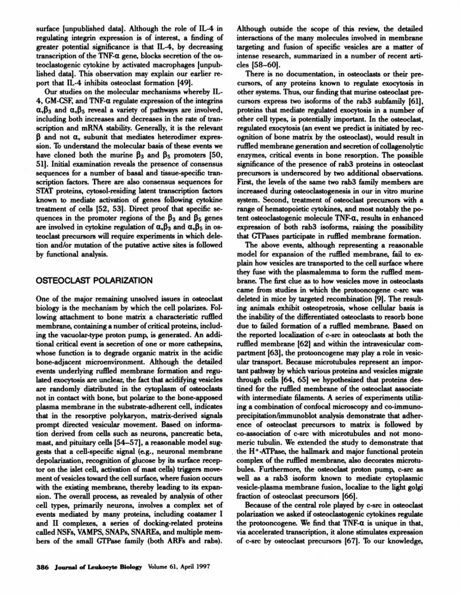
OSTEOCLAST POLARIZATION
One of the major remaining unsolved issues in osteoclast
biology is the mechanism by which the cell polarizes. Fol-
lowing attachment to bone matrix a characteristic ruffled
membrane, containing a number ofcritical proteins, includ-
ing the vacuolar-type proton pump, is generated. An addi-
tional critical event is secretion of one or more cathepsins,
whose function is to degrade organic matrix in the acidic
bone-adjacent microenvironment. Although the detailed
events underlying ruffled membrane formation and regu-
lated exocytosis are unclear, the fact that acidifying vesicles
are randomly distributed in the cytoplasm of osteoclasts
not in contact with bone, but polarize to the bone-apposed
plasma membrane in the substrate-adherent cell, indicates
that in the resorptive polykaryon, matrix-derived signals
prompt directed vesicular movement. Based on informa-
tion derived from cells such as neurons, pancreatic beta,
mast, and pituitary cells [54-57], a reasonable model sug-
gests that a cell-specific signal (e.g., neuronal membrane
depolarization, recognition of glucose by its surface recep-
tor on the islet cell, activation of mast cells) triggers move-
ment of vesicles toward the cell surface, where fusion occurs
with the existing membrane, thereby leading to its expan-
sion. The overall process, as revealed by analysis of other
cell types, primarily neurons, involves a complex set of
events mediated by many proteins, including coatamer I
and II complexes, a series of docking-related proteins
called NSFs, VAMPS, SNAPs, SNAREs, and multiple mem-
bers of the small GTPase family (both ARFs and rabs).
Although outside the scope of this review, the detailed
interactions of the many molecules involved in membrane
targeting and fusion of specific vesicles are a matter of
intense research, summarized in a number of recent arti-
des [58-60].
There is no documentation, in osteoclasts or their pre-
cursors, of any proteins known to regulate exocytosis in
other systems. Thus, our finding that murine osteoclast pre-
cursors express two isoforms of the rab3 subfamily [61J,
proteins that mediate regulated exocytosis in a number of
other cell types, is potentially important. In the osteoclast,
regulated exocytosis (an event we predict is initiated by rec-
ognition of bone matrix by the osteoclast), would result in
ruffled membrane generation and secretion of collagenolytic
enzymes, critical events in bone resorption. The possible
significance of the presence of rab3 proteins in osteoclast
precursors is underscored by two additional observations.
First, the levels of the same two rab3 family members are
increased during osteoclastogenesis in our in vitro murine
system. Second, treatment of osteoclast precursors with a
range of hematopoietic cytokines, and most notably the po-
tent osteoclastogenic molecule TNF-a, results in enhanced
expression of both rab3 isoforms, raising the possibility
that GTPases participate in ruffled membrane formation.
The above events, although representing a reasonable
model for expansion of the ruffled membrane, fail to ex-
plain how vesicles are transported to the cell surface where
they fuse with the plasmalemma to form the ruffled mem-
brane. The first clue as to how vesicles move in osteoclasts
came from studies in which the protooncogene c-src was
deleted in mice by targeted recombination [91. The result-
ing animals exhibit osteopetrosis, whose cellular basis is
the inability of the differentiated osteoclasts to resorb bone
due to failed formation of a ruffled membrane. Based on
the reported localization of c-src in osteoclasts at both the
ruffled membrane [62] and within the intravesicular com-
partment [63J, the protooncogene may play a role in vesic-
ular transport. Because microtubules represent an impor-
tant pathway by which various proteins and vesicles migrate
through cells [64, 65] we hypothesized that proteins des-
tined for the ruffled membrane of the osteoclast associate
with intermediate filaments. A series of experiments utiliz-
ing a combination of confocal microscopy and co-immuno-
precipitation/immunoblot analysis demonstrate that adher-
ence of osteoclast precursors to matrix is followed by
co-association of c-src with microtubules and not mono-
meric tubulin. We extended the study to demonstrate that
the H�-ATPase, the hallmark and major functional protein
complex of the ruffled membrane, also decorates microtu-
bules. Furthermore, the osteoclast proton pump, c-src as
well as a rab3 isoform known to mediate cytoplasmic
vesicle-plasma membrane fusion, localize to the light golgi
fraction of osteoclast precursors [661.
Because of the central role played by c-src in osteoclast
polarization we asked if osteoclastogenic cytokines regulate
the protooncogene. We find that TNF-a is unique in that,
via accelerated transcription, it alone stimulates expression
of c-src by osteoclast precursors [67]. To our knowledge,
ACKNOWLEDGMENTS
Teitelbaum et a!. Molecular mechanisms of hone resorption 387
Fig. 4. Model ofosteoclast polarization. The osteoclast attaches to bone
via the integrin a433, resulting in tubulin polymerization. Unidentified
signals stimulate movement along microtubules of intracellular vesicles,
which bear functional proteins targeted to the bone-apposed plasma mem-
brane. Vesicle generation and targeting is a complex set of events involv-
ing a large number of facilitatory proteins. Fusion of vesicles results in
production of the characteristic ruffled membrane.
this represents the first report of transcriptional regulation
of c-src. Taken together, our findings suggest a model in
which movement of vesicles containing functionally impor-
tant proteins destined for the osteoclast ruffled membrane,
move along a track comprised of microtubules (Fig. 4).
SUMMARY
Recognition that the osteoclast is a member ofthe monocyte/
macrophage family has prompted development of meaning-
ful experimental models to study the cellular mechanisms
of bone resorption. These efforts have delineated molecu-
lar targets to inhibit the resorptive process and thus prevent
diseases such as osteoporosis. Many concerns remain to be
addressed. Among the most provocative is the means by
which the osteoclast polarizes and the impact of matrix rec-
ognition and cytokines on this event. The role of tissue-
specific transcription factors in osteoclast commitment is
also fertile area for investigation. The tools are at hand to
address these issues and one may expect progress in under-
standing osteoclast biology to continue to impact patient
care.
This study was supported in part by National Institutes of
Health Grants DE05413, AR32788 (S. L. T.), AR42404,
AR42378 (F. P. R.), AR44089 (M. M. T.) and a grant from
the Shriners Hospital for Crippled Children, St. Louis Unit,
St. Louis, MO (S. L. T.).
REFERENCES
1. Teitelbaum, S. L., Tondravi, M. M., Ross, F. P. (1996) Osteodast biology.
In Osteopomsi.s (Marcus, R.. Feldman, D., and Kelsey, J. eds.), San Diego:Academic Press, 61-94.
2. Coccia, P. F., Krivit, W., Cervenka, J. (1980) Successful bone marrow trans-
plantation for infantile malignant osteopetrosis. N. Engl. J. Med. 302,702-708.
3. Kahn, A. J., Simmons, D. J. (1975) Investigation of cell lineage in boneusing a chimaera of chick and quail embryonic tissue. Nature 258,
325-327.
4. Walker, D. G. (1973) Osteopetrosis cured by temporary parabiosis. Science180, 275.
5. Shioi, A., Ross, F. P., Teitelbaum, S. L. (1994) Enrichment of generatedmurine osteoclasts. Calc�f Tissue hit. 55, 387-394.
6. Tondravi, M. M., McKercher, S., Anderson, K., Erdmann, J. M., Quiroz,M., Maki, R., Teitelbaum, S. L. (1996) Targeted deletion of PU.l inducesosteopetrosis rescuable by bone marrow transplantation. Nature, in press.
7. Grigoriadis, A. E., Wang, Z., Cecchini, M. G., Hofstetter. W., Felix, R.,
Fleisch, H. A., Wagner, E. F. (1994) c-Fos: A key regulator of osteoclast-
macrophage lineage determination and bone remodeling. Science 266.443-448.
8. Felix, R., Cecchini, M. G., Fleisch, H. (1990) Macrophage colony stimulat-ing factor restores in vivo bone resorption in the op/op mouse. Endocrinol.
127, 2592-2594.9. Soriano, P., Montgomery, C., Geske, R., Bradley, A. (1991) Targeted disrup-
tion of the c-sir proto-oncogene leads to osteopetrosis in mice. Cell 64,693-702.
10. Yamamoto, T., Kurihara, N., Yamaoka, K., Ozono, K., Okada, M., Yama-moto, K., Matsumoto, S., Michigami, T., Ono, J., Okada, S. (1993) Bone
marrow-derived osteoclast-like cells from a patient with craniometaphyseal
dysplasia lack expression of osteoclast-reactive vacuolar proton pump. .1
Clin. Invest. 91, 362-367.1 1 . Sly, W. S., Hewett-Emmett, D., Whyte. M. P., Yu, 1’., Tashian, R. E. (1983)
Carbonic anhydrase II deficiency identified as the primary defect in theautosomal recessive syndrome of osteopetrosis with renal tubular acidosisand cerebral calcification. hoc. Nail. Aco4. Sci. USA 80, 2752-2756.
12. Kodama, H., Yamasaki, A., Nose, M., Niida, S., Ohgama, Y., Abe. M., Ku-megawa, M., Suds, T. (1991) Congenital osteoclast deficiency in osteopetro-tic (op/op) mice is cured by injections of macrophage colony-stimulating fac-
tor. j Exp. Med. 173, 269-272.13. Wiktor-Jedrzejczak, W., Bartocci, A., Ferrante, A. W. J., Ahmed-Ansari, A..
Sell, K. W., Pollard, J. W., Stanley, E. R. (1990) Total absence of colony.
stimulating factor 1 in the macrophage-deficient osteopetrotic (op/op)
mouse. Proc. Nail. Aced. Sci. USA 87, 4828-4832.14. Zhang, D.-E., Hetherington, C. J., Chen, H.-M., Tenen, D. G. (1994) The
macrophage transcription factor PU.1 directs tissue-specific expression of
the macrophage colony-stimulating factor receptor. MoL Cell. Biol. 14,373-381.
15. Boyce, B. F., Yoneda, T., Lowe, C., Soriano, P., Mundy, G. R. (1992) Re-
quirement of pp60c-src expression for osteoclasts to form ruffled borders
and resorb bone in mice. J. Clin. Invest. 90, 1622-1627.16. Alvarez, J. I., Teitelbaum, S. L., Blair, H. C., Greenfield, E. M., Athanasou,
N. A., Ross, F. P. (1991) Generation of avian cells resembling osteoclastsfrom mononuclear phagocytes. Endocrinol. 128, 2324-2335.
17. Blair, H. C., Kahn, A. J., Crouch, E. C., Jeffrey, J. J., Teitelbaum, S. 1.
(1986) Isolated osteoclasts resorb the organic and inorganic componentsof bone� J. Cell Bail. 102, 1164-1172.
18. Blair, H. C., Teitelbaum, S. L., Ghiselli, R., Gluck, S. (1989) Osteoclasticbone resorption by a polarized vacuolar proton pump. Science 245,855-857.
19. Chatterjee, D., Chakraborty, M., Leit, M., Neff, L., Jamsa-Kellokumpu, S.,
Fuchs, R., Baron, R. (1992) Sensitivity to vanadate and isoforms of sub-
units A and B distinguish the osteoclast proton pump from other vacuolarH� ATPases. Proc. Nail. Acad� Sci. USA 89, 6257-6261.
20. Mattsson, J. P., Schlesinger, P. H., Keeling, D. J., Teitelbaum, S. L., Stone,D. K., Xie, X. (1994) Isolation and reconstitution of a vacuolar-type protonpump of osteoclast membranes. J. Bail. Chem. 269, 24979-24982.

388 Journal of Leukocyte Biology Volume 61, April 1997
21. Teti, A., Blair, H. C., Teitelbaum, S. L., Kahn, A. J., Koziol, C. M., Konsek,J., Zambonin-Zallone, A., Schlesinger, P. (1989) Cytoplasmic pH regulationand chloride/bicarbonate exchange in avian osteoclasts. J. Clin. Invest. 83,227-233.
22. Blair, H. C., Teitelbaum, S. L., Tan, H., Koziol, C. M., Schlesinger, P. H.
(1991) Passive chloride permeability charge coupled to H�-ATPase of avianosteoclast ruffled membrane. Am. J. Physiol. 260, C1315-C1324.
23. Robey, P. G., Boskey, A. L. (1996) The biochemistry of bone. In Osteo-
porosi.c (Marcus, R. , Feldman, D., and Kelsey, J. eds.), San Diego: Aca-
demic Press, 95-183.
24. Blair, H. C., Teitelbaum, S. L., Grosso, L. E., Lacey, D. L., Tan, H., McCort,D. W., Jeffrey, J. J. (1993) Extracellular matrix degradation at acid pH: Avianosteoclast acid collagenase isolation and characterization. Biochem. .1 290,873-884.
25. Sasaki, 1., Ueno-Matsuda, E. (1993) Cysteine-proteinase localization in os-teoclasts: an immunocytochemical study. Cell Tissue Res. 271, 177-179.
26. Drake, F. H., Dodds, R. A., James, I. E., Connor, J. R., Debouck, C.,
Richardson, S., Leerykaczewski, E., Coleman, L., Rieman, D., Barthlow,
R., Hastings, G., Gowen, M. (1996) Cathepsin K, but not Cathepsins B, L,or S, is abundantly expressed in human osteoclasts. J. Bail. Chem. 271,12511- 12516.
27. Gelb, B. D., Shi, G. P., Chapman, H. A., Desnick, R. J. (1996) Pycnodysos-
tosis, a lysosomal disease caused by cathepsin K deficiency. Science 273,1236-1238.
28. Blair, H. C., Teitelbaum, S. L., Schimke, P. A., Konsek, J. D., Koziol,
C. M., Schlesinger, P. H. (1988) Receptor-mediated uptake of mannose-6-phosphate bearing glycoprotein by isolated chicken osteoclasts. J. Cell
PhysioL 137, 476-482.29. Ross, F. P., Alvarez, J. I., Chappel, J., Sander, D., Butler, W. T., Farach-
Carson, M. C., Mintz, K. A., Robey, P. G., Teitelbaum, S. L., Cheresh, D. A.(1993) Interactions between the bone matrix proteins osteopontin and bonesialoprotein and the osetoclast integrin a��3 potentiate bone resorption. J.BaiL Chem. 268, 9901-9907.
30. Engleman, V. W., Nickols, G. A., Ross, F. P., Horton, M. A., Settle, S. L.,
Ruminski, P. G., Teitelbaum, S. L. (1996) A peptidomimetic antagonist ofthe a,�l3 integrin inhibits bone resorption in vitro and prevents osteoporo-sis in vivo. _t Clin. Invest. , in press.
31. Clohisy, D. R., Bar-Shavit, Z., Chappel, J., Teitelbaum, S. L. (1987)
1,25-dihydroxyvitamin D3 modulates bone marrow macrophage precursorproliferation and differentiation: Upregulation of the mannose receptor. J.Biol. Chem. 262, 15922-15929.
32. Perkins, S. L., Teitelbaum, S. L. (1991) 1,25-dihydroxyvitamin D3 modu-lates colony stimulating factor-i receptor binding by murine bone marrowmacrophage precursors. Endocrinol. 128, 303-311.
33. Bar-Shavit, Z., Teitelbaum, S. L., Reitsma, P. Hall, A., Pegg, L. E., Trial,
J., Kahn, A. J. (1983) Induction of monocytic differentiation and bone re-sorption by i,25-dihydroxyvitamin D3. Proc. Nail. Acad. Sci. USA 80,5907-5911.
34. Medhora, M. M., Teitelbaum, S. 1., Chappel, J., Alvarez, J., Mimura, H.,Ross, F. P., Hruska, K. (1993) la.25-dihydroxyvitamin D3 up-regulates cx-pression of the osteoclast integrin a413. J. Bail. Chem. 268, 1456-1461.
35. Mimura, H., Cao, X., Ross, F P., Chiba, M., Teitelbaum, S. L. (1994)1,25(OH)2D3 vitamin D3 transcriptionally activates the �33-integrin subunitgene in avian osteoclast precursors. Endocrinol. 134, 1061-1066.
36. Chiba, M., Teitelbaum, S. L., Cao, X., Ross, F. P. (1996) Retinoic acid stim-ulates expression of the functional osteoclast integrin a433. Transcriptional
activation of the 133 but not a� gene. J. Cell Biochem., in press.
37. Li, C., Ross, F. P., Cao, X., Teitelbaum, S. L. (1995) Estrogen enhances
a,�33-integnn expression by avian osteoclast precursors via stabilization of
I33-integnn mRNA. MoL Endocrinol., in press.38. Can, X., Ross, F. P., Zhang. L., MacDonald, P. N., Chappel, J., Teitelbaum,
S. L. (1993) Cloning of the promoter for the avian integrin p3-subunit gene
and its regulation by 1,25-dihydroxyvitamin D3. I. Bail. Chem. 268,27371-27380.
39. Cao, X., Teitelbaum, S. L., Zhu, H., Zhang, L., Feng, X., Ross, F. P. (1996)Competition for a unique response element mediates retinoic acid inhibi-tion of vitamin D3-stimulated transcription. I. Bail. Chem. 271, 20650-20654.
40. Matayoshi, A., Brown, C., DePersio, J. F., Haug, J., Abu-Amer, Y.. Liapis,H., Kuestner, R., Pacifici, R. (1996) Human blood-mobilized hematopoi-etic precursors differentiate into osteoclasts in the absence of stromal cells.Proc. Nail. Acad. Sci. USA 93, 10785-10790.
41. Kitazawa, R., Kimble, R. B., Vannice, J. L., Kung, V. T., Pacifici, R. (1994)
Interleukin-i receptor antagonist and tumor necrosis factor binding protein
decrease osteoclast formation and bone resorption in ovariectomized mice.
J. Clin. Invest. 94, 2397-2406.
42. Ammann, P., Garcia, I., Rizolli. R., Meyer, J.. Vassalli. P.. Bonjour. J.(1995) Transgenic mice expressing high levels of soluble tumor necrosis
factor receptor-i fusion protein are protected from bone loss caused by estro-
gen deficiency. J. Bone Mm. Res. 10, 5139. (Abstract)43. Inoue, M., Teitelbaum, S. L., Abu-Amer, Y., Ross, F. P. (1996) Tumor ne-
crosis factor a regulates a435 integrin expression by osteoclast precursors
in vitro and in vivo. J. Bone Mm. Res. 11, 5159. (Abstract)44. Lewis, D. R, Liggitt, H. D., Effmann, E. L., Motley, S. T., Teitelbaum, S. L.,
Jepsen, K. J. , Goldstein, S. A., Bonadio, J. , Carpenter, J., Perlmutter, R. M.
(1993) Osteoporosis induced in mice by overproduction of interleukin 4.Proc. Nail. Acad. Sci. USA 90, 11618-11622.
45. Shioi, A., Teitelbaum, S. L., Ross, F. P., Welgus, H. G., Ohara, J., Suzuki,
H., Lacey, D. L. (1991) Interleukin 4 inhibits murine osteoclast formationin vitro. J. l2e11. Biochem. 47. 1-6.
46. Lacey, D. L., Erdmann, J. M., Teitelbaum, S. L., Tan, H.. Ohara, J., Shioi,A. (1995) Interleukin-4, interferon gamma and prostaglandin E impact theosteoclast forming potential of murine bone marrow macrophages in vitro.
Endocrinol. 136, 2367-2376.47. Bizzarri, C., Shioi, A., Teitelbaum. S. L., Ohara, J., Harwalker. V. A., Erd-
mann, J. M., Lacey, D. L., Civitelli, R. (1994) Interleukin-4 inhibits boneresorption and acutely increases cytosolic Ca2� in murine osteoclasts. J.Bail. Chem. 269, 13817-13824.
48. Kitazawa, S., Ross, F. P., McHugh, K., Teitelbaum, S. L. (1995) Interleukin-4 induces expression of the integrin a433 via transactivation of the L�3
gene. J. Bail. Chem. 270, 4115-4120.49. Lacey, D. L., Erdmann, J. M., Tan, H. (1994) Interleukin 4 increases type
5 acid phosphatase mRNA expression in murine bone marrow macro-
phages. J. Cell. Biochem. 54, 365-371.50. Feng, X., Teitelbaum, S. L., Tondravi, M., Ross, F. P. (1996) Cloning the
promoter of the mouse integrin � subunit gene. J Bone Mm. Res. 11,5145. (Abstract)
Si. McHugh, K., Teitelbaum, S. L., Kitazawa, S., Ross, F. P. (1994) Cloning
and characterization of the murine integrin �3 gene promoter. J. Bone Mm.Res. 9, 5248. (Abstract)
52. Darnell, J. E., Jr., Kerr, I. M., Stark, G. R. (1994) Jak-STAT pathways and
transcriptional activation in response to IFNs and other extracellular signal-
ing proteins IReviewj. Science 264, 1415-1421.
53. Ihle, J. N. (1995) Cytokine receptor signalling. Naiwe 377. 591-594.54. Geppert. M., Bolshakow, V. Y., Siegelbaum, S. A., Takei, K., Dc Camilli,
P., Hammer, R. E., Sudhof, 1’. C. (1994) The role of Rab3A in neurotrans-
mifter release. Nature 369, 493-497.55. Lledo, P., Vernier, P., Vincent, J., Mason, W. T., Zorec, R. (1993) Inhibition
of rab3B expression attenuates Ca2+.dependent exocytosis in rat anterior
pituitary cells. Nature 364, 540-544.56. Oberhauser, A. F., Monck, J. R., Balch, W. E., Fernandez, J. M. (1992)
Exocytotic fusion is activated by Rab3a peptides. Nature 360, 270-273.57. Piiper, A., Stryjek-Kaminska, D., Jahn, R., Zeusem, S. (1995) Stimulation
of inositol 1,4,5-trisphosphate production by peptides corresponding to the
effector domain of different Rab3 isoforms and cross-linking of an effector
domain peptide target. Biochem. J. 309, 621-627.58. Fisher Von Mollard, C., Stahl, R. Li, C., Sudhof, T., Jahn, R. (1994) Rab
proteins in regulated exocytosis. Trends Bail. Sci. 19, 164-168.59. Schekman, R., Orci, L. (1996) Coat proteins and vesicle budding. Science
271, 1526-1538.60. Sudhof, T. C. (1995) The synaptic vesicle cycle: a cascade of protein-protein
interactins. Nature 375, 645-653.61. Abu-Amer, Y., Teitelbaum, S. L, Chappel, J., Zong, Q.. Ross, F. P. (1995)
Regulation of a Rab protein which mediates regulates exocytosis and co-
localizes with ruffled membrane-associated proteins. J. Bone Mm. Res. 10,Si50. (Abstract)
62. Tanaka, S., Takahashi, N., Udagawa, N., Sasaki, T., Fukui, Y., Kurokawa,
T., Suda, T. (1992) Osteodasts express high levels of pp60�, preferen-tially on ruffled border membranes. FEBS Len. 313, 85-89.
63. Home, W. C., Neff, L., Chatterjee, D., Lomri, A., Levy, J. R, Baron, R.(1992) Osteoclasts express high levels of pp6Oe� in association with intra-cellular membranes. J. Cell BaiL 119. 1003-1013.
64. Fath, K. R., Mamajiwalla, S. N., Burgess. D. R. (1993) The cytoskeletonin development of epithelial cell polarity. J. Cell Sci. 17 (Suppl.), 65-73.
65. Raff, E. C. (1994) The role of multiple tubulin isoforms in cellular micro-tubule function. In Micmtubules, New York: Wiley-Liss. 85-109.
66. Abu-Amer, Y., Ross, F. P., Schlesinger, P.. Tondravi, M. M., Teitelbaum,
S. L. (1996) Substrate recognition by osteoclast precursors induces s-crc/
microtubule association. J. Cell BaiL, in press.
67. Abu-Amer, Y., Ross, F. P., Zhu, H., Chappel, J., Teitelbaum, S. L. (1996)
Tumor necrosis factor induces pp60c� expression in murine osteoclast
precursors. J. Bone Mm. Res. 11. 5312. (Abstract)


