Embed Size (px)
Citation preview

NeuroImage 83 (2013) 837–848
Contents lists available at SciVerse ScienceDirect
NeuroImage
j ourna l homepage: www.e lsev ie r .com/ locate /yn img
Overlapping and parallel cerebello-cerebral networks contributing tosensorimotor control: An intrinsic functional connectivity study
Judy A. Kipping a, Wolfgang Grodd e, Vinod Kumar e, Marco Taubert a,Arno Villringer a,c,d, Daniel S. Margulies b,⁎a Department of Neurology, Max Planck Institute for Human Cognitive and Brain Sciences, Stephanstrasse 1a, 04103 Leipzig, Germanyb Max Planck Research Group: Neuroanatomy & Connectivity, Max Planck Institute for Human Cognitive and Brain Sciences, Stephanstrasse 1a, D-04103 Leipzig, Germanyc Center for Stroke Research Berlin, Charité–Universitätsmedizin Berlin, Charitéplatz 1, 10117 Berlin, Germanyd Berlin School of Mind and Brain, Humboldt University, Luisenstrasse 56, 10117 Berlin, Germanye Psychotherapy and Psychosomatics, University Hospital Aachen, Department of Psychiatry, Pauwelsstraße 30, 52074 Aachen, Germany
⁎ Correspondingauthor at:Max Planck Institute forHumStephanstrasse 1a, D-04103 Leipzig, Germany.
E-mail address: [email protected] (D.S. Margul
1053-8119/$ – see front matter © 2013 Elsevier Inc. Allhttp://dx.doi.org/10.1016/j.neuroimage.2013.07.027
a b s t r a c t
a r t i c l e i n f oArticle history:Accepted 9 July 2013Available online 16 July 2013
Keywords:CerebellumfMRIParietalPrefrontalResting state
In concert with sensorimotor control areas of the cerebrum, the cerebellum shows differential activation pat-terns during a variety of sensorimotor-related tasks. However, the spatial details and extent of the complexand heterogeneous cerebello-cerebral systems involved in action control remain uncertain. In this study, weuse intrinsic functional connectivity (iFC) to examine cerebello-cerebral networks of five cerebellar lobules(I–IV, V, VI, and VIIIa/b) that have been empirically identified to form the functional basis of sensorimotor pro-cesses. A refined cerebellar seed-region selection allowed us to identify a network of primary sensorimotorand supplementary motor areas (I–V), a network of prefrontal, premotor, occipito-temporal and inferior-parietal regions (VI), and two largely overlapping networks involving premotor and superior parietal regions,the temporo-parietal junction as well as occipito-temporal regions (VIIIa/b). All networks involved themedialprefrontal/cingulate cortex. These cerebral clusters were used in a partial correlation analysis to systematicallymap cerebral connectivity throughout the entire cerebellum. We discuss these findings in the framework ofaffective and cognitive control, sensorimotor, multisensory systems, and executive/language systems. Withinthe cerebellum we found that cerebro-cerebellar systems seem to run in parallel, as indicated by distinctsublobular functional topography of prefrontal, parietal, sensorimotor, cingulate, and occipito-temporal re-gions. However, all areas showed overlapping connectivity to various degrees in both hemispheres. The re-sults of both analyses demonstrate that different sublobular parts of the cerebellar lobules may dominate indifferent aspects of primary or higher-order sensorimotor processing. This systems-level cerebellar organiza-tion provides a more detailed structure for cerebello-cerebral interaction which contributes to our under-standing of complex motor behavior.
© 2013 Elsevier Inc. All rights reserved.
Introduction
Throughout the evolution of mammals, the cerebellum and thecerebral cortex have evolved in parallel. Their respective sizes have pro-portionally increased, and the connections between both have also sub-stantially grown during phylogeny (Sultan, 2002; Sultan and Glickstein,2007). Thefirst combined cerebellar-cerebral functionwas coordinationof movement involving both motor planning and execution (Holmes,1939; Thach, 1997). In humans and great apes, the demands for sensorydiscrimination, sensorimotor integration and motor coordination haveincreased, requiring additional cerebellar contributions (Manto et al.,2012). The cerebellum contributes to diverse functions such as flexiblemotor action, execution of complexmovements and successful learning
an Cognitive and Brain Sciences,
ies).
rights reserved.
of new movement strategies (Smaers et al., 2011; Stein and Glickstein,1992; Thach, 1998). One cerebellar role is to provide predictive valuesabout the outcomeof an intended goal, which allows for the preparationof neural systems in the cerebrum that are likely to be responsive to thetask at hand (Courchesne and Allen, 1997). Both the estimated predic-tion of outcomes as well as the preparation of neural systems enablecost-optimal and goal-accomplishing movements.
In this study we specifically focused on widespread cerebral con-nectivity, which has been investigated in the primate brain usingtranssynaptic tract-tracing studies in the macaque monkey (Kellyand Strick, 2003; Lu et al., 2007; Middleton and Strick, 2000; Orioliand Strick, 1989). It has been demonstrated that the cerebellar outputis composed of a number of separate but parallel “output channels”(Middleton and Strick, 1997). The underlying anatomical relationshipof the sensorimotor system mainly comprises afferent connections ofcerebellar lobules III–VI and VIIb–VIII via the deep cerebellar nuclei tothe midbrain and thalamus, and subsequently to premotor and motor

838 J.A. Kipping et al. / NeuroImage 83 (2013) 837–848
cortex. Efferent connections project back via the pontine nuclei tolobules V–VI and VIIb–VIII in a closed-loop fashion (Kelly and Strick,2003).
Anatomical connections established in the macaque monkey havebeen indirectly validated in humans by corresponding activation pat-terns in imaging studies using positron emission tomography and func-tional magnetic resonance imaging (fMRI). fMRI movement paradigmsin humans, which involve simple and natural muscular flexion and ex-tension, have revealed movement-related cerebellar representationswhich mapped foot, tongue and hand, which are restricted to thelobules IV, V, VI and VIII (Grodd et al., 2001; Rijntjes et al., 1999).
Lobules IV and V are consistently activated during the planning andexecution of simple movements of body parts (Grodd et al., 2001;Schlerf et al., 2010). With respect to lower-level motor functions theseareas appear to have somatotopic organization,which is similar inmon-keys and cats (Snider and Eldred, 1952), aswell as in humans (Grodd etal., 2001). However, in addition to the movement-related cerebral in-puts, lobule V also receives proprioceptive information from spinal in-puts, whose signals both converge on the same Purkinje cells asshown in cats (Allen et al., 1974). Consequently, lobule V has beenfound to be activated during passive and active movements (Jueptneret al., 1997; Thickbroom et al., 2003; Wiestler et al., 2011), tactile stim-ulation (Wiestler et al., 2011) and in the prediction of the sensory con-sequences of a movement (Blakemore et al., 1998), indicating aninvolvement in sensory as well as motor processes.
With increasing complexity of coordinated movements as evokedby higher rates of tapping (Jancke et al., 1999) or sequential move-ments (Schlerf et al., 2010), parts of lobule VI have been shown to ac-company the motor-evoked activation in lobule V and more anteriorlobules using fMRI in humans. Lobule VI has also been found to bespecifically activated during the storage of acquired kinematic tooluse (Imamizu et al., 2000). An index finger movement task withself-paced delay showed a temporal dissociation in sensorimotor pro-cessing (Hulsmann et al., 2003). Whereas lobule VI was recruited 3 sprior to movement onset, activation in lobule V followed close to andafter movement onset. In contrast, Cui et al. (2000) report a lack offunctional dissociation between preparation and execution of coordi-nated movements within bilateral lobules V–VII. These diverse fMRIresults show that lobules IV, V, and VI may contribute to differentphases of sensorimotor events.
Lobule VIII is activated during simple active and passive sensorimo-tor tasks, and therefore has been proposed to possess similar functionsas lobule V, which is the representation of motor and sensory processes(Thickbroom et al., 2003). In both lobules, active performance aswell astactile stimulation reveal superimposed sensory and motor maps(Wiestler et al., 2011). However, despite this similarity, lobule VIII hasbeen found to play a role in sensory feedback accompanying basicmotor processing in lobule V (Habas and Cabanis, 2008) and in thetiming of more complex and voluntary movements (Habas et al.,2004). Recent exploration of lobule VIII found a functional contributionof lobular segments VIIIa andVIIIb tofinger tapping and verb generation(Stoodley et al., 2012).
The fMRI findings of overlapping cerebro-cerebellar networks in-volved in sensorimotor tasks complicate the functional assignmentsof cerebellar lobules IV, V, VI, and VIII. These cerebellar regions likelyreveal differential involvement in domains involved in planning, exe-cution, and control of movements and are thus engaged in variousbrain networks.
Evaluating human cerebello-cerebral connectivity using iFC
Three studies published in 2009 (Habas et al., 2009; Krienen andBuckner, 2009; O'Reilly et al., 2010) used iFC to depict major cerebro-cerebellar iFC networks within the sensorimotor as well as non-motordomains in humans. In addition to dedicated cortical areas, subcorticalcomponents—including the midbrain and thalamus (Middleton and
Strick, 1997; Sommer, 2003)—have been shown to participate in vari-ous iFC networks (Habas et al., 2009). The sensorimotor nature ofcerebellar lobules V, VI, and VIII (and particularly VIIIb; according to apeak cerebellar coordinate in Krienen and Buckner (2009)) has beenvalidated using iFC (Krienen and Buckner, 2009; O'Reilly et al., 2010).Buckner et al. (2011) recently demonstrated a further differentiation.They show that lobules IV and V are sensorimotor related, lobule VI ispart of the sensorimotor, ventral attention and fronto-parietal network,and lobule VIII is part of the sensorimotor and the ventral attention net-work. Very recently, Sang et al. (2012) investigated the cerebellar con-nectivity of the hemispheres and vermis. They found that lobules I–VIare connected to the visual and sensorimotor network; lobule VI tothe auditory, the salience and sensorimotor network; and lobule VIIIbto the salience, sensorimotor, task-positive and the default mode net-work. However, if the cerebellum is connected to various regionswithinthe cerebrum, the question remains as to what extent these cerebellarregions have overlapping or distinct connectivity patterns. The studyby Buckner et al. (2011) applied a winner-take-all algorithm, whichseparates regions based on their strongest connectivity to large-scalecerebral resting-state networks, but does not allow for overlapping con-nectivity patterns. In contrast, the study by Sang et al. (2012) usedone-sample t-tests for each lobule, which does not account for commonvariance across adjacent lobules, potentially introducing false positiveoverlapping connectivity results. Even though they quantified the con-nectivity maps of lobules VII and VIII, the more anterior cerebellar lob-ules were not addressed. Due to the variable methodology, it remainsunclear whether the cerebellar regions have overlapping or separateconnectivity patterns.
We therefore extended the examination of cerebellar lobules by in-troducing a refined seed definition for seed-based iFC analysis whichaddresses the false positive connectivity to adjacent cerebral andnon-brain tissue (see also Buckner et al. (2011)). This refinementconsisted of removing all voxels which were contaminated by adjacentnon-cerebellar tissue. To enhance our understanding of the separateand overlapping cerebello-thalamo-cerebral pathways, we first investi-gated connectivity of cerebellar lobules I–IV, V, VI, and VIIIa/b withlarge-scale cerebral networks. This cerebello-centric approach wasthen complemented by a voxelwise iFC analysis beginning in the cere-brum. We were thus able to systematically identify overlapping andparallel functional regions on a sublobular level. We interpret the cere-bellar mapping in the context of their associated cerebral functionalsubsystems.
Material and methods
Participants
We included 38 MR data sets from healthy participants (25female; mean age of 30.34 ± 7.51 years), distributed by the 1000Functional Connectome Project (http://fcon_1000.projects.nitrc.org/indi/pro/Berlin.html). Two resting state scans were acquired duringa single scanning session for each participant. Functional imageswere visually inspected to ensure that the ventral parts of the cere-bellum (esp. lobule VIIIa/b) were acquired. Only those data sets thatincluded the inferior portion of the cerebellum were included, thusrestricting this study to 38 of 50 data sets.
Data acquisition
MR images were acquired at a 3 T Magnetom Tim Trio Siemensscanner using a 12 channel phased-array head coil. In each of thetwo resting state scanning sessions, 200 whole brain volumes wereacquired including the entire cerebellum using echo-planar image(EPI) pulse sequences with 34 axial slices (TR = 2300 ms, TE =30 ms, flip angle 90°, slice thickness = 4 mm, 3 × 3 × 4 mm voxels,interleaved ascending slice acquisition, acquisition matrix = 64 × 64,

839J.A. Kipping et al. / NeuroImage 83 (2013) 837–848
bandwidth = 2232 Hz, duration = ~7.5 min). During the resting statescan participants were instructed to remain relaxedwhile keeping theireyes open. A high-resolution T1-weighted scan was acquired with aMPRAGE (magnetization-prepared rapid acquisition gradient echo) se-quence (TR = 2.3 s; TE = 2.98 ms; flip angle = 9°, FOV = 256; 176sagittal slices; voxel size = 1 × 1 × 1 mm) covering the whole brainfor anatomical reference.
Data preprocessing
Image preprocessing of the resting state data was performed withFSL (Smith et al., 2004), AFNI (Cox, 1996), SPM8 (http://www.fil.ion.ucl.ac.uk/SPM, Welcome Department of Imaging Neuroscience, Lon-don, UK), and in-house scripts written in MatLab (http://www.mathworks.com, Cambridge, UK).
The main preprocessing steps are described here, and are also avail-able in further detail in the scripts distributed by 1000 FunctionalConnectome Project (http://fcon_1000.projects.nitrc.org). The firstfour time points (9.2 s) of the two scanning sessions were discardedto reach equilibrium of magnetization. In SPM, the origins of functionaland anatomical data were reoriented to the anterior commissure forwithin-subject registration. Anatomical images were realigned toreoriented functional images. Sequential slice acquisition during EPI ac-quisition was accounted for by shifting each slice in time to be alignedwith the middle slice (FSL). Correction for small movement artifactswas performed by estimating the spatial deviations between themean functional reference image and each functional image (AFNI).The frequency spectrum was band-pass filtered (0.01–0.1 Hz) to re-move nuisance signals (AFNI). Drifts originating from the MR scannerwere removed by linearly detrending the functional data. In order to re-move signal fromnon-graymatter tissue, sixmotion parameters aswellas signals from white matter (WM) and cerebro-spinal fluid (CSF)masks were removed using multiple regression (FSL). Masks of WMand CSF resulted from tissue segmentation (FSL), were inspected visu-ally and modified such that only the tissue of interest was included.Global signal was not removed due to previous findings that global sig-nal is also partially of neuronal origin (Scholvinck et al., 2010). UsingSPM, anatomical data was segmented into gray matter, WM and CSF.By means of the transformation matrix from the segmentation step,functional images were brought into standard stereotactic MontrealNeurological Institute (MNI) space using spatial non-linear normaliza-tion (SPM) with a 2 mm3 voxel size. SPM normalization was used be-cause we wished to take advantage of the cerebellar normalizationprovided with the SPM toolbox SUIT (see below). Before smoothingthe cerebellumwasmasked tominimize the cross-structure signal con-tamination. Afterwards, a Gaussian kernel with 4 mm full-widthhalf-maximum (FWHM) was applied separately to the cerebrum (FSL).
Cerebellar normalization
Diedrichsen (2006) developed a spatially unbiased atlas templateof the cerebellum and brainstem (SUIT) and a cerebellum-only nor-malization (provided by SUIT) because whole-brain spatial normali-zation to MNI space can cause elongation along the cerebellar z-axis.Furthermore, the highly smoothed ICBM152 MNI template leavesthe fine cerebellar folia almost invisible. Therefore, cerebella wereseparately normalized using the SPM toolbox SUIT, which is in thespace defined byMNI152 template and uses the anatomical labels pro-vided by the MRI atlas from Schmahmann et al. (1999). The segmen-tation of the cerebellum was visually inspected and hand-correctedto ensure the accuracy of the mask. A spatial non-linear normalization(same as used for cerebral normalization) to the SUIT template wasperformed utilizing the individual cerebellar masks. The preprocessedfunctional data were resliced using the deformation matrix of the an-atomical normalization process and afterwards smoothed with a4 mm FWHM Gaussian kernel.
Cerebellar ROI definition and cerebello-cerebral signal separation
The iFC analysis is based on the hypothesis that different cerebellarlobules are involved in action control. Due to a lack of sublobularparcellation, we have utilized anatomical masks of cerebellar lobulesfor a voxelwise seed-based correlation analysis. Even though wewere interested in above mentioned ten anatomical lobules, all 20 an-atomical lobular ROIs (aROIs) were chosen from the SUIT template(Diedrichsen et al., 2009) in order to account for common varianceacross adjacent lobules (Fig. 2B). Lobular binary masks are based onthe maximum probability maps of the SUIT atlas (Diedrichsen et al.,2009). The signals within each aROI includes voxels that are contami-nated by non-cerebellar signals from the surrounding tissue such asCSF or adjacent visual cortex. The following method aimed to removesuch voxels from further analysis. In order to determine whether avoxel contained more cerebellar than cerebral signal, we took advan-tage of the high correlation between interhemispheric cerebral homo-logs (Stark et al., 2008). The following analysis was thus based on theassumption that pure cerebellar signals are more correlated acrosshemispheres than with adjacent cerebral signals. A higher correlationbetween time series of cerebellar homologs as compared to non-homologs provides support for a similar interhemispheric synchronyin the cerebellum as previously observed in the cerebrum (Stark et al.,2008). Supplementary Fig. 1 demonstrates the mean intracerebellarcorrelation.
20 anatomical cerebellar masks were functionally constrained byseparating the voxels, whose time series correlated more with either acerebellar or cerebral signal. Themean BOLD signal from the homotopiccerebellar region (Fig. 1, step 1b) was used as the “cerebellar signal”whereas the mean BOLD signals from ipsilateral adjacent cerebralareas (lingual gyrus, fusiform gyrus, parahippocampal gyrus, and lateraloccipital gyrus) were utilized as “noise signals” (Fig. 1, step 1c): Let xnbe a voxel,where n is the index of a voxel in a cerebellarmaskof interest(Fig. 1, step 1a and 2), y is themean signal of the contralateral homolog(Fig. 1, step 3a). Then voxel xc is a functionally relevant cerebellar voxel,if the correlation between xn and y was stronger than between xn andany of the mean signals of adjacent cerebral areas (Fig. 1, step 3b)using awinner-take-all algorithm (Fig. 1, step 4). For each of the two in-dividual scans, two binary masks with voxels, which were either morestrongly correlated to the “cerebellar” signal or to a “noise” signal,were created (Fig. 1, step 5). The intersection of the two “cerebellar”masks entered a group-level comparison. On the group-level, a probabi-listic cerebellar map was created and arbitrarily thresholded at 85% (32out of 38 subjects), i.e. those remaining “cerebellar” voxelswere consid-ered as functionally relevant within the anatomical mask (Fig. 1, step 6and 7). After labeling the functionally relevant voxels according to theanatomical masks, the cerebello-cerebral separation processwas iterat-ed a second time (Fig. 1, repetition of steps 1a, 1c, 2–7). But instead ofcorrelating cerebellar voxels' time series to a mean signal of thehomotopic cerebellar lobule, the mean signals of the new set of func-tionally relevant cerebellar voxels' time series were taken as references(Fig. 1, step 8).
Seed-based correlation analysis
Within the anatomical masks (aROIs) only functionally-relevantcerebellar voxels were used to examine cerebello-cerebral iFC ofcerebellar lobules I–IV, V, VI, and VIIIa/b (scheme in Fig. 2A, left). How-ever, all 20 cerebellar fROIs were used as weights in the followingANOVA. Therefore, the mean time series of functionally relevantvoxels within each of the 20 lobules (fROIs) were correlated with allvoxels within the cerebrum (Fig. 1, steps 9 and 10). The resulting ma-trices of Pearson's product moment correlation coefficients wereFisher's r-to-z transformed (two per individual) and averaged withinindividuals for each of the 20 fROIs (Fig. 1, step 10). For the group-level, the r-to-z transformed maps of each lobule entered a one-way

Fig. 1. Methods schematic for seed selection on the single-subject level (brown areas) and cerebral and cerebellar iFC map generation (gray areas). Numbers indicate the stepwiseprocesses (see text). TS, time series; iFC, intrinsic functional connectivity.
840 J.A. Kipping et al. / NeuroImage 83 (2013) 837–848
mixed-effects ANOVA (analysis of variance) with 20 levels of a singlefactor, which represented the 20 lobules (Fig. 1, step 11). While thisfactor remained fixed, subjects were considered to be random.We in-vestigated the fraction of the total variance that can be explained byeach lobule. Therefore, each lobule was contrasted against all otherlobules in a weighted-ANOVA. The contrast values for each lobule rep-resented weights which indicated how strongly a mean lobular signalwas correlated to the signals of the other lobules. Therefore, all 20 sig-nals from the cerebellum were correlated within each individual.Pearson's product moment correlation coefficients were Fisher'sr-to-z transformed and averaged across individuals (Fig. 1, step 12).
Fig. 2. (A) Analysis scheme of a voxelwise cerebello-cerebral analysis and a subsequent voxefirst subcomponents of large-scale cerebral networks, which are connected to different cerbeen identified in the cerebellum. (B) Anatomical cerebellar masks on a partially inflated suence (Diedrichsen, 2006; Van Essen et al., 2001). (C) Anatomical masks (aROIs) and functioncoronal slices to show the reduction of voxels at cerebellar borders.
For the purpose of contrasting, r-to-z transformed values were set toa range between 0 and 1 by dividing each r-to-z value by the sum ofr-to-z values (Fig. 1, step 13). A lobule of interest received value 1and all others the negative of the normalized z-value. High correla-tions yielded a high contrast value and low correlation values a lowcontrast value. The same analysis was performed with aROIs (insteadof fROIs). The r-to-z matrices using signals of fROIs and aROIs aredisplayed in Supplementary Fig. 1. All connectivity maps were multi-plied with a subcortico-cortical Harvard-Oxford mask (Desikan et al.,2006) and corrected for multiple tests over the voxels within thegray-matter mask (p b 0.001, voxelwise FDR corrected).
lwise cerebro-cerebellar analysis. The combination of both analyses aimed at revealingebellar lobules. Secondly, overlapping and unique connectivity of cerebral regions hasrface map (top) and on a cerebellar flat map (bottom) displayed for anatomical refer-al masks (fROIs) created by cerebello-cerebral signal separation are displayed on single

Table 1Number of voxels in anatomical and functional ROIs. Volume sizes, center of mass co-ordinates, and location of cerebral areas, which showed iFC to the respective cerebellarfROIs are reported and represent areas that have been used for partial correlationanalysis.
Lobule Size ofaROIs(voxels)
Size of fROIs(2nd run)
Size ofcerebralclusters(voxels)
MNI coordinates(x, y, z)
Cerebralclusters
Left I–IV 630 427 (67.78%) 46 10, −22, 71 Right SM*Right I–IV 662 404 (61.03%) – – –
Left V 779 730 (93.71%) 2656 34, −23, 60 Right SM1303 −28, −25, 62 Left SM848 1, −17, 53 Bilateral
SMA/CCRight V 738 696 (94.31%) 622 −31, −23, 61 Left SM
27 −2, −16, 52 Left SMA/CCLeft VI 1561 1436 (92%) 1546 50, −40, 43 Right IPL
455 −60, −34, 35 Left IPL1005 37, 46, 23 Right MFG494 −37, 43, 26 Left MFG987 44, 13, 6 Right IFG/INS501 −39, 9, 5 Left INS604 3, 17, 38 Bilateral CC136 25, 7, 61 Right PM104 56, −54, −3 Right OTC
Right VI 1418 1274 (98.84%) 1168 −40, 42, 17 Left MFG1045 −46, 11, 13 Left IFG/INS76 43, 7, 1 Right INS475 −4, 18 42 Bilateral CC364 −49, −42, 46 Left IPL139 −14, 5, 69 Left PM27 −54, −60, −4 Left OTC
Left VIIIa 847 426 (50.30%) 2423 54, −13, 18 Right TPJ1955 −54, −17, 17 Left TPJ1900 4, −2, 55 Bilateral
CC/PM1659 23, −50, 63 Right SPL897 −22, −49, 64 Left SPL318 −2, −85, 28 Bilateral OCC135 53, −61, 1 Right OTC65 −51, −67, 4 Left OTC119 28, −82, 31 Right OCC
Right VIIIa 798 356 (44.61%) 4155 −49, −13, 28 Left TPJ2380 55, −9, 18 Right TPJ2151 3, 2, 51 Bilateral CC1528 −25, −51, 61 Left SPL1189 24, −50, 63 Right SPL309 −46, −72, 5 Left OTC87 54, −63, 1 Right OTC66 −5, −85, 36 Left OCC54 21, −82, 40 Right OCC
Left VIIIb 712 183 (25.70%) 1040 22, −46, 65 Right SPL588 −21, −48, 66 Left SPL337 3, −5, 52 Bilateral CC227 58, −25, 30 Right TPJ206 −51, −29, 20 Left TPJ171 20, −7, 68 Right PM69 55, −64, 1 Right OTC
Right VIIIb 697 198 (28.41%) 2922 −35, −37, 49 Left SPL/TPJ1412 21, −47, 64 Right SPL581 56, −22, 23 Right TPJ588 1, −8, 52 Bilateral CC461 −20, −12, 67 Left PM158 25, −10, 63 Right PM139 −49, −74, 5 Left OTC42 53, −63, 0 Right OTC
*before erosion/dilation; A voxel has a volume of 2 mm3; aROIs, anatomical regions ofinterest; fROIs, functional regions of interest; CC, cingulate cortex; IFG, inferiorfrontal gyrus; INS, insula; IPL, inferior parietal lobe; MFG, middle frontal cortex; PM,premotor cortex; OCC, extrastriate areas of occipital cortex; OTC, occipito-temporalcortex; SMA, supplementary motor cortex; SM, pre- and postcentral gyrus; SPL,superior parietal lobe; TPJ, temporo-parietal junction.
841J.A. Kipping et al. / NeuroImage 83 (2013) 837–848
Cerebral connectivity within the cerebellum
The contrast analysis revealed connectivity patterns to cerebralareas. The subsequent analysis addressedwhether and how the connec-tivity of cerebral areas is distributed within and across cerebellar ana-tomical borders (Fig. 1, step 14). For this, the cerebral clusters servedas seed regions (scheme in Fig. 2A, right) and the analysis wasperformed for left and right cerebellar hemispheres separately. As afirst step, cerebral clusters were eroded and dilated to receive the larg-est supra-threshold clusters and to remove bridges between clusters.The following criteria have been chosen for cluster selection. First, werestricted our analysis to cerebral regions that were found for both leftand right cerebellar homologs. Second, for each cerebello-cerebral iFCmap separately, we collapsed the signals of homologous cerebral clus-ters, if present. Each of the sets of cerebral clusters (as defined by thefive cerebellar fROIs) was entered separately into a partial correlationanalysis to reduce contributions from global and third-party effects tospecific cerebro-cerebellar correlations (Hampson et al., 2002). Foreach of the two scans of each individual, the mean time series ofcerebral clusters which were connected to the five left fROIs was corre-lated with cerebellar time series of all ten left fROIs. The same correla-tion analysis was performed for cerebral clusters connected to theright cerebellum. Pearson's correlation coefficients were Fisher's r-to-ztransformed and averaged across the two individual scans. Voxelwiset-tests determined connectivity of each cerebral cluster at every cere-bellar voxel, so that we could identify how the connectivity is distribut-ed at the sublobular level, while allowing for separate or overlappingcerebral connectivity. To differentiate spatial patterns of those highlyconnected cerebellar and cerebral sets of regions, a more stringentthreshold was required (p b 0.00001, voxelwise FDR corr.). At a moreliberal threshold of p b 0.001 cerebral clusters showed substantial over-lap, some ofwhich occurred between regions as functionally separate asprefrontal and sensorimotor areas. For the purposes of visualization, wemapped cerebral cluster combinations in the cerebellar hemispheres, ifa combination was present in at least five voxels (b0.1% of all hemi-spheric fROIs) in the corresponding cerebellar hemisphere.
Results
Optimized functional connectivity of cerebellar lobules
All cerebellar lobules in both hemispheres were targeted to undergocerebello-cerebral signal separation. The number of voxels included inour five fROIs and aROIs (I–IV, V, VI, and VIIIa/b) are presented inTable 1, and the masks are presented in Fig. 2C. fROIs within lobules Vand VI included ~90% of voxels from the anatomical masks and seemedto be the least contaminated. In lobules I–IV ~70%, in lobule VIIIa ~50%,and in lobule VIIIb ~30% of voxels survived the cerebello-cerebral sepa-ration. The voxels remaining in the fROIs, irrespective of the lobule,were distant from the cerebellar border as can be seen in Fig. 2C. Fur-thermore, the mean SNR values for every lobule increased with reduc-tion in the number of “noise” voxels (Supplementary Fig. 2).
Delineating cerebello-cerebral subsystems
The results of this analysis represent the unique cerebello-thalamo-cerebral functional networks within the sensorimotor and associatedhigher-order sensorimotor systems. We found a sensorimotor networkinvolving cerebellar lobules I–V. Lobule VI was connected to a higher-order executive network. Lobules VIIIa/b were part of a multisensorynetwork. Lobules V, VI, and VIIIa/b were connected to various parts ofthe medial prefrontal/cingulate cortex, regions involved in affectiveand cognitive control.
Fig. 3 represents thresholded and color-coded cerebral and tha-lamic iFC patterns derived from the weighted-ANOVA using fROIs,where color labels indicate the corresponding cerebellar fROIs.
Table 2 contains the thalamic center-of-mass (CoM) coordinatesand their probability of belonging to a structurally defined thalamicregion based on the Thalamic Connectivity Atlas (Johansen-Berg et

Fig. 3. iFC maps based on the five different fROIs in each hemisphere. Thresholded and color-coded cerebral connectivity patterns are represented on partially inflated cerebral sur-face maps. Cerebral overlaps are displayed by means of translucent colors. (A) displays cerebral iFC maps of left cerebellar lobules I–IV, V, VI, and VIIIa/b (top) and correspondingcolor-coded thalamic maps (bottom). (B) shows iFC maps of right cerebellar lobules V, VI, and VIIIa/b (top) and corresponding color-coded thalamic maps (bottom). Left lobulesI–IV are connected to bilateral ventral primary sensorimotor areas, which are displayed in the enlarged circles. Left lobule V is connected to bilateral primary sensorimotor cortex(SM), supplementary motor cortex (SMA), and cingulate cortex (CC). Right lobule V is connected to left SM, and left SMA/CC. Left lobule VI is connected to bilateral inferior andmiddle frontal gyrus (IFG/MFG), bilateral superior and inferior posterior parietal lobe (IPL/SPL), occipito-temporal gyrus (OTC), bilateral medial prefrontal cortex (mPFC), CC,and right premotor cortex (PM). Right lobule VI shows iFC to left MFG, IFG, PM, IPL, mPFC/CC, and bilateral OCC. Left and right lobule VIIIa shows iFC to temporo-parietal junction(TPJ), OTC, SPL, PM, and mPFC/CC, OCC. Left and right lobule VIIIb is connected to SPL, TPJ, PM, mPFC/CC, OTC, OCC. Right lobule VIIIb also shows connectivity to left MFG.Color-coded regions within the thalamus are displayed below on six slices. The outlines represent the group of medial (green) and lateral (black) thalamic nuclei according tothe Morel et al. (1997). The arrows indicate the thalamic connectivity of left and right I–IV.
842 J.A. Kipping et al. / NeuroImage 83 (2013) 837–848
al., 2005) as well as a medial or lateral group of thalamic nuclei as de-fined by a multiarchitectonic atlas (Morel et al., 1997). In addition wereport the connectivity to subcortical structures. Unthresholded con-trast images of the weighted-ANOVA are presented in SupplementaryFig. 3.
Lobules I–IV
The iFC pattern of left lobules I–IV included ventral parts of theprecentral gyrus (SM) and the brain stem. The iFC of right and leftlobules I–IV was found in cross-lateralized lateral thalamus.

Table 2Connectivity to thalamic and subcortical areas of cerebellar lobules I–IV, V, VI, and VIIIa.Center of mass coordinates for thalamic regions are reported, along with the closestdistance to medial or lateral thalamic nuclei of the Morel atlas, as well as the probabi-listic connectivity to cerebral areas of these thalamic coordinates according to theOxford thalamic atlas. CA, caudate; GP, globus pallidus; M, primary motor cortex;PFC, pre-frontal cortex; PM, pre-motor cortex; PP, posterior-parietal cortex; PU, putamen;S, sensory cortex.
Lobule Coordinates(x, y, z)
ThalamicREGION
Probabilisticcerebralconnectivity
Subcortical regions
Right I–IV −18, −19, 4 Lateral 65% M, 35% PM –
– –
Left I–IV 20, −20, 2 Lateral 50% S, 39% M Brain stem– – –
Right V −14, −21, 3 Lateral 59% M, 38% S Bilat. PU, bilat. GP,brain stem
16, −19, 4 Lateral 45% PM, 41% MLeft V 15, −21, 3 Lateral 59% M, 38% M Bilat. PU, bilat. GP,
brain stem−13, −22, 2 Lateral 48% PM, 37% M
Right VI −9, −14, 8 Lateral 93% PFC Bilat. CA, bilat. PU,bilat. GP, brain stem
14, −10, 7 Lateral 76% PFC, 26% PMLeft VI 10, −14, 8 Lateral 90% PFC Bilat. CA, bilat. PU,
bilat. GP, brain stem−11, −13, 8 Lateral 93% PFC
Right VIIIa −10, −21, −2 Medial 36% PM, 36% M –
– – –
Left VIIIa 14, −23, 3 Lateral 34% S, 31% PP –
−12, −23, 2 Medial 44% M, 31% PP
843J.A. Kipping et al. / NeuroImage 83 (2013) 837–848
Lobule V
Left lobule V was connected to regions within the bilateral senso-rimotor cortex engulfing pre- and postcentral areas (SM), bilateralsupplementary motor area (SMA), and cingulate cortex (CC). iFC ofright lobule V was found in the left SM, SMA/CC. Left and right lobuleV included iFC to brainstem, bilateral lateral thalamus, bilateralglobus pallidus, and putamen (extending into the insular cortex).
Lobule VI
Left lobule VI connectivity was found in the bilateral middle fron-tal gyrus (MFG), bilateral inferior frontal gyrus (IFG; with extensiontowards the insular cortex), right premotor cortex (PM), bilateral in-ferior parietal lobe (IPL), bilateral occipito-temporal cortex (OTC), aswell as in bilateral medial occipital cortex (OCC). Right lobule VIwas connected to left MFG, left IFG (with extension towards the insu-lar cortex), left PM, left IPL, left OTC, and bilateral OCC. Both left andright lobule VI showed iFC to bilateral CC andmedial prefrontal cortex(mPFC) as well as subcortical connections including bilateral lateralthalamus, caudate, putamen, globus pallidus, and brain stem.
Lobule VIIIa
iFC of left and right lobule VIIIa was found in bilateral superior pa-rietal lobe (SPL), bilateral temporo-parietal junction (TPJ), bilateralCC/mPFC, which extended bilaterally towards PM, bilateral OTC, andbilateral OCC. Subcortical iFC was found in left lateral thalamus(right VIIIa) and bilateral lateral thalamus (left VIIIa).
Lobule VIIIb
iFC of left and right lobule VIIIb was found in bilateral SPL, TPJ, PM,mPFC/CC, and OTC. No subcortical iFC was found for either left orright lobule VIIIb.
We also performed the same analysis with aROIs (instead of fROIs)and carried out a two-way ANOVA in which we contrasted the iFCmaps of fROIs and aROIs. Despite high connectivity between cerebellarhomologs, connectivity that was lobular-specific and not mirrored bythe signal in the homologous lobulemight be superimposed by strongerconnectivity to one of the adjacent cerebral areas. The cerebello-cerebral signal separation excluded these voxels and might have beenoverly conservative. However, the direct contrast between aROIs andfROIs revealed that mainly noise-susceptible areas showed decreasesin connectivity. In Supplementary Fig. 4 we show decreased iFC inareas proximate to CSF, air, skull, the interhemispheric fissure and thetransverse and frontal sinuses, when using fROIs. Further, cerebralareas in parietal and prefrontal cortex, which have been found to becontaminated by physiological signal, also showed a decrease in iFC(Tong et al., 2011). Increases in iFC using fROIswere found in subcorticalareas such as thalamus, midbrain and basal ganglia as well as in corticalareas. These findings can be taken as an indication that fROIs improvethe functional relevance of cerebellar lobules.
Overlapping and separate topography of cerebral regions in thecerebellum
The detailed voxelwise correlation analysis between 18 cerebralclusters, which were defined based on the initial iFC analysis, andvoxels within cerebellar fROIs, determined cerebellar mapping of sep-arate and overlapping cerebral connectivity (Fig. 4). The cerebralclusters used in the partial correlation analysis included for left andright lobule V: SM and CC; lobule VI: IPL, MFG, IFG, OTC, CC, andPM; lobule VIIIa: TPJ, SPL, CC, OTC, and two regions in the OCC; andlobule VIIIb: SPL/TPJ, CC, PM, and OTC. For left and right lobules I–IV, no cluster survived the erosion/dilation procedure. The CoM coor-dinates and sizes of cerebral clusters are represented in Table 1.
Cerebellar voxels which showed connectivity from overlappingmPFC/CC regions of lobules V, VI, and VIIIa/b were collapsed (purplecolor in Fig. 4B). Partially overlapping parietal areas (temporo-parietaland posterior-parietal regions), which were connected with lobules VIand VIIIa/b were grouped together (Fig. 4B, cyan color). Connectivityof occipital and overlapping premotor regions were also grouped, butdid not survive the threshold and are not represented in the cerebellarmap. Supplementary Table 1 includes the 13 and 28 different cerebralcluster combinations for left and right cerebellar hemispheres respec-tively, and demonstrates how they were color-coded in Fig. 4B.
The mapping of left cerebellar regions I–X showed distributedconnectivity from parietal regions (35% of the entire cerebellar hemi-spheric fROIs), middle frontal cortex (16%), cingulate regions (13%),overlapping sensorimotor, cingulate, occipito-temporal and parietalregions (12%), sensorimotor regions (11%), overlapping sensorimotor,occipito-temporal, and parietal regions (4%), occipito-temporal (3%),overlapping occipito-temporal and parietal regions (3%), overlappingcingulate and parietal regions (2%), and overlapping middle frontalcortex and cingulate regions (1%).
In the right cerebellar hemisphere we identified connectivity fromsensorimotor regions (30% of the entire cerebellar hemisphericfROIs), middle frontal cortex (23%), overlapping sensorimotor, cingu-late, occipito-temporal and parietal regions (14%), overlapping senso-rimotor, prefrontal, occipito-temporal, and parietal regions (9%),cingulate regions (8%), overlapping sensorimotor, occipito-temporal,and parietal regions (6%), parietal regions (2%), overlapping middlefrontal and parietal regions (2%), overlapping inferior frontal, cingu-late, parietal, and occipito-temporal regions (2%), occipito-temporalregions (1%), overlapping middle frontal and cingulate regions (1%)and other combinations of cerebral connectivity (b1%).
Lobularwise, in left lobules I–Vwe found separate iFC from the sen-sorimotor cortex, cingulate, and occipito-temporal regions, whichoverlapped partially with parietal regions. Separate and overlappingiFC of cingulate and sensorimotor extended towards the superior
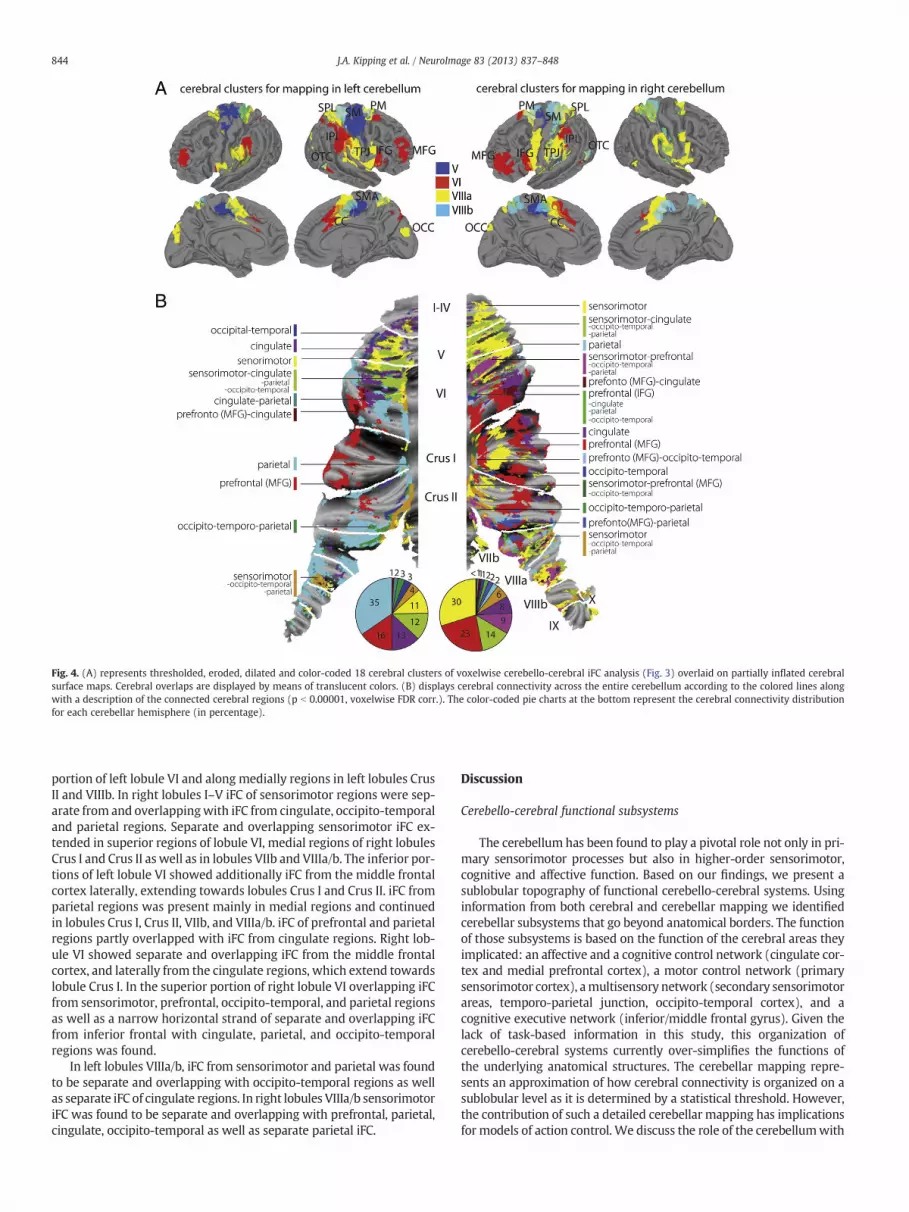
Fig. 4. (A) represents thresholded, eroded, dilated and color-coded 18 cerebral clusters of voxelwise cerebello-cerebral iFC analysis (Fig. 3) overlaid on partially inflated cerebralsurface maps. Cerebral overlaps are displayed by means of translucent colors. (B) displays cerebral connectivity across the entire cerebellum according to the colored lines alongwith a description of the connected cerebral regions (p b 0.00001, voxelwise FDR corr.). The color-coded pie charts at the bottom represent the cerebral connectivity distributionfor each cerebellar hemisphere (in percentage).
844 J.A. Kipping et al. / NeuroImage 83 (2013) 837–848
portion of left lobule VI and along medially regions in left lobules CrusII and VIIIb. In right lobules I–V iFC of sensorimotor regions were sep-arate from and overlappingwith iFC from cingulate, occipito-temporaland parietal regions. Separate and overlapping sensorimotor iFC ex-tended in superior regions of lobule VI, medial regions of right lobulesCrus I and Crus II as well as in lobules VIIb and VIIIa/b. The inferior por-tions of left lobule VI showed additionally iFC from the middle frontalcortex laterally, extending towards lobules Crus I and Crus II. iFC fromparietal regions was present mainly in medial regions and continuedin lobules Crus I, Crus II, VIIb, and VIIIa/b. iFC of prefrontal and parietalregions partly overlapped with iFC from cingulate regions. Right lob-ule VI showed separate and overlapping iFC from the middle frontalcortex, and laterally from the cingulate regions, which extend towardslobule Crus I. In the superior portion of right lobule VI overlapping iFCfrom sensorimotor, prefrontal, occipito-temporal, and parietal regionsas well as a narrow horizontal strand of separate and overlapping iFCfrom inferior frontal with cingulate, parietal, and occipito-temporalregions was found.
In left lobules VIIIa/b, iFC from sensorimotor and parietal was foundto be separate and overlapping with occipito-temporal regions as wellas separate iFC of cingulate regions. In right lobules VIIIa/b sensorimotoriFC was found to be separate and overlapping with prefrontal, parietal,cingulate, occipito-temporal as well as separate parietal iFC.
Discussion
Cerebello-cerebral functional subsystems
The cerebellum has been found to play a pivotal role not only in pri-mary sensorimotor processes but also in higher-order sensorimotor,cognitive and affective function. Based on our findings, we present asublobular topography of functional cerebello-cerebral systems. Usinginformation from both cerebral and cerebellar mapping we identifiedcerebellar subsystems that go beyond anatomical borders. The functionof those subsystems is based on the function of the cerebral areas theyimplicated: an affective and a cognitive control network (cingulate cor-tex and medial prefrontal cortex), a motor control network (primarysensorimotor cortex), amultisensory network (secondary sensorimotorareas, temporo-parietal junction, occipito-temporal cortex), and acognitive executive network (inferior/middle frontal gyrus). Given thelack of task-based information in this study, this organization ofcerebello-cerebral systems currently over-simplifies the functions ofthe underlying anatomical structures. The cerebellar mapping repre-sents an approximation of how cerebral connectivity is organized on asublobular level as it is determined by a statistical threshold. However,the contribution of such a detailed cerebellar mapping has implicationsfor models of action control. We discuss the role of the cerebellumwith

845J.A. Kipping et al. / NeuroImage 83 (2013) 837–848
respect to the cerebral networks and present evidence for cross-lobularintegration of network information at the cerebellar level.
Cerebellar affective and cognitive control system
A cerebellar contribution to motivation and intention (Devinsky etal., 1995), error monitoring (Shane et al., 2008), and autonomic func-tions such as cardiovascular control of stressors (Critchley et al., 2000)and emotional processing (Devinsky et al., 1995; Schmahmann et al.,2007; Stoodley and Schmahmann, 2009) is supported by the anatom-ical connectivity between cingulate cortex and pontine nuclei in ma-caque monkeys (Glickstein et al., 1985; Schmahmann and Pandya,1997; Vilensky and van Hoesen, 1981). Our results extend the viewof cerebello-cingulate interaction and implicate a strong connectivitybetween mPFC/CC and lobules V, VI and VIIIa/b. Using seed based iFCanalysis Margulies et al. (2007) showed that anterior CC regions (s3and s4), which are in the vicinity of the lobule VI-connected CC region,are connected to the prefrontal and inferior parietal cortex. LobulesVIIIa/b-connected CC regions were connected to a fronto-parietal net-work involved in sensorimotor processes (regions s1–s2). When wedecreased the threshold for lobule V (p b 0.01, FDR corr.), we founda caudal anterior CC region, which is associatedwith affective process-es (region i5). At the cerebellar level, the strong lateral as well aswidespread overlapping cingulate connectivity with sensorimotor,multisensory, and cognitive executive systems might demonstratean integration of affective/cognitive control information to variousstages during action performances, conflict detection, and error-driven motor learning (Lehericy et al., 2005; Margulies et al.,2007). Note that in the current study only a subset of cerebellar re-gions were investigated and shown to be connected to areas in-volved in affective and cognitive processes. In addition, parts ofposterior cerebellar lobules are also connected to the cerebral lim-bic system (Schmahmann, 2004).
Cerebellar sensorimotor system
As expected, cerebellar lobules I–V showed connectivity to the pre-and postcentral cortex, as well as SMA/CC and lateral thalamic regionswhich are connected to primary sensorimotor regions. The cerebello-thalamic connectivity was recently shown using fiber tracking inhumans (Salmi et al., 2010). While lobules I–IV were connected toventral primary motor areas, representing the leg area, lobule V wasconnected to dorsal primary motor cortex (Buckner et al., 2011;Grodd et al., 2001), representing the upper body. A recruitment of lob-ule V has also been described during state-dependent coordination,where predictive state values of one effector enable the coordinationof another (Diedrichsen et al., 2007). The cerebellar mapping of cere-bral connectivity reveals also a contribution of lobules VI, Crus I, CrusII, VIIb, and VIIIa/b to sensorimotor processes, which is supported bytract-tracing in animals (Kelly and Strick, 2003; Lu et al., 2007). LobuleVI has been shown to represent the tongue and lips in humans(Buckner et al., 2011; Grodd et al., 2001). To what extent the connec-tivity to SMA is driven by the cerebellum (lobules V/VI) or by basalganglia is unclear, as both structures are interconnected (Bostan andStrick, 2010) and are targeted by the SMA (Akkal et al., 2007).
Lobules VIIIa/b were found to be connected to primary and second-ary sensorimotor thalamic (only lobule VIIIa) and cerebral regions. Thisis in line with iFC studies (Buckner et al., 2011; Krienen and Buckner,2009; O'Reilly et al., 2010) and task fMRI (Grodd et al., 2001; Stoodleyand Schmahmann, 2010; Wiestler et al., 2011). The first cerebello-cerebral iFC analysis revealed connectivity of lobules VIIIa/b to second-ary sensory, premotor, cingulate, and occipito-temporal areas, but notprimary motor cortex. This suggests an additional role of lobules VIIIa/b in higher-order processes (Prevosto et al., 2010). Furthermore,according to Schmahmann (2007) lesions in the posterior lobules VII–
X do not lead to cerebellar motor syndrome as compared to anteriorlobules I–V.
The overlap of sensorimotor with parietal and occipito-temporalconnectivity in cerebellar bilateral lobules I–V and VI, along the mid-line of lobules Crus I and II, left VIIIa, and right VIIb–IX provides evi-dence for internal models of proprioception as well as visuallyderived information, which allow for the coordination of movementstowards targets (Ramnani et al., 2001) and goal-directed action(Shadmehr and Krakauer, 2008). However, in future connectivitystudies the weight of primary and secondary sensorimotor connectiv-ity to these regions should be addressed in detail.
Lobule VIIIb lacked connectivity to subcortical regions, which wasalso absent in a study of diffusion-weighted imaging that did not findfiber tracks between lobule VIII and thalamic endpoints (Salmi et al.,2010). As the ventral thalamus receives efferent connections from thedentate nucleus and projects to parietal regions, thalamic connectivityof lobule VIIIb might be weak comparable to the signal of the other lob-ules. Furthermore, the low SNR in particularly the inferior lobules VIIIbmight have influenced the cerebello-cerebral signal separation and sub-sequent iFC results.
Lobules I–IV also posed a methodological concern, as the masks ineach hemisphere contained four adjacent lobules. This was due to theinability to distinguish the lobules given the comparatively low spa-tial resolution of the data. The heterogeneity of the extracted signalmay have weakened connectivity to primary motor areas, especiallyin right lobules I–IV (see unthresholded iFC maps in SupplementaryFig. 3). In the future, higher resolution functional images would be re-quired to segment cerebellar lobules I–IV and to accurately distin-guish their connectivity patterns.
Cerebellar multisensory system
The strong connectivity of posterior parietal connectivity through-out the entire cerebellum, and particularly in the left cerebellum,stresses the substantial contribution to the integration of contextualinformation from various proprioceptive sources for coordinatingand integrating the dynamics of targets and executed movements asshown in animal and human studies (Prevosto et al., 2010; Stein andGlickstein, 1992) as well as in recalibrating sensory representationsduring motor adaptation (Clower et al., 2001). In addition, a strongoccipito-temporal iFC, prominently overlapping with iFC from senso-rimotor, prefrontal, and parietal regions in bilateral lobules I–VI, CrusII, VIIb, VIIIa, and right lobules Crus I, VIIIb, and IX might be attributedto various visuomotor coordinatory processes as well as visual track-ing of motion (Miall et al., 2000; Sokolov et al., 2012; Stein andGlickstein, 1992). In the primate brain, ponto-cerebellar connectivityhas been found to mediate visual processes (Brodal, 1979; Glicksteinet al., 1994) from the occipito-temporal as well as parietal cortex(Schmahmann and Pandya, 1991). This overlapping connectivitymight be also particularly important for a language-related visual sys-tem in reading and writing, auditory processes, as well as articulation(Stoodley and Stein, 2013).
Cerebellar control system
In humans, the cerebellar lobules VI, Crus I, and Crus II have beenimplicated in executive functions such as selecting stored internalmodels after acquired tool use (Imamizu and Kawato, 2012;Imamizu et al., 2000), task accomplishment (Ramnani, 2012), workingmemory, verb generation and fluency, and set shifting (Schmahmannet al., 2007; Stoodley et al., 2012). As revealed by ameta-analysis and aseed-based iFC analysis, middle and inferior frontal gyri are part of anetwork – which resembled the network involving lobule VI – beingresponsible for working memory, flexibility, inhibition, and language(Sundermann and Pfleiderer, 2012). These large-scale executive net-works, which are connected to contralateral cerebellar lobules VI,

846 J.A. Kipping et al. / NeuroImage 83 (2013) 837–848
Crus I and Crus II (Habas, 2012), involve posterior parietal and pre-frontal regions.
In this study the connectivity profiles of parietal and prefrontal re-gions within the cerebellum appeared to be asymmetrically distribut-ed. First, we observed stronger connectivity of the prefrontal regionsin the right cerebellum, whereas parietal were connected strongly toregions in the left hemisphere. Recent work by Wang et al. (2013)suggests that an asymmetry in the cerebellum can be explained bythe lateralization of the cerebrum. They demonstrated that the rightcerebellar lobules VI, Crus I and II are highly lateralized according toa lateralized cerebral network comprising left inferior frontal gyrus,superior temporal gyrus and temporal pole. Left cerebellar regionswere connected to a right cerebral network of insula, parietal opercu-lum and angular gyrus. Secondly, in the left cerebellar hemisphere wefound that parietal regions were connected stronger to medial cere-bellar regions in lobules Crus I, Crus II, and VIIb, whereas prefrontalregions were connected more to lateral areas in lobules VI and CrusI. In nonhuman primates, posterior parietal regions have been foundto be connected to translobular bands along the medial cerebellumspanning lobules V, VI, Crus I, and VIIb (Prevosto et al., 2010). Kellyand Strick (2003) demonstrated that prefrontal connectivity occursmainly along lateral portions of lobules Crus I, Crus II, VIIb and IX.
Even though left and right cerebellar mapping was generated fromcerebral clusters that were connected to both left and right cerebellarhomologs, they varied in size and laterality. It is beyond the scope ofthis study to statistically validate the extent to which prefronto-parietal separation represents distinct functional sublobular zones, orwhether this connectivity is lateralized.However, this detailedmappingshows that cerebellar lobules consist of homogeneous sublobular func-tional zones which show specifically stronger connectivity for differentcerebral regions.
Methodological approaches to study cerebellar connectivity
Restriction of the anatomical regions to relevant voxels substan-tially increased its functional specificity. The functional definitionwithin anatomical ROIs is beneficial for capturing meaningful iFC ofcerebello-cerebral networks. Especially when for group-level analysisa certain region is not transformed to a region-only template, but to aglobal template as the entire brain, the smoothness inherent by nor-malization leads to distorted connectivity. This is crucial becausebrain regions are often in the vicinity of functionally unrelated ana-tomical structures. Thus, functional separation also offers a more gen-eral possibility for defining other brain structures as well. We usedvisual and parahippocampal structures as cerebral reference regionsfor all lobules. A different reference region may have improved thecerebellar signal in lobule VIIIb, as it is more adjacent to CSF and thebrainstem.
Unique connectivity patterns of every cerebellar lobule werepresented based on results of a weighted-ANOVA. The main effect ofeach lobule by itself yielded rather global and non-specific cerebraliFC patterns (data not shown). The removal of common variancewas found to be a crucial step in determining cerebello-cerebral iFCpatterns. Only for the thalamus, the main effects of cerebellar regionsrevealed strong connectivity for every lobule (Supplementary Fig. 5).
iFC results likely include direct cerebro-subcortico-cerebellar con-nectivity as well as indirect cerebro-cerebral and cerebro-subcorticalconnectivity. In this study we have addressed the influence of thirdparties by means of partial correlation. However, partial correlationwas performed between cerebral regions that were connected to asingle lobule, but not between regions connected to different lobules.Adjacent regions such as superior parietal lobe (connected to lobulesVIIIa/b) and primary sensorimotor cortex (connected to lobules IV/V)might have common variance that was not accounted for in thisstudy.
For the cerebellar mapping we only represent the spatial distribu-tion of the strongest cerebral connectivity, as this is clearly dependenton the chosen threshold. As we only selected highly connected cere-bral and cerebellar regions, we cannot rule out false negatives, butcan rather assess differential connectivity to sublobular regions (aspresented in Fig. 4B). To what extent the amount of overlap or theweight at each cerebellar voxel demonstrates unique input requiresfurther research with the use of high-resolution data.
Even though the representation of a cerebellar flat map optimizesvisualization, the spatial correspondence remains an approximationof anatomical correspondence due to the rather low-resolution thatunderlies the surface reconstruction. The low resolution of the func-tional data (3 × 3 × 4 mm) might have additionally influenced thespatial distribution. Studies of higher resolution data are required tonot only obtain more anatomical precision, but also to provide bettervisualization of the intricate cerebellar surface.
Conclusion
Methodological challenges inherent in studying the densely foldedcerebellumwith fMRI should be carefully considered in order to devel-op methods to reveal detailed cerebello-cerebral network information.We have identified five cerebellar lobules involved in action control,and described their separate (lobules I–IV, V, VI) as well as overlapping(lobules VIIIa/b) cerebral connectivity. The cerebral clusters, that wehave found to be involved in action control, however, seem to beunevenly distributed across and within cerebellar lobules. A spatiallydistinct as well as overlapping topography of sensorimotor, parietal,prefrontal, occipito-temporal and cingulate connectivity suggest asublobular functional organization, which confirms models that extendorganization beyond the anatomical lobular divisions (Buckner et al.,2011). Taken together, the detail of this cerebellar mapping shows afunctionally overlapping and parallelized cerebral topography thatspansmultiple lobules. The findings of functionally distinct areaswithinand across lobules increase our understanding of its spatial organiza-tion, and provide a basis for functional parcellation that is independentof anatomical divisions. However, higher-resolution data are clearlynecessary to understand and utilize the cross-lobular arrangement ofcerebral connectivity as well as how these network interactions repre-sent the underlying key processes of motor behavior.
Funding
This work was supported by the funding of the Max Planck Societyand by the German Research Society (GR833/9-1).
Conflict of interest
None declared.
Appendix A. Supplementary data
Supplementary data to this article can be found online at http://dx.doi.org/10.1016/j.neuroimage.2013.07.027.
References
Akkal, D., Dum, R.P., Strick, P.L., 2007. Supplementary motor area and pre-supplementary motor area: targets of basal ganglia and cerebellar output.J. Neurosci. 27, 10659–10673.
Allen, G.I., Azzena, G.B., Ohno, T., 1974. Somatotopically organized inputs from fore-and hindlimb areas of sensorimotor cortex to cerebellar Purkyne cells. Exp. BrainRes. 20, 255–272.
Blakemore, S.J., Wolpert, D.M., Frith, C.D., 1998. Central cancellation of self-producedtickle sensation. Nat. Neurosci. 1, 635–640.
Bostan, A.C., Strick, P.L., 2010. The cerebellum and basal ganglia are interconnected.Neuropsychol. Rev. 20, 261–270.

847J.A. Kipping et al. / NeuroImage 83 (2013) 837–848
Brodal, P., 1979. The pontocerebellar projection in the rhesus monkey: an experimentalstudy with retrograde axonal transport of horseradish peroxidase. Neuroscience 4,193–208.
Buckner, R.L., Krienen, F.M., Castellanos, A., Diaz, J.C., Yeo, B.T., 2011. The organizationof the human cerebellum estimated by intrinsic functional connectivity.J. Neurophysiol. 106, 2322–2345.
Clower, D.M., West, R.A., Lynch, J.C., Strick, P.L., 2001. The inferior parietal lobule is thetarget of output from the superior colliculus, hippocampus, and cerebellum.J. Neurosci. 21, 6283–6291.
Courchesne, E., Allen, G., 1997. Prediction and preparation, fundamental functions ofthe cerebellum. Learn. Mem. 4, 1–35.
Cox, R.W., 1996. AFNI: software for analysis and visualization of functional magneticresonance neuroimages. Comput. Biomed. Res. 29, 162–173.
Critchley, H.D., Corfield, D.R., Chandler, M.P., Mathias, C.J., Dolan, R.J., 2000. Cerebralcorrelates of autonomic cardiovascular arousal: a functional neuroimaging investi-gation in humans. J. Physiol. 523 (Pt 1), 259–270.
Cui, S.Z., Li, E.Z., Zang, Y.F., Weng, X.C., Ivry, R., Wang, J.J., 2000. Both sides of humancerebellum involved in preparation and execution of sequential movements.Neuroreport 11, 3849–3853.
Desikan, R.S., Segonne, F., Fischl, B., Quinn, B.T., Dickerson, B.C., Blacker, D., Buckner,R.L., Dale, A.M., Maguire, R.P., Hyman, B.T., Albert, M.S., Killiany, R.J., 2006. An auto-mated labeling system for subdividing the human cerebral cortex on MRI scansinto gyral based regions of interest. NeuroImage 31, 968–980.
Devinsky, O., Morrell, M.J., Vogt, B.A., 1995. Contributions of anterior cingulate cortexto behaviour. Brain 118 (Pt 1), 279–306.
Diedrichsen, J., 2006. A spatially unbiased atlas template of the human cerebellum.NeuroImage 33, 127–138.
Diedrichsen, J., Criscimagna-Hemminger, S.E., Shadmehr, R., 2007. Dissociating timingand coordination as functions of the cerebellum. J. Neurosci. 27, 6291–6301.
Diedrichsen, J., Balsters, J.H., Flavell, J., Cussans, E., Ramnani, N., 2009. A probabilisticMR atlas of the human cerebellum. NeuroImage 46, 39–46.
Glickstein, M., May III, J.G., Mercier, B.E., 1985. Corticopontine projection in themacaque: the distribution of labelled cortical cells after large injections of horse-radish peroxidase in the pontine nuclei. J. Comp. Neurol. 235, 343–359.
Glickstein, M., Gerrits, N., Kralj-Hans, I., Mercier, B., Stein, J., Voogd, J., 1994. Visualpontocerebellar projections in the macaque. J. Comp. Neurol. 349, 51–72.
Grodd, W., Hulsmann, E., Lotze, M., Wildgruber, D., Erb, M., 2001. Sensorimotor map-ping of the human cerebellum: fMRI evidence of somatotopic organization. Hum.Brain Mapp. 13, 55–73.
Habas, C., 2012. Functional imaging and the cerebellum: recent developments andchallenges. Editorial. Cerebellum 11, 311–313.
Habas, C., Cabanis, E.A., 2008. Neural correlates of simple unimanual discrete andcontinuous movements: a functional imaging study at 3 T. Neuroradiology 50,367–375.
Habas, C., Axelrad, H., Cabanis, E.A., 2004. The cerebellar second homunculus remainssilent during passive bimanual movements. Neuroreport 15, 1571–1574.
Habas, C., Kamdar, N., Nguyen, D., Prater, K., Beckmann, C.F., Menon, V., Greicius, M.D.,2009. Distinct cerebellar contributions to intrinsic connectivity networks. J. Neurosci.29, 8586–8594.
Hampson, M., Peterson, B.S., Skudlarski, P., Gatenby, J.C., Gore, J.C., 2002. Detection offunctional connectivity using temporal correlations in MR images. Hum. BrainMapp. 15, 247–262.
Holmes, G., 1939. The cerebellum of man. Brain 62, 1–30.Hulsmann, E., Erb, M., Grodd, W., 2003. From will to action: sequential cerebellar con-
tributions to voluntary movement. NeuroImage 20, 1485–1492.Imamizu, H., Kawato, M., 2012. Cerebellar internal models: implications for the dexter-
ous use of tools. Cerebellum 11, 325–335.Imamizu, H., Miyauchi, S., Tamada, T., Sasaki, Y., Takino, R., Putz, B., Yoshioka, T.,
Kawato, M., 2000. Human cerebellar activity reflecting an acquired internalmodel of a new tool. Nature 403, 192–195.
Jancke, L., Specht, K., Mirzazade, S., Peters, M., 1999. The effect of finger-movementspeed of the dominant and the subdominant hand on cerebellar activation: a func-tional magnetic resonance imaging study. NeuroImage 9, 497–507.
Johansen-Berg, H., Behrens, T.E., Sillery, E., Ciccarelli, O., Thompson, A.J., Smith, S.M.,Matthews, P.M., 2005. Functional–anatomical validation and individual variationof diffusion tractography-based segmentation of the human thalamus. Cereb.Cortex 15, 31–39.
Jueptner, M., Ottinger, S., Fellows, S.J., Adamschewski, J., Flerich, L., Muller, S.P., Diener,H.C., Thilmann, A.F., Weiller, C., 1997. The relevance of sensory input for the cere-bellar control of movements. NeuroImage 5, 41–48.
Kelly, R.M., Strick, P.L., 2003. Cerebellar loops with motor cortex and prefrontal cortexof a nonhuman primate. J. Neurosci. 23, 8432–8444.
Krienen, F.M., Buckner, R.L., 2009. Segregated fronto-cerebellar circuits revealed by in-trinsic functional connectivity. Cereb. Cortex 19, 2485–2497.
Lehericy, S., Benali, H., Van de Moortele, P.F., Pelegrini-Issac, M., Waechter, T.,Ugurbil, K., Doyon, J., 2005. Distinct basal ganglia territories are engaged inearly and advanced motor sequence learning. Proc. Natl. Acad. Sci. U. S. A. 102,12566–12571.
Lu, X., Miyachi, S., Ito, Y., Nambu, A., Takada, M., 2007. Topographic distribution of out-put neurons in cerebellar nuclei and cortex to somatotopic map of primary motorcortex. Eur. J. Neurosci. 25, 2374–2382.
Manto, M., Bower, J.M., Conforto, A.B., Delgado-Garcia, J.M., da Guarda, S.N., Gerwig, M.,Habas, C., Hagura, N., Ivry, R.B., Marien, P., Molinari, M., Naito, E., Nowak, D.A.,Oulad Ben Taib, N., Pelisson, D., Tesche, C.D., Tilikete, C., Timmann, D., 2012. Con-sensus paper: roles of the cerebellum in motor control—the diversity of ideas oncerebellar involvement in movement. Cerebellum 11, 457–487.
Margulies, D.S., Kelly, A.M., Uddin, L.Q., Biswal, B.B., Castellanos, F.X., Milham, M.P., 2007.Mapping the functional connectivity of anterior cingulate cortex. NeuroImage 37,579–588.
Miall, R.C., Imamizu, H., Miyauchi, S., 2000. Activation of the cerebellum in co-ordinatedeye and hand tracking movements: an fMRI study. Exp. Brain Res. 135, 22–33.
Middleton, F.A., Strick, P.L., 1997. Cerebellar output channels. Int. Rev. Neurobiol. 41,61–82.
Middleton, F.A., Strick, P.L., 2000. Basal ganglia and cerebellar loops: motor and cogni-tive circuits. Brain Res. Brain Res. Rev. 31, 236–250.
Morel, A., Magnin, M., Jeanmonod, D., 1997. Multiarchitectonic and stereotactic atlas ofthe human thalamus. J. Comp. Neurol. 387, 588–630.
O'Reilly, J.X., Beckmann, C.F., Tomassini, V., Ramnani, N., Johansen-Berg, H., 2010. Dis-tinct and overlapping functional zones in the cerebellum defined by resting statefunctional connectivity. Cereb. Cortex 20, 953–965.
Orioli, P.J., Strick, P.L., 1989. Cerebellar connections with the motor cortex and thearcuate premotor area: an analysis employing retrograde transneuronal transportof WGA-HRP. J. Comp. Neurol. 288, 612–626.
Prevosto, V., Graf, W., Ugolini, G., 2010. Cerebellar inputs to intraparietal cortexareas LIP and MIP: functional frameworks for adaptive control of eye move-ments, reaching, and arm/eye/head movement coordination. Cereb. Cortex 20,214–228.
Ramnani, N., 2012. Frontal lobe and posterior parietal contributions to the cortico-cerebellar system. Cerebellum 11, 366–383.
Ramnani, N., Toni, I., Passingham, R.E., Haggard, P., 2001. The cerebellum and parietalcortex play a specific role in coordination: a PET study. NeuroImage 14, 899–911.
Rijntjes, M., Buechel, C., Kiebel, S., Weiller, C., 1999. Multiple somatotopic representa-tions in the human cerebellum. Neuroreport 10, 3653–3658.
Salmi, J., Pallesen, K.J., Neuvonen, T., Brattico, E., Korvenoja, A., Salonen, O., Carlson, S.,2010. Cognitive and motor loops of the human cerebro-cerebellar system. J. Cogn.Neurosci. 22, 2663–2676.
Sang, L., Qin, W., Liu, Y., Han, W., Zhang, Y., Jiang, T., Yu, C., 2012. Resting-state function-al connectivity of the vermal and hemispheric subregions of the cerebellum withboth the cerebral cortical networks and subcortical structures. NeuroImage 61,1213–1225.
Schlerf, J.E., Verstynen, T.D., Ivry, R.B., Spencer, R.M., 2010. Evidence of a novel somatopicmap in the human neocerebellum during complex actions. J. Neurophysiol. 103,3330–3336.
Schmahmann, J.D., 2004. Disorders of the cerebellum: ataxia, dysmetria of thought, andthe cerebellar cognitive affective syndrome. J. Neuropsychiatry Clin. Neurosci. 16,367–378.
Schmahmann, J.D., 2007. The primary motor cerebellum is in the anterior lobe but notthe posterior lobe. Evidence from stroke patients. Neurology 68, A357.
Schmahmann, J.D., Pandya, D.N., 1991. Projections to the basis pontis from the superiortemporal sulcus and superior temporal region in the rhesus monkey. J. Comp.Neurol. 308, 224–248.
Schmahmann, J.D., Pandya, D.N., 1997. The cerebrocerebellar system. Int. Rev.Neurobiol. 41, 31–60.
Schmahmann, J.D., Doyon, J., McDonald, D., Holmes, C., Lavoie, K., Hurwitz, A.S., Kabani,N., Toga, A., Evans, A., Petrides, M., 1999. Three-dimensional MRI atlas of thehuman cerebellum in proportional stereotaxic space. NeuroImage 10, 233–260.
Schmahmann, J.D., Weilburg, J.B., Sherman, J.C., 2007. The neuropsychiatry of the cere-bellum — insights from the clinic. Cerebellum 6, 254–267.
Scholvinck, M.L., Maier, A., Ye, F.Q., Duyn, J.H., Leopold, D.A., 2010. Neural basis of glob-al resting-state fMRI activity. Proc. Natl. Acad. Sci. U. S. A. 107, 10238–10243.
Shadmehr, R., Krakauer, J.W., 2008. A computational neuroanatomy for motor control.Exp. Brain Res. 185, 359–381.
Shane, M.S., Stevens, M., Harenski, C.L., Kiehl, K.A., 2008. Neural correlates of the pro-cessing of another's mistakes: a possible underpinning for social and observationallearning. NeuroImage 42, 450–459.
Smaers, J.B., Steele, J., Zilles, K., 2011. Modeling the evolution of cortico-cerebellar sys-tems in primates. Ann. N. Y. Acad. Sci. 1225, 176–190.
Smith, S.M., Jenkinson, M., Woolrich, M.W., Beckmann, C.F., Behrens, T.E., Johansen-Berg, H., Bannister, P.R., De Luca, M., Drobnjak, I., Flitney, D.E., Niazy, R.K.,Saunders, J., Vickers, J., Zhang, Y., De Stefano, N., Brady, J.M., Matthews, P.M.,2004. Advances in functional and structural MR image analysis and implementa-tion as FSL. NeuroImage 23 (Suppl. 1), S208–S219.
Snider, R.S., Eldred, E., 1952. Cerebrocerebellar relationships in the monkey.J. Neurophysiol. 15, 27–40.
Sokolov, A.A., Erb, M., Gharabaghi, A., Grodd, W., Tatagiba, M.S., Pavlova, M.A., 2012.Biological motion processing: the left cerebellum communicates with the right su-perior temporal sulcus. NeuroImage 59, 2824–2830.
Sommer, M.A., 2003. The role of the thalamus in motor control. Curr. Opin. Neurobiol.13, 663–670.
Stark, D.E., Margulies, D.S., Shehzad, Z.E., Reiss, P., Kelly, A.M., Uddin, L.Q., Gee, D.G.,Roy, A.K., Banich, M.T., Castellanos, F.X., Milham, M.P., 2008. Regional variation ininterhemispheric coordination of intrinsic hemodynamic fluctuations. J. Neurosci.28, 13754–13764.
Stein, J.F., Glickstein, M., 1992. Role of the cerebellum in visual guidance of movement.Physiol. Rev. 72, 967–1017.
Stoodley, C.J., Schmahmann, J.D., 2009. Functional topography in the human cerebellum:a meta-analysis of neuroimaging studies. NeuroImage 44, 489–501.
Stoodley, C.J., Schmahmann, J.D., 2010. Evidence for topographic organization in thecerebellum of motor control versus cognitive and affective processing. Cortex 46,831–844.
Stoodley, C.J., Stein, J.F., 2013. Cerebellar function in developmental dyslexia. Cerebellum12, 267–276.

848 J.A. Kipping et al. / NeuroImage 83 (2013) 837–848
Stoodley, C.J., Valera, E.M., Schmahmann, J.D., 2012. Functional topography of the cer-ebellum for motor and cognitive tasks: an fMRI study. NeuroImage 59, 1560–1570.
Sultan, F., 2002. Brain evolution — analysis of mammalian brain architecture. Nature415, 133–134.
Sultan, F., Glickstein, M., 2007. The cerebellum: comparative and animal studies. Cere-bellum 6, 168–176.
Sundermann, B., Pfleiderer, B., 2012. Functional connectivity profile of the human inferiorfrontal junction: involvement in a cognitive control network. BMC Neurosci. 13, 119.
Thach, W.T., 1997. Context-response linkage. Int. Rev. Neurobiol. 41, 599–611.Thach, W.T., 1998. A role for the cerebellum in learning movement coordination.
Neurobiol. Learn. Mem. 70, 177–188.Thickbroom, G.W., Byrnes, M.L., Mastaglia, F.L., 2003. Dual representation of the hand
in the cerebellum: activation with voluntary and passive finger movement.NeuroImage 18, 670–674.
Tong, Y., Lindsey, K.P., de, B.F.B., 2011. Partitioning of physiological noise signals in thebrain with concurrent near-infrared spectroscopy and fMRI. J. Cereb. Blood FlowMetab. 31, 2352–2362.
Van Essen, D.C., Drury, H.A., Dickson, J., Harwell, J., Hanlon, D., Anderson, C.H., 2001. Anintegrated software suite for surface-based analyses of cerebral cortex. J. Am. Med.Inform. Assoc. 8, 443–459.
Vilensky, J.A., van Hoesen, G.W., 1981. Corticopontine projections from the cingulatecortex in the rhesus monkey. Brain Res. 205, 391–395.
Wang, D., Buckner, R.L., Liu, H., 2013. Cerebellar asymmetry and its relation to cerebralasymmetry estimated by intrinsic functional connectivity. J. Neurophysiol. 109,46–57.
Wiestler, T., McGonigle, D.J., Diedrichsen, J., 2011. Integration of sensory and motorrepresentations of single fingers in the human cerebellum. J. Neurophysiol. 105,3042–3053.