Embed Size (px)
Citation preview

Ovotransferrin and Ovotransferrin Receptor Expression during Chondrogenesis and Endochondral Bone Formation in Developing Chick Embryo Chia ra Gentili,* Rober to Doliana,§ Paola Bet,* Giul iano Campani le ,* Alfonso Colombat t i ,§ Fiorella Descalzi C ancedda ,* I and Ranier i Cancedda**
* Istituto Nazionale per la Ricerca sul Cancro and *Istituto di Oncologia Clinica e Sperimentale, Universita' di Genova, Genova, Italy; and §Centro di Riferimento Oncologico, Divisione Oncologica Sperimentale 2, Aviano (Pn), Italy; and I Istituto Internazionale di Genetica e Biofisica, Consiglio Nazionale delle Ricerche, Napoli, Italy
Abstract. Ovotransferrin expression during chick em- bryo tibia development has been investigated in vivo by immunocytochemistry and in situ hybridization. Ovotransferrin was first observed in the 7 day car- tilaginous rudiment. At later stages, the factor was localized in the articular zone of the bone epiphysis and in the bone diaphysis where it was concentrated in hypertrophic cartilage, in zones of cartilage erosion and in the osteoid at the chondro-bone junction. When the localization of the ovotransferrin receptors was in- vestigated, it was observed that chondrocytes at all stages of differentiation express a low level of the oviduct (tissue) specific receptor. Interestingly, high levels of the receptor were detectable in the 13-d old tibia in the diaphysis collar of stacked-osteoprogenitor cells and in the layer of derived osteoblasts. High lev- els of oviduct receptor were also observed in the primordia of the menisci.
Metabolic labeling of proteins secreted by cultured chondrocytes and osteoblasts and Northern blot analy-
sis of RNA extracted from the same cells confirmed and completed the above information. Ovotransferrin was expressed by in vitro differentiating chondrocytes in the early phase of the culture and, at least when culture conditions allowed extracellular matrix assem- bly, also by hypertrophic chondrocytes and derived osteoblast-like cells. Osteoblasts directly obtained from bone chips produced ovotransferrin only at the time of culture mineralization. By Western blot analy- sis, oviduct receptor proteins were detected at a very low level in extract from differentiating and hyper- trophic chondrocytes and at a higher level in extract from hypertrophic chondrocytes undergoing differentia- tion to osteoblast-like cells and from mineralizing os- teoblasts.
Based on these results, the existence of autocrine and paracrine loops involving ovotransferrin and its receptor during chondrogenesis and endochondral bone formation is discussed.
NDOCHONDRAL bone formation during embryo de- velopment is characterized by the formation of hy- pertrophic cartilage followed by its calcification, ero-
sion, invasion by blood vessels, and substitution by bone tissue. It has been generally believed that chondrocytes of the hypertrophic cartilage are terminally differentiated cells whose ultimate fate is death. Nevertheless hypertrophic chon- drocytes in organ cultures start expressing proteins consid- ered to be bone specific markers (Richman and Diewert, 1988; Strauss et al., 1990; Thesingh et al., 1991). We have reported that chick growth plate hypertrophic chondrocytes, when transferred to anchorage dependent cultures in the pres- ence of ascorbic acid, further differentiate to osteoblast-like
Address all correspondence to Professor Ranieri Caneedda, Differenzia- mento Cellulare, Istituto Nazionale per la Ricerca sul Cancro, Viale Bene- detto XV, 10, 16132, Genova, Italy.
cells (Descalzi Cancedda et al., 1992; Gentili et al., 1993). This differentiation process is characterized by resumption of proliferative activity, switch from the synthesis of type II and X collagen and other cartilage specific extracellular ma- trix macromolecules to synthesis of type I collagen and bone- specific proteins, alkaline phosphatase expression and even- tually deposition of calcium mineral on the newly formed matrix. This process is enhanced by retinoic acid and is strictly dependent upon cell-substratum interactions. More recently we have proposed that differentiation of hyper- trophic chondrocytes toward an osteoblast-like phenotype may occur also in vivo underneath early/prospective peri- osteum (Galotto, M., G. Campanile, G. Robino, F. Descalzi Cancedda, E Bianco, and R. Cance~da~ manuscript submitted for publication). In the course of these studies, we have shown that hypertrophic chondrocytes differentiating in vitro
© The Rockefeller University Press, 0021-9525/94/02/579/10 $2.00 The Journal of Cell Biology, Volume 124, Number 4, February 1994 579-588 579

to osteoblast-like cells express relatively large amounts of an 82-kD glycoprotein, subsequently identified as ovotransferrin, a member of the transferrin family (Gentili et al., 1993).
Transferrins are monomeric, glycosylated, 80 kDa pro- teins with the property of reversibly binding iron (de Jong et al., 1990). Liver is the major site of transferrin production in vertebrates and hepatocyte the main cell involved in its synthesis. Other cells synthesizing transferrin are Sertoli cells (Skinner et al., 1989), oviduct cells (McKnight et al., 1980) and, in the brain, ependymal cells and oligoden- droglial cells (Tsutsumi et al., 1989; Bloch et al., 1985). Transferrins are characterized by a large heterogeneity and microheterogeneity due to variations in the polypeptide chain and linked glycan chains and to differences in iron con- tent. It is widely accepted that the major role of transferrin is iron transport and delivery to cells and tissues, neverthe- less it has also been proposed that transferrin is important for growth and differentiation of several cell types. In partic- ular, transferrin seems to play a significant role as a mito- genic factor (Trowbridge and Omary, 1981), as a neuro- (Aizenman et al., 1986) and myotrophic agent (Li et al., 1982) and in embryonic morphogenesis (Ekblom et al., 1983; Partanen et al., 1984).
By immunolocalization and in situ hybridization, in the present report we have deeply investigated the expression of ovotransferrin in the developing chick embryo tibia in vivo. The results obtained are corroborated by additional observa- tions made by studying the expression of the factor in cul- tured chondrocytes and osteoblasts. The expression of ovo- transferrin receptors was also investigated. Based on our results, the occurrence of autocrine and paracrine loops of ovotransferrin and its receptor during chondrogenesis and endochondral bone formation may be postulated.
Materials and Methods
lmmunohistochemistry For ovotransferrin and oviduct ovotransferrin receptor immunolocalization, deparaffinized sections of limbs of day 7, 9, 10, and 13 chick embryos were treated with methanol:H202 (49:1) for 30 rain to inhibit endogenous perox- idases, then the sections were treated with I mg/ml of hyaluronidase in PBS for 20 rain at 37°C, washed in PBS, and exposed to undiluted normal goat serum for 20 rain at room temperature. After thorough washing in PBS, the sections were exposed to the specific antiserum for I h at room temperature and, after additional washing, incubation was continued with a biotinylated- goat anti-rabbit IgG (Jackson Immunoresearch Laboratories Inc. West Grove, PA; diluted 1:200 in PBS, 0.1% BSA) for 30 rain at room temper- ature.
To detect sites of antibody binding, sections were challenged with perox- idase-conjugated egg-white avidin (Jackson Immunoresearch Laboratories Inc.). Sections were then washed in PBS and 50 mM Na acetate pH 5 and the peroxidase activity was visualized during a 15-rain incubation in the dark at room temperature by the enzymatic modification of the 3-amino-9- ethylcarbazole substratum (1 ml of a 0.4% solution of 3-amino-9- ethylcarbazole in dimethylformamide; 9 ml of 50 mM Na acetate pH 5; 0.01 1111 of 30% H202). Sections were then counterstained with Harris' hema- toxylin and mounted with Gel/mount from Biomeda Corp (Foster City, CA). Sections were studied and photographed in a Zeiss Axiophot (Ober- kochen, Germany).
Antibodies. An antiserum directed against chicken ovotransferrin was obtained in our laboratory by injecting a rabbit with the protein purified from conditioned culture medium as described by Descalzi Cancedda et al. (1992). Before use the antiserum was affinity purified on commercially available ovotransferrin (Sigma Chem. Co.) bound to Sepbarose 4B-CNBr (Pbarmacia LKB Biotechnology, Uppsula, Sweden). The antiserum against the chick oviduct ovotransferrin receptor was kindly provided by Dr. John
J. Lucas (Poola and Lucas, 1988; Fuerkranz et al., 1991). The author stated that the antiserum used for the published experiments was no longer avail- able; continuing to inject the same rabbit with antigen, antisera preparations detected on Western blots few more bands, in addition to the doublet of,,o96 kD characteristic of the oviduct transferrin receptor. We therefore preliminarily tested the antibodies specificity by immunoblot experiments with chondrocyte extracts (see Results). Monoclonal antibodies (IgGl, k chain) specific for the chick embryo red blood cell receptor were prepared in the lab of Dr. Anne B. Mason and kindly provided to us before publi- cation.
Preparation of the Probe for In Situ Hybridization The template used to synthesize the single stranded RNA complementary to ovotransferrin was from ovotransferrin cDNA (Jeltsch and Chambon, 1982). Probes, labeled with 35S-UTP to specific activity of l0 s cpm//~g, were synthesized using SP6 or T7 polymerase. The 440-bp long probes were used after alkaline hydrolysis to reduce the size to "~150 bp and after selective ethanol and ammonium acetate precipitation to eliminate the unin- corporated label.
In Situ Hybridization
Slides were dewaxed and passed sequentially through 10 mg/ml proteinase K, 4% paraformaldehyde for 5 rain, 1/400 (vol/vol) acetic anhydride in 0.1 M triethanolamine, pH 8.0 for 10 rain followed by addition of the hybrid- ization mixture. Hybridization buffer was 50% deionized formamide, 10% dextran sulfate, 0.3 M NaCI, 20 mM Tris-HCl, pH 7.6, 5 mM EDTA, Den- hart solution Ix , 0.5 mg/ml of tRNA, and 10 mM DTT containing 1.5 × l0 s cpm/pl of the appropriate probe. After hybridization overnight at 50°C, slides were rinsed for 2 h at 50oc in hybridization buffer, digested with 20 ag/ml RNAse A for 30 rain at 37°C and then washed a~,ain, de- hydrated, and dried in air. Slides were then dipped in NTB-2 emulsion (diluted 1:1 in water) and exposed for 12 d at 4oc, aRer which time they were developed, fixed, and mounted with DPX mountant.
Cell Culture Cultures of chondrocytes were performed as described (Castngnoia et al., 1986). Cells derived from 6-d chick embryo tibiae, expanded as adherent dedifferentiated cells for 2 wk, were transferred to suspension culture for 3-4 wk until a homogeneous population of single isolated hypertrophic chondrocytes was visible. To obtain osteoblast-like cells as described (Des- calzi Cancedda et al., 1992), hypertrophic chondmcytes ware filtered through a nylon filter Nitex 42 pm mesh in order to remove cells still ag- gregated, digested with hyaluronidase (1 mg/ml), and plated (2 X los in 35-ram dish) in Coon's modified F12 culture medium (Ambesi Impiombato et al., 1980) supplemented with 10% FCS. After 3 d the medium was sup- plemented with 100 #g/ml ascorbic acid and 10 mM//-glycerophosphate (complete medium). Culture was continued without cell passaging. When indicated retinoic acid was added to the culture medium at 500 nM final concentration. Fresh retinoic acid was added and the culture medium was changed every day.
Cultures of chick embryo osteoblasts were obtained from cells outgrown from bone chips according to Manduca et al. (1992).
Cell Labeling and Protein Analysis Cells were labeled with [35S]methionine as described (Descalzi Cancedda et al., 1988). Samples of culture media were run for protein analysis on SDS-PAGE in reducing and unreducing conditions (Bonatti and Descalzi Cancedda, 1982). Immunoprecipitation of specific proteins was performed as in Descalzi Cancedda et al. (1988).
Western Blot Analysis Cell lysates were prepared by adding 0.1% SDS in PBS to the cells. Sam- pies, containing ,,o300 pg of proteins, were applied to a 10% SDS-POly- acrylamide gel. Electrophoresis was performed in reducing conditions. Af- ter electrophoresis, the gel was blotted to a BA85 nitrocellulose membrane (Schleicher and Schuell GmbH, Dassel, Germany) as described by Towbin et al. (1979). After saturation for 16 h with 2% BSA in TTBS buffer (20 ram Tris HC1 pH 7.5, 500 mM NaCl, 0.05% Tween 20), the blot was in- cubated for 2-16 h at room temperature either with the polyclonal antiserum directed against the oviduct transferrin receptor or with the monoclonal antibodies directed against red blood cell transferrin receptor. After wash- ing, bound antibodies were detected by a biotin conjugated anti-rabbit IgG
The Journal of Cell Biology, Volume 124, 1994 580

Figure 1. Immunolocalization of ovotransferrin in the de- veloping chick embryo tibia. Sections of 7 day (a), 10 day (b-f), and 13 day (g-i) tibiae were stained with ovotransfer- rin antiserum, b and g refer to the epiphysis region, c-f and h-i refer to the diaphysis re- gion. d-f and i are enlarge- ment of regions indicated by arrows in b and h. Ovotrans- ferrin is present in the car- tilaginous rudiment at day 7 and epiphysis of early em- bryos; at later stages, it is as- sociated with frank chondro- cytes in the hypertrophic cartilage and by cells (possi- bly stage m chondrocytes and osteocytes) within the first bone at the chondro-bone junction. HC, hypertrophic c h o ~ ; MC, marrow cav- ity; BT, bone trabecule. Bars: (A) 100/~m; (B-C, G-H) 200 #m; (D-F, I) 50 ttm.
(H + L) (Jackson lmmunoresearch Laboratories Inc.) and avidine coupled- horseradish peroxidase (Jackson Immunoresearch Laboratories Inc.) using 4-chlor-l-napthoi (Merck Darmstadt, Germany) as substrate. Antibody and avidine-peroxidase solutions were made in TTBS containing 2% BSA.
R N A Extraction and Northern Analysis
Total RNA was extracted from cells using the guanidinium thiocyanate method (Cbomczynski and Sacchi, 1987). For Northern analysis ~10 pg of total RNA, eleotrophoresed through 1% agarose gel in the presence of formal- dehyde, was blotted onto Hybond-N-membranes. Hybridization and washing conditions were as recommended by Amersham Corp. Probes were BST XI- BST XI fragment of ovotransferrin cDNA (Jeltsch and Chambon, 1982); Eco
RI-Eco [] fragment of clone CHKCTR3 for red blood cell receptor cDNA (Chanetal., 1989; Gerhardtetal., 1991);andpXCR7 for rRNA(gift fromDr. F. Amaldi, Universit~ di Tor Vergata, Roma, Italy).
Results
lmmunolocalization o f Ovotransferrin in the De . l op ing Tibia
To investigate ovotransferrin expression in vivo, sections of tibiae from 7-13-d embryos were stained with antibodies
Gentili e! al. Ovotransferrin Expression in the Developing Tibia 581

Figure 2. Localization of ovotransferrin mRNA in the developing chick embryo tibia. In situ hybridization was performed on sections of 13 day (A-D) and 10 day (E-F) hindlimb with ovotransferrin antisense (A-C, E) and sense (D and F) riboprobes. In A is shown a detail of a digit cartilage rudiment. Other panels refer to diaphysis (B-D) and epiphysis (E-F) of the tibia. Bars: (A-B, D-F) 200 #m; (C) 100 #m.
raised against the purified protein. Ovotransferrin was de- tected in the cartilaginous rudiment of day 7 embryos (Fig. 1 a). During development the ovotransferrin was readily identified at early stages of development in the tibia epi- physeal regions (Fig. 1 b), but progressively diminished and remained restricted to areas immediately adjacent to articu- lar surface (Fig. 1 g). In the bone diaphysis ovotransferrin was located in hypertrophic cartilage and in osteoid at the
chondro-osseous junction of both 10 (Fig. 1, c-f) and 13- (Fig. 1, h-i) d old embryos. Especially at later stages of de- velopment, the ovotransferrin was not uniformly distributed in the hypertrophic cartilage and was more concentrated in the central part and in areas of cartilage erosion. In the os- teoid at the chondro-bone junction, elongated ceils trapped within the extracellular matrix presented a frank positivity (Fig. 1 e); these positive cells were located in both the carti-
The Journal of Cell Biology, Volume 124, 1994 582

Figure 3. Protein secreted by in vitro differentiating chondrocytes. Aliquots of [ssS]methionine-labeled culture media from dediffer- entiated chondrocytes grown as adherent cells (lane 1) and trans- ferred to suspension culture for 1 day (lane 2), 2 days (lane 3), 3 days (lane 4), 4 days (lane 5), 7 days (lane 6), and 2 wk (lane 7) were analyzed on 12.5 % polyacrylamide gel. In lane 8 an aliquot of labeled culture medium equivalent to the one analyzed in lane 6 was immunoprecipitated with specific anti-ovotransferrin antise- rum. Culture medium was not supplemented with ascorbic acid. Electrophoresis was performed in reducing conditions. OTF refers to ovotransferrin.
lage and the bone side of the first osteoid deposited and were tentatively identified as osteoblast-like cells (Stage III chon- drocytes) and osteocytes.
Localization of Ovotransferrin mRNA by In Situ Hybridization In situ hybridization on paraffin embedded sections of 10- and 13-d old hindlimbs was performed to analyze the ovo- transferrin mRNA distribution in the forming bones. Similar results were obtained when the sections from the 10- and the 13-d old embryos were analyzed. In Fig. 2 A is shown a de- tail of a digit cartilage rudiment from the 13-d old embryo. Due to the different timing of limb bone development, this image represents an earlier development stage than the one of tibia in the same embryo. Other panels refer to diaphysis (B-D) and epiphysis (E-F) of the 13- and 10-d old tibia. A strong hybridization signal was displayed by the hypertrophic
chondrocytes, but not by the more immature chondrocytes (A-C). In the epiphyseal region, also the prearticular carti- lage presented a strong positive signal (E). As expected no signal was detected when the hybridization was performed with the control sense riboprobe (D and F).
Expression of Ovotransferrin by In Vitro Differentiating Chondrocytes: Importance of Cell-Extracellular Matrix Interactions We have previously shown production of ovotransferrin by cultured chondrocytes undergoing differentiation to osteo- blast-like cells (Descalzi Cancedda et al., 1992; Gentili et al., 1993). We therefore focused our attention on the initial phase of the in vitro chondrogenesis. Labeled culture media were collected from cultures of dedifferentiated chondro- cytes grown as adherent cells and transferred and maintained for different time in suspension culture. Aliquots of media were analyzed on SDS polyacrylamide gel (Fig. 3). Ovotrans- ferrin was identified based on the different electrophoretic mobility in reducing and non-reducing conditions (not shown) and on its immunoprecipitability by specific antibodies. Ovo- transferrin was not produced by dedifferentiated cells, was secreted by differentiating chondrocytes during the first week of the in vitro chondrogenesis, but it was absent in conditioned media from hypertrophic chondrocytes after 2 wk suspension culture.
This last observation was in apparent contrast with the presence of ovotransferrin in the hypertrophic cartilage zone of embryo tibiae (Fig. I). Therefore we reasoned that the lack of ovotransferrin production by cultured hypertrophic chon- drocytes might be due to the absence of a properly organized extracellular matrix (obviously present in vivo) in our stan- dard culture conditions due to the absence of ascorbic acid in the medium. In fact, only in the presence of ascorbic acid the collagen chains are correctly hydroxylated and the ex- tracellular matrix fully assembled. To test this hypothesis we performed one experiment in which a population of dedif- ferentiated chondrocytes, split at the time it was transferred to suspension culture, was maintained either in the presence or it the absence ofascorbic acid. Culture media from 9- and 13-d culture were analyzed (Fig. 4 A). Only when the culture medium was supplemented with ascorbic acid, ovotransfer- rin was secreted by the cells.
In another series of experiments, hypertrophic chondro- cytes were obtained after 3 wk suspension culture in the ab- sence of ascorbic acid. At that time the chondrocyte popula- tion was divided and the suspension culture was continued for 5 and 20 additional days either in the presence or in the absence of ascorbic acid (Fig. 4 B). Also in this case the presence of ascorbic acid in the culture medium was an abso- lute requirement for the production of ovotransferrin.
Northern blot analysis of mRNAs extracted from chondro- cytes confirmed the activation of the ovotransferrin gene transcription in the initial phase of the chondrogenesis (Fig. 5 A). After 12 d the signal, although at low level, was present only in chondrocytes cultured in the presence of ascorbic acid (Fig. 5 B), in agreement with the observed presence of the protein after 13 d (Fig. 4 A). Ovotransferrin mRNA was induced by retinoic acid in the presence of ascorbic acid in hy- pertrophic chondrocytes replated as adherent cells (Fig. 5 C).
Gentili et al. Ovotransferrin ~xpression in the Developing Tibia 583

Figure 4. Extracellular matrix assembly and ovotransferrin production by cultured chon- drocytes. Aliquots of [~S]- methionine-labeled culture me- dia were analyzed on 12.5% polyacrylamide gel. (.4) Dedif- ferentiated chondrocytes trans- ferred to suspension culture and maintained in culture me- dium non supplemented (lanes 1-2) or supplemented (lanes 3-4) with ascorbic acid for 9 (lanes I and 3) or 13 d (lanes 2 and 4). (B) Hypertrophic chondrocytes grown in suspen- sion culture for 3 wk in me- dium non supplemented with ascorbic acid (lane 1), filtered, and maintained for 5 additional days (lanes 2 and 4) and 20 d (lanes 3 andS) in medium non- supplemented with ascorbic acid (lanes 2-3) and supple- mented (lanes 4-5) with ascor- bic acid. Electrophoresis was performed in non-reducing conditions. OTF refers to ovo- transferrin.
Figure 5. Northern blot of RNAs extracted from cultured chondrocytes. (A) Dedifferen- tiated chondrocytes grown as adherent cells (lane 1) were transferred to suspension cul- ture for 36 h (lane 2), 3 d (lane 3), 11 d (lane 4), and 3 wk (lane 5) without ascorbic acid. (B) Dedifferentiated chondro- cytes were transferred to sus- pension culture for 3 days (lanes 1 and 3) and 12 days (lanes 2 and 4); culture me- dium was either not supple- merited (lanes 1-2) or supple- merited (lanes 3-4) with ascorbic acid. (C) Hyper- trophic chondrocytes grown in suspension for 4 wk (lane 1), replated as adherent cells and grown in the presence of ascorbic acid and retinoic acid for additional 3 (lane 2), 7 (lane 3), and 15 d (lane 4). At
the bottom the 28 S rRNA region of the same filter after rehybridization with the probe for rRNA is shown. Probes were as follows: BST XI-BST XI fragment of ovotransferrin cDNA, pXCR7 for rRNA. Number on the left refers to RNA size in kb.
The Journal of Cell Biology, Volume 124, 1994 584
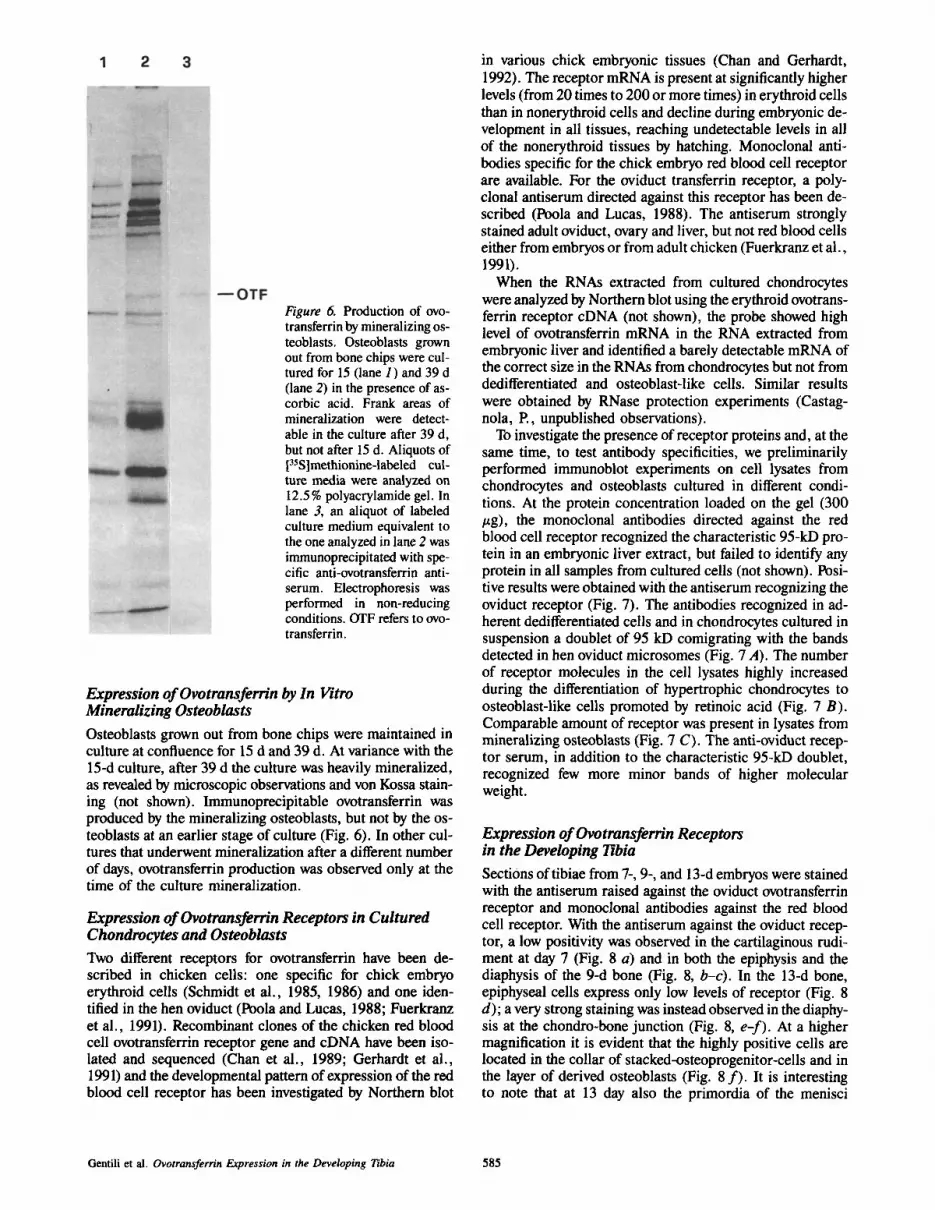
Figure 6. Production of ovo- transferrin by mineralizing os- teoblasts. Osteoblasts grown out from bone chips were cul- tured for 15 (lane 1) and 39 d (lane 2) in the presence of as- corbic acid. Frank areas of mineralization were detect- able in the culture after 39 d, but not after 15 d. Aliquots of [35S]methionine-labeled cul- ture media were analyzed on 12.5 % polyacrylamide gel. In lane 3, an aliquot of labeled culture medium equivalent to the one analyzed in lane 2 was immunoprecipitated with spe- cific anti-ovotransferrin anti- serum. Electrophoresis was performed in non-reducing conditions. OTF refers to ovo- transferrin.
Expression of Ovotransferrin by In Vitro Mineralizing Osteoblasts Osteoblasts grown out from bone chips were maintained in culture at confluence for 15 d and 39 d. At variance with the 15-d culture, after 39 d the culture was heavily mineralized, as revealed by microscopic observations and von Kossa stain- ing (not shown). Immunoprecipitable ovotransferrin was produced by the mineralizing osteoblasts, but not by the os- teoblasts at an earlier stage of culture (Fig. 6). In other cul- tures that underwent mineralization after a different number of days, ovotransferrin production was observed only at the time of the culture mineralization.
Expression of Ovotransferrin Receptors in Cultured Chondrocytes and Osteoblasts Two different receptors for ovotransferrin have been de- scribed in chicken cells: one specific for chick embryo erythroid cells (Schmidt et al., 1985, 1986) and one iden- tiffed in the hen oviduct (Poola and Lucas, 1988; Fuerkranz et al., 1991). Recombinant clones of the chicken red blood cell ovotransferrin receptor gene and cDNA have been iso- lated and sequenced (Chan et al., 1989; Gerhardt et ai., 1991) and the developmental pattern of expression of the red blood cell receptor has been investigated by Northern blot
in various chick embryonic tissues (Chan and Gerhardt, 1992). The receptor mRNA is present at significantly higher levels (from 20 times to 200 or more times) in erythroid cells than in nonerythroid cells and decline during embryonic de- velopment in all tissues, reaching undetectable levels in all of the nonerythroid tissues by hatching. Monoclonal anti- bodies specific for the chick embryo red blood cell receptor are available. For the oviduct transferrin receptor, a poly- clonal antiserum directed against this receptor has been de- scribed (Poola and Lucas, 1988). The antiserum strongly stained adult oviduct, ovary and liver, but not red blood cells either from embryos or from adult chicken (Fuerkranz et al., 1991).
When the RNAs extracted from cultured chondrocytes were analyzed by Northern blot using the erythroid ovotrans- ferrin receptor cDNA (not shown), the probe showed high level of ovotransferrin mRNA in the RNA extracted from embryonic liver and identified a barely detectable mRNA of the correct size in the RNAs from chondrocytes but not from dedifferentiated and osteoblast-like cells. Similar results were obtained by RNase protection experiments (Castag- nola, P., unpublished observations).
To investigate the presence of receptor proteins and, at the same time, to test antibody specificities, we preliminarily performed immunoblot experiments on cell lysates from chondrocytes and osteoblasts cultured in different condi- tions. At the protein concentration loaded on the gel (300 #g), the monoclonal antibodies directed against the red blood cell receptor recognized the characteristic 95-kD pro- tein in an embryonic liver extract, but failed to identify any protein in all samples from cultured cells (not shown). Posi- tive results were obtained with the antiserum recognizing the oviduct receptor (Fig. 7). The antibodies recognized in ad- herent dedifferentiated cells and in chondrocytes cultured in suspension a doublet of 95 kD comigrating with the bands detected in hen oviduct microsomes (Fig. 7 A). The number of receptor molecules in the cell lysates highly increased during the differentiation of hypertrophic chondrocytes to osteoblast-like cells promoted by retinoic acid (Fig. 7 B). Comparable amount of receptor was present in lysates from mineralizing osteoblasts (Fig. 7 C). The anti-oviduct recep- tor serum, in addition to the characteristic 95-kD doublet, recognized few more minor bands of higher molecular weight.
Expression of Ovotransferrin Receptors in the Developing Tibia Sections of tibiae from 7-, 9-, and 13-d embryos were stained with the antiserum raised against the oviduct ovotransferrin receptor and monoclonal antibodies against the red blood cell receptor. With the antiserum against the oviduct recep- tor, a low positivity was observed in the cartilaginous rudi- ment at day 7 (Fig. 8 a) and in both the epiphysis and the diaphysis of the 9-d bone (Fig. 8, b-c). In the 13-d bone, epiphyseal cells express only low levels of receptor (Fig. 8 d); a very strong staining was instead observed in the diaphy- sis at the chondro-bone junction (Fig. 8, e-f). At a higher magnification it is evident that the highly positive cells are located in the collar of stacked-osteoprogenitor-cells and in the layer of derived osteoblasts (Fig. 8 f ) . It is interesting to note that at 13 day also the primordia of the menisci
Gentili et al. Ovotransferrin Expression in the Developing Tibia 585

Figure 7. Immunoblot analysis of transferrin receptor. Ali- quots of cell lysates contain- ing 300/~g proteins were run on SDS-polyaerylamide gel, blotted to nitrocellulose filter and subjected to immunoblot with antiserum directed against oviduct ovotransferrin recep- tor. (A) Deditferentiated chon- drocytes grown as adherent ceils (lane 2) and transferred to suspension culture for 15 d (lane 3). In lane 1 an aliquot of hen oviduct microsomes (a gift of Dr. J. J. Lucas) is run for comparison. (B) Hyper- trophic chondrocytes replated as adherent cells and treated with 500 nM retinoic acid for 3 (lane I ), 6 (lane 2), 10 (lane 3), 13 (lane 4), and 27 (lane 5) d. Mineral deposits were observed in the cell layer at the last time of the culture. (C) Osteoblasts maintained in cul- ture for 39 d, a time when mineralization has already occurred.
is stained by the anti-ovotransferrin receptor antibodies (Fig. 8 d).
In the 13-d embryo, when the antibodies directed against the red blood cell receptor were used, only a faint peroxidase activity was observed in the epiphysis and in the hyper- trophic cartilage zone. Enrichment of this receptor was never observed in osteoprogenitor cells and derived osteoblasts, as in the case of the oviduct receptor (not shown).
Discus s ion
In this study we have investigated the ovotransferrin expres- sion during development of the chick embryo tibia. Ovo- transferrin is present in the first cartilaginous rudiment. While development continues, in the epiphyseal region, ovo- transferrin decreases and remains restricted to the articular zone. In the diaphysis the factor, which is abundant in the hypertrophic cartilage, progressively concentrates in zones of cartilage erosion and in the osteoid at the chondro-bone junction. The studies with cultured chondrocytes and osteo- blasts corroborated the morphological observations. In cul- ture, ovotransferrin was expressed by the in vitro differentiat- ing chondrocytes in the initial phase of the culture and by hypertrophic chondrocytes in the presence of ascorbic acid. Ovotransferrin is also expressed by hypertrophic chondro- cytes undergoing differentiation to osteoblast-like cells and by mineralizing osteoblasts. The two last processes occur in the presence of ascorbic acid.
Two types of transferrin receptors have been described in chick cells. One is expressed in cells of the erythroid lineage (Schmidt et al., 1985, 1986), the other is known as the oviduct receptor, since was first identified in oviduct ceils (Poola and Lucas, 1988), but could be better defined as the
tissue type transferrin receptor since it is expressed in sev- eral tissues (Fuerkranz et al., 1991; our work in this manu- script). When the presence of the ovotransferrin receptors was investigated in the developing tibia, it was found that, at all developmental stages, the oviduct receptor, was ex- pressed at a low level and almost evenly distributed in all cartilages. High level of oviduct receptor expression was ob- served only in the 13 day tibia, in the collar of stacked- osteoprogenitor-cells, and in the layer of derived osteoblasts. The Western blot analysis on the extracts from cultured chondrocytes and osteoblasts were in agreement with the im- munolocalization experiments. Low levels of oviduct recep- tor were detectable in almost all samples but high levels of receptor expression were reached only in extracts from hy- pertrophic chondrocytes differentiating to osteoblast-like cells and from mineralizing osteoblasts.
Since hypertrophic and differentiating chondrocytes coex- press the ovotransferrin factor and its receptor, the existence of autocrine loops playing a role in the control of chondro- genesis and hypertrophy could be postulated. At late stage of bone development high expression of the receptor is ob- served in the osteogenic collar surrounding the hypertrophic cartilage. Since the cells in the osteogenic collar do not ex- press the ovotransferrin, a paracrine loop leading to the first bone deposition, as part of the interplay between chondro- cytes and osteogenlc cells, should be considered. In princi- ple chondrocyte specific factors, as ovotransferrin, could participate in the activation of preosteoblastic cell differenti- ation program. With continuing development, differentiated mineralized osteoblasts start producing ovotransferrin in turn. Therefore, at this stage, an additional autocrine loop leading to the activation of more preosteoblastic cells could be envisaged. In the embryo limbs, at the time the initial car-
The Journal of Cell Biology, Volume 124, 1994 586

Figure 8. Immunolocalization of ovotransferrin receptor in the developing chick embryo tibia. Sections of 7-d (a), 9-d (b-c), and 13-d (d-f) tibiae were stained with an antiserum directed against the oviduct ovotransferrin receptor. (b and d) Epiphyseal region of tibia. (c, e-f) Mid-diaphysis region. Oviduct ovotransferrin recep- tors are present in the cartilag- inous rudiment at 7 days and in all cartilages at other devel- opmental stages, but they are highly enriched only in the 13-d tibia at the osteoblast side of the chondro-bone junction. At 13 day also the primordia of the menisci, which is nega- tive for ovotransferrin (see Fig. 1), is stained by the anti-oviduct-ovotransferrin re- ceptor antibodies. HC, hyper- trophic chondrocyte; OB, os- teoblast; SC, stacked cell layer. Bars: (A) 100/am; (B-E) 200/am; (F) 50 lam.
tilage and bone are formed, blood vessels are not present and the bone rudiment is not vascularized, a local production of ovotransferrin appears to be the only way to make this factor available to the differentiating cartilage and bone cells.
Interestingly high levels of oviduct receptor were observed at 13 day also in the primordia of the menisci. At this de- velopmental stage, these cells do not express ovotransferrin, but, on the contrary, ovotransferrin is detectable in the re- gion of the prearticular cartilage. This finding suggests the existence of paracrine mechanisms also during bone joint morphogenesis.
Ovotransferrin function during the cartilage and bone for- mation can be only hypothesized. Transferrins reversibly bind iron. Transferrin receptors are expressed at particularly high levels in the erythroid cell lineage between the early and intermediate normoblast stages when the iron requirement is at the highest level; subsequently their number decreases and finally disappears in the mature red blood cells (New- man et al., 1982; Horton, 1983; Chan and Gerhardt, 1992). In addition or in association with their role as iron trans- porters, transferrins play a role as mitogenic factors. Trans- ferrin receptors are in fact associated with actively prolifer- ating ceils, both in vertebrates and invertebrates (Huebers and Finch, 1987). A neuro- and myotrophic activity of trans-
ferrin has also been proposed. Transferrin is essential for myotube development in tissue culture (Li et al., 1982; Beach et al., 1985) and is required for the maintenance of the differentiated state of the striated muscle by the nerve (Stamatos and Fine, 1986). A similar situation may occur in brain where the transferrin produced by oligodendrocytes and other cells probably plays atrophic role on developing neurons and astrocytes (Aizenman et al., 1986). Finally a role for transferrin and its receptor in embryonic morpho- genesis has been shown by Ekblom et al. (1983) in mouse organ cultures of developing kidneys and teeth (Partanen et al., 1984). Transferrin is necessary to cell proliferation and cell differentiation. According to our results ovotransferrin is expressed in regions of the embryonic cartilage and bone characterized both by cell proliferation and cell differentia- tion. It should be recalled that often cell differentiation is preceded by a burst of cell proliferation. Therefore, in de- veloping bone ovotransferrin might play a role in the control of both differentiation and proliferation of chondrocytes and osteoblasts.
Ovotransferrin is particularly expressed in regions where tissue morphogenesis occurs. Retinoic acid is a well known morphogenetic factor. Retinoic acid responsive elements have been identified on transferrin promoters (Ham and
Gentili et al. Ovotransferrin Expression in the Developing Tibia 587

Griswald, 1986). We have shown an induct ive effect o f reti- noic acid on ovotransferr in express ion in cul tured chondro- cytes (Descalz i Cancedda et a l . , 1992; Gent i l i et a l . , 1993). Levels o f transferrin and transferrin m R N A are responsive to added ret inoids also in cul tured Ser tol i cel ls (Hugly and Griswold, 1987). It is interest ing to note that the Ch21, a pro- tein belonging to the l ipocal in family (extracel lular t ransport proteins for small hydrophobic l igands, such as steroids, reti- noids, and o ther small hydrophobic regula tory molecules) (Descalz i Cancedda et a l . , 1990), is p roduced by chondro- cytes and is local ized in the same regions where ovotransfer- rin is observed (Manduca et a l . , 1989). We would l ike to suggest that ovotransferr in is a m e m b e r o f a whole group o f proteins coordinate ly expressed in areas o f chondrogenes is and osteogenesis .
The finding that at late s tage o f deve lopment the presence o f ascorbic acid, i .e . , the assembly o f an ext racel lu lar ma- trix, is an absolute r equ i rement for the express ion o f ovo- transferr in by chondrocytes is pe r se an interest ing observa- t ion worth addit ional comments . Ovotransferr in is not the only example where the interact ion o f the chondrocyte with an organized extracel lu lar mat r ix is necessary for gene acti- vation. A control over gene express ion depending upon the chondrocyte-ext race l lu lar mat r ix interact ion has been shown also in the case o f the R I H B (Retinoic Induced Hepar in Bind- ing factor (Castagnola , P., S. Tavella, M. Gennar i , R. van der Werken , P. Raffo, D. Ravlais, M. Vigny, and R. Cancedda , manuscr ip t submit ted for publicat ion) and in the case of a newly descr ibed angiogenic act ivi ty (Descalz i Cancedda , E , A. Melch io r i , R. Benel l i , C. Genti l i , L. Masiello, G. Campani le , R. Cancedda, and A. Albini , manu- script submit ted for publicat ion) . It therefore appears that, in order to fully express their differentiated phenotype, chon- drocytes need to interact wi th an organized extracel lu lar ma- trix that they deposi t by themselves.
This work was supported by grants from Progetti Finalizzati: "Ingegneria Genetica" and "Applicazioni cliniche della ricerca oncoiogica", Consiglio Nazionale Delle Ricerche, Rome and by funds from the Assoclazione Italiana per la Ricerca sul Cancro, Italy. We thank Dr. Anne Mason for the monoclonal antibody against the red blood cell ovotransferrin receptor; Dr. John J. Lucas for the antiserum against the oviduct ovotransferrin receptor; Drs. Pierre Chambon, Lee-Nien L. Chan, and Francesco Amaldi for the DNA probes; Dr. Sara Tavella for providing with some chondrocyte RNA; and Dr. Patrizio Castagnola for helpful discussions.
Received for publication 17 August 1993 and in revised form 4 November 1993.
References
Aizenman, Y., M. E. Weichsel, and J. de Vellis. 1986. Changes in insulin and transferrin requirements of pure brain neuronal cultures during embryonic development. Proc. Natl. Acad. Sci. USA. 83:2263-2266.
Ambesi-Impiombato, F. S., L. A. Parks, and H. F. Coon. 1980. Culture of hormone-dependant functional epithelial cells from rat thyroids. Proc. Natl. Acad. Sci. USA. 77:3455-3459.
Beach, R. L., H. Popiela, and B. W. Festoff. 1985. Specificity of chicken and mammalian transferrins in myogenesis. Cell Differ. 16:93-100.
Bloch, B., T. Popovici, M. J. Levin, D. Tuil, and A. Kahn. 1985. Transferrin gene expression visualized in oligodendrocytes of the rat brain by using in situ hybridization and immunohistochemistry. Proc. Natl. Acad. Sci. USA. 82:6706-6710.
Bonatti, S., and F. Descalzi Cancedda. 1982. Posttranslation modification of Sindbis virus glycoproteins: electrophoretic analysis of pulse-chase labeled infected cells. J. Virol. 42:64-70.
Castaguola, P., G. Moro, F. Descalzi Cancedda, and R. Cancedda. 1986. Type X collagen synthesis during in vitro development of chick embryo tibial chondrocytes. J. Cell Biol. 102:2310-2317.
Chan, L. N., N. Grammatikak!s, J. M. Banks, and E. M. Gerhardt. 1989. Chicken transferrin receptor gane: conservation 3' non coding sequences and expression in erythroid cells. Nucleic Acids Res. 17:3763-3771.
Chan, L. N., and E. M. Gerhardt. 1992. Transferrin receptor gene is hyperex-
The Journal of Cell Biology, Volume 124, 1994
pressed and transcriptionally regulated in differentiating erythroid cells. J. Biol. Chem. 267:8254-8259.
Chomczynski, P., and N. Sacchi. 1987. Single-step method of RNA isolation by acid guanidium thiocyanate-phenol-chloroform extraction. Anal. Bio- chem. 162:156-159.
de Jong, G., J. P. van Dijk, and H. G. van Eijk. 1990. The biology of transfer- sin. Clinica Chim. Acta. 190:1--46.
Descalzi Cancedda, F., B. Dozin, F. Rossi, F. Molina, R. Cancedda, A. Negri, and S. Ronchi. 1990. The Ch21 protein, developmentally regulated in chick embryo, belongs to the supeffamily of lipophilic molecule carrier proteins. J. Biol. Chem. 265:19060-19064.
Descalzi Cancedda, F., C. Gentili, P. Manduca, and R. Cancedda. 1992. HypeRrophic chondrocytes undergo further differentiation in culture. J. Cell Biol. 117:427-435.
Descalzi Cancedda, F., P. Manduca, C. Tacchetli, P. Fossa, R. Quarto, and R. Cancedda. 1988. Developmentally regulated synthesis of a low molecular weight protein (Ch21) by differentiating chondrocytes. J. Cell Biol. 107: 2445-2463.
Ekblom, P., I. Thesleff, L. Saxen, A. Micttinen, and R. Timpl. 1983. Transfer- rin as a fetal growth factor: acquisition of responsiveness related to em- bryonic induction. Proc. Natl. Acad. Sci. USA, 80:2651-2655.
Fnernkranz, H. A., J. E. Schwob, and J. J. Lucas. 1991. Differential tissue lo- calization of oviduct and erythroid transferrin receptors. Proc. Natl. Acad. Sc/. USA. 88:7505-7508.
Gentili, C., P. Bianco, M. Neri, M. Malpeli, G. Campanile, P. Castaguola, R. Cancedda, and F. Descalzi Cancedda. 1993. Cell proliferation, extracel- iular matrix mineralization, and ovotransferrin transient expression during "in vitro ~ differentiation of chick hypertrophic chondrocytes into osteoblast- like cell. J. Cell Biol. 122:703-712.
Gerhardt, E. M., L. N. Chats, S. Q. Jing, and M. Y. Qi. 1991. The eDNA se- quence and primary structure of the chicken transferrin receptor. C-ene (Amst.). 102:249-254.
Ham, G. H., and G. N. Griswald. 1986. The EMBL data library. Nucleic Acids Res. 14:5-9.
Horton, M. A. 1983. Expression oftransferrin receptors during erythroid matu- ration. Exp. Cell Res. 144:361-366.
Huebers, H. A., and C. A. Finch. 1987. The physiology of transferrin and transferrin receptors, Physiol. Rev. 67:520-582.
Hugly, S., and M. Griswold. 1987. Regulation of levels of specific Sertoli cell mRNAs by Vitamin A. Dev. Biol. 121:316-324.
Jeltsch, J. M., and P. Chambon. 1982. The complete nucleotide sequence of the chicken ovotransferrin mRNA. Fur. J. Biochem. 112:291-295.
Li, I., I. Kimira, and E. A. Ozawe. 1982. A myotrophic protein from chick embryo extracts; its purification, identity to transferrin and indispensibility of avian myogenesis. Dev. Growth Differ. 94:366-377.
Manduca, P., F. Descalzi Cancedda, and R. Cancedda. 1992. Chondrogenic differentiation in chick embryo osteohlast cultures. Eur. J. Cell Biol. 57: 193-201.
Manduca, P., F. Descalzi Cancedda, C. Tacchetti, R. Quarto, P. Fossa, and R. Cancedda. 1989. Synthesis and secretion of Ch 21 protein in embryonic chick skeletal tissue. Eur. J. Cell Biol. 50:154-161.
McKnight, G. S., D. C. Lee, D. Hemmaplardh, C. A. Finch, and R. D. Palmiter. 1980. Transferrin gene expression. Effects of nutritional iron deficiency. J. Biol. Chem. 255:144-147.
Newman, R., C. Schneider, R. Sutherland, L. Vodinelich, and M. Greaves. 1982. The transferring receptor. Trends Biochem. Sci. 376:397-400.
Partanen, A. M., I. Thesleff, and P. Ekblom. 1984. Transferrin is required for early tooth morphogenesis. Differentiation. 27:59-66.
Poola, I., and J. J. Lucas. 1988. Purification and characterization of an estrogen- inducible membrane glycoprotein. Evidence that is a transferrin receptor. J. Biol. Chem. 263:19137-19146.
Richman, J. M., and V. M. Diewert. 1988. The fate of Meckel's cartilage chon- drocytes in ocular culture. Dev. Biol. 129:48-60.
Schmidt, J. A., J. Marshall, M. J. Hayman, and H. Beug. 1986. Primitive se- ries embryonic chick erythrocytes express the transferrin receptor. Exp. Cell Res. 164:71-78.
Schmidt, J. A., J. Marshall, and M. J. Hayman. 1985. Identification and char- acterization of the chicken transferrin recep~r. Biochem. J. 232:735-741.
Skinner, M. K., S. M. Schlitz, and C. T. Anthony. 1989. Regulation of Sertoli cell differentiated function: testicular transferrin and androgen binding pro- tein expression. Endocrinology. 124:3015-3024.
Stamatos, C., and R. E. Fine. 1986. Chick embryo myotubes contain transfer- tin receptors and internalize and recycle transferrin. J. Neurosc. Res. 15: 529-542.
Strauss, P. G., E. I. Closs, J. Schmidt, and V. Erfle. 1990. Gene expression during osteogenic differentiation in mandibular condyles in vitro. J. Cell Biol. 110:1369-1377.
Thesingh, C. W., C. G. Groot, and A. M. Wassenaar. 1991. Transdifferentla- tion of hypertrophic chondrocytes in routine metatarsal bones, induced by co-cultured cerebrum. Bone Miner. 12:25--40.
Towbin, H., T. Stachelin, andJ. Gordon. 1979. Electrophoretic transfer of pro- teins from polyacrylamide gels to nitrocellulose sheets: procedure and some applications. Proc. Natl. Acad. Sci. USA. 76:4350-4354.
Trowbridge, I. S., and B. Omary. 1981. Human cell surface glycoproteins related to cell proliferation is the receptor for transferrin. Proc. Natl. Acad. $ci. USA. 78:3039-3043.
Tsutsumi, M., M. K. Skinner, and E. Sanders-Bush. 1989. Transferrin gene expression and synthesis by cultured choroid plexus epithefial cells. J. Biol. Chem. 264:9626-9631.
588


