Embed Size (px)
Citation preview

This article was downloaded by:[INFLIBNET, India order 2005]On: 19 December 2007Access Details: [subscription number 772801163]Publisher: Informa HealthcareInforma Ltd Registered in England and Wales Registered Number: 1072954Registered office: Mortimer House, 37-41 Mortimer Street, London W1T 3JH, UK
Drug and Chemical ToxicologyPublication details, including instructions for authors and subscription information:http://www.informaworld.com/smpp/title~content=t713597244
Chemopreventive Effects of NonsteroidalAnti-Inflammatory Drugs in the Membrane LipidComposition and Fluidity Parameters of the1,2-Dimethylhydrazine-Induced Colon Carcinogenesis inRatsShailender Singh Kanwar a; Kim Vaiphei b; Bimla Nehru a; Sankar N. Sanyal aa Department of Biophysics, Panjab University, Chandigarh, Indiab Department of Histopathology, Post Graduate Institute of Medical Education &Research, Chandigarh, India
Online Publication Date: 01 October 2007To cite this Article: Kanwar, Shailender Singh, Vaiphei, Kim, Nehru, Bimla and
Sanyal, Sankar N. (2007) 'Chemopreventive Effects of Nonsteroidal Anti-Inflammatory Drugs in the Membrane LipidComposition and Fluidity Parameters of the 1,2-Dimethylhydrazine-Induced Colon Carcinogenesis in Rats', Drug andChemical Toxicology, 30:4, 293 - 309To link to this article: DOI: 10.1080/01480540701522106URL: http://dx.doi.org/10.1080/01480540701522106
PLEASE SCROLL DOWN FOR ARTICLE
Full terms and conditions of use: http://www.informaworld.com/terms-and-conditions-of-access.pdf
This article maybe used for research, teaching and private study purposes. Any substantial or systematic reproduction,re-distribution, re-selling, loan or sub-licensing, systematic supply or distribution in any form to anyone is expresslyforbidden.
The publisher does not give any warranty express or implied or make any representation that the contents will becomplete or accurate or up to date. The accuracy of any instructions, formulae and drug doses should beindependently verified with primary sources. The publisher shall not be liable for any loss, actions, claims, proceedings,demand or costs or damages whatsoever or howsoever caused arising directly or indirectly in connection with orarising out of the use of this material.

Dow
nloa
ded
By:
[IN
FLIB
NE
T, In
dia
orde
r 200
5] A
t: 07
:09
19 D
ecem
ber 2
007
Drug and Chemical Toxicology, 30:293–309, 2007Copyright © Informa Healthcare USA, Inc.ISSN: 0148-0545 print / 1525-6014 onlineDOI: 10.1080/01480540701522106
LDCT0148-05451525-6014Drug and Chemical Toxicology, Vol. 30, No. 4, August 2007: pp. 1–25Drug and Chemical ToxicologyChemopreventive Effectsof Nonsteroidal Anti-Inflammatory Drugs in the Membrane Lipid Compositionand Fluidity Parametersof the 1,2-Dimethylhydrazine-Induced Colon Carcinogenesis in RatsChemopreventive Effects of NSAIDs in colonic membranesKanwar et al.
Shailender Singh Kanwar,1 Kim Vaiphei,2 Bimla Nehru,1 and Sankar N. Sanyal1
1Department of Biophysics, Panjab University, Chandigarh, India2Department of Histopathology, Post Graduate Institute of Medical Education &Research, Chandigarh, India
Nonsteroidal anti-inflammatory drugs (NSAIDs) such as aspirin, celecoxib, and etori-coxib are reported to act as chemopreventive agents in experimental colon cancer inducedby 1,2-dimethylhydrazine (DMH) as they are known cyclooxygenase (COX) enzymeinhibitors. To determine whether NSAIDs can also effectively modulate the membranelipid compositions and the fluidity parameters of colonic brush border membrane, ratswere injected subcutaneously (s.c.) with DMH 30 mg/kg body weight per week for 6weeks. The animals were simultaneously treated with NSAIDs orally at the dose of aspi-rin, 60 mg/kg body weight; celecoxib, 6 mg/kg body weight; and etoricoxib, 0.6 mg/kg bodyweight. The animals were sacrificed after 6 weeks of treatments. Brush border mem-brane was isolated from proximal and distal portions of the colon. Membrane lipids wereextracted and analyzed while the fluidity parameters were assessed by steady-state fluo-rescence polarization technique using the membrane extrinsic fluorophore 1,6-diphenyl-1,3,5-hexatriene (DPH). The translational diffusion was measured by using the excimerformation of pyrene incorporated in the membrane. Colonic mucosal changes in DMHalone and DMH + NSAID treated animals were assessed histologically. The results dem-onstrate that (a) there is a distinct occurrence of premalignant alterations in
Address correspondence to Dr. Sankar N. Sanyal, Professor, Department of Biophysics,Panjab University, Chandigarh, 160 014, India; E-mail: [email protected]

Dow
nloa
ded
By:
[IN
FLIB
NE
T, In
dia
orde
r 200
5] A
t: 07
:09
19 D
ecem
ber 2
007
294 Kanwar et al.
DMH-induced colon in the form of multiple plaque lesions (MPLs), which were greatlyreduced by the NSAIDs used, (b) the membrane lipid changes in DMH-induced colonwere completely restored back, (c) the alterations in membrane fluorescence polarizationand the fluidity parameters are partially recovered, particularly with etoricoxib, and (d)the pyrene excimer formation process was completely restored. It may be concluded thatthe NSAIDs, particularly the coxib group of the drugs (COX-2 selective), are effective inchemoprevention in the DMH-induced colon carcinogenesis and membrane alterations.
Keywords Colon cancer, Dimethylhydrazine, Fluidity, Membrane lipid, Nonsteroidalanti-inflammatory drugs.
INTRODUCTION
Colorectal cancer is one of the leading causes of death worldwide (Potter,1996). Among the treatment modalities actively pursued at present, chemo-prevention by nonsteroidal anti-inflammatory drugs (NSAIDs), which blockeicosanoid production by inhibiting the enzyme cyclooxygenase (COX), demon-strates remarkable potential in cancer regression (Kune et al., 1988). Aspirin,a known pain reliever and anti-inflammatory agent, has been shown to beeffective in reducing the incidence of colon and other cancers in epidemiologicstudies (Thun et al., 1991) but its use is limited owing to the associated gas-trointestinal ulceration and bleeding. Newer-generation NSAIDs like cycloox-ygenase-2 inhibitors have shown promising results, as in the case of familialadenomatous polyposis (Giardielle et al., 1993). COX-2 is not normally presentin most tissues and is inducible by inflammatory cytokines and cellular trans-formations (Masferer et al., 1996). NSAIDs have been reported to affect cellmembrane dynamics, lymphocyte and neutrophil functions, and lysosomalenzyme release (Langman et al., 2000; Lucio et al., 2004). In our previousstudies, we have shown in experimental model that NSAIDs can alter thenutrient transport (Kaushal and Sanyal, 2006), affect membrane fluidity(Sanyal et al., 2007), affect the activities of disaccharide hydrolases (Kaushaland Sanyal, 2007), recover the antioxidant enzymes (Nair et al., 2006), andremarkably reduce the precancerous lesions in rat colon induced by 1,2-dimethylhydrazine (DMH) (Kanwar et al., 2007). DMH is a colonic procarcino-gen that requires metabolic activation within the host to become active (LaMont and O’Gorman, 1978), and DMH-induced tumors closely parallel humancancers in clinical and pathologic features (Luigeman and Garner, 1972).
Considerable attention has been focused on membrane lipid compositionand membrane fluidity and its relationship to malignant transformation(Brasitus et al., 1986). The membrane fluidity is generally understood as thefreedom of motion of a membrane extrinsic fluorescent probe, which can beassessed by rotational and translational diffusion of the fluorescent probes(Brasitus et al., 1988). Membrane lipid compositions and fluidity parametershave been correlated in the current study with incidence of premalignant
Dow
nloa
ded
By:
[IN
FLIB
NE
T, In
dia
orde
r 200
5] A
t: 07
:09
19 D
ecem
ber 2
007
Chemopreventive Effects of NSAIDs in Colonic Membranes 295
mucosal changes in colon in DMH-treated rat and their prevention in NSAIDadministration. In view of the regional differences and proliferation for thedevelopment of tumors with DMH, both proximal and distal brush bordermembranes (BBMs) of the colon were examined.
MATERIALS AND METHODS
Animal TreatmentMale rats of the Sprague-Dawley strain weighing 140–150 g were pro-
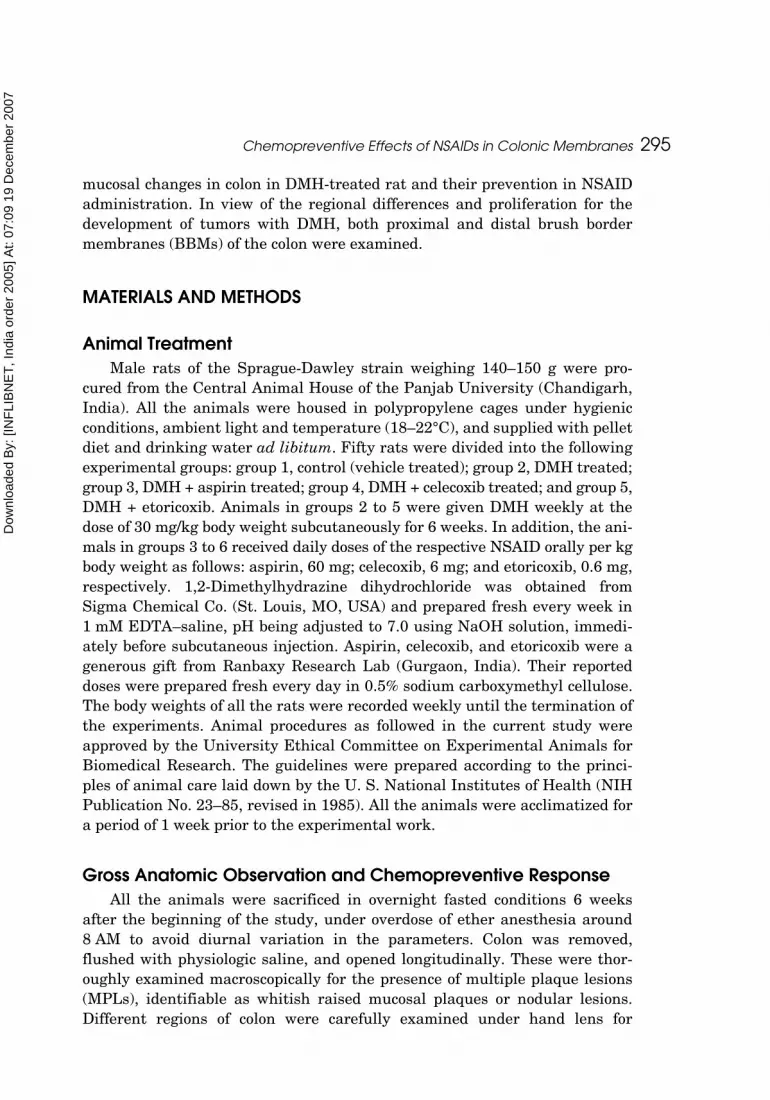
cured from the Central Animal House of the Panjab University (Chandigarh,India). All the animals were housed in polypropylene cages under hygienicconditions, ambient light and temperature (18–22°C), and supplied with pelletdiet and drinking water ad libitum. Fifty rats were divided into the followingexperimental groups: group 1, control (vehicle treated); group 2, DMH treated;group 3, DMH + aspirin treated; group 4, DMH + celecoxib treated; and group 5,DMH + etoricoxib. Animals in groups 2 to 5 were given DMH weekly at thedose of 30 mg/kg body weight subcutaneously for 6 weeks. In addition, the ani-mals in groups 3 to 6 received daily doses of the respective NSAID orally per kgbody weight as follows: aspirin, 60 mg; celecoxib, 6 mg; and etoricoxib, 0.6 mg,respectively. 1,2-Dimethylhydrazine dihydrochloride was obtained fromSigma Chemical Co. (St. Louis, MO, USA) and prepared fresh every week in1 mM EDTA–saline, pH being adjusted to 7.0 using NaOH solution, immedi-ately before subcutaneous injection. Aspirin, celecoxib, and etoricoxib were agenerous gift from Ranbaxy Research Lab (Gurgaon, India). Their reporteddoses were prepared fresh every day in 0.5% sodium carboxymethyl cellulose.The body weights of all the rats were recorded weekly until the termination ofthe experiments. Animal procedures as followed in the current study wereapproved by the University Ethical Committee on Experimental Animals forBiomedical Research. The guidelines were prepared according to the princi-ples of animal care laid down by the U. S. National Institutes of Health (NIHPublication No. 23–85, revised in 1985). All the animals were acclimatized fora period of 1 week prior to the experimental work.
Gross Anatomic Observation and Chemopreventive ResponseAll the animals were sacrificed in overnight fasted conditions 6 weeks
after the beginning of the study, under overdose of ether anesthesia around8 AM to avoid diurnal variation in the parameters. Colon was removed,flushed with physiologic saline, and opened longitudinally. These were thor-oughly examined macroscopically for the presence of multiple plaque lesions(MPLs), identifiable as whitish raised mucosal plaques or nodular lesions.Different regions of colon were carefully examined under hand lens for

Dow
nloa
ded
By:
[IN
FLIB
NE
T, In
dia
orde
r 200
5] A
t: 07
:09
19 D
ecem
ber 2
007
296 Kanwar et al.
counting the MPLs. Chemopreventive response was assessed on the basis ofMPL incidence, burden, and multiplicity that were calculated as follows:
MPL incidence: percentage of animals having MPLs.
MPL burden: total number of MPLs counted ÷ total number of rats.
MPL multiplicity: total number of MPLs counted ÷ number of MPLs bearing rats.
Histopathologic ObservationsThe colonic segments divided into proximal, middle, and distal containing
MPLs were dissected, fixed immediately in 10% formalin, and processed rou-tinely. Paraffin-embedded sections were stained with hematoxylin and eosin(H&E) for histopathologic examinations. All of the specimens were blindlyevaluated by a GE histopathologist (K.V.) who was blinded to the treatedgroups. Aberrant crypt foci (ACF) of hyperplastic and dysplastic types, aggre-gates of lymphoid tissues, and mucosal inflammation were observed in thestained sections of colon under the light microscope. ACF are raised clusters ofabnormally large crypts on colon mucosal surface and are considered as earlycenters of tumorigenesis.
Isolation of the Proximal and Distal ColonicBrush Border MembraneFor each treatment group and the control, 10 animals were sacrificed by
overdose of ether anesthesia, and the colons were excised. Cecal portion wasdiscarded and the remaining large intestine was divided into two parts: proxi-mal and distal. Intestinal BBM was isolated first by homogenizing the tissuein chilled sodium maleate buffer (10% homogenate) in a waring blender fittedwith a Teflon pestle and using a glass homogenizing tube with 0.05-mm clear-ance. All the procedures were done strictly at 4°C including the excision of thecolon, flushing with physiologic saline, and dissection into several small piecesfor homogenization. The homogenate was passed through two layers of surgi-cal bandage, and to the filtrate anhydrous CaCl2 was added with constantstirring on a magnetic stirrer to a final concentration of 10 mM and left for10–15 min in the cold. Later it was centrifuged at 2000 × g for 10 min at 4°Cand the supernatant further recentrifuged at 42,000 × g for 20 min. The pelletwas suspended in 20 vol of 50 mM sodium maleate buffer (pH 6.5–6.8) andrecentrifuged at 42,000 × g for 20 min. The final pellet obtained was sus-pended in the same buffer but containing 0.02% sodium azide (NaN3). Themethod has been based on the procedure of Brasitus and Keresztes (1984).
The purity of each membrane preparation was assessed by markerenzymes, total alkaline phosphatase (p-nitro phenyl phosphatase), andcysteine-sensitive alkaline phosphatase (Brasitus et al., 1986, 1988). The

Dow
nloa
ded
By:
[IN
FLIB
NE
T, In
dia
orde
r 200
5] A
t: 07
:09
19 D
ecem
ber 2
007
Chemopreventive Effects of NSAIDs in Colonic Membranes 297
specific activity ratio, purified membrane/original homogenate, ranged from16 to 20 for these enzymes. The corresponding values for glucose-6-phos-phatase, succinic dehydrogenase, and sodium-potassium dependent adenosinetriphosphatase, the marker enzymes for micrososmal, mitochondrial, andbasolateral membrane, respectively, ranged from 0.5 to 1.5 in these mem-brane preparations. Protein was estimated by a modified method of Lowry(Lees and Paxman, 1972).
Fluorescence Polarization Studies in BBM
The membrane fluidity was assessed in the current study by steady-statefluorescence polarization technique using the hydrocarbon fluorophore DPHaccording to the method of Schachter and Shinitzky (1977). The steady-statefluorescence anisotropy (rs) and polarization (P) were determined by the emis-sion intensities through an analyzer, oriented parallel (I||) and perpendicular(I⊥) to the direction of polarization of the excitation light as follows:
The membrane suspension (50 μL) was mixed with an aliquot of probe disper-sion (DPH; 25 mM in absolute EtOH) and incubated for 30 min in a waterbath at 37°C with continuous shaking. The total volume was made 3 mL withthe buffer (5 mM HEPES, 10 mM NaCl, pH 7.4). Also, the membrane samplewithout DPH was prepared, and the correction for light scattering was madewith appropriate filter. The fluorescence intensity measurements were con-ducted in a Perkin Elmer LS 55 Fluorescence Spectrophotometer (Beconsfield,Bucks, UK) with polarizer/analyzer attachment. The excitation wavelengthused was 352 nm to measure the emission fluorescence at 435 nm. The steady-state fluorescence anisotropy (rs) was used to calculate the membrane orderparameter (S) as follows:
where ro is maximum fluorescence anisotropy in the absence of any rotationalmotion and taken as 0.362 for DPH as the calculated value (Shinitzky andBarenholz, 1978).
Measurement of Membrane Microviscosity Using Pyrene as an Extrinsic FluorophorePyrene fluorescence excimer (dimer) formation was used to study the lat-
eral diffusion in the membrane following the method of Massey et al. (1982).
rI I
I II II I
II
II
IIs
IIP=
−+
=−+
⊥
⊥
⊥
⊥2,
S = −{( / ) . } / ,4 3 0 1r ro

Dow
nloa
ded
By:
[IN
FLIB
NE
T, In
dia
orde
r 200
5] A
t: 07
:09
19 D
ecem
ber 2
007
298 Kanwar et al.
One hundred microliters of the BBM was taken and to it was added 1 mLstock solution (5 mM in acetone) of pyrene and 100 μL of sodium maleatebuffer. The samples were incubated at 37°C for 1 hr, vortexed in between, andthen centrifuged at 10,000 × g for 1 hr. To the pellet was added sodium male-ate buffer until the solution was clear. Finally, the fluorescence intensity wasread in a fluorescence spectrophotometer as above using the excitation wave-length of 365 nm while the emission wavelengths used were 515 nm and 475nm for the excimer and monomer fluorescences, respectively. The viscosity (η)was calculated from the monomer/excimer fluorescence intensity ratios usingthe following relationship:
where T is the absolute temperature, k is the Boltzmann constant (1.38062 ×10⁻²³ Joules/Kelvin), and η is the microviscosity while the pyrene concentra-tion was kept at 5 mM.
Membrane Lipid Composition StudiesTotal lipids were extracted from the BBM by the method of Folch et al.
(1957) in the chloroform:methanol (2:1, v/v), phase separated with 0.9% KCl,washed with Folch upper phase (chlo:meth:0.9% KCl, 3:48:47) and dried andredistributed in chloroform–methanol in a rotary evaporator. Cholesterol andphospholipids in the lipid extract were measured by the method of Zlatkiset al. (1953) and Ames (1966), respectively. The ganglioside was estimated interms of the sialic acid content in the upper phase of the lipid extract followingthe method of Warren (1976).
RESULTS
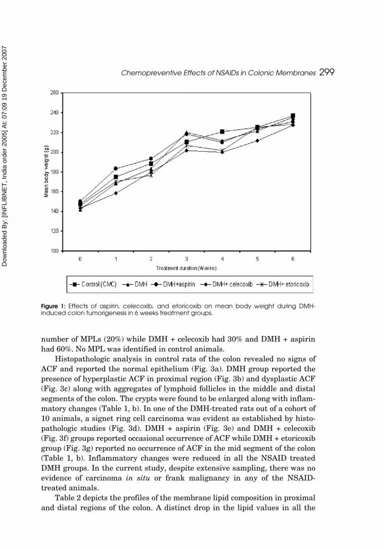
Body WeightFigure 1 shows the profiles of mean body weight during the 6 weeks treat-
ment schedule. All the groups showed normal body growth and weight gainwhen compared with their respective initial body weight. The mean bodyweight of the control animals recorded an increment of 60.5% while the DMH-treated group showed 66%. Animals in DMH + aspirin group showed 54%increase in body weight, DMH + celecoxib group 59% increase, and DMH +etoricoxib group 56% increase over the initial body weight.
Figure 2 shows gross anatomy of the colonic mucosa of control (a), DMH (b),DMH + aspirin (c), DMH + celecoxib (d), and DMH + etoricoxib (e). MPLs wereobserved in all the DMH-treated rats (100%) with 43% located in the proximalregion of the colon (Table 1, a). DMH + etoricoxib treated group had minimum
E M/ ( ) ( / ,excimer/monomer Pyrene)= Tk h
Dow
nloa
ded
By:
[IN
FLIB
NE
T, In
dia
orde
r 200
5] A
t: 07
:09
19 D
ecem
ber 2
007
Chemopreventive Effects of NSAIDs in Colonic Membranes 299
number of MPLs (20%) while DMH + celecoxib had 30% and DMH + aspirinhad 60%. No MPL was identified in control animals.
Histopathologic analysis in control rats of the colon revealed no signs ofACF and reported the normal epithelium (Fig. 3a). DMH group reported thepresence of hyperplastic ACF in proximal region (Fig. 3b) and dysplastic ACF(Fig. 3c) along with aggregates of lymphoid follicles in the middle and distalsegments of the colon. The crypts were found to be enlarged along with inflam-matory changes (Table 1, b). In one of the DMH-treated rats out of a cohort of10 animals, a signet ring cell carcinoma was evident as established by histo-pathologic studies (Fig. 3d). DMH + aspirin (Fig. 3e) and DMH + celecoxib(Fig. 3f) groups reported occasional occurrence of ACF while DMH + etoricoxibgroup (Fig. 3g) reported no occurrence of ACF in the mid segment of the colon(Table 1, b). Inflammatory changes were reduced in all the NSAID treatedDMH groups. In the current study, despite extensive sampling, there was noevidence of carcinoma in situ or frank malignancy in any of the NSAID-treated animals.
Table 2 depicts the profiles of the membrane lipid composition in proximaland distal regions of the colon. A distinct drop in the lipid values in all the
Figure 1: Effects of aspirin, celecoxib, and etoricoxib on mean body weight during DMH-induced colon tumorigenesis in 6 weeks treatment groups.

Dow
nloa
ded
By:
[IN
FLIB
NE
T, In
dia
orde
r 200
5] A
t: 07
:09
19 D
ecem
ber 2
007
300 Kanwar et al.
treated groups was observed compared with the control. Total lipid, phospho-lipid, cholesterol, ganglioside-sialic acid, and protein values declined consider-ably in DMH-treated group, which recovered with NSAID treatment,particularly in the etoricoxib-treated animals. Because there was a decline inthe individual lipid value, the ratio of all the different lipids, the cholesterol/phospholipid, protein/lipid, cholesterol/protein, and phospholipid/proteinratios were reduced. There was no significant difference in the pattern ofchanges in the lipid compositions in BBM in the two colonic regions, proximaland distal in DMH and DMH + NSAIDs groups.
Membrane lipid fluidity from the proximal and distal colon in the control,DMH, and DMH + NSAID treated groups were assessed by steady-state fluo-rescence polarization technique using the rotational movement of the hydro-phobic fluorophore probe, diphenyl hexatriene. Results of these studies aresummarized in Table 3 where the calculated order parameter (S) did indicatesignificant change in the hydrocarbon phase of the membrane and may there-fore emphasize less rotational diffusion of the probe.
The translational fluidity was measured by the excimer formation of thepyrene (Table 4), which however showed a distinct decrease in microviscosityin DMH-treated tissue, both in the proximal and distal part of the colon.NSAIDs in general did not change the fluidity parameter except for celecoxib,which showed slight improvement.
Figure 2: Effects of aspirin, celecoxib, and etoricoxib on colonic mucosal surface during DMH-induced colon tumorigenesis in 6 weeks treatment groups (locations showing mucosal plaquelesions are circled).

Dow
nloa
ded
By:
[IN
FLIB
NE
T, In
dia
orde
r 200
5] A
t: 07
:09
19 D
ecem
ber 2
007
301
Tab
le 1
:C
he
mo
pre
ven
tive
resp
on
se o
f asp
irin
, ce
lec
oxi
b, a
nd
eto
rico
xib
in D
MH
-ind
uc
ed
co
lon
ca
rcin
og
en
esis
in 6
we
eks
tr
ea
tme
nt
gro
up
.
(a)
Inc
ide
nce
of m
uco
sal p
laq
ue le
sio
ns (
MPL
s)
No
. of M
PLs
in d
iffe
rent
re
gio
n o
f co
lon
Tota
l num
be
ro
f MPL
sN
o. o
f ra
ts w
ithM
PL/t
ota
l no
. of r
ats
MPL
in
cid
enc
e (
%)
MPL
b
urd
en
MPL
mul
tiplic
ityA
nim
al g
roup
sPr
oxi
ma
lM
idd
leD
ista
l
Co
ntr
ol
Nil
Nil
Nil
Nil
No
MPL
0N
ilN
ilD
MH
1611
1037
10/1
010
03.
73.
7D
MH
+ a
spiri
n5
29
166/
1060
1.6
2.66
DM
H +
ce
lec
oxi
b2
14
73/
1030
0.7
2.3
DM
H +
eto
rico
xib
10
23
2/10
200.
31.
5
(b)
Oc
cur
renc
e o
f hyp
erp
last
ic/d
ysp
last
ic a
be
rra
nt c
ryp
t fo
ci (
AC
F), a
gg
reg
ate
s o
f lym
pho
id fo
llic
le, a
nd in
flam
ma
tory
sig
ns
in d
iffe
rent
reg
ions
of c
olo
n
Hyp
erp
lasi
aD
ysp
lasi
aA
gg
reg
ate
s o
fly
mp
hoid
folli
cle
Infla
mm
ato
ry s
igns
Ani
ma
l gro
ups
Pro
xim
al
Mid
dle
Dis
tal
Pro
xim
al
Mid
dle
Dis
tal
Pro
xim
al
Mid
dle
Dis
tal
Pro
xim
al
Mid
dle
Dis
tal
Co
ntr
ol
−−
−−
−−
−−
−−
−−
DM
H+
++
++
++
++
++
++
++
++
++
++
++
++
++
++
++
+D
MH
+ A
spiri
n+
++
+−
++
+−
++
++
++
DM
H +
Ce
lec
oxi
b+
++
+−
−+
−−
++
+D
MH
+ E
toric
oxi
b+
−+
−−
−−
−−
+−
+
MP
L in
cid
en
ce
= t
he
pe
rce
nta
ge
of
an
ima
ls h
avi
ng
MP
Ls;
MP
L b
urd
en
= t
ota
l nu
mb
er
of
MP
Ls c
ou
nte
d/t
ota
l nu
mb
er
of
rats
; M
PL
mu
ltip
licity
=to
tal n
um
be
r of
MP
Ls c
ou
nte
d/n
um
be
r of
MP
Ls b
ea
ring
rats
.+
++
/++
/+ in
dic
ate
s h
igh
oc
cu
rre
nc
e/m
od
era
te o
cc
urr
en
ce
/lo
w o
cc
urr
en
ce
, re
spe
ctiv
ely
, an
d ‘
−’ in
dic
ate
s a
bse
nc
e.

Dow
nloa
ded
By:
[IN
FLIB
NE
T, In
dia
orde
r 200
5] A
t: 07
:09
19 D
ecem
ber 2
007
302 Kanwar et al.
DISCUSSION
The current study clearly establishes presence of precancerous lesions mark-ing the initiation stage of colon cancer in experimental model in procarcinogenDMH-treated rats. Further, the chemopreventive effects of NSAIDs such asaspirin, celecoxib, and etoricoxib were evaluated at their clinically relevantanti-inflammatory dose level. Both from gross and histopathologic observa-tions, it was apparent that the premalignant signs such as occurrence of ACF(hyperplastic and dysplastic types) along with aggregates of lymphoid folliclesoccurred along the colonic length with slightly higher incidence in the proxi-mal region. It was earlier reported that DMH-induced colon carcinogenesis inthe rat model as indicated by the morphologic alterations such as proliferativeactivity and dysplasia in colonic mucosal epithelium indicates the initial stageof carcinogenesis (Melen-Mucha and Niewiadowska, 2002). Dysplastic ACFoverlaying lymphoid aggregates are considered as putative premalignant
Figure 3: (a) Normal mucosal epithelium of control rat (magnification ×100); (b) rat adminis-tered DMH exhibiting various hyperplastic ACFs (magnification ×100); (c) rat administeredDMH exhibiting various dysplastic ACFs (magnification ×100); (d) rat administered DMHrevealing a signet ring cell carcinoma (magnification ×100); (e) mucosal surface of rat admin-istered DMH + aspirin indicating regression in ACF occurrence and inflammation (magnification×100); (f & g) mucosal surface of rat administered DMH + celecoxib and DMH + etoricoxib,respectively, both exhibiting the normal mucosal surface (magnification ×100).

Dow
nloa
ded
By:
[IN
FLIB
NE
T, In
dia
orde
r 200
5] A
t: 07
:09
19 D
ecem
ber 2
007
303
Tab
le 2
:Ef
fec
ts o
f va
riou
s tr
ea
tme
nts
on
me
mb
ran
e li
pid
co
mp
osit
ion
of
pro
xim
al a
nd
dist
al c
olo
nic
BBM
.
Reg
ion
Co
ntro
lD
MH
DM
H +
asp
irin
DM
H +
ce
lec
oxi
bD
MH
+ e
toric
oxi
b
Tota
l lip
ids
(mg
/g t
issu
e)
Pro
xim
al
37.8
97 ±
3.0
5323
.115
± 3
.244
***
31.7
68 ±
2.5
98**
/###
34.9
71 ±
3.5
72##
#35
.549
± 2
.655
###
Dist
al
39.5
70 ±
4.2
5427
.143
± 3
.261
***
31.6
62 ±
2.7
79**
*/#34
.540
± 2
.222
*/###
34.5
96 ±
2.1
29*/#
##
Ch
ole
ste
rol
(mg
/g t
issu
e)
Pro
xim
al
9.59
5 ±
1.56
74.
650
± 0.
450*
**8.
547
± 0.
649
8.90
3 ±
0.51
392
9.71
6 ±
0.59
2D
ista
l9.
547
± 1.
215
7.59
8 ±
0.71
1**
7.97
7 ±
0.70
58.
837
± 0.
678##
9.55
7 ±
0.54
2###
Pho
sph
olip
ids
(mg
/g t
issu
e)
Pro
xim
al
28.2
13 ±
2.5
0619
.382
± 2
.127
***
27.5
29 ±
1.6
1###
26.3
32 ±
3.7
06##
#31
.605
± 3
.278
###
Dist
al
34.6
68 ±
2.7
1625
.337
± 2
.261
***
28.8
63 ±
2.5
26**
*/#31
.422
± 2
.477
###
32.3
43 ±
2.3
99##
#
Pro
tein
(m
g/g
tiss
ue
)P
roxi
ma
l19
.655
± 3
.246
11.0
29 ±
1.5
3416
.956
± 2
.437
17.8
47 ±
2.6
5318
.857
± 3
.042
Dist
al
21.2
6 ±
3.53
112
.562
± 2
.812
*15
.698
± 2
.032
17.2
62 ±
1.9
2419
.353
± 3
.048
#
Pro
tein
:lip
id
(w/w
)P
roxi
ma
l0.
518
± 0.
034
0.47
7 ±
0.00
10.
532
± 0.
033
0.50
9 ±
0.02
40.
525
± 0.
041
Dist
al
0.53
5 ±
0.03
20.
459
± 0.
049*
0.49
5 ±
0.02
10.
499
± 0.
024
0.55
7 ±
0.05
4##
Ch
ol:P
LP
roxi
ma
l0.
339
± 0.
026
0.24
0 ±
0.00
3***
0.31
0 ±
0.00
5###
0.34
1 ±
0.02
9###
0.30
8 ±
0.01
3*/#
##
Dist
al
0.27
6 ±
0.03
10.
299
± 0.
0018
0.27
7 ±
0.00
10.
281
± 0.
001
0.29
6 ±
0.00
5C
ho
l:pro
tein
Pro
xim
al
0.49
2 ±
0.00
60.
423
± 0.
018*
**0.
507
± 0.
035##
#0.
255
± 0.
011*
**/#
##0.
274
± 0.
004*
**/#
##
Dist
al
0.45
1 ±
0.01
80.
617
± 0.
084*
**0.
510
± 0.
021#
0.51
3 ±
0.01
8#0.
499
± 0.
051##
PL:
pro
tein
Pro
xim
al
1.45
8 ±
0.09
91.
762
± 0.
053*
*1.
637
± 0.
142
1.47
7 ±
0.01
2##1.
687
± 0.
099*
Dist
al
1.64
7 ±
0.14
82.
059
± 0.
288
1.83
9 ±
0.06
91.
825
± 0.
060
1.68
6 ±
0.14
3##
GSA
(n
mo
l/g
tiss
ue
)P
roxi
ma
l0.
134
± 0.
021
0.20
8 ±
0.02
9**
0.14
0 ±
0.02
0##0.
135
± 0.
020##
0.14
0 ±
0.02
0##
Dist
al
0.08
8 ±
0.01
50.
131
± 0.
030*
0.12
6 ±
0.01
6*0.
100
± 0.
011
0.10
3 ±
0.01
7
Da
ta re
pre
sen
ts m
ea
n ±
SD
of 1
0 d
iffe
ren
t o
bse
rva
tion
s. C
ho
l, c
ho
lest
ero
l; PL
, ph
osp
ho
lipid
s; G
SA, g
an
glio
sid
e-s
ialic
ac
id.
*p <
0.0
5, *
*p <
0.0
1, *
**p
< 0
.001
in c
om
pa
riso
n w
ith c
on
tro
l; # p
< 0
.05,
##p
< 0
.01,
### p
< 0
.001
in c
om
pa
riso
n w
ith D
MH
.

Dow
nloa
ded
By:
[IN
FLIB
NE
T, In
dia
orde
r 200
5] A
t: 07
:09
19 D
ecem
ber 2
007
304 Kanwar et al.
precursor in human and experimental colon cancer (Bird and Good, 2000).DMH-induced ACF associated with lymphoid follicles in Sprague-Dawley ratshas been reported by Cameron et al. (1996) and also in other strain such asFischer F-344 (Ward, 1974) and Wistar (Deasy et al., 1983) and in mice(Carter et al., 1994). Bland and Britton (1981) suggested carcinoma arisingfrom the epithelium covering the lymphoid follicles, and hence, occurrences ofACF associated with the aggregates of lymphoid follicles are significant prog-nostic biomarkers in colon carcinogenesis. Furthermore, presence of aggre-gates of lymphoid follicles associated with ACF is thought to play a crucialrole in either promoting conversion of ACF to adenoma to carcinoma and/or inpromoting de novo adenoma carcinoma (Cameron, 1996).
DMH with different NSAIDs administered simultaneously resulted inlessening of mucosal inflammation, occurrence of ACF dysplasia, and associ-ated aggregates of lymphoid follicles indicating chemopreventive potential ofthese drugs. Our results clearly show selective COX-2 inhibitors (celecoxiband etoricoxib) proved to be better than the nonselective inhibitor like aspirinin preventing DMH-induced colonic mucosal alterations. Earlier reports havealso shown reduced occurrence of ACF in an animal model treated withNSAIDs (Tsuji and Dubois, 1995). Thus, blockage of COX enzymes could sup-press the eicosanoid production that affects the cell proliferation, tumorgrowth, and immune responsiveness (Marnett, 1992; Rigas et al., 1993). Theresults of the current study support the hypothesis of COX-2 mediated chemo-preventive action of NSAIDs as the inhibition of the occurrence of lymphoidfollicle associated ACF, and hence, the likely action against the carcinogenesisprocess can be initiated well before the proliferative stage.
Table 3: Effects of various treatments on DPH fluorescence anisotropy and order parameter in colonic BBMs.
Colonregion
Polarization(p)
Anisotropy(r)
Order parameter(S)
Control Proximal 0.3717 ± 0.004 0.2822 ± 0.003 0.8660 ± 0.005Distal 0.3985 ± 0.002 0.3063 ± 0.001 0.9167 ± 0.005
DMH Proximal 0.4320 ± 0.004 0.3387 ± 0.0005 0.9800 ± 0.000***Distal 0.4355 ± 0.003 0.3262 ± 0.007 0.9517 ± 0.012***
DMH + aspirin Proximal 0.4515 ± 0.002 0.3540 ± 0.001 1.0100 ± 0.000***/###
Distal 0.2633 ± 0.007 0.2023 ± 0.002 0.6783 ± 0.004***/###
DMH + celecoxib Proximal 0.4513 ± 0.001 0.3552 ± 0.0007 1.0100 ± 0.000***/###
Distal 0.4217 ± 0.007 0.3253 ± 0.005 0.9500 ± 0.010***/###
DMH + etoricoxib Proximal 0.4488 ± 0.006 0.3530 ± 0.002 1.0083 ± 0.004***/###
Distal 0.4430 ± 0.005 0.3450 ± 0.001 0.9900 ± 0.000***/###
Data represents mean ± SD of five different observations; order parameter
; ro = maximum fluorescence anisotropy in the absence of any rota-tional motion = 0.365 for DPH.***p < 0.001 in comparison with control; ###p < 0.001 in comparison with DMH.
( ) {( / ) . }/S r roDPH = −4 3 0 1

Dow
nloa
ded
By:
[IN
FLIB
NE
T, In
dia
orde
r 200
5] A
t: 07
:09
19 D
ecem
ber 2
007
305
Tab
le 4
:Ef
fec
ts o
f va
riou
s tr
ea
tme
nts
on
pyr
en
e fl
uo
resc
en
ce
exc
ime
r an
d m
on
om
er r
atio
an
d m
icro
visc
osit
y in
co
lon
ic B
BMs.
Co
lon
reg
ion
Exc
ime
r(E
)M
ono
me
r(M
)E/
MM
icro
visc
osi
ty(h
)
Co
ntr
ol
Pro
xim
al
60.3
015
± 0.
2124
170.
3927
± 0
.387
60.
3538
± 0
.000
85.
3227
± 0
.012
2D
ista
l63
.926
2 ±
0.17
9717
3.20
85 ±
0.7
661
0.36
90 ±
0.0
009
5.10
38 ±
0.0
124
DM
HP
roxi
ma
l96
.314
25 ±
0.8
874
233.
4625
± 3
.045
60.
4125
± 0
.006
24.
5660
± 0
.068
9***
Dist
al
87.6
010
± 0.
5784
197.
2205
± 0
.687
60.
4441
± 0
.001
44.
2409
± 0
.013
8***
DM
H +
asp
irin
Pro
xim
al
187.
6047
± 1
.250
530
2.26
27 ±
0.8
816
0.62
06 ±
0.0
033
3.03
47 ±
0.0
165*
**/#
##
Dist
al
140.
982
± 0.
3464
267.
5305
± 0
.266
10.
5269
± 0
.000
83.
5745
± 0
.005
8***
/###
DM
H +
ce
lec
oxi
bP
roxi
ma
l91
.500
5 ±
0.19
0522
4.85
22 ±
0.1
210
0.40
69 ±
0.0
009
4.62
89 ±
0.01
02**
*/#
Dist
al
64.1
842
± 0.
5625
168.
1272
± 1
.563
60.
3817
± 0
.002
14.
9342
± 0
.028
0***
/###
DM
H +
eto
rico
xib
Pro
xim
al
124.
0207
± 0
.339
221
2.19
52 ±
0.5
111
0.58
44 ±
0.0
029
3.22
28 ±
0.0
165*
**/#
##
Dist
al
79.4
457
± 0.
8462
178.
3875
± 1
.302
50.
4453
± 0
.006
54.
2301
± 0
.061
7***
Da
ta re
pre
sen
ts m
ea
n ±
SD
of f
ive
diff
ere
nt
ob
serv
atio
ns.
***p
< 0
.001
in c
om
pa
riso
n w
ith c
on
tro
l; ##
# p <
0.0
01 in
co
mp
aris
on
with
DM
H; # p
< 0
.05
in c
om
pa
riso
n w
ith D
MH
.

Dow
nloa
ded
By:
[IN
FLIB
NE
T, In
dia
orde
r 200
5] A
t: 07
:09
19 D
ecem
ber 2
007
306 Kanwar et al.
It was thought that such chemopreventive effects at an early stage of car-cinogenesis might result from the membrane lipid changes and the fluidityparameters. The results demonstrate that the dynamic nature of the membranefluidity can be detected in the BBM both by rotational as well as translationaldiffusion. Fluorescence polarization of optical probes has been used in the studyof viscosity of membrane lipids earlier in the cell systems (Fuchs, 1975) as wellas in the isolated membranes (Ghosh and Mukherjee, 1975). The BBM from theDMH-treated membranes showed an increase in order parameter of DPH com-pared with the control, which was further changed to a significant extent by thetreatment with NSAIDs. This may therefore indicate a membrane solidifyingeffect. However, the translational fluidity as measured by the excimer forma-tion of the fluorophore, pyrene, showed a different result. Here the microviscos-ity (inverse of fluidity) decreased in the DMH-treated membrane significantly,which was further dropped by the NSAID treatment. The membranes weretherefore interpreted as more fluid during the carcinogenesis process, whichhowever to some extent recovered back by the COX-2 specific inhibitors likecelecoxib and etoricoxib. Definite indication of lateral diffusion in the hydropho-bic region of the membrane by the use of pyrene excimer formation as an opticalprobe had been obtained by the earlier workers (Ghosh and Mukherjee, 1975).The differences in the two results of DPH order parameter and pyrene excimerformation may be interpreted in terms of the different size and differentialcharacterization of the two probes (Fuchs et al., 1975; Lucio et al., 2004).
The results on the membrane fluidity were further corroborated by lipidcompositional study. The ratio of cholesterol/phospholipid decreased to anappreciable extent in the DMH-induced rats indicating more fluidity as alsoshown by others (Garder and Brennes, 1985), which recovered to certainextent by the NSAID treatments. Supporting the fluidity observations asabove, a lowered cholesterol/phospholipid ratio is indicative of higher mem-brane fluidity. Also, an unaltered ganglioside-sialic acid value points out thatthe fine-tuned activities of the membranes such as the signal transductionand receptor functions are largely unaffected in the early days of carcinogene-sis. Gangliosides are known mediators of such processes (Fischman andBrady, 1976). There was no difference in the lipid compositional data betweenthe proximal and distal regions of the colon and the pattern of change in theDMH and DMH + NSAIDs compared with the control. However, there is amarginal indication that the distal colonic tissue showed slightly higher lipidvalues, in particular the phospholipids, which also corroborates well with thefluidity parameters in the tissue.
CONCLUSION
Chemical carcinogenesis in rat colon adenoma-carcinoma model appears torepresent a multistage process, and although speculative, the current data

Dow
nloa
ded
By:
[IN
FLIB
NE
T, In
dia
orde
r 200
5] A
t: 07
:09
19 D
ecem
ber 2
007
Chemopreventive Effects of NSAIDs in Colonic Membranes 307
would suggest that alterations in the lipid fluidity and composition of plasmamembrane lipid may play a role in the early stage (initiation) of malignanttransformation process. The change in the membrane structure, function, andthe dynamic behavior itself may not be sufficient to progress further andtherefore needs other processes for proliferation and tumor development suchas the recruitment of growth factors and angiogenesis activities.
ACKNOWLEDGMENTS
Thanks are due to the Indian Council of Medical research for providing thefinancial support.
REFERENCES
Ames, B. N. (1969). Assay of inorganic phosphate, total phosphorous and phosphatase.vol. III. Method in Enzymology. Eds; Colowick, S. P., Kaplan, N. D. New York: Aca-demic Press, 8:115–118.
Bird, R. P., Good, C. K. (2000). The significance of aberrant crypt foci. in understandingthe pathogenesis of colon cancer. Toxicol. Lett. 112–113:395–402.
Bland, P. W., Britton, D. C. (1981). Colonic lymphoid tissue and its influence on tumorinduction in dimethylhydrazine treated rats. Br. J. Cancer 44:275–276.
Brasitus, T. A., Dudeja, P. K., Dahiya, R. (1986). Premalignant alterations in the lipidcomposition and fluidity of colonic brush border membranes of rats administered1,2-dimethylhydrazine. J. Clin. Invest. 77:831–840.
Brasitus, T. A., Dudeja, P. K., Foster, E. S. (1988). 1,2-Dimethylhydrazine-inducedalterations in Na+/H+ exchange in rat colonic brush border membrane vesicles.Biochim. Biophys. Acta 938:483–488.
Brasitus, T. A., Kersztes, R. S. (1984). Protein-lipid interactions in antipodal plasmamembranes of rat colonocytes. Biochim. Biophys. Acta 728:11–19.
Cameron, I. L., Garza, J., Hardman,W. E. (1996). Distribution of lymphoid nodules,aberrant crypt foci and tumors in the colon of carcinogen treated rats. Br. J.Cancer 7:893–898.
Carter, J. W., Lancaster, H. K., Hardman, W. E., Cameron, I. L. (1994). Distribution ofintestine associated lymphoid tissue, aberrant crypt foci and tumors in the largebowel of 1,2-dimethylhydrazine treated mice. Cancer Res. 54:4304–4307.
Deasy, J. M., Steele, G., Ross, D. S., Kahey, S. J., Wilson, R. E., Madora, J. (1983). Gutassociated lymphoid tissue and dimethyl hydrazine induced colorectal carcinomain the Wister/Furth. Rat. J. Surg. Oncol. 24:36–40.
Fischman, P. H., Brady, R. O. (1976). Biosynthesis and function of ganglioside. Science194:906–915.
Folch, J., Lees, M., Sloane-Stanley, G. A. (1957). A simple method of the isolation andpurification of total lipids from animal tissues. J. Biol. Chem. 226:497–509.
Fuchs, P., Parola, P., Robbins, W., Blout, E. R. (1975). Fluorescence polarizationand viscosities of membrane lipids of 373 cells. Proc. Natl. Acad. Sci. U.S.A.72:3351–3354.

Dow
nloa
ded
By:
[IN
FLIB
NE
T, In
dia
orde
r 200
5] A
t: 07
:09
19 D
ecem
ber 2
007
308 Kanwar et al.
Garder, R. A., Brennes, R. R. (1985). In-vitro modifications of cholesterol content of ratliver microsomes. Effect upon membrane fluidity and activities of glucose-6-phos-phatase and folic acid disruptions system. Biochim. Biophys. Acta 819:45–51.
Ghosh, P. K., Mukherjee, M. (1995). Increase in fluidity of human placental syncy-tiotrophoblastic brush border membrane with advancement of gestational age: afluorescence polarization study. Biochim. Biophys. Acta. 1236:317–322.
Giardielle, F. M., Hamilton, S. R., Krush, A. J., Piantoadosis, S., Hylimd, L. M., Celavo, P.,Booker, S. V., Robinson, C. R., Offerhaus, G. J. (1993). Treatment of colonic andrectal adenomas with sulindac in familial adenomatous polyposis. N. Engl. J. Med.328:1313–1316.
Kanwar, S. S., Vaiphei, K., Nehru, B., Sanyal, S. N. (2007). Chemopreventive effects ofnon-steroidal anti-inflammatory drugs on 1,2-dimethylhydrazine-induced coloncarcinogenesis in rats. Toxicol. Mech. Methods. 17:1–8.
Kaushal, N., Sanyal, S. N. (2006). Alterations in L-Histidine transport in response toaspirin and nimesulide-indiced toxicity in rat intestine using everted intestinalsacs. Toxicol. Mech. Methods 16:379–384.
Kaushal, S., Sanyal, S. N. (2007). Modulations in the intestinal disaccharide hydro-lases and membrane dynamics: effect of non-steroidal anti-inflammatory drugsaspirin and nimesulide. Mol. Cell. Biochem, 294:107–115.
Kune, G., Kune, S., Watson, L. (1988). Colorectal cancer risk, chronic illness, opera-tions and indications: Case control results from the Melbourne colorectal cancerstudy. Cancer Res. 4:4399–4404.
La Mont, J. T., O’Gorman, T. A. (1978). Experimental colon cancer. Gastroenterology75:1157–1169.
Langman, M. J., Cheng, K. K., Gilman, E. A., Lancashire, R. J. (2000). Effect of anti-inflammatory drugs on overall risk of common cancer: case-control study in gen-eral practice research database. BMJ 320:1642–1646.
Lees, M., Paxman, S. (1972). Modifications of the Lowry procedure for the analysis ofproteolipid protein. Anal. Biochem. 47:184–194.
Lingeman, C. H., Garner, F. M. (1972). Comparative study of intestinal adenocarcino-mas of animals and man. J. Natl. Cancer Inst. 48:325–346.
Lucio, M., Ferreira, H., Lima, J. L. F. C., Matos, C., deCastro, B., Reis, Salette. (2004).Influence of some anti-inflammatory drugs in membrane fluidity studied by fluo-rescence anisotropy measurements. Phys. Chem. Chem. Phys. 6:1493–1498.
Marnett, L. J. (1992). Aspirin and the potential role of prostaglandins in colon cancer.Cancer Res. 52:5575–5589.
Masferer, J. L., Isakson, P. C., Seibert, K. (1996). Cyclooxygenase-2 inhibitors: a newclass of anti-inflammatory agents that spare the gastrointestinal tract. Gastroen-terol. Clin. North Am. 25:363–372.
Massey, J. B., Gotto, A. M., Pownell, J. H. (1982). Kinetics and mechanism of the spon-taneous transfer of fluorescent phosphatidyl choline between apolipoprotein-phospholipid recombinants. Biochemistry 21:3630–3636.
Melen-Mucha, G., Niewiadowska, H. (2002). Frequency of proliferation, apoptosis, andtheir ratio during rat colon carcinogenesis and their characteristic pattern in thedimethylhydrazine-induced colon adenoma and carcinoma. Cancer Invest. 20:700–712.
Nair, P., Kanwar, S. S., Sanyal, S. N. (2006). Effects of non steroidal anti-inflammatorydrugs on the antioxidant defense system and the membrane functions in the ratintestine. Nutricion Hospitalaria 21:638–649.
Dow
nloa
ded
By:
[IN
FLIB
NE
T, In
dia
orde
r 200
5] A
t: 07
:09
19 D
ecem
ber 2
007
Chemopreventive Effects of NSAIDs in Colonic Membranes 309
Potter, J. D. (1996). Risk factor for colon neoplasia. Epidemiology and Biology. Eur. J.Cancer 314:1033–1038.
Rigas, B., Goldman, I. S., Levine, L. (1993). Altered eicosanoid levels in human coloncancer. J. Lab. Clin. Med. 122:518–523.
Sanyal, S. N., Sood, N., Kaushal, N. (2007). Alterations in the lipid profile and mem-brane dynamics of rat intestinal brush border membrane induced by differentclasses of nonsteroidal anti-inflammatory drugs. Toxicol. Mech. Methods. 17:1–8.
Schachter, D., Shinitzky, M. (1977). Fluorescence polarization studies of rat intestinalmicrovillous membranes. J. Clin. Invest. 59:536–548.
Shinitzky, M., Barenholz, Y. (1978). Fluidity parameters of lipid regions determined byfluorescence polarization. Biochim. Biophys. Acta 515:367–394.
Thun, M. J., Namboodiri, M. M., Heath, C. W., Jr. (1991). Aspirin use and reduced riskof fatal colon cancer. N. Engl. J. Med. 325:1593–1596.
Tsuji, M., Dubois, R. N. (1995). Alterations in cellular adhesion and apoptosis in epithe-lial cells over expressing prostaglandin endoperoxide synthase-2. Cell 83:493–501.
Ward, J. M. (1974). Morphogenesis of chemically induced neoplasms of the colon andsmall intestines in rats. Lab. Invest. 30:505–513.
Warren, L. (1976). The thiobarbituric acid assay of sialic acids. J. Biol. Chem.234:1971–1976.
Zlatkis, A., Zak, B., Boyle, A. J. (1953). A new method for the determination of serumcholesterol. J. Lab. Clin. Med. 41:485–492.

Dow
nloa
ded
By:
[IN
FLIB
NE
T, In
dia
orde
r 200
5] A
t: 07
:09
19 D
ecem
ber 2
007


