Embed Size (px)
Citation preview

p107 is a suppressor of retinoblastomadevelopment in pRb-deficient miceEls Robanus-Maandag,1 Marleen Dekker,2 Martin van der Valk,1 Maria-Luisa Carrozza,1,3
Jean-Claude Jeanny,4 Jan-Hermen Dannenberg,2 Anton Berns,1 and Hein te Riele2,5
1Division of Molecular Genetics and 2Division of Molecular Carcinogenesis, The Netherlands Cancer Institute, Amsterdam,The Netherlands; 4Institut National de la Sante et de la Recherche Medicale (INSERM) U450, Affiliee Centre Nationalde la Recherche Scientifique (CNRS), Paris, France
Hemizygosity for the retinoblastoma gene RB in man strongly predisposes to retinoblastoma. In the mouse,however, Rb hemizygosity leaves the retina normal, whereas in Rb−/− chimeras pRb-deficient retinoblastsundergo apoptosis. To test whether concomitant inactivation of the Rb-related gene p107 is required tounleash the oncogenic potential of pRb deficiency in the mouse retina, we inactivated both Rb and p107 byhomologous recombination in embryonic stem cells and generated chimeric mice. Retinoblastomas werefound in five out of seven adult pRb/p107-deficient chimeras. The retinal tumors showed amacrine celldifferentiation, and therefore originated from cells committed to the inner but not the outer nuclear layer.Retinal lesions were already observed at embryonic day 17.5. At this stage, the primitive nuclear layerexhibited severe dysplasia, including rosette-like arrangements, and apoptosis. These findings provide formalproof for the role of loss of Rb in retinoblastoma development in the mouse and the first in vivo evidence thatp107 can exert a tumor suppressor function.
[Key Words: Retinoblastoma; apoptosis; Rb; p107; tumor suppressor gene; chimeric mice]
Received February 20, 1998; revised version accepted April 3, 1998.
Hereditary retinoblastoma, a childhood tumor of the eye,has served as a paradigm for studies concerning the roleof tumor suppressor genes in cancer predisposition. Therate-limiting step in the initiation of both the hereditaryand sporadic form of this tumor is loss of function of theretinoblastoma gene RB in the developing retina. Inher-itance of one mutant RB allele not only predisposes toretinoblastoma (90%) early in life but also to osteosar-comas (2%) later on (Draper et al. 1986; Friend et al.1987). In addition, loss of function of RB has been fre-quently found in lung, breast, and bladder carcinomas(Harbour et al. 1988; Lee et al. 1988; Horowitz et al.1990). Also, upstream regulators of pRB are repeatedlyfound mutated (p16 and CDK4) or overexpressed (cyclinD1) in human tumors (Hall and Peters 1996). Thus, de-regulation of pRB function appears to be a commonevent in the development of many tumor types.
pRB plays an important role during the G1 phase of thecell cycle, when cells are responsive to extracellularpositive and negative proliferation signals (Sherr 1994).pRB functions in a pathway that transduces such signalsto the cell nucleus modulating the activity of, for ex-ample, E2F transcription factors. In G1, the transactivat-ing potential of these proteins is suppressed by their as-
sociation with hypophosphorylated pRB. The E2Fs arereleased upon phosphorylation of pRB by cyclin D-de-pendent kinases whose activity depends on mitogenicstimuli. After passing a restriction point, pRB stays inthe hyperphosphorylated, inactive conformationthroughout the autonomous program that carries the cellthrough the remaining of G1, S, and G2 phases of the cellcycle (Weinberg 1995). In both sequence and functionpRB is closely related to two other nuclear phosphopro-teins, p107 and p130 (Ewen et al. 1991; Hannon et al.1993; Li et al. 1993; Mayol et al. 1993). Extensive struc-tural homology is found in their so called pocket do-main, the binding site for many viral oncoproteins, in-cluding adenovirus E1A, simian virus 40 large T antigen,and human papillomavirus E7 (DeCaprio et al. 1988;Whyte et al. 1988; Dyson et al. 1989). Like pRB, p107 andp130 may also act as negative regulators of cell prolifera-tion through interaction with E2F transcription factors(Zhu et al. 1993; Claudio et al. 1994; Qin et al. 1995).However, different pRB family proteins associate withdifferent E2Fs at different times during the cell cycle(Bernards 1997). In mice, the Rb gene family membersshare a wide expression pattern, with high and overlap-ping expression of Rb and p107 in embryonic liver andCNS (Jiang et al. 1997).
Retinoblastomas have not been described to occurspontaneously in species other than man. In addition toloss of RB, a limited number of karyotypic rearrange-ments with unknown functional significance have been
3Present address: Instituto di Neurofisiologia del Consiglio NazionaleRicerche, Pisa, Italy.5Corresponding author.E-MAIL [email protected]; FAX 31 205121954.
GENES & DEVELOPMENT 12:1599–1609 © 1998 by Cold Spring Harbor Laboratory Press ISSN 0890-9369/98 $5.00; www.genesdev.org 1599
found in retinal tumors (Kusnetsova et al. 1982; Squire etal. 1984). In the mouse, hemizygosity for Rb does notlead to retinoblastoma. Instead, Rb+/− mice succumb topituitary gland tumors from 6–8 months on. Rb−/− em-bryos show severe defects in central neurogenesis, fetalliver erythropoiesis, lens development, and myogenesis,and die around days 12–15 of gestation when the devel-oping retina appears normal (Clarke et al. 1992; Jacks etal. 1992; Lee et al. 1992; Morgenbesser et al. 1994; Ro-banus Maandag et al. 1994; Williams et al. 1994b; Zack-senhaus et al. 1996). However, evidence for a function ofpRb at later stages of retinal development has come fromthe analysis of chimeric Rb−/− mice. Apoptosis was ob-served in the developing retina beyond day 16 of gesta-tion and the number of Rb−/− cells in the adult retina wassignificantly reduced (Robanus Maandag et al. 1994).These observations suggest that in the mouse loss of Rbduring development of the retina results in cell deathrather than enhanced cell proliferation. Therefore, addi-tional mutations may be required to unleash the onco-genic potential of pRb deficiency in mouse retinoblasts.This hypothesis is supported by the analyses of varioustransgenic and knock-out mouse lines. Retinoblastomasdevelop in transgenic mice with retina-specific expres-sion [using the human interphotoreceptor retinoid-bind-ing protein (IRBP) promoter] of SV40 Tag or HPV-16 E7,the latter exclusively in a p53−/− background (Al-Ubaidiet al. 1992; Howes et al. 1994). These results suggest arequirement for multiple inactivations that possibly in-clude one or more of the pocket proteins and p53. How-ever, which specific proteins need to be inactivated hasnot been answered and the oncoproteins may elicit otheroncogenic alterations as well. In addition, the use of spe-cific promoters to drive SV40 Tag or HPV-16 E7 limitsinactivation to those cells in which the oncoproteins areexpressed. As a consequence, analyses have remained re-stricted to a subset of cells within a specific differentia-tion window characterized by expression of IRBP.Knockout mouse models lack these limitations of thetransgenic mouse models. Mice have been generated
with (various combinations of) inactivated candidategenes that may be required for retinoblastoma develop-ment. Rb+/−;p107−/− mice do not show any altered tumorpredisposition when compared with Rb+/− mice but de-velop multiple dysplastic lesions of the retina that areabsent in Rb+/− and p107−/− mice (Lee et al. 1996). Inaddition, retinal dysplasias have been observed in 40% ofthe Rb+/−;p53−/− mice as well as pinealoblastomas thatshow loss of heterozygosity for Rb (Williams et al.1994a).
The lethality of pRb/p107-deficient embryos at day11.5 of gestation precludes to study the effect of con-comitant pRb and p107 deficiency at later stages of de-velopment and during adult life (Lee et al. 1996). To cir-cumvent this problem, we investigated the tumorigenicand developmental potential of Rb−/−;p107−/− cells in theretina of chimeric mice generated with Rb−/−;p107−/− embryonic stem cells. We report here that loss offunction of both Rb and p107 in murine retinoblastsleads to retinoblastoma originating from cells commit-ted to the amacrine cell compartment of the innernuclear layer but not from those committed to the outernuclear layer of the retina.
Results
Generation of mutant Rb;p107 chimeras
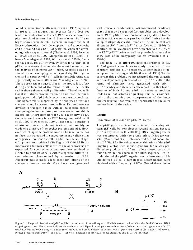
The p107 gene was inactivated in murine embryonicstem (ES) cells by homologous recombination. Becausep107 is expressed in ES cells (Fig. 1B), a targeting vectorwas constructed with the promoterless IRESbgeo cas-sette (Mountford et al. 1994) inserted behind codon 145of p107 (Fig. 1A). Homologous recombination of the p107targeting vector with mouse genomic DNA was pre-dicted to produce a p107 null allele caused by an in-frame termination codon in the IRES sequence. On in-troduction of the p107 targeting vector into strain 129/Ola-derived ES cells homologous recombinants wereobtained with a frequency of 65%. One of these clones
Figure 1. Targeted disruption of p107. (A) Restriction map of the wild-type p107 allele around codon 145 at the EcoRV site and DNAtargeting construct. Black boxes indicate determined exons. Upon homologous recombination a fusion transcript is generated of p107,truncated behind codon 145, with IRESbgeo. Probe A and probe B detect modifications at p107. (B) Western blot analysis of p107 inlysates prepared from p107+/+ and p107−/− ES cells. Positions of molecular mass standards and p107 are indicated.
Robanus-Maandag et al.
1600 GENES & DEVELOPMENT
carried IRESbgeo in both p107 alleles giving a p107−/− EScell line. To confirm full inactivation of p107, the levelof p107 protein was examined in extracts of ES cells.Whereas p107 could be readily detected in wild-type EScells, no p107 protein was detected in p107−/− ES cells byWestern blot analysis using the anti-p107 rabbit anti-body C-18 (Fig. 1B).
Subsequently, in this p107−/− ES cell line both allelesof the retinoblastoma gene Rb were inactivated by tworounds of homologous recombination with the isogenictargeting vectors 129Rb-hyg (Te Riele et al. 1992) and129Rb-his. These vectors carry the hygromycin and his-tidinol resistance genes, respectively, inserted into exon19 of Rb, which on homologous recombination lead toRb null alleles (Clarke et al. 1992).
In an attempt to investigate the effect of combined lossof Rb, p107, and p53 in the retina, we also introducedinto Rb−/−;p107−/− ES cells the dominant-negative p53mutant minigene p53DD (Shaulian et al. 1992) driven bythe 1.3-kb human interphotoreceptor retinoid-bindingprotein (hIRBP) promoter fragment (Liou et al. 1990).
p53DD has been shown to elicit a biological effect cor-responding to genetic loss of p53 including a reduction inapoptosis and acceleration of tumorigenesis (Bowmanet al. 1996). We expected Rb−/−;p107−/−;hIRBPp53DDchimeras to mimic hIRBP-E7;p53−/− transgenic mice,which developed retinoblastoma (Howes et al. 1994).Multiple copies of hIRBPp53DD were introduced intoRb−/−;p107−/− ES cells by coelectroporation with the se-lection marker PGKpur.
Rb+/−;p107−/−, Rb−/−;p107−/−, and Rb−/−;p107−/−;hIRBPp53DD ES cell clones were verified for the correctkaryotype and injected into C57BL/6 blastocysts to gen-erate chimeras. The level of pigmentation in the retinalpigment epithelium (RPE) served as a rough indicationfor the extent of chimerism of the eye (nonpigmentedareas result from ES cell contribution).
Poor chimerism in Rb−/−;p107−/− chimeras
Chimeric Rb+/−;p107−/− mice were readily obtained (28/62 births) and were able to transmit ES cell-derived al-
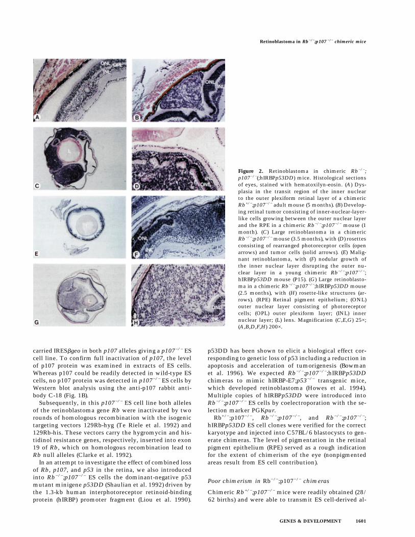
Figure 2. Retinoblastoma in chimeric Rb−/−;p107−/−(;hIRBPp53DD) mice. Histological sectionsof eyes, stained with hematoxilyn-eosin. (A) Dys-plasia in the transit region of the inner nuclearto the outer plexiform retinal layer of a chimericRb+/−;p107−/− adult mouse (5 months). (B) Develop-ing retinal tumor consisting of inner-nuclear-layer-like cells growing between the outer nuclear layerand the RPE in a chimeric Rb−/−;p107−/− mouse (1month). (C) Large retinoblastoma in a chimericRb−/−;p107−/− mouse (3.5 months), with (D) rosettesconsisting of rearranged photoreceptor cells (openarrows) and tumor cells (solid arrows). (E) Malig-nant retinoblastoma, with (F) nodular growth ofthe inner nuclear layer disrupting the outer nu-clear layer in a young chimeric Rb−/−;p107−/−;hIRBPp53DD mouse (P15). (G) Large retinoblasto-ma in a chimeric Rb−/−;p107−/−;hIRBPp53DD mouse(2.5 months), with (H) rosette-like structures (ar-rows). (RPE) Retinal pigment epithelium; (ONL)outer nuclear layer consisting of photoreceptorcells; (OPL) outer plexiform layer; (INL) innernuclear layer; (L) lens. Magnification (C,E,G) 25×;(A,B,D,F,H) 200×.
Retinoblastoma in Rb−/−;p107 −/− chimeric mice
GENES & DEVELOPMENT 1601
leles through the germ line. In 6/56 chimeric eyes, theretina showed some dysplasia. For example, Figure 2Ashows a small lesion in the transition region of the innernuclear to the outer plexiform layer. In contrast, Rb−/−;p107−/− chimeras were obtained with low efficiency (7/56 births) and only when a low number of ES cells (4–6)per blastocyst was injected. In general, the ES cell con-tribution in the tissue samples of these animals was re-duced twofold with respect to that of Rb−/− chimeras(Robanus Maandag et al. 1994) (not shown).
Retinoblastoma in Rb−/−;p107−/− chimeras
Retinoblastomas were found in five of seven Rb−/−;p107−/−(;hIRBPp53DD) chimeras: Two of six eyes inRb−/−;p107−/− and four of eight eyes (one bilateral case)in Rb−/−;p107−/−;hIRBPp53DD chimeras. Histologicalexamination of the eyes of Rb−/−;p107−/− chimeras re-vealed in a 1-month-old chimera a developing retinal tu-mor between the photoreceptor layer and the RPE, con-sisting of inner-nuclear-layer-like cells (Fig. 2B). In a chi-mera of 3.5 months, one of the eyes contained a largetumor process. Microscopically, this appeared to be aretinoblastoma that had invaded into the anterior eyechamber (Fig. 2C,D). The tumor cells often formed smallirregular circles. Also, invasion of tumor cells into theouter nuclear layer apparently induced the rearrange-ment of normal photoreceptor cells into rosettes (Fig.2D). In a chimeric Rb−/−;p107−/−;hIRBPp53DD mouse ofpostnatal day 15 (P15) we observed a malignant nodulargrowth of inner-nuclear-layer-like cells at multiple re-gions (Fig. 2E,F); similar to the tumor in Figure 2B, thetumor cells tended to invade between the photoreceptorlayer and RPE (Fig. 2F). We found three large tumors inchimeric Rb−/−;p107−/−;hIRBPp53DD mice of 2.5 (Fig.2G,H) and 4 months. In the four large tumors, 3–10 mi-totic figures were counted per high power field with a40× objective (not shown). Thus, both types of chimerasdeveloped retinoblastoma with similar incidence (al-though the numbers were small), inner-nuclear-layer-like appearance, and tendency to grow between the pho-toreceptor layer and RPE.
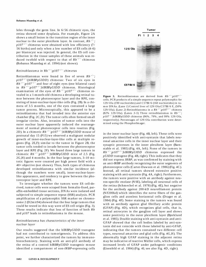
To investigate whether the tumors were ES cell-de-rived, tumor cells were scraped from formalin-fixed, par-affin-embedded tissue sections, DNAs were isolated andsubjected to simple sequence repeat (SSR) analysis. PCRamplification of a polymorphic SSR marker on chromo-some 2 (D2mit94) showed that the four large tumors thatcould be tested in this way were of ES cell origin (Fig. 3).These results indicate that loss of function of both Rband p107 leads to retinoblastoma in the mouse.
Retinoblastoma has characteristics of the innernuclear layer
Our results suggested that the hIRBPp53DD transgenehad not contributed to tumorigenesis. To address thispoint, we further characterized the tumors by immuno-histochemistry. Staining with an anti-p53 antibody ofthe retina of a control hIRBPp53DD transgenic mouseidentified a compartment of non-IRBP-expressing cells
in the inner nuclear layer (Fig. 4B, left). These cells werepositively identified with anti-syntaxin that labels neu-ronal amacrine cells in the inner nuclear layer and theirsynaptic processes in the inner plexiform layer (Barn-stable et al. 1985) (Fig. 4A, left). None of the tumors inRb−/−;p107−/−;hIRBPp53DD chimeras expressed thep53DD transgene (Fig. 4B, right). This indicates that theydid not express IRBP, as was confirmed by staining withan anti-IRBP antibody recognizing the outer segments ofphotoreceptor cells (Carter-Dawson et al. 1986) (Fig. 4C).Instead, all retinal tumors showed extensive positivestaining with anti-syntaxin (Fig. 4A, right). Furthermore,the tumors were positive with an antibody against neu-ron-specific enolase (NSE), labeling all neuronal cells ofthe retina (Schmechel et al. 1978) (Fig. 4E), but negativefor the antibody against 200-kD neurofilament protein(NF200kd) which identifies the nerve fibers of the gan-glion cells and axonless horizontal cells (Drager et al.1984) (Fig. 4F). Some staining in the tumors was foundwith an antibody against glial fibrillary acidic protein(GFAP) (Fig. 4D), which recognizes glial cells, that is,retinal astrocytes in the ganglion cell layer and showssome positivity in the outer plexiform layer (Bjorklundet al. 1985). Double staining with anti-syntaxin and anti-GFAP showed that the cell bodies labeled by anti-syn-taxin did not coincide with those labeled by anti-GFAP,indicating that the tumors contained two different celltypes, neuronal amacrine and glial cells (Fig. 4G,H). Theabnormally high GFAP staining in the adjacent retinamay be indicative of reactive Muller cells, which expressincreased levels of GFAP under pathogenic conditions(Eisenfeld et al. 1984) (Fig. 4I; see also Fig. 4D, right).
Figure 3. Retinoblastomas are derived from Rb−/−;p107−/−
cells. PCR products of a simple sequence repeat polymorphic for129/Ola (190 nucleotides) and C57Bl/6 (160 nucleotides) in tu-mor DNAs. (Lane 1) Control liver of 129/Ola:C57Bl/6 F1 (50%129/Ola). (Lane 2) Retinoblastoma in a Rb−/−;p107−/− chimera(82% 129/Ola). (Lanes 3–5) Three retinoblastomas in Rb−/−;p107−/−;hIRBPp53DD chimeras (96%, 79%, and 99% 129/Ola,respectively). Percentages of 129/Ola contribution were deter-mined using the PhosphorImager.
Robanus-Maandag et al.
1602 GENES & DEVELOPMENT
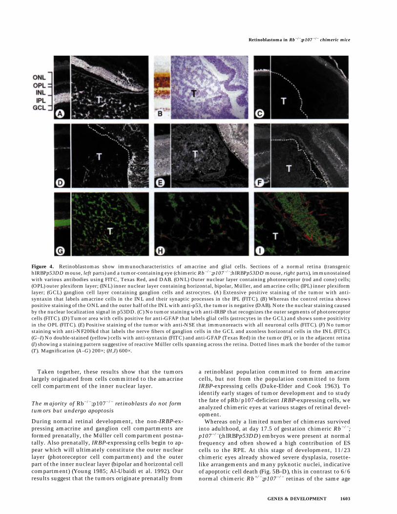
Taken together, these results show that the tumorslargely originated from cells committed to the amacrinecell compartment of the inner nuclear layer.
The majority of Rb−/−;p107−/− retinoblasts do not formtumors but undergo apoptosis
During normal retinal development, the non-IRBP-ex-pressing amacrine and ganglion cell compartments areformed prenatally, the Muller cell compartment postna-tally. Also prenatally, IRBP-expressing cells begin to ap-pear which will ultimately constitute the outer nuclearlayer (photoreceptor cell compartment) and the outerpart of the inner nuclear layer (bipolar and horizontal cellcompartment) (Young 1985; Al-Ubaidi et al. 1992). Ourresults suggest that the tumors originate prenatally from
a retinoblast population committed to form amacrinecells, but not from the population committed to formIRBP-expressing cells (Duke-Elder and Cook 1963). Toidentify early stages of tumor development and to studythe fate of pRb/p107-deficient IRBP-expressing cells, weanalyzed chimeric eyes at various stages of retinal devel-opment.
Whereas only a limited number of chimeras survivedinto adulthood, at day 17.5 of gestation chimeric Rb−/−;p107−/−(;hIRBPp53DD) embryos were present at normalfrequency and often showed a high contribution of EScells to the RPE. At this stage of development, 11/23chimeric eyes already showed severe dysplasia, rosette-like arrangements and many pyknotic nuclei, indicativeof apoptotic cell death (Fig. 5B–D), this in contrast to 6/6normal chimeric Rb+/−;p107−/− retinas of the same age
Figure 4. Retinoblastomas show immunocharacteristics of amacrine and glial cells. Sections of a normal retina (transgenichIRBPp53DD mouse, left parts) and a tumor-containing eye (chimeric Rb−/−;p107−/−;hIRBPp53DD mouse, right parts), immunostainedwith various antibodies using FITC, Texas Red, and DAB. (ONL) Outer nuclear layer containing photoreceptor (rod and cone) cells;(OPL) outer plexiform layer; (INL) inner nuclear layer containing horizontal, bipolar, Muller, and amacrine cells; (IPL) inner plexiformlayer; (GCL) ganglion cell layer containing ganglion cells and astrocytes. (A) Extensive positive staining of the tumor with anti-syntaxin that labels amacrine cells in the INL and their synaptic processes in the IPL (FITC). (B) Whereas the control retina showspositive staining of the ONL and the outer half of the INL with anti-p53, the tumor is negative (DAB). Note the nuclear staining causedby the nuclear localization signal in p53DD. (C) No tumor staining with anti-IRBP that recognizes the outer segments of photoreceptorcells (FITC). (D) Tumor area with cells positive for anti-GFAP that labels glial cells (astrocytes in the GCL) and shows some positivityin the OPL (FITC). (E) Positive staining of the tumor with anti-NSE that immunoreacts with all neuronal cells (FITC). (F) No tumorstaining with anti-NF200kd that labels the nerve fibers of ganglion cells in the GCL and axonless horizontal cells in the INL (FITC).(G–I) No double-stained (yellow) cells with anti-syntaxin (FITC) and anti-GFAP (Texas Red) in the tumor (H), or in the adjacent retina(I) showing a staining pattern suggestive of reactive Muller cells spanning across the retina. Dotted lines mark the border of the tumor(T). Magnification (A–G) 200×; (H,I) 600×.
Retinoblastoma in Rb−/−;p107 −/− chimeric mice
GENES & DEVELOPMENT 1603
(Fig. 5A). Clear anti-p53DD antibody staining in the ven-tricular layer of the developing retina identified Rb−/−;p107−/−;hIRBPp53DD retinoblasts that had apparentlyreached the differentiation stage of IRBP-expressing cells(Fig. 6B). These cells were virtually absent at P15, even atretinal regions that were highly chimeric as deducedfrom the presence of malignant nodular growths of theinner nuclear layer (Fig. 6C, see also Fig. 4B). Apparently,pRb/p107-deficient cells can contribute to the IRBP-ex-pressing compartment, but are excluded from the retinabetween E17.5 and P15. Indeed, apoptotic cell death inchimeric Rb−/−;p107−/− retinas could be detected as earlyas day 17.5 of gestation and continued at least until P11(Fig. 7B,D).
These results allow us to draw two conclusions.Firstly, oncogenic transformation of pRb/p107-deficientretinoblasts occurs as early as day 17.5 of gestation andinvolves cells committed to the non-IRBP-expressingamacrine cell compartment of the inner nuclear layer.Secondly, pRb/p107-deficient cells can contribute tothe IRBP-expressing compartment, but these cells donot grow out to retinoblastoma. Instead, they undergoapoptosis before P15, likely at the stage of differentia-
tion to mature bipolar, horizontal, and photoreceptorcells.
Discussion
In contrast to the situation in humans, mice hemizygousfor the retinoblastoma gene Rb do not develop retino-blastoma, but pituitary gland tumors. Also, chimericRb−/− mice do not form retinoblastomas but, instead,undergo retinal apoptosis during development. Appar-ently, the murine retina is better protected against tu-morigenesis than the human retina. For example, in themouse, additional mutations may be required for the on-cogenic transformation of pRb-deficient retinoblasts. In-dications for this have come from studies in transgenicmice in which concomitant inactivation of pocket pro-teins and p53 by overexpression of HPV-16 E7 (in a p53null background) or SV40 Tag led to development of ret-inoblastoma. However, these transgenic mouse modelsdo not accurately specify the mutational requirementsfor retinal tumorigenesis. On the other hand, the use ofknockout mice carrying specific (combinations of) mu-tations is limited by the early death of pRb-deficient em-
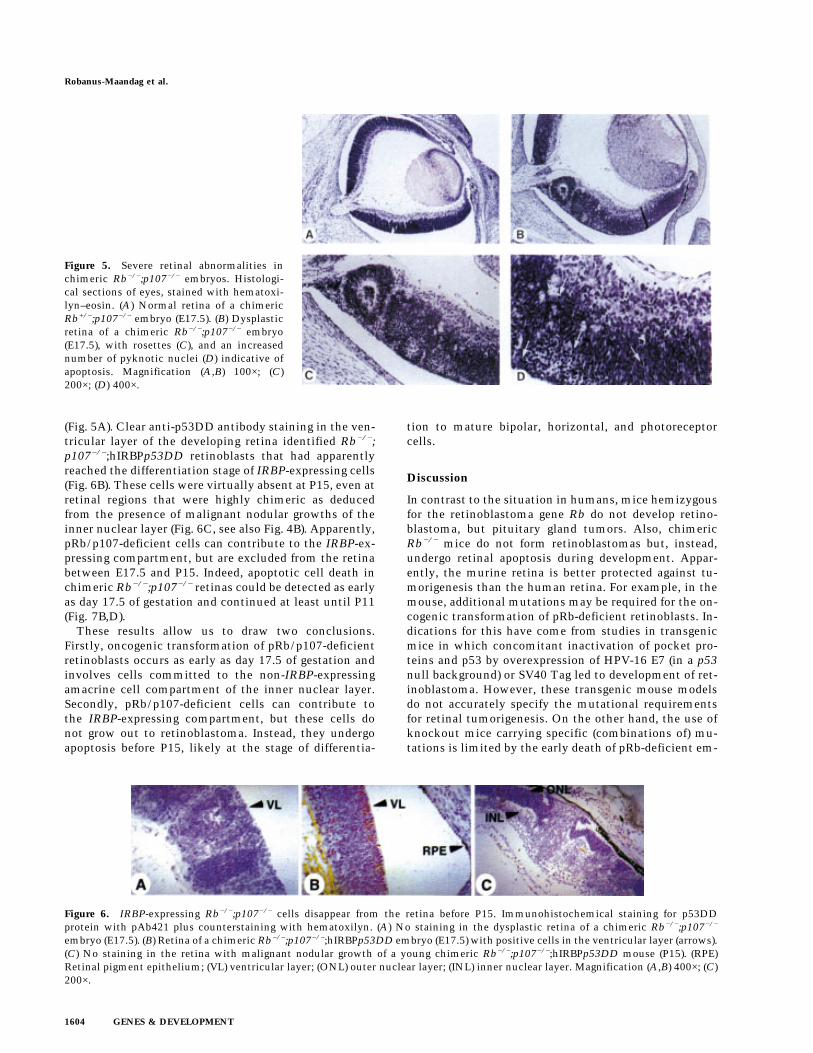
Figure 5. Severe retinal abnormalities inchimeric Rb−/−;p107−/− embryos. Histologi-cal sections of eyes, stained with hematoxi-lyn–eosin. (A) Normal retina of a chimericRb+/−;p107−/− embryo (E17.5). (B) Dysplasticretina of a chimeric Rb−/−;p107−/− embryo(E17.5), with rosettes (C), and an increasednumber of pyknotic nuclei (D) indicative ofapoptosis. Magnification (A,B) 100×; (C)200×; (D) 400×.
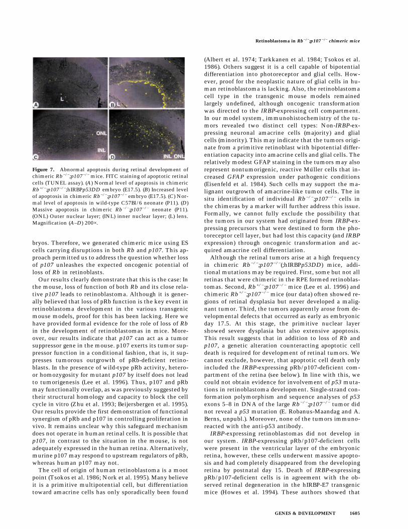
Figure 6. IRBP-expressing Rb−/−;p107−/− cells disappear from the retina before P15. Immunohistochemical staining for p53DDprotein with pAb421 plus counterstaining with hematoxilyn. (A) No staining in the dysplastic retina of a chimeric Rb−/−;p107−/−
embryo (E17.5). (B) Retina of a chimeric Rb−/−;p107−/−;hIRBPp53DD embryo (E17.5) with positive cells in the ventricular layer (arrows).(C) No staining in the retina with malignant nodular growth of a young chimeric Rb−/−;p107−/−;hIRBPp53DD mouse (P15). (RPE)Retinal pigment epithelium; (VL) ventricular layer; (ONL) outer nuclear layer; (INL) inner nuclear layer. Magnification (A,B) 400×; (C)200×.
Robanus-Maandag et al.
1604 GENES & DEVELOPMENT
bryos. Therefore, we generated chimeric mice using EScells carrying disruptions in both Rb and p107. This ap-proach permitted us to address the question whether lossof p107 unleashes the expected oncogenic potential ofloss of Rb in retinoblasts.
Our results clearly demonstrate that this is the case: Inthe mouse, loss of function of both Rb and its close rela-tive p107 leads to retinoblastoma. Although it is gener-ally believed that loss of pRb function is the key event inretinoblastoma development in the various transgenicmouse models, proof for this has been lacking. Here wehave provided formal evidence for the role of loss of Rbin the development of retinoblastomas in mice. More-over, our results indicate that p107 can act as a tumorsuppressor gene in the mouse. p107 exerts its tumor sup-pressor function in a conditional fashion, that is, it sup-presses tumorous outgrowth of pRb-deficient retino-blasts. In the presence of wild-type pRb activity, hetero-or homozygosity for mutant p107 by itself does not leadto tumorigenesis (Lee et al. 1996). Thus, p107 and pRbmay functionally overlap, as was previously suggested bytheir structural homology and capacity to block the cellcycle in vitro (Zhu et al. 1993; Beijersbergen et al. 1995).Our results provide the first demonstration of functionalsynergism of pRb and p107 in controlling proliferation invivo. It remains unclear why this safeguard mechanismdoes not operate in human retinal cells. It is possible thatp107, in contrast to the situation in the mouse, is notadequately expressed in the human retina. Alternatively,murine p107 may respond to upstream regulators of pRb,whereas human p107 may not.
The cell of origin of human retinoblastoma is a mootpoint (Tsokos et al. 1986; Nork et al. 1995). Many believeit is a primitive multipotential cell, but differentiationtoward amacrine cells has only sporadically been found
(Albert et al. 1974; Tarkkanen et al. 1984; Tsokos et al.1986). Others suggest it is a cell capable of bipotentialdifferentiation into photoreceptor and glial cells. How-ever, proof for the neoplastic nature of glial cells in hu-man retinoblastoma is lacking. Also, the retinoblastomacell type in the transgenic mouse models remainedlargely undefined, although oncogenic transformationwas directed to the IRBP-expressing cell compartment.In our model system, immunohistochemistry of the tu-mors revealed two distinct cell types: Non-IRBP-ex-pressing neuronal amacrine cells (majority) and glialcells (minority). This may indicate that the tumors origi-nate from a primitive retinoblast with bipotential differ-entiation capacity into amacrine cells and glial cells. Therelatively modest GFAP staining in the tumors may alsorepresent nontumorigenic, reactive Muller cells that in-creased GFAP expression under pathogenic conditions(Eisenfeld et al. 1984). Such cells may support the ma-lignant outgrowth of amacrine-like tumor cells. The insitu identification of individual Rb−/−;p107−/− cells inthe chimeras by a marker will further address this issue.Formally, we cannot fully exclude the possibility thatthe tumors in our system had originated from IRBP-ex-pressing precursors that were destined to form the pho-toreceptor cell layer, but had lost this capacity (and IRBPexpression) through oncogenic transformation and ac-quired amacrine cell differentiation.
Although the retinal tumors arise at a high frequencyin chimeric Rb−/−;p107−/−(;hIRBPp53DD) mice, addi-tional mutations may be required. First, some but not allretinas that were chimeric in the RPE formed retinoblas-tomas. Second, Rb+/−;p107−/− mice (Lee et al. 1996) andchimeric Rb+/−;p107−/− mice (our data) often showed re-gions of retinal dysplasia but never developed a malig-nant tumor. Third, the tumors apparently arose from de-velopmental defects that occurred as early as embryonicday 17.5. At this stage, the primitive nuclear layershowed severe dysplasia but also extensive apoptosis.This result suggests that in addition to loss of Rb andp107, a genetic alteration counteracting apoptotic celldeath is required for development of retinal tumors. Wecannot exclude, however, that apoptotic cell death onlyincluded the IRBP-expressing pRb/p107-deficient com-partment of the retina (see below). In line with this, wecould not obtain evidence for involvement of p53 muta-tions in retinoblastoma development. Single-strand con-formation polymorphism and sequence analyses of p53exons 5–8 in DNA of the large Rb−/−;p107−/− tumor didnot reveal a p53 mutation (E. Robanus-Maandag and A.Berns, unpubl.). Moreover, none of the tumors immuno-reacted with the anti-p53 antibody.
IRBP-expressing retinoblastomas did not develop inour system. IRBP-expressing pRb/p107-deficient cellswere present in the ventricular layer of the embryonicretina, however, these cells underwent massive apopto-sis and had completely disappeared from the developingretina by postnatal day 15. Death of IRBP-expressingpRb/p107-deficient cells is in agreement with the ob-served retinal degeneration in the hIRBP-E7 transgenicmice (Howes et al. 1994). These authors showed that
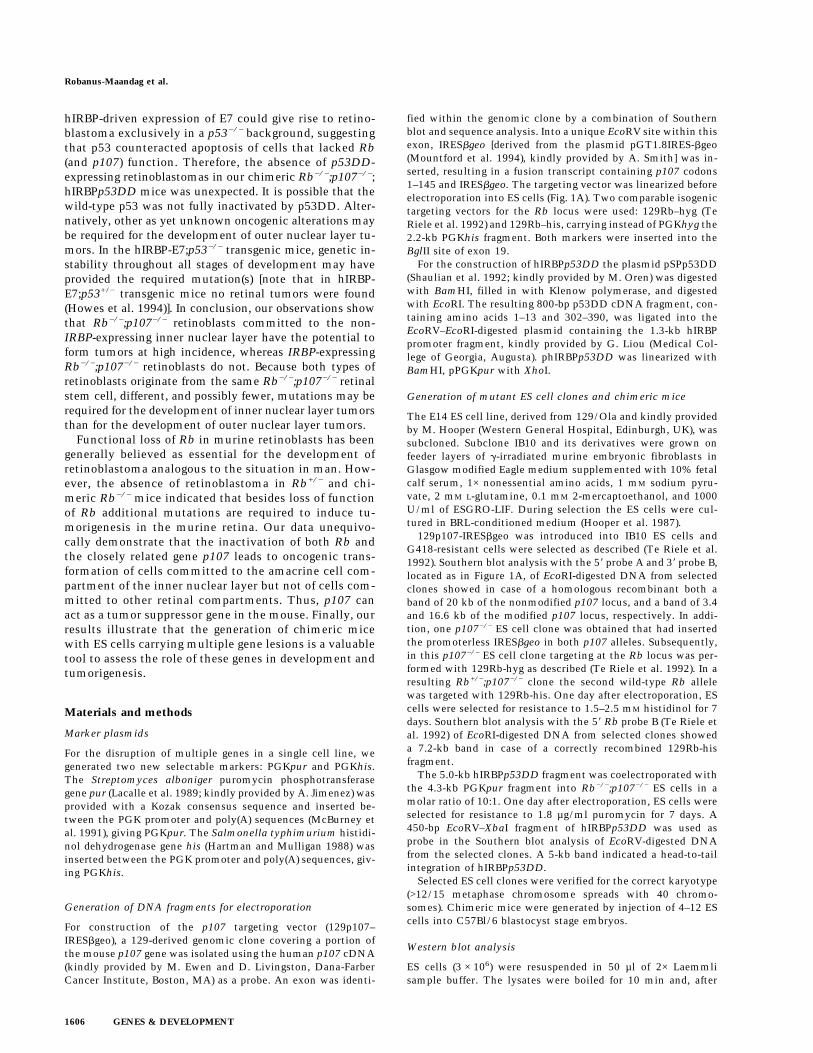
Figure 7. Abnormal apoptosis during retinal development ofchimeric Rb−/−;p107−/− mice. FITC staining of apoptotic retinalcells (TUNEL assay). (A) Normal level of apoptosis in chimericRb+/−;p107−/−;hIRBPp53DD embryo (E17.5). (B) Increased levelof apoptosis in chimeric Rb−/−;p107−/− embryo (E17.5). (C) Nor-mal level of apoptosis in wild-type C57Bl/6 neonate (P11). (D)Massive apoptosis in chimeric Rb−/−;p107−/− neonate (P11).(ONL) Outer nuclear layer; (INL) inner nuclear layer; (L) lens.Magnification (A–D) 200×.
Retinoblastoma in Rb−/−;p107 −/− chimeric mice
GENES & DEVELOPMENT 1605
hIRBP-driven expression of E7 could give rise to retino-blastoma exclusively in a p53−/− background, suggestingthat p53 counteracted apoptosis of cells that lacked Rb(and p107) function. Therefore, the absence of p53DD-expressing retinoblastomas in our chimeric Rb−/−;p107−/−;hIRBPp53DD mice was unexpected. It is possible that thewild-type p53 was not fully inactivated by p53DD. Alter-natively, other as yet unknown oncogenic alterations maybe required for the development of outer nuclear layer tu-mors. In the hIRBP-E7;p53−/− transgenic mice, genetic in-stability throughout all stages of development may haveprovided the required mutation(s) [note that in hIRBP-E7;p53+/− transgenic mice no retinal tumors were found(Howes et al. 1994)]. In conclusion, our observations showthat Rb−/−;p107−/− retinoblasts committed to the non-IRBP-expressing inner nuclear layer have the potential toform tumors at high incidence, whereas IRBP-expressingRb−/−;p107−/− retinoblasts do not. Because both types ofretinoblasts originate from the same Rb−/−;p107−/− retinalstem cell, different, and possibly fewer, mutations may berequired for the development of inner nuclear layer tumorsthan for the development of outer nuclear layer tumors.
Functional loss of Rb in murine retinoblasts has beengenerally believed as essential for the development ofretinoblastoma analogous to the situation in man. How-ever, the absence of retinoblastoma in Rb+/− and chi-meric Rb−/− mice indicated that besides loss of functionof Rb additional mutations are required to induce tu-morigenesis in the murine retina. Our data unequivo-cally demonstrate that the inactivation of both Rb andthe closely related gene p107 leads to oncogenic trans-formation of cells committed to the amacrine cell com-partment of the inner nuclear layer but not of cells com-mitted to other retinal compartments. Thus, p107 canact as a tumor suppressor gene in the mouse. Finally, ourresults illustrate that the generation of chimeric micewith ES cells carrying multiple gene lesions is a valuabletool to assess the role of these genes in development andtumorigenesis.
Materials and methods
Marker plasmids
For the disruption of multiple genes in a single cell line, wegenerated two new selectable markers: PGKpur and PGKhis.The Streptomyces alboniger puromycin phosphotransferasegene pur (Lacalle et al. 1989; kindly provided by A. Jimenez) wasprovided with a Kozak consensus sequence and inserted be-tween the PGK promoter and poly(A) sequences (McBurney etal. 1991), giving PGKpur. The Salmonella typhimurium histidi-nol dehydrogenase gene his (Hartman and Mulligan 1988) wasinserted between the PGK promoter and poly(A) sequences, giv-ing PGKhis.
Generation of DNA fragments for electroporation
For construction of the p107 targeting vector (129p107–IRESbgeo), a 129-derived genomic clone covering a portion ofthe mouse p107 gene was isolated using the human p107 cDNA(kindly provided by M. Ewen and D. Livingston, Dana-FarberCancer Institute, Boston, MA) as a probe. An exon was identi-
fied within the genomic clone by a combination of Southernblot and sequence analysis. Into a unique EcoRV site within thisexon, IRESbgeo [derived from the plasmid pGT1.8IRES-bgeo(Mountford et al. 1994), kindly provided by A. Smith] was in-serted, resulting in a fusion transcript containing p107 codons1–145 and IRESbgeo. The targeting vector was linearized beforeelectroporation into ES cells (Fig. 1A). Two comparable isogenictargeting vectors for the Rb locus were used: 129Rb–hyg (TeRiele et al. 1992) and 129Rb–his, carrying instead of PGKhyg the2.2-kb PGKhis fragment. Both markers were inserted into theBglII site of exon 19.
For the construction of hIRBPp53DD the plasmid pSPp53DD(Shaulian et al. 1992; kindly provided by M. Oren) was digestedwith BamHI, filled in with Klenow polymerase, and digestedwith EcoRI. The resulting 800-bp p53DD cDNA fragment, con-taining amino acids 1–13 and 302–390, was ligated into theEcoRV–EcoRI-digested plasmid containing the 1.3-kb hIRBPpromoter fragment, kindly provided by G. Liou (Medical Col-lege of Georgia, Augusta). phIRBPp53DD was linearized withBamHI, pPGKpur with XhoI.
Generation of mutant ES cell clones and chimeric mice
The E14 ES cell line, derived from 129/Ola and kindly providedby M. Hooper (Western General Hospital, Edinburgh, UK), wassubcloned. Subclone IB10 and its derivatives were grown onfeeder layers of g-irradiated murine embryonic fibroblasts inGlasgow modified Eagle medium supplemented with 10% fetalcalf serum, 1× nonessential amino acids, 1 mM sodium pyru-vate, 2 mM L-glutamine, 0.1 mM 2-mercaptoethanol, and 1000U/ml of ESGRO-LIF. During selection the ES cells were cul-tured in BRL-conditioned medium (Hooper et al. 1987).
129p107-IRESbgeo was introduced into IB10 ES cells andG418-resistant cells were selected as described (Te Riele et al.1992). Southern blot analysis with the 58 probe A and 38 probe B,located as in Figure 1A, of EcoRI-digested DNA from selectedclones showed in case of a homologous recombinant both aband of 20 kb of the nonmodified p107 locus, and a band of 3.4and 16.6 kb of the modified p107 locus, respectively. In addi-tion, one p107−/− ES cell clone was obtained that had insertedthe promoterless IRESbgeo in both p107 alleles. Subsequently,in this p107−/− ES cell clone targeting at the Rb locus was per-formed with 129Rb-hyg as described (Te Riele et al. 1992). In aresulting Rb+/−;p107−/− clone the second wild-type Rb allelewas targeted with 129Rb-his. One day after electroporation, EScells were selected for resistance to 1.5–2.5 mM histidinol for 7days. Southern blot analysis with the 58 Rb probe B (Te Riele etal. 1992) of EcoRI-digested DNA from selected clones showeda 7.2-kb band in case of a correctly recombined 129Rb-hisfragment.
The 5.0-kb hIRBPp53DD fragment was coelectroporated withthe 4.3-kb PGKpur fragment into Rb−/−;p107−/− ES cells in amolar ratio of 10:1. One day after electroporation, ES cells wereselected for resistance to 1.8 µg/ml puromycin for 7 days. A450-bp EcoRV–XbaI fragment of hIRBPp53DD was used asprobe in the Southern blot analysis of EcoRV-digested DNAfrom the selected clones. A 5-kb band indicated a head-to-tailintegration of hIRBPp53DD.
Selected ES cell clones were verified for the correct karyotype(>12/15 metaphase chromosome spreads with 40 chromo-somes). Chimeric mice were generated by injection of 4–12 EScells into C57Bl/6 blastocyst stage embryos.
Western blot analysis
ES cells (3 × 10⁶) were resuspended in 50 µl of 2× Laemmlisample buffer. The lysates were boiled for 10 min and, after
Robanus-Maandag et al.
1606 GENES & DEVELOPMENT
centrifugation for 2 min, 30% of the supernatant was loaded ona 10% SDS–polyacrylamide gel. After resolution, the gel wastransferred to a Protran membrane (Schleicher & Schull) byelectroblotting. For the antibody incubation with anti-p107,performed in 5% Blotto dissolved in TBST (Tris-buffered saline;0.1% Tween-20), the polyclonal rabbit anti-human antibody C-18 was used that recognizes amino acids 1052–1068 of p107(Santa Cruz Biotechnology). Subsequently, the membrane wasincubated with goat anti-rabbit horseradish peroxidase-labeledantibody. Antigen–antibody complexes were detected by en-hanced chemoluminescence (ECL; Amersham).
Generation of transgenic hIRBPp53DD mice
The 2.1-kb ClaI–BamHI fragment of phIRBPp53DD was micro-injected into FVB zygotes. Southern blot analysis of EcoRV-digested DNA from tail biopsies was performed as described(Laird et al. 1991) using the 450-bp EcoRV–XbaI fragment ofphIRBPp53DD as probe.
ES cell contribution in (tumor) tissues
DNA was isolated from tissue samples as described by Laird etal. (1991). The extent of chimerism was determined by detec-tion of the Rb wild-type and mutated EcoRI fragments withprobe A on Southern blots as described before (Te Riele et al.1992) using the PhosphorImager. Cells of the tumor areas in theunstained 10 µm tissue sections were scraped off with a scalpelfrom the plain glass slides and transferred to 0.5 ml of xylene todissolve the paraffin for 5 min. One volume of 100% ethanolwas mixed with the supernatants and after 5 min the tissueswere pelleted, dried at 55°C, and incubated in 50 mM Tris (pH8.5), 1 mM EDTA, 0.5% Tween-20, and 200 µg/ml proteinase Kovernight at 55°C and for 10 min at 95°C. To determine thepercentage of ES cell-derived cells in the tumors, simple se-quence repeat analyses were performed on the DNA solutionswith the primer set D2mit94 (Mouse MapPairs, Research Ge-netics, Huntsville, AL) as described (Dietrich et al. 1992).
Histological analysis and immunostaining
Embryos and tissues were fixed in phosphate-buffered formalin,embedded in paraffin, sectioned at 5 µm, and stained with he-matoxylin and eosin according to standard procedures.
For immunohistochemical detection of antigens, the rehy-drated tissue sections were boiled for 15 min in citrate buffer atpH 6.0 and cooled down slowly before preincubation with 1%normal goat serum. The following primary antibodies wereused: (1) mouse monoclonal anti-human p53 that recognizesamino acids 370–378 (pAb421, Harlow et al. 1981; OncogeneScience); (2) mouse monoclonal anti-rat syntaxin (HPC-1, SigmaBiosciences); (3) rabbit polyclonal anti-bovine IRBP (kindly pro-vided by Yvonne De Kozak, U450 INSERM, Paris, France); (4)rabbit polyclonal anti-cow glial fibrillary acidic protein (GFAP;DAKO); (5) rabbit polyclonal anti-bovine neuron-specific eno-lase (NSE; Chemicon International); and (6) rabbit polyclonalanti-bovine neurofilament, 200-kD subunit (NF200kd; Sigma).
Expression of the p53DD transgene was detected by the indi-rect immunoperoxidase assay with DAB substrate as described(Ivanyi et al. 1989). Expression of the endogenous retinal anti-gens was determined by the indirect immunofluorescence assaywith goat anti-mouse or pig anti-rabbit FITC (DAKO) and, incase of double staining, goat anti-rabbit Texas Red (MolecularProbes, Leiden, The Netherlands). Incorporated fluorescein wasdetected by confocal laser scan microscopy.
In situ detection of apoptosis
TUNEL analyses (Gavrieli et al. 1992) were performed on 8-µmtissue sections as described (In Situ Cell Death Detection kit,Boehringer Mannheim). Incorporated fluorescein was detectedby confocal laser scan microscopy.
Acknowledgments
We thank Paul Krimpenfort, Karin van Veen-Buurman, andRene Bobeldijk for assistance in zygote and blastocyst injec-tions; Jurjen Bulthuis, Kees de Goeij, Lia Kuijper-Pietersma, andEva van Muylwijk for histotechnical assistance; Rein Regnerusfor tail DNA analysis; Fina van der Ahe, Kwame Ankama, NelBosnie, Halfdan Raasø, Loes Rijswijk, and Auke Zwerver foranimal care; Lauran Oomen for assistance with the confocallaser scan microscope; Rene Bernards, Gabriel Gil-Gomez, andMarc Vooijs for critically reading the manuscript. This workwas supported by The Netherlands Organization for ScientificResearch (NWO) through a program grant to A.B. (E.R.-M.), theEuropean Community (E.R.-M.), and the Netherlands CancerFoundation (J.-H.D.).
The publication costs of this article were defrayed in part bypayment of page charges. This article must therefore be herebymarked ‘‘advertisement’’ in accordance with 18 USC section1734 solely to indicate this fact.
References
Albert, D.M., M. Lahav, R. Lesser, and J. Craft. 1974. Recentobservations regarding retinoblastoma, I: Ultrastructure, tis-sue culture growth, incidence, and animal models. Trans.Ophthalmol. Soc. U.K. 94: 909–928.
Al-Ubaidi, M.R., R.L. Font, A.B. Quiambao, M.J. Keener, G.I.Liou, P.A. Overbeek, and W. Baehr. 1992. Bilateral retinaland brain tumors in transgenic mice expressing simian virus40 large T antigen under control of the human interphoto-receptor retinoid-binding protein promoter. J. Cell Biol.119: 1681–1687.
Barnstable, C.J., R. Hofstein, and K. Akagawa. 1985. A marker ofearly amacrine cell development in rat retina. Brain Res.20: 286–290.
Beijersbergen, R.L., L. Carlee, R.M. Kerkhoven, and R. Bernards.1995. Regulation of the retinoblastoma protein-related p107by G1 cyclin complexes. Genes & Dev. 9: 1340–1353.
Bernards, R. 1997. E2F: A nodal point in cell cycle regulation.Biochim. Biophys. Acta 1333: 33–40.
Bjorklund, H., A. Bignami, and D. Dahl. 1985. Immunohisto-chemical demonstration of glial fibrillary acidic protein innormal rat Muller glia and retinal astrocytes. Neurosci. Lett.54: 363–368.
Bowman, T., H. Symonds, L. Gu, C. Yin, M. Oren, and T. VanDyke. 1996. Tissue-specific inactivation of p53 tumor sup-pression in the mouse. Genes & Dev. 10: 826–835.
Carter-Dawson, L., R.A. Alvarez, S.-L. Fong, G.I. Liou, H.G.Sperling, and C.D.B. Bridges. 1986. Rhodopsin, 11-cis vita-min A, and interstitial retinol-binding protein (IRBP) duringretinal development in normal and rd mutant mice. Dev.Biol. 116: 431–438.
Clarke, A.R., E. Robanus Maandag, M. van Roon, N.M.T. vander Lugt, M. van der Valk, M.L. Hooper, A. Berns, and H. teRiele. 1992. Requirement for a functional Rb-1 gene in mu-rine development. Nature 359: 328–330.
Claudio, P.P., C.M. Howard, A. Baldi, A. De Luca, Y. Fu, G.Condorelli, Y. Sun, N. Colburn, B. Calabretta, and A. Gior-
Retinoblastoma in Rb−/−;p107 −/− chimeric mice
GENES & DEVELOPMENT 1607
dano. 1994. p130/pRB2 has growth suppressive propertiessimilar to yet distinctive from those of retinoblastoma fam-ily members pRB and p107. Cancer Res. 54: 5556–5560.
DeCaprio, J.A., J.W. Ludlow, J. Figge, J.-Y. Shew, C.-M. Huang,W.-H. Lee, E. Marsilio, E. Paucha, and D.M. Livingston.1988. SV40 large tumor antigen forms a specific complexwith the product of the retinoblastoma susceptibility gene.Cell 54: 275–283.
Dietrich, W., H. Katz, S.E. Lincoln, H.-S. Shin, J. Friedman, N.C.Dracopoli, and E.S. Lander. 1992. A genetic map for themouse suitable for typing intraspecific crosses. Genetics131: 423–447.
Drager, U.C., D.L. Edwards, and C.J. Barnstable. 1984. Antibod-ies against filamentous proteins in discrete cell types of themouse retina. J. Neurosci. 4: 2025–2042.
Draper, G.J., B.M. Sanders, and J.E. Kingston. 1986. Second pri-mary neoplasms in patients with retinoblastoma. Br. J. Can-cer 53: 661–671.
Duke-Elder, S. and C. Cook. 1963. Embryology. In System ofophthalmology (ed. S. Duke-Elder), pp. 81–109. Kimpton,London, UK.
Dyson, N., P.M. Howley, K. Munger, and E. Harlow. 1989. Thehuman papillomavirus-16 E7 oncoprotein is able to bind tothe retinoblastoma gene product. Science 243: 934–936.
Eisenfeld, A.J., A.H. Bunt-Milam, and P.V. Sarthy. 1984. Mullercell expression of glial fibrillary acidic protein after geneticand experimental photoreceptor degeneration in the ratretina. Invest. Ophthalmol. Vis. Sci. 25: 1321–1328.
Ewen, M.E., Y. Xing, J.B. Lawrence, and D.M. Livingston. 1991.Molecular cloning, chromosomal mapping, and expressionof the cDNA for p107, a retinoblastoma gene product-relatedprotein. Cell 66: 1155–1164.
Friend, S.H., J.M. Horowitz, M.R. Gerber, X.-F. Wang, E. Bogen-mann, F.P. Li, and R.A. Weinberg. 1987. Deletions of a DNAsequence in retinoblastomas and mesenchymal tumors: Or-ganization of the sequence and its encoded protein. Proc.Natl. Acad. Sci. 84: 9059–9063.
Gavrieli, Y., Y. Sherman, and S.A. Ben-Sasson. 1992. Identifica-tion of programmed cell death in situ via specific labeling ofnuclear DNA fragmentation. J. Cell Biol. 119: 493–501.
Hall, M. and G. Peters. 1996. Genetic alterations of cyclins,cyclin-dependent kinases, and Cdk inhibitors in human can-cer. Adv. Cancer Res. 68: 67–108.
Hannon, G.J., D. Demetrick, and D. Beach. 1993. Isolation ofthe Rb-related p130 through its interaction with CDK2 andcyclins. Genes & Dev. 7: 2378–2391.
Harbour, J.W., S.-L. Lai, J. Whang-Peng, A.F. Gazdar, J.D. Minna,and F.J. Kaye. 1988. Abnormalities in structure and expres-sion of the human retinoblastoma gene in SCLC. Science241: 353–357.
Harlow, E., L.V. Crawford, D.C. Pim, and N.M. Williamson.1981. Monoclonal antibodies specific for simian virus 40 tu-mor antigens. J. Virol. 39: 861–869.
Hartman, S. and R.C. Mulligan. 1988. Two dominant-actingselectable markers for gene transfer studies in mammaliancells. Proc. Natl. Acad. Sci. 85: 8047–8051.
Hooper, M., K. Hardy, A. Handyside, S. Hunter, and M. Monk.1987. HPRT-deficient (Lesch-Nyhan) mouse embryos de-rived from germline colonization by cultured cells. Nature326: 292–295.
Horowitz, J.M., S.-H. Park, E. Bogenmann, J.-C. Cheng, D.W.Yandell, F.J. Kaye, J.D. Minna, T.P. Drya, and R.A. Weinberg.1990. Frequent inactivation of the retinoblastoma anti-on-cogene is restricted to a subset of human tumor cells. Proc.Natl. Acad. Sci. 87: 2775–2779.
Howes, K.A., N. Ransom, D.S. Papermaster, J.G.H. Lasudry,
D.M. Albert, and J.J. Windle. 1994. Apoptosis or retinobla-soma: Alternative fates of photoreceptors expressing theHPV-16 E7 gene in the presence or absence of p53. Genes &Dev. 8: 1300–1310.
Ivanyi, D., A. Ansink, E. Groeneveld, P.C. Hageman, W.J. Mooi,and A.P.M. Heintz. 1989. New monoclonal antibodies rec-ognizing epidermal differentiation-associated keratins in for-malin-fixed, paraffin-embedded tissue. Keratin 10 expressionin carcinoma of the vulva. J. Pathol. 159: 7–12.
Jacks, T., A. Fazeli, E.M. Schmitt, R.T. Bronson, M.A. Goodell,and R.A. Weinberg. 1992. Effects of an Rb mutation in themouse. Nature 359: 295–300.
Jiang, Z., E. Zacksenhaus, B.L. Gallie, and R.A. Phillips. 1997.The retinoblastoma gene family is differentially expressedduring embryogenesis. Oncogene 14: 1789–1797.
Kusnetsova, L.E., E.L. Prigogina, H.E. Pogosianz, and B.M. Bel-kina. 1982. Similar chromosomal abnormalities in severalretinoblastomas. Hum. Genet. 61: 201–204.
Lacalle, R.A., D. Pulido, J. Vara, M. Zalacaın, and A. Jimenez.1989. Molecular analysis of the pac gene encoding a puro-mycin N-acetyl transferase from Streptomyces alboniger.Gene 79: 375–380.
Laird, P.W., A. Zijderveld, K. Linders, M.A. Rudnicki, R. Jae-nisch, and A. Berns. 1991. Simplified mammalian DNA iso-lation procedure. Nucleic Acids Res. 19: 4293.
Lee, E.Y.-H.P., H. To, J.-Y. Shew, R. Bookstein, P. Scully, andW.-H. Lee. 1988. Inactivation of the retinoblastoma suscep-tibility gene in human breast cancers. Science 241: 218–221.
Lee, E.Y.-H.P., C.-Y. Chang, N. Hu, Y.-C. Wang, C.-C. Lai, K.Herrup, W.-H. Lee, and A. Bradley. 1992. Mice deficient forRb are nonviable and show defects in neurogenesis and hae-matopoiesis. Nature 359: 288–294.
Lee, M.-H., B.O. Williams, G. Mulligan, S. Mukai, R.T. Bronson,N. Dyson, E. Harlow, and T. Jacks. 1996. Targeted disruptionof p107: Functional overlap between p107 and Rb. Genes &Dev. 10: 1621–1632.
Li, Y., C. Graham, S. Lacy, A.M.V. Duncan, and P. Whyte. 1993.The adenovirus E1A-associated 130-kD protein is encodedby a member of the retinoblastoma gene family and physi-cally interacts with cyclins A and E. Genes & Dev. 7: 2366–2377.
Liou, G.I., L. Geng, M.R. Al-Ubaidi, S. Matragoon, G. Hanten,W. Baehr, and P.A. Overbeek. 1990. Tissue-specific expres-sion in transgenic mice directed by the 58-flanking sequencesof the human gene encoding interphotoreceptor retinoid-binding protein. J. Biol. Chem. 265: 8373–8376.
Mayol, X., X. Grana, A. Baldi, N. Sang, Q. Hu, and A. Giordano.1993. Cloning of a new member of the retinoblastoma genefamily (pRb2) which binds to the E1A transforming domain.Oncogene 8: 2561–2566.
McBurney, M.W., L.C. Sutherland, C.N. Adra, B. Leclair, M.A.Rudnicki, and K. Jardine. 1991. The mouse Pgk-1 gene pro-moter contains an upstream activator sequence. Nucleic Ac-ids Res. 19: 5755–5761.
Morgenbesser, S.D., B.O. Williams, T. Jacks, and R.A. DePinho.1994. p53-dependent apoptosis produced by Rb-deficiency inthe developing mouse lens. Nature 371: 72–74.
Mountford, P., B. Zevnik, A. Duwel, J. Nichols, M. Li, C. Dani,M. Robertson, I. Chambers, and A. Smith. 1994. Dicistronictargeting constructs: Reporters and modifiers of mammaliangene expression. Proc. Natl. Acad. Sci. 91: 4303–4307.
Nork, T.M., T.L. Schwartz, H.M. Doshi, and L.L. Millecchia.1995. Retinoblastoma: Cell of origin. Arch. Ophthalmol.113: 791–802.
Qin, X.-Q., D.M. Livingston, M. Ewen, W.R. Sellers, Z. Arany,and W.G. Kaelin Jr. 1995. The transcription factor E2F-1 is a
Robanus-Maandag et al.
1608 GENES & DEVELOPMENT
downstream target of RB action. Mol. Cell. Biol. 15: 742–755.
Robanus Maandag, E.C., M. van der Valk, M. Vlaar, C. Felt-kamp, J. O’Brien, M. van Roon, N. van der Lugt, A. Berns,and H. te Riele. 1994. Developmental rescue of an embry-onic-lethal mutation in the retinoblastoma gene in chimericmice. EMBO J. 13: 4260–4268.
Schmechel, D., P.J. Marangos, and M. Brightman. 1978. Neu-ron-specific enolase is a molecular marker for peripheral andcentral neuroendocrine cells. Nature 276: 834–836.
Shaulian, E., A. Zauberman, D. Ginsberg, and M. Oren. 1992.Identification of a minimal transforming domain of p53:Negative dominance through abrogation of sequence-spe-cific DNA binding. Mol. Cell. Biol. 12: 5581–5592.
Sherr, C.J. 1994. G1 phase progression: Cycling on cue. Cell79: 551–555.
Squire, J., R.A. Phillips, S. Boyce, R. Godbout, B. Rogers, andB.L. Gallie. 1984. Isochromosome 6p, a unique chromosomalabnormality in retinoblastoma: Verification by standardstaining techniques, new densitometric methods, and so-matic cell hybridization. Hum. Genet. 66: 46–53.
Tarkkanen, A., T. Tervo, K. Tervo, and P. Panula. 1984. Immu-nohistochemical evidence for preproenkephalin A synthesisin human retinoblastoma. Invest. Ophthalmol. Vis. Sci.25: 1210–1212.
Te Riele, H., E. Robanus Maandag, and A. Berns. 1992. Highlyefficient gene targeting in embryonic stem cells through ho-mologous recombination with isogenic DNA constructs.Proc. Natl. Acad. Sci. 89: 5128–5132.
Tsokos, M., A.P. Kyritsis, G.J. Chader, and T.J. Triche. 1986.Differentiation of human retinoblastoma in vitro into celltypes with characteristics observed in embryonal or matureretina. Am. J. Pathol. 123: 542–552.
Weinberg, R.A. 1995. The retinoblastoma protein and cell cyclecontrol. Cell 81: 323–330.
Whyte, P., K.J. Buchkovich, J.M. Horowitz, S.H. Friend, M. Ray-buck, R.A. Weinberg, and E. Harlow. 1988. Association be-tween an oncogene and an anti-oncogene: The adenovirusE1A proteins bind to the retinoblastoma gene product. Na-ture 334: 124–129.
Williams, B.O., L. Remington, D.M. Albert, S. Mukai, R.T.Bronson, and T. Jacks. 1994a. Cooperative tumorigenic ef-fects of germline mutations in Rb and p53. Nature Genet.7: 480–484.
Williams, B.O., E.M. Schmitt, L. Remington, R.T. Bronson,D.M. Albert, R.A. Weinberg, and T. Jacks. 1994b. Extensivecontribution of Rb-deficient cells to adult chimeric micewith limited histopathological consequences. EMBO J.13: 4251–4259.
Young, R.W. 1985. Cell differentiation in the retina of themouse. Anat. Rec. 212: 199–205.
Zacksenhaus, E., Z. Jiang, D. Chung, J.D. Marth, R.A. Phillips,and B.L. Gallie. 1996. pRb controls proliferation, differentia-tion, and death of skeletal muscle cells and other lineagesduring embryogenesis. Genes & Dev. 10: 3051–3064.
Zhu, L., S. van den Heuvel, K. Helin, A. Fattaey, M. Ewen, D.Livingston, N. Dyson, and E. Harlow. 1993. Inhibition of cellproliferation by p107, a relative of the retinoblastoma pro-tein. Genes & Dev. 7: 1111–1125.
Retinoblastoma in Rb−/−;p107 −/− chimeric mice
GENES & DEVELOPMENT 1609


