Embed Size (px)
Citation preview
Pancreatic and duodenal homeobox 1 (PDX1) phosphorylation atserine-269 is HIPK2-dependent and affects PDX1 subnuclearlocalization
Rong Ana,1, Gabriela da Silva Xaviera,1, Francesca Semplicia,1, Saharnaz Vakhshouria,Huai-Xiang Haob, Jared Rutterb, Mario A. Paganoc, Flavio Meggioc, Lorenzo A. Pinnac, andGuy A. Ruttera,⁎aSection of Cell Biology, Division of Diabetes, Endocrinology and Metabolism, Department ofMedicine, Imperial College London, London SW7 2AZ, UKbDepartment of Biochemistry, University of Utah, Salt Lake City, UT 84112-5650, USAcDepartment of Biological Chemistry, University of Padova, viale G. Colombo 3, 35131 Padova, Italy
Research highlights► Mouse PDX1 Ser-269 is phosphorylated in pancreatic islets of Langerhans and beta cells. ► Highglucose concentration decreases the degree of phosphorylation on PDX1 Ser-269, as assessed by aphospho-specific anti-phospho-Ser269 antibody. ► HIPK2 is a potential kinase for PDX1 Ser-269.► Phosphorylation at Ser-269 affects the subnuclear distribution of PDX1.
AbstractPancreatic and duodenal homeobox 1 (PDX1) regulates pancreatic development and mature β-cellfunction. We demonstrate by mass spectrometry that serine residue at position 269 in the C-terminaldomain of PDX1 is phosphorylated in β-cells. Besides we show that the degree of phosphorylation,assessed with a phospho-Ser-269-specific antibody, is decreased by elevated glucose concentrationsin both MIN6 β-cells and primary mouse pancreatic islets. Homeodomain interacting protein kinase2 (HIPK2) phosphorylates PDX1 in vitro; phosphate incorporation substantially decreases in PDX1S269A mutant. Silencing of HIPK2 led to a 51 ± 0.2% decrease in Ser-269 phosphorylation in MIN6β-cells. Mutation of Ser-269 to phosphomimetic residue glutamic acid (S269E) or de-phosphomimetic residue alanine (S269A) exerted no effect on PDX1 half-life. Instead, PDX1 S269Emutant displayed abnormal changes in subnuclear localization in response to high glucose. Ourresults suggest that HIPK2-mediated phosphorylation of PDX1 at Ser-269 might be a regulatorymechanism connecting signals generated by changes in extracellular glucose concentration todownstream effectors via changes in subnuclear localization of PDX1, thereby influencing islet celldifferentiation and function.
© 2010 Elsevier Inc.⁎Corresponding author. Address: Department of Medicine, Imperial College London, Exhibition Road, London SW7 2AZ, UK. Fax:+44 020 7594 3351. [email protected] authors contributed equally to the work described in this manuscript.This document was posted here by permission of the publisher. At the time of deposit, it included all changes made during peer review,copyediting, and publishing. The U.S. National Library of Medicine is responsible for all links within the document and for incorporatingany publisher-supplied amendments or retractions issued subsequently. The published journal article, guaranteed to be such by Elsevier,is available for free, on ScienceDirect.
Sponsored document fromBiochemical and BiophysicalResearch Communications
Published as: Biochem Biophys Res Commun. 2010 August 20; 399(2): 155–161.
Sponsored Docum
ent Sponsored D
ocument
Sponsored Docum
ent
KeywordsPDX1; Islet; β-Cell; Phosphorylation; Glucose; HIPK2
1 IntroductionControl by glucose of preproinsulin gene transcription in pancreatic islet β-cells involves theinterplay of multiple trans- and cis-activating factors ([1] and references therein). Pancreaticand duodenal homeobox 1 (PDX1) was first described as a preproinsulin promoter A3-sitebinding factor [2,3]. PDX1 is involved in both the development and function of pancreatic βand other islet cells [4].
Deletion or inactivation of the PDX1 gene in the mouse [5] or in man [6] results in the completefailure of normal pancreatic development. Moreover, β-cell selective disruption of murinePDX1 expression [7] or function [8] lead respectively to defective insulin secretion and glucosesignaling. On the other hand, heterozygosity for a defective PDX1 gene leads to defectivepreproinsulin gene expression [7] and abnormal insulin secretion [9] in mice and to maturity-onset-diabetes of the young 4 (MODY4) in man [6].
In contrast to the relatively well-studied functions of the N-terminus and the homeodomain ofPDX1, the role of the conserved C-terminus is less well defined. Mutations which affect theC-terminus of PDX1 are associated with the development of type 2 diabetes in humans [10–12], while other findings indicate that the C-terminal domain may serve as both repressor andactivator of PDX1 function [13,14].
Humphrey and colleagues [15] reported that PDX1 phosphorylation in primary rat islets isdecreased by high glucose levels. These authors described Ser-268 and Ser-272 of rat PDX1(corresponding to Ser-269 and Ser-273 of mouse PDX1) as a novel C-terminal atypical non-primed GSK-3 consensus site which regulates PDX1 protein stability in response to glucose.Importantly, homeodomain interacting protein kinase 2 (HIPK2) ([16] and references therein)has been shown to co-localize with PDX1 in both the developing and adult pancreas and tomodulate positively PDX1 transcriptional activity, possibly by phosphorylation of the C-terminal domain [17]. We have previously observed that, in clonal β-cells, elevated glucoseconcentrations lead to translocation of PDX1 between the nuclear periphery and thenucleoplasm, accompanied by increased preproinsulin promoter activity [18]. Although themolecular basis for the enhanced nucleoplasmic accumulation of PDX1 is unclear, this processmay involve interaction of PDX1 homeodomain with the nuclear import receptor familymember importin-β1 [19]. In the present study we used mass spectrometry and generated ananti-phospho-serine-specific antibody to confirm Ser-269 as a bona fide phosphorylation sitein mouse PDX1 that is regulated by glucose in MIN6 β-cells and in primary mouse islets ofLangerhans. We show that Ser-269 is phosphorylated by homeodomain interacting proteinkinase 2 (HIPK2) in vitro. The analysis of (de)phospho-Ser-269-specific mutants suggest thatphosphorylation at this site, whilst having no effect on PDX1 protein stability or PDX1 DNA-binding property, is involved in nucleoplasmic (versus nuclear-peripheric) localization in theβ-cell in response to glucose.
2 Materials and methodsThe work described in this article has been carried out in accordance with the EC Directive86/609/EEC for animal experiments http://europa.eu.int/scadplus/leg/en/s23000.htm; and theUniform Requirements for manuscripts submitted to Biomedical journalshttp://www.nejm.org/general/text/requirements/1.htm.
An et al. Page 2
Published as: Biochem Biophys Res Commun. 2010 August 20; 399(2): 155–161.
Sponsored Docum
ent Sponsored D
ocument
Sponsored Docum
ent
2.1 Cell culture and reagentsHuman embryonic kidney (HEK) 293 cells were cultured in Dulbecco’s modified Eagle’smedium (DMEM) (Lonza) containing 10% (v/v) fetal bovine serum (FBS), 100 IU/mlpenicillin and 100 IU/ml streptomycin. INS-1(832/13) cells (kindly provided by Dr. C.Newgard, Duke University) and MIN6 β (mouse insulinoma pancreatic beta) cells werecultured as in [20].
Anti-c-myc antibody was from Roche. Rabbit polyclonal anti-PDX1 antibody was as described[18]. Anti-phospho-Ser-269-PDX1 antibody was raised in rabbits by immunization withsynthetic phospho-peptide: L262PSGLSVpSPQPSSIAPLRPQEPR284 (Pacific ImmunologyInc, USA). HIPK2 was purchased from Upstate (Lake Placid, NY).
2.2 Mouse islet isolation and cultureIslets were isolated from CD1 mice and cultured as previously described [21].
2.3 PlasmidsPlasmid pcDNA3-PDX1-c-myc has been described [18]. Mutant plasmids pcDNA3-PDX1-S269A-c-myc and pcDNA3-PDX1-S269E-c-myc were generated using a QuikChange site-directed mutagenesis kit (Stratagene). Wild-type and mutant PDX1 myc-tagged codingsequences were inserted (HindIII/EcoRV) into the shuttle vector pAdTrack-CMV multiplecloning site [22]. Mouse wild-type PDX1 sequence PCR amplified from plasmid pcDNA3-PDX1-c-myc was cloned (NcoI/BamHI) into 6xHis-MBP (maltose binding protein) plasmid.Mutant plasmid His-MBP-PDX1 (S269A) was generated as above.
2.4 His-MBP-PDX1 production and purificationHis-MBP-PDX1 proteins expression was induced in Escherichia coli BL21 with 0.2 mMisopropyl-β-D-thiogalactopyranoside (IPTG). Proteins were purified on a nickel–nitrilotriaceticacid column according to Qiagen and dialyzed for 16 h at 4 °C in 50 mM Tris pH 7.9, 150 mMNaCl, 5 mM MgCl2, 1 mM β-mercaptoethanol. The MBP moiety was cut with Tobacco EtchVirus (TEV) protease, AcTEVTM protease (Invitrogen). MBP, histidine tag and histidine-tagged Ac-TEV protease were removed respectively with Amylose beads (New EnglandBioLab) and Ni–NTA agarose beads.
2.5 Recombinant adenoviruses and viral infectionRecombinant adenoviruses expressing wild-type (WT) and mutant (S269A, S269E) PDX1 andcontrol adenovirus, expressing green fluorescent protein (Ad-GFP) were prepared using theAdEasy system [22]. Cells were infected with various adenoviruses at a multiplicity of infection(MOI) of 50 for 5 h and maintained in 25 mM glucose for 24 h before subsequent experiments.
2.6 Real-time RT-PCRTotal mRNA and real-time quantitative RT-PCR analysis was as [23]. Primer sequences areas follows: cyclophilin A fwd, 5′-TAT CTG CAC TGC CAA GAC TGA-3′; cyclophilin A rev,5′-CCA CAA TGC TCA TGC CTT CTT TCA-3′; HIPK2 fwd, 5′-TGC TTG ACT TCC CCCATA GTG -3′; HIPK2 rev, 5′-CTT GCA AAT CTC CAT GTT TTG G -3′.
Data were analyzed by ABR PRISM SDS v1.3.1 (Applied Biosystems).
An et al. Page 3
Published as: Biochem Biophys Res Commun. 2010 August 20; 399(2): 155–161.
Sponsored Docum
ent Sponsored D
ocument
Sponsored Docum
ent
2.7 ImmunocytochemistryMIN6 β-cells infected with wild-type or mutant forms of PDX1 viruses at 50 MOI (multiplicityof infection). Immunocytochemical analysis was performed as described in the figure legendsand in [20].
2.8 ImmunoprecipitationCells lyzed on ice with 750 μl of IP buffer [1% (v/v) NP40 (Nonidet P40); 50 mM NaCl; 1%(w/v) sodium deoxycholate; 0.1% (w/v) sodium monododecyl sulfate (SDS); 50 mM Tris–HClpH 7.5; 2 mM EDTA; 10 mM sodium phosphate; 50 mM NaF (sodium fluoride); 1 m PMSF;200 μM Na3VO4 (sodium orthovanadate); 1 × CompleteTM protease inhibitor cocktail solution(Roche) and 1 × phosphatase inhibitor cocktail 1 & 2 (Sigma)] were rotated on wheel at 4 °Cfor 30 min. The lysate was clarified by centrifugation at 16,000g for 5 min. c-myc antibody-conjugated beads (Santa-Cruz; 40 μl) were added and tubes rotated at 4 °C overnight. Aftercentrifugation at 1,000g at 4 °C for 30 s, beads were washed five times with lysis buffer.
2.9 In vitro phosphorylation by HIPK2Purified wild-type (WT) or serine to alanine mutant (S269A) PDX1 (2 μg) was subjected toin vitro phosphorylation with HIPK2 (Upstate) following the manufacturer’s instruction.
2.10 Western blottingNuclear proteins were extracted as in [24]. Protein samples were separated on SDS–PAGE,and analyzed by Western blotting with the indicated antibodies.
2.11 Silencing of HIPK2siRNA were made with the AMBION Silencer siRNA construction kit. Starting primer pairswere designed as suggested in the AMBION protocol and according to the guidelines publishedin [25]. Sense and anti-sense primers were derived from mouse cDNA sequence for HIPK2 inthe ENSEMBL database (transcript name: Hipk2-201; transcript Id:ENSMUST00000038777). Target sequence was from nucleotide 788 to nucleotide 808 asfollows: 5′-AAA CGG GGC ACC AAT GAA ATT CCT GTC TC-3′). Control siRNA weremade with primers derived by scrambling the siRNA sequence for HIPK2. All sequences weresearched against known mouse cDNA sequences using BLAST(http://blast.ncbi.nlm.nih.gov/Blast.cgi). Transfection was with TransIT-TKO transfectionreagent (Mirus Bio Corporation).
2.12 Pulse-chaseMIN6 β-cells were infected with PDX1 wild-type, S269A and S269E adenoviruses. 24 h afterinfection, media were replaced with fresh DMEM containing 3 mM glucose and cells werecultured for 16 h. Cells were transferred for 1 h to cysteine/methionine-free medium, pulsedfor 3 h with 0.6 mCi [35S]cysteine/methionine (Perkin–Elmer), after which cells were washedand cultured for 2, 4, 8, and 16 h in 5 ml chase media with 10,000-fold molar excess of coldL-cysteine, L-methionine or harvested as the zero time point. Cells were lyzed in 1 ml RIPA[50 mM Tris (pH 7.5), 150 mM NaCl, 1% (v/v) Nonidet P-40 (NP-40) 0.5% (w/v) sodiumdeoxycholate (DOC), 0.1% (v/v) SDS, 0.4 mg/ml AESBF {4-(2-Aminoethyl) benzenesulfonylfluoride hydrochloride}, 10 μg/ml Leupeptin, 10 μg/ml Pepstatin, 5 μg/ml Aprotinin,1 × CompleteTM (Roche) and 1 × phosphatase inhibitor cocktail 1 and 2 (Sigma)]. Lysateswere immunoprecipitated with anti-myc antibody (Roche). After five washes with RIPAbuffer, proteins were eluted and analyzed by SDS–PAGE. PDX1 proteins were detected byfluorography and quantified in a Cyclone (Perkin–Elmer) phosphoimager storage system.
An et al. Page 4
Published as: Biochem Biophys Res Commun. 2010 August 20; 399(2): 155–161.
Sponsored Docum
ent Sponsored D
ocument
Sponsored Docum
ent
2.13 StatisticsData are given as means ±SE of three to five individual experiments. Comparisons betweenmeans were performed using one-tailed Student’s t-test for paired data with Bonferronicorrection for multiple sampling as appropriate.
3 Results3.1 Ser-269 phosphorylation is regulated by glucose in living β-cells (MIN6) and primarymouse islets
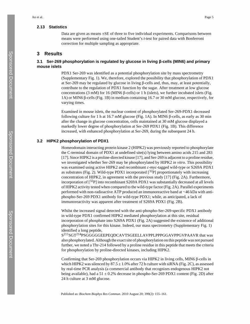
PDX1 Ser-269 was identified as a potential phosphorylation site by mass spectrometry(Supplementary Fig. 1). We, therefore, explored the possibility that phosphorylation of PDX1at Ser-269 may be regulated by glucose in living β-cells and, thus, may, at least potentially,contribute to the regulation of PDX1 function by the sugar. After treatment at low glucoseconcentrations (3 mM) for 16 (MIN6 β-cells) or 1 h (islets), we further incubated islets (Fig.1A) or MIN6 β-cells (Fig. 1B) in medium containing 16.7 or 30 mM glucose, respectively, forvarying times.
Examined in mouse islets, the nuclear content of phosphorylated Ser-269-PDX1 decreasedfollowing culture for 1 h at 16.7 mM glucose (Fig. 1A). In MIN6 β-cells, as early as 30 minafter the change in glucose concentration, cells maintained at 30 mM glucose displayed amarkedly lower degree of phosphorylation at Ser-269 PDX1 (Fig. 1B). This differenceincreased, with enhanced phosphorylation at Ser-269, during the subsequent 24 h.
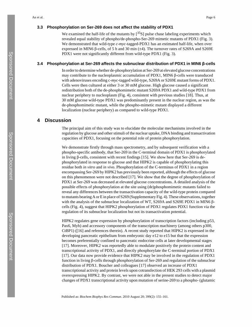
3.2 HIPK2 phosphorylation of PDX1Homeodomain interacting protein kinase 2 (HIPK2) was previously reported to phosphorylatethe C-terminal domain of PDX1 at undefined site(s) lying between amino acids 215 and 283[17]. Since HIPK2 is a proline-directed kinase [17], and Ser-269 is adjacent to a proline residue,we investigated whether Ser-269 may be phosphorylated by HIPK2 in vitro. This possibilitywas examined using active HIPK2 and recombinant c-myc-tagged wild-type or S269A PDX1as substrates (Fig. 2). Wild-type PDX1 incorporated [32P] proportionately with increasingconcentration of HIPK2, in agreement with the previous study [17] (Fig. 2A). Furthermore,incorporation of [32P] into recombinant S269A PDX1 was substantially decreased at all levelsof HIPK2 activity tested when compared to the wild-type factor (Fig. 2A). Parallel experimentsperformed with non-radioactive ATP produced an immunoreactive band at ∼46 kDa with anti-phospho-Ser-269 PDX1 antibody for wild-type PDX1; while, as anticipated, a lack ofimmunoreactivity was apparent after treatment of S269A PDX1 (Fig. 2B).
Whilst the increased signal detected with the anti-phospho-Ser-269-specific PDX1 antibodyin wild-type PDX1 confirmed HIPK2 mediated phosphorylation at this site, residualincorporation of phosphate into S269A PDX1 (Fig. 2A) suggested the existence of additionalphosphorylation sites for this kinase. Indeed, our mass spectrometry (Supplementary Fig. 1)identified a long peptide,S211SGT214PSGGGGGEEPEQDCAVTSGEELLAVPPLPPPGGAVPPGVPAAVR that wasalso phosphorylated. Although the exact site of phosphorylation on this peptide was not pursuedfurther, we noted a Thr-214 followed by a proline residue in this peptide that meets the criteriafor phosphorylation by proline-directed kinases, including HIPK2.
Confirming that Ser-269 phosphorylation occurs via HIPK2 in living cells, MIN6 β-cells inwhich HIPK2 was silenced by 87.5 ± 1.0% after 72 h culture with siRNA (Fig. 2C), as assessedby real-time PCR analysis (a commercial antibody that recognizes endogenous HIPK2 notbeing available), had a 51 ± 0.2% decrease in phospho-Ser-269 PDX1 content (Fig. 2D) after24 h culture at 3 mM glucose.
An et al. Page 5
Published as: Biochem Biophys Res Commun. 2010 August 20; 399(2): 155–161.
Sponsored Docum
ent Sponsored D
ocument
Sponsored Docum
ent
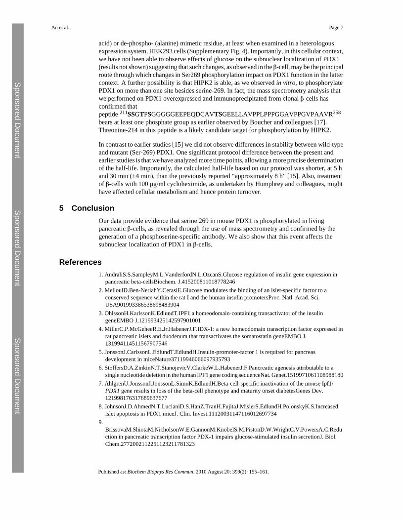
3.3 Phosphorylation on Ser-269 does not affect the stability of PDX1We examined the half-life of the mutants by [35S] pulse chase labeling experiments whichrevealed equal stability of phospho/de-phospho-Ser-269 mimetic mutants of PDX1 (Fig. 3).We demonstrated that wild-type c-myc-tagged-PDX1 has an estimated half-life, when overexpressed in MIN6 β-cells, of 5 h and 30 min (±4). The turnover rates of S269A and S269EPDX1 were not significantly different from wild-type PDX1 (Fig. 3).
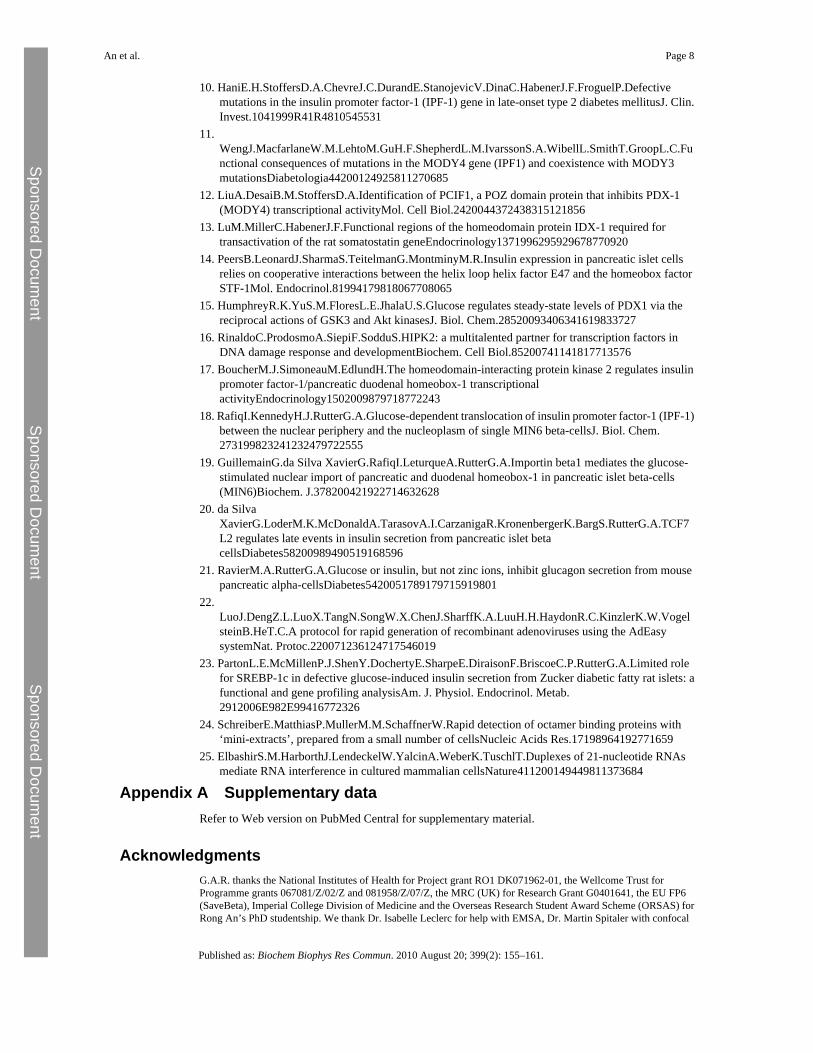
3.4 Phosphorylation at Ser-269 affects the subnuclear distribution of PDX1 in MIN6 β-cellsIn order to determine whether de-phosphorylation at Ser-269 at elevated glucose concentrationsmay contribute to the nucleoplasmic accumulation of PDX1, MIN6 β-cells were transducedwith adenoviruses encoding c-myc-tagged wild-type, S269A or S269E mutant forms of PDX1.Cells were then cultured at either 3 or 30 mM glucose. High glucose caused a significantredistribution both of the de-phosphomimetic mutant S269A PDX1 and wild-type PDX1 fromnuclear periphery to nucleoplasm (Fig. 4), consistent with previous studies [18]. Thus, at30 mM glucose wild-type PDX1 was predominantly present in the nuclear region, as was thede-phosphomimetic mutant, while the phospho-mimetic mutant displayed a differentlocalization (nuclear periphery) as compared to wild-type PDX1.
4 DiscussionThe principal aim of this study was to elucidate the molecular mechanisms involved in theregulation by glucose and other stimuli of the nuclear uptake, DNA binding and transactivationcapacities of PDX1, focusing on the potential role of protein phosphorylation.
We demonstrate firstly through mass spectrometry, and by subsequent verification with aphospho-specific antibody, that Ser-269 in the C-terminal domain of PDX1 is phosphorylatedin living β-cells, consistent with recent findings [15]. We show here that Ser-269 is de-phosphorylated in response to glucose and that HIPK2 is capable of phosphorylating thisresidue both in vitro and in vivo. Phosphorylation of the C-terminus of PDX1 in a regionencompassing Ser-269 by HIPK2 has previously been reported, although the effects of glucoseon this phenomenon were not described [17]. We show that the degree of phosphorylation ofPDX1 at Ser-269 was decreased at elevated glucose concentrations. A detailed analysis of thepossible effects of phosphorylation at the site using (de)phosphomimetic mutants failed toreveal any differences between the transactivation capacity of the wild-type protein comparedto mutants bearing A or E in place of S269 (Supplementary Fig. 4). These observations, togetherwith the analysis of the subnuclear localization of WT, S269A and S269E PDX1 in MIN6 β-cells (Fig. 4), suggest that HIPK2 phosphorylation of PDX1 regulates PDX1 function via theregulation of its subnuclear localization but not its transactivation potential.
HIPK2 regulates gene expression by phosphorylation of transcription factors (including p53,Pax6, Myb) and accessory components of the transcription machinery (among others p300,CtBP1) ([16] and references therein). A recent study reported that HIPK2 is expressed in thedeveloping pancreatic epithelium from embryonic day e12 to e15 but that the expressionbecomes preferentially confined to pancreatic endocrine cells at later developmental stages[17]. Moreover, HIPK2 was reportedly able to modulate positively the protein content andtranscriptional activity of PDX1, and directly phosphorylate the C-terminal portion of PDX1[17]. Our data now provide evidence that HIPK2 may be involved in the regulation of PDX1function in living β-cells through phosphorylation of Ser-269 and regulation of the subnucleardistribution of PDX1. Boucher and colleagues [17] observed an increase of PDX1transcriptional activity and protein levels upon cotransfection of HEK 293 cells with a plasmidoverexpressing HIPK2. By contrast, we were not able in the present studies to detect majorchanges of PDX1 transcriptional activity upon mutation of serine-269 to a phospho- (glutamic
An et al. Page 6
Published as: Biochem Biophys Res Commun. 2010 August 20; 399(2): 155–161.
Sponsored Docum
ent Sponsored D
ocument
Sponsored Docum
ent
acid) or de-phospho- (alanine) mimetic residue, at least when examined in a heterologousexpression system, HEK293 cells (Supplementary Fig. 4). Importantly, in this cellular context,we have not been able to observe effects of glucose on the subnuclear localization of PDX1(results not shown) suggesting that such changes, as observed in the β-cell, may be the principalroute through which changes in Ser269 phosphorylation impact on PDX1 function in the lattercontext. A further possibility is that HIPK2 is able, as we observed in vitro, to phosphorylatePDX1 on more than one site besides serine-269. In fact, the mass spectrometry analysis thatwe performed on PDX1 overexpressed and immunoprecipitated from clonal β-cells hasconfirmed thatpeptide 211SSGTPSGGGGGEEPEQDCAVTSGEELLAVPPLPPPGGAVPPGVPAAVR258
bears at least one phosphate group as earlier observed by Boucher and colleagues [17].Threonine-214 in this peptide is a likely candidate target for phosphorylation by HIPK2.
In contrast to earlier studies [15] we did not observe differences in stability between wild-typeand mutant (Ser-269) PDX1. One significant protocol difference between the present andearlier studies is that we have analyzed more time points, allowing a more precise determinationof the half-life. Importantly, the calculated half-life based on our protocol was shorter, at 5 hand 30 min (±4 min), than the previously reported “approximately 8 h” [15]. Also, treatmentof β-cells with 100 μg/ml cycloheximide, as undertaken by Humphrey and colleagues, mighthave affected cellular metabolism and hence protein turnover.
5 ConclusionOur data provide evidence that serine 269 in mouse PDX1 is phosphorylated in livingpancreatic β-cells, as revealed through the use of mass spectrometry and confirmed by thegeneration of a phosphoserine-specific antibody. We also show that this event affects thesubnuclear localization of PDX1 in β-cells.
References1. AndraliS.S.SampleyM.L.VanderfordN.L.OzcanS.Glucose regulation of insulin gene expression in
pancreatic beta-cellsBiochem. J.4152008110187782462. MelloulD.Ben-NeriahY.CerasiE.Glucose modulates the binding of an islet-specific factor to a
conserved sequence within the rat I and the human insulin promotersProc. Natl. Acad. Sci.USA901993386538698483904
3. OhlssonH.KarlssonK.EdlundT.IPF1 a homeodomain-containing transactivator of the insulingeneEMBO J.121993425142597901001
4. MillerC.P.McGeheeR.E.Jr.HabenerJ.F.IDX-1: a new homeodomain transcription factor expressed inrat pancreatic islets and duodenum that transactivates the somatostatin geneEMBO J.131994114511567907546
5. JonssonJ.CarlssonL.EdlundT.EdlundH.Insulin-promoter-factor 1 is required for pancreasdevelopment in miceNature37119946066097935793
6. StoffersD.A.ZinkinN.T.StanojevicV.ClarkeW.L.HabenerJ.F.Pancreatic agenesis attributable to asingle nucleotide deletion in the human IPF1 gene coding sequenceNat. Genet.1519971061108988180
7. AhlgrenU.JonssonJ.JonssonL.SimuK.EdlundH.Beta-cell-specific inactivation of the mouse Ipf1/PDX1 gene results in loss of the beta-cell phenotype and maturity onset diabetesGenes Dev.121998176317689637677
8. JohnsonJ.D.AhmedN.T.LucianiD.S.HanZ.TranH.FujitaJ.MislerS.EdlundH.PolonskyK.S.Increasedislet apoptosis in PDX1 miceJ. Clin. Invest.11120031147116012697734
9.BrissovaM.ShiotaM.NicholsonW.E.GannonM.KnobelS.M.PistonD.W.WrightC.V.PowersA.C.Reduction in pancreatic transcription factor PDX-1 impairs glucose-stimulated insulin secretionJ. Biol.Chem.2772002112251123211781323
An et al. Page 7
Published as: Biochem Biophys Res Commun. 2010 August 20; 399(2): 155–161.
Sponsored Docum
ent Sponsored D
ocument
Sponsored Docum
ent
10. HaniE.H.StoffersD.A.ChevreJ.C.DurandE.StanojevicV.DinaC.HabenerJ.F.FroguelP.Defectivemutations in the insulin promoter factor-1 (IPF-1) gene in late-onset type 2 diabetes mellitusJ. Clin.Invest.1041999R41R4810545531
11.WengJ.MacfarlaneW.M.LehtoM.GuH.F.ShepherdL.M.IvarssonS.A.WibellL.SmithT.GroopL.C.Functional consequences of mutations in the MODY4 gene (IPF1) and coexistence with MODY3mutationsDiabetologia44200124925811270685
12. LiuA.DesaiB.M.StoffersD.A.Identification of PCIF1, a POZ domain protein that inhibits PDX-1(MODY4) transcriptional activityMol. Cell Biol.2420044372438315121856
13. LuM.MillerC.HabenerJ.F.Functional regions of the homeodomain protein IDX-1 required fortransactivation of the rat somatostatin geneEndocrinology1371996295929678770920
14. PeersB.LeonardJ.SharmaS.TeitelmanG.MontminyM.R.Insulin expression in pancreatic islet cellsrelies on cooperative interactions between the helix loop helix factor E47 and the homeobox factorSTF-1Mol. Endocrinol.81994179818067708065
15. HumphreyR.K.YuS.M.FloresL.E.JhalaU.S.Glucose regulates steady-state levels of PDX1 via thereciprocal actions of GSK3 and Akt kinasesJ. Biol. Chem.28520093406341619833727
16. RinaldoC.ProdosmoA.SiepiF.SodduS.HIPK2: a multitalented partner for transcription factors inDNA damage response and developmentBiochem. Cell Biol.85200741141817713576
17. BoucherM.J.SimoneauM.EdlundH.The homeodomain-interacting protein kinase 2 regulates insulinpromoter factor-1/pancreatic duodenal homeobox-1 transcriptionalactivityEndocrinology1502009879718772243
18. RafiqI.KennedyH.J.RutterG.A.Glucose-dependent translocation of insulin promoter factor-1 (IPF-1)between the nuclear periphery and the nucleoplasm of single MIN6 beta-cellsJ. Biol. Chem.273199823241232479722555
19. GuillemainG.da Silva XavierG.RafiqI.LeturqueA.RutterG.A.Importin beta1 mediates the glucose-stimulated nuclear import of pancreatic and duodenal homeobox-1 in pancreatic islet beta-cells(MIN6)Biochem. J.378200421922714632628
20. da SilvaXavierG.LoderM.K.McDonaldA.TarasovA.I.CarzanigaR.KronenbergerK.BargS.RutterG.A.TCF7L2 regulates late events in insulin secretion from pancreatic islet betacellsDiabetes58200989490519168596
21. RavierM.A.RutterG.A.Glucose or insulin, but not zinc ions, inhibit glucagon secretion from mousepancreatic alpha-cellsDiabetes5420051789179715919801
22.LuoJ.DengZ.L.LuoX.TangN.SongW.X.ChenJ.SharffK.A.LuuH.H.HaydonR.C.KinzlerK.W.VogelsteinB.HeT.C.A protocol for rapid generation of recombinant adenoviruses using the AdEasysystemNat. Protoc.220071236124717546019
23. PartonL.E.McMillenP.J.ShenY.DochertyE.SharpeE.DiraisonF.BriscoeC.P.RutterG.A.Limited rolefor SREBP-1c in defective glucose-induced insulin secretion from Zucker diabetic fatty rat islets: afunctional and gene profiling analysisAm. J. Physiol. Endocrinol. Metab.2912006E982E99416772326
24. SchreiberE.MatthiasP.MullerM.M.SchaffnerW.Rapid detection of octamer binding proteins with‘mini-extracts’, prepared from a small number of cellsNucleic Acids Res.17198964192771659
25. ElbashirS.M.HarborthJ.LendeckelW.YalcinA.WeberK.TuschlT.Duplexes of 21-nucleotide RNAsmediate RNA interference in cultured mammalian cellsNature411200149449811373684
Appendix A Supplementary dataRefer to Web version on PubMed Central for supplementary material.
AcknowledgmentsG.A.R. thanks the National Institutes of Health for Project grant RO1 DK071962-01, the Wellcome Trust forProgramme grants 067081/Z/02/Z and 081958/Z/07/Z, the MRC (UK) for Research Grant G0401641, the EU FP6(SaveBeta), Imperial College Division of Medicine and the Overseas Research Student Award Scheme (ORSAS) forRong An’s PhD studentship. We thank Dr. Isabelle Leclerc for help with EMSA, Dr. Martin Spitaler with confocal
An et al. Page 8
Published as: Biochem Biophys Res Commun. 2010 August 20; 399(2): 155–161.
Sponsored Docum
ent Sponsored D
ocument
Sponsored Docum
ent

imaging, Dr C. Newgard (Duke University) for INS-1 832/13 cells, Dr. Chris Wright (Vanderbilt University) for theanti PDX1 antiserum. L.A.P was supported by Associazione Italiana per la Ricerca sul Cancro (AIRC) and EuropeanCommission (PROKINASERESEARCH503467). We thank Drs Danielle Melloul (Hadassah University Hospital,Jerusalem), Ulupi Jhala (Whittier Institute, San Diego) for useful discussion and Dr Gargi Meur for proof-reading themanuscript.
An et al. Page 9
Published as: Biochem Biophys Res Commun. 2010 August 20; 399(2): 155–161.
Sponsored Docum
ent Sponsored D
ocument
Sponsored Docum
ent
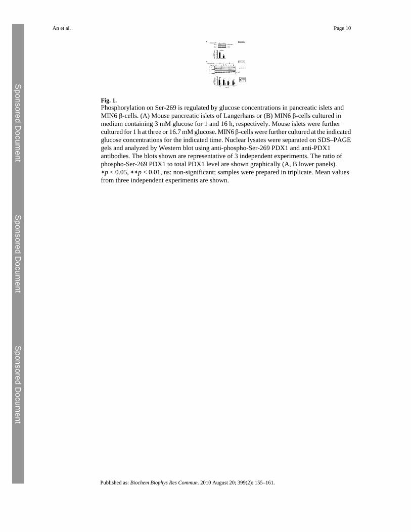
Fig. 1.Phosphorylation on Ser-269 is regulated by glucose concentrations in pancreatic islets andMIN6 β-cells. (A) Mouse pancreatic islets of Langerhans or (B) MIN6 β-cells cultured inmedium containing 3 mM glucose for 1 and 16 h, respectively. Mouse islets were furthercultured for 1 h at three or 16.7 mM glucose. MIN6 β-cells were further cultured at the indicatedglucose concentrations for the indicated time. Nuclear lysates were separated on SDS–PAGEgels and analyzed by Western blot using anti-phospho-Ser-269 PDX1 and anti-PDX1antibodies. The blots shown are representative of 3 independent experiments. The ratio ofphospho-Ser-269 PDX1 to total PDX1 level are shown graphically (A, B lower panels).∗p < 0.05, ∗∗p < 0.01, ns: non-significant; samples were prepared in triplicate. Mean valuesfrom three independent experiments are shown.
An et al. Page 10
Published as: Biochem Biophys Res Commun. 2010 August 20; 399(2): 155–161.
Sponsored Docum
ent Sponsored D
ocument
Sponsored Docum
ent
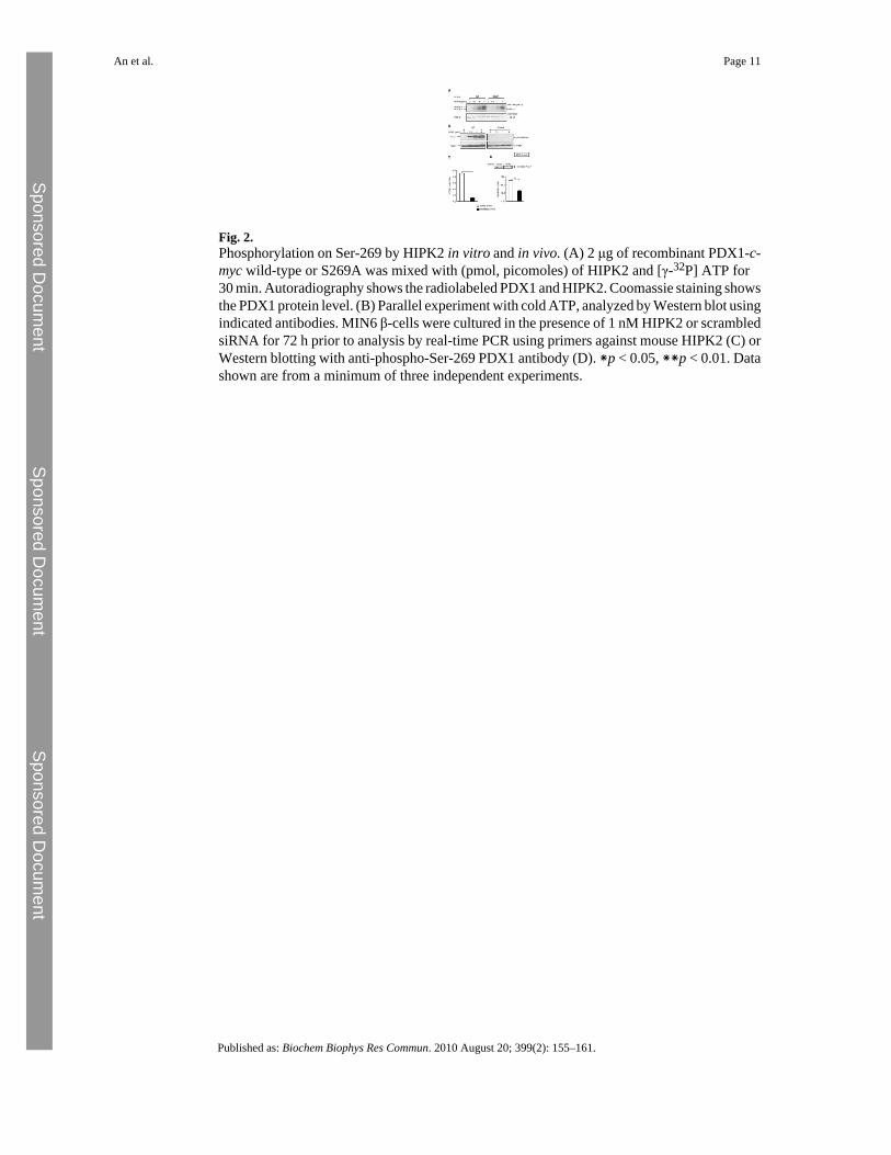
Fig. 2.Phosphorylation on Ser-269 by HIPK2 in vitro and in vivo. (A) 2 μg of recombinant PDX1-c-myc wild-type or S269A was mixed with (pmol, picomoles) of HIPK2 and [γ-32P] ATP for30 min. Autoradiography shows the radiolabeled PDX1 and HIPK2. Coomassie staining showsthe PDX1 protein level. (B) Parallel experiment with cold ATP, analyzed by Western blot usingindicated antibodies. MIN6 β-cells were cultured in the presence of 1 nM HIPK2 or scrambledsiRNA for 72 h prior to analysis by real-time PCR using primers against mouse HIPK2 (C) orWestern blotting with anti-phospho-Ser-269 PDX1 antibody (D). ∗p < 0.05, ∗∗p < 0.01. Datashown are from a minimum of three independent experiments.
An et al. Page 11
Published as: Biochem Biophys Res Commun. 2010 August 20; 399(2): 155–161.
Sponsored Docum
ent Sponsored D
ocument
Sponsored Docum
ent
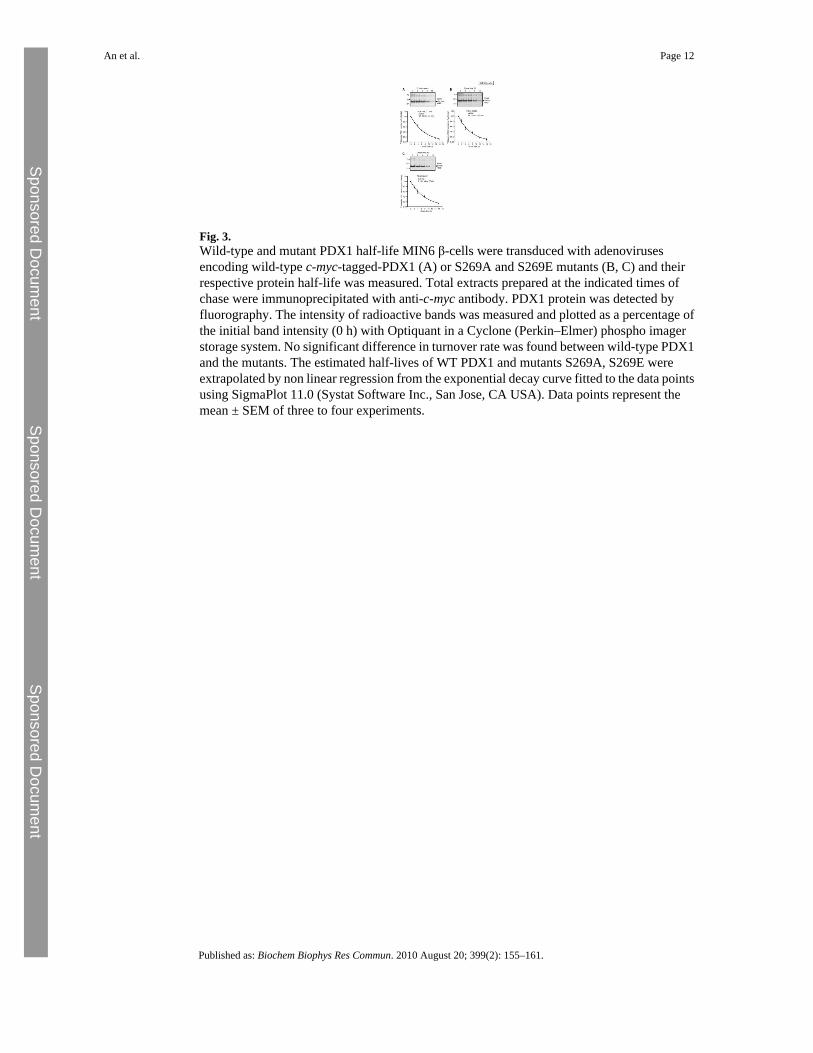
Fig. 3.Wild-type and mutant PDX1 half-life MIN6 β-cells were transduced with adenovirusesencoding wild-type c-myc-tagged-PDX1 (A) or S269A and S269E mutants (B, C) and theirrespective protein half-life was measured. Total extracts prepared at the indicated times ofchase were immunoprecipitated with anti-c-myc antibody. PDX1 protein was detected byfluorography. The intensity of radioactive bands was measured and plotted as a percentage ofthe initial band intensity (0 h) with Optiquant in a Cyclone (Perkin–Elmer) phospho imagerstorage system. No significant difference in turnover rate was found between wild-type PDX1and the mutants. The estimated half-lives of WT PDX1 and mutants S269A, S269E wereextrapolated by non linear regression from the exponential decay curve fitted to the data pointsusing SigmaPlot 11.0 (Systat Software Inc., San Jose, CA USA). Data points represent themean ± SEM of three to four experiments.
An et al. Page 12
Published as: Biochem Biophys Res Commun. 2010 August 20; 399(2): 155–161.
Sponsored Docum
ent Sponsored D
ocument
Sponsored Docum
ent

Fig. 4.Ser-269 phosphorylation and PDX1 subnuclear localization (A) Immunocytochemical analysisof the subcellular distribution of PDX1-c-myc. MIN6 β-cells were transduced withadenoviruses encoding for the indicated PDX1 molecules. Cells were cultured at 3 mM glucosefor 16 h and then treated with 3 or 20 mM glucose for 6 h. c-myc-tagged PDX1 was detectedusing anti-c-myc antibody (Roche) and Alexa-568 (Molecular Proves). Nuclear staining wasachieved using DAPI. Confocal images were captured using a Leica SP2 upright laser scanningconfocal microscope (x63/1.32 oil-immersion lens) equipped with a krypton/argon laser (488and 568 nm excitation lines) and UV light. Images were analyzed off-line using VolocityTM
4.0 software. Each sample was prepared in triplicate. (B) The average number of cellsdisplaying PDX1 localization predominantly at nuclear periphery is expressed as a percentageof the total number of cells analyzed. Mean values from three independent experiments areshown.
An et al. Page 13
Published as: Biochem Biophys Res Commun. 2010 August 20; 399(2): 155–161.
Sponsored Docum
ent Sponsored D
ocument
Sponsored Docum
ent


